Embed Size (px)
Citation preview
© 2003 Nature Publishing Group
496 | JUNE 2003 | VOLUME 4 www.nature.com/reviews/neuro
R E V I E W S
Following their description more than 50 years ago1,reeler mice were the only known animal mutants withmalformations of the cerebral cortex. Understandably,they generated a lot of interest, which increased furtherafter the cloning of the affected gene — reelin (Reln)2.As the reeler mouse model has been exhaustivelyreviewed3, and the Reln signalling pathway has recentlybeen discussed4, we will not attempt to cover the fieldextensively, but instead will focus on recent findings that concern the role of Reln during brain development.Our understanding of the action of Reln remainssketchy, and we will try to point out caveats as well asadvances in this field. After a presentation of the reelermalformation, we will summarize present views on thebiochemistry of Reln signalling, and discuss observationsand hypotheses about the putative mechanism of actionof this interesting protein.
Reeler and reeler-like phenotypesIn rodents, Reln deficiency results in the reeler pheno-type, several alleles of which have been described in mice and rats3,5. In humans, it is the cause of theNorman–Roberts type of LISSENCEPHALY6. The human phenotype is only known from medical imaging, but itseems to be similar to its murine counterpart. In mice,mutations of Disabled 1 (Dab1)7–9 and double mutationsof two lipoprotein receptors, very-low-density lipo-protein receptor (VLDLR) and apolipoprotein E receptortype 2 (ApoER2), generate similar phenotypes that willbe referred to as reeler-like throughout this review10,11.
In reeler and reeler-like mice, neurons are generatedin germinative zones (VENTRICULAR ZONE, SUBVENTRICULAR
ZONE and RHOMBIC LIP) as in wild-type animals, and their
migration is initially normal. However, as they approachtheir destination, reeler neurons fail to form normalarchitectonic structures. This is illustrated in FIGS 1 and 2for the embryonic cortex, and in FIG. 3 for the cerebellumand inferior olivary complex. It seems that there is aninstruction at the end of migration that is lacking in reeler mutants, so that neurons do not recognize their proper location and orientation at the end of theirmigration pathway. The rest of the differentiation program is unaffected — normal neuronal classes areformed, dendritic trees and axons ramify and connectalmost normally with their physiological targets, and gliogenesis and myelination are not directly altered.However, due to abnormal neuronal positioning, thedendritic trees and initial axonal pathways are oftendistorted. The defect is most severe in the cerebral cortex,hippocampus and cerebellar cortex, but subtle anomalieshave been identified at every location that has beensearched, including the inferior olive, olfactory bulb,cochlear nuclei, facial nerve nucleus, thalamus and tectum (reviewed in REF. 3). Defects were also recentlydemonstrated in the retina12 and spinal cord13.
Reln, the protein that is defective in reeler mice, issecreted by some neurons, such as Cajal–Retzius cells inthe cortical marginal zones and cerebellar granule cells,and it acts through the extracellular milieu on neigh-bouring target cells — cortical plate cells and Purkinjecells, respectively — to provide an architectonic signal.Reception of the Reln signal requires the presence of atleast one of two receptors of the lipoprotein receptorfamily on the surface of target cells, namely VLDLR andApoER2. The signal is then transduced by tyrosine phos-phorylation of the intracellular adaptor Dab1. In some
REELIN AND BRAIN DEVELOPMENTFadel Tissir and André M. Goffinet
Over the last 50 years, the reeler mutant mouse has become an important model for studyingnormal and abnormal development in the cerebral cortex and other regions of the brain.However, we are only just beginning to understand the actions of reelin — the protein that isaffected by the reeler mutation — at the molecular and cellular level. This review discusses themost recent advances in this research field, and considers the merits of the various models thathave been put forward to explain how reelin works.
LISSENCEPHALY
Literally meaning ‘smoothbrain’. Lissencephaly is a humanbrain disorder that ischaracterized by absence orreduction of the cerebralconvolutions.
VENTRICULAR ZONE
The proliferative inner layer of the developing brain andspinal cord.
SUBVENTRICULAR ZONE
A layer of cells in the developingbrain that is generated by themigration of neuroblasts fromthe adjoining ventricular zone.
RHOMBIC LIP
A specialized germinal matrixlocated at the posterior edge ofthe cerebellar anlage that givesrise to the granule cells of thecerebellum.
Developmental GeneticsUnit, Université Catholiquede Louvain, UCL 7382,73 Avenue E. Mounier,B1200 Brussels, Belgium.e-mail: [email protected]:10.1038/nrn1113
© 2003 Nature Publishing Group
NATURE REVIEWS | NEUROSCIENCE VOLUME 4 | JUNE 2003 | 497
R E V I E W S
TATA BOX
A DNA sequence with theconsensus TATAAAA that ispresent in many eukaryotic genepromoters and specifies the sitewhere transcription is initiated.
5′RACE
(5′ rapid amplification of cDNAends). RACE is a PCR-basedmethod for amplifyingunknown cDNA sequences byusing primers that correspond toa known sequence.
ALTERNATIVE SPLICING
During splicing, introns areexcised from RNA aftertranscription and the cut endsare rejoined to form acontinuous message. Alternativesplicing allows the production ofdifferent messages from thesame DNA molecule.
(I. Bar and A.G., unpublished data), and the genomicregion upstream of the Reln gene contains mostly repeatedsequences that lack specific features. This implies that reg-ulatory elements are located in introns, but this remainsto be analyzed. A better definition of the transcriptionalcontrol of Reln will be required if we want to gain a betterunderstanding of the roles of Reln and Cajal–Retziuscells in cortical development and evolution26.
The Reln mRNA is about 12 kb in length and con-sists of 65 exons22. Two ALTERNATIVE SPLICING events havebeen found27: a micro-exon of 6 nucleotides (exon 64,which encodes Val–Ser) is included in the neuronalmRNA but not in mRNA that is made in non-neuronalcells, and the use of an alternative polyadenylation sitein intron 63 produces an mRNA limited to exons 1–63that codes for a protein that lacks a carboxy (C)-terminalregion. Variability in the 5’ untranslated region (UTR)of the human RELN gene has been tentatively correlatedwith a genetic susceptibility to autism (BOX 1).
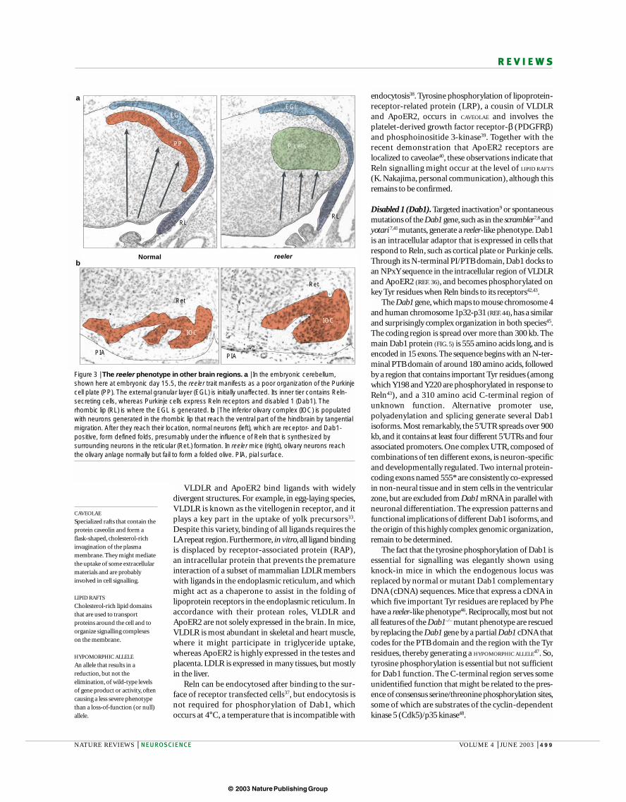
The Reln protein is 3461 amino acids long2. Asschematized in FIG. 4, the sequence begins with a signal peptide, followed by a region with similarity to F-spondin, a unique region and then eight repeats, eachof around 350 amino acids. Each repeat contains anepidermal growth factor (EGF) motif at the centre,flanked by two subrepeats, A and B, which show weaksimilarity to one another. The protein terminates with abasic stretch of 33 amino acids, which are absent fromthe Reln that is coded by alternatively polyadenylatedtranscripts. Although the predicted molecular mass ofthe Reln polypeptide is 387,497 Da, the estimated size
locations, such as the cerebral cortex, hippocampus, cere-bellum and inferior olive, Reln-producing and Reln targetcells are spatially segregated. In other regions, such as theretina and spinal cord, the source and target cells are closeto each other or even mixed. In a few cases, for example inhuman Cajal–Retzius cells, the same cells are positive forReln and Dab1, implying that there is an autocrineloop14,15. Intriguingly, the reeler phenotype is most evidentwhen the source and target cells are segregated, and thismight be relevant to the mechanism of action of Reln. Inaddition to the brain, Reln is also expressed at lower levelsin peripheral organs, such as the liver16, kidney and a few others17–19. However, the function of the pathway innon-neural tissues is still largely unexplored.
Reln and its partnersThe Reln gene is about 450 kb long and maps to mousechromosome 5 and human 7q22 (REFS 20–22). It is similarin mouse, man and probably other species. Reln containshuge introns — in particular, introns one, two and three,which are 57, 59 and 54 kb long, respectively, in themouse, and even larger in man. So far, almost nothing isknown about the regulation of Reln mRNA expression.The proximal promoter is CG-rich and contains no TATA
BOX. Its methylation pattern has received some attentionrecently23–25, but there is still considerable scope for fur-ther investigation. This promoter has significant activityin reporter systems, but is probably not sufficient toaccount for the fine regulation of Reln mRNA expressionin neurons such as the Cajal–Retzius cells. 5’RACE analysisfrom exon 2 did not show any additional promoter
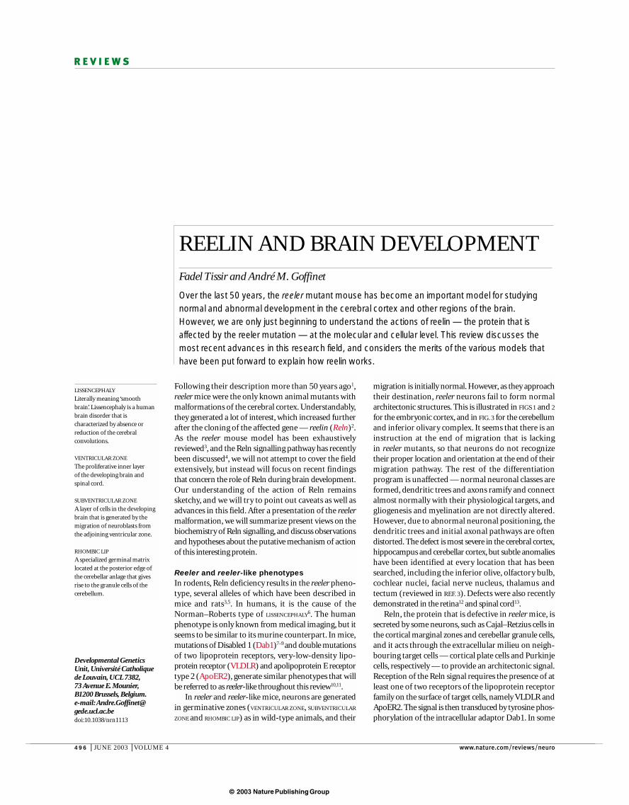
Figure 1 | Schematic view of early cortical development in mice. At embryonic day (E) 13.5 (the preplate stage) pioneerneurons migrate — probably by somal translocation76 — and form a loose horizontal network. Some are reelin (Reln)-positive (red)and others are Reln-negative (pink). At this stage, the reeler phenotype is not evident. At E14.5, as migratory distances and thecomplexity of the environment increase, cells migrate along radial glial fibres. The first cohort of cells condenses in the cortical plate(green), splitting the preplate into two contingents. The reeler cortical plate is populated with cells in an oblique orientation and doesnot split the preplate into two components. At E15.5, a second cohort of cells (blue) migrates through the normal cortical plate andsettles superficially, forming an inside to outside gradient. In reeler mutants, the second cohort settles in the deep tiers of the corticalplate, forming an outside to inside gradient.
PiaPia
Pia
PreplateE12.5–E13.5
Early cortical plateE14.5
E15.5
Pia
Normal or reeler-like Normal reeler-like Normal reeler-like
Pia

© 2003 Nature Publishing Group
498 | JUNE 2003 | VOLUME 4 www.nature.com/reviews/neuro
R E V I E W S
Reln can be studied using antibodies against N- andC-terminal epitopes29,30 (FIG. 4). The full-length proteinpredominates in the supernatant of transfected cells,but several other fragments are also consistently found.N-terminal antibodies disclose two fragments ofaround 180 and 320 kDa in length, whereas C-terminalantibodies disclose fragments of around 100 and 240 kDa.In adult and embryonic brain extracts and body fluids(such as cerebrospinal fluid or plasma18), almost no full-length Reln is detected,and the main polypeptides arethe N-terminal 180 kDa and the C-terminal 100 kDafragments (N. Ignatova and A. G., unpublished data).Comparison with partial recombinant Reln constructsshows that Reln is cleaved at two main locations —between repeats 2 and 3, and between repeats 6 and 7(arrows in FIG 4). Unfortunately, there are no antibodiesavailable to probe the central region. In embryonicbrain explant cultures, Reln processing was blocked byzinc chelators but not by other proteinase inhibitors,implying that METALLOPROTEINASE activity is involved31. Onthe other hand, Reln cleavage is observed in differentsettings, implying that the processing pattern mightreflect access of proteinases to exposed loops of the protein, whereas folding stabilizes other domains. Thequestion of processing is not trivial, as the central regionof Reln is essential for receptor binding and to triggerDab1 phosphorylation (see later discussion).
Lipoprotein receptors: VLDLR and ApoER2. TheVLDLR gene maps to mouse chromosome 19 andhuman chromosome 9p24. ApoER2, which is alsonamed LRP8, maps to mouse chromosome 4 andhuman chromosome 1p34. The structure and biologyof these receptors have been discussed elsewhere32,33,and only the features that are relevant to Reln signallingwill be summarized here. The VLDLR and ApoER2genes and proteins are similar to the low densitylipoprotein receptor (LDLR) gene, although they aremore similar to one another than to LDLR.
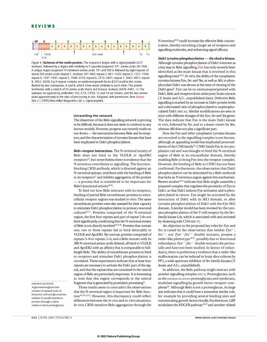
All three receptor genes code for proteins with similarorganizations, but only VLDLR and ApoER2 bind Reln.Mice with inactivation of VLDLR and ApoER2 have areeler-like phenotype11. Their structure is schematizedin FIG. 5. The extracellular ligand binding region contains 7 or 8 cysteine-rich repeats (leucine-alanine(LA) repeats) of around 40 amino acids, each with threedisulphide bonds and a coordinated Ca2+ ion. This isfollowed by two EGF repeats, a β-PROPELLER segment withYWTD motifs (leucine-tyrosine (LY) repeats)34, anotherEGF repeat, a paramembrane region of O-linked glyco-sylation, the transmembrane segment, and a cytoplasmicregion of around 60 amino acids with an NPxYsequence (where x is any amino acid). In ApoER2, alter-native splicing can result in an insertion of 59 residuesin this region, and this sequence serves as a docking sitefor Jun N-terminal kinase (JNK)-interacting proteins35.The cytoplasmic NPxY sequence is the docking site forthe protein interaction (PI)/phosphotyrosine binding(PTB) domain of the Dab1 adapter. In contrast to otherPTB domain proteins, such as Shc, Dab1 binds withhigher affinity to unphosphorylated NPxY sequences36.
in polyacrylamide gel electrophoresis (PAGE) is around427 kDa (M. Sikorska, personal communication), andthis ~40 kDa difference might indicate that the proteinis glycosylated28. A similar difference is found whenmammalian cells express a construct that codes for theamino (N)-terminal part of Reln, up to the end ofrepeat 2. This results in the production of a 180 kDaprotein, although the predicted mass is 136,107 Da.This indicates that some glycosylation might reside inthe N-terminal region, but so far this has not beenthoroughly investigated.
METALLOPROTEINASE
A proteinase that has a metal ionat its active site.
β-PROPELLER
A protein domain that consistsof an array of β-sheet motifs,which are configured in a ring toresemble the blades of apropeller.
Figure 2 | Photomicrography of the normal and reeler telencephalon at embryonic day 14.5. 2µm thick plastic section, stained with toluidine blue. Note the poor organization of thecortical plate (CP) and absence of a subplate (SP) in the reeler mouse cortex. IZ, intermediatezone; MZ, marginal zone; PIA, pial surface; VZ, ventricular zone.
PIAPIA
VZ
IZ
SP
CP
VZ
IZ
CP
MZ
Normal reeler-like
Box 1 | Reelin and susceptibility to psychoses
In addition to the well-established function of reelin (Reln) during brain development,some studies point to a possible role for the Reln gene in conditions such asschizophrenia89–92 and autism93–98. The human RELN 5’UTR contains a low andvariable number of CGG repeats, and this has been tentatively correlated with geneticsusceptibility to autism97. This association should be regarded as preliminary, however,as it was not confirmed by other studies98.
In brain samples from patients with chronic psychosis, Reln protein and mRNA levelswere reduced by approximately 50%. There were no changes in the levels of disabled 1(Dab1) mRNA and protein; this is unexpected, as a marked upregulation of Dab1protein levels is observed in Reln- and lipoprotein-receptor-deficient mice. Theproximal Reln promoter is CG-rich, and like other such promoters, methylation isassociated with decreased expression. Therefore, it was proposed thathypermethylation of the promoter might be the origin of decreased expression inpsychiatric patients23–25. Promoter methylation is known to influence long-term rather than dynamic regulation of expression, so this hypothesis needs to beinvestigated further.
Reln mRNA is expressed in a subset of GABA (γ-aminobutyric acid)-expressingcortical interneurons, indicating that downregulation of Reln expression might disturbthe delicate balance of cortical excitability, although Reln-deficient mice have no overtepilepsy. Heterozygous reeler mice have a 50% reduction in Reln levels, and have somebehavioural features that are evocative of human psychosis99. Given the difficulty ofbehavioural studies in mice, these results should be independently confirmed. Takentogether, these findings can be tentatively interpreted within a neurodevelopmental/vulnerability ‘multi-hit’ model of schizophrenia100.
© 2003 Nature Publishing Group
NATURE REVIEWS | NEUROSCIENCE VOLUME 4 | JUNE 2003 | 499
R E V I E W S
endocytosis38. Tyrosine phosphorylation of lipoprotein-receptor-related protein (LRP), a cousin of VLDLR and ApoER2, occurs in CAVEOLAE and involves theplatelet-derived growth factor receptor-β (PDGFRβ)and phosphoinositide 3-kinase39. Together with therecent demonstration that ApoER2 receptors arelocalized to caveolae40, these observations indicate thatReln signalling might occur at the level of LIPID RAFTS
(K. Nakajima, personal communication), although thisremains to be confirmed.
Disabled 1 (Dab1). Targeted inactivation9 or spontaneousmutations of the Dab1 gene, such as in the scrambler7,8 andyotari7,41 mutants, generate a reeler-like phenotype. Dab1is an intracellular adaptor that is expressed in cells thatrespond to Reln, such as cortical plate or Purkinje cells.Through its N-terminal PI/PTB domain, Dab1 docks toan NPxY sequence in the intracellular region of VLDLRand ApoER2 (REF. 36), and becomes phosphorylated onkey Tyr residues when Reln binds to its receptors42,43.
The Dab1 gene, which maps to mouse chromosome 4and human chromosome 1p32-p31 (REF. 44), has a similarand surprisingly complex organization in both species45.The coding region is spread over more than 300 kb. Themain Dab1 protein (FIG. 5) is 555 amino acids long, and isencoded in 15 exons. The sequence begins with an N-ter-minal PTB domain of around 180 amino acids, followedby a region that contains important Tyr residues (amongwhich Y198 and Y220 are phosphorylated in response toReln43), and a 310 amino acid C-terminal region ofunknown function. Alternative promoter use,polyadenylation and splicing generate several Dab1 isoforms. Most remarkably, the 5’UTR spreads over 900kb, and it contains at least four different 5’UTRs and fourassociated promoters. One complex UTR, composed ofcombinations of ten different exons, is neuron-specificand developmentally regulated. Two internal protein-coding exons named 555* are consistently co-expressedin non-neural tissue and in stem cells in the ventricularzone, but are excluded from Dab1 mRNA in parallel withneuronal differentiation. The expression patterns andfunctional implications of different Dab1 isoforms, andthe origin of this highly complex genomic organization,remain to be determined.
The fact that the tyrosine phosphorylation of Dab1 isessential for signalling was elegantly shown usingknock-in mice in which the endogenous locus wasreplaced by normal or mutant Dab1 complementaryDNA (cDNA) sequences. Mice that express a cDNA inwhich five important Tyr residues are replaced by Phehave a reeler-like phenotype46. Reciprocally, most but notall features of the Dab1–/– mutant phenotype are rescuedby replacing the Dab1 gene by a partial Dab1 cDNA thatcodes for the PTB domain and the region with the Tyrresidues, thereby generating a HYPOMORPHIC ALLELE47. So,tyrosine phosphorylation is essential but not sufficientfor Dab1 function. The C-terminal region serves someunidentified function that might be related to the pres-ence of consensus serine/threonine phosphorylation sites,some of which are substrates of the cyclin-dependentkinase 5 (Cdk5)/p35 kinase48.
VLDLR and ApoER2 bind ligands with widelydivergent structures. For example, in egg-laying species,VLDLR is known as the vitellogenin receptor, and itplays a key part in the uptake of yolk precursors33.Despite this variety, binding of all ligands requires theLA repeat region.Furthermore, in vitro, all ligand binding is displaced by receptor-associated protein (RAP),an intracellular protein that prevents the prematureinteraction of a subset of mammalian LDLR memberswith ligands in the endoplasmic reticulum, and whichmight act as a chaperone to assist in the folding oflipoprotein receptors in the endoplasmic reticulum. Inaccordance with their protean roles, VLDLR andApoER2 are not solely expressed in the brain. In mice,VLDLR is most abundant in skeletal and heart muscle,where it might participate in triglyceride uptake,whereas ApoER2 is highly expressed in the testes andplacenta. LDLR is expressed in many tissues, but mostlyin the liver.
Reln can be endocytosed after binding to the sur-face of receptor transfected cells37, but endocytosis isnot required for phosphorylation of Dab1, whichoccurs at 4°C, a temperature that is incompatible with
CAVEOLAE
Specialized rafts that contain theprotein caveolin and form aflask-shaped, cholesterol-richinvagination of the plasmamembrane. They might mediatethe uptake of some extracellularmaterials and are probablyinvolved in cell signalling.
LIPID RAFTS
Cholesterol-rich lipid domainsthat are used to transportproteins around the cell and toorganize signalling complexeson the membrane.
HYPOMORPHIC ALLELE
An allele that results in areduction, but not theelimination, of wild-type levelsof gene product or activity, oftencausing a less severe phenotypethan a loss-of-function (or null)allele.
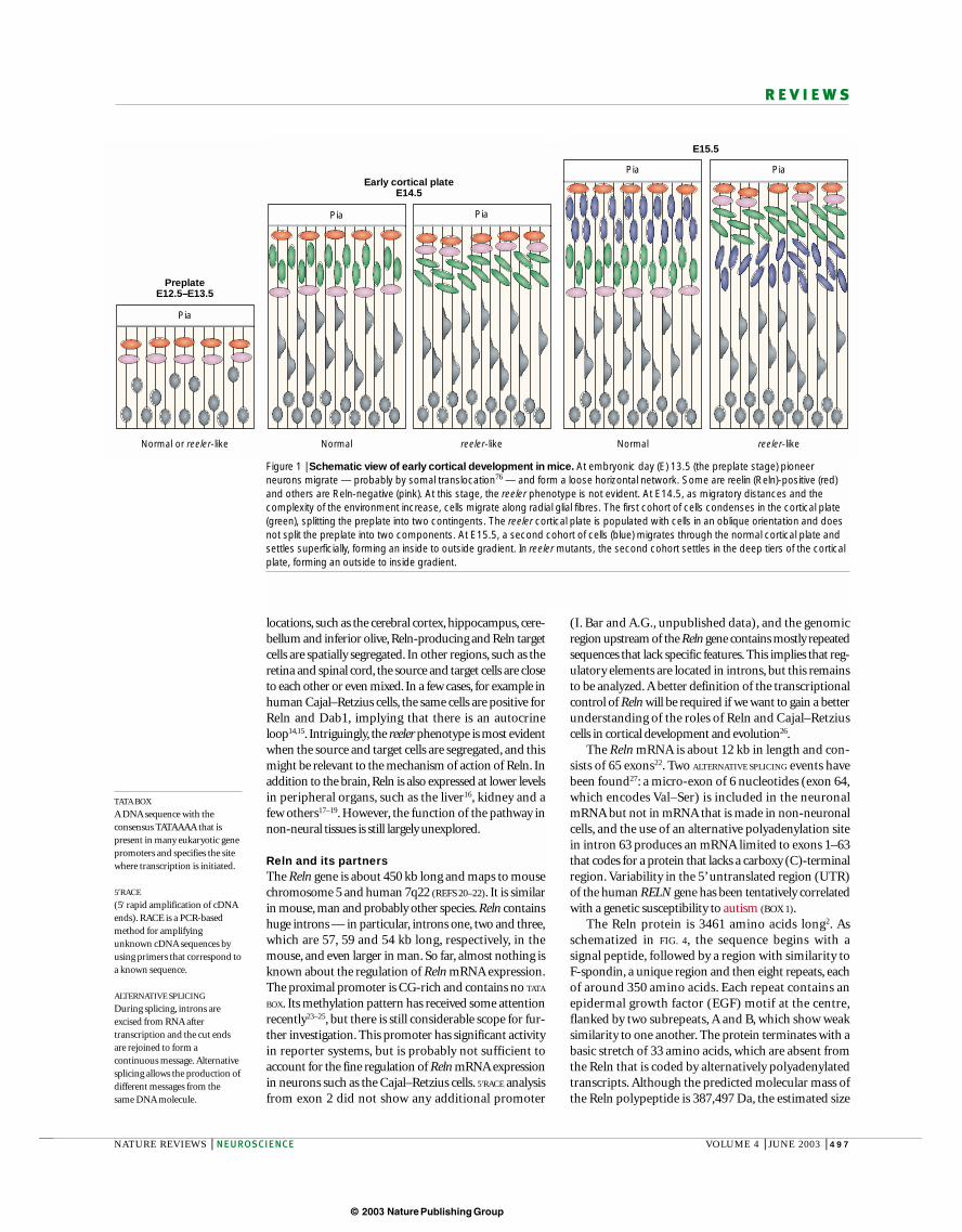
Figure 3 | The reeler phenotype in other brain regions. a | In the embryonic cerebellum,shown here at embryonic day 15.5, the reeler trait manifests as a poor organization of the Purkinjecell plate (PP). The external granular layer (EGL) is initially unaffected. Its inner tier contains Reln-secreting cells, whereas Purkinje cells express Reln receptors and disabled 1 (Dab1). Therhombic lip (RL) is where the EGL is generated. b | The inferior olivary complex (IOC) is populatedwith neurons generated in the rhombic lip that reach the ventral part of the hindbrain by tangentialmigration. After they reach their location, normal neurons (left), which are receptor- and Dab1-positive, form defined folds, presumably under the influence of Reln that is synthesized bysurrounding neurons in the reticular (Ret.) formation. In reeler mice (right), olivary neurons reachthe olivary anlage normally but fail to form a folded olive. PIA, pial surface.
reelerNormal
PIA
EGL
Ret
RL
P cells
IOC
PIA
RL
PP
Ret
EGL
IOC
a
b
© 2003 Nature Publishing Group
500 | JUNE 2003 | VOLUME 4 www.nature.com/reviews/neuro
R E V I E W S
HEPARAN SULPHATE
A glycosaminoglycan thatconsists of repeated units ofhexuronic acid and glucosamineresidues. It usually attaches toproteins through a xyloseresidue to form proteoglycans.
N terminus49,50 could increase the effective Reln concen-tration, thereby recruiting a larger set of receptors andsignalling molecules, and enhancing signal efficacy.
Dab1 tyrosine phosphorylation — the elusive kinase.Although tyrosine phosphorylation of Dab1 is known asa key step in Reln signalling, Fyn has only recently beenidentified as the main kinase that is involved in this signalling event54,55. In vitro, the ability of the cytoplasmictyrosine kinases Fyn, Src and Yes, as well as Abl, to phos-phorylate Dab1 was shown at the time of cloning of theDab1 gene9. Fyn can be co-immunoprecipitated withDab1, Reln and receptors from embryonic brain extracts(Y. Jossin and A.G., unpublished data). Defective Relnsignalling is marked by an increase in Dab1 protein levelsand a decreased ratio of phosphorylated to unphospho-rylated Dab1 (REF. 42). Similar modifications are seen inmice with different dosages of the Fyn, Src and Yes genes.The data indicate that Fyn is the main Dab1 kinase in vivo, followed by Src and to a lesser extent by Yes,whereas Abl does not play a significant part.
How the Fyn and other cytoplasmic tyrosine kinasesare recruited to the signalling complex is not known,although an appealing model has implicated protocad-herins of the CNR family56. CNR1 binds Fyn in its cyto-plasmic tail and was thought to bind the N-terminalregion of Reln in its extracellular domain, therebyenabling Reln to bring Fyn into the receptor complex.However, the binding of Reln to CNR1 has not beenconfirmed. Furthermore, the observation that Dab1phosphorylation can be stimulated by a Reln moleculethat lacks an N terminus argues against this mechanism.Recent studies54,55 indicate that Reln might assemble aprepared complex that regulates the proximity of Fyn toDab1, so that Dab1 induces Fyn activation and is phos-phorylated in return. Fyn might be activated by theinteraction of Dab1 with its SH3 domain, or after tyrosine phosphorylation of Dab1 with the Fyn SH2domain. A similar model has been invoked for the tyro-sine phosphorylation of the T-cell receptor by the Src-family kinase Lck, which is associated with and activatedby clustering with CD4 (REF. 57).
An objection to the proposed key roles for Fyn andSrc is raised by the observation that neither Fyn–/–,Src –/–, nor Fyn–/–;Src+/– double mutants, possess a reeler-like phenotype54,55, possibly due to functionalredundancy. Fyn–/–;Src –/– double mutants die perina-tally and have not been studied. In favour of redun-dancy, there is preliminary evidence that a reeler-likemalformation can be induced in brain slice cultures byPP2, a wide spectrum inhibitor of Src family kinases (Y.Jossin and A.G., unpublished).
In addition, the Reln pathway might interact withanother signalling complex (FIG 5). Proteoglycans, suchas the HEPARAN SULPHATE proteoglycans and syndecans,modulate signalling by growth factor-receptor com-plexes58. Although Reln is not a proteoglycan, its largesize indicates that it could have a somewhat similar role,for example by providing several binding sites and concentrating growth factors locally. Furthermore, LRPmodulates the PDGFR pathway39,59 and another related
Unravelling the networkThe dissection of the Reln signalling network is provingto be difficult, because it does not seem to conform to anyknown models. However, progress was recently made ontwo fronts — the interactions between Reln and its recep-tors, and the characterization of tyrosine kinases that havebeen implicated in Dab1 phosphorylation.
Reln–receptor interactions. The N-terminal moiety ofReln does not bind to the VLDLR or ApoER2receptors10, but nevertheless there is evidence that theN terminus contributes to signalling. The function-blocking CR50 antibody, which is directed against anN-terminal epitope, interferes with the binding of Relnto its receptors37 and inhibits aggregation of the protein— a process that is considered to be important forReln’s functional activity49,50.
To find out how Reln interacts with its receptors,binding of partial Reln recombinant proteins to extra-cellular receptor regions was studied in vitro. The samerecombinant proteins were also assessed for their capacityto stimulate Dab1 phosphorylation in primary neuronalcultures42,51. Proteins comprised of the N-terminalregion, the first four repeats and part of repeat 5 do notbind significantly, confirming that the N-terminal moietyof Reln is not directly involved10,15,42. Proteins that containone, two or three repeats fail to bind detectably toVLDLR and ApoER2. By contrast, proteins comprised ofrepeats 3–8 or repeats 3–6, and a Reln mutant with its388 N-terminal amino acids deleted, all bind to VLDLRand ApoER2 with an affinity that is comparable to full-length Reln. The ability of recombinant proteins to bindto receptors and stimulate Dab1 phosphorylation is correlated. These experiments indicate that at least fourrepeats are necessary to activate the Dab1 part of the sig-nal, and that the repeats that are contained in the centralregion of Reln are particularly important. It is interestingto note that this region corresponds to the central fragment that is generated by proteolytic processing51.
These results seem to contradict the observationsthat the N-terminal region is important for Reln func-tion49,50,52,53. However, this discrepancy could reflect differences between the in vivo and in vitro situations.In vivo, CR50-sensitive Reln aggregation through the
Figure 4 | Schema of the reelin protein. The sequence begins with a signal peptide of 27residues, followed by a region with similarity to F-spondin (segment ‘SP’, amino acids 28–190). A unique region (segment ‘H’) between amino acids 191 and 500 is followed by eight repeats ofabout 350 amino acids (repeat 1, residues 501–860; repeat 2, 861–1220; repeat 3, 1221–1596;repeat 4, 1597–1947; repeat 5, 1948–2314; repeat 6, 2315–2661; repeat 7, 2662–3051; repeat8, 3052–3428). Each repeat contains an epidermal growth factor (EGF) motif at the centre,flanked by two subrepeats, A and B, which show weak similarity to each other. The proteinterminates with a stretch of 33 amino acids that is rich in basic residues (3429–3461, +). Theepitopes recognized by antibodies 142, G10, CR50, 12 and 14 are shown, and the two arrowspoint approximately to the sites of processing in vivo. Adapted, with permission, from NatureREF. 2 (1995) Macmillan Magazines Ltd. s, signal peptide.
s SP H
A
1
B A
2
B A
3
B A
4
B A
5
B A
6
B A
7
B A
8
B
+
142 12 14CR50G10
EGF motif

© 2003 Nature Publishing Group
NATURE REVIEWS | NEUROSCIENCE VOLUME 4 | JUNE 2003 | 501
R E V I E W S
migration to the olfactory bulb62. Expression of Dab1 andReln receptors in the ventricular zone agrees with the proposed role for Reln in the maturation or migratorycapacity of neural stem cells63. Reln assists in the forma-tion of the radial glial scaffold in the dentate gyrus andpromotes branching of radial glial cells in vitro through aDab1-dependent mechanism64, further pointing to a rolefor Reln signalling in radial precursor cells.
Unlike other species such as chick and crocodile65, noReln-positive cells are detected in the vicinity of thetelencephalic ventricular zone in mammals, raising the question of the origin of the ligand that binds to thereceptors in precursor cells. Reln secreted fromCajal–Retzius cells could bind to lipoprotein receptors onthe radial processes of precursor cells that expandthrough the whole thickness of the tissue. The observa-tion that Reln promotes branching of these radialprocesses64 concurs with this view, which also predictsthat the VLDLR and/or ApoER2 protein(s) should bepresent on radial processes in the marginal zone, close toReln-producing cells.Alternatively, Reln or its active cen-tral fragment could diffuse from the marginal zone to theventricular zone. In principle, these ideas should be easyto check. However, so far, it has proved difficult to detectextracellular Reln reliably. In addition, antibodies are notyet available to allow immunohistochemical studies of thecentral fragment of Reln and the lipoprotein receptors.
Given the potential links between Reln signalling,psychoses and Alzheimer’s disease (BOXES 1 and 2), theeffect of this pathway on neural stem cells is potentiallyimportant, especially if it continues to operate in theadult brain and influence regeneration, as indicated bythe observation that decreased Reln expression seems toinfluence granule cell dispersion in epilepsy66.
Cell biological mechanisms of Reln action. Reln is presentin the nervous system in all vertebrates, from lamprey67,and even Amphioxus (G. Meyer, personal commun-ication), to fish, Xenopus, reptiles, birds and mam-mals26,65,68, and its expression is widespread.Comparative studies of Reln expression in the embryoniccortex indicate that Reln-positive Cajal–Retzius cells arepresent in the marginal zone in all amniotes, indicatingthat these cells are evolutionarily homologous.However, mammalian Cajal–Retzius cells are charac-terized by a striking amplification of Reln production,pointing to the spatiotemporal control of Reln expression as a key feature of cortical evolution26,65. LikeCajal–Retzius cells, cerebellar granule cells synthesizelarge amounts of Reln in all species, and this is consistentwith the cerebellar malformation that is observed inReln-deficient mice and humans. On the other hand,the reeler malformation is subtle, and until recently hadnot been detected in many areas where Reln expressionis now known to be strong, such as the retina69 andspinal cord13. The same contrast between high Relnexpression and almost no alterations in reeler mice iseven seen in some laminar structures, such as olfactorymitral cells. However, the parsimony principle dictatesthat a single molecular mechanism should explain allobservations, until proven otherwise.
molecule, LRP6, is a key regulator of Wnt signalling60.The failure to identify such a ‘missing component’among the many spontaneous or induced mousemutants could be due to embryonic lethality or redun-dancy. This view is admittedly highly speculative, andthe identity of this putative signalling system, whichmight be coupled to Reln, remains open to debate.
The action of Reln in the developing brainThe observations summarized earlier leave the questionof what Reln is actually doing in the developing brainrelatively untouched. Although our view is still fuzzy, wewill discuss recent observations that point to a possiblerole for Reln in radial precursor cells, before ending withsome speculations on the cellular action of Reln.
Reln and neural precursor cells. In man and rodents, Dab1mRNA, as well as mRNAs for VLDLR and the ApoER2receptor, are expressed in the ventricular zone — presum-ably in neuronal precursor cells. The Dab1 protein isdetected in the same cells as Dab1 mRNA, but data on theVLDLR and ApoER2 proteins are sketchy45,61. The relativeintensity of VLDLR and ApoER2 mRNA hybridization inthe ventricular zone differs among brain areas, as well asbetween developmental stages. These preliminary resultsindicate that precursor cells possess the machinery torespond to Reln. One target of Reln in the ventricularzone could be the future olfactory interneurons that follow the rostral migratory stream. Interestingly, it wasshown that Reln facilitates the detachment of theseinterneurons from the stream, and promotes their
VLDLR/ApoER2
VLDLR/ApoER2
Dab1
Fyn
Dab1
Fyn
PI/PTB
NPxYLA LYE E O
X
Y198 Y220
SH3 SH2 Kinase
Rx
a b
Figure 5 | The partners of reelin signalling. a | The very-low-density lipoprotein receptor(VLDLR) and apolipoprotein E receptor type 2 (ApoER2) extracellular ligand binding regioncontains 7 or 8 imperfect cysteine-rich repeats (leucine-arginine (LA) repeats, yellow) of around 40amino acids, each with three disulphide bonds and a coordinated Ca2+ ion. This is followed bytwo epidermal growth factor (EGF) repeats (blue), a β-propeller segment (leucine-tyrosine (LY)repeats), usually made of 5 repeats of about 50 residues with a F/YWxD consensus (where x isany amino acid), another EGF repeat, a paramembrane region of O-linked glycosylation (O), thetransmembrane segment, and a cytoplasmic region of about 60 amino acids with the key NPxYsequence. The main disabled 1 (Dab1) protein isoform has 555 residues and is composed of anamino-terminal PI/PTB domain of about 180 amino acids that docks to the NPxY sequence inreceptors, followed by a region with Tyr residues, two of which (Y198/220) are essential forsignalling. Fyn family kinases have one SH3, one SH2 and a kinase domain as shown. b | Thecentral part of reelin docks to a complex of lipoprotein receptors and Dab1, and tyrosine kinasesof the Fyn group play a key part in signal transmission. There is a possibility that another ligand(denoted by ‘X’) and corresponding receptor (‘Rx’), would interact with the reelin signal.
© 2003 Nature Publishing Group
502 | JUNE 2003 | VOLUME 4 www.nature.com/reviews/neuro
R E V I E W S
more important at later stages, when migration distances increase.
Clearly, the idea of a ‘stop’ signal requires formula-tion in cell biological terms. Although reeler mice haveabnormal entorhinal–hippocampal connections78,79,this is probably secondary to architectonic distur-bances, as Reln does not seem to influence leading edgeextension or growth cone progression80,81. However,Reln could negatively regulate nucleokinesis, and influ-ence the position of the nucleus relative to processesand ramifications82. The idea that Reln might act as aninhibitor of nucleokinesis could account for severalobservations. For example, in the tectum and cochlearnuclei, inhibition of the progression of neuronal nucleiby Reln might explain why, in reeler mutants, abnor-mally orientated neurons invade a cell-poor subpialzone. In the reeler spinal cord, spinal presympatheticneurons are in an ectopic position, close to the centralcanal13,83. However, the reason is not a failure tomigrate, but rather a late back-movement from outsideto inside. This back-movement does not occur in nor-mal animals, in which a Reln-positive layer is founddeep in migrated presympathetic neurons, implyingthat Reln inhibits this back nuclear translation. In thereeler inferior olive, the long tangential migration fromthe rhombic lip occurs normally, but neurons seem tomigrate too far, too close to the midline. This indicatesthat Reln secreted by the reticular formation mightgovern the location of the olivary neuronal nucleus andsoma in relation to their processes.
On the other hand, some aspects of the reeler pheno-type do not fit easily with this action on nucleokinesis. Inthe hippocampal formation, the reeler trait consistsmostly of a poor lamination of the pyramidal cell layerand dentate granule cell layer, but this is due to failure of neurons to reach their respective layers rather thaninvasion of the marginal zone. In the reeler cerebellum,radially migrating Purkinje cells stop migration prema-turely and fail to condense into a normal Purkinje cellplate. The defect in the molecular layer is due to thesecondary degeneration of granule cells and not to over-migration of Purkinje cells. Similarly, in the reeler facialnerve nucleus, ectopic neurons settle in the tegmentum,and there is little or no overmigration towards the pia.The subtle malformations in the reeler retina are also notobviously explained by an effect on nucleokinesis.
Furthermore, an action of Reln as a direct negativeregulator of nucleokinesis is difficult to reconcile withtwo important sets of observations. First, expression of the Reln cDNA under the control of the nestin promoter, which results in ectopic Reln expression inprecursor cells in the ventricular zone, can partially but significantly rescue the reeler phenotype84. It is difficultto imagine how Reln, in the ventricular zone, coulddirectly influence the terminal migration of neurons thathave already left it. However, it raises the possibility thatReln could make neurons responsive to an unidentifiedsignal in the marginal zone.
A second question has arisen from studies of normaland Dab1–/– chimaeric mice85,86. In the cerebral cortex ofchimaeras produced by BLASTOCYST INJECTION, Dab1+/+ cells
In the reeler cortex, cortical plate cells keep a pro-longed contact with radial glial guides and invade themarginal zone to pile up close to the pia. This led to the proposal that Reln could provide a ‘stop’ signal to neurons at the end of their migration pathway70,71.Like the migration of other cells, neuronal migrationproceeds by the extension of a leading edge, followed byprogression of the nucleus in this cytoplasmic furrow(nucleokinesis)72. Leading edge extension requires actinpolymerization/depolymerization, with formation offilopodia and lamellipodia in some cells, followed by consolidation of stress fibres. These processes are controlled by the small GTPases Cdc42 (filopodia form-aton), Rac (lamellipodia formation) and Rho (con-solidation of stress fibres). Nucleokinesis depends onmicrotubule dynamics, in addition to microfilaments.
Recent findings on radial neuronal migration haveintroduced an additional level of complexity into the‘stop signal’ model. The new data indicate that radialneuroepithelial or glial cells are direct neuronal precur-sors73–75 and that early migrating neurons might useSOMAL TRANSLOCATION rather than gliophilic migration76,77.The latter mode of radial migration seems to become
YEAST TWO-HYBRID SCREENS
System used to determine theexistence of direct interactionsbetween proteins. It involves theuse of plasmids that encode twohybrid proteins; for example,one of them is fused to the GAL4DNA-binding domain and theother one is fused to the GAL4activation domain. The twoproteins are expressed togetherin yeast and, if they interact, thenthe resulting complex will drivethe expression of a reportergene, commonly β-galactosidase.
SOMAL TRANSLOCATION
Displacement of the cell body, asopposed to migration of thewhole cell.
BLASTOCYST INJECTION
The introduction of embryonicstem cells into a blastocyst-stageembryo of a different genotypeto generate a chimaeric embryo.
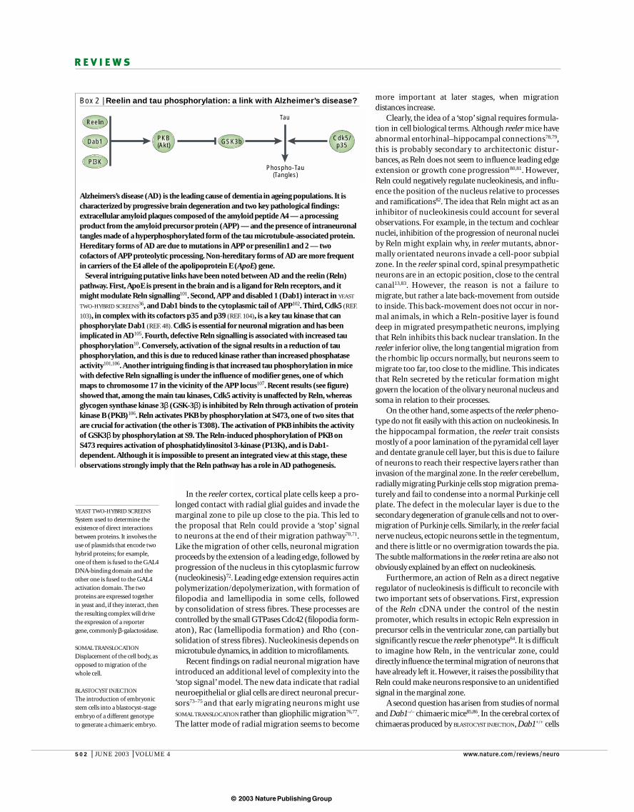
Box 2 | Reelin and tau phosphorylation: a link with Alzheimer’s disease?
Alzheimers’s disease (AD) is the leading cause of dementia in ageing populations. It ischaracterized by progressive brain degeneration and two key pathological findings:extracellular amyloid plaques composed of the amyloid peptide A4 — a processingproduct from the amyloid precursor protein (APP) — and the presence of intraneuronaltangles made of a hyperphosphorylated form of the tau microtubule-associated protein.Hereditary forms of AD are due to mutations in APP or presenilin1 and 2 — twocofactors of APP proteolytic processing. Non-hereditary forms of AD are more frequentin carriers of the E4 allele of the apolipoprotein E (ApoE) gene.
Several intriguing putative links have been noted between AD and the reelin (Reln)pathway. First, ApoE is present in the brain and is a ligand for Reln receptors, and itmight modulate Reln signalling101. Second, APP and disabled 1 (Dab1) interact in YEAST
TWO-HYBRID SCREENS36, and Dab1 binds to the cytoplasmic tail of APP102. Third, Cdk5 (REF.
103), in complex with its cofactors p35 and p39 (REF. 104), is a key tau kinase that canphosphorylate Dab1 (REF. 48). Cdk5 is essential for neuronal migration and has beenimplicated in AD105. Fourth, defective Reln signalling is associated with increased tauphosphorylation10. Conversely, activation of the signal results in a reduction of tauphosphorylation, and this is due to reduced kinase rather than increased phosphataseactivity101,106. Another intriguing finding is that increased tau phosphorylation in micewith defective Reln signalling is under the influence of modifier genes, one of whichmaps to chromosome 17 in the vicinity of the APP locus107. Recent results (see figure)showed that, among the main tau kinases, Cdk5 activity is unaffected by Reln, whereasglycogen synthase kinase 3β (GSK-3β) is inhibited by Reln through activation of proteinkinase B (PKB)106. Reln activates PKB by phosphorylation at S473, one of two sites thatare crucial for activation (the other is T308). The activation of PKB inhibits the activityof GSK3β by phosphorylation at S9. The Reln-induced phosphorylation of PKB onS473 requires activation of phosphatidylinositol 3-kinase (P13K), and is Dab1-dependent. Although it is impossible to present an integrated view at this stage, theseobservations strongly imply that the Reln pathway has a role in AD pathogenesis.
Reelin
Dab1 PKB(Akt) GSK3b Cdk5/
p35
Tau
Phospho-Tau(Tangles)
PI3K
© 2003 Nature Publishing Group
NATURE REVIEWS | NEUROSCIENCE VOLUME 4 | JUNE 2003 | 503
R E V I E W S
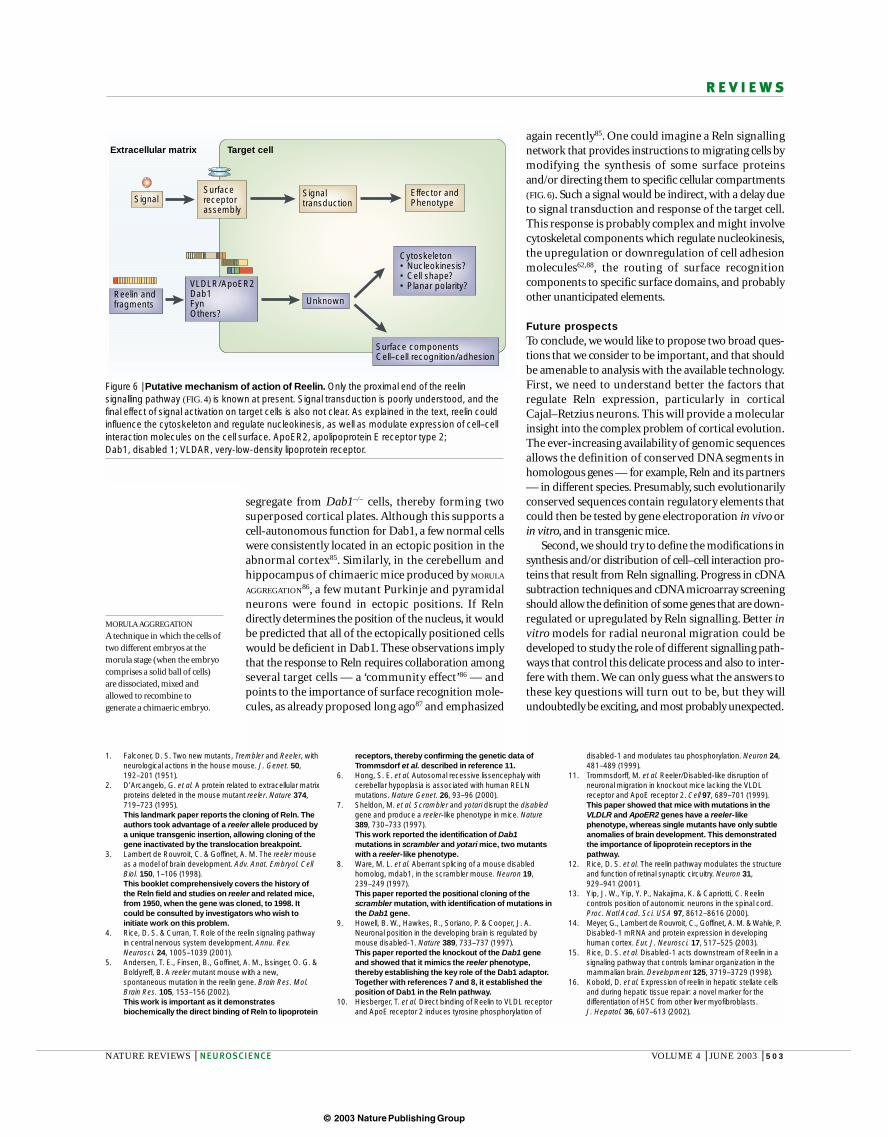
again recently85. One could imagine a Reln signallingnetwork that provides instructions to migrating cells bymodifying the synthesis of some surface proteinsand/or directing them to specific cellular compartments(FIG. 6). Such a signal would be indirect, with a delay dueto signal transduction and response of the target cell.This response is probably complex and might involvecytoskeletal components which regulate nucleokinesis,the upregulation or downregulation of cell adhesionmolecules62,88, the routing of surface recognition components to specific surface domains, and probablyother unanticipated elements.
Future prospectsTo conclude, we would like to propose two broad ques-tions that we consider to be important, and that shouldbe amenable to analysis with the available technology.First, we need to understand better the factors that regulate Reln expression, particularly in corticalCajal–Retzius neurons. This will provide a molecularinsight into the complex problem of cortical evolution.The ever-increasing availability of genomic sequencesallows the definition of conserved DNA segments inhomologous genes — for example, Reln and its partners— in different species. Presumably, such evolutionarilyconserved sequences contain regulatory elements thatcould then be tested by gene electroporation in vivo orin vitro, and in transgenic mice.
Second, we should try to define the modifications insynthesis and/or distribution of cell–cell interaction pro-teins that result from Reln signalling. Progress in cDNAsubtraction techniques and cDNA microarray screeningshould allow the definition of some genes that are down-regulated or upregulated by Reln signalling. Better invitro models for radial neuronal migration could bedeveloped to study the role of different signalling path-ways that control this delicate process and also to inter-fere with them. We can only guess what the answers tothese key questions will turn out to be, but they willundoubtedly be exciting, and most probably unexpected.
segregate from Dab1–/– cells, thereby forming twosuperposed cortical plates. Although this supports acell-autonomous function for Dab1, a few normal cellswere consistently located in an ectopic position in theabnormal cortex85. Similarly, in the cerebellum andhippocampus of chimaeric mice produced by MORULA
AGGREGATION86, a few mutant Purkinje and pyramidalneurons were found in ectopic positions. If Relndirectly determines the position of the nucleus, it wouldbe predicted that all of the ectopically positioned cellswould be deficient in Dab1. These observations implythat the response to Reln requires collaboration amongseveral target cells — a ‘community effect’86 — andpoints to the importance of surface recognition mole-cules, as already proposed long ago87 and emphasized
MORULA AGGREGATION
A technique in which the cells oftwo different embryos at themorula stage (when the embryocomprises a solid ball of cells)are dissociated, mixed andallowed to recombine togenerate a chimaeric embryo.
1. Falconer, D. S. Two new mutants, Trembler and Reeler, withneurological actions in the house mouse. J. Genet. 50,192–201 (1951).
2. D’Arcangelo, G. et al. A protein related to extracellular matrixproteins deleted in the mouse mutant reeler. Nature 374,719–723 (1995).This landmark paper reports the cloning of Reln. Theauthors took advantage of a reeler allele produced bya unique transgenic insertion, allowing cloning of thegene inactivated by the translocation breakpoint.
3. Lambert de Rouvroit, C. & Goffinet, A. M. The reeler mouseas a model of brain development. Adv. Anat. Embryol. CellBiol. 150, 1–106 (1998).This booklet comprehensively covers the history ofthe Reln field and studies on reeler and related mice,from 1950, when the gene was cloned, to 1998. Itcould be consulted by investigators who wish toinitiate work on this problem.
4. Rice, D. S. & Curran, T. Role of the reelin signaling pathwayin central nervous system development. Annu. Rev.Neurosci. 24, 1005–1039 (2001).
5. Andersen, T. E., Finsen, B., Goffinet, A. M., Issinger, O. G. &Boldyreff, B. A reeler mutant mouse with a new,spontaneous mutation in the reelin gene. Brain Res. Mol.Brain Res. 105, 153–156 (2002).This work is important as it demonstratesbiochemically the direct binding of Reln to lipoprotein
receptors, thereby confirming the genetic data ofTrommsdorf et al. described in reference 11.
6. Hong, S. E. et al. Autosomal recessive lissencephaly withcerebellar hypoplasia is associated with human RELNmutations. Nature Genet. 26, 93–96 (2000).
7. Sheldon, M. et al. Scrambler and yotari disrupt the disabledgene and produce a reeler-like phenotype in mice. Nature389, 730–733 (1997).This work reported the identification of Dab1mutations in scrambler and yotari mice, two mutantswith a reeler-like phenotype.
8. Ware, M. L. et al. Aberrant splicing of a mouse disabledhomolog, mdab1, in the scrambler mouse. Neuron 19,239–249 (1997).This paper reported the positional cloning of thescrambler mutation, with identification of mutations inthe Dab1 gene.
9. Howell, B. W., Hawkes, R., Soriano, P. & Cooper, J. A.Neuronal position in the developing brain is regulated bymouse disabled-1. Nature 389, 733–737 (1997).This paper reported the knockout of the Dab1 geneand showed that it mimics the reeler phenotype,thereby establishing the key role of the Dab1 adaptor.Together with references 7 and 8, it established theposition of Dab1 in the Reln pathway.
10. Hiesberger, T. et al. Direct binding of Reelin to VLDL receptorand ApoE receptor 2 induces tyrosine phosphorylation of
disabled-1 and modulates tau phosphorylation. Neuron 24,481–489 (1999).
11. Trommsdorff, M. et al. Reeler/Disabled-like disruption ofneuronal migration in knockout mice lacking the VLDLreceptor and ApoE receptor 2. Cell 97, 689–701 (1999).This paper showed that mice with mutations in theVLDLR and ApoER2 genes have a reeler-likephenotype, whereas single mutants have only subtleanomalies of brain development. This demonstratedthe importance of lipoprotein receptors in thepathway.
12. Rice, D. S. et al. The reelin pathway modulates the structureand function of retinal synaptic circuitry. Neuron 31,929–941 (2001).
13. Yip, J. W., Yip, Y. P., Nakajima, K. & Capriotti, C. Reelincontrols position of autonomic neurons in the spinal cord.Proc. Natl Acad. Sci. USA 97, 8612–8616 (2000).
14. Meyer, G., Lambert de Rouvroit, C., Goffinet, A. M. & Wahle, P.Disabled-1 mRNA and protein expression in developinghuman cortex. Eur. J. Neurosci. 17, 517–525 (2003).
15. Rice, D. S. et al. Disabled-1 acts downstream of Reelin in asignaling pathway that controls laminar organization in themammalian brain. Development 125, 3719–3729 (1998).
16. Kobold, D. et al. Expression of reelin in hepatic stellate cellsand during hepatic tissue repair: a novel marker for thedifferentiation of HSC from other liver myofibroblasts. J. Hepatol. 36, 607–613 (2002).
Figure 6 | Putative mechanism of action of Reelin. Only the proximal end of the reelinsignalling pathway (FIG. 4) is known at present. Signal transduction is poorly understood, and thefinal effect of signal activation on target cells is also not clear. As explained in the text, reelin couldinfluence the cytoskeleton and regulate nucleokinesis, as well as modulate expression of cell–cellinteraction molecules on the cell surface. ApoER2, apolipoprotein E receptor type 2; Dab1, disabled 1; VLDAR, very-low-density lipoprotein receptor.
Target cellExtracellular matrix
Effector andPhenotype
Cytoskeleton• Nucleokinesis?• Cell shape?• Planar polarity?
Surface componentsCell–cell recognition/adhesion
Signaltransduction
Unknown
VLDLR/ApoER2Dab1FynOthers?
Surfacereceptorassembly
Signal
Reelin andfragments

© 2003 Nature Publishing Group
504 | JUNE 2003 | VOLUME 4 www.nature.com/reviews/neuro
R E V I E W S
17. Buchaille, R., Couble, M. L., Magloire, H. & Bleicher, F. A substractive PCR-based cDNA library from humanodontoblast cells: identification of novel genes expressed intooth forming cells. Matrix Biol. 19, 421–430 (2000).
18. Smalheiser, N. R. et al. Expression of reelin in adultmammalian blood, liver, pituitary pars intermedia, andadrenal chromaffin cells. Proc. Natl Acad. Sci. USA 97,1281–1286 (2000).
19. Ikeda, Y. & Terashima, T. Expression of reelin, the generesponsible for the reeler mutation, in embryonicdevelopment and adulthood in the mouse. Dev. Dyn. 210,157–172 (1997).
20. Bar, I. et al. A YAC contig containing the reeler locus withpreliminary characterization of candidate gene fragments.Genomics 26, 543–549 (1995).
21. DeSilva, U. et al. The human reelin gene: isolation,sequencing, and mapping on chromosome 7. GenomeRes. 7, 157–164 (1997).
22. Royaux, I., Lambert de Rouvroit, C., D’Arcangelo, G.,Demirov, D. & Goffinet, A. M. Genomic organization of themouse reelin gene. Genomics 46, 240–250 (1997).
23. Tremolizzo, L. et al. An epigenetic mouse model formolecular and behavioral neuropathologies related toschizophrenia vulnerability. Proc. Natl Acad. Sci. USA 99,17095–17100 (2002).
24. Chen, Y., Sharma, R. P., Costa, R. H., Costa, E. & Grayson,D. R. On the epigenetic regulation of the human reelinpromoter. Nucleic Acids Res. 30, 2930–2939 (2002).
25. Chen, M. L., Chen, S. Y., Huang, C. H. & Chen, C. H.Identification of a single nucleotide polymorphism at the 5’promoter region of human reelin gene and association studywith schizophrenia. Mol. Psychiatry 7, 447–448 (2002).
26. Bar, I., Lambert de Rouvroit, C. & Goffinet, A. M. Theevolution of cortical development. An hypothesis based onthe role of the Reelin signaling pathway. Trends Neurosci.23, 633–638 (2000).
27. Lambert de Rouvroit, C., Bernier, B., Royaux, I., de Bergeyck, V.& Goffinet, A. M. Evolutionarily conserved, alternativesplicing of reelin during brain development. Exp. Neurol.156, 229–238 (1999).
28. D’Arcangelo, G. et al. Reelin is a secreted glycoproteinrecognized by the CR-50 monoclonal antibody. J. Neurosci.17, 23–31 (1997).
29. de Bergeyck, V., Naerhuyzen, B., Goffinet, A. M. & Lambertde Rouvroit, C. A panel of monoclonal antibodies againstreelin, the extracellular matrix protein defective in reelermutant mice. J. Neurosci. Methods 82, 17–24 (1998).
30. Ogawa, M. et al. The reeler gene-associated antigen onCajal–Retzius neurons is a crucial molecule for laminarorganization of cortical neurons. Neuron 14, 899–912(1995).Before the Reln gene was cloned, these authorsgenerated the CR50 antibody against Cajal–Retziuscells by immunizing reeler mice with brain extracts.The antibody was later shown to recognize an N-terminal epitope of Reln and is thought to perturb thefunction of Reln.
31. Lambert de Rouvroit, C. et al. Reelin, the extracellular matrixprotein deficient in reeler mutant mice, is processed by ametalloproteinase. Exp. Neurol. 156, 214–217 (1999).
32. Herz, J. & Bock, H. H. Lipoprotein receptors in the nervoussystem. Annu. Rev. Biochem. 71, 405–434 (2002).
33. Nimpf, J. & Schneider, W. J. From cholesterol transport tosignal transduction: low density lipoprotein receptor, verylow density lipoprotein receptor, and apolipoprotein Ereceptor-2. Biochim. Biophys. Acta 1529, 287–298 (2000).
34. Rudenko, G. et al. Structure of the LDL receptorextracellular domain at endosomal pH. Science 298,2337–2339 (2002).
35. Stockinger, W. et al. The reelin receptor ApoER2 recruitsJNK-interacting proteins-1 and-2. J. Biol. Chem. 275,25625–25632 (2000).
36. Howell, B. W., Lanier, L. M., Frank, R., Gertler, F. B. &Cooper, J. A. The disabled 1 phosphotyrosine-bindingdomain binds to the internalization signals oftransmembrane glycoproteins and to phospholipids. Mol.Cell. Biol. 19, 5179–5188 (1999).
37. D’Arcangelo, G. et al. Reelin is a ligand for lipoproteinreceptors. Neuron 24, 471–479 (1999).
38. Benhayon, D., Magdaleno, S. & Curran, T. Binding of purifiedReelin to ApoER2 and VLDLR mediates tyrosinephosphorylation of Disabled-1. Brain Res. Mol. Brain Res.112, 33–45 (2003).
39. Boucher, P. et al. Platelet-derived growth factor mediatestyrosine phosphorylation of the cytoplasmic domain of thelow Density lipoprotein receptor-related protein in caveolae.J. Biol. Chem. 277, 15507–15513 (2002).
40. Riddell, D. R., Sun, X. M., Stannard, A. K., Soutar, A. K. &Owen, J. S. Localization of apolipoprotein E receptor 2 tocaveolae in the plasma membrane. J. Lipid Res. 42,998–1002 (2001).
41. Kojima, T., Nakajima, K. & Mikoshiba, K. The disabled 1gene is disrupted by a replacement with L1 fragment inyotari mice. Brain Res. Mol. Brain Res. 75, 121–127 (2000).
42. Howell, B. W., Herrick, T. M. & Cooper, J. A. Reelin-inducedtryosine phosphorylation of disabled 1 during neuronalpositioning. Genes Dev. 13, 643–648 (1999).
43. Keshvara, L., Benhayon, D., Magdaleno, S. & Curran, T.Identification of reelin-induced sites of tyrosylphosphorylation on disabled 1. J. Biol. Chem. 276,16008–16014 (2001).
44. Lambert de Rouvroit, C. & Goffinet, A. M. Cloning of humanDAB1 and mapping to chromosome 1p31-p32. Genomics53, 246–247 (1998).
45. Bar, I., Tissir, F., Lambert De Rouvroit, C., De Backer, O. &Goffinet, A. M. The gene encoding disabled-1 (DAB1), theintracellular adaptor of the reelin pathway, reveals unusualcomplexity in human and mouse. J. Biol. Chem. (2002).
46. Howell, B. W., Herrick, T. M., Hildebrand, J. D., Zhang, Y. &Cooper, J. A. Dab1 tyrosine phosphorylation sites relaypositional signals during mouse brain development. Curr.Biol. 10, 877–885 (2000).
47. Herrick, T. M. & Cooper, J. A. A hypomorphic allele of dab1reveals regional differences in reelin-Dab1 signaling duringbrain development. Development 129, 787–796 (2002).This paper shows that a Dab1 null mutation can berescued fully by a knock-in of the normal Dab1 cDNA,and partially by a partial cDNA that contains thePI/PTB domain and the adjacent region that encodeskey Tyr residues, but not by a cDNA in which five Tyrresidues are replaced by Phe.
48. Keshvara, L., Magdaleno, S., Benhayon, D. & Curran, T.Cyclin-dependent kinase 5 phosphorylates disabled 1independently of Reelin signaling. J. Neurosci. 22,4869–4877 (2002).
49. Kubo, K., Mikoshiba, K. & Nakajima, K. Secreted Reelinmolecules form homodimers. Neurosci. Res. 43, 381–388(2002).
50. Utsunomiya-Tate, N. et al. Reelin molecules assembletogether to form a large protein complex, which is inhibitedby the function-blocking CR-50 antibody. Proc. Natl Acad.Sci. USA 97, 9729–9734 (2000).
51. Jossin, Y. et al. The Reelin signaling pathway: some recentdevelopments. Cereb. Cortex (in the press).
52. Miyata, T., Nakajima, K., Mikoshiba, K. & Ogawa, M.Regulation of Purkinje cell alignment by reelin as revealedwith CR-50 antibody. J. Neurosci. 17, 3599–3609 (1997).
53. Nakajima, K., Mikoshiba, K., Miyata, T., Kudo, C. & Ogawa, M.Disruption of hippocampal development in vivo by CR-50mAb against reelin. Proc. Natl Acad. Sci. USA. 94,8196–8201 (1997).
54. Bock, H. H. & Herz, J. Reelin activates SRC family tyrosinekinases in neurons. Curr. Biol. 13, 18–26 (2003).
55. Arnaud, L., Ballif, B. A., Forster, E. & Cooper, J. A. Fyntyrosine kinase is a critical regulator of disabled-1 duringbrain development. Curr. Biol. 13, 9–17 (2003).References 54 and 55 show that tyrosine kinases ofthe Src family, particularly Fyn and Yes, are keyelements in the transduction of the Reln signal, andthat they mediate Dab1 tyrosine phosphorylation.These two studies also indicate that there isredundancy among this kinase family.
56. Senzaki, K., Ogawa, M. & Yagi, T. Proteins of the CNR familyare multiple receptors for Reelin. Cell 99, 635–647 (1999).
57. Weiss, A. & Littman, D. R. Signal transduction bylymphocyte antigen receptors. Cell 76, 263–274 (1994).
58. Zhang, Z., Coomans, C. & David, G. Membrane heparansulfate proteoglycan-supported FGF2-FGFR1 signaling:evidence in support of the ‘cooperative end structures’model. J. Biol. Chem. 276, 41921–41929 (2001).
59. Loukinova, E. et al. Platelet-derived growth factor (PDGF)-induced tyrosine phosphorylation of the low densitylipoprotein receptor-related protein (LRP). Evidence forintegrated co-receptor function between LRP and thePDGF. J. Biol. Chem. 277, 15499–15506 (2002).
60. Pinson, K. I., Brennan, J., Monkley, S., Avery, B. J. &Skarnes, W. C. An LDL-receptor-related protein mediatesWnt signalling in mice. Nature 407, 535–538 (2000).
61. Meyer, G., De Rouvroit, C. L., Goffinet, A. M. & Wahle, P.Disabled-1 mRNA and protein expression in developinghuman cortex. Eur. J. Neurosci. 17, 517–525 (2003).
62. Hack, I., Bancila, M., Loulier, K., Carroll, P. & Cremer, H. Reelinis a detachment signal in tangential chain-migration duringpostnatal neurogenesis. Nature Neurosci. 5, 939–945 (2002).
63. Kim, H. M. et al. Reelin function in neural stem cell biology.Proc. Natl Acad. Sci. USA 99, 4020–4025 (2002).
64. Forster, E. et al. Reelin, Disabled 1, and β1 integrins arerequired for the formation of the radial glial scaffold in thehippocampus. Proc. Natl Acad. Sci. USA 99, 13178–13183(2002).
65. Tissir, F., Lambert de Rouvroit, C., Sire, J. Y., Meyer, G. &Goffinet, A. M. Reelin expression during embryonic brain
development in Crocodylus niloticus. J. Comp. Neurol. 457,250–262 (2003).
66. Haas, C. A. et al. Role for reelin in the development ofgranule cell dispersion in temporal lobe epilepsy. J. Neurosci.22, 5797–5802 (2002).
67. Perez-Costas, E. et al. Reelin immunoreactivity in the larvalsea lamprey brain. J. Chem. Neuroanat. 23, 211–221 (2002).
68. Costagli, A., Kapsimali, M., Wilson, S. W. & Mione, M.Conserved and divergent patterns of Reelin expression inthe zebrafish central nervous system. J. Comp. Neurol. 450,73–93 (2002).
69. Rice, D. S. & Curran, T. Disabled-1 is expressed in type AIIamacrine cells in the mouse retina. J. Comp. Neurol. 424,327–338 (2000).
70. Pearlman, A. L., Faust, P. L., Hatten, M. E. & Brunstrom, J. E.New directions for neuronal migration. Curr. Opin. Neurobiol.8, 45–54 (1998).
71. Frotscher, M. Cajal-Retzius cells, Reelin, and the formationof layers. Curr. Opin. Neurobiol. 8, 570–575 (1998).
72. Lambert de Rouvroit, C. & Goffinet, A. M. Neuronalmigration. Mech. Dev. 105, 47–56 (2001).
73. Malatesta, P., Hartfuss, E. & Gotz, M. Isolation of radial glialcells by fluorescent-activated cell sorting reveals a neuronallineage. Development 127, 5253–5263 (2000).
74. Noctor, S. C., Flint, A. C., Weissman, T. A., Dammerman, R. S.& Kriegstein, A. R. Neurons derived from radial glial cellsestablish radial units in neocortex. Nature 409, 714–720(2001).
75. Miyata, T., Kawaguchi, A., Okano, H. & Ogawa, M.Asymmetric inheritance of radial glial fibers by corticalneurons. Neuron 31, 727–741 (2001).
76. Nadarajah, B., Brunstrom, J. E., Grutzendler, J., Wong, R. O.& Pearlman, A. L. Two modes of radial migration in earlydevelopment of the cerebral cortex. Nature Neurosci. 4,143–150 (2001).
77. Nadarajah, B. & Parnavelas, J. G. Modes of neuronalmigration in the developing cerebral cortex. Nature Rev.Neurosci. 3, 423–432 (2002).
78. Del Rio, J. A. et al. A role for Cajal–Retzius cells and reelin inthe development of hippocampal connections. Nature 385,70–74 (1997).
79. Deller, T. et al. The hippocampus of the reeler mutantmouse: fiber segregation in area CA1 depends on theposition of the postsynaptic target cells. Exp. Neurol. 156,254–267 (1999).
80. Jossin, Y. & Goffinet, A. M. Reelin does not directly influenceaxonal growth. J. Neurosci. 21, RC183 (2001).
81. Teillon, S. M., Yiu, G. & Walsh, C. A. Reelin is expressed inthe accessory olfactory system, but is not a guidance cuefor vomeronasal axons. Brain Res. Dev. Brain Res. 140,303–307 (2003).
82. Walsh, C. A. & Goffinet, A. M. Potential mechanisms ofmutations that affect neuronal migration in man and mouse.Curr. Opin. Genet. Dev. 10, 270–274 (2000).
83. Phelps, P. E., Rich, R., Dupuy-Davies, S., Rios, Y. & Wong, T.Evidence for a cell-specific action of Reelin in the spinalcord. Dev. Biol. 244, 180–198 (2002).
84. Magdaleno, S., Keshvara, L. & Curran, T. Rescue of ataxiaand preplate splitting by ectopic expression of Reelin inreeler mice. Neuron 33, 573–586 (2002).This is the first attempt to rescue the reeler phenotypeby transgenesis with a Reln cDNA under the control ofthe nestin promoter. A partial rescue was obtained,even though Reln was expressed ectopically in theventricular zone instead of Cajal–Retzius cells. Thisprovides an argument for an indirect action of Relnthat would make neurons responsive to anothersignal in the marginal zone.
85. Hammond, V., Howell, B., Godinho, L. & Tan, S. S.Disabled-1 functions cell autonomously during radialmigration and cortical layering of pyramidal neurons. J. Neurosci. 21, 8798–8808 (2001).
86. Yang, H., Jensen, P. & Goldowitz, D. The community effectand Purkinje cell migration in the cerebellar cortex: analysis ofscrambler chimeric mice. J. Neurosci. 22, 464–470 (2002).By studying chimaeras produced by aggregation ofnormal and Dab1 mutant morulae, these authors showthat some actions of Dab1 are not fully explained by acell-autonomous effect of this intracellular adaptor,implying a ‘community effect’ that hints at theimportance of cell–cell interaction events.
87. Goffinet, A. M. An early development defect in the cerebralcortex of the reeler mouse. A morphological study leading toa hypothesis concerning the action of the mutant gene.Anat. Embryol. (Berl.) 157, 205–216 (1979).
88. Dulabon, L. et al. Reelin binds α3β1 integrin and inhibitsneuronal migration. Neuron 27, 33–44 (2000).
89. Costa, E., Davis, J., Pesold, C., Tueting, P. & Guidotti, A. Theheterozygote reeler mouse as a model for the developmentof a new generation of antipsychotics. Curr. Opin.Pharmacol. 2, 56–62 (2002).
© 2003 Nature Publishing Group
NATURE REVIEWS | NEUROSCIENCE VOLUME 4 | JUNE 2003 | 505
R E V I E W S
90. Fatemi, S. H., Kroll, J. L. & Stary, J. M. Altered levels ofReelin and its isoforms in schizophrenia and mooddisorders. Neuroreport 12, 3209–3215 (2001).
91. Guidotti, A., Pesold, C. & Costa, E. New neurochemicalmarkers for psychosis: a working hypothesis of theiroperation. Neurochem. Res. 25, 1207–1218 (2000).
92. Impagnatiello, F. et al. A decrease of reelin expression as aputative vulnerability factor in schizophrenia. Proc. NatlAcad. Sci. USA 95, 15718–15723 (1998).
93. Fatemi, S. H. The role of Reelin in pathology of autism. Mol.Psychiatry 7, 919–920 (2002).
94. Andres, C. Molecular genetics and animal models in autisticdisorder. Brain Res. Bull. 57, 109–119 (2002).
95. Zhang, H. et al. Reelin gene alleles and susceptibility to autismspectrum disorders. Mol. Psychiatry 7, 1012–1017 (2002).
96. Stokstad, E. Development. New hints into the biologicalbasis of autism. Science 294, 34–37 (2001).
97. Persico, A. M. et al. Reelin gene alleles and haplotypes as afactor predisposing to autistic disorder. Mol. Psychiatry 6,150–159 (2001).
98. Krebs, M. O. et al. Absence of association between apolymorphic GGC repeat in the 5’ untranslated region of thereelin gene and autism. Mol. Psychiatry 7, 801–804 (2002).
99. Tueting, P. et al. The phenotypic characteristics ofheterozygous reeler mouse. Neuroreport 10, 1329–1334(1999).
100. Lewis, D. A. & Levitt, P. Schizophrenia as a disorder ofneurodevelopment. Annu. Rev. Neurosci. 25, 409–432(2002).
101. Ohkubo, N. et al. Apolipoprotein E and Reelin ligandsmodulate tau phosphorylation through an apolipoprotein Ereceptor/disabled-1/glycogen synthase kinase-3β cascade.FASEB J. 17, 295–297 (2003).
102. Homayouni, R., Rice, D. S., Sheldon, M. & Curran, T.Disabled-1 binds to the cytoplasmic domain of amyloidprecursor-like protein 1. J. Neurosci. 19, 7507–7515(1999).
103. Ohshima, T. & Mikoshiba, K. Reelin signaling and Cdk5 inthe control of neuronal positioning. Mol. Neurobiol. 26,153–166 (2002).
104. Ko, J. et al. p35 and p39 are essential for cyclin-dependentkinase 5 function during neurodevelopment. J. Neurosci. 21,6758–6771 (2001).
105. Tseng, H. C., Zhou, Y., Shen, Y. & Tsai, L. H. A survey ofCdk5 activator p35 and p25 levels in Alzheimer’s diseasebrains. FEBS Lett. 523, 58–62 (2002).
106. Beffert, U. et al. Reelin-mediated signaling locally regulatesprotein kinase B/Akt and glycogen synthase kinase 3β. J. Biol. Chem. 277, 49958–49964 (2002).
107. Brich, J. et al. Genetic modulation of tau phosphorylation inthe mouse. J. Neurosci. 23, 187–192 (2003).
AcknowledgementsWe wish to thank C. Lambert de Rouvroit for her help in the prepara-tion of this review, as well as I. Bar, Y. Jossin and N. Ignatova for dis-cussions and inclusion of unpublished results. We apologize to col-leagues for the inevitable selection of citations due to space limitations.This work was supported by the Fondation Médicale Reine Elisabeth,and by the fifth framework program of the European Union.
Online links
DATABASESThe following terms in this article are linked online to:LocusLink: http://www.ncbi.nlm.nih.gov/LocusLink/ApoER2 | Dab1 | Fyn | Reln | VLDLROMIM: http://www.ncbi.nlm.nih.gov/Omim/autismAccess to this interactive links box is free online.