Embed Size (px)
Citation preview
S1
Supporting Information for
Production and Electrical Characterization of the Reflectin A2 Isoform from
Doryteuthis (Loligo) pealeii
David D. Ordinario1,†, Long Phan1,†, Ward G. Walkup IV1,2,†, Yegor Van Dyke1, Erica M.
Leung1, Michael Nguyen1, Amanda G. Smith1, Justin Kerr1, Mahan Naeim1, Ioannis
Kymissis3, Alon A. Gorodetsky1,4,*
1Department of Chemical Engineering and Materials Science, University of California, Irvine,
Irvine, CA 92697, USA
2Current Address: Department of Biology and Biological Engineering, California Institute of
Technology, Pasadena, CA 91125, USA
3Department of Electrical Engineering, Columbia University, New York, NY 10027, USA
4Department of Chemistry, University of California, Irvine, Irvine, CA 92697, USA
†Denotes equal contribution
*To whom correspondence may be addressed. Email: [email protected]
Electronic Supplementary Material (ESI) for RSC Advances.This journal is © The Royal Society of Chemistry 2016
S2
Experimental Methods and Procedures:
(I) Expression and Purification of Reflectin A2. Histidine-tagged wild type reflectin A2
(RfA2) was expressed and purified via a protocol that was modified from an analogous one
previously reported for wild type reflectin A1 (RfA1)S1,S2. In brief, an E. coli codon
optimized gene coding for histidine-tagged wild type RfA2 from Doryteuthis (Loligo) pealeii
(Genbank: ACZ57765.1) was synthesized and cloned into the pJExpress414 vector (DNA2.0).
The vector was transformed into BL21(DE3) cells (Novagen). RfA2 was expressed at 37 °C
using Overnight Express Instant Terrific Broth (TB) media (Novagen) supplemented with
100 µg mL −1 Carbenicillin. RfA2 was insoluble when expressed at 37 °C and was
sequestered in inclusion bodies. The inclusion bodies were then extracted by using
BugBuster® (Novagen) according to the manufacturer's suggested protocols. The inclusion
bodies were subsequently solubilized in denaturing buffer (pH 7.4, 50 mM sodium phosphate,
300 mM sodium chloride, 6 M guanidine hydrochloride) through repeated manual agitation
and sequentially filtered through 5, 0.45, and 0.22 µm filters. The protein was next purified
by high-performance liquid chromatography (HPLC) on an Agilent 1260 Infinity system
using an Agilent reverse phase C18 column with a gradient evolved from 95% Buffer A:5%
Buffer B to 5% Buffer A:95% Buffer B at a flow rate of 1 mL min-1 over 30 minutes (Buffer
A: 99.9% H2O, 0.1% TFA; Buffer B: 95% acetonitrile, 4.9% H2O, 0.1% TFA) (Supporting
Figure S1). The fractions containing RfA2 were pooled, flash frozen in liquid nitrogen, and
S3
lyophilized, yielding > 200 mg of pure RfA2 protein per liter of E. coli cell culture.
(II) Characterization of Reflectin A2. Wild type RfA2 was characterized according to a
general protocol, which was adopted from the literatureS1-S4. Throughout the purification
process, purified and unpurified reflectin samples were analyzed by sodium dodecyl sulfate
polyacrylamide gel electrophoresis (SDS-PAGE) and GelCode Blue Staining (Thermo) using
an Invitrogen XCell SureLock Mini using NuPAGE Novex 4-12% Bis-Tris gels, with
NuPAGE MOPS as the running buffer under reducing conditions. Stained protein bands were
routinely subjected to in-gel tryptic digestion, as confirmation of protein identityS1-S3. After
digestion, the peptides were separated on a reverse phase C18 chromatography column and
analyzed by mass spectrometry either on a Synapt G2 instrument (Waters) outfitted with an
electrospray ionization source or on an EASY-nLC system (Proxeon Biosystems, now
Thermo Scientific) connected to a hybrid LTQ-FT spectrometer (Thermo Scientific)
equipped with a nanoelectrospray ion source (Proxeon Biosystems, now Thermo
Scientific)S1,S4. The resulting sequence coverages routinely exceeded > 80 % for RfA2
(Supporting Figure S2).
(III) Fabrication of Reflectin A2-based Devices. The two-terminal devices were fabricated
using a protocol modified from established proceduresS2. In brief, silicon dioxide/silicon or
glass substrates (International Wafer Service, Inc.) were first cleaned in Piranha solution (1:3
hydrogen peroxide to sulfuric acid) and washed thoroughly. To fabricate devices for
S4
two-terminal direct current measurements, arrays of paired electrodes consisting of a 4 nm
chromium adhesion layer overlaid with a 40 nm palladium layer were electron-beam
evaporated onto SiO2/Si substrates through a shadow mask. The dimensions of the palladium
paired electrodes were 100 µm wide by 400 µm long, with an inter-electrode separation of 50
µm. To fabricate devices for two-terminal alternating current measurements, arrays of paired
electrodes consisting of a 4 nm chromium adhesion layer overlaid with a 40 nm gold layer
were electron-beam evaporated onto glass substrates through a shadow mask. The dimensions
of the gold paired electrodes were 2.5 cm wide by 3 cm long with an inter-electrode
separation of 100 µm. For all devices, aqueous solutions containing HPLC-purified RfA2
were prepared and subsequently dropcast onto the electrodes. The resulting films were dried
in ambient conditions, and the excess material was scribed away mechanically, leaving the
desired completed devices. To convert electron-injecting palladium (Pd) electrodes to
proton-injecting palladium hydride (PdHx) electrodes, the devices were exposed to a 5%
hydrogen/95% argon atmosphere both before and during the electrical measurements.
(IV) Physical Characterization of Reflectin A2-based Devices. The devices were
characterized with optical and atomic force microscopy, as previously describedS1,S2. The
dimensions of the reflectin films were determined from analysis of optical images obtained
with a Zeiss Axio Imager A1 Microscope. The thicknesses of both dry and humidified
reflectin films were determined from the analysis of topographical scans obtained with an
S5
Asylum Research MFP-3D Atomic Force microscope outfitted with an Asylum Research
Humidity Sensing Cell.
(V) Electrical Characterization of Reflectin A2-based Devices. The completed devices were
characterized electrically in two different configurations according to established
proceduresS2. The direct current measurements were performed on a Cascade Microtech
PM-5 Probe Station outfitted with an Agilent 4156C Semiconductor Parameter Analyzer,
with the current was recorded as a function of voltage at a scan rate of ~ 0.6 V/s. The direct
current measurements for palladium-contacted and palladium hydride-contacted devices were
performed under 100% argon and 5% hydrogen/95% argon atmospheres, respectively. The
alternating current measurements were performed with a 4294A Impedance Analyzer
(Agilent) at various frequencies with a constant applied voltage of 500 mV. The alternating
current measurements for gold-contacted devices were performed under a 100% argon
atmospheres. All electrical experiments were performed at an 80% relative humidity, which
was monitored with a Fisher Scientific hygrometer.
S6
Supporting Figures:
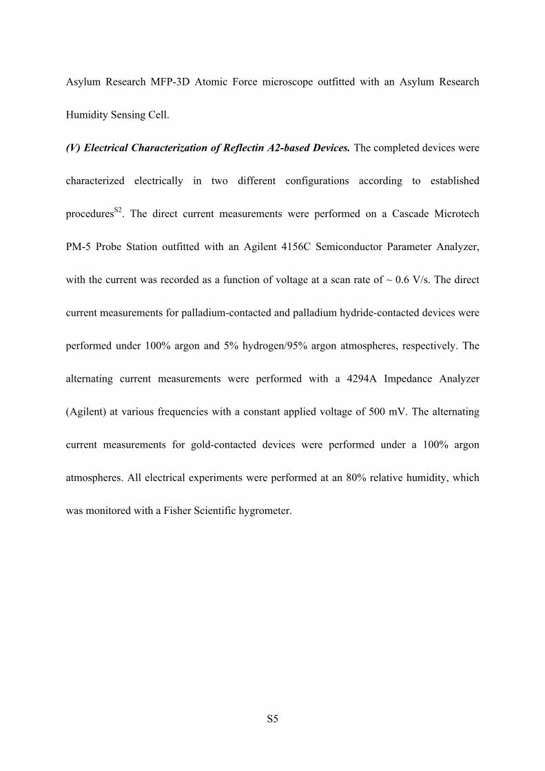
Supporting Figure S1: A typical analytical reverse phase HPLC chromatogram for RfA2
obtained after inclusion body filtration and concentration. The elution of the protein was
monitored at a wavelength of 280 nm. The peak indicates excellent purity.
S7
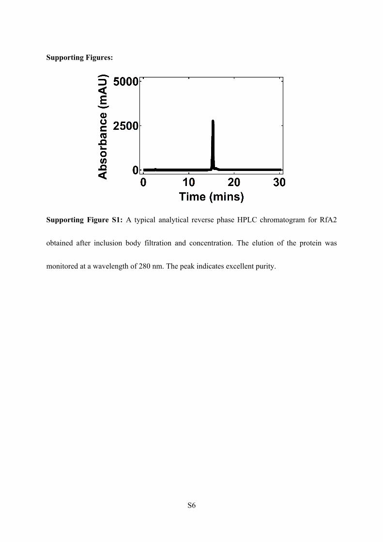
Supporting Figure S2: A tryptic peptide sequence coverage map obtained from tandem
mass spectrometry analysis of the trypsin-digested of the histidine-tagged RfA2 protein.
Bolded amino acids with a yellow background correspond to amino acids comprising tryptic
peptides. Bolded amino acids with a green background correspond to oxidized amino acids
comprising tryptic peptides. The total sequence coverage of ~ 83 % confirmed the purified
protein’s identity as RfA2 from D. pealeii.
S8
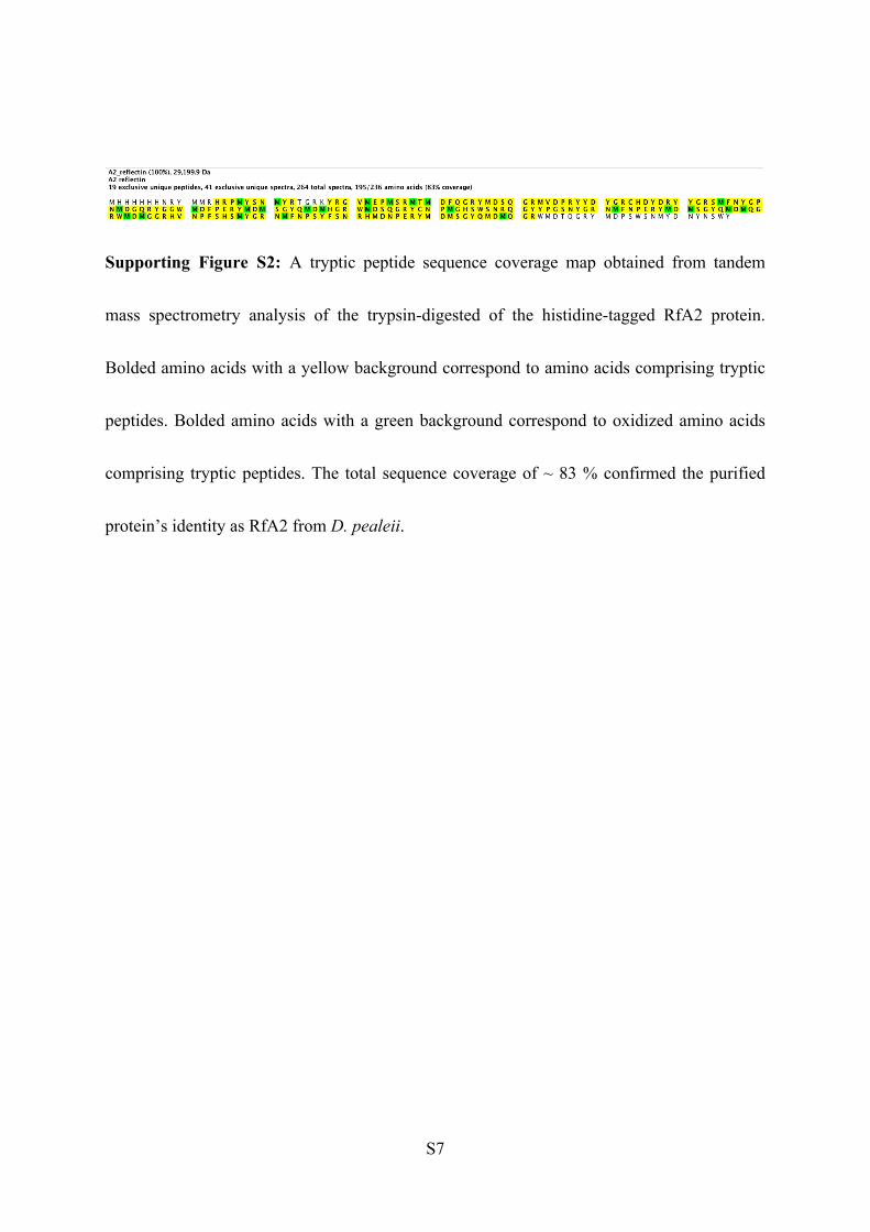
Supporting Figure S3: (A) General scheme for the fabrication of RfA2-based devices. (B) A
representative optical image of a completed device for which an RfA2 film bridges two
palladium electrodes. (C) A representative AFM image of an RfA2 active layer. The
additional typical images correspond to Figure 2 in the main text.
S9
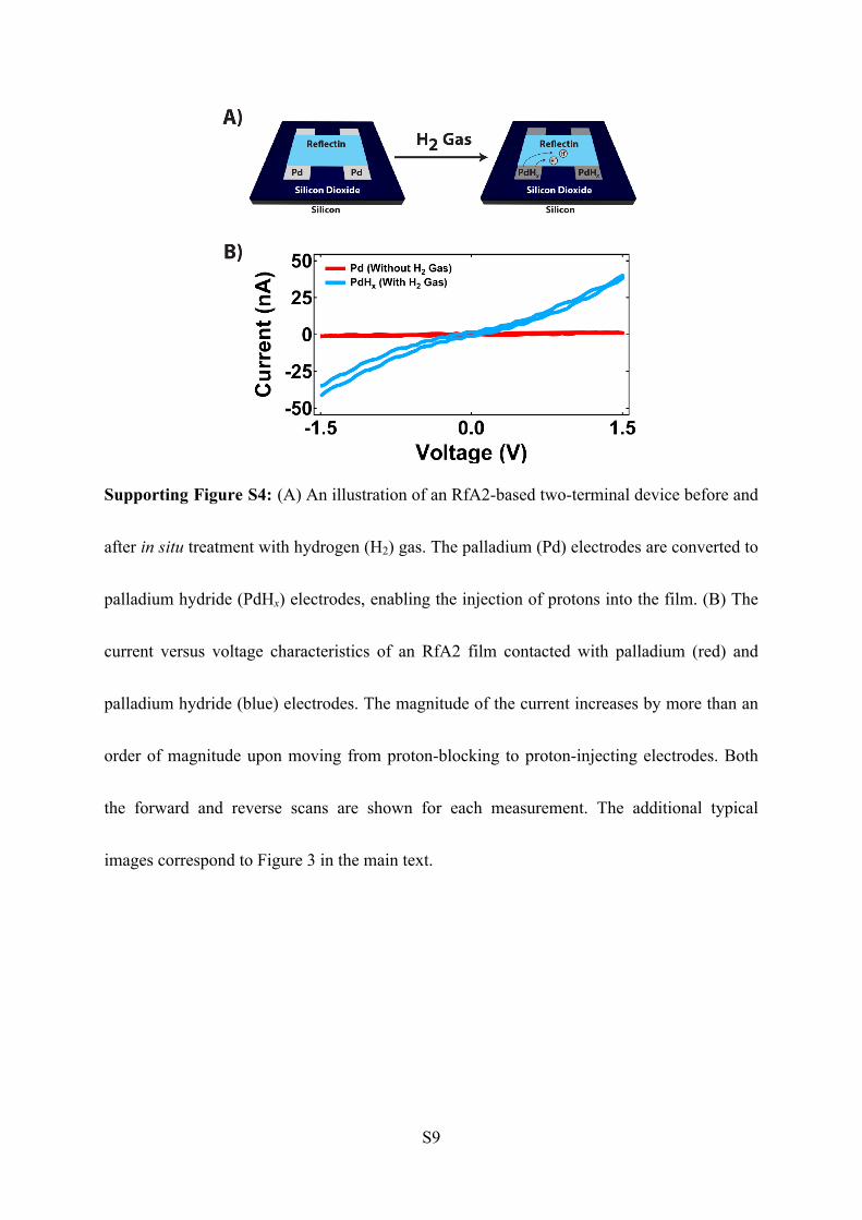
Supporting Figure S4: (A) An illustration of an RfA2-based two-terminal device before and
after in situ treatment with hydrogen (H2) gas. The palladium (Pd) electrodes are converted to
palladium hydride (PdHx) electrodes, enabling the injection of protons into the film. (B) The
current versus voltage characteristics of an RfA2 film contacted with palladium (red) and
palladium hydride (blue) electrodes. The magnitude of the current increases by more than an
order of magnitude upon moving from proton-blocking to proton-injecting electrodes. Both
the forward and reverse scans are shown for each measurement. The additional typical
images correspond to Figure 3 in the main text.
S10
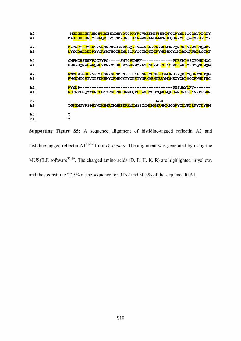
Supporting Figure S5: A sequence alignment of histidine-tagged reflectin A2 and
histidine-tagged reflectin A1S1,S2 from D. pealeii. The alignment was generated by using the
MUSCLE softwareS5,S6. The charged amino acids (D, E, H, K, R) are highlighted in yellow,
and they constitute 27.5% of the sequence for RfA2 and 30.3% of the sequence RfA1.
S11
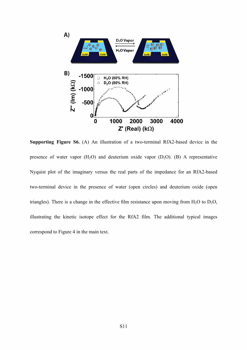
Supporting Figure S6. (A) An illustration of a two-terminal RfA2-based device in the
presence of water vapor (H2O) and deuterium oxide vapor (D2O). (B) A representative
Nyquist plot of the imaginary versus the real parts of the impedance for an RfA2-based
two-terminal device in the presence of water (open circles) and deuterium oxide (open
triangles). There is a change in the effective film resistance upon moving from H2O to D2O,
illustrating the kinetic isotope effect for the RfA2 film. The additional typical images
correspond to Figure 4 in the main text.
S12
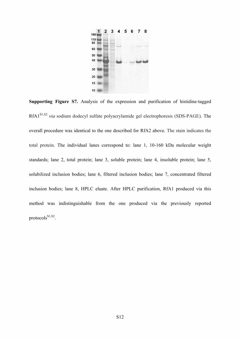
Supporting Figure S7. Analysis of the expression and purification of histidine-tagged
RfA1S1,S2 via sodium dodecyl sulfate polyacrylamide gel electrophoresis (SDS-PAGE). The
overall procedure was identical to the one described for RfA2 above. The stain indicates the
total protein. The individual lanes correspond to: lane 1, 10-160 kDa molecular weight
standards; lane 2, total protein; lane 3, soluble protein; lane 4, insoluble protein; lane 5,
solubilized inclusion bodies; lane 6, filtered inclusion bodies; lane 7, concentrated filtered
inclusion bodies; lane 8, HPLC eluate. After HPLC purification, RfA1 produced via this
method was indistinguishable from the one produced via the previously reported
protocolsS1,S2.
S13
Supporting References:
(S1) L. Phan, W. G. Walkup IV, D. D. Ordinario, E. Karshalev, J.-M. Jocson, A. M. Burke
and A. A. Gorodetsky, Adv. Mater., 2013, 25, 5621 – 5625.
(S2) D. D. Ordinario, L. Phan, W. G. Walkup IV, J.-M. Jocson, E. Karshalev, N. Hüsken, and
A. A. Gorodetsky, Nat. Chem., 2014, 6, 596 – 602.
(S3) A. Kalli, and S. Hess, Proteomics, 2012, 12, 21 – 31.
(S4) A. Shevchenko, H. Tomas, J. Havlis, J. V. Olsen, and M. Mann, Nat. Protoc., 2006, 1,
2856 – 2860.
(S5) R. C. Edgar, Nucleic Acids Res., 2004, 32, 1792 – 1797.
(S6) R.C. Edgar, BMC Bioinf., 2004, 5, 113.