Embed Size (px)
Citation preview

This article was downloaded by: [Naturalis]On: 18 June 2012, At: 02:00Publisher: Taylor & FrancisInforma Ltd Registered in England and Wales Registered Number: 1072954 Registeredoffice: Mortimer House, 37-41 Mortimer Street, London W1T 3JH, UK
Journal of Natural HistoryPublication details, including instructions for authors andsubscription information:http://www.tandfonline.com/loi/tnah20
Reflections on the genus AmagaOgren and Kawakatsu 1990, anddescription of a new genus of landplanarian (Platyhelminthes: Tricladida:Geoplanidae)José Horacio Grau a , Ronald Sluys b , Eudóxia Maria Froehlich c &Fernando Carbayo c da Museum für Naturkunde, Leibniz Institute for Research onEvolution and Biodiversity, Humboldt University Berlin, 10115,Berlin, Germanyb Institute for Biodiversity and Ecosystem Dynamics, Universityof Amsterdam and Netherlands Centre for Biodiversity Naturalis(section ZMA), P.O. Box 9514, 2300 RA, Leiden, Netherlandsc Departamento de Zoologia, Instituto de Biociências, Rua doMatão, 114, travessa 332. Universidade de São Paulo, SP, Brazild Escola de Artes, Ciências e Humanidades, Universidade de SãoPaulo, Av. Arlindo Bettio, 1000, São Paulo, SP, 03828-000, Brazil
Available online: 18 Jun 2012
To cite this article: José Horacio Grau, Ronald Sluys, Eudóxia Maria Froehlich & Fernando Carbayo(2012): Reflections on the genus Amaga Ogren and Kawakatsu 1990, and description of a new genusof land planarian (Platyhelminthes: Tricladida: Geoplanidae), Journal of Natural History, 46:25-26,1529-1546
To link to this article: http://dx.doi.org/10.1080/00222933.2012.691996
PLEASE SCROLL DOWN FOR ARTICLE
Full terms and conditions of use: http://www.tandfonline.com/page/terms-and-conditions
This article may be used for research, teaching, and private study purposes. Anysubstantial or systematic reproduction, redistribution, reselling, loan, sub-licensing,systematic supply, or distribution in any form to anyone is expressly forbidden.
The publisher does not give any warranty express or implied or make any representationthat the contents will be complete or accurate or up to date. The accuracy of anyinstructions, formulae, and drug doses should be independently verified with primarysources. The publisher shall not be liable for any loss, actions, claims, proceedings,demand, or costs or damages whatsoever or howsoever caused arising directly orindirectly in connection with or arising out of the use of this material.
Dow
nloa
ded
by [
Nat
ural
is]
at 0
2:00
18
June
201
2
Journal of Natural HistoryVol. 46, Nos. 25–26, July 2012, 1529–1546
Reflections on the genus Amaga Ogren and Kawakatsu 1990, anddescription of a new genus of land planarian (Platyhelminthes:Tricladida: Geoplanidae)
José Horacio Graua, Ronald Sluysb*, Eudóxia Maria Froehlichc
and Fernando Carbayoc,d
aMuseum für Naturkunde, Leibniz Institute for Research on Evolution and Biodiversity,Humboldt University Berlin, 10115 Berlin, Germany; bInstitute for Biodiversity and EcosystemDynamics, University of Amsterdam and Netherlands Centre for Biodiversity Naturalis (sectionZMA), P.O. Box 9514, 2300 RA Leiden, Netherlands; cDepartamento de Zoologia, Instituto deBiociências, Rua do Matão, 114, travessa 332. Universidade de São Paulo, SP, Brazil; dEscola deArtes, Ciências e Humanidades, Universidade de São Paulo, Av. Arlindo Bettio, 1000, São Paulo,SP, 03828-000, Brazil
(Received 12 September 2011; final version received 4 May 2012; printed 14 June 2012)
Amaga amagensis, the type species of the genus Amaga, and Amaga bogotensisare re-described. Detailed analysis of the morphology of A. amagensis revealedimportant taxonomic features, such as testes located dorsally to the supraintestinalparenchymal muscular layer, and secretory accumulations opening through the lat-eral margins of the body. These characters, as well as other morphological features,are discussed, culminating in an emendation of the generic diagnosis of Amaga.Amaga bogotensis exhibits a characteristic set of morphological features, namely aneversible penis, a male atrium lined with large musculosecretory papillae, and inde-pendent muscular coats around both male and female atrium. Therefore, a newgenus is proposed for this species.
Keywords: Platyhelminthes; Tricladida; Geoplaninae; Amaga; Bogga; Colombia.
Introduction
Many land planarian taxa are taxonomically poorly characterized because of insuffi-cient knowledge of their anatomical features, hindering phylogenetic and evolutionarystudies on this group of organisms. An example of this situation is the genus AmagaOgren and Kawakatsu, 1990 of the subfamily Geoplaninae, which has been erectedsolely on information from the literature and not on detailed examination of speci-mens (cf. Ogren and Kawakatsu 1990). The type species Amaga amagensis (Fuhrmann,1914) has not been studied since its original description (Fuhrmann 1914). As the cur-rent diagnosis of Amaga Ogren and Kawakatsu, 1990 fails to provide unique diagnosticfeatures, it embraces a heterogeneous group of 11 species and because the diagnosticfeatures of Amaga are ambiguous and inconclusive, this situation greatly hampers theelucidation of new taxa.
Geoplana bogotensis Von Graff, 1899, currently placed in the genus Amaga, wasoriginally described from a single worm from Bogotá (Colombia). Von Graff (1899)
*Corresponding author. Email: [email protected]
ISSN 0022-2933 print/ISSN 1464-5262 online© 2012 Taylor & Francishttp://dx.doi.org/10.1080/00222933.2012.691996http://www.tandfonline.com
Dow
nloa
ded
by [
Nat
ural
is]
at 0
2:00
18
June
201
2
1530 J.H. Grau et al.
described only the external characters and sent the single worm to the Museum fürNaturkunde in Berlin. A few years later he received another seven specimens fromthe environs of Bogotá. This material was studied by his student Busson (1903). Twoworms from the new material were sectioned and Busson considered his descriptionof the anatomy to complement the description of G. bogotensis given by Von Graffand also described the new variety G. bogotensis var. buergeri, which was merely basedon differences in colour pattern and body shape. Later, Fuhrmann (1914) collected38 specimens of Geoplana near Bogotá. This material was identified as Geoplanabogotensis because the general characteristics and the copulatory organs agreed withBusson’s account of the species. Fuhrmann also considered the external differencesthat Busson mentioned in support of his new variety to be the result of preservation orfixation artefacts.
Hyman (1955) identified one of her specimens from Tuicochchaca (Peru) as G.bogotensis on the basis of external features and found that the copulatory appara-tus differed from that described by both Busson and Fuhrmann. Therefore, Hymanconsidered her animal to represent the true G. bogotensis, in contrast to Busson’s andFuhrmann’s specimens. Du Bois-Reymond Marcus (1957) provided a succinct accountof this confusing situation and, to avoid further confusion, considered Hyman’sworm to represent a new species. One year later she called it Geoplana libbieae (DuBois-Reymond Marcus 1958) and considered Busson’s and Fuhrmann’s specimens torepresent the true G. bogotensis.
Ogren and Kawakatsu (1990) split the genus Geoplana into several taxa, basedon the informal groups delineated by Froehlich (1967), to facilitate further taxo-nomic studies. The authors erected the genus Amaga and included G. bogotensis VonGraff, 1899 and G. bogotensis var. buergeri sensu Hyman (1955) as separate species,Amaga bogotensis and Amaga buergeri, respectively. Subsequently, Kawakatsu et al.(1992) transferred G. bogotensis Von Graff, 1899 to the collective genus PseudogeoplanaOgren and Kawakatsu, 1990.
The present paper details the anatomy of two species of land planarian fromColombia. Their general characteristics and the gross morphology of their copula-tory apparatuses are concordant with the current broad diagnosis of Amaga, albeitthat they also have new structures. As any attempt to coin a new genus would be pre-mature without first re-examining the type species, we here also provide a detailedre-description of the type material, so as to enable a re-evaluation of the genericcharacters of the genus Amaga.
Materials and methods
The type material of A. amagensis was received on loan from the Natural HistoryMuseum, London (NHM) as a series of histological sections on glass slides. Accordingto Fuhrmann (1914) the material was stained in acid Haemalum and treated with amixture of picric acid and fuchsin. The type specimen of A. bogotensis was obtainedfrom the Museum für Naturkunde, Berlin (ZMB), preserved in alcohol. It was cuttransversally into several pieces, according to body regions; each of these parts wasdehydrated in a graded series of ethanol, treated with isopropyl alcohol and embeddedinto paraffin wax. The same procedures were followed in the case of one specimen fromBogotá (ZMA V.Pl. 6904.1), housed in the Zoological Museum Amsterdam (ZMA),
Dow
nloa
ded
by [
Nat
ural
is]
at 0
2:00
18
June
201
2
Journal of Natural History 1531
except for the isopropyl alcohol treatment, and clove oil was used as clearing agent.Serial sections 8 µm thick were made of the various regions of the body. The sectionswere stained with Mallory–Cason stain. The slides of G. bogotensis made by Bussonwere studied and photographed at the Naturhistorisches Museum Wien (NHW). Thesubepidermal muscular index (SMI; given by the subepidermal musculature thickness:body height ratio) was calculated at the pre-pharyngeal region, after Froehlich (1955).Drawings were prepared using a camera lucida.
Abbreviations used in the figuresThe following abbreviations are used in the figures: cm, circular musculature; cs, creep-ing sole; dep, dorsal epidermis; dm, diagonal muscle; e, eye; ed, ejaculatory duct;es, erythrophil secretion; fa, female atrium; gl, glands; go, gonopore; gr, glandularridge; in, intestinal branch; lm, longitudinal muscle; ma, male atrium; od, oviduct; ov,ovary; ph, pharynx; pm, parenchymal muscle; pp, penis papilla; pv, prostatic vesicle;sb, subintestinal transverse muscle layer; sep, secretory papilla; sg; shell glands; sp,supraintestinal transverse muscle layer; te, testis; va, vagina; vd, vas deferens; vep,ventral epidermis; vi, vitellarium; vnp, ventral nerve plate.
Systematic account
Order TRICLADIDA Lang, 1884Family GEOPLANIDAE Stimpson, 1857
Subfamily GEOPLANINAE Stimpson, 1857Genus Amaga Ogren and Kawakatsu, 1990
Emended diagnosisGeoplaninae of large broad and flattened body with well-developed glandular bodymargins. SMI: 5–7%. Testes placed above supra-intestinal parenchymal transversemuscular layer; male atrium folded, with ejaculatory duct opening through a smallpapilla-like fold; penis eversible; prostatic vesicle extrabulbar, bifurcate and elongate.Ovovitelline ducts approaching copulatory apparatus from anterodorsal aspect andopening at the same point into the vagina. No cephalic specializations; subepidermalmusculature not insunk. Parenchymal longitudinal musculature absent. Adenodactylsor glandulo-muscular organs absent. Type species: Geoplana amagensis Fuhrmann,1914.
DistributionPeru, Colombia and Amazonian Brazil.
Amaga amagensis (Fuhrmann, 1914)
Material examinedType material: NHM 1928.1.4.83–88 + 1928.1.4.90–91 + 1928.1.4.112 + 1928.1.4.92–95, sagittal sections of the pharynx and copulatory organs; 1928.1.4.110–111+ 1928.1.4.113 + 1928.1.4.89 + 1928.1.4.114–115, sagittal sections of the posterior
Dow
nloa
ded
by [
Nat
ural
is]
at 0
2:00
18
June
201
2
1532 J.H. Grau et al.
end of a second animal; 1928.1.4.96–109, transversal section of pre-pharyngeal region;1928.1.4.116–120, horizontal sections of the anterior end; Cafetal la Camelia, south ofthe Amagá river valley, Colombia, altitude 1600–1800 m (note that there is a mix upin the numbering and assignment of slides to specimens concerning the two sagittallysectioned animals; the order given above represents the original, continuous series ofsections).
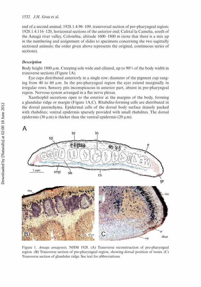
DescriptionBody height 1800 µm. Creeping sole wide and ciliated, up to 90% of the body width intransverse sections (Figure 1A).
Eye cups distributed anteriorly in a single row; diameter of the pigment cup rang-ing from 40 to 60 µm. In the pre-pharyngeal region the eyes extend marginally inirregular rows. Sensory pits inconspicuous in anterior part, absent in pre-pharyngealregion. Nervous system arranged in a flat nerve plexus.
Xanthophil secretions open to the exterior at the margins of the body, forminga glandular ridge or margin (Figure 1A,C). Rhabdite-forming cells are distributed inthe dorsal parenchyma. Epidermal cells of the dorsal body surface densely packedwith rhabdites; ventral epidermis sparsely provided with small rhabdites. The dorsalepidermis (30 µm) is thicker than the ventral epidermis (20 µm).
Figure 1. Amaga amagensis. NHM 1928. (A) Transverse reconstruction of pre-pharyngealregion. (B) Transverse section of pre-pharyngeal region, showing dorsal position of testes. (C)Transverse section of glandular ridge. See text for abbreviations.
Dow
nloa
ded
by [
Nat
ural
is]
at 0
2:00
18
June
201
2
Journal of Natural History 1533
Subepidermal musculature tripartite, composed of circular, double diagonal andlongitudinal layers. Longitudinal layer packed in small, separate bundles, strongerdorsally (60–75 µm high) than ventrally (40–50 µm high). In the anterior region thesubepidermal musculature has the same arrangement as in the rest of the body, sodoes not exhibit any specialization. Parenchymal musculature weak, composed of afew thin, obliquely running fibres, and other fibres arranged in three layers: supra-intestinal and subintestinal transversal layers, and a dorsal double layer of diagonalfibres. Longitudinal parenchymal muscle fibres absent.
Pharynx incompletely present in the type material. Mouth located at least 5.6 mmfrom the posterior end of the pharyngeal pouch. Pharynx of the collar-type, longand flat and highly folded. Oesophagus not observed, as the root of the pharynxwas not present in the slides of the type material. Pharyngeal pouch mainly sur-rounded by circular muscle fibres, with a few interspersed longitudinal fibres. Outerpharynx epithelium ciliated. Outer pharyngeal musculature composed of thin layersof intermingled longitudinal and circular muscles. Inner pharyngeal musculature notobserved, as only the external pharynx layers were sectioned.
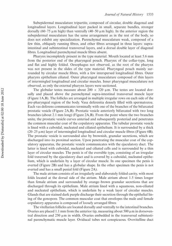
The globular testes measure about 200 × 320 µm. The testes are located dor-sally and placed above the parenchymal supra-intestinal transversal muscle layer(Figure 1A,B). The follicles are arranged in multiple irregular rows on each side of thepre-pharyngeal region of the body. Vasa deferentia densely filled with spermatozoa.Each vas deferens communicates terminally with one of the branches of the bifurcatedprostatic vesicle (Figure 2A,B). Prostatic vesicle anteriorly bifurcated with two longbranches (about 2.1 mm long) (Figure 2A,B). From the point where the two branchesunite, the prostatic vesicle curves anteriad and subsequently posteriad and penetratesthe common muscular coat of the copulatory apparatus. The tubular prostatic vesicleis lined with a cuboidal, nucleated and ciliated epithelium. It is surrounded by a thick(20–25 µm) layer of intermingled longitudinal and circular muscle fibres (Figure 6B).The prostatic vesicle is surrounded also by brownish, granular secretions, which aredischarged into its proximal section. Upon penetrating the muscular coat of the cop-ulatory apparatus, the prostatic vesicle communicates with the ejaculatory duct. Thelatter is lined with cuboidal, nucleated and ciliated cells and is surrounded by a thinlayer of circular muscles. The penis is of the eversible type, consisting of an irregularfold traversed by the ejaculatory duct and is covered by a cuboidal, nucleated epithe-lium, which is underlain by a layer of circular muscle. In one specimen the penis iseverted (Figure 2B) and has a globular shape. In the other specimen the penis is noteverted and has a small penial fold (Figure 2A).
The male atrium consists of an irregularly and elaborately folded cavity, with mostfolds located at the dorsal side of the atrium. Male atrium about 1.5 times longerthan female atrium and surrounded by orange–brown granular secretions that aredischarged through its epithelium. Male atrium lined with a squamous, non-ciliatedand nucleated epithelium, which is underlain by a weak layer of circular muscles.Glands that are stained dark purple discharge their secretion through the epithelial lin-ing of the gonopore. The common muscular coat that envelopes the male and femalecopulatory apparatus is composed of loosely arranged fibres
The vitellarian follicles are located dorsally and ventrally to the intestinal branches.Ovaries are placed 1.1 mm from the anterior tip, measuring about 500 µm in dorsoven-tral direction and 250 µm in width. Ovaries embedded in the transversal subintesti-nal parenchymatic muscle layer. Oviducal tubes not conspicuous. Ovovitelline duct
Dow
nloa
ded
by [
Nat
ural
is]
at 0
2:00
18
June
201
2

1534 J.H. Grau et al.
Figure 2. Amaga amagensis. NHM 1928. (A) Sagittal reconstruction of copulatory apparatusof specimen B. (B) Sagittal reconstruction of copulatory apparatus of specimen A. See text forabbreviations.
emerges from the ventrolateral part of each ovary. At the level of the gonopore theovovitelline ducts ascend at a 45◦ angle towards the dorsal body surface and even-tually open separately into the vagina. Just before communicating with the vagina,the oviducts receive the secretion of shell glands. The nucleated epithelium of theoviducts is surrounded by a thin layer of circular muscles. The female atrium consistsof a cavity lined with a nucleated, non-ciliated epithelium. The vagina emerges fromthe posterodorsal section of the female atrium and curves anterodorsally, attaining asomewhat horizontal orientation.
DiscussionThe histological sections of the type material were in good condition but, unfor-tunately, the animal was incompletely sectioned. Sections from the pre-pharyngealregion, anterior part of the pharynx and the region posterior to the copulatory appa-ratus are not available. Our observations on the type material are basically concordantwith those of Fuhrmann (1914). A few detailed measurements made by Fuhrmann,such as the size of the ovaries, are different from ours but may simply result from
Dow
nloa
ded
by [
Nat
ural
is]
at 0
2:00
18
June
201
2

Journal of Natural History 1535
the precision of our modern equipment. Furthermore, Fuhrmann did not describethat the prostatic vesicle extends anteriorly for a considerable distance. He consideredboth the branched and unbranched sections of the prostatic vesicle to be part of thevasa deferentia. He described these presumed vasa deferentia as being surrounded by astrong musculature and receiving the secretion of numerous glands. But our examina-tion revealed that there are no histological differences between the muscular prostaticvesicle and its anterior muscularized branches, and that they actually belong to thesame organ, namely the prostatic vesicle. It is only at a still farther anterior position,on the sides of the pharyngeal pouch, that each of the branches communicates withthe vas deferens (Figure 2A). The latter is narrower than the prostatic vesicle branches,does not receive any secretions and is surrounded by a weak layer of circular muscles.
Genus Bogga Grau and Sluys, gen. nov.
DiagnosisGeoplaninae with large, broad, and flattened body; anterior third gradually narrow-ing; posterior end rounded or obtuse. Eyes and sensory pits encircle the anterior endof the body. Sensory pits distributed in a single row. SMI: 9–10%. Subepidermal andparenchymal musculature without cephalic specializations. Parenchymal longitudinalmuscles absent. Male copulatory apparatus with eversible penis. Male atrium linedwith multiple, large musculo-secretory papillae. Vagina emerging from the posterodor-sal section of the female atrium; ovovitelline ducts approaching female canal from theanterodorsal side. Shell glands opening into the distal region of the oviducts. Commonglandular duct very reduced or absent. Adenodactyls absent. Type species: Geoplanabogotensis Von Graff, 1899.
DistributionBogotá, Colombia.
EtymologyThe generic epithet refers to a combination of the names Bogotá and Amaga. Gender:feminine.
Bogga bogotensis (Von Graff, 1899), comb. nov.
DiagnosisBroad leaf-like body; grey–black dorsal surface with a dark yellow marginal stripe thatencircles the body and a dark yellow mid-dorsal stripe; both stripes about the samewidth; eyes located marginally; SMI: 9–10%; pharynx of the collar type; oesophagussmall; globular prostatic vesicle located immediately posterior to pharyngeal pouch;small eversible penis; male atrium twice as long as female atrium; shell glands openinginto the distal region of the oviducts. Oviducts ascending laterally to the gonopore.Vagina emerging from the posterodorsal wall of the genital atrium, with dorsal oranterodorsal orientation.
Dow
nloa
ded
by [
Nat
ural
is]
at 0
2:00
18
June
201
2
1536 J.H. Grau et al.
Material examinedHolotype: ZMB 743. Preserved in 70% ethanol. Sections were prepared as follows:anterior region 1: transverse sections on seven slides; anterior region 2: sagittal sectionson 11 slides; pre-pharyngeal region: transverse sections on four slides; pharynx:sagittal sections on 14 slides; copulatory apparatus: sagittal sections on 24 slides.
NHW 2741. Original material of G. bogotensis var. buergeri studied by Busson.Sagittal sections of the copulatory apparatus on 167 slides.
ZMA V.Pl. 6904.1. National Park, Bogotá, Colombia, 15 August 1975, coll. J.Tamsitt; front end: horizontal sections on 37 slides; pre-pharyngeal region: transversesections on 13 slides; pharynx and copulatory apparatus: sagittal sections on 78 slides.
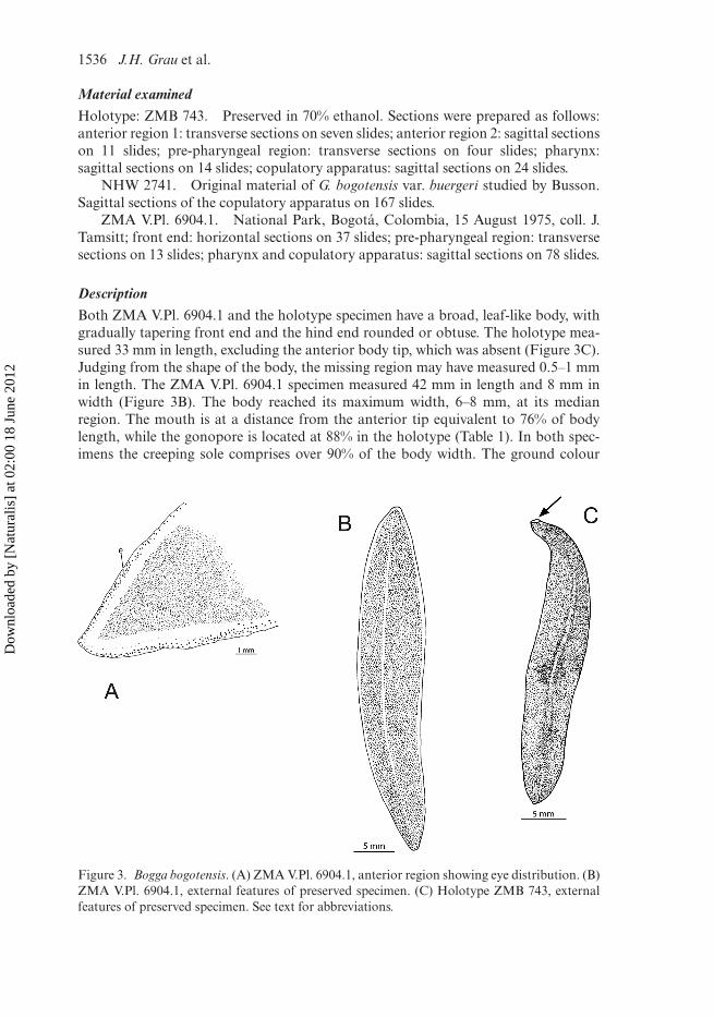
DescriptionBoth ZMA V.Pl. 6904.1 and the holotype specimen have a broad, leaf-like body, withgradually tapering front end and the hind end rounded or obtuse. The holotype mea-sured 33 mm in length, excluding the anterior body tip, which was absent (Figure 3C).Judging from the shape of the body, the missing region may have measured 0.5–1 mmin length. The ZMA V.Pl. 6904.1 specimen measured 42 mm in length and 8 mm inwidth (Figure 3B). The body reached its maximum width, 6–8 mm, at its medianregion. The mouth is at a distance from the anterior tip equivalent to 76% of bodylength, while the gonopore is located at 88% in the holotype (Table 1). In both spec-imens the creeping sole comprises over 90% of the body width. The ground colour
Figure 3. Bogga bogotensis. (A) ZMA V.Pl. 6904.1, anterior region showing eye distribution. (B)ZMA V.Pl. 6904.1, external features of preserved specimen. (C) Holotype ZMB 743, externalfeatures of preserved specimen. See text for abbreviations.
Dow
nloa
ded
by [
Nat
ural
is]
at 0
2:00
18
June
201
2
Journal of Natural History 1537
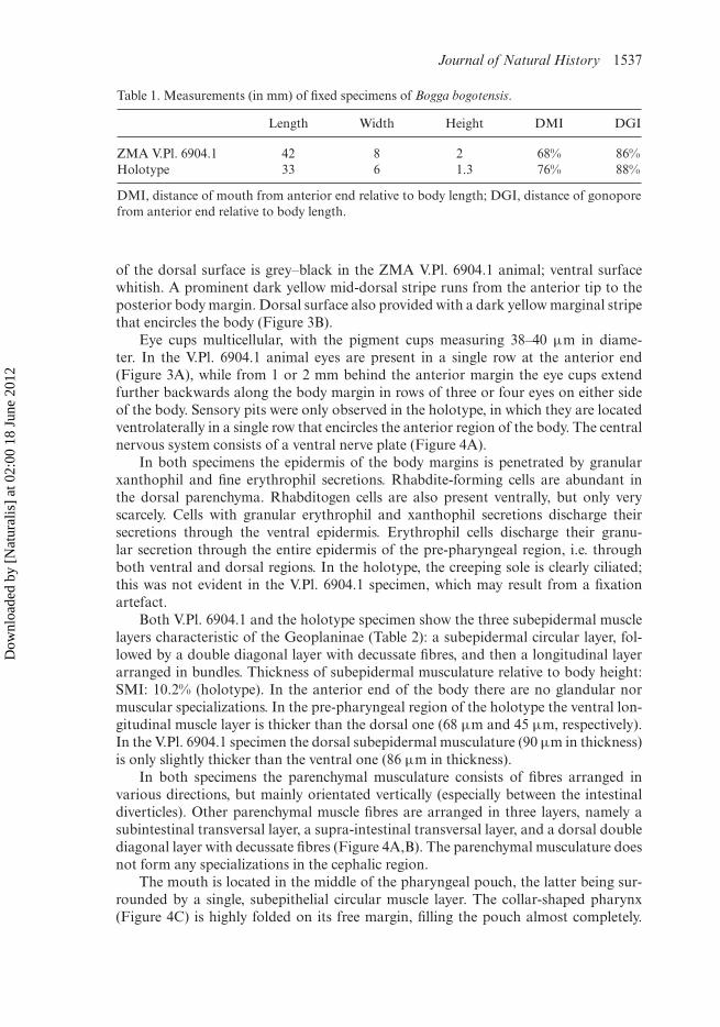
Table 1. Measurements (in mm) of fixed specimens of Bogga bogotensis.
Length Width Height DMI DGI
ZMA V.Pl. 6904.1 42 8 2 68% 86%Holotype 33 6 1.3 76% 88%
DMI, distance of mouth from anterior end relative to body length; DGI, distance of gonoporefrom anterior end relative to body length.
of the dorsal surface is grey–black in the ZMA V.Pl. 6904.1 animal; ventral surfacewhitish. A prominent dark yellow mid-dorsal stripe runs from the anterior tip to theposterior body margin. Dorsal surface also provided with a dark yellow marginal stripethat encircles the body (Figure 3B).
Eye cups multicellular, with the pigment cups measuring 38–40 µm in diame-ter. In the V.Pl. 6904.1 animal eyes are present in a single row at the anterior end(Figure 3A), while from 1 or 2 mm behind the anterior margin the eye cups extendfurther backwards along the body margin in rows of three or four eyes on either sideof the body. Sensory pits were only observed in the holotype, in which they are locatedventrolaterally in a single row that encircles the anterior region of the body. The centralnervous system consists of a ventral nerve plate (Figure 4A).
In both specimens the epidermis of the body margins is penetrated by granularxanthophil and fine erythrophil secretions. Rhabdite-forming cells are abundant inthe dorsal parenchyma. Rhabditogen cells are also present ventrally, but only veryscarcely. Cells with granular erythrophil and xanthophil secretions discharge theirsecretions through the ventral epidermis. Erythrophil cells discharge their granu-lar secretion through the entire epidermis of the pre-pharyngeal region, i.e. throughboth ventral and dorsal regions. In the holotype, the creeping sole is clearly ciliated;this was not evident in the V.Pl. 6904.1 specimen, which may result from a fixationartefact.
Both V.Pl. 6904.1 and the holotype specimen show the three subepidermal musclelayers characteristic of the Geoplaninae (Table 2): a subepidermal circular layer, fol-lowed by a double diagonal layer with decussate fibres, and then a longitudinal layerarranged in bundles. Thickness of subepidermal musculature relative to body height:SMI: 10.2% (holotype). In the anterior end of the body there are no glandular normuscular specializations. In the pre-pharyngeal region of the holotype the ventral lon-gitudinal muscle layer is thicker than the dorsal one (68 µm and 45 µm, respectively).In the V.Pl. 6904.1 specimen the dorsal subepidermal musculature (90 µm in thickness)is only slightly thicker than the ventral one (86 µm in thickness).
In both specimens the parenchymal musculature consists of fibres arranged invarious directions, but mainly orientated vertically (especially between the intestinaldiverticles). Other parenchymal muscle fibres are arranged in three layers, namely asubintestinal transversal layer, a supra-intestinal transversal layer, and a dorsal doublediagonal layer with decussate fibres (Figure 4A,B). The parenchymal musculature doesnot form any specializations in the cephalic region.
The mouth is located in the middle of the pharyngeal pouch, the latter being sur-rounded by a single, subepithelial circular muscle layer. The collar-shaped pharynx(Figure 4C) is highly folded on its free margin, filling the pouch almost completely.
Dow
nloa
ded
by [
Nat
ural
is]
at 0
2:00
18
June
201
2

1538 J.H. Grau et al.
Figure 4. Bogga bogotensis. (A) Holotype ZMB 743. Transverse section of the pre-pharyngealregion. (B) ZMA V.Pl. 6904.1, transverse section of the pre-pharyngeal region. (C) HolotypeZMB 743, sagittal reconstruction of pharynx. See text for abbreviations.
An oesophagus is present, but it was not completely observable in either of the speci-mens because it was not completely sectioned. The outer pharyngeal lining consistsof a flat, cuboidal epithelium. The outer pharynx musculature is composed of alayer of intermingled longitudinal and circular fibres (about 5 µm in thickness). Theouter epithelium of the distal region of the pharynx is penetrated by two types ofsecretory cells, producing erythrophil and xanthophil granules, respectively. The innerpharyngeal and oesophageal epithelia are underlain by a thin layer of longitudinalmuscles (6 µm in thickness), followed by a much thicker layer of circular fibres (60 µmin thickness).
Dow
nloa
ded
by [
Nat
ural
is]
at 0
2:00
18
June
201
2
Journal of Natural History 1539
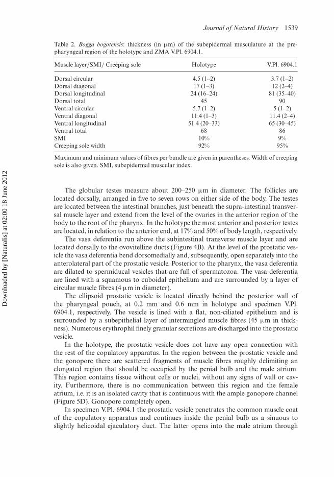
Table 2. Bogga bogotensis: thickness (in µm) of the subepidermal musculature at the pre-pharyngeal region of the holotype and ZMA V.Pl. 6904.1.
Muscle layer/SMI/ Creeping sole Holotype V.Pl. 6904.1
Dorsal circular 4.5 (1–2) 3.7 (1–2)Dorsal diagonal 17 (1–3) 12 (2–4)Dorsal longitudinal 24 (16–24) 81 (35–40)Dorsal total 45 90Ventral circular 5.7 (1–2) 5 (1–2)Ventral diagonal 11.4 (1–3) 11.4 (2–4)Ventral longitudinal 51.4 (20–33) 65 (30–45)Ventral total 68 86SMI 10% 9%Creeping sole width 92% 95%
Maximum and minimum values of fibres per bundle are given in parentheses. Width of creepingsole is also given. SMI, subepidermal muscular index.
The globular testes measure about 200–250 µm in diameter. The follicles arelocated dorsally, arranged in five to seven rows on either side of the body. The testesare located between the intestinal branches, just beneath the supra-intestinal transver-sal muscle layer and extend from the level of the ovaries in the anterior region of thebody to the root of the pharynx. In the holotype the most anterior and posterior testesare located, in relation to the anterior end, at 17% and 50% of body length, respectively.
The vasa deferentia run above the subintestinal transverse muscle layer and arelocated dorsally to the ovovitelline ducts (Figure 4B). At the level of the prostatic ves-icle the vasa deferentia bend dorsomedially and, subsequently, open separately into theanterolateral part of the prostatic vesicle. Posterior to the pharynx, the vasa deferentiaare dilated to spermiducal vesicles that are full of spermatozoa. The vasa deferentiaare lined with a squamous to cuboidal epithelium and are surrounded by a layer ofcircular muscle fibres (4 µm in diameter).
The ellipsoid prostatic vesicle is located directly behind the posterior wall ofthe pharyngeal pouch, at 0.2 mm and 0.6 mm in holotype and specimen V.Pl.6904.1, respectively. The vesicle is lined with a flat, non-ciliated epithelium and issurrounded by a subepithelial layer of intermingled muscle fibres (45 µm in thick-ness). Numerous erythrophil finely granular secretions are discharged into the prostaticvesicle.
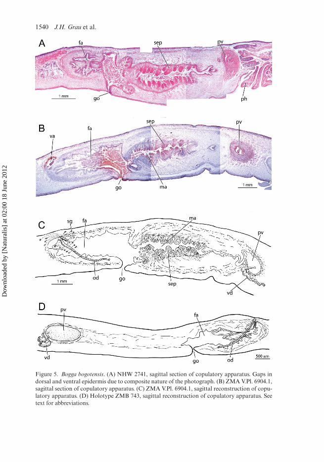
In the holotype, the prostatic vesicle does not have any open connection withthe rest of the copulatory apparatus. In the region between the prostatic vesicle andthe gonopore there are scattered fragments of muscle fibres roughly delimiting anelongated region that should be occupied by the penial bulb and the male atrium.This region contains tissue without cells or nuclei, without any signs of wall or cav-ity. Furthermore, there is no communication between this region and the femaleatrium, i.e. it is an isolated cavity that is continuous with the ample gonopore channel(Figure 5D). Gonopore completely open.
In specimen V.Pl. 6904.1 the prostatic vesicle penetrates the common muscle coatof the copulatory apparatus and continues inside the penial bulb as a sinuous toslightly helicoidal ejaculatory duct. The latter opens into the male atrium through
Dow
nloa
ded
by [
Nat
ural
is]
at 0
2:00
18
June
201
2
1540 J.H. Grau et al.
Figure 5. Bogga bogotensis. (A) NHW 2741, sagittal section of copulatory apparatus. Gaps indorsal and ventral epidermis due to composite nature of the photograph. (B) ZMA V.Pl. 6904.1,sagittal section of copulatory apparatus. (C) ZMA V.Pl. 6904.1, sagittal reconstruction of copu-latory apparatus. (D) Holotype ZMB 743, sagittal reconstruction of copulatory apparatus. Seetext for abbreviations.
Dow
nloa
ded
by [
Nat
ural
is]
at 0
2:00
18
June
201
2
Journal of Natural History 1541
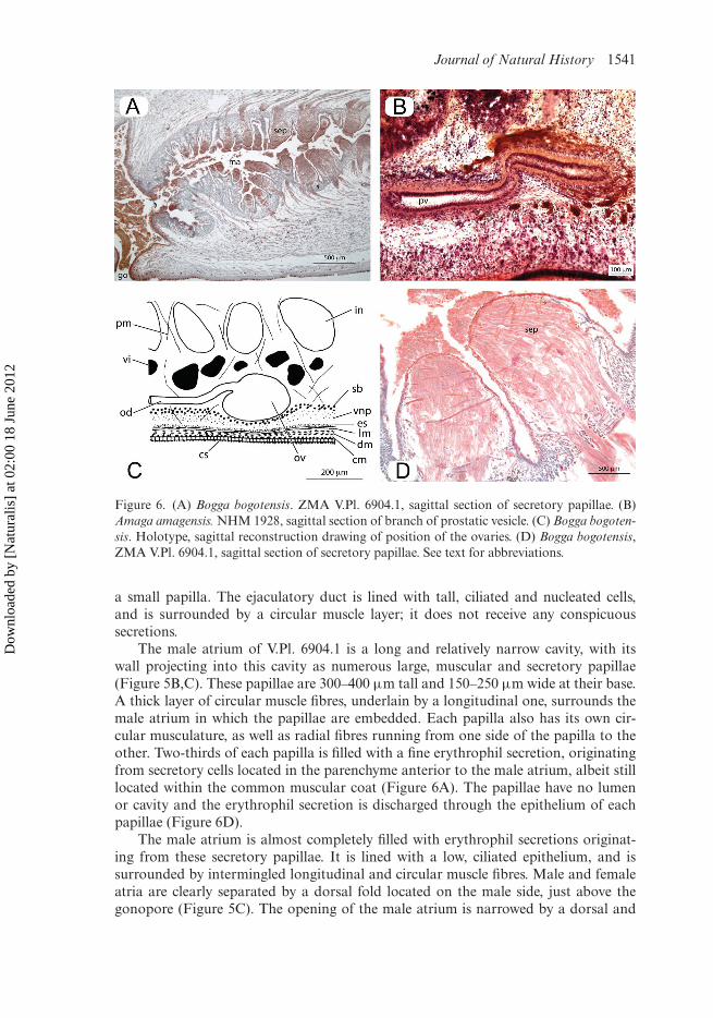
Figure 6. (A) Bogga bogotensis. ZMA V.Pl. 6904.1, sagittal section of secretory papillae. (B)Amaga amagensis. NHM 1928, sagittal section of branch of prostatic vesicle. (C) Bogga bogoten-sis. Holotype, sagittal reconstruction drawing of position of the ovaries. (D) Bogga bogotensis,ZMA V.Pl. 6904.1, sagittal section of secretory papillae. See text for abbreviations.
a small papilla. The ejaculatory duct is lined with tall, ciliated and nucleated cells,and is surrounded by a circular muscle layer; it does not receive any conspicuoussecretions.
The male atrium of V.Pl. 6904.1 is a long and relatively narrow cavity, with itswall projecting into this cavity as numerous large, muscular and secretory papillae(Figure 5B,C). These papillae are 300–400 µm tall and 150–250 µm wide at their base.A thick layer of circular muscle fibres, underlain by a longitudinal one, surrounds themale atrium in which the papillae are embedded. Each papilla also has its own cir-cular musculature, as well as radial fibres running from one side of the papilla to theother. Two-thirds of each papilla is filled with a fine erythrophil secretion, originatingfrom secretory cells located in the parenchyme anterior to the male atrium, albeit stilllocated within the common muscular coat (Figure 6A). The papillae have no lumenor cavity and the erythrophil secretion is discharged through the epithelium of eachpapillae (Figure 6D).
The male atrium is almost completely filled with erythrophil secretions originat-ing from these secretory papillae. It is lined with a low, ciliated epithelium, and issurrounded by intermingled longitudinal and circular muscle fibres. Male and femaleatria are clearly separated by a dorsal fold located on the male side, just above thegonopore (Figure 5C). The opening of the male atrium is narrowed by a dorsal and
Dow
nloa
ded
by [
Nat
ural
is]
at 0
2:00
18
June
201
2
1542 J.H. Grau et al.
ventral fold, slightly anterior to the gonopore. A muscular coat separately covers eachatrium, as suggested by a clear muscular division at the level of the dorsal atrial foldthat separates male and female atria.
In both animals the vitellarian follicles are relatively small, located dorsally andventrally to the intestine in the parenchymal space delimited by the supra-intestinaland subintestinal parenchymal muscle layers, without invading the space between theintestinal branches.
The ovaries are large and elongate, up to 500 µm long by 200 µm wide. Theyare ventrally located above the subintestinal parenchymal muscle layer (Figure 6C) at5.6 mm from the anterior tip in the holotype. The ovovitelline ducts arise from theposterodorsal surface of the ovaries.
The oviducts run between the subintestinal muscle layer and the ventral nerveplate. At the level of the female atrium the ducts run dorsomedially and, subsequently,open into the vagina; common oviduct very small or absent. Ovovitelline ducts linedby a ciliated epithelium and surrounded by a layer of interwoven muscle fibres. Shellglands, producing a granular xanthophil secretion, open into the distal half of theascending sections of the oviducts. After it has received the openings of the oviducts,the vagina dilates to form a cavity that is three to four times as wide as the oviducts.The female atrium is lined with a secretory epithelium. Granular xanthophil secretionsfill the lumen of the female atrium, which is principally surrounded by longitudinal andcircular muscle fibres.
DiscussionWe agree with Fuhrman (1914) that G. bogotensis and G. bogotensis var. buergeri con-cern one and the same species, a conclusion that is supported by the fact that theV.Pl. 6904.1 and the holotype specimen examined by us conform to the publisheddescriptions of both of these taxa.
The copulatory apparatus of the holotype was never described in the literature.Furthermore, the copulatory complex is only partly present in the holotype becausethe section between the prostatic vesicle and the female atrium is fully disintegrated,hence complicating taxonomic assignment of this specimen (Figure 5D). However, theV.Pl. 6904.1 animal and the holotype share two rare characteristics that strongly sug-gest their conspecificity: (1) the internal pharynx epithelium being underlain by a thinlayer of longitudinal muscles, followed by a much thicker layer of circular fibres; (2) theovaries being located above the subintestinal transversal parenchymal muscle layer(Figure 6C). Such a pharyngeal muscle arrangement is presently only known fromone other Andean species, Notogynaphallia andina (Hyman, 1961) (Carbayo 2003),while such a location of the ovaries is only known from Notogynaphallia sexstriata(Von Graff, 1899) (Carbayo 2003). Furthermore, another suite of features also pointsto their conspecificity: (1) similar size, body shape and colour pattern; (2) xanthophiland erythrophil glands opening through the body margins; (3) collar-shaped pharynx;(4) close proximity between the pharyngeal pouch and the prostatic vesicle; (5) ellipti-cal, extra-bulbar prostatic vesicle; (6) similar size ratio of male–female atria; (7) vaginalocated in posterodorsal section of female atrium; (8) ovovitelline ducts ascendingposterior to the gonopore; (9) direct communication between the oviducts and thevagina. Additionally, the animals were collected from the same locality (Bogotá,Colombia). Differences in ratio of dorsal versus ventral subepidermal longitudinal
Dow
nloa
ded
by [
Nat
ural
is]
at 0
2:00
18
June
201
2

Journal of Natural History 1543
musculature between the two animals may be the result of the more contracted stateof the V.Pl. 6904.1 specimen.
The NHW 2741 animal described by Busson (1903) is most probably also con-specific with the holotype. First, the copulatory apparatus of this animal is morpho-logically identical to the V.Pl. 6904.1 specimen (Figure 5A). Second, a series of othersimilarities between V.Pl. 6904.1 and the animal described by Busson (1903) as the vari-ety buergeri support our conclusion that the latter should be considered as a member ofthat species. These features do not only concern the gross morphology but also charac-ters such as: (1) secretory muscular papillae projecting into the male atrium, (2) zone ofcircular muscles at the bases of these papillae surrounding the male atrium, (3) locationof the ovaries above the subintestinal muscle layer, (4) direct communication betweenthe oviducts and the vagina.
In contrast to Busson (1903), we do not consider the penis-like fold at the end ofthe ejaculatory duct to be a true penis papilla, because it is not morphologically dif-ferentiated from the rest of the male atrium by the presence, for example, of associatedmuscle layers or glandular secretions. Earlier, it was already noted that the “ . . . pre-sumed intra-antral papilla of Amaga, which appears more like folds of the atrial wall,is not homologous with the penis papilla of the genus Geoplana . . . ” (E.M. Froehlich,in Ogren and Kawakatsu 1990, p. 87).
General discussion
Re-evaluation of the diagnostic features of Amaga
The diagnostic features of many genera in the Geoplaninae need to be re-evaluated,preferably in the context of a phylogenetic analysis of the entire group of land planari-ans. However, it is not our intention to present in this paper a complete revision of thegenus Amaga but rather to discuss the morphological features of its type species andto propose an emendation of the generic diagnosis.
The type species Amaga amagensis has a rather simple and conventional anatomyas it has few or no anatomical novelties that can easily be used to distinguish it fromother species of Geoplaninae. The main diagnostic feature, originally proposed, albeittentatively, by Froehlich (1967) and subsequently formalized by Ogren and Kawakatsu(1990) is the presence of a small intra-antral penis papilla. This papilla is actually aprojection from the anterior wall of the male atrium. Other characters mentioned byOgren and Kawakatsu (1990), such as body shape and size, and absence of musculo-glandular organs, are features to be found in many other taxa and therefore unableto uniquely define the genus Amaga. On the basis of our study of the type species aswell as other species of Geoplaninae, we do not consider the intra-antral penis papillaas forming a good diagnostic character for generic rank because the presumed papilladoes not show any independent structural features, such as the presence of its ownmuscle layers or the openings of special kinds of secretory cells. This presumed papillais not even a permanent structure, as is evident from Fuhrmann’s figures of the cop-ulatory apparatus of both his worms. For example, his fig. 24 shows a worm with itsintromittent organ clearly formed by a fold of the male atrial wall. Therefore, the cur-rent diagnosis, mentioning the presence of an intra-antral papilla, leads to confusionin cases of other animals in which the male atrium exhibits a highly folded wall, soresulting in a poorly defined papilla. In view of the above, we interpret the presumed
Dow
nloa
ded
by [
Nat
ural
is]
at 0
2:00
18
June
201
2
1544 J.H. Grau et al.
intra-penial papilla of Amaga actually to be the tip of an eversible penis, housed in afolded male atrium.
Another feature proposed by Ogren and Kawakatsu (1990) for Amaga concernsthe situation that the female canal opens into the dorsal section of the female atriumand that the oviducts approach the canal from an anterodorsal direction. Besidesbeing common to Geoplana, this condition applies to species of other genera of theGeoplaninae, such as Notogynaphallia Ogren and Kawakatsu, 1990, and PolycladusBlanchard, 1845. It is worth noting that the ovovitelline ducts open directly intothe vagina and do not fuse to form a common glandular duct, as in most othergenera of the Geoplaninae, such as Cephaloflexa Carbayo and Leal-Zanchet, 2003,Choeradoplana Von Graff, 1896, Issoca Froehlich, 1955, Supramontana Carbayo andLeal-Zanchet, 2003, and in some species of Geoplana. Therefore, we propose thisfeature as a diagnostic character for the genus Amaga.
One taxonomic character we wish to emphasize is the position of the testes inrelation to the parenchymal muscle layers. In most members of the Geoplaninae thetestes are placed underneath the supra-intestinal transversal muscle layer. The fact thatin Amaga the testes are located above the supra-intestinal layer of transverse musclesdeserves special attention. The testes occur dorsally to this muscle layer as well as atthe level of this layer or shortly underneath the layer in some Andean species, such asNotogynaphallia andina (Hyman, 1962), Gigantea urubambensis Negrete et al., 2010,and in Polycladus gayi Blanchard, 1845 (personal observation J.H.G.).
Systematic position of Bogga Grau and Sluys, gen. nov.Dorsal testes, broad creeping sole and subepidermal longitudinal muscles arrangedin bundles provide substantial evidence that the animals from Bogotá belong to thesubfamily Geoplaninae. Currently, the Geoplaninae comprises 17 genera, plus a collec-tive group, Pseudogeoplana Ogren and Kawakatsu, 1990, which was specifically createdto include all species for which morphological knowledge is insufficient to place themin one of the other genera of the subfamily.
Within this subfamily there are no genera into which we could comfortably fit ourspecimens from Bogotá and therefore the new genus Bogga was created to taxonomi-cally accommodate the animals. The genus Bogga is characterized by a unique feature,i.e. a male atrium lined with secretory papillae, a feature not known from other gen-era of the Geoplaninae. Musculo-glandular accessory organs are not common in theGeoplaninae. Currently there is only one genus that is known for its glandular struc-tures in the male copulatory organs, namely Gigantea Ogren and Kawakatsu, 1990.Carbayo (2008) suggested that the secretory structures in Gigantea are not homolo-gous between the different species in the genus. Furthermore, the author divided thesestructures into two types, muscularized and non-muscularized, and also discussedtheir position inside the copulatory apparatus. However, the monophyletic statusof Gigantea has been questioned, based on the fine morphology of these glandularstructures (cf. Carbayo 2008). Nonetheless, Bogga and Gigantea are different in theirmorphological aspects. The male atrium of Gigantea is filled with a large penis papillafrom which the embedded secretory papillae project outwards. In Bogga, however, thesecretory papillae are embedded in the wall of the male atrium and project towardsits lumen. Furthermore, a permanent penial structure is absent in Bogga because thepenis is merely represented by a very small papilla at the proximal region of the male
Dow
nloa
ded
by [
Nat
ural
is]
at 0
2:00
18
June
201
2
Journal of Natural History 1545
atrium. Additionally, the genus Gigantea is characterized by a posterior approach ofthe female canal, whereas in Bogga the female canal approaches the female atriumfrom an anterior direction.
The secretory papillae of Bogga appear be to unique among the known glandulo-muscular organs. Many different types of muscularized glandular organs are known tooccur in other taxa of Geoplanidae, but these are different from the papillae in Boggabecause the latter lack a duct or small canal.
The presence of secretory papillae projecting into the male atrium is not part of anyof the current generic diagnoses within the Geoplaninae. On the basis of other charac-ters, either separately or combined (e.g. subepidermal and parenchymal muscle layers,distribution of eyes and sensory pits), we were unable to comfortably fit the specimensfrom Bogotá into any of the present genera of the Geoplaninae and therefore a newgenus was erected.
Presently, the phylogenetic relationships within the Geoplaninae, and land planari-ans in general, have not been analysed in any detail, and the type species of many SouthAmerican genera await detailed taxonomic re-description. From that perspective, oneshould remain reluctant to propose monotypic genera. Nevertheless, we felt compelledto propose a new genus to accommodate the specimens from Bogotá, thus avoiding theunnatural groupings of species in very large genera that have so much troubled landplanarian taxonomy – see for example, the several genera that have been split off fromthe large genus Geoplana (cf. Ogren and Kawakatsu 1990). In this way we hope toprovide future explorations of the Colombian land planarian fauna with a stable andnatural taxonomic basis.
Acknowledgements
We are grateful to Dr Tim Littlewood (Natural History Museum, London, UK) for providing aloan of the type specimen of Amaga amagensis. Dr Birger Neuhaus (Museum für Naturkunde,Berlin, Germany) is thanked for the loan of the holotype of Bogga bogotensis and for givingpermission to histologically section this animal. Mr J. van Arkel (University of Amsterdam,the Netherlands) is thanked for his assistance in digitally rendering the figures. F.C. was sup-ported by FAPESP. Dr Helmut Sattmann is thanked for hosting J.H.G. at the NaturhistorischesMuseum Wien (NHW). Completion of the manuscript was made possible by a grant from theNetherlands Centre for Biodiversity Naturalis to R. Sluys.
References
Busson B. 1903. Ueber einige Landplanarien. Sitzungsber Akad Wien. 112(1):375–429.Carbayo F. 2003. Revisión de Notogynaphallia (Ogren & Kawakatsu, 1990). PhD the-
sis. Departamento de Biología Animal. Parasitología, Ecología, Edafología y QuímicaAgrícola. Universidad de Salamanca.
Carbayo F, Leal-Zanchet AM. 2003. Two new genera of geoplaninid land planarians(Platyhelminthes: Tricladida: Terricola) of Brazil in the light of cephalic specializations.Invertbr Syst. 17(3):449–468.
Du Bois-Reymond Marcus E. 1957. On Turbellaria. Acad Brasil Ciênc. 29(1):153–159.Froehlich C.G. 1967. A contribution to the zoogeography of neotropical land planarians. Acta
Zool Lill. 23:153–162.Fuhrmann O. 1914. Planaires terrestres de Colombie. In: Fuhrmann O, Mayor E. editors.
“Voyage d’Exploration Scientifique en Colombie”. Mém Soc Sci Natur de Neuchatel. 5(2):748–792 + 3 pls.
Dow
nloa
ded
by [
Nat
ural
is]
at 0
2:00
18
June
201
2
1546 J.H. Grau et al.
Froehlich CG. 1955. Sôbre morfologia e taxonomia das Geoplanidae. Bol Fac Filos Cienc LetrUniv Sao Paulo, Ser. Zool. 19:195–279.
Hyman LH. 1955. Miscellaneous marine and terrestrial flatworms from South America. AmMus Novit. 1742:1–33.
Ogren RE, Kawakatsu M. 1990. Index to the species of the Geoplanidae (Turbellaria,Tricladida, Terricola). Part I: Geoplaninae. Bull Fuji Women’s Coll, Ser. II. 28:79–166.
Von Graff L. 1899. Monographie der Turbellarien. II. Tricladida Terricola (Landplanarien).I–XII+574 pp. Atlas von Achtundfünfzig Tafeln zur Monographie der TurbellarienII. Tricladida Terricola (Landplanarien). Pls I–LVIII. Leipzig (Germany): WilhelmEngelmann.
Dow
nloa
ded
by [
Nat
ural
is]
at 0
2:00
18
June
201
2