Embed Size (px)
Citation preview
RESEARCH ARTICLE Open Access
Regional heterogeneity in response ofairway epithelial cells to cigarette smokeHario Baskoro1, Tadashi Sato1* , Keiko Karasutani1, Yohei Suzuki1, Aki Mitsui1, Naoko Arano1, Fariz Nurwidya1,2,Motoyasu Kato1, Fumiyuki Takahashi1, Yuzo Kodama1, Kuniaki Seyama1 and Kazuhisa Takahashi1
Abstract
Background: Cigarette smoke (CS) exposure causes an abnormal inflammatory response, which can result inchronic obstructive pulmonary disease (COPD). Previous studies show that this disorder predominantly occurs inperipheral or small-airway areas, whereas the same condition has not been identified in the larger airways duringthe course of COPD. However, the different biochemical and genetic alterations occurring in response to CSexposure among airway epithelial cells from different sites in the lungs have not been fully investigated.
Methods: Human small airway epithelial cells (SAECs) and normal human bronchial epithelial cells (NHBEs) wereexposed to CS extract (CSE), and microarray analysis was used to determine gene- and protein-expression profilesand identify alterations following CSE exposure in both cell types. An in vivo smoking experiment was alsoperformed to confirm differential responses to CS between sites in the lung.
Results: Microarray analysis of SAECs and NHBEs following 24 h of CSE exposure showed that inflammatory relatedpathways and terms, including the tumor necrosis factor-signaling pathway, were overrepresented, especially inSAECs. Clustering analysis highlighted prostaglandin-endoperoxide synthase-2 [also known as cyclooxygenase (COX)-2] as a gene specifically upregulated in SAECs, with COX-2 mRNA and protein expression significantly elevated byCSE exposure in SAECs (3.1- and 3.1-fold, respectively), but not in NHBEs. Furthermore, time-course analysis of COX-2 expression revealed earlier increases in SAECs compared with NHBEs following CS exposure. Short-term exposureof mouse lungs to CS was found to predominantly induce COX-2 expression in the small airway.
Conclusions: The small airway is more susceptible to CSE than the large airway and could be the initial site ofdevelopment of CS-related respiratory diseases, such as COPD.
Keywords: Cigarette smoke, Airway epithelial cells, Small airway, Inflammation, Cyclooxygenase-2, Chronicobstructive pulmonary disease
BackgroundCigarette smoke (CS) produces oxidant and reactive oxygenspecies in the lungs [1] and the unfavorable impacts of CSrepresent a primary cause of respiratory diseases, such aschronic obstructive pulmonary disease (COPD) and a lead-ing contributor to preventable morbidity and mortality [2].Preventing CS-related diseases can be best achieved byavoiding smoking initiation [3]; however, when the smokinghabit has already started, smoking cessation considerablydecreases numerous adverse health effects [4]. Intervention
by smoking cessation during the early stages of COPD candecrease the rate of lung-function loss [5]. Moreover,CS-induced oxidant burden can irreversibly changeendogenous antioxidant protection in airways. CS-derivedoxidants injure airway epithelial cells by affectinglipid-membrane, carbohydrate, protein, and DNA struc-tures and lead to the persistent inflammation evident inCOPD development [6].In addition to its status as a leading cause of mortality
worldwide, COPD is also the only major cause of deaththat continues to increase annually [2]. Two main featuresof COPD include progressive limitation of expiratory air-flow, which is not completely reversible, and an abnormalinflammatory response in the lungs [7]. Hogg et al. [8]
* Correspondence: [email protected] of Respiratory Medicine, Juntendo University Graduate Schoolof Medicine, 3-1-3 Hongo, Bunkyo-ku, Tokyo 113-8431, JapanFull list of author information is available at the end of the article
© The Author(s). 2018 Open Access This article is distributed under the terms of the Creative Commons Attribution 4.0International License (http://creativecommons.org/licenses/by/4.0/), which permits unrestricted use, distribution, andreproduction in any medium, provided you give appropriate credit to the original author(s) and the source, provide a link tothe Creative Commons license, and indicate if changes were made. The Creative Commons Public Domain Dedication waiver(http://creativecommons.org/publicdomain/zero/1.0/) applies to the data made available in this article, unless otherwise stated.
Baskoro et al. BMC Pulmonary Medicine (2018) 18:148 https://doi.org/10.1186/s12890-018-0715-4

suggested that the initial locus of inflammation in COPDis the small airways represented by membranous (diam-eter, < 2 mm), terminal and respiratory bronchioles. Insmokers, the small airways show structural abnormalities,with or without COPD. The relationship between inflam-matory mucus exudates from airway lumen and COPD se-verity was investigated by analyzing lung tissue frompatients with all clinical stages of COPD [9], revealing thatsmall-airway obstruction associated with COPD is relatedto thickening of the airway wall as a consequence of theremodelling process marked by mucociliary clearance-component malfunction and tissue repair related toinnate-immune mechanisms, finally resulting in acculu-mation of lumen inflammatory exudates [9, 10]. A morerecent study from the same group, based on multidetectorcomputed tomography obtained from patients at variousstages of COPD, showed that narrowing and loss of smallconducting airways occurs before the emphysematouschanges associated with COPD [11]. However, the geneticand biochemical alterations involved in responses to CSexposure by airway epithelial cells from different sites inthe lungs have not been fully investigated.In this study, we investigated the response to CS in
two commercially available human airway epithelial celllines [small airway epithelial cells (SAECs) and normalhuman bronchial epithelial cells (NHBEs)]. SAECs wereisolated from the distal portion of a healthy lung in the1-mm bronchiole area, and NHBEs were derived fromthe epithelial lining of airways above the bifurcation ofthe lung (according to documentation from Lonza, Ba-sel, Switzerland). We explored the genetic differencesbetween SAECs and NHBEs during the initial phase ofCS-exposure using microarray analysis and performed invivo smoking experiments using a mouse model to con-firm that cells from different lung sites respond differ-ently to CS exposure.
MethodsPreparation of CS extractCS extract (CSE) was prepared using commercially mar-keted Peace unfiltered cigarettes (29 mg of tar and 2.5 mgof nicotine per cigarette; Japan Tobacco, Tokyo, Japan).The smoke from one cigarette was bubbled through15 mL of sterilized purified water, and this solution passedthrough a 0.22-μm pore filter (Pall Corporation, PortWashington, NY, USA), to generate 100% CSE. Pilotcell-growth experiments showed that concentrations of≤2.5% CSE were not cytotoxic; therefore, cultured cellswere exposed to 2.5% CSE.
Cell cultureSAECs and NHBEs were purchased from Lonza. Threebatches of each cell type, each originally isolated fromthree different healthy never-smoker donors, were used.
SAECs were cultured with small airway epithelial cellgrowth medium (Lonza), and NHBEs were cultured withbronchial epithelial cell growth medium (Lonza), accord-ing to the manufacturer’s instructions (SAECs andNHBEs were seeded at 2500 and 3500 cells/cm2, respect-ively, in 25 cm2 plastic flasks). Both cell types were usedat passages three to six and assayed at the same passagesfor direct comparisons. After reaching approximately80% confluence, cells were treated with or without 2.5%CSE until assayed.
RNA extraction and real-time quantitative polymerasechain reaction (qPCR)Total RNA was extracted using miRNeasy mini kits (Qia-gen, Hilden, Germany). For cDNA synthesis, total RNAwas transcribed using ReverTra Ace qPCR RT master mixwith gDNA remover (Toyobo, Osaka, Japan). Real-timeqPCR for cyclooxygenase (COX)-2 mRNA detection wasperformed using Thunderbird SYBR qPCR mix (Toyobo),an ABI 7500 Fast real-time PCR instrument (Applied Bio-systems, Foster City, CA, USA), and COX-2-specificprimers [(5′→ 3′) Fw: CTGGCGCTCAGCCATACAG;Rv: CCGGGTACAATCGCACTTATACT; Thermo FisherScientific, Waltham, MA, USA] according to manufacturerinstructions. The expression of β-actin was measured usingspecific primers [(5′→ 3′) Fw: CTCTTCCAGCCTTCCTTCCT; Rv: AGCACTGTGTTGGCGTACAG; ThermoFisher Scientific] as an internal control.
Microarray analysisTwo different batches of SAECs and NHBEs were used formicroarray analysis. RNA samples (2 μg) were labeled usingthe low input Quick Amp labeling kit (Agilent Technolo-gies, Waldbronn, Germany). Labeled RNA was hybridizedto an oligonucleotide microarray (Whole Human Genome4× 44 K; Agilent Technologies) at 60 °C for 17 h. Afterwashing with reagents from the gene expression wash bufferkit (Agilent Technologies) and drying, slides were scannedwith an Agilent microarray scanner and analyzed using Fea-ture Extraction software (v9.5.1; Agilent Technologies).Normalization was performed using global normalizationmethods. Data from the microarray analysis have been de-posited in the Gene Expression Omnibus database (acces-sion ID: GSE107200). Differentially expressed genes weredefined based on unpaired t-test p values < 0.01 and foldchange ≥2. Gene-set enrichment analysis (GSEA) ofdifferentially expressed genes was conducted usingthe DAVID functional annotation tool (https://david.ncifcrf.gov/home.jsp). Gene sets from theKyoto Encyclopedia of Genes and Genomes (KEGG;http://www.genome.jp/kegg/) and the Gene Ontology(GO) biological process database (http://www.ge-neontology.org/) were used. Pathways and ontologyterms with a p < 0.05 were considered significant.
Baskoro et al. BMC Pulmonary Medicine (2018) 18:148 Page 2 of 11

The Benjamini-Hochberg procedure was used in theanalysis to control the false discovery rate.
Western blot analysisWhole-cell lysates were prepared, and the total proteincontent of each measured using a BCA kit (Thermo FisherScientific). Protein aliquots (10 μg) were separated by elec-trophoresis on 10% sodium dodecyl sulfate polyacrylamidegels. After transferring the proteins to Immobilion-Pmembranes (Merck Millipore Corporation, Darmstadt,Germany), membranes were blocked with 5% skim milkin Tris-buffered saline with Tween-20 and allowed to reactwith an anti-COX-2 (1:400; Santa Cruz Biotechnology,Dallas, TX, USA) and anti-β-actin (1:10,000; Wako,Tokyo, Japan) monoclonal antibodies at 4 °C overnight.Membranes were then incubated with the appropriatehorseradish-peroxidase-conjugated secondary antibody(1:20,000; Sigma-Aldrich, St. Louis, MO, USA), followedby detection with the Clarity western enhanced chemilu-minescence substrate (Bio-Rad Laboratories, Hercules,CA, USA). Western blot signals were captured using anImageQuant LAS-4000 mini fluorescence imager (GEHealthcare, Pittsburgh, PA, USA) and quantified usingMulti Gauge image analysis software (Fujifilm Corpor-ation, Tokyo, Japan).
In vivo exposure to CS and preparation of lung sectionsC57BL/6 J male mice (3 months old; n= 6) were purchasedfrom Oriental Yeast Co., Ltd. (Tokyo, Japan) and used forthe smoking experiment. The procedures for animal experi-mentation were approved by the Animal Care and UseCommittee of Juntendo University School of Medicine(Tokyo, Japan). Mice were maintained in a limited-accessbarrier facility and housed in a humidity (55% ± 10%) andtemperature (24 °C ± 2 °C) controlled room under 12-hlight/dark cycles. Mice were fed with standard commercialchow (CRF-1; Oriental Yeast) and provided water ad libi-tum. Mice were exposed to CS using Peace unfiltered ciga-rettes and a tobacco smoke-inhalation experimental systemfor small animals (Model SIS-CS; Shibata Scientific Tech-nology, Tokyo, Japan) [12–15]. The experimental settingswere as follows: 15 mL stroke volume, six puffs per minute,and 3.5% CS diluted with compressed air (the mass concen-tration of total particulate matter; 1430 ± 100 mg/m3). Micewere exposed to either diluted CS or fresh air (as a control,n = 3 each) for 30 min/day over 5 days.Mice were sacrificed at day 5 and their lungs subsequently
processed as described previously [13–15]. Briefly, micewere anesthetized and then sacrificed by exsanguination ofthe left atrium with perfusion of phosphate-buffered saline(PBS) through the main pulmonary artery. Lungs were in-flated and fixed by intratracheal instillation of 10% bufferedformalin (pH 7.4; Wako) at a constant pressure of 25 cmH2O for 48 h. Frontal plane sections of the lungs were taken
at the depth of the hilum, followed by further sectioning ofeach block into two equal-sized pieces, and from the frontalplane, and embedded in paraffin.
ImmunohistochemistryParaffin-embedded lung sections (4 μm) were deparaffi-nized in xylene and rehydrated in ethanol. Heat-inducedepitope retrieval (10 min at 125 °C) was performed incitrate buffer (pH 6.0; LSI Medience Corporation, Tokyo,Japan). After blocking with 2% bovine serum albumin inPBS, tissue sections were incubated at 4 °C overnightwith anti-COX-2 antibody (1:300; BD Biosciences, SanJose, CA, USA). Tissue sections were then incubatedwith biotin-labeled anti-mouse IgG antibody (1:300; Agi-lent Technologies) and labeled with avidin (1:50; VectorLaboratories, Burlingame, CA, USA) for 40 min at be-tween 25 °C and 28 °C. Signals were detected usinghydrogen peroxide and 3,3-diaminobenzidine tetrahy-drochloride. Tissue sections were counterstained withhematoxylin and dehydrated in xylene. The ratio of posi-tively immuno-stained nuclei to the total count of nucleipresent in a field at 400× magnification was determinedin 10 different lung areas per mouse.
Statistical analysisData are expressed as the mean ± standard error of themean (SEM) and were analyzed using GraphPad Prism 6(GraphPad Software, San Diego, CA, USA). Analysis ofvariance was performed using a non-parametric Krus-kal–Wallis test. When applicable, the Mann–Whitney Utest was used for comparisons between groups. Differ-ences were considered significant at p < 0.05.
ResultsGene-expression profiles altered by CSE exposure inSAECs and NHBEsAs shown in the scatter plots, exposure to CSE for 24 haltered the expression of several probes in NHBEs andSAECs (Fig. 1a and b). In NHBEs, 83 probes were upreg-ulated (≥ 2-fold) by CSE exposure, while in SAECs 116probes were upregulated (Fig. 1c). Among upregulatedprobes, 72 were upregulated only in SAECs (46% of thetotal upregulated probes), whereas 39 were upregulatedonly in NHBEs (25%). Ninety-four and 78 probes weredownregulated (≤ 2-fold) by CSE exposure in NHBEs andSAECs, respectively, with 71 probes (48% of the totaldownregulated probes) downregulated in NHBEsalone and 55 (37%) downregulated in SAECs alone(Fig. 1d). Differentially expressed probes are listed inAdditional file 1: Tables S1–S6.To elucidate the biological significance of altered
gene-expression profiles in NHBEs and SAECs, we per-formed GSEA based on two different gene sets chosenusing KEGG and GO biological-process analyses. As
Baskoro et al. BMC Pulmonary Medicine (2018) 18:148 Page 3 of 11

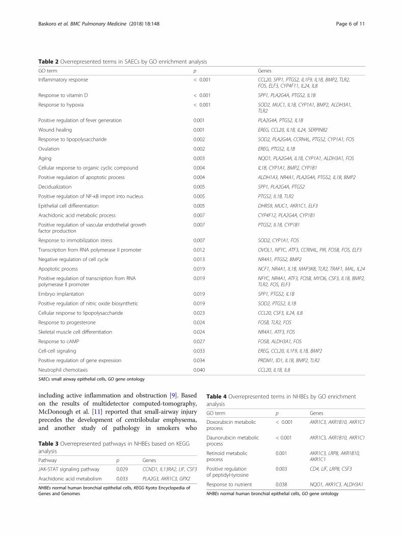
shown in Table 1, KEGG analysis showed that 10 pathways,including the tumor necrosis factor (TNF)- and nuclear fac-tor (NF)-κB-signaling pathways, were significantly and spe-cifically overrepresented in SAECs subjected to CSEexposure. Additionally, GO analysis revealed that 29 GOterms, mostly related to inflammation, were significantlyenriched in SAECs (Table 2). In contrast, no pathways or
terms were identified by analysis of enrichment of genesdownregulated in either CSE-exposed SAECs or NHBEs. Incontrast, two pathways, including the JAK-STAT signalingpathway and arachidonic acid metabolism, and five GOterms related to oxidative stress metabolism, were signifi-cantly overrepresented in NHBEs subjected to CSEexposure (Tables 3, 4).
A B
NHBE: 83 probes SAEC: 116 probes
44 probesupregulate39 probes
upregulate72 probesupregulate
NHBE: 94 probes SAEC: 78 probes
23 probesdownregulate
71 probesdownregulate
55 probesdownregulate
C
D
Fig. 1 Gene-expression profiles after exposure to CSE in SAECs and NHBEs. Two different batches of SAECs and NHBEs were used. Cells wereexposed to 2.5% CSE for 24 h or not (controls). Scatter plot of (a) NHBEs and (b) SAECs. The vertical axis represents the relative signal intensity inCSE-exposed cells, and the horizontal axis represents the control (without CSE exposure). Both axes show log scales. c Venn diagram of probesshowing significant upregulation in SAECs and NHBEs after CSE exposure. d Venn diagram of probes showing significant downregulation inSAECs and NHBEs after CSE exposure. CSE, cigarette smoke extract; SAECs, small airway epithelial cells; NHBEs, normal human bronchialepithelial cells.
Baskoro et al. BMC Pulmonary Medicine (2018) 18:148 Page 4 of 11
Next, we focused on the TNF-signaling pathway andperformed hierarchical clustering analysis of the 82 iden-tified genes included in this pathway. As shown in Fig. 2,CSE exposure significantly upregulated genes in a spe-cific cluster in SAECs compared with NHBEs, and thecluster included several inflammation-related genes, in-cluding TNF-receptor-associated factor-1 and prostaglan-din-endoperoxide synthase-2 (PTGS2).
COX-2 expression is altered by CSE exposure in SAECsand NHBEsHierarchical clustering analysis revealed that PTGS2 ishighly upregulated after CSE exposure, especially inSAECs. PTGS2 encodes a key enzyme (COX-2) involvedin prostaglandin (PG) biosynthesis. COX-2 is notexpressed under normal conditions in most cells; however,elevated levels are observed during inflammation. Further-more, our previous studies identified a relationship be-tween COX-2 and the abnormal inflammatory response inCOPD [16, 17]. Therefore, we evaluated COX-2 mRNAand protein expression following exposure to CSE. After24 h of exposure, COX-2 mRNA and protein levels wereboth significantly elevated in SAECs (3.1- and 3.1-foldcompared with no CSE treatment, respectively), but not inNHBEs (1.2- and 1.4-fold, respectively) (Fig. 3a and b). Toexplore the mechanism of COX-2 overproduction inSAECs, we performed time-course analysis of COX-2 ex-pression in response to CSE exposure in NHBEs andSAECs. Our results showed that COX-2 mRNA levelsbegan to increase 1 h earlier in SAECs than in NHBEs,and that COX-2 production was significantly higher inSAECs than in NHBEs (Fig. 3c). Time-course results for
COX-2 protein expression were similar to those obtainedfor COX-2 mRNA expression (Fig. 3d).
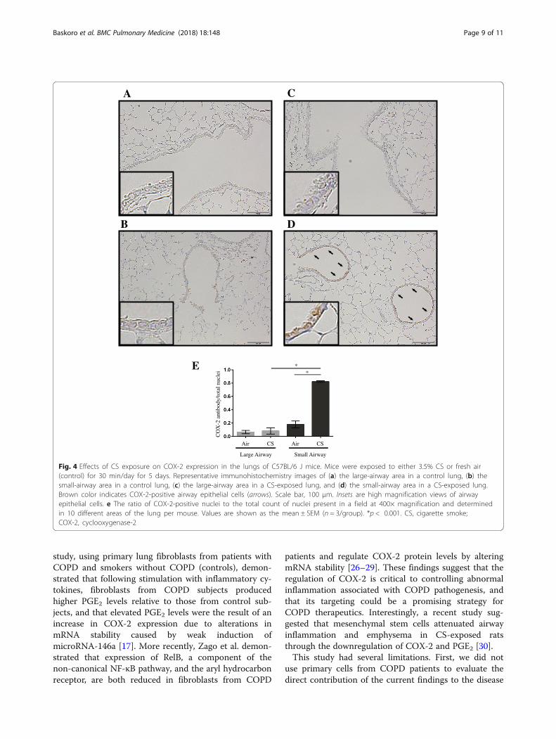
COX-2 expression in the lungs of CS-exposed miceTo investigate the initial effect of CS exposure on COX-2expression in vivo, we performed a short-duration in vivosmoking experiment using C57BL/6 J mice. Immunohisto-chemical analysis revealed that, after 5 days of exposure toCS, COX-2 expression increased compared with that inair-exposed mice, with more immuno-stained cells ob-served in small airways than in large airways (Fig. 4a–d).After 5 days of CS exposure, COX-2-positive cells were sig-nificantly increased in the small airways of mouse lungs,but not in the large airways (4.6-fold vs. 1.2-fold; Fig. 4e).
DiscussionThis study demonstrated that small and large airways dif-fered in their initial responses to CS exposure. Microarrayanalysis revealed that small airways showed higher suscepti-bility to CS compared with large airways and displayed en-hanced expression of genes associated with inflammation-related pathways, including TNF-signaling. AmongTNF-pathway related genes, PTGS2, also known as COX-2,showed the greatest difference in expression levels, withhigher CSE-induced increases in both mRNA and proteinexpression in SAECs compared with NHBEs. In vivoshort-duration smoking experiments also showed that initialCOX-2 expression occurred in lung small-airways. This isthe first study clarifying initial genetic and biochemical alter-ations associated with inflammatory responses to CS expos-ure in the small and large airways.CS is the most typical source of exogenous oxidants
[18], and oxidants derived from CS can injure airway epi-thelial cells, leading to persistent inflammation. Oxidativestress and persistent inflammation combine to result inincreases in airway free radicals and amplification of theexpression of proinflammatory genes that contribute tothe release of inflammatory proteins, thereby promotinginflammatory cell infiltration [6]. The present study dem-onstrated that the expression of proinflammatory genes,and initiation of signaling associated with their relatedpathways, were more greatly enhanced in SAECs relativeto NHBEs following a comparatively short period of CSEexposure. Further, we identified initial induction of COX-2expression in the small airway of CS-exposed mice. Theseresults suggest that SAECs represent the initial targets ofCS and play an important role in the development ofCS-mediated inflammation, which might contribute toCS-related respiratory diseases, such as COPD.Accumulating evidence suggests that the small airway is
the initial locus of inflammation in COPD and plays a crit-ical role in disease development [19]. Based on clinicalfindings, even patients with mild COPD already showsome pathological abnormalities in the small-airway area,
Table 1 Overrepresented pathways in SAECs based on KEGGanalysis
Pathway p Genes
Rheumatoid arthritis < 0.001 CCL20, MMP1, IL1B,TLR2, FOS, IL8
TNF-signaling pathway 0.001 CCL20, PTGS2, IL1B,MAP3K8, FOS, TRAF1
Toll-like receptor-signaling pathway
0.001 SPP1, IL1B, MAP3K8,TLR2, FOS, IL8
Leishmaniasis 0.001 NCF1, PTGS2, IL1B,TLR2, FOS
Malaria 0.004 CSF3, IL1B, TLR2, IL8
Amoebiasis 0.005 IL1B, IL1R2, TLR2, IL8,SERPINB2
NF-κB-signaling pathway 0.020 PTGS2, IL1B, TRAF1, IL8
MAPK-signaling pathway 0.026 NR4A1, PLA2G4A, IL1B,IL1R2, MAP3K8, FOS
Chagas disease(Americantrypanosomiasis)
0.032 IL1B, TLR2, FOS, IL8
SAECs small airway epithelial cells, KEGG Kyoto Encyclopedia of Genesand Genomes
Baskoro et al. BMC Pulmonary Medicine (2018) 18:148 Page 5 of 11
including active inflammation and obstruction [9]. Basedon the results of multidetector computed-tomography,McDonough et al. [11] reported that small-airway injuryprecedes the development of centrilobular emphysema,and another study of pathology in smokers who
Table 2 Overrepresented terms in SAECs by GO enrichment analysis
GO term p Genes
Inflammatory response < 0.001 CCL20, SPP1, PTGS2, IL1F9, IL1B, BMP2, TLR2,FOS, ELF3, CYP4F11, IL24, IL8
Response to vitamin D < 0.001 SPP1, PLA2G4A, PTGS2, IL1B
Response to hypoxia < 0.001 SOD2, MUC1, IL1B, CYP1A1, BMP2, ALDH3A1,TLR2
Positive regulation of fever generation 0.001 PLA2G4A, PTGS2, IL1B
Wound healing 0.001 EREG, CCL20, IL1B, IL24, SERPINB2
Response to lipopolysaccharide 0.002 SOD2, PLA2G4A, CCRN4L, PTGS2, CYP1A1, FOS
Ovulation 0.002 EREG, PTGS2, IL1B
Aging 0.003 NQO1, PLA2G4A, IL1B, CYP1A1, ALDH3A1, FOS
Cellular response to organic cyclic compound 0.004 IL1B, CYP1A1, BMP2, CYP1B1
Positive regulation of apoptotic process 0.004 ALDH1A3, NR4A1, PLA2G4A, PTGS2, IL1B, BMP2
Decidualization 0.005 SPP1, PLA2G4A, PTGS2
Positive regulation of NF-κB import into nucleus 0.005 PTGS2, IL1B, TLR2
Epithelial cell differentiation 0.005 DHRS9, MUC1, AKR1C1, ELF3
Arachidonic acid metabolic process 0.007 CYP4F12, PLA2G4A, CYP1B1
Positive regulation of vascular endothelial growthfactor production
0.007 PTGS2, IL1B, CYP1B1
Response to immobilization stress 0.007 SOD2, CYP1A1, FOS
Transcription from RNA polymerase II promoter 0.012 OVOL1, NFYC, ATF3, CCRN4L, PIR, FOSB, FOS, ELF3
Negative regulation of cell cycle 0.013 NR4A1, PTGS2, BMP2
Apoptotic process 0.019 NCF1, NR4A1, IL1B, MAP3K8, TLR2, TRAF1, MAL, IL24
Positive regulation of transcription from RNApolymerase II promoter
0.019 NFYC, NR4A1, ATF3, FOSB, MYO6, CSF3, IL1B, BMP2,TLR2, FOS, ELF3
Embryo implantation 0.019 SPP1, PTGS2, IL1B
Positive regulation of nitric oxide biosynthetic 0.019 SOD2, PTGS2, IL1B
Cellular response to lipopolysaccharide 0.023 CCL20, CSF3, IL24, IL8
Response to progesterone 0.024 FOSB, TLR2, FOS
Skeletal muscle cell differentiation 0.024 NR4A1, ATF3, FOS
Response to cAMP 0.027 FOSB, ALDH3A1, FOS
Cell-cell signaling 0.033 EREG, CCL20, IL1F9, IL1B, BMP2
Positive regulation of gene expression 0.034 PRDM1, ID1, IL1B, BMP2, TLR2
Neutrophil chemotaxis 0.040 CCL20, IL1B, IL8
SAECs small airway epithelial cells, GO gene ontology
Table 3 Overrepresented pathways in NHBEs based on KEGGanalysis
Pathway p Genes
JAK-STAT signaling pathway 0.029 CCND1, IL13RA2, LIF, CSF3
Arachidonic acid metabolism 0.033 PLA2G3, AKR1C3, GPX2
NHBEs normal human bronchial epithelial cells, KEGG Kyoto Encyclopedia ofGenes and Genomes
Table 4 Overrepresented terms in NHBEs by GO enrichmentanalysis
GO term p Genes
Doxorubicin metabolicprocess
< 0.001 AKR1C3, AKR1B10, AKR1C1
Daunorubicin metabolicprocess
< 0.001 AKR1C3, AKR1B10, AKR1C1
Retinoid metabolicprocess
0.001 AKR1C3, LRP8, AKR1B10,AKR1C1
Positive regulationof peptidyl-tyrosine
0.003 CD4, LIF, LRP8, CSF3
Response to nutrient 0.038 NQO1, AKR1C3, ALDH3A1
NHBEs normal human bronchial epithelial cells, GO gene ontology
Baskoro et al. BMC Pulmonary Medicine (2018) 18:148 Page 6 of 11

underwent surgical resection showed that the number ofneutrophils and mast cells infiltrating the small airwaywas higher than that in the large airway [20]. In terms ofgenetic analysis, Mercer et al. [21] performed microarrayanalyses using CSE-exposed SAECs and showed differen-tial regulation of 425 genes involved in various biologicalprocesses, including immune function, signal transduc-tion, apoptosis, the cell cycle, cell proliferation, and anti-oxidation. The results of the present study were consistentwith the findings of Mercer et al. [21], and further demon-strate that CSE exposure affects gene groups related to di-verse biological processes in SAECs, including theinflammatory response, wound healing, aging, apoptosis,cell differentiation, and neutrophil chemotaxis (Table 2).Importantly, these findings extend previous results bydemonstrating that SAECs exhibit greater alterations ingene regulation and a higher susceptibility to CSE expos-ure than NHBEs.A recent study by Yang et al. demonstrated that smok-
ing induced distal-to-proximal repatterning of the adulthuman small airway epithelium, which is a pathologic fea-ture of COPD [22] . The authors showed that the prox-imal airway epithelium is enriched for molecular featuresrelated to oxidative stress, xenobiotic metabolism, and
nicotine degradation, suggesting that the proximal airwayepithelium can more robustly tolerate oxidants and toxinscompared with the small airway epithelium. In the currentstudy, the GSEA in NHBEs indicated that a smaller num-ber of pathways and GO terms were overrepresented fol-lowing CSE exposure compared to SAECs (Tables 3, 4);however, GPX-2 [glutathione peroxidase] was one of theenriched genes in the arachidonic acid metabolism path-way, according to KEGG analysis (Table 3). Furthermore,GO enrichment analysis showed that several AKR [aldo--keto reductase] family members, which encode enzymesthat detoxify oxidative stress-induced carbonyl proteins,were enriched (Table 4). These findings are consistentwith those of Yang et al. [22], showing that GPX-2 is aproximal airway epithelial signature gene. Interestingly,Yang et al. [22] also showed that the expression of prox-imal signature genes was increased in the small airwayepithelium of healthy smokers and COPD patients com-pared with nonsmokers. They named this phenomenon,which may be relevant to the pathogenesis of small airwaydisorder in COPD, distal-to-proximal repatterning. Here,we investigated the initial response towards CS-exposureto determine whether the small-airway is the initial site ofthe CS-induced lung damage; however, investigations
NHBE SAEC2.5 CSE NHBE SAEC
Relative expression
Low High
2.5 CSE
Fig. 2 Hierarchical clustering analysis of the TNF-signaling pathway. SAECs and NHBEs were exposed to 2.5% CSE for 24 h or not (controls). Twodifferent batches of SAECs and NHBEs were used. The specific cluster containing genes upregulated by CSE exposure predominantly in SAECs ishighlighted (right). High relative expression is indicated in green, and low relative expression is indicated in red. CSE, cigarette smoke extract;SAECs, small airway epithelial cells; NHBEs, normal human bronchial epithelial cells.
Baskoro et al. BMC Pulmonary Medicine (2018) 18:148 Page 7 of 11
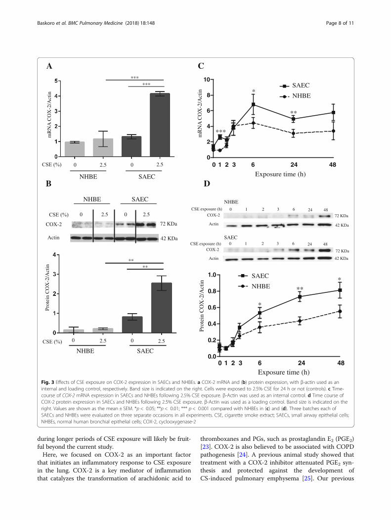
during longer periods of CSE exposure will likely be fruit-ful beyond the current study.Here, we focused on COX-2 as an important factor
that initiates an inflammatory response to CSE exposurein the lung. COX-2 is a key mediator of inflammationthat catalyzes the transformation of arachidonic acid to
thromboxanes and PGs, such as prostaglandin E2 (PGE2)[23]. COX-2 is also believed to be associated with COPDpathogenesis [24]. A previous animal study showed thattreatment with a COX-2 inhibitor attenuated PGE2 syn-thesis and protected against the development ofCS-induced pulmonary emphysema [25]. Our previous
0
2
4
6
8
10
0 1 2 3 6 24 48
***
*
**
Exposure time (h)
nitcA/2-
XO
CA
NR
m
0.0
0.2
0.4
0.6
0.8
1.0
0 1 2 3 6 24 48
**
*
*
Exposure time (h)
nitcA/2-
XO
Cnietor
P
C
NHBE
SAEC
COX-2
Actin
COX-2
Actin
CSE exposure (h)
CSE exposure (h) 0 1 2 3 6 24 48
0 1 2 3 6 24 48
72 KDa
72 KDa
42 KDa
42 KDa
D
0
NHBE SAEC
nitcA/2-
XO
CA
NR
m
***
2.5 2.50
***
A
NHBE
nitcA/2-
XO
Cnietor P
**
SAEC
0 2.5 0 2.5
**
B
COX-2
Actin 42 KDa
72 KDa
NHBE SAEC
0 2.5 2.50
SAEC
NHBE
SAEC
NHBE
Fig. 3 Effects of CSE exposure on COX-2 expression in SAECs and NHBEs. a COX-2 mRNA and (b) protein expression, with β-actin used as aninternal and loading control, respectively. Band size is indicated on the right. Cells were exposed to 2.5% CSE for 24 h or not (controls). c Time-course of COX-2 mRNA expression in SAECs and NHBEs following 2.5% CSE exposure. β-Actin was used as an internal control. d Time course ofCOX-2 protein expression in SAECs and NHBEs following 2.5% CSE exposure. β-Actin was used as a loading control. Band size is indicated on theright. Values are shown as the mean ± SEM. *p < 0.05; **p < 0.01; *** p < 0.001 compared with NHBEs in (c) and (d). Three batches each ofSAECs and NHBEs were evaluated on three separate occasions in all experiments. CSE, cigarette smoke extract; SAECs, small airway epithelial cells;NHBEs, normal human bronchial epithelial cells; COX-2, cyclooxygenase-2
Baskoro et al. BMC Pulmonary Medicine (2018) 18:148 Page 8 of 11
study, using primary lung fibroblasts from patients withCOPD and smokers without COPD (controls), demon-strated that following stimulation with inflammatory cy-tokines, fibroblasts from COPD subjects producedhigher PGE2 levels relative to those from control sub-jects, and that elevated PGE2 levels were the result of anincrease in COX-2 expression due to alterations inmRNA stability caused by weak induction ofmicroRNA-146a [17]. More recently, Zago et al. demon-strated that expression of RelB, a component of thenon-canonical NF-κB pathway, and the aryl hydrocarbonreceptor, are both reduced in fibroblasts from COPD
patients and regulate COX-2 protein levels by alteringmRNA stability [26–29]. These findings suggest that theregulation of COX-2 is critical to controlling abnormalinflammation associated with COPD pathogenesis, andthat its targeting could be a promising strategy forCOPD therapeutics. Interestingly, a recent study sug-gested that mesenchymal stem cells attenuated airwayinflammation and emphysema in CS-exposed ratsthrough the downregulation of COX-2 and PGE2 [30].This study had several limitations. First, we did not
use primary cells from COPD patients to evaluate thedirect contribution of the current findings to the disease
A C
B D
E
Large Airway Small Airway
ielcunlatot/ydobit na2-
XO
C
Air AirCS CS
**
Fig. 4 Effects of CS exposure on COX-2 expression in the lungs of C57BL/6 J mice. Mice were exposed to either 3.5% CS or fresh air(control) for 30 min/day for 5 days. Representative immunohistochemistry images of (a) the large-airway area in a control lung, (b) thesmall-airway area in a control lung, (c) the large-airway area in a CS-exposed lung, and (d) the small-airway area in a CS-exposed lung.Brown color indicates COX-2-positive airway epithelial cells (arrows). Scale bar, 100 μm. Insets are high magnification views of airwayepithelial cells. e The ratio of COX-2-positive nuclei to the total count of nuclei present in a field at 400× magnification and determinedin 10 different areas of the lung per mouse. Values are shown as the mean ± SEM (n = 3/group). *p < 0.001. CS, cigarette smoke;COX-2, cyclooxygenase-2
Baskoro et al. BMC Pulmonary Medicine (2018) 18:148 Page 9 of 11
condition. Second, the SAECs and NHBEs used in thecurrent study were not from matched donors. Instead,we confirmed COX-2 expression in the lungs ofCS-exposed mice as an experimental model of COPD.Thirdly, we used different media approved by the sup-plier for SAECs and NHBEs. Their components arealmost identical; however, there is a possibility that theyinfluenced the current findings. Additionally, we did notperform air-liquid interface (ALI) culture to promotedifferentiation. The phenotypes of each cell type ob-served under non-differentiating conditions could beaffected by contamination with different cell types.Therefore, differentiating SAECs and NHBEs using ALIculture system first and then exposing to CSE will makeexperimental findings more applicable to human air-ways. However, our findings were consistent with thosefrom a previous study using ALI culture [22], indicatingthat commercially available cells cultured using conven-tional methods can be suitable for these types of experi-ment. Lastly, we did not define the mechanisms bywhich inflammation spreads from the initial site to thewhole lung, leading to development of pulmonary em-physema. Further investigation of the transition ofCOX-2 expression in the airways, following variousdurations of CS exposure, and the interactions betweensmall-airway cells and other cells will be beneficial.
ConclusionsThis study demonstrated that the small airway is moresusceptible to CS than the large airway and might repre-sent an initial target site for development of CS-relatedrespiratory diseases, such as COPD. These findings sup-port the results of previous pathological investigations.
Additional file
Additional file 1: Table S1. A list of 44 probes upregulated by CSEexposure in both NHBEs and SAECs. Table S2. A list of 72 probesupregulated by CSE exposure only in SAECs. Table S3. A list of 39 probesupregulated by CSE exposure only in NHBEs. Table S4. A list of 23probes downregulated by CSE exposure in both NHBEs and SAECs. TableS5. A list of 55 probes downregulated by CSE exposure only in SAECs.Table S6. A list of 71 probes downregulated by CSE exposure only inNHBEs. (DOCX 3206 kb)
AbbreviationsALI: Air-liquid interface; COPD: Chronic obstructive pulmonary disease; COX-2: Cyclooxygenase-2; CS: Cigarette smoke; CSE: Cigarette smoke extract;GO: Gene ontology; GSEA: Gene set enrichment analysis; KEGG: KyotoEncyclopedia of Genes and Genomes; NF-κB: Nuclear factor-κB;NHBE: Normal human bronchial epithelial cells; PBS: Phosphate-bufferedsaline; PG: Prostaglandin; PGE2: Prostaglandin E2; PTGS2: Prostaglandin-endoperoxide synthase 2; SAEC: Small airway epithelial cells; SEM: Standarderror of the mean; TNF: Tumor necrosis factor
AcknowledgmentsThe authors wish to thank Mr. Dai Ogura for excellent support withmicroarray analysis.
FundingThis work was supported in part by JSPS KAKENHI Grant Number 26461199(T. Sato) and the Institute for Environmental and Gender-Specific Medicine,Juntendo University Graduate School of Medicine, Grant Number E2920 (T.Sato). The funder had no role in the design of the study and collection, ana-lysis, and interpretation of data and in writing the manuscript.
Availability of data and materialsThe datasets generated and analyzed during the current study are availablein the NCBI Gene Expression Omnibus (accession ID: GSE107200; https://www.ncbi.nlm.nih.gov/geo/query/acc.cgi?acc=GSE107200).
Authors’ contributionsConception and experimental design: HB, TS, and KT; data analysis andinterpretation: HB and KS; animal experiments: KK and AM; microarrayanalysis: TS and FT; molecular studies: FN and MK; immunohistochemistry: HBand YS; western blot analysis: NA and YK; and drafting of the manuscript: HBand TS. All authors read and approved the final manuscript.
Ethics approval and consent to participateThe procedures for animal experimentation were approved by the AnimalCare and Use Committee of Juntendo University School of Medicine(reference number: 632). Regarding studies using SAECs and NHBEs, theethics committee of Juntendo University School of Medicine ruled that noformal approval was required in the current study.
Consent for publicationNot applicable.
Competing interestsThe authors have no competing interests to declare.
Publisher’s NoteSpringer Nature remains neutral with regard to jurisdictional claims inpublished maps and institutional affiliations.
Author details1Department of Respiratory Medicine, Juntendo University Graduate Schoolof Medicine, 3-1-3 Hongo, Bunkyo-ku, Tokyo 113-8431, Japan. 2Departmentof Pulmonology and Respiratory Medicine, Universitas Indonesia Faculty ofMedicine, Jalan Persahabatan Raya No. 1, Rawamangun, Jakarta 13230,Indonesia.
Received: 28 December 2017 Accepted: 28 August 2018
References1. Lin JL, Thomas PS. Current perspectives of oxidative stress and its measurement
in chronic obstructive pulmonary disease. COPD. 2010;7:291–306.2. Vogelmeier CF, Criner GJ, Martinez FJ, Anzueto A, Barnes PJ, Bourbeau J,
et al. Global strategy for the diagnosis, management, and prevention ofchronic obstructive lung disease 2017 report. GOLD Executive Summary.Am J Respir Crit Care Med. 2017;195:557–82.
3. Rennard SI, Daughton DM. Smoking cessation. Clin Chest Med. 2014;35:165–76.4. Rennard SI, Drummond MB. Early chronic obstructive pulmonary disease:
definition, assessment, and prevention. Lancet. 2015;385:1778–88.5. Anthonisen NR, Connett JE, Murray RP. Smoking and lung function of lung health
study participants after 11 years. Am J Respir Crit Care Med. 2002;166:675–9.6. Lee W, Thomas PS. Oxidative stress in COPD and its measurement through
exhaled breath condensate. Clin Transl Sci. 2009;2:150–5.7. Barnes PJ. Cellular and molecular mechanisms of chronic obstructive
pulmonary disease. Clin Chest Med. 2014;35:71–86.8. Hogg JC, Macklem PT, Thurlbeck WM. Site and nature of airway obstruction
in chronic obstructive lung disease. N Engl J Med. 1968;278:1355–60.9. Hogg JC, Chu F, Utokaparch S, Woods R, Elliott WM, Buzatu L, et al. The
nature of small-airway obstruction in chronic obstructive pulmonary disease.N Engl J Med. 2004;350:2645–53.
10. Hogg JC. Pathophysiology of airflow limitation in chronic obstructivepulmonary disease. Lancet. 2004;364:709–21.
Baskoro et al. BMC Pulmonary Medicine (2018) 18:148 Page 10 of 11
11. McDonough JE, Yuan R, Suzuki M, Seyednejad N, Elliott WM, Sanchez PG,et al. Small-airway obstruction and emphysema in chronic obstructivepulmonary disease. N Engl J Med. 2011;365:1567–75.
12. Kasagi S, Seyama K, Mori H, Souma S, Sato T, Akiyoshi T, et al. Tomato juiceprevents senescence-accelerated mouse P1 strain from developingemphysema induced by chronic exposure to tobacco smoke. Am J PhysiolLung Cell Mol Physiol. 2006;290:L396–404.
13. Sato T, Seyama K, Sato Y, Mori H, Souma S, Akiyoshi T, et al. Senescencemarker protein-30 protects mice lungs from oxidative stress, aging, andsmoking. Am J Respir Crit Care Med. 2006;174:530–7.
14. Koike K, Ishigami A, Sato Y, Hirai T, Yuan Y, Kobayashi E, et al. Vitamin Cprevents cigarette smoke-induced pulmonary emphysema in mice andprovides pulmonary restoration. Am J Respir Cell Mol Biol. 2014;50:347–57.
15. Suzuki Y, Sato T, Sugimoto M, Baskoro H, Karasutani K, Mitsui A, et al.Hydrogen-rich pure water prevents cigarette smoke-induced pulmonaryemphysema in SMP30 knockout mice. Biochem Biophys Res Commun.2017;492:74–81.
16. Togo S, Holz O, Liu X, Sugiura H, Kamio K, Wang X, et al. Lung fibroblast repairfunctions in patients with chronic obstructive pulmonary disease are alteredby multiple mechanisms. Am J Respir Crit Care Med. 2008;178:248–60.
17. Sato T, Liu X, Nelson A, Nakanishi M, Kanaji N, Wang X, et al. Reduced miR-146a increases prostaglandin E2 in chronic obstructive pulmonary diseasefibroblasts. Am J Respir Crit Care Med. 2010;182:1020–9.
18. Domej W, Oettl K, Renner W. Oxidative stress and free radicals in COPD– implications and relevance for treatment. Int J Chron ObstructPulmon Dis. 2014;9:1207–24.
19. O'Donnell DE, Laveneziana P, Webb K, Neder JA. Chronic obstructive pulmonarydisease: clinical integrative physiology. Clin Chest Med. 2014;35:51–69.
20. Battaglia S, Mauad T, van Schadewijk AM, Vignola AM, Rabe KF, Bellia V,et al. Differential distribution of inflammatory cells in large and smallairways in smokers. J Clin Pathol. 2007;60:907–11.
21. Mercer BA, Lemaitre V, Powell CA, D'Armiento J. The epithelial cell in lunghealth and emphysema pathogenesis. Curr Respir Med Rev. 2006;2:101–42.
22. Yang J, Zuo WL, Fukui T, Chao I, Gomi K, Lee B, et al. Smoking-dependentdistal-to-proximal Repatterning of the adult human small airway epithelium.Am J Respir Crit Care Med. 2017;196:340–52.
23. Montuschi P, Macagno F, Parente P, Valente S, Lauriola L, Ciappi G, et al.Effects of cyclo-oxygenase inhibition on exhaled eicosanoids in patientswith COPD. Thorax. 2005;60:827–33.
24. Xaubet A, Roca-Ferrer J, Pujols L, Ramirez J, Mullol J, Marin-Arguedas A, et al.Cyclooxygenase-2 is up-regulated in lung parenchyma of chronicobstructive pulmonary disease and down-regulated in idiopathic pulmonaryfibrosis. Sarcoidosis Vasc Diffuse Lung Dis. 2004;21:35–42.
25. Roh GS, Yi CO, Cho YJ, Jeon BT, Nizamudtinova IT, Kim HJ, et al. Anti-inflammatory effects of celecoxib in rat lungs with smoke-inducedemphysema. Am J Physiol Lung Cell Mol Physiol. 2010;299:L184–91.
26. Zago M, Sheridan JA, Nair P, Rico de Souza A, Gallouzi IE, Rousseau S, et al.Aryl hydrocarbon receptor-dependent retention of nuclear HuR suppressescigarette smoke-induced cyclooxygenase-2 expression independent ofDNA-binding. PLoS One. 2013;8:e74953.
27. Zago M, Rico de Souza A, Hecht E, Rousseau S, Hamid Q, Eidelman DH,et al. The NF-kappaB family member RelB regulates microRNA miR-146a tosuppress cigarette smoke-induced COX-2 protein expression in lungfibroblasts. Toxicol Lett. 2014;226:107–16.
28. Sheridan JA, Zago M, Nair P, Li PZ, Bourbeau J, Tan WC, et al. Decreasedexpression of the NF-kappaB family member RelB in lung fibroblasts fromsmokers with and without COPD potentiates cigarette smoke-induced COX-2 expression. Respir Res. 2015;16:54.
29. Zago M, Sheridan JA, Traboulsi H, Hecht E, Zhang Y, Guerrina N, et al. Lowlevels of the AhR in chronic obstructive pulmonary disease (COPD)-derivedlung cells increases COX-2 protein by altering mRNA stability. PLoS One.2017;12:e0180881.
30. Gu W, Song L, Li XM, Wang D, Guo XJ. Xu WG. Mesenchymal stemcells alleviate airway inflammation and emphysema in COPD throughdown-regulation of cyclooxygenase-2 via p38 and ERK MAPK pathways.Sci Rep. 2015;5:8733.
Baskoro et al. BMC Pulmonary Medicine (2018) 18:148 Page 11 of 11


![Testing arXiv:1611.06739v2 [stat.ME] 23 Oct 2017 · 2017. 10. 24. · signal is present, FDR-controlling procedures such as BH (Benjamini and Hochberg, 1995) reject each false hypothesiswith](https://img.pdfslide.net/doc/110x75/602d1a066b6abe62d1132a32/testing-arxiv161106739v2-statme-23-oct-2017-2017-10-24-signal-is-present.jpg)