Embed Size (px)
Citation preview
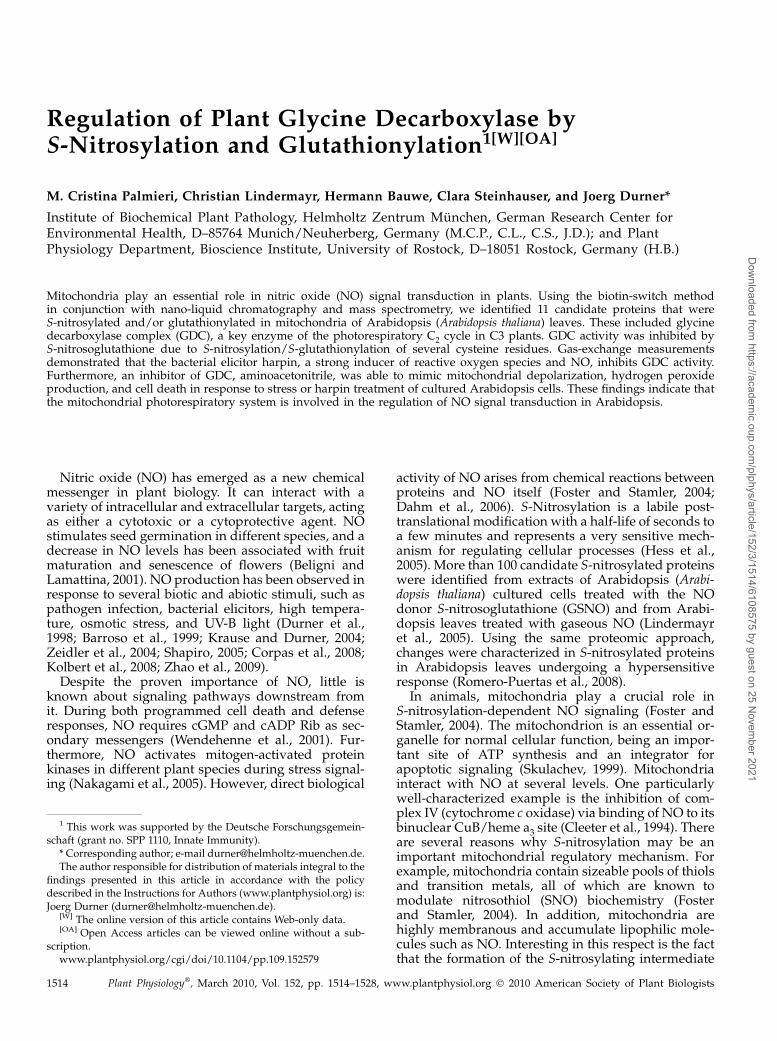
Regulation of Plant Glycine Decarboxylase byS-Nitrosylation and Glutathionylation1[W][OA]
M. Cristina Palmieri, Christian Lindermayr, Hermann Bauwe, Clara Steinhauser, and Joerg Durner*
Institute of Biochemical Plant Pathology, Helmholtz Zentrum Munchen, German Research Center forEnvironmental Health, D–85764 Munich/Neuherberg, Germany (M.C.P., C.L., C.S., J.D.); and PlantPhysiology Department, Bioscience Institute, University of Rostock, D–18051 Rostock, Germany (H.B.)
Mitochondria play an essential role in nitric oxide (NO) signal transduction in plants. Using the biotin-switch methodin conjunction with nano-liquid chromatography and mass spectrometry, we identified 11 candidate proteins that wereS-nitrosylated and/or glutathionylated in mitochondria of Arabidopsis (Arabidopsis thaliana) leaves. These included glycinedecarboxylase complex (GDC), a key enzyme of the photorespiratory C2 cycle in C3 plants. GDC activity was inhibited byS-nitrosoglutathione due to S-nitrosylation/S-glutathionylation of several cysteine residues. Gas-exchange measurementsdemonstrated that the bacterial elicitor harpin, a strong inducer of reactive oxygen species and NO, inhibits GDC activity.Furthermore, an inhibitor of GDC, aminoacetonitrile, was able to mimic mitochondrial depolarization, hydrogen peroxideproduction, and cell death in response to stress or harpin treatment of cultured Arabidopsis cells. These findings indicate thatthe mitochondrial photorespiratory system is involved in the regulation of NO signal transduction in Arabidopsis.
Nitric oxide (NO) has emerged as a new chemicalmessenger in plant biology. It can interact with avariety of intracellular and extracellular targets, actingas either a cytotoxic or a cytoprotective agent. NOstimulates seed germination in different species, and adecrease in NO levels has been associated with fruitmaturation and senescence of flowers (Beligni andLamattina, 2001). NO production has been observed inresponse to several biotic and abiotic stimuli, such aspathogen infection, bacterial elicitors, high tempera-ture, osmotic stress, and UV-B light (Durner et al.,1998; Barroso et al., 1999; Krause and Durner, 2004;Zeidler et al., 2004; Shapiro, 2005; Corpas et al., 2008;Kolbert et al., 2008; Zhao et al., 2009).
Despite the proven importance of NO, little isknown about signaling pathways downstream fromit. During both programmed cell death and defenseresponses, NO requires cGMP and cADP Rib as sec-ondary messengers (Wendehenne et al., 2001). Fur-thermore, NO activates mitogen-activated proteinkinases in different plant species during stress signal-ing (Nakagami et al., 2005). However, direct biological
activity of NO arises from chemical reactions betweenproteins and NO itself (Foster and Stamler, 2004;Dahm et al., 2006). S-Nitrosylation is a labile post-translational modification with a half-life of seconds toa few minutes and represents a very sensitive mech-anism for regulating cellular processes (Hess et al.,2005). More than 100 candidate S-nitrosylated proteinswere identified from extracts of Arabidopsis (Arabi-dopsis thaliana) cultured cells treated with the NOdonor S-nitrosoglutathione (GSNO) and from Arabi-dopsis leaves treated with gaseous NO (Lindermayret al., 2005). Using the same proteomic approach,changes were characterized in S-nitrosylated proteinsin Arabidopsis leaves undergoing a hypersensitiveresponse (Romero-Puertas et al., 2008).
In animals, mitochondria play a crucial role inS-nitrosylation-dependent NO signaling (Foster andStamler, 2004). The mitochondrion is an essential or-ganelle for normal cellular function, being an impor-tant site of ATP synthesis and an integrator forapoptotic signaling (Skulachev, 1999). Mitochondriainteract with NO at several levels. One particularlywell-characterized example is the inhibition of com-plex IV (cytochrome c oxidase) via binding of NO to itsbinuclear CuB/heme a3 site (Cleeter et al., 1994). Thereare several reasons why S-nitrosylation may be animportant mitochondrial regulatory mechanism. Forexample, mitochondria contain sizeable pools of thiolsand transition metals, all of which are known tomodulate nitrosothiol (SNO) biochemistry (Fosterand Stamler, 2004). In addition, mitochondria arehighly membranous and accumulate lipophilic mole-cules such as NO. Interesting in this respect is the factthat the formation of the S-nitrosylating intermediate
1 This work was supported by the Deutsche Forschungsgemein-schaft (grant no. SPP 1110, Innate Immunity).
* Corresponding author; e-mail [email protected] author responsible for distribution of materials integral to the
findings presented in this article in accordance with the policydescribed in the Instructions for Authors (www.plantphysiol.org) is:Joerg Durner ([email protected]).
[W] The online version of this article contains Web-only data.[OA] Open Access articles can be viewed online without a sub-
scription.www.plantphysiol.org/cgi/doi/10.1104/pp.109.152579
1514 Plant Physiology�, March 2010, Vol. 152, pp. 1514–1528, www.plantphysiol.org � 2010 American Society of Plant Biologists
Dow
nloaded from https://academ
ic.oup.com/plphys/article/152/3/1514/6108575 by guest on 25 N
ovember 2021

N2O3 is enhanced within membranes (Burwell et al.,2006).The role of mitochondria in stress-related responses
has been investigated in both animals and plants.Endogenous nitrosylation of the catalytic Cys site of asubset of mitochondrial caspases serves as an on/offswitch regulating caspase activity during apoptosis(Mannick et al., 2001).Moreover, cytochrome c,which ismodified by NO at its heme iron during apoptosis, isreleased from mitochondria into the cytoplasm, whichplays a critical role in many forms of apoptosis bystimulating apoptosome formation and subsequentcaspase activation (Schonhoff et al., 2003). We previ-ously showed that a prime target of NO in plants is themitochondrial apparatus, causing an inhibition ofKCN-sensitive respiration and an activation of alternativerespiration via alternative oxidase (AOX; Huang et al.,2002; Krause and Durner, 2004; Livaja et al., 2008).
The aim of this study was to identify possible targetsfor S-nitrosylation in mitochondria of Arabidopsisleaves in order to gain more insight into the regulatoryfunction of NO at the protein level. Using a proteomicapproach involving the highly specific biotin-switchmethod for detection and purification of S-nitrosylatedproteins (Jaffrey and Snyder, 2001) in conjunction withliquid chromatography and tandem mass spectrome-try (nanoLC/MS/MS), we could identify 11 mitochon-drial proteins as targets for S-nitrosylation. Amongthese identified proteins, we focused our attention onthe P-subunit of the Gly decarboxylase complex(GDC), which is an integral part of the photorespi-ratory system. Since the release of apoptotic factorsfrom mitochondria may be a result of inhibition ofrespiration, transition of mitochondrial permeability,and formation of reactive oxygen species (ROS; Savianiet al., 2002; Taylor et al., 2004; Chen and Gibson, 2008),
Figure 1. Harpin-dependent NO production inmitochondria. Arabidopsis cell cultures weretreated either with extracts of nontransformed E.coli (A) or with 35 mg mL21 recombinant partiallypurified harpin (B–D). Thirty minutes before mi-croscopic analysis, cells were incubated withMitoTracker Red 580 as a mitochondria-specificdye (red fluorescence, center column) and DAF-2FM DA as an NO probe (green fluorescence,right column) and analyzed using a fluorescenceconfocal microscope (Zeiss LSM 510 NLO; 403water lens). For NO scavenging, cells were incu-bated with 0.5 mM cPTIO 20 min before applica-tion of the NO probe. Colocalization of bothfluorescent signals appears yellow (left column).A, Cells treated with extracts of nontransformedE. coli. The images were taken 6 h after treatment.B and C, Cells were treated with recombinantharpin. The images were taken 1 h (B) and 6 h (C)after treatment. D, Cells were treated with recom-binant harpin followed by incubation with 0.5mM cPTIO and DAF-2FM DA. The images weretaken 6 h after treatment. NO fluorescence wascompletely scavenged by the cPTIO preincuba-tion.
Nitric Oxide Regulates Glycine Decarboxylase
Plant Physiol. Vol. 152, 2010 1515
Dow
nloaded from https://academ
ic.oup.com/plphys/article/152/3/1514/6108575 by guest on 25 N
ovember 2021

we investigated the molecular mechanism and thefunction of GDC-Cys modification in Arabidopsis.
RESULTS
Harpin-Induced Generation of NO in Mitochondria
While a strict relationship between harpin-inducedgeneration of NO in mitochondria was proven indifferent organisms and in different signaling events(Millar and Day, 1996; Cooper and Davies, 2000;Huang et al., 2002; Krause and Durner, 2004), directevidence for NO production in plant leaf mitochondriais still missing. To elucidate NO localization in mito-chondria, we treated Arabidopsis cultured cells withthe bacterial elicitor harpin because we previouslyshowed that this is able to induce NO productionwithin a few hours (Krause and Durner, 2004). Toanalyze NO production in mitochondria, Arabidopsiscultured cells were loaded with the NO-specific fluo-rescent dye diaminofluorescein (DAF-2FM DA) andwith the mitochondria-specific dye MitoTracker Red580. Fluorescence was visualized by confocal lasermicroscopy (Fig. 1) as described (Foissner et al., 2000;Pedroso et al., 2000; Beligni et al., 2002; Yao et al., 2002).As a control, we applied carboxy-2-phenyl-4,4,5,5-tetramethylimidazolinone-3-oxide-1-oxyl (cPTIO), anNO scavenger that does not react with any ROS(Barchowsky et al., 1999). After a 1-h treatmentwith 35 mg mL21 harpin, an NO burst became visiblein Arabidopsis mitochondria, with increasing inten-sity during the next hours (Fig. 1). The NO scavengercPTIO was able to inhibit the elicited burst.
Detection of S-Nitrosylated Proteins in Mitochondria
To identify S-nitrosylated proteins in mitochondriaof Arabidopsis leaves, we used a modified biotin-switch method (Jaffrey and Snyder, 2001). The mi-tochondrial fraction from the leaves was preparedaccording to Keech et al. (2005). Since the mitochon-drial proteome of Arabidopsis is well established(Kruft et al., 2001; Millar et al., 2001; Eubel et al.,2007), partially purified mitochondria were a usefulexperimental system to study posttranslational pro-tein modification (Supplemental Fig. S1).
After treating mitochondrial proteins either with thetrans-nitrosylatingagentGSNOorwithglutathione(GSH)as a negative control, S-nitrosylated proteins werebiotinylated and separated by SDS-PAGE (Fig. 2; Sup-plemental Fig. S2). In order to identify S-nitrosylatedproteins, GSNO- and GSH-derived eluates were sub-jected to nanoLC/MS/MS analysis. It was possible toidentify 25 proteins with a significant score as judgedby the Mascot search algorithm (score . 30). Five ofthese were chloroplast protein contaminations, whilenine were not listed in the mitochondrial proteome(Millar et al., 2001) and did not harbor the mitochon-drial targeting signal as identified by analysis usingeither TargetP 1.1 or iPSORT (Table I). Furthermore,
mitochondrial fractions seemed to be contaminated byperoxisomes, as indicated by detection of catalase 3.Among 11 mitochondrial proteins in Table I, we couldidentify three subunits of the GDC, the key enzyme ofthe photorespiratory C2 cycle in plants, namely Glydehydrogenase subunit P2 (15223217), Gly decarbox-ylase H1 (15226973), and Gly dehydrogenase subunitP1 (14596025).
Mass Spectrometric Analyses of GSNO-Treated P Protein
GSNO is able to S-nitrosylate or S-glutathiolate Cysresidues, while both modifications can have differenteffects on protein conformation and activity. We in-vestigated which kind of modification(s) resulted fromthe GSNO treatment of GDC, focusing our attention onthe decarboxylating subunit, the P protein. In Arabi-dopsis, there are two genes encoding the P subunit,which share high homology. Since the production ofrecombinant P protein was not successful, we partiallypurified the P subunit from Arabidopsis mitochondriaas described (Bourguignon et al., 1988) and tested its
Figure 2. Detection of S-nitrosylated proteins in Arabidopsis mito-chondria. Fifteen milligrams of protein of mitochondria-enriched frac-tions (MEF) was treated with 1 mM GSNO or GSH and labeled withbiotin according to the biotin-switch method. Proteins were purified byaffinity chromatography using neutravidin-agarose beads. A part ofeluates was separated by 12% SDS-PAGE and visualized by SYPRORuby staining. Protein bands corresponding to predominant bands ofthe immunoblot analysis were identified by nanoLC/MS/MS. Addition-ally, to compare the amount of proteins in the different treatments,aliquots of mitochondria-enriched fractions were separated by SDS-PAGE (right gel). The relative masses of protein standards (M) are shownon the left (kD). The experiment was repeated three times with similarresults.
Palmieri et al.
1516 Plant Physiol. Vol. 152, 2010
Dow
nloaded from https://academ
ic.oup.com/plphys/article/152/3/1514/6108575 by guest on 25 N
ovember 2021

integrity by assaying it for the CO2-exchange reaction(Supplemental Fig. S3). S-Nitrosylation of this par-tially purified P protein was confirmed using thebiotin-switch method (data not shown). In addition,we used mass spectrometric analyses to detect GSNO-mediated modifications of the Cys residues, since thismethod allows the detection of both S-nitrosylationand S-glutathionylation. Protein S-glutathionylationis promoted by nitrosative stress and transientS-nitrosylation, but it also occurs in unstressed cells(Dalle-Donne et al., 2009). After treating the P proteinwith GSNO and digesting it with trypsin, we analyzedthe Cys-containing peptides for their modifications(Fig. 3; Supplemental Fig. S4). C402 and C463 were foundto be S-glutathionylated after all treatments. In con-trast, C98, C943, C777, and C1022 were S-glutathionylatedonly after treatment with 1 mM GSNO (Table II). NoS-nitrosylated Cys residues were detected by massspectrometric analysis, probably because of the tran-sient nature of this modification at the P protein, asreported for several other proteins (Martinez-Ruiz andLamas, 2007).
Inhibition of Gly Decarboxylase Activity by GSNO
To elucidate the effects of S-nitrosylation/S-gluta-thionylation on GDC activity, we treated mitochondriawith GSNO and monitored the GDC activity (Walkeret al., 1982; Walker and Oliver, 1986). Following 14CO2release from [1-14C]Gly (i.e. the activity of P and Hsubunits), we could observe a significant reduction ofdecarboxylation, which was dependent on GSNOconcentration (Fig. 4A). After 20 min, the GDC activityof mitochondria incubated with 0.25 mM GSNOshowed an inhibition of 60% as compared with theGDC activity of untreated organelles. Treatment with1 mM GSNO further decreased the decarboxylation toabout 30%, similar to the inhibition obtained with theGDC inhibitor aminoacetonitrile (AAN; 10 mM). Theincubation of mitochondria with the reducing agents
dithiothreitol (DTT) and GSH increased decarboxyla-tion activity by 76% and 15% in vivo, respectively,demonstrating the redox-dependent regulation of theP protein and suggesting a change in the mitochon-drial redox status during purification. To further char-acterize the redox dependency of GDC activity, wetested the effects of two Cys-modifying agents on theenzymatic activity:N-ethylmaleimide, a highly reactiveagent that covalently and irreversibly alkylates freeCys thiol groups, and 5,5#-dithiobis-(2-nitrobenzoicacid), an oxidizing reagent. Both treatments resulted ina complete inhibition of P/H protein activity within20 min (Fig. 4A).
Modulation of GDC activity by GSNO was con-firmed using Arabidopsis leaf slices as described byNavarre and Wolpert (1995). Incubation with 1 mM
GSNO for 20 min resulted in an inhibition of GDCactivity by 75%, which could be restored by 1 mM DTT(Fig. 4B). Moreover, to test if modulation of GDCactivity was due to a direct effect of GSNO on thephotorespiratory system, Arabidopsis leaf slices wereincubated with inhibitors of complex I of the mito-chondrial respiration system (rotenone) and of AOX(salicylhydroxamic acid [SHAM]). In animal systems,complex I has been shown to be regulated byS-nitrosylation and to be involved in reversible ROSproduction (Borutaite and Brown, 2006; Burwell et al.,2006). In plants, it was shown that the NO-inducedAOX suppressed the production of ROS and cell deathinduced by KCN-sensitive respiration inhibition(Huang et al., 2002). Therefore, the inhibition of GDCactivity after GSNO treatment could have been anindirect effect of ROS induced by inhibition of com-plex I. It is well known that the activity of complex I ofthe mitochondrial electron transport chain is inhibitedby nitrosothiols and peroxinitrite (Carreras et al., 2004;Dahm et al., 2006). This inhibition results in increasedproduction of ROS, which can cause oxidative damageof GDC (Taylor et al., 2002). However, blocking ofcomplex I in mitochondria via rotenone did not affect
Table I. S-Nitrosylated proteins from Arabidopsis mitochondria
Mitochondria extracts treated with GSNO or GSH were subjected to the biotin-switch method and analyzed by nanoLC/MS/MS after trypticdigestion. The Mascot search engine was used to parse MS data to identify proteins from primary sequence databases. The best-matching peptideidentifying the protein is given. If there were additional peptides found, the number of the peptides is given. The asterisks indicate proteins not listedin the mitochondrial proteome (Millar et al., 2001) but with mitochondrial targeting signal (TargetP). Proteome analysis was carried out by TopLab.
Protein Identifier Protein Name Molecular Mass Identified Peptides (Score)
D
15223217 Gly dehydrogenase subunit P2 18,000 K.LTESPGLINSSPYEDGWMIK.V (87) +215221119 Aminomethyltransferase 44,759 R.TGYTGEDGFEISVPDEHAVDLAK.A (80) +1015235745 Ser hydroxymethyltransferase 57,535 K.LIVAGASAYAR.L (70) +915226973 Gly decarboxylase H1 18,050 K.LTESPGLINSSPYEDGWMIK.V (87) +314916970 ATP synthase subunit a 55,296 K.AVDSLVPIGR.G (44) +418394888 Catalase 3 57,059 R.LGPNYLQLPVNAPK.C (54) +430684419 Lipoamide dehydrogenase 2 54,237 K.HIIVATGSDVK.S (47) +715221044 Lipoamide dehydrogenase 1 54,239 K.HIIVATGSDVK.S (47) +479401911 Unknown protein* 78,638 K.GSFSSVVSDK.S (42) +714596025 Gly dehydrogenase subunit P1 113,852 R.EYAAFPAPWLR.S (35) +1121537215 Unknown protein* 32,979 K.SNMSNCETSSEIQKPDYIHVR.A (28) +1
Nitric Oxide Regulates Glycine Decarboxylase
Plant Physiol. Vol. 152, 2010 1517
Dow
nloaded from https://academ
ic.oup.com/plphys/article/152/3/1514/6108575 by guest on 25 N
ovember 2021

GDC activity within 30 min (Fig. 4B). Furthermore,neither increased ROS levels due to AOX inhibitionnor reduced ROS levels due to catalase-dependentscavenging of ROS (data not shown) influenced themodulation of GDC activity.
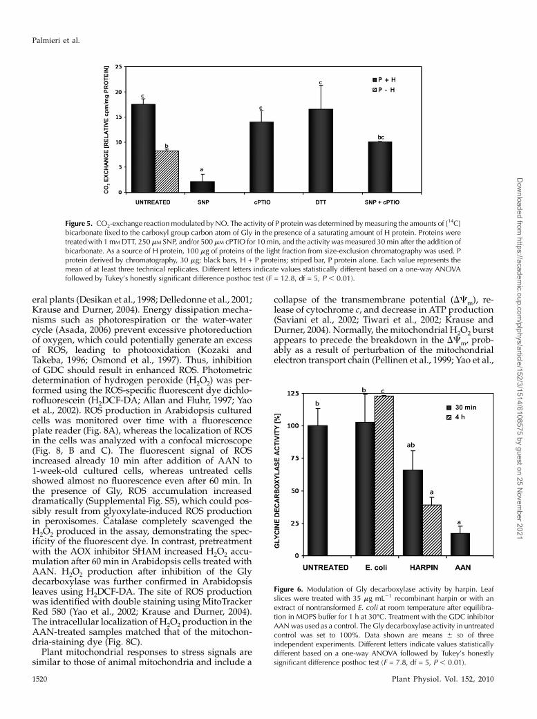
Since it was not possible to demonstrate GSNO-dependent S-nitrosylation of the P protein via MS, wetested the effect of sodium nitroprusside (SNP) on theactivity of partially purified P protein. SNP is an NOdonor that is not able to S-glutathionylate Cys resi-dues. As shown by Fig. 5, 250 mM SNP was able toinhibit the CO2-exchange reaction, while the NO scav-enger cPTIO (500 mM) almost completely prevented it.These data suggest that the activity of the P subunit ofthe GDC is inhibited by NO.
Inhibition of Gly Decarboxylase Activity by Harpin
Since protein S-nitrosylation is emerging as aspecific and fundamental mechanism in NO signal
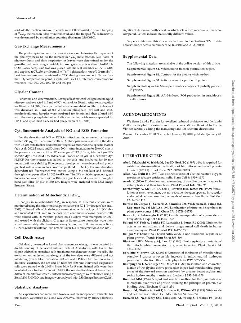
transduction (e.g. during plant-pathogen interactions;Romero-Puertas et al., 2004), we tested the involve-ment of GDC inhibition in a stress-related response.Harpin is a proteinaceous bacterial elicitor of Erwinia,Pseudomonas, and Xanthomonas strains that can inducean oxidative burst and programmed cell death invarious host plants. Leaf slices were incubated atroom temperature with 35 mg mL21 recombinantharpin protein or with an extract of untransformedEscherichia coli DH5a as a control. GDC activity wasrecorded over a 4-h period. An inhibition of Glydecarboxylation in harpin-treated leaves was detectedafter 30 min and increased after 4 h to 61% in com-parison with untreated and E. coli-treated samples(Fig. 6).
The Gly-Ser interconversion, catalyzed by GDC inconjunction with the Ser hydroxymethyltransferase, isan integral part of the photorespiratory metabolicpathway (Douce et al., 2001; Bauwe and Kolukisaoglu,2003). Therefore, we investigated the role of GDC
Figure 3. Mass spectrometric analysesof partially purified GSNO-treated Pprotein. For mass spectrometric analy-ses, partially purified P protein wasdigested with trypsin at 37�C for 1 h,pH 6.5. Digested proteins were ana-lyzed by nanoLC/MS/MS, with an au-tomatic switch between MS, MS2, andMS3 acquisition. A, The S-glutathiony-lation was detected in the MS2 as again of 305 kD in mass. The spectrarepresent the MS3 fragmentation ofthe peptide FCDALISIR containing theglutathionylated C943. The spectrumis characterized by y-ion (y1–y5) andb-ion (b1–b5) series and shows thatC943 was modified with GSH, as indi-cated by the presence of a b2-ion atm/z 556.2 (mass shift +305 D). B,Sequencing table showing the shift ofC943. The table details peptide frag-ments (AA, amino acids; B, b-ionshaving the charge retained on theN-terminal fragment; Y, y-ions havingthe charge retained on the C-terminalfragment), b-ions and y-ions (singleand double charged), and the neutrallosses of ammonia and water for b-ionsand y-ions.
Palmieri et al.
1518 Plant Physiol. Vol. 152, 2010
Dow
nloaded from https://academ
ic.oup.com/plphys/article/152/3/1514/6108575 by guest on 25 N
ovember 2021

during plant defense responses in vivo by monitor-ing the amino acid content in Arabidopsis leaves(Blackwell et al., 1990; Abe et al., 1997; Heineke et al.,2001). The concentration of harpin used was able tocause cell death after 24 h when infiltrated intoArabidopsis leaves, but not after 30 min (in vitroexperiments; data not shown). The inhibition of GDCactivity observed after harpin treatment led to a dis-tinctly elevated leaf Gly-Ser ratio after 4 h relative tothe control treatment with E. coli extract (Fig. 7). As asecond control, leaves were infiltrated with the GDCinhibitor AAN, which also resulted in a fast increase inGly-Ser within 30 min (Fig. 7).
Mitochondrial and Cellular Responses toGDC Inhibition
To confirm the role of GDC in the harpin-inducedplant defense response, Arabidopsis plants and cul-tured cells were treated with the GDC inhibitor AAN,which has no side effects on dark respiration and CO2
fixation (Usuda and Edwards, 1980; Creach andStewart, 1982) and is able to mimic the toxin victorin-induced mitochondrial oxidative burst in oat (Avenasativa) cells (Yao et al., 2002).
Stress-induced ROS generation has been reported asan essential signal for activation of resistance in sev-
Table II. Mass spectrometric analyses of the partially purified P protein
P protein (20 mg) was treated with 0.5 or 1 mM GSNO for 15 min. After removing the excess of GSNO, the protein was digested with trypsin andanalyzed as described in “Materials and Methods.” Six peptides belonging to the P subunit (P1 gene) of the GDC showed a mass differencecompared with the expected one of 305 D, which corresponds to the addition of one 2GS group. No peptide showed a shift of 29 D (2NO) in themass. The asterisks indicate peptides that showed a shift in mass after both the GSNO treatments.
Peptide SequencePeptide Identification
Probability
Modifications Identified
by Spectrum
Actual Peptide
MassPeptide Start Peptide Stop
%
FCDALISIR 95 GSH (+305) 1,341.61 942 950FCGFDHIDSLIDATVPK 95 GSH (+305) 2,181.97 97 113TFCIPHGGGGPGMGPIGVK 95 GSH (+305) 2,085.94 775 793DKATSNICTAQALLANMAAMYAVYHGPAGLK* 95 GSH (+305) 3,498.65 395 425CSDAHAIADAASK* 95 GSH (+305) 1,563.63 463 475LVCTLLPEEEQVAAAVSA 95 GSH (+305) 2,147.02 1,020 1,037
Figure 4. ModulationofGlydecarboxyl-ase activity by thiol-modifying agents.A, Mitochondria-enriched fractionswere preincubated with the indicatedamounts of GSNO or SH-modifyingagents for 20 min at room temperaturein the dark, and enzyme activity wasdetermined subsequently. The activityof the control (untreated) was set to100%. B, Leaf slices were treated for20 min with GSNO, rotenone, orSHAM at room temperature after equili-bration in a MOPS buffer for 1 h at30�C. For restoring GDC activity, 1 mM
DTT was added to the inhibited en-zymes and incubated for an additional10 min. Treatment with the GDC in-hibitor AAN (10 mM) served as a con-trol. The activity of controls (untreated)was set to 100%. Presented data aremeans 6 SD of three independent ex-periments. Different letters indicatevalues statistically different based ona one-way ANOVA followed by Tu-key’s honestly significant differenceposthoc test (A, F = 22.46, df = 9,P, 0.01; B, F = 6.16, df = 4, P, 0.01).
Nitric Oxide Regulates Glycine Decarboxylase
Plant Physiol. Vol. 152, 2010 1519
Dow
nloaded from https://academ
ic.oup.com/plphys/article/152/3/1514/6108575 by guest on 25 N
ovember 2021
eral plants (Desikan et al., 1998; Delledonne et al., 2001;Krause and Durner, 2004). Energy dissipation mecha-nisms such as photorespiration or the water-watercycle (Asada, 2006) prevent excessive photoreductionof oxygen, which could potentially generate an excessof ROS, leading to photooxidation (Kozaki andTakeba, 1996; Osmond et al., 1997). Thus, inhibitionof GDC should result in enhanced ROS. Photometricdetermination of hydrogen peroxide (H2O2) was per-formed using the ROS-specific fluorescent dye dichlo-rofluorescein (H2DCF-DA; Allan and Fluhr, 1997; Yaoet al., 2002). ROS production in Arabidopsis culturedcells was monitored over time with a fluorescenceplate reader (Fig. 8A), whereas the localization of ROSin the cells was analyzed with a confocal microscope(Fig. 8, B and C). The fluorescent signal of ROSincreased already 10 min after addition of AAN to1-week-old cultured cells, whereas untreated cellsshowed almost no fluorescence even after 60 min. Inthe presence of Gly, ROS accumulation increaseddramatically (Supplemental Fig. S5), which could pos-sibly result from glyoxylate-induced ROS productionin peroxisomes. Catalase completely scavenged theH2O2 produced in the assay, demonstrating the spec-ificity of the fluorescent dye. In contrast, pretreatmentwith the AOX inhibitor SHAM increased H2O2 accu-mulation after 60 min in Arabidopsis cells treated withAAN. H2O2 production after inhibition of the Glydecarboxylase was further confirmed in Arabidopsisleaves using H2DCF-DA. The site of ROS productionwas identified with double staining using MitoTrackerRed 580 (Yao et al., 2002; Krause and Durner, 2004).The intracellular localization of H2O2 production in theAAN-treated samples matched that of the mitochon-dria-staining dye (Fig. 8C).
Plant mitochondrial responses to stress signals aresimilar to those of animal mitochondria and include a
collapse of the transmembrane potential (DCm), re-lease of cytochrome c, and decrease in ATP production(Saviani et al., 2002; Tiwari et al., 2002; Krause andDurner, 2004). Normally, the mitochondrial H2O2 burstappears to precede the breakdown in the DCm, prob-ably as a result of perturbation of the mitochondrialelectron transport chain (Pellinen et al., 1999; Yao et al.,
Figure 5. CO2-exchange reaction modulated byNO. The activity of P protein was determined bymeasuring the amounts of [14C]bicarbonate fixed to the carboxyl group carbon atom of Gly in the presence of a saturating amount of H protein. Proteins weretreated with 1 mM DTT, 250 mM SNP, and/or 500 mM cPTIO for 10 min, and the activity was measured 30 min after the addition ofbicarbonate. As a source of H protein, 100 mg of proteins of the light fraction from size-exclusion chromatography was used. Pprotein derived by chromatography, 30 mg; black bars, H + P proteins; striped bar, P protein alone. Each value represents themean of at least three technical replicates. Different letters indicate values statistically different based on a one-way ANOVAfollowed by Tukey’s honestly significant difference posthoc test (F = 12.8, df = 5, P , 0.01).
Figure 6. Modulation of Gly decarboxylase activity by harpin. Leafslices were treated with 35 mg mL21 recombinant harpin or with anextract of nontransformed E. coli at room temperature after equilibra-tion in MOPS buffer for 1 h at 30�C. Treatment with the GDC inhibitorAANwas used as a control. The Gly decarboxylase activity in untreatedcontrol was set to 100%. Data shown are means 6 SD of threeindependent experiments. Different letters indicate values statisticallydifferent based on a one-way ANOVA followed by Tukey’s honestlysignificant difference posthoc test (F = 7.8, df = 5, P , 0.01).
Palmieri et al.
1520 Plant Physiol. Vol. 152, 2010
Dow
nloaded from https://academ
ic.oup.com/plphys/article/152/3/1514/6108575 by guest on 25 N
ovember 2021

2002). JC-1 was used as a dye to probe the mitochon-drial membrane potential in AAN-treated culturedcells of Arabidopsis. At 30 min after treatment withAAN, approximately 50% of cells showed a decreasein DCm compared with 30% of the control cells (Fig.9A). The depolarization observed in the control wasmost likely due to a stress condition of cells in the platereader and was completely suppressed by pretreat-ment with cyclosporine A, which is an inhibitor of themitochondrial transition pore (Saviani et al., 2002). Inaddition, catalase was able to reduce the DCm inuntreated or AAN-treated cells, confirming that theoxidative burst is essential for the breakdown of themitochondrial membrane potential (Fig. 9A). Further-more, blocking of the ROS-scavenging ability of AOX(via SHAM) resulted in increased loss of red fluores-cence, with approximately 90% of mitochondrialmembranes depolarized (Fig. 9).From experiments with toxins of plant pathogens or
mutants of mitochondrial proteins, it seems that loss ofmitochondrial function and concomitant mitochon-drial ROS formation and signaling are necessary forsubsequent cell death (Robson and Vanlerberghe,2002; Vanlerberghe et al., 2002; Yao et al., 2002; Krauseand Durner, 2004). To monitor AAN-induced celldeath, we measured the loss of plasma membraneintegrity by double staining of Arabidopsis suspen-sion cells with Evans blue and fluorescein diacetate(Huang et al., 2002). Both fluorescent signals were
analyzed by confocal microscopy. Untreated cellsshowed clear green fluorescence, while 15 to 30 minafter treatment with AAN, a decrease in fluoresceinsignal and an increase in red fluorescence could beobserved (Fig. 10).
DISCUSSION
Programmed cell death as observed in the form of ahypersensitive response in several plant-pathogen in-teractions exhibits similarities to programmed celldeath as seen in animal apoptosis, including chroma-tin condensation and fragmentation (Mur et al., 2008)and caspase-like proteolytic activity (Lam and delPozo, 2000). Furthermore, it has been shown that NOand ROS play major regulatory and antimicrobial rolesin plant defense responses (Delledonne et al., 2001). Inanimal cells, mitochondria are major sites of the for-mation of redox compounds and major targets of NO/ROS-induced damage (Skulachev, 1996). By contrast,large quantities of ROS and NO are produced inplastids and peroxisomes of plants, especially in pho-tosynthetic cells (del Rio et al., 2003). Nonetheless,plant mitochondria also are responsible for the pro-duction of ROS and are a site of oxidative damage(Taylor et al., 2002). In this paper, we not only showedthat NO and ROS accumulate in Arabidopsis mito-chondria during stress responses but also that NO iscapable of modulating an essential mitochondrialfunction, photorespiratory metabolism, as an integralpart of this response.
Inhibition of GDC by Harpin and NO
GDC, together with Ser hydroxymethyltransferase,is responsible for the conversion of Gly to Ser duringphotorespiration. During this metabolic process, CO2and NH3 are produced while ATP and reducingequivalents are consumed, which makes photorespi-ration an energetically wasteful process (Wingler et al.,2000). It has been described that oat GDC is involvedin the response to the toxin victorin produced by thefungus Cochliobolus victoriae (Navarre and Wolpert,1995; Yao et al., 2002). The P subunit of the GDC wasfound to bind victorin in vivo, but only in susceptiblegenotypes. The H subunit bound victorin in vivo inboth susceptible and resistant genotypes (Navarre andWolpert, 1995). In addition, application of the GDCinhibitor AAN resulted in apoptosis-like cell deathand disease symptoms (Yao et al. 2002). In this study,we observed a clear modulation of the GDC activity byredox agents in both intact mitochondria and plantleaves following 14CO2 release from [1-14C]Gly (i.e. theH-dependent P protein reaction). The decarboxylationactivity of GDC was significantly inhibited by GSNOas well as after blocking of the Cys residues with Cys-modifying chemicals. By contrast, strongly reducingagents such as DTT promoted GDC activity (Fig. 4A).The requirement of a reduced environment for both
Figure 7. Determination of Gly-Ser ratio to monitor GDC inhibition.The inhibition of the activity of the GDC was monitored following theincrease in Gly content and decrease in Ser content in Arabidopsisleaves. Amino acid content analysis was performed using 100 mg offresh tissue and a HPLC system. Each value represents the ratio betweenGly and Ser (mmol g21 fresh weight). Leaves were treated for theindicated times with extracts of nontransformed E. coli or recombinantharpin (35 mg mL21). The GDC inhibitor AAN was used as a control.Experiments were repeated three times with similar results. Differentletters indicate values statistically different based on a one-wayANOVA followed by Tukey’s honestly significant difference posthoctest (F = 8.5, df = 5, P , 0.01).
Nitric Oxide Regulates Glycine Decarboxylase
Plant Physiol. Vol. 152, 2010 1521
Dow
nloaded from https://academ
ic.oup.com/plphys/article/152/3/1514/6108575 by guest on 25 N
ovember 2021

CO2 exchange and decarboxylase activity of GDC in vi-tro has been described previously (Hiraga and Kikuchi,1980; Walker and Oliver, 1986). Inactivation of GDCafter treatment of mitochondria withN-ethylmaleimideor 5,5#-dithiobis-(2-nitrobenzoic acid) showed that Cysresidues are important for the enzymatic activity.
In animal systems, the toxicity of NO has beenassociated with inhibition of cellular respiration(Clementi et al., 1998). Interaction of NO with cyto-chrome c oxidase (complex I) has dramatic biologicalconsequences, such as the increase in the mitochon-drial ROS level and the induction of apoptosis(Brunori et al., 1999). Therefore, the reason for NOtoxicity is not a shutdown of respiration per se but anoverreduction of the ubiquinone pool. This inhibitoryeffect of NO on cytochrome c oxidase has also beendescribed for soybean (Glycine max) and mung bean(Vigna radiata) mitochondria (Millar and Day, 1996;Yamasaki et al., 2001). Consequently, the modulationof GDC activity observed after GSNO treatment couldbe a secondary effect due to inhibition of the mito-chondrial electron transport chain and subsequentaccumulation of ROS. Using inhibitors of complex I
(rotenone) and AOX (SHAM), we showed that a directaction of NO on the GDC is responsible for its de-creased activity (Fig. 4B).
Posttranslational Modification of GDC by NO
MS analysis of partially purified P protein (Table II)suggested the possibility that the modulation of theGDC was due to an S-glutathionylation and not to anS-nitrosylation of specific Cys residues. Although thebiotin-switch method is specific for detection of SNO-modified Cys, we tested the activity of the purified Pprotein after treatment with the nonglutathionylatingagent SNP and analyzed if NO is able to modify GDCactivity (Fig. 5). The inhibition of GDC activity ob-served after SNP treatment and the ability of cPTIO toscavenge this effect proved that NO is able to modu-late GDC activity per se, confirming the strict connec-tion between the two posttranslational modifications.In general, the role of S-nitrosothiols as primers forS-glutathionylation should be considered, with specialemphasis given to GSNO as a reagent that can inducenot only S-nitrosylation but also S-glutathionylation of
Figure 8. AAN-dependent ROS production in cultured cells and leaves of Arabidopsis. A, AAN, an inhibitor of the GDC, wasused to study the effect of GDC inhibition in cultured cells of Arabidopsis. The ROS-dependent fluorescence of the H2DCF-DAdye was monitored with a Tecan GENios fluorescence plate reader. Catalase was used to scavenge H2O2 production in the assay,and its relative fluorescence unit (RFU) value, corresponding to the dye background, was subtracted from other samples. SHAM,2 mM; catalase, 100 units mL21; AAN, 10 mM. Data shown are means 6 SD of three independent experiments. Different lettersindicate values statistically different based on a one-way ANOVA followed by Tukey’s honestly significant difference posthoc test(F = 166.2, df = 7, P, 0.01). B, Arabidopsis leaf slices were incubated with 10 mM ROS-sensitive dye (H2DCF-DA). After 20 min,the leaf slices were treated with 10 mM AAN for 20 min (middle) or buffer (control; left). Produced H2O2 was scavenged bypreincubation with 100 units mL21 catalase (right). Confocal microscopy images were obtained using Zeiss LSM 510 NLOwith a403 water lens. C, Colocalization of mitochondria and ROS production in Arabidopsis. Cells were incubated with 0.5 mM
MitoTracker Red 580 as a specific mitochondrial marker (red fluorescence, center column) and subsequently with H2DCF-DA(10 mM; green fluorescence, right column). After 20 min, cells were treated either with 10 mM AAN or buffer for 10 min. Confocalmicroscopy images were obtained using Zeiss LSM 510 NLO with a 403 water lens. The experiment was repeated three timeswith similar results. D, Model proposed to explain the relation between GDC and AOX in modulating ROS production.
Palmieri et al.
1522 Plant Physiol. Vol. 152, 2010
Dow
nloaded from https://academ
ic.oup.com/plphys/article/152/3/1514/6108575 by guest on 25 N
ovember 2021

proteins (Martinez-Ruiz and Lamas, 2007). In severalproteins, “competition” between S-glutathionylationand S-nitrosylation has been compared, which showedthat there is considerable variation among differentproteins regarding their tendencies to undergo eithermodification.The modulation of a key enzyme of the photores-
piratory system via S-nitrosylation suggests a directrole of this metabolic pathway in the regulation of theredox state of the cell. In addition, GDC was found inroot and etiolated tissue of different plants, in C4plants, and in a wide range of organisms from bacteriato humans. Taken together, the data suggest a non-photorespiratory role of the GDC complex (Navarreand Wolpert, 1995). For this reason, we investigatedthe state of GDC during a plant stress response. Usingthe Arabidopsis-harpin interaction as a model system(Xie and Chen, 2000; Krause and Durner, 2004; Livajaet al., 2008), we demonstrated that GDC activity isinhibited in Arabidopsis leaves after harpin treatment(Figs. 6 and 7). Thus, harpin inhibits not only thecytochrome c-dependent respiration (Xie and Chen,2000; Livaja et al., 2008) but also the photorespiratorysystem in Arabidopsis. This reduces the cellular ca-pacity of ATP synthesis and increases the productionof ROS, which leads to an overreduction and over-energization of the entire cell. Recent studies revealedthe importance of energy dissipation in mitochondria.
Oxidative stress by excess light is involved in theregulation of respiratory gene expression and themodulation of respiratory properties, especially up-regulation of AOX (Yoshida et al., 2006, 2007, 2008).
Potential Involvement of GDC in PlantDefense Responses
The possible role of GDC in pathogen responses hasbeen investigated upon victorin treatment of oat. Inthis system, application of the GDC-specific inhibitorAAN resulted in a highly localized accumulation ofH2O2 within mitochondria followed by disease symp-toms similar to those induced by victorin and duringapoptosis-like cell death (Yao et al., 2002). Here, wedemonstrated that AAN is able to induce an oxidativeburst in cultured cells and in leaves of Arabidopsis(Fig. 8). Whereas catalase was able to scavenge AAN-induced ROS, inhibition of AOX (by SHAM) furtherincreased the ROS signal 1 h after incubation (Fig. 8).The use of a mitochondria-specific dye allowed us tolocalize the ROS burst in Arabidopsis mitochondria,although peroxisomal ROS production cannot by ex-cluded, since peroxisomes are localized next to mito-chondria. The direct relationship between inhibition ofGDC activity and ROS levels is difficult to explain. Theoxidation of Gly by GDC provides NADH for themitochondrial electron transport chain. Inhibition of
Figure 9. AAN-induced depolarization of mitochondrial membranes. AAN was used to study the effect of GDC inhibition inArabidopsis. A, One-week-old cultured Arabidopsis cells were treated for 30 min with different inhibitors on the 96-well plate inthe grow chamber. Afterward, JC-1 dye (5 mg mL21), to probe the mitochondrial membrane potential, and 10 mM AAN wereadded and the measurement was started. The mitochondrial depolarization was indicated by a decrease in the red-greenfluorescence intensity ratio. The data represent the status of the mitochondrial membrane 30min after AAN treatment. Black barsindicate the percentage of depolarization. SHAM, 2 mM; catalase, 100 units mL21; cyclosporine A, 50 mM. Cyclosporine A wasused as a control for complete polarization (100%). The experiment was repeated three times with similar results. Different lettersindicate values statistically different based on a one-way ANOVA followed by Tukey’s honestly significant difference posthoc test(F = 532.6, df = 7, P , 0.01). B, Model proposed to explain the relation between GDC and AOX in modulating depolarizationmitochondrial membranes.
Nitric Oxide Regulates Glycine Decarboxylase
Plant Physiol. Vol. 152, 2010 1523
Dow
nloaded from https://academ
ic.oup.com/plphys/article/152/3/1514/6108575 by guest on 25 N
ovember 2021

GDC would lead to undersupply of NADH to therespiratory chain. This shortage of the electron donorpool might cause the production of partially reducedoxygen species. However, there might be other, stillunknown connections between the inhibition of GDCon the one hand and ROS production as a result ofperturbation of the energy metabolism in mitochon-dria on the other hand.
In animal cells, the increase of mitochondrial ROS,togetherwith an elevation of cytosolic Ca2+, contributesto the opening of the mitochondrial permeability tran-sition pore (PTP). The PTP depolarizes mitochondria,leading to mitochondrial swelling and subsequent re-lease of cytochrome c from the intermembrane space(Goldstein et al., 2000). It has been demonstrated thatthe mitochondrial PTP participates in NO-induced celldeath in plants (Saviani et al., 2002). In concurrence, ourdata show a 50% loss of membrane potential in Arabi-dopsis cells 30 min after treatment with AAN, whereasthis loss is limited by the action of the alternativerespiratory pathway (Fig. 9). Moreover, the scavengingeffect of catalase proves that ROS are directly involvedin activating depolarization of mitochondrial mem-branes in plants. Among the proteins released frommitochondria after depolarization of themitochondrialmembrane are cytochrome c and apoptosis-inducingfactor. The latter protein moves directly to the nucleus,where it causes chromatin condensation and nuclear
fragmentation. Cytochrome c activates a caspase sig-naling cascade that selectively cleaves vital substratesin the cell, including the nuclease responsible for DNAfragmentation (Green and Reed, 1998). In agreementwith this scheme of actions, inhibition of ArabidopsisGDC by AAN resulted in cell death (Fig. 10).
In conclusion, we showed that GSNO is able tomodulate the photorespiratory pathway by S-nitrosy-lation/S-gluthationylation of critical Cys residues ofthe P subunit of the GDC in Arabidopsis leaves. More-over, we observed that this inhibition is part of thestress-related response of Arabidopsis to the bacterialelicitor harpin and that GDC inhibition alone is able toactivate a redox response that triggers mitochondriaperturbation and cell death. Taken together, these datareinforce themodel of cross talk betweenNO/ROS andmitochondria in the activation of stress-related re-sponses in plants.
MATERIALS AND METHODS
Harpin Purification
The bacterial elicitor harpin was purified by Escherichia coli DH5a trans-
formed with a pBluescript SK+ vector carrying the full-length Pseudomonas
syringae 61 hrpZ open reading frame as described by Krause and Durner
(2004). Proteins of nontransformed Escherichia coli were purified as the one
carrying the harpin-encoding vector and used as a control.
Figure 10. Visualization of AAN-induced celldeath in Arabidopsis cell cultures. Confocal mi-croscopy images were obtained using Zeiss LSM510 NLO with a 403 water lens. Cell death,measured as loss of plasma membrane integrity,was detected by double staining with Evans blue(red fluorescence, dead cells) and fluoresceindiacetate (green fluorescence, living cells). Afterincubation with the dyes for 5 min, 10 mM AANwas added to the cell cultures and the fluores-cence was visualized. The experiment was re-peated three times with similar results.
Palmieri et al.
1524 Plant Physiol. Vol. 152, 2010
Dow
nloaded from https://academ
ic.oup.com/plphys/article/152/3/1514/6108575 by guest on 25 N
ovember 2021

Growth Conditions of Cell Culture and Plant
Arabidopsis (Arabidopsis thaliana) plants (ecotype Columbia-0) were culti-
vated as described by Lindermayr et al. (2005). Five- to 6-week-old plants were
used for experiments. Cultured cells of Arabidopsis, originating from the
ecotype Columbia-0, were grown as described by Krause and Durner (2004).
All experiments were performed using cells in the logarithmic growth phase,
5 to 6 d after subculturing.
Partial Purification of Mitochondria
Crude, well-coupled mitochondria were isolated and purified by differ-
ential centrifugation as described by Keech et al. (2005), with a nonreducing
medium replacing the extraction buffer to avoid interference with the
biotinylation process. All procedures were carried out at 4�C in detergent-
free vessels. Arabidopsis leaves (15 g, from 5- to 6-week-old plants) were
ground with 20 mL of grinding medium (0.3 M Suc, 60 mM TES, 10 mM EDTA,
25 mM tetrasodium pyrophosphate, 1% [w/v] polyvinylpyrrolidone-40, 0.5%
[w/v] defatted bovine serum albumin, 10 mM potassium phosphate [pH 8.0],
1 mM phenylmethylsulfonyl fluoride, 1 mM leupeptin, and 1 mM pepstatin)
using a pestle and a small amount of quartz (fine granular, washed and
calcined, guaranteed reagent grade; Merck). The extract was filtered through a
20-mm nylon mesh and centrifuged at 2,500g for 5 min to remove most of the
intact chloroplasts and thylakoid membranes. The supernatant was trans-
ferred to a new tube and centrifuged at 15,000g for 15 min. The pellet obtained
was suspended in wash medium (0.3 M Suc, 10 mM TES, 10 mM potassium
phosphate [pH 7.5], and Complete Protease Inhibitor Cocktail [Roche Applied
Science]) and centrifuged at 15,000g for 15 min. The resulting supernatant was
discarded, and the pellet containing the crude mitochondria was resuspended
in 0.5 mL of wash medium or a specific assay medium. Cytochrome c oxidase
activity, succinate-dependent respiration rate, and total protein concentration
were estimated as described (Bradford, 1976; Neuburger et al., 1985; Livaja
et al., 2008). Mitochondria were used immediately after preparation.
Detection of S-Nitrosylated Proteins
To detect S-nitrosylated proteins, we adopted the biotin-switch method, a
three-step procedure that converts S-nitrosylated Cys residues into biotiny-
lated Cys residues (Jaffrey and Snyder, 2001). Mitochondria (around 15 mg
total wet weight) were incubated with the S-nitrosylating agent GSNO (1 mM;
Alexis) or with GSH (1 mM; Sigma-Aldrich) in the dark at room temperature
for 20 min with frequent vortexing. Mitochondrial proteins were precipitated
with 10 volumes of cold acetone for 20 min at 220�C to remove the excess of
GSNO/GSH. The samples were centrifuged at 10,000g for 10 min, and pellets
were resuspended at a final concentration of 1 mg mL21 and assayed with the
biotin-switch method. The remaining proteins were analyzed by nanoLC/
MS/MS essentially as described (Lindermayr et al., 2005).
Purification of P Protein from Mitochondria Extract
P protein purification from mitochondria extract was essentially as de-
scribed by Bourguignon et al. (1988). Arabidopsis mitochondria (about 70 mg
of protein) were diluted in dilution buffer (5 mM MOPS, 5 mM Tris, 1 mM
b-mercaptoethanol, 1 mM EGTA, 20 mM pyridoxal phosphate, 1 mM Ser, 4 mM
leupeptin, and 0.1% Triton X-100, pH 7.0) to 40 mg mL21. Total release of the
matrix protein was achieved by three cycles of freeze thaw (liquid N2 for 2 min
followed by 30�C until thawed). The suspension of broken mitochondria was
centrifuged at 100,000g for 2 h to remove all the mitochondrial membranes.
The supernatant-containing soluble protein was concentrated with a Centri-
con Plus-20 Pl 10 kD (Millipore) by centrifugation at 3,600g for 30 min in a
swilling rotor (Rotanta 460R; Hettich Zentrifugen). P protein was purified as
described (Bourguignon et al., 1988). After an initial size-exclusion chroma-
tography step with a HiPrep 16/60 Sephacryl S-300 HR (GE Healthcare)
column connected to an AKTAexplorer FPLC device, an anion-exchange step
was applied (HiTRAP DEAE FF; GE Healthcare). Final fractions were com-
bined, concentrated with Amicon Ultra-4 10 kD (Millipore), and stored at
280�C (on addition of 20% glycerol).
Identification of S-Nitrosylated Proteins: Matrix-Assisted
Laser-Desorption Ionization Time of Flight MSand NanoLC/MS/MS
Proteins were dissolved in 50 mL of 0.1 M NH4HCO3 containing 10%
acetonitrile and digested with 3 mg of trypsin at 37�C overnight. Protein spots
were visualized by colloidal Coomassie Brilliant Blue staining, excised from
SDS gels, and completely destained by washing in 25 mM NH4HCO3. Digested
peptides were extracted by vortexing for 3 h with 100 mL of 5% formic acid.
All tryptic peptide samples were dried and redissolved in 50 mL of 0.1%
trifluoroacetic acid and 5% acetonitrile. Peptides were separated by reverse-
phase chromatography using an UltiMate Capillary/Nano liquid chromatog-
raphy system (LC Packings) and eluted and fractionated on a self-packed
analytical column (75 mm 3 120 mm) packed with YMC-Gel ODS-A (3 mm
C18; YMC) with a gradient of 5% to 55% acetonitrile at a flow rate of 150 nL
over 40 min. Eluted peptides were continuously delivered to a Q-Tof Ultima
mass spectrometer (Waters/Micromass) by electrospray and analyzed byMS/
MS employing data-dependent analysis (three most abundant ions in each
cycle; 0.3 s MS, mass-to-charge ratio [m/z] 400–2,000, and maximum 4.8 s MS/
MS,m/z 50–3,000; continuummode, 60 s dynamic exclusion). The MS/MS raw
data were processed and converted into Micromass pkl format using
MassLynx 4.0 ProteinLynx. Resulting pkl files were compared with those of
theoretical trypsin digestions and searched against predicted masses derived
from the National Center for Biotechnology Information genomic database
using ProFound software (Genomic Solutions). The Mascot search engine was
used to parse MS data to identify proteins from primary sequence databases.
Proteome analysis was carried out by TopLab.
Posttranslational Modifications of GDC:
Nano-HPLC-MS2/3 and Data Analysis
For mass spectrometric analyses, partially purified P protein was digested
by trypsin at 37�C for 1 h in 100 mM Tris-HCl, pH 6.5. The reaction was done in
the dark to avoid light-dependent decomposition of the modifications. The
used trypsin:protein ratio was 1:20. Protein digests were analyzed by online
nanoLC/MS/MS. The samples were separated on an in-house-made 10-cm
reverse-phase capillary emitter column (i.d. 75 mm, 5 mmProntoSIL 120–5-C18
ace-EPS) using 120-min linear gradients from 0% to 40% acetonitrile/0.1%
formic acid with a Flux Rheos 2200 nanoflow system (Flux Instruments) at a
flow rate of 270 nL min21. The LC setup was connected to an LTQ-Orbitrap
classic (Thermo Fisher) equippedwith a nanoelectrospray ion source (Proxeon
Biosystems). The mass spectrometer was operated in the data-dependent
mode to automatically switch between MS, MS2, and MS3 acquisition. Survey
full-scan MS spectra (m/z 350–1,800) were acquired in the Orbitrap with
resolution R = 7,500 at m/z 400. The six most intense ions were then
sequentially fragmented in the linear ion trap using collisionally induced
dissociation at normalized collision energy of 35. In the case of a resulting
neutral loss of 9.7m/z or 14.5m/z in theMS2 spectra in the three most abundant
peaks, indicating the loss of NO, these fragments were selected for furtherMS3
fragmentation. Former target ions selected for MS2 were dynamically ex-
cluded for 30 s. Total cycle time was approximately 3 s. The general mass
spectrometric conditions were as follows: spray voltage, 1.4 kV; no sheath and
auxiliary gas flow; ion transfer tube temperature, 200�C. Ion selection thresh-
olds were as follows: 500 counts for MS2 and 500 counts for MS3. An activation
q = 0.25 and activation time of 30 ms were applied in both MS2 and MS3
acquisitions.
Peptides and proteins were identified via automated database searching
(Bioworks 3.3.1; SP1) of all MS2 and MS3 against an in-house-curated database
(36,361 protein sequences). Spectra were normally searched with a mass
tolerance of 1.5 atomic mass units for the parent mass and 1 atomic mass unit
for the MS2 and MS3 fragment masses, with semitryptic specificity allowing
two miscleavages. All modifications were set to be variable: oxidation of Met,
nitrosylation and glutathionylation of Cys. Proteome analysis was carried out
in cooperation with the Helmholtz Zentrum Munich core facility.
Gly Decarboxylase Activity
GDC activity in Arabidopsis leaf slices and intact mitochondria was
determined following the Gly decarboxylase activity in vitro, by means of the
P/H subunit activity (Somerville and Ogren, 1981). Leaf slices were prepared
as described (Navarre and Wolpert, 1995; 2-3 7-mm slices). GDC activity was
assayed with 8 mM [14C]Gly (8.75 mCi, labeled at the carboxyl group) to 300 mL
of assay buffer (4 mM sodium phosphate, pH 7.2, 0.3 M sorbitol, 20 mM MOPS,
pH 7.2, 8 mM KCl, 4 mM MgCl2, and 0.1% bovine serum albumin) containing
100 mg of mitochondrial protein or 10 leaf slices. Reaction mixtures were
placed in 2-mL Eppendorf tubes suspended over 1.4 mL of Oxysolve C400
scintillator fluid (Zinsser Analytic) in 20-mL scintillation vials. Reactions were
initiated by adding [14C]Gly and terminated by injecting 100 mL of 6 M acetic
Nitric Oxide Regulates Glycine Decarboxylase
Plant Physiol. Vol. 152, 2010 1525
Dow
nloaded from https://academ
ic.oup.com/plphys/article/152/3/1514/6108575 by guest on 25 N
ovember 2021
acid into the reaction mixture. The vials were left overnight to permit trapping
of 14CO2, the reaction tubes were removed, and the trapped 14C radioactivity
was determined by scintillation counting (Beckman LS6000SC).
Gas-Exchange Measurements
The photorespiration rate in vivo was monitored following the response of
the photosynthesis (A) to the intracellular CO2 mole fraction (Ci). Rates of
photosynthesis and dark respiration in leaves were determined under the
growth conditions using a portable infrared gas analyzer system (LI-6400; LI-
COR Biosciences). One leaf was placed into the leaf chamber of the LI-6400
and exposed to 75, 250, or 400 mmol m22 s21 light at a flow rate of 300mmol s21.
Leaf temperature was maintained at 25�C during measurement. To calculate
the CO2 compensation point, a cycle with six CO2 reference concentrations
was used: 400, 300, 200, 100, 50, and 400 pM.
Gly-Ser Content
For amino acid determination, 100 mg of leaf material was ground in liquid
nitrogen and extracted in 1 mL of 80% ethanol for 30 min. After centrifugation
for 10 min at 24,000g, the supernatant was vacuum dried and the dried extract
was dissolved in 1 mL of 0.3 M sodium phosphate (pH 6.8) and 0.4%
tetrahydroflurane. Samples were incubated for 30 min and then diluted 1:50
with the same phosphate buffer. Individual amino acids were separated by
HPLC and quantified as described (Hagemann et al., 2005).
Cytofluorometric Analysis of NO and ROS Formation
For the detection of NO or ROS in mitochondria, untreated or harpin-
treated (35 mg mL21) cultured cells of Arabidopsis were stained in the dark
with 0.5 mM MitoTracker Red 580 (Invitrogen) as mitochondria-specific marker
(Yao et al., 2002; Krause and Durner, 2004). After incubation for 20 to 30 min in
the presence or absence of the NO scavenger cPTIO (0.5 mM; Alexis), 2 mM NO-
specific dye DAF-2FM DA (Molecular Probe) or 10 mM ROS-sensitive dye
H2DCF-DA (Invitrogen) was added to the cells and incubated for 10 min
under continuous shaking. Fluorescence development was observed and photo-
graphed with a Zeiss confocal microscope (LSM 510 NLO). The mitochondria-
dependent red fluorescence was excited using a 543-nm laser and detected
through a long-pass filter LP 565 to 615 nm. The NO- or ROS-dependent green
fluorescence was excited with a 488-nm argon laser and visualized through a
band-pass filter BP 500 to 550 nm. Images were analyzed with LSM Image
Browser (Zeiss).
Determination of Mitochondrial DCm
Changes in mitochondrial DCm in response to different elicitors were
monitored using themitochondrial potential sensor JC-1 (Invitrogen; Yao et al.,
2002). Cultured cells of Arabidopsis were stained with the 5 mg mL21 JC-1 dye
and incubated for 30 min in the dark with continuous shaking. Stained cells
were diluted with PS medium, placed on a black 96-well microplate (Nunc),
and treated with the elicitors. Changes in fluorescence intensities were mea-
sured immediately after treatment, every 5 min over 100 min, using a Tecan
GENios reader (excitation, 488 nm; emission 1, 535 nm; emission 2, 595 nm).
Cell Death Assay
Cell death, measured as loss of plasma membrane integrity, was detected by
double staining of harvested cultured cells of Arabidopsis with Evans blue
(Sigma-Aldrich) to staindeadcells andfluoresceindiacetate to stain live cells. The
excitation and emission wavelengths of the two dyes were different and not
interfering (Evans blue: excitation, 543 nm and LP filter 650 nm; fluorescein
diacetate: excitation, 488 nm and BP filter 505–530 nm). Harvested suspension
cells were stained with 0.005% Evans blue for 5 min. Stained cells were then
incubated for a further 5 min with 0.01% fluorescein diacetate and treated with
different inhibitors or water. Confocal microscopy images were obtained using a
ZeissLSM510NLO, and imageswere analyzedwithLSMImageBrowser (Zeiss).
Statistical Analysis
All experiments had more than two levels of the independent variable. For
this reason, we carried out a one-way ANOVA, followed by Tukey’s honestly
significant difference posthoc test, in which sets of two means at a time were
compared. Letters indicate statistically different values.
Sequence data from this article can be found in the GenBank/EMBL data
libraries under accession numbers AT4G33010 and AT2G26080.
Supplemental Data
The following materials are available in the online version of this article.
Supplemental Figure S1. Mitochondria fraction purification degree.
Supplemental Figure S2. Controls for the biotin-switch method.
Supplemental Figure S3. Activity assay for purified P protein.
Supplemental Figure S4.Mass spectrometric analyses of partially purified
P protein.
Supplemental Figure S5. AAN-induced ROS production in Arabidopsis
cell cultures.
ACKNOWLEDGMENTS
We thank Jahnke Kathrin for excellent technical assistance and Benjamin
Zeitler for helpful discussions and instructions. We are thankful to Corina
Vlot for carefully editing the manuscript and for scientific discussions.
Received December 22, 2009; accepted January 18, 2010; published January 20,
2010.
LITERATURE CITED
Abe J, Takahashi M, Ishida M, Lee JD, Berk BC (1997) c-Src is required for
oxidative stress-mediated activation of big mitogen-activated protein
kinase 1 (BMK1). J Biol Chem 272: 20389–20394
Allan AC, Fluhr R (1997) Two distinct sources of elicited reactive oxygen
species in tobacco epidermal cells. Plant Cell 9: 1559–1572
Asada K (2006) Production and scavenging of reactive oxygen species in
chloroplasts and their functions. Plant Physiol 141: 391–396
Barchowsky A, Klei LR, Dudek EJ, Swartz HM, James PE (1999) Stimu-
lation of reactive oxygen, but not reactive nitrogen species, in vascular
endothelial cells exposed to low levels of arsenite. Free Radic Biol Med
27: 1405–1412
Barroso JB, Corpas FJ, Carreras A, Sandalio LM, Valderrama R, Palma JM,
Lupianez JA, del Rio LA (1999) Localization of nitric-oxide synthase in
plant peroxisomes. J Biol Chem 274: 36729–36733
Bauwe H, Kolukisaoglu U (2003) Genetic manipulation of glycine decar-
boxylation. J Exp Bot 54: 1523–1535
Beligni MV, Fath A, Bethke PC, Lamattina L, Jones RL (2002) Nitric oxide
acts as an antioxidant and delays programmed cell death in barley
aleurone layers. Plant Physiol 129: 1642–1650
Beligni MV, Lamattina L (2001) Nitric oxide: a non-traditional regulator of
plant growth. Trends Plant Sci 6: 508–509
Blackwell RD, Murray AJ, Lea PJ (1990) Photorespiratory mutants of
the mitochondrial conversion of glycine to serine. Plant Physiol 94:
1316–1322
Borutaite V, Brown GC (2006) S-Nitrosothiol inhibition of mitochondrial
complex I causes a reversible increase in mitochondrial hydrogen
peroxide production. Biochim Biophys Acta 1757: 562–566
Bourguignon J, Neuburger M, Douce R (1988) Resolution and character-
ization of the glycine-cleavage reaction in pea leaf mitochondria: prop-
erties of the forward reaction catalysed by glycine decarboxylase and
serine hydroxymethyltransferase. Biochem J 255: 169–178
Bradford MM (1976) A rapid and sensitive method for the quantitation of
microgram quantities of protein utilizing the principle of protein-dye
binding. Anal Biochem 77: 248–254
Brunori M, Giuffre A, Sarti P, Stubauer G, Wilson MT (1999) Nitric oxide
and cellular respiration. Cell Mol Life Sci 56: 549–557
Burwell LS, Nadtochiy SM, Tompkins AJ, Young S, Brookes PS (2006)
Palmieri et al.
1526 Plant Physiol. Vol. 152, 2010
Dow
nloaded from https://academ
ic.oup.com/plphys/article/152/3/1514/6108575 by guest on 25 N
ovember 2021

Direct evidence for S-nitrosation of mitochondrial complex I. Biochem J
394: 627–634
Carreras MC, Franco MC, Peralta JG, Poderoso JJ (2004) Nitric oxide,
complex I, and the modulation of mitochondrial reactive species in
biology and disease. Mol Aspects Med 25: 125–139
Chen Y, Gibson SB (2008) Is mitochondrial generation of reactive oxygen
species a trigger for autophagy? Autophagy 4: 246–248
Cleeter MW, Cooper JM, Darley-Usmar VM, Moncada S, Schapira AH
(1994) Reversible inhibition of cytochrome c oxidase, the terminal
enzyme of the mitochondrial respiratory chain, by nitric oxide: impli-
cations for neurodegenerative diseases. FEBS Lett 345: 50–54
Clementi E, Brown GC, Feelisch M, Moncada S (1998) Persistent inhibi-
tion of cell respiration by nitric oxide: crucial role of S-nitrosylation of
mitochondrial complex I and protective action of glutathione. Proc Natl
Acad Sci USA 95: 7631–7636
Cooper CE, Davies NA (2000) Effects of nitric oxide and peroxynitrite on
the cytochrome oxidase K(m) for oxygen: implications for mitochon-
drial pathology. Biochim Biophys Acta 1459: 390–396
Corpas FJ, Chaki M, Fernandez-Ocana A, Valderrama R, Palma JM,
Carreras A, Begara-Morales JC, Airaki M, del Rio LA, Barroso JB
(2008) Metabolism of reactive nitrogen species in pea plants under
abiotic stress conditions. Plant Cell Physiol 49: 1711–1722
Creach E, Stewart CR (1982) Effects of aminoacetonitrile on net photosyn-
thesis, ribulose-1,5-bisphosphate levels, and glycolate pathway inter-
mediates. Plant Physiol 70: 1444–1448
Dahm CC, Moore K, Murphy MP (2006) Persistent S-nitrosation of com-
plex I and other mitochondrial membrane proteins by S-nitrosothiols
but not nitric oxide or peroxynitrite: implications for the interaction of
nitric oxide with mitochondria. J Biol Chem 281: 10056–10065
Dalle-Donne I, Rossi R, Colombo G, Giustarini D, Milzani A (2009)
Protein S-glutathionylation: a regulatory device from bacteria to hu-
mans. Trends Biochem Sci 34: 85–96
Delledonne M, Zeier J, Marocco A, Lamb C (2001) Signal interactions
between nitric oxide and reactive oxygen intermediates in the plant
hypersensitive disease resistance response. Proc Natl Acad Sci USA 98:
13454–13459
del Rio LA, Corpas FJ, Sandalio LM, Palma JM, Barroso JB (2003) Plant
peroxisomes, reactive oxygen metabolism and nitric oxide. IUBMB Life
55: 71–81
Desikan R, Reynolds A, Hancock JT, Neill SJ (1998) Harpin and hydrogen
peroxide both initiate programmed cell death but have differential
effects on defence gene expression in Arabidopsis suspension cultures.
Biochem J 330: 115–120
Douce R, Bourguignon J, Neuburger M, Rebeille F (2001) The glycine
decarboxylase system: a fascinating complex. Trends Plant Sci 6:
167–176
Durner J, Wendehenne D, Klessig DF (1998) Defense gene induction in
tobacco by nitric oxide, cyclic GMP, and cyclic ADP-ribose. Proc Natl
Acad Sci USA 95: 10328–10333
Eubel H, Lee CP, Kuo J, Meyer EH, Taylor NL, Millar AH (2007) Free-flow
electrophoresis for purification of plant mitochondria by surface charge.
Plant J 52: 583–594
Foissner I, Wendehenne D, Langebartels C, Durner J (2000) In vivo
imaging of an elicitor-induced nitric oxide burst in tobacco. Plant J 23:
817–824
Foster MW, Stamler JS (2004) New insights into protein S-nitrosylation:
mitochondria as a model system. J Biol Chem 279: 25891–25897
Goldstein JC, Waterhouse NJ, Juin P, Evan GI, Green DR (2000) The
coordinate release of cytochrome c during apoptosis is rapid, complete
and kinetically invariant. Nat Cell Biol 2: 156–162
Green DR, Reed JC (1998) Mitochondria and apoptosis. Science 281:
1309–1312
Hagemann M, Vinnemeier J, Oberpichler I, Boldt R, Bauwe H (2005) The
glycine decarboxylase complex is not essential for the cyanobacterium
Synechocystis sp. strain PCC 6803. Plant Biol (Stuttg) 7: 15–22
Heineke D, Bykova N, Gardestrom P, Bauwe H (2001) Metabolic response
of potato plants to an antisense reduction of the P-protein of glycine
decarboxylase. Planta 212: 880–887
Hess DT, Matsumoto A, Kim SO, Marshall HE, Stamler JS (2005) Protein
S-nitrosylation: purview and parameters. Nat Rev Mol Cell Biol 6:
150–166
Hiraga K, Kikuchi G (1980) The mitochondrial glycine cleavage system:
functional association of glycine decarboxylase and aminomethyl car-
rier protein. J Biol Chem 255: 11671–11676
Huang X, von Rad U, Durner J (2002) Nitric oxide induces transcriptional
activation of the nitric oxide-tolerant alternative oxidase in Arabidopsis
suspension cells. Planta 215: 914–923
Jaffrey SR, Snyder SH (2001) The biotin switch method for the detection of
S-nitrosylated proteins. Sci STKE 2001: PL1
Keech O, Dizengremel P, Gardestrom P (2005) Preparation of leaf mito-
chondria from Arabidopsis thaliana. Physiol Plant 124: 403–409
Kolbert Z, Bartha B, Erdei L (2008) Osmotic stress- and indole-3-butyric
acid-induced NO generation are partially distinct processes in root
growth and development in Pisum sativum. Physiol Plant 133: 406–416
Kozaki A, Takeba G (1996) Photorespiration protects C3 plants from
photooxidation. Nature 384: 557–560
Krause M, Durner J (2004) Harpin inactivates mitochondria in Arabidopsis
suspension cells. Mol Plant Microbe Interact 17: 131–139
Kruft V, Eubel H, Jansch L, Werhahn W, Braun HP (2001) Proteomic
approach to identify novel mitochondrial proteins in Arabidopsis. Plant
Physiol 127: 1694–1710
Lam E, del Pozo O (2000) Caspase-like protease involvement in the control
of plant cell death. Plant Mol Biol 44: 417–428
Lindermayr C, Saalbach G, Durner J (2005) Proteomic identification of
S-nitrosylated proteins in Arabidopsis. Plant Physiol 137: 921–930
Livaja M, Palmieri MC, von Rad U, Durner J (2008) The effect of the
bacterial effector protein harpin on transcriptional profile and mito-
chondrial proteins of Arabidopsis thaliana. J Proteomics 71: 148–159
Mannick JB, Schonhoff C, Papeta N, Ghafourifar P, Szibor M, Fang K,
Gaston B (2001) S-Nitrosylation of mitochondrial caspases. J Cell Biol
154: 1111–1116
Martinez-Ruiz A, Lamas S (2007) Signalling by NO-induced protein
S-nitrosylation and S-glutathionylation: convergences and divergences.
Cardiovasc Res 75: 220–228
Millar AH, Day DA (1996) Nitric oxide inhibits the cytochrome oxidase but
not the alternative oxidase of plant mitochondria. FEBS Lett 398:
155–158
Millar AH, Sweetlove LJ, Giege P, Leaver CJ (2001) Analysis of the
Arabidopsis mitochondrial proteome. Plant Physiol 127: 1711–1727
Mur LA, Kenton P, Lloyd AJ, OughamH, Prats E (2008) The hypersensitive
response: the centenary is upon us but howmuch do we know? J Exp Bot
59: 501–520
Nakagami H, Pitzschke A, Hirt H (2005) Emerging MAP kinase pathways
in plant stress signalling. Trends Plant Sci 10: 339–346
Navarre DA, Wolpert TJ (1995) Inhibition of the glycine decarboxylase
multienzyme complex by the host-selective toxin victorin. Plant Cell 7:
463–471
Neuburger M, Day DA, Douce R (1985) Transport of NAD in Percoll-
purified potato tuber mitochondria: inhibition of NAD influx and efflux
by N-4-azido-2-nitrophenyl-4-aminobutyryl-3#-NAD. Plant Physiol 78:
405–410
Osmond B, Badger M, Maxwell K, Bjorkman O, Leegood R (1997) Too
many photons: photorespiration, photoinhibition and photooxidation.
Trends Plant Sci 2: 119–121
Pedroso MC, Magalhaes JR, Durzan D (2000) A nitric oxide burst precedes
apoptosis in angiosperm and gymnosperm callus cells and foliar tissues.
J Exp Bot 51: 1027–1036
Pellinen R, Palva T, Kangasjarvi J (1999) Short communication: subcellular
localization of ozone-induced hydrogen peroxide production in birch
(Betula pendula) leaf cells. Plant J 20: 349–356
Robson CA, Vanlerberghe GC (2002) Transgenic plant cells lacking
mitochondrial alternative oxidase have increased susceptibility to
mitochondria-dependent and -independent pathways of programmed
cell death. Plant Physiol 129: 1908–1920
Romero-Puertas MC, Campostrini N, Matte A, Righetti PG, Perazzolli M,
Zolla L, Roepstorff P, Delledonne M (2008) Proteomic analysis of
S-nitrosylated proteins in Arabidopsis thaliana undergoing hypersen-
sitive response. Proteomics 8: 1459–1469
Romero-Puertas MC, Perazzolli M, Zago ED, Delledonne M (2004) Nitric
oxide signalling functions in plant-pathogen interactions. Cell Micro-
biol 6: 795–803
Saviani EE, Orsi CH, Oliveira JF, Pinto-Maglio CA, Salgado I (2002)
Participation of the mitochondrial permeability transition pore in nitric
oxide-induced plant cell death. FEBS Lett 510: 136–140
Nitric Oxide Regulates Glycine Decarboxylase
Plant Physiol. Vol. 152, 2010 1527
Dow
nloaded from https://academ
ic.oup.com/plphys/article/152/3/1514/6108575 by guest on 25 N
ovember 2021

Schonhoff CM, Gaston B, Mannick JB (2003) Nitrosylation of cytochrome
c during apoptosis. J Biol Chem 278: 18265–18270
Shapiro AD (2005) Nitric oxide signaling in plants. Vitam Horm 72:
339–398
Skulachev VP (1996) Why are mitochondria involved in apoptosis? Per-
meability transition pores and apoptosis as selective mechanisms to
eliminate superoxide-producing mitochondria and cell. FEBS Lett
397: 7–10
Skulachev VP (1999) Mitochondrial physiology and pathology: concepts of
programmed death of organelles, cells and organisms. Mol Aspects Med
20: 139–184
Somerville CR, Ogren WL (1981) Photorespiration-deficient mutants of
Arabidopsis thaliana lacking mitochondrial serine transhydroxymethyl-
ase activity. Plant Physiol 67: 666–671
Taylor NL, Day DA, Millar AH (2002) Environmental stress causes oxida-
tive damage to plant mitochondria leading to inhibition of glycine
decarboxylase. J Biol Chem 277: 42663–42668
Taylor NL, Day DA, Millar AH (2004) Targets of stress-induced oxidative
damage in plant mitochondria and their impact on cell carbon/nitrogen
metabolism. J Exp Bot 55: 1–10
Tiwari BS, Belenghi B, Levine A (2002) Oxidative stress increased respi-
ration and generation of reactive oxygen species, resulting in ATP
depletion, opening of mitochondrial permeability transition, and pro-
grammed cell death. Plant Physiol 128: 1271–1281
Usuda H, Edwards GE (1980) Localization of glycerate kinase and some
enzymes for sucrose synthesis in C(3) and C(4) plants. Plant Physiol 65:
1017–1022
Vanlerberghe GC, Robson CA, Yip JY (2002) Induction of mitochondrial
alternative oxidase in response to a cell signal pathway down-regulating
the cytochrome pathway prevents programmed cell death. Plant Physiol
129: 1829–1842
Walker GH, Sarojini G, Oliver DJ (1982) Identification of a glycine
transporter from pea leaf mitochondria. Biochem Biophys Res Commun
107: 856–861
Walker JL, Oliver DJ (1986) Glycine decarboxylase multienzyme complex:
purification and partial characterization from pea leaf mitochondria.
J Biol Chem 261: 2214–2221
Wendehenne D, Pugin A, Klessig DF, Durner J (2001) Nitric oxide:
comparative synthesis and signaling in animal and plant cells. Trends
Plant Sci 6: 177–183
Wingler A, Lea PJ, Quick WP, Leegood RC (2000) Photorespiration:
metabolic pathways and their role in stress protection. Philos Trans R
Soc Lond B Biol Sci 355: 1517–1529
Xie Z, Chen Z (2000) Harpin-induced hypersensitive cell death is associ-
ated with altered mitochondrial functions in tobacco cells. Mol Plant
Microbe Interact 13: 183–190
Yamasaki H, Shimoji H, Ohshiro Y, Sakihama Y (2001) Inhibitory effects
of nitric oxide on oxidative phosphorylation in plant mitochondria.
Nitric Oxide 5: 261–270
Yao N, Tada Y, Sakamoto M, Nakayashiki H, Park P, Tosa Y, Mayama S
(2002) Mitochondrial oxidative burst involved in apoptotic response in
oats. Plant J 30: 567–579
Yoshida K, Terashima I, Noguchi K (2006) Distinct roles of the cytochrome
pathway and alternative oxidase in leaf photosynthesis. Plant Cell
Physiol 47: 22–31
Yoshida K, Terashima I, Noguchi K (2007) Up-regulation of mitochondrial
alternative oxidase concomitant with chloroplast over-reduction by
excess light. Plant Cell Physiol 48: 606–614
Yoshida K, Watanabe C, Kato Y, Sakamoto W, Noguchi K (2008) Influence
of chloroplastic photo-oxidative stress on mitochondrial alternative
oxidase capacity and respiratory properties: a case study with Arabi-
dopsis yellow variegated 2. Plant Cell Physiol 49: 592–603
Zeidler D, Zahringer U, Gerber I, Dubery I, Hartung T, Bors W, Hutzler P,
Durner J (2004) Innate immunity in Arabidopsis thaliana: lipopolysac-
charides activate nitric oxide synthase (NOS) and induce defense genes.
Proc Natl Acad Sci USA 101: 15811–15816
Zhao MG, Chen L, Zhang LL, Zhang WH (2009) Nitric reductase-dependent
nitric oxide production is involved in cold acclimation and freezing toler-
ance in Arabidopsis. Plant Physiol 151: 755–767
Palmieri et al.
1528 Plant Physiol. Vol. 152, 2010
Dow
nloaded from https://academ
ic.oup.com/plphys/article/152/3/1514/6108575 by guest on 25 N
ovember 2021