Embed Size (px)
Citation preview

REGULATION OF SNARE-DEPENDENT
FUSION IN AN IN VITRO SYSTEM
A thesis submitted to the
FACULTY OF BIOMEDICAL AND LIFE SCIENCES
For the degree of
DOCTOR OF PHILOSOPHY
By
Veronica Aran-Ponte
Division of Biochemistry and Molecular Biology
Institute of Biomedical and Life Sciences
University of Glasgow
February 2009

2
Abstract
Glucose homeostasis depends on the ability of insulin to stimulate glucose uptake into both
muscle and adipose tissue by promoting the translocation of glucose transporters (GLUT4)
from intracellular sites to the plasma membrane (PM). In individuals with Type 2 diabetes
the ability of insulin to stimulate glucose transport is impaired. The incidence of Type 2
diabetes is increasing worldwide, highlighting the need to understand the molecular basis
of insulin-stimulated glucose uptake. GLUT4 translocation is a specialised example of
vesicular trafficking.
Within the context of vesicle trafficking, all eukaryotic cells contain a common set of
conserved components responsible for the execution of membrane fusion. Central to this
machinery are members of the SNARE (soluble NSF attachment protein receptor) family
of proteins. The process of SNARE-mediated membrane fusion needs to be tightly
regulated and the SNARE proteins are partially responsible for the specificity in
communication between eukaryotic subcellular organelles. Other proteins such as the
Sec1p/ Munc18 (SM) proteins were shown to be essential for SNARE-mediated membrane
fusion. Several methods were used to test the ability of SNARE proteins to drive
membrane fusion, and one of the most important methods described to date is the in vitro
fusion assay used in this study.
The first topic addressed in this thesis was related to the molecular interactions between the
regulatory SM protein Munc18c and the SNARE proteins VAMP2 and syntaxin 4. The use
of pull-down assays revealed the novel fact that Munc18c interacts not only with the t-
SNARE syntaxin 4 but also with the v-SNARE VAMP2 via its SNARE motif. The SM:v-
SNARE interaction was disrupted by the presence of syntaxin 4 revealing that these two
SNARE proteins compete for binding to Munc18c. Next, the role of Munc18c in
membrane fusion driven by four different versions of syntaxin 4 plus SNAP23 and
VAMP2 liposomes, was investigated using the well-characterised in vitro fusion assay.
Results suggested that Munc18c negatively regulates SNARE- mediated membrane fusion
by inhibiting the formation of SNARE complexes. Interestingly, deletion of the first 36
amino acids of syntaxin 4 was not sufficient to suppress Munc18c negative regulation of
fusion indicating that this inhibition might involve other interactions apart from the short
N-terminal peptide of syntaxin 4. Finally, the role of phosphorylation in SNARE complex
formation was assessed using several techniques such as site-directed mutagenesis, pull-
down assays and radiolabelling studies. Data obtained revealed that both syntaxin 4 and

3
Munc18c become phosphorylated in vitro by a recombinant cytoplasmic insulin receptor
kinase (CIRK). Munc18c phosphorylated by CIRK was unable to bind syntaxin 4 in vitro.
Furthermore, phosphomimetic mutations were also introduced on both proteins and pull-
down assays indicated that phosphorylated Munc18c is unable to interact with both
syntaxin 4 and VAMP2, whereas phosphomimetic mutations in syntaxin 4 did not affect
the interaction with its cognate SNARE proteins and Munc18c. These results were very
useful to further understand and confirm the importance of phosphorylation in SNARE
complex formation.
Collectively, these data suggest that Munc18c acts through different modes of interaction
with its cognate SNARE proteins, and support a model in which Munc18c negatively
regulates SNARE complex formation. However, this regulation might also be dependent
on other factors such as phosphorylation upon insulin signalling.

4
Table of Contents
Abstract……………………………………………………………………………………………………...2
Abbreviations……………………………………………………………………………..13
Chapter 1 ........................................................................................................................ 18
1 Introduction............................................................................................................ 18
1.1 The importance of membrane trafficking .......................................................... 19
1.1.1 The secretory and endocytic pathways ...................................................... 19
1.1.2 Membrane fusion ...................................................................................... 20
1.2 SNARE proteins ............................................................................................... 21
1.2.1 Syntaxins .................................................................................................. 22
1.2.2 SNAP25 family ........................................................................................ 23
1.2.3 VAMP family ........................................................................................... 24
1.2.4 The SNARE hypothesis ............................................................................ 24
1.2.5 Common structural features of SNARE proteins ....................................... 25
1.2.6 Classification of SNAREs ......................................................................... 26
1.2.7 The different locations of SNARE proteins ............................................... 27
1.2.8 SNAREs: The minimal machinery for fusion ............................................ 28
1.2.9 SNARE-mediated membrane fusion regulation ......................................... 32
1.3 Glucose transport.............................................................................................. 38
1.3.1 Types of glucose transporters .................................................................... 38
1.3.2 GLUT4 ..................................................................................................... 39
1.3.3 Insulin action and GLUT4 trafficking ....................................................... 39
1.3.4 SNARE-mediated GLUT4 fusion with the plasma membrane ................... 42
1.3.5 Regulation of GLUT4 fusion .................................................................... 44
1.3.6 Diabetes .................................................................................................... 48
1.3.7 Relationship between GLUT4 trafficking and Diabetes ............................. 49
1.4 Aims of this thesis ............................................................................................ 50
Chapter 2 ........................................................................................................................ 51
2 Materials and Methods .......................................................................................... 51 2.1 Materials .......................................................................................................... 52
2.1.1 Escherichia coli (E. coli) strains .............................................................. 56
2.1.2 Primary antibodies .................................................................................... 56
2.1.3 General solutions ...................................................................................... 57
2.2 Methods ........................................................................................................... 58
2.2.1 Molecular Biology .................................................................................... 58
2.2.2 Biochemical Methods ............................................................................... 64
2.2.3 In vitro fusion studies ............................................................................... 68
2.2.4 M18c protein purification ......................................................................... 72
2.2.5 Cytoplasmic Insulin Receptor (tyrosine) Kinase - “CIRK” ........................ 73
2.2.6 Immunoprecipitation using anti-phosphotyrosine agarose ......................... 74
Chapter 3 ........................................................................................................................ 76
3 In vitro studies of Munc18c binding modes to cognate SNARE proteins ............. 76 3.1 Introduction ...................................................................................................... 77
3.2 Aims of this chapter ......................................................................................... 78
3.3 Results ............................................................................................................. 78
3.3.1 M18c interaction with non-syntaxin SNAREs ........................................... 78
3.3.2 M18c interacts with the SNARE motif of VAMP2 .................................... 80
3.3.3 GST-Sx 4 displaces GST-VAMP2 from His6-M18c in a dose dependent
manner ……………………………………………………………………………..81

5
3.3.4 GST-VAMP2 is not able to displace GST-Sx 4 from His6-M18c .............. 83
3.3.5 The importance of the GST tags present in both VAMP2 and Sx 4 in the
competing assay results ............................................................................................ 84
3.3.6 M18c and Sx 4 different modes of interaction ........................................... 86
3.4 Discussion ........................................................................................................ 91
Chapter 4 ........................................................................................................................ 94
4 Negative regulation of Sx 4/SNAP23/VAMP2-mediated fusion by Munc18c in
vitro ………………………………………………………………………………………..94 4.1 Introduction ...................................................................................................... 95
4.1.1 The N-terminal domain of Sx 4 ................................................................. 96
4.2 Aims of this chapter ....................................................................................... 100
4.3 Results ........................................................................................................... 100
4.3.1 Expression and purification of t-SNARE complexes ............................... 100
4.3.2 Reconstitution of recombinant t-SNAREs into liposomes ....................... 105
4.3.3 VAMP2 expression, purification and reconstitution into liposomes ......... 109
4.3.4 In vitro fusion assays using reconstituted liposomes ................................ 110
4.3.5 Effects of the addition of M18c directly into the in vitro fusion assay ..... 115
4.4 Discussion ...................................................................................................... 135
Chapter 5 ...................................................................................................................... 137
5 The consequences of Syntaxin 4 and Munc18c phosphorylation on the regulation
of SNARE complex formation ..................................................................................... 137 5.1 Introduction .................................................................................................... 138
5.2 Aims of this chapter ....................................................................................... 139
5.3 Results ........................................................................................................... 140
5.3.1 Sx 4 phosphomimetic mutants ................................................................ 140
5.3.2 Munc18c phosphomimetic mutant Y521E .............................................. 149
5.3.3 The Recombinant Cytoplasmic Insulin Receptor Kinase (CIRK) ............ 158
5.3.4 Phosphorylation of endogenous M18c in response to insulin ................... 170
5.4 Discussion ...................................................................................................... 172
Chapter 6 ...................................................................................................................... 177
6 Discussion ............................................................................................................. 177
Appendix…………………………………………………………………………………………………………….185
Supplementary figures for chapter 4:.............................................................................. 185
Supplementary figures for chapter 5:.............................................................................. 199
References……………………………………………………………………………….203
Publications……………………………………………………………………………...215

6
List of Figures
Chapter 1
Figure 1.1 The secretory and endocytic pathways ........................................................... 20
Figure 1.2 Model of the OPEN and CLOSED conformation of syntaxins ........................ 23
Figure 1.3 Schematic representation of the SNARE hypothesis ....................................... 25
Figure 1.4 General comparison between domains of different SNARE proteins .............. 26
Figure 1.5 The different locations of mammalian SNAREs within the cell ...................... 28
Figure 1.6 Hypothetical model of the SNARE core complex ........................................... 30
Figure 1.7 Ribon representation of M18a, Sx 1A and the complex formed by them ......... 34
Figure 1.8 Diagram of the GLUT protein family ............................................................. 38
Figure 1.9 Schematic model of both PI3K dependent and independent pathways ............ 42
Chapter 3
Figure 3.1 M18c interacts specifically with the non-syntaxin SNARE VAMP2 ............... 79
Figure 3.2 His6-M18c binds the SNARE motif of GST-VAMP2 ..................................... 81
Figure 3.3 GST- Sx 4 displaces GST-VAMP2 from His6-M18c. ..................................... 82
Figure 3.4 GST-VAMP2 is not able to displace GST-Sx 4 from His6-M18c. ................... 83
Figure 3.5 Untagged versions of both VAMP2 and Sx 4, when used in the competition
experiments, give the same results as their tagged versions .............................................. 85
Figure 3.6 CLUSTALW multiple sequence alignment between Vps45p and M18c .......... 87
Figure 3.7 SDS-PAGE analysis of His6-M18cF119A protein purification ....................... 88
Figure 3.8 A hydrophobic pocket mutation in His6-M18c disrupts its binding to SNARE
proteins in vitro ................................................................................................................ 90
Chapter 4
Figure 4.1 The predicted two modes of interaction between M18c and Sx 4. ................... 97
Figure 4.2 Quantifications of the amount of M18c bound to either N- (A) or C-tagged
versions (B) of cytosolic Sx 4 (Figure taken from Dr Fiona Brandie, University of
Glasgow) ......................................................................................................................... 99
Figure 4.3 Purified Sx 4/SNAP23 t-SNARE complex from E.coli lysates ..................... 101
Figure 4.4 Purification of Sx 4 N∆36/SNAP23 complex from E.coli ............................. 102

7
Figure 4.5 Purification of Sx 4 OPEN N∆36/SNAP23 complex from E.coli .................. 103
Figure 4.6 Purification of Sx 4 OPEN/SNAP23 complex from E.coli ............................ 104
Figure 4.7 Schematic representation of the different Sx 4 constructs used in this study. 105
Figure 4.8 Schematic representation of the incorporation of SNARE proteins into
liposomes (kindly given by Prof James A. McNew, Rice University) ............................. 107
Figure 4.9 Reconstitution of recombinant SNAREs into liposomes ............................... 108
Figure 4.10 Purification and reconstitution of VAMP2 into liposomes .......................... 109
Figure 4.11 Fusion assay model (Prof James A. McNew, Rice University, USA) .......... 111
Figure 4.12 In vitro fusion assays of liposomes containing either full-length Sx 4/SNAP23
(A) or Sx 4 N∆36/SNAP23 (B) or Sx 4 OPEN/SNAP23 (C) or Sx 4 OPEN N∆36/SNAP23
(D) with liposomes containing VAMP2 ......................................................................... 114
Figure 4.13 His6-M18c protein purification ................................................................... 116
Figure 4.14 His6-M18c inhibits SNARE-mediated membrane fusion in vitro in a dose
dependent manner .......................................................................................................... 119
Figure 4.15 Proteoliposomes used for fusion assays with M18c .................................... 120
Figure 4.16 In vitro fusion of wild-type Sx 4/SNAP23 with VAMP2 vesicles (pre-docked
or not) in the presence and in the absence of His6-M18c ................................................ 123
Figure 4.17 In vitro fusion of Sx 4 N∆36/SNAP23 with VAMP2 vesicles (pre-docked
overnight or not) in the presence and in the absence of His6-M18c ................................. 125
Figure 4.18 In vitro fusion of Sx 4 N∆36/SNAP23 liposomes with VAMP2 liposomes
(pre-docked or not) in the presence or in the absence of His6-M18c for 2 h .................... 127
Figure 4.19 In vitro fusion of Sx 4 OPEN/SNAP23 with VAMP2 vesicles (pre-docked
overnight or not) in the presence and in the absence of M18c ......................................... 129
Figure 4.20 In vitro fusion of Sx 4 OPEN N∆36/SNAP23 with VAMP2 vesicles (pre-
docked overnight and not docked) in the presence and in the absence of M18c .............. 131
Figure 4.21 In vitro fusion of Sx 4 OPEN N∆36/SNAP23 with VAMP2 vesicles (pre-
docked overnight or not) in the presence and in the absence of M18c for 2 h ................. 133
Figure 4.22 Quantifications of the percentages of fusion inhibited by M18c when added at
the same time as the SNARE liposomes or added after pre-incubation of SNAREs ........ 134

8
Chapter 5
Figure 5.1 Purification of Sx 4 phosphomimetic mutants .............................................. 142
Figure 5.2 Far UV CD spectra of the cytosolic domains of wild-type Sx 4, Y115E, Y251E
and Y115E/Y251E ......................................................................................................... 144
Figure 5.3 Near UV CD spectra of wild-type GST-Sx 4 and Sx 4 Y115E, Sx4 Y251E, and
Y115E/Y251E ............................................................................................................... 145
Figure 5.4 Binding of His6-M18c to GST-Sx 4 wild-type (WT), Sx 4 Y115E, Sx 4 Y251E
and Sx 4Y115E/Y251E .................................................................................................. 146
Figure 5.5 Binding of His6-SNAP23 to GST-Sx 4 wild-type (WT), Sx 4 Y115E, Sx 4
Y251E and Sx 4 Y115E/Y251E ..................................................................................... 148
Figure 5.6 M18cY521E protein purification .................................................................. 150
Figure 5.7 Comparison between wild-type M18c and M18cY521E ............................... 151
Figure 5.8 Far UV CD spectra of wild-type M18c (blue) and mutant M18cY521E (red)152
Figure 5.9 Tryptophan fluorescence spectra of His6-M18c and His6-M18c Y521E ......... 154
Figure 5.10 Pull-down assay of the different versions of cytosolic GST-Sx4 with both
wild-type His6- M18c and His6-M18cY521E ................................................................. 155
Figure 5.11 GST pull-down of GST-VAMP2 and His6- M18cY521E............................ 157
Figure 5.12 SDS-PAGE analysis of recombinant CIRK ................................................ 159
Figure 5.13 Autoradiography studies using M18c and Sx 4/SNAP23 complex which were
pre-incubated with CIRK ............................................................................................... 160
Figure 5.14 M18c and Sx 4 phosphorylation by CIRK .................................................. 161
Figure 5.15 M18c phosphorylation by CIRK using different amounts of CIRK and
different times of incubation .......................................................................................... 163
Figure 5.16 M18c phosphorylated by CIRK loses affinity for Sx 4 in vitro ................... 166
Figure 5.17 Pull-down assay of GST- VAMP2 and M18c phosphorylated by CIRK ..... 167
Figure 5.18 Negative-ion ESI-MS of phosphorylated M18c……………………………169
Figure 5.19 Negative-ion ESI-MS of non-phosphorylated M18c……………………….170
Figure 5.20 Endogenous M18c undergoes insulin-stimulated tyrosine phosphorylation.171

9
Appendix
Figure I (chapter 4, figure 4.12) Raw fluorescence data from in vitro fusion assays of
liposomes containing either full-length Sx 4/SNAP23 (A) or Sx 4 N∆36/SNAP23 (B) or Sx
4 OPEN/SNAP23 (C) or Sx 4 OPEN N∆36/SNAP23 (D) with liposomes containing
VAMP2 ......................................................................................................................... 187
Figure II (chapter 4, figure 4.16) Raw fluorescence data from in vitro fusion of wild-type
Sx 4/SNAP23 with VAMP2 vesicles (pre-docked and not docked) in the presence and in
the absence of His6-M18c .............................................................................................. 189
Figure III (chapter 4, figure 4.17) Raw fluorescence data from in vitro fusion of Sx 4
N∆36/SNAP23 with VAMP2 vesicles (pre-docked overnight or not) in the presence and in
the absence of His6-M18c .............................................................................................. 191
Figure IV (chapter 4, figure 4.18) Raw fluorescence data from in vitro fusion of Sx 4
N∆36/SNAP23 liposomes with VAMP2 liposomes (pre-docked or not) in the presence or
in the absence of His6-M18c for only 2 h ....................................................................... 193
Figure V (chapter 4, figure 4.19) Raw fluorescence data from in vitro fusion of Sx 4
OPEN/SNAP23 with VAMP2 vesicles (pre-docked overnight and not docked) in the
presence and in the absence of M18c ............................................................................. 195
Figure VI (chapter 4, figure 4.20) Raw fluorescence data in vitro fusion of Sx 4 OPEN
N∆36/SNAP23 with VAMP2 vesicles (pre-docked overnight and not docked) in the
presence and in the absence of M18c ............................................................................. 197
Figure VII (chapter 4, figure 4.21) Raw fluorescent data from in vitro fusion of Sx 4
OPEN N∆36/SNAP23 with VAMP2 vesicles (pre-docked overnight or not) in the presence
and in the absence of M18c for 2 h................................................................................. 199
Figure VIII Positive-ion ESI-MS/MS of Peptide 1 (residues Thr 519- Arg 526) ............ 199
Figure IX Positive-ion ESI-MS/MS spectrum of the phosphorylated peptide 2 (residues
Thr 519-Lys 527) of M18c ............................................................................................. 200
Figure X Sequences of the phosphorylated tryptic peptides 1 and 2 from M18c ............ 201

10
Acknowledgements
There are a number of people I would like to thank for helping me to make this project
possible. Firstly, I am very thankful to my supervisor Prof Gwyn W Gould, for being a
great supervisor, always willing to help and encourage me throughout my studies. I will
always be grateful for the opportunity to work in his lab. Secondly, thanks to Diabetes UK
for funding this project. I would also like to thank collaborator Dr Nia Bryant for her
scientific advice and for always being helpful and friendly (and of course, for saving my
overnight cultures from death!).
I would like to thank everybody in lab 241 (past and present) for their moral support,
friendship and happy tea breaks. Special thanks to Dr Scott Shanks, Dr Lindsay Carpp and
Dr Amber Yu for technical advice and friendship. Thanks to Dr Fiona Brandie for
introducing me into the world of in vitro fusion assays, for her technical advice and
collaboration. Special thanks to best pals Marion Struthers and Dr Becky McCann for
being such wonderful friends and trip companions (we will always be the three
musketeers). Thanks to “curly” Chris, for being so kind in helping me with the French
press torture machine! Thanks to Clare Miller for informing me of the latest football
scores and for being so friendly. Thanks to Dr Ian Salt for his help and comments on this
thesis. Thanks must also go to, Dr Sharon Kelly and Prof James Milner-White for their
assistance with CD analysis and modelling studies.
Outside the lab I would like to thank my friends from all over the world for making my life
so special and for listening to my moans on the phone. I specially thank Alvaro for all his
moral support, patience, invaluable scientific advice and love. And finally, I don‟t have
enough words to thank my family for always believing in me and giving me support to
achieve my goals in life.
Muito Obrigada, Muchas Gracias, Thank you very much!!!

11
Dedication
To Mum: the most intelligent, loving and brave woman I have ever met in my life
To my brother Jose Carlos for all his love and support
In memory of Dad

12
Declaration
I declare that the work presented in this thesis has been carried out by myself, unless
otherwise stated. It is entirely of my own composition and has not, in whole or in part,
been submitted for any other degree.
Veronica Aran-Ponte
February 2009

13
Abbreviations
AEBSF 4-(2-Aminoethyl)benzenesulphonyl fluoride
APS Ammonium persulfate
ADP Adenosine diphosphate
Amp Ampicillin
ATP Adenosine 5'-triphosphate
~ Approximately
β-cell Beta cell
bp Base pairs
DMSO Dimethlysulphoxide
DNA Deoxyribonucleic acid
DNase Deoxyribonuclease
dNTP Deoxynucleoside (5‟)-triphosphate
DOPS 1,2-dioleoyl phosphatidylserine
DTT Dithiothreitol
E. coli Escherichia coli
ECL Enhanced chemiluminescence
EDTA Ethylenediaminetetraacetic acid
ER Endoplasmic reticulum
14
g Gram
g Gravitational force
GDP Guanosine diphosphate
GLUT Glucose transporter
GST Glutathione S-transferase
GSV GLUT4 storage vesicle
GTP Guanosine-5'-triphosphate
h Hour
HAc Acetic acid
HCl Hydrochloric acid
HEPES 2-[4-(2-Hydroxyethyl)-1-piperazine]ethanesulfonic acid
His6 six-histidine residue tag
HRP Horseradish peroxidise
IgG Immunoglobulin G
IP Immunoprecipitation
IPTG Isopropyl- -D-Thiogalactopyranoside
IRS Insulin receptor substrate
L Litre
µ Micro

15
Kan Kanamycin
kb Kilobase
kDa Kilodalton
M Molar
mA Milliamp
mg Milligram
ml Millilitre
min Minute(s)
M18c Munc18c
MWCO Molecular Weight Cut-Off
n Nano
NBD-DPPE (N-(7-nitro-2,1,3-benzoxadiazol-4-yl)-1,2-dipalmitoyl
phosphatidylethanolamine
NSF N-ethylmaleimide sensitive factor
Ni-NTA Nickel-nitrilotriacetic acid
OD600 Optical density at 600 nm
OG n-octyl- -D-glucopyranoside
KOAc Potassium acetate
KOH Potassium hydroxide
PAGE Polyacrylamide gel electrophoresis

16
PBS Phosphate buffered saline
PBS-T 0.1 % Tween-20 in PBS
PCR Polymerase chain reaction
Pfu Pyrococcus furiosis
PH Pleckstrin homology domain
PI3K Phosphatidylinositol 3-kinase
PIP2 Phosphatidylinositol 4,5-bisphosphate
PIP3 Phosphatidylinositol 3,4,5-triphosphate
PKB Protein kinase B
PKC Protein kinase C
PM Plasma membrane
PMSF Phenylmethylsulfonyl fluoride
POPC Palmitoyl oleoyl phosphatidyl choline
PrA Protein A
Rhodamine-DPPE N-(Lissamine rhodamine B sulfonyl)-1,2-dipalmitoyl
phosphatidylethanolamine
rpm Rotations per minute
S. cerevisiae Saccharomyces cerevisiae
SDS Sodium dodecyl sulfate
SDS-PAGE Sodium dodecyl sulfate polyacrylamide gel electrophoresis

17
SM Sec1/Munc18
SNAP Soluble NSF attachment protein
SNAP25 25 kDa synaptosome-associated protein
SNAP23 23 kDa synaptosome-associated protein
SNARE soluble NSF attachment protein receptor
Sx 4 Syntaxin 4
TAE Tris-acetate EDTA
Taq Thermus aquaticus
TEM Transmission Electron Microscopy
TEMED N, N, N‟, N‟, - tetramethyl ethylene diamine
Tris 2-Amino-2-(hydroxymethyl)-1,3-propanediol
t-SNARE target SNARE
TGN Trans-Golgi network
Tris Tris(hydroxymethyl)aminoethane
Tween 20 Polyoxyethylene sorbitan monolaurate
VAMP Vesicle associated membrane protein
v/v volume/volume ratio
v-SNARE vesicle SNARE
w/v weight/volume ratio

18
Chapter 1
1 Introduction

Veronica Aran-Ponte, 2009 Chapter 1, 19
1.1 The importance of membrane trafficking
The plasma membrane (PM) is a structured bilayer of phospholipids, cholesterol and
protein molecules which serves as a barrier between the cell and the extracellular
environment. The selective permeability of the membrane allows only lipophilic molecules
in or out of the cell. Other molecules can only cross the cell membrane if specific
transporters are involved. This process allows the PM to maintain a stable internal
environment.
Eukaryotic cells are compartmentalised into different membrane-bound organelles
containing specific proteins and lipids. In order to maintain homeostasis of these
organelles, membrane trafficking is essential. Cells require two main trafficking pathways
to maintain cellular integrity: the secretory and endocytic pathways.
1.1.1 The secretory and endocytic pathways
More than 30 years ago, it was demonstrated that newly synthesised secretory proteins pass
through several membrane-enclosed organelles such as the endoplasmic reticulum (ER),
the Golgi complex, and secretory granules on their way to the PM where vesicles finally
fuse releasing their contents extracellularly (Palade, 1975). This process corresponds to the
secretory pathway, which is also known as exocytosis. There are two types of secretory
pathway: constitutive and regulated. The first pathway delivers proteins to the PM from the
Golgi apparatus. In the second pathway, both soluble and integral membrane proteins are
initially stored in secretory vesicles for later release. These vesicles accumulate at the
target membrane in preparation for fusion, until triggered to fuse in response to
extracellular signals. This regulated exocytic pathway is found mainly in cells specialised
for secreting products such as hormones, neurotransmitters, or digestive enzymes (van
Vliet et al., 2003).
In the endocytic pathway, defined regions of the PM invaginate to form endocytic vesicles,
which pinch off to form intracellular vesicles, so allowing molecules to be internalised and
then transported to the early endosome where sorting occurs. Many of the endocytosed
molecules end up in lysosomes, where they are degraded. Endocytosed molecules can also
be recycled to the PM (e.g. recycling receptors) or transported to the Trans-Golgi network
(TGN) via the late endosomes (van Vliet et al., 2003). Figure 1.1 summarises the
secretory and endocytic pathways.

Veronica Aran-Ponte, 2009 Chapter 1, 20
Figure 1.1 The secretory and endocytic pathways
The red arrows correspond to the biosynthetic-secretory pathway where protein molecules
are transported from the ER to the PM. They can also be transported via late endosomes to
lysosomes (where molecules are digested). Green arrows correspond to the endocytic
pathway where a portion of the PM is invaginated and pinched off generating a membrane-
bounded vesicle termed endosome. Molecules are packed in these vesicles and delivered to
the early endosomes and then to lysosomes via late endosomes. Blue arrows correspond to
the retrieval pathways. For example, some endocytosed molecules are retrieved from early
endosomes and returned to the cell surface for recycling; Other molecules are retrieved
from the late endosome and recycled to the Golgi apparatus, and finally, some are retrieved
from the Golgi apparatus and returned to the ER.
Figure taken from the book Molecular Biology of the Cell, fourth edition, 2002.
1.1.2 Membrane fusion
In all trafficking pathways, vesicles bud from a “donor” compartment by a process that
allows incorporation of cargo into the forming vesicles. These vesicles are then targeted to
a specific “acceptor” compartment, into which they release their cargo upon fusion of their
limiting membranes. The proper function of vesicle targeting and fusion is crucial for
maintenance of cellular integrity, normal growth, and for intercellular signalling events,
such as neurotransmission and insulin signalling.
Membrane fusion occurs when two separate lipid bilayers come close together and fuse to
become one lipid bilayer. Fusion can be homotypic (occurs when similar compartments
fuse) or heterotypic (when different compartments fuse). The mechanism of membrane
Veronica Aran-Ponte, 2009 Chapter 1, 21
fusion is thought to occur via an intermediate stage before complete fusion. This stage is
called hemifusion, which occurs when the apposing monolayers merge, however the distal
monolayers do not (Chernomordik and Kozlov, 2005). After hemifusion, a fusion stalk is
formed which then develops into a fusion pore, resulting in the complete membrane fusion
of the two bilayers (Chernomordik and Kozlov, 2005).
Membrane fusion is not a simple process to achieve since the energy barrier resulting from
the close apposition of two membranes needs to be overcome. The main candidates that
help to overcome this energy barrier are the SNARE (soluble N-ethylmaleimide-sensitive
factor attachment protein receptor) proteins, which are assisted by tethering factors and
other regulatory molecules. Interestingly, SNARE-mediated membrane fusion was shown
to transit through a hemifusion intermediate (Xu et al., 2005).
1.2 SNARE proteins
SNARE proteins are the main protein mediators of membrane fusion events. Their finding
was a result of studies using both genetic and biochemical approaches which sought to
define proteins involved in membrane trafficking. The identification of NSF (N-
ethylmaleimide-Sensitive Factor), a protein that is present in cytosolic or membrane bound
forms (Glick and Rothman, 1987) and its requirement for membrane fusion (Beckers et al.,
1989; Diaz et al., 1989; Malhotra et al., 1988) together with the identification of its partner
called α-SNAP (Soluble NSF Association Protein) (Clary et al., 1990), led scientists to the
discovery of SNARE proteins. An affinity column containing both NSF and α-SNAP was
used to isolate proteins from brain lysates capable of binding to these proteins. This
resulted in the identification of three membrane associated “SNAP receptors” - proteins
subsequently known as SNARE proteins (Sollner et al., 1993b). Further studies revealed
the interesting observation that “SNAP receptors” comprised one type of protein present in
the synaptic vesicle (v-SNARE) and another type in the PM (t-SNARE).
The SNARE proteins identified by this study were the v-SNARE VAMP, a member of the
synaptobrevin family present on the synaptic vesicle, and the t-SNAREs SNAP25
(synaptosomal protein of 25 kDa) and Syntaxin 1A (Sx 1A) present in the presynaptic PM.
These are the most thoroughly characterized SNARE proteins and are described in sections
1.2.1, 1.2.2 and 1.2.3.

Veronica Aran-Ponte, 2009 Chapter 1, 22
In the years since these proteins were identified, much research has established that these
SNARE proteins interact to form a so-called SNARE complex, which acts to drive fusion
of membranes. The structure of this complex is discussed further below. It is now equally
clear that that the proteins purified from brain by virtue of their binding to NSF and α-
SNAP are examples of families of similar proteins. Genome sequencing projects have
established that SNAP receptors exist in all species and across phyla. In addition, higher
eukaryotes have evolved more SNAREs. Of these SNARE proteins there were more
syntaxins in all the genomes than the other types of SNARE-coils (Bock et al., 2001).
Before elaborating further on the mechanism and function of SNARE proteins, the
individual families of SNAP-receptors (syntaxin, VAMP and SNAP-families) will first be
described.
1.2.1 Syntaxins
Syntaxins were first identified as 35 kDa proteins present in the nervous system
concentrated to the PM of pre-synaptic neurons (Bennett et al., 1992), but since then many
homologues have been identified in many phyla. There are currently 15 syntaxins in
mammals and 8 in yeast which localise to different intracellular compartments and are
cytoplasmically oriented (Hong, 2005; Teng et al., 2001).
Mammalian syntaxins contain 288-301 amino acids and are attached to membranes via
highly hydrophobic trans-membrane domains at the extreme carboxyl terminus (Bennett et
al., 1993). Apart from this C-terminal domain, syntaxins contain a SNARE motif
(discussed in more depth in section 1.2.5) and an N-terminal domain. This N-terminal
region is conserved in all isoforms which are localised at the PM such as syntaxins 1A, 2, 3
and 4 (Bennett et al., 1993; Bock et al., 1996; Wong et al., 1998). Nuclear magnetic
resonance (NMR) spectroscopy analysis of neuronal Sx 1A revealed the N-terminal
peptide as an autonomously folded region comprising three anti-parallel helices (termed
Habc domain) connected via a flexible linker to the SNARE motif (also called H3 domain)
(Fernandez et al., 1998). Interestingly, this Habc domain can fold over the C-terminus of
the protein containing the SNARE motif, making several contacts with the H3 domain
(Dulubova et al., 1999; Misura et al., 2000). This confers syntaxin the ability to flip
between 2 different conformations: “open” and “closed” (Figure 1.2), which is an
important regulatory step in the formation of the SNARE core complex (Dulubova et al.,
1999), since syntaxins need to adopt an open conformation to form SNARE complexes
(Sutton et al., 1998). This ability was observed in other syntaxins such as yeast syntaxins

Veronica Aran-Ponte, 2009 Chapter 1, 23
Sso1p, which is functionally homologous to Sx 1A and also contain a similar N-terminal
domain (Fiebig et al., 1999). Recently, the closed conformation of Sx 1A was shown to
stimulate the initiation of the synaptic vesicle fusion reaction (Gerber et al., 2008).
NClosed
N
H3
Habc
Hinge
Open
C C
NClosed
N
H3
Habc
Hinge
Open
C C
Figure 1.2 Model of the OPEN and CLOSED conformation of syntaxins
This model was based on the model described by Dulubova and colleagues, which suggests
that Sx 1A adopts two different conformations (Dulubova et al., 1999). The N-terminal
domain, also known as Habc domain, is shown. This terminal autonomously folds and
contain three α-helices arranged in parallel (highlighted in red) (Fernandez et al., 1998).
The SNARE motif, also called H3 domain, is highlighted in blue and the hinge region that
connects the SNARE motif to the N-terminal domain, is also shown. In the closed
conformation, the Habc domain folds back into the SNARE motif (Dulubova et al., 1999;
Misura et al., 2000). Figure made by Prof. Gwyn W. Gould.
1.2.2 SNAP25 family
The first SNARE protein from the SNAP family to be characterised was SNAP25 which is
present in neurons and neuroendocrine cells (Oyler et al., 1989). Later, another three
members of the SNAP25 family were identified and described as SNAP23, SNAP29 and
SNAP47 (Holt et al., 2006; Ravichandran et al., 1996; Steegmaier et al., 1998), which

Veronica Aran-Ponte, 2009 Chapter 1, 24
unlike SNAP25, are ubiquitously expressed. These SNARE proteins are thought to be
directed to the targeted membranes post-translationally and most of them are peripheral
membrane proteins. SNAREs like SNAP25, 23 and 29 do not contain trans-membrane
domains and, unlike VAMP and synaxins, contain two SNARE motifs, which participate
in the formation of the SNARE core complex. SNAP25 and SNAP23 were shown to be
palmitoylated in vivo at cysteine residues located in the linker region which connects their
two coiled-coil domains helping these proteins to attach to membranes (Lane and Liu,
1997; Vogel and Roche, 1999). SNAP29, unlike SNAP25 and SNAP23 is not
palmitoylated and associates with membranes via its direct interaction with syntaxins
(Steegmaier et al., 1998).
1.2.3 VAMP family
VAMP (vesicle associated membrane proteins) or synaptobrevins were first described as
integral membrane components of synaptic vesicles (Trimble et al., 1988). They are small
conserved proteins of ~120 amino acids which contain a proline-rich amino terminus, a
highly charged internal region, and a hydrophobic carboxyl-terminal domain (trans-
membrane domain) which anchors the protein to the membrane and, like the syntaxin
family, a SNARE motif of ~70 amino acids (Trimble et al., 1988).
The mammalian VAMP subfamily contains seven members: VAMP1 (synaptobrevin 1),
VAMP2 (synaptobrevin 2), VAMP3 (cellubrevin), VAMP4, VAMP5 (myobrevin),
VAMP7 (tetanus toxin insensitive), and VAMP8 (endobrevin) (Hong, 2005). VAMP1 is
present primarily in neurons, whereas other VAMP isoforms are more ubiquitously
expressed (Lin and Scheller, 2000). VAMP proteins are localised to several sites and one
isoform can be present in multiple intracellular compartments, such as VAMP2 which is
present both in neurons and in GLUT4-containing vesicles (Volchuk et al., 1995). Other
examples that show overlapping in trafficking steps include VAMP5 and 7 which are both
present at the PM. However, VAMP7 is also localised in late endosomes and lysosomes
(Advani et al., 1999; Zeng et al., 1998).
1.2.4 The SNARE hypothesis
The identification of the SNARE proteins gave rise to the SNARE hypothesis which stated
that the formation of a complex consisting of v-SNAREs on the vesicle and t-SNAREs on
the target membrane mediates docking of the membranes leading to membrane fusion

Veronica Aran-Ponte, 2009 Chapter 1, 25
(Sollner et al., 1993a). Although this hypothesis has been widely accepted in the field,
some studies have suggested that membrane docking can occur in the absence of SNAREs
and that SNAREs are promiscuous in vitro (Gagescu, 2000). However, reconstitution of
yeast SNAREs into liposomes in several possible combinations have confirmed the
specificity in SNARE pairing and fusion (McNew et al., 2000).
Figure 1.3 shows a simple schematic representation of the SNARE hypothesis.
Figure 1.3 Schematic representation of the SNARE hypothesis
v- and t-SNAREs present in opposite membranes (v- on the vesicle and t- on the target
membrane) come into close proximity which favours the formation of SNARE complexes,
that will eventually drive membrane fusion by overcoming the energy barrier for fusion.
1.2.5 Common structural features of SNARE proteins
The majority of SNARE proteins have a C-terminal membrane-spanning region (apart
from SNAP25 family), an N-terminal domain and a region characterised by a heptad-
repeats of approximately 60-70 amino acids referred to as the SNARE motif (Hong, 2005).
This region is common to all SNAREs and it is critical for SNARE complex formation and
important for fusion specificity (Paumet et al., 2004). Monomeric SNARE proteins present
unstructured SNARE motifs, nevertheless upon SNARE complex formation these SNARE
motifs associate forming helical SNARE core complexes (Antonin et al., 2002; Fasshauer
et al., 1997). The SNARE core complex is described in detail in section 1.2.8.1.

Veronica Aran-Ponte, 2009 Chapter 1, 26
Figure 1.4 shows a general comparison of overall structure of SNARE proteins.
Figure 1.4 General comparison between domains of different SNARE proteins
(A) Proteins from the VAMP family have a short N-terminal region (pink), one SNARE
domain (light blue) and a C-terminal trans-membrane domain which anchors VAMP to the
vesicle membrane (red). (B) Members from the SNAP25 family are composed of two
SNARE domains connected by a linker (pink) that is post-translationally modified by
addition of palmitate groups to conserved central cysteine residues. (C) Some SNAREs
that do not possess trans-membrane domains (Syntaxin 11 and Yky6); (D) Most of
syntaxins contain a C-terminal trans-membrane region (red), a SNARE motif (light blue)
and a N-terminal domain which is autonomously folded (pink). (E) Comparisons between
the N-terminal domains of different SNARE proteins (This figure was taken and adapted
from Hong, 2005)
1.2.6 Classification of SNAREs
SNARE proteins used to be classified according to their preferred localisation inside the
cell. This could be either on the vesicle (v-SNARE) or at the target membrane (t-SNARE).
However, this classification can not easily be applied when v- and t-SNAREs are present
in the same compartment (i.e. homotypic fusion). This led scientists to further classify
SNAREs according to their structure. Since the four-helix bundle structure of the SNARE
complex is well conserved among the SNARE family, SNAREs were re-classified as Q- or

Veronica Aran-Ponte, 2009 Chapter 1, 27
R-SNAREs (see below) taking in consideration the most highly conserved residues at the
centre of the fusion complex (i.e. SNARE motifs) (Fasshauer et al., 1998).
1.2.6.1 Q-SNAREs
Q-SNAREs correspond to SNARE proteins that provide a glutamine to the formation of
the central layer of the SNARE complex (also called ionic 0 layer). Examples come from
the Sx 1A and SNAP25 homologues (Fasshauer et al., 1998). In addition, this group of
SNAREs have been further classified into Qa, Qb and Qc SNAREs according to their
position in the SNARE complex. Syntaxins form part of the Qa subgroup, the SNAP25
homologues are part of the Qb (corresponds to the N-terminal SNARE motif of SNAP25
family) and Qc (corresponds to the C-terminal SNARE motif of the SNAP25 family) sub
groups (Hong, 2005).
1.2.6.2 R-SNAREs
Since all proteins from the synaptobrevin family share a common structure, the observation
of an arginine common to the central residue within the SNARE domain of these proteins
suggested their new classification as R-SNAREs (Fasshauer et al., 1998). Since all rules
have exceptions, the exceptions in these cases are yeast Bet1p (which provides a serine the
central layer) and leech synaptobrevin (which provides a lysine to the layer) (Fasshauer et
al., 1998).
1.2.7 The different locations of SNARE proteins
To date, 21 SNARE proteins have been found in yeast and over 35 in mammals. They are
usually found in specific cellular compartments within the cell. This indicates that their
localisation might influence their function and specificity in membrane trafficking (Chen
and Scheller, 2001). The SNARE proteins can be found at the PM (facing the cytosol), or
on intracellular vesicles and organelles (figure 1.5).

Veronica Aran-Ponte, 2009 Chapter 1, 28
Figure 1.5 The different locations of mammalian SNAREs within the cell
The different locations of mammalian SNAREs are depicted. Red = Syntaxin family; Blue
= VAMP family; Green = SNAP-25 family; black = others (Taken from Chen and
Scheller, 2001).
1.2.8 SNAREs: The minimal machinery for fusion
SNARE proteins were shown to be the minimal machinery that mediates membrane fusion
in vitro (Weber et al., 1998). The cytoplasmic domains of SNAREs self-assemble into a
rod-like SNARE complex (Sutton et al., 1998) stimulating the docking of vesicles to target
membranes, and the use of artificial lipid bilayers has confirmed that SNARE pairing
triggers membrane fusion (Weber et al., 1998).
1.2.8.1 The SNARE core complex
Sx 1A, SNAP25 and VAMP2 were the proteins originally identified as SNAP-receptors
(see above in section 1.2). Subsequent studies have shown that these proteins form a highly
stable complex, resistant to proteases and SDS and also resistant to cleavage by clostridial

Veronica Aran-Ponte, 2009 Chapter 1, 29
neurotoxin (Hayashi et al., 1994; Poirier et al., 1998a). This structure (and its relatives
comprised of other cognate SNARE proteins) has been termed the core complex.
The SNARE complex is a parallel four-stranded helical bundle (Poirier et al., 1998b)
where one helix is contributed from syntaxin, two helices from the SNAP25 family and the
other helix from the VAMP family. This four helices bundle contains a central layer
termed “zero-layer” of interaction between one R- and three Q- hydrophilic residues
forming the core of the SNARE complex (hence the R-SNARE and Q-SNARE
nomenclature outlined previously) (Sutton et al., 1998). Although this well characterised
SNARE complex comes from neurones, comparisons of sequences, length and positions of
some interacting layers of SNARE proteins that act in other tissues suggest that the major
hallmarks of the neuronal SNARE complex are conserved for all SNAREs (Antonin et al.,
2002).
Circular dichroism (CD) analysis have indicated that, apart from syntaxin, SNARE
proteins are unstructured and that SNARE complex formation is associated with an
increase in -helicity and thermal stability of the complex (Fasshauer et al., 1997). The
change from unstructured monomers to a tightly packed ternary complex is a favourable
process that helps to overcome energy barriers for membrane fusion (Fasshauer et al.,
1997). Figure 1.6 shows a general model of the SNARE complex.

Veronica Aran-Ponte, 2009 Chapter 1, 30
Figure 1.6 Hypothetical model of the SNARE core complex
Adaptation of the hypothetical model of the synaptic fusion complex based on the X-ray
crystal structure at 2.4 Å resolution containing Sx 1A, VAMP2 and SNAP25 made by
Sutton and colleagues (Sutton et al., 1998). Sx 1A is shown in red; VAMP2 is shown in
blue; SNAP25 is shown in green. The trans-membrane domains of VAMP2 (vesicle) and
Sx 1A (target membrane) are shown in yellow. The overall structure shows 4 α-helices
highly twisted and parallel to each other forming a bundle.
1.2.8.2 In vitro fusion assay
The ability of SNAREs to form a highly ordered and energetically favourable complex led
to the suggestion that the formation of the SNARE complex may be sufficient to overcome
the energy barrier to fuse two lipid bilayers. In an effort to test this hypothesis, Rothman
and colleagues used a liposome fusion assay that was first described by Struck and
colleagues (Struck et al., 1981). Reconstitution experiments using purified recombinant
SNARE proteins have shown that they act as fusogens promoting the fusion of two
different populations of liposomes: one containing reconstituted v- and the other
reconstituted t-SNAREs (Weber et al., 1998). Such experiments offer conclusive evidence
that SNAREs constitute the basic membrane fusion apparatus. However, this SNARE-
mediated liposome fusion occurs at a much slower rate than vesicle fusion in vivo,

Veronica Aran-Ponte, 2009 Chapter 1, 31
suggesting that in the cell additional factors are employed to accelerate the rate of fusion
(Weber et al., 1998).
Although, in vitro fusion can be mediated by one SNARE complex, where each SNARE
protein is separately reconstituted into liposomes, in vivo each exocytic event between
vesicles and membranes are thought to involve more than one set of SNARE proteins.
Interestingly, SNARE proteins are able to form clusters suggesting that more than one
complex of SNARE proteins might be necessary for SNARE-mediated membrane fusion.
A model was proposed where a cluster of three t-SNAREs is positioned opposite to a
cluster of three v-SNAREs, then the complexes form originating a fusion pore which is
composed of three VAMP2 trans-membrane domains, and three syntaxin trans-membrane
domains (Hua and Scheller, 2001). This hypothesis was later confirmed when native brain
SNARE complexes were isolated from bovine brain detergent extract (and also
recombinant neuronal SNARE complexes were generated) and these SNARE complexes
were analysed using negative stain transmission electron microscopy since this method is a
useful tool to visualise SNARE bundles (Rickman et al., 2005). This study identified
oligomers of SNARE protein complexes which acquire forms of star shaped bundles
containing 3 or 4, but also up to 6 SNARE complexes (Rickman et al., 2005). The
oligomeric appearance that the SNARE bundles acquire could be a result of the interaction
of the SNARE trans-membrane domains (Laage et al., 2000). Apart from the trans-
membrane domain, the SNARE motif was shown to be also important for the formation of
SNARE clusters in vivo (Sieber et al., 2006).
The liposome fusion assay has been a very important tool to unravel two of the major
functions of SNARE proteins, the first being their role as mediators of membrane fusion
and the second being the fact that SNAREs helps to confer specificity to membrane fusion
(McNew et al., 2000; Weber et al., 1998). The latter function was doubted when
experiments using recombinant purified SNAREs indicated that they can pair
promiscuously in vitro (Gagescu, 2000). In contrast, when purified SNAREs were tested
in the liposome fusion assay, the formation of trans-SNARE complexes was mostly
restricted to specific v- and t-SNARE combinations (McNew et al., 2000). For example, in
yeast, of the 300 different combinations of SNARE proteins tested using in vitro fusion
assays, only nine mediate fusion (Shen et al., 2007).
Fidelity in membrane fusion in vivo is not such a simple process to achieve. It requires
other regulatory mechanisms to ensure that specific pairs of t- and v-SNAREs will

Veronica Aran-Ponte, 2009 Chapter 1, 32
participate in specific fusion reactions at different intracellular locations. Thus, further
specificity is provided by other proteins. These include tethering proteins that link the
apposing membranes prior to SNARE complex formation, Rab family of GTPases which
promote the initial association of two membranes and Sec1/Munc18 (SM) proteins which
still have a controversial function (Hong, 2005).
1.2.9 SNARE-mediated membrane fusion regulation
SNARE proteins alone are not the sole mediators of membrane fusion. The fact that they
are sufficient to catalyse membrane fusion events in vitro does not mean that under
physiological conditions they are similarly sufficient. Fusion is a very rapid process where
several factors need to work in concert to maintain membrane fusion control and
specificity.
1.2.9.1 Sec1/Munc18 (SM) proteins
Sec1/Munc18 (SM) proteins play an important role in the process of membrane fusion
control. The UNC-18 gene was originally identified through studies performed in C.
elegans (Brenner, 1974), but homologues have now been described in all organisms
examined. For example, a temperature-sensitive mutation in the SEC1 gene (sec1-1)
induced an accumulation of intracellular vesicles demonstrating that this gene is important
for vesicle exocytosis in yeast (Novick and Schekman, 1979). In Drosophila, the Rop
protein was shown to be homologous to the products of the S. cerevisiae SLY1 and SEC1
genes (Salzberg et al., 1993). Subsequent studies have revealed that all intracellular
membrane fusion events involve a Munc18 homologue (Dulubova et al., 1999).
SM proteins are hydrophilic proteins of around 60-70 kDa which can be present in the
cytosol or attached to membranes via their high affinity interaction with syntaxins. Four
SM proteins in yeast (i.e. Sly1p, Vps45p, Vps33p and Sec1p) and seven SM proteins in
mammals (i.e. Munc18a, Munc18b, and Munc18c, VPS33A, VPS33B, VPS45, and SLY1)
have been identified (Hong, 2005). Munc18a (also termed n-Sec1 or Munc18-1), b (also
termed Munc18-2) and c (also termed Munc18-3) are functionally homologous to yeast
Sec1p and act at the PM. Munc18a (M18a) was found to be predominantly expressed in the
brain (Hata et al., 1993), whereas the Munc18b (M18b) and c (M18c) isoforms appeared to
be more ubiquitously expressed throughout different tissues (Tellam et al., 1995). VPS33A
and VPS33B correspond to yeast Vps33p and function in the endocytic pathway. VPS45

Veronica Aran-Ponte, 2009 Chapter 1, 33
and SLY1 correspond to yeast Vps45p and Sly1p, respectively, and are involved in
trafficking at the trans- and cis-faces of the Golgi apparatus (Hong, 2005).
Different trafficking events are thought to involve specific SM isoforms. For example, in
mice lacking M18a, synaptic transmission is abolished even with the presence of other SM
isoforms such as M18b and M18c (Verhage et al., 2000). Thus, one SM protein cannot
compensate the loss of another. Null mutations in other isoforms were shown to cause a
reduction in vesicle exocytosis, suggesting that SM proteins are essential, not only in
mammals, but also in different organisms (Cowles et al., 1994; Harrison et al., 1994).
It is known that SM proteins are binding partners of syntaxins, and one of the best
Syntaxin/SM interactions studied to date is the one formed by Sx 1A/M18a, which were
shown to form a tight complex (Pevsner et al., 1994a; Pevsner et al., 1994b). A detailed
study performed by Misura and colleagues solved the crystal structure of this interaction
showing that Mc18a “holds” Sx 1A when this syntaxin is in a “closed” conformation,
thereby preventing Sx 1A from forming SNARE complexes (Misura et al., 2000). Figure
1.7, shows the three-dimensional structure of the complex between Sx 1A/M18a and also
the structures of M18a and Sx1A separately. Both the Habc and H3 regions of Sx 1A
interact with M18a which forms a central cavity which is formed by domains 1 and 3
(Misura et al., 2000). These two domains provide the binding surfaces for Sx 1A.
Nevertheless, domain 1 contributes more contacts and generates a lager contact area than
domain 3, contacting Sx 1A via the Habc, H3 and the following C-terminal extended
region of H3 (Misura et al., 2000).

Veronica Aran-Ponte, 2009 Chapter 1, 34
Figure 1.7 Ribon representation of M18a, Sx 1A and the complex formed by them
(A) Ribbon representation of the M18a structure. Neuronal M18a is an arch-shaped
molecule with a central cavity (~ 15 Å wide). Its peptide chain can be divided into three
domains: domain 1 (blue), domain 2 (green) and domain 3 (yellow). (B) Ribbon diagram
of Sx 1A showing Habc domain (red), the Habc/H3 linker (orange) and H3 region (purple).
(C) and (D) Ribbon representation of the M18a/Sx 1A complex. (C)View looking down
the long Sx 1A helices (D) Similar figure as c but rotated 90˚C. Figure adapted from
Misura and colleagues (Misura et al., 2000).
Interestingly, one of the possible regulators of the formation of Sx 1A/M18a complex is
protein kinase C (PKC). This kinase phosphorylates both Ser 306 and Ser 313 (located in
domain 3, near the contact are for Sx 1A) of M18a inhibiting formation of the Sx 1A/M18a
complex, however if the complex is already formed, these sites can no longer be
phosphorylated (Fujita et al., 1996). Other kinases were also shown to be involved in
SM/syntaxin interactions (see more details in chapter 5).

Veronica Aran-Ponte, 2009 Chapter 1, 35
It is controversial whether SM proteins play a positive or a negative role in membrane
fusion events. This subject is addressed in more detail in chapter 4.
1.2.9.2 NSF and SNAP
NSF and α-SNAP proteins are important regulators of SNARE-mediated membrane fusion.
Once membrane fusion is achieved, SNARE proteins remain as a complex in the same
membrane (cis-SNARE complex). In order to recycle these SNAREs for further rounds of
fusion, disassembly of these cis-complexes is required and this is performed by the action
of α-SNAP and NSF. Three α-SNAP molecules bind to the centre of the cis-SNARE core
complex, and these molecules recruit the hexameric NSF forming a transient 20 S
complex, which then stimulates the ATPase activity of NSF (Hohl et al., 1998; Marz et al.,
2003; Wimmer et al., 2001). NSF only binds SNARE complexes in the presence of α-
SNAP and uses ATP hydrolysis to disassemble the SNARE complexes (Hayashi et al.,
1995). The use of temperature-sensitive NSF mutants in Drosophila has shown that the
inactivation of NSF causes the accumulation of SNARE complexes and a block in synaptic
transmission (Littleton et al., 1998).
1.2.9.3 Rab proteins
Ras-associated binding (Rab) proteins are small GTPases (20-29 kDa) of the Ras
superfamily which are ubiquitously expressed and cycle between the cytosol and different
membranes (Bock et al., 2001). They form the largest family of membrane trafficking
proteins, with the number of isoforms varying depending on the species analysed. There
are 11 Rabs in budding yeasts, 29 in Caenorhabditis elegans and Drosophila
melanogaster, and more than 60 in humans and mice (Fukuda, 2008). Because several
organelles contain multiple Rabs, they might help in the regulation of membrane transport
specificity (Zerial and McBride, 2001).
Rab proteins can exist in two different states: a GDP-bound inactive state and a GTP-
bound active state. The switch between these two states is controlled by two classes of
enzymes: guanine nucleotide exchange factors (GEF, which stimulates the binding of
GTP) and GTPase-activating proteins (GAP, which accelerate hydrolysis of the bound
GTP to GDP). When Rab is active, it promotes vesicle trafficking through the interaction
with specific effector molecules (Fukuda, 2008). Rab effectors bind specifically to the
GTP-bound conformation of Rab proteins (Novick and Zerial, 1997). These proteins

Veronica Aran-Ponte, 2009 Chapter 1, 36
undergo a membrane insertion and extraction cycle, for example a GDP dissociation
inhibitor (GDI) is responsible for maintaining the Rab protein in the cytosol and also to
deliver Rabs to their specific membrane compartments, whereas a GDI displacement factor
(GDF) is needed to maintain membrane attachment (Dirac-Svejstrup et al., 1997; Pfeffer et
al., 1995; Shisheva et al., 1999).
Several membrane trafficking steps have been shown to require the function of Rab
proteins, which are believed to be involved in the initial contact between vesicles and
membranes (tethering and docking) (Pfeffer and Aivazian, 2004). For example, in
mammals, Rab10 and Rab4 have been implicated in the regulation of GLUT4 trafficking
(Sano et al., 2007; Vollenweider et al., 1997). AS160 (Akt substrate of 160 kDa) was also
shown to contribute to the regulation of GLUT4 trafficking since it is a substrate of the
insulin-dependent kinase Akt (Larance et al., 2005; Sano et al., 2003; Zeigerer et al., 2004)
and serve as a Rab-GAP for Rab10 (Miinea et al., 2005), Rab8a and Rab14 (Ishikura et al.,
2007). Interestingly, siRNA directed against Rab10 in adipocytes was shown to cause
inhibition of GLUT4 translocation (Sano et al., 2007). Another example of Rab involved in
membrane trafficking is the function of the the Rab GTPase Sec4p, which is thought to
control the final stage of the exocytic pathway in yeast (Novick et al., 2006).
1.2.9.4 The Exocyst
In S. cerevisiae, a protein complex termed the “ exocyst” was identified at sites of vesicle
fusion (TerBush et al., 1996). The components of the exocyst were initially identified as
products of yeast sec genes, but mammalian homologues were later identified indicating its
importance in all eukaryotes (Kee et al., 1997; TerBush et al., 1996; TerBush and Novick,
1995). The yeast exocyst complex consists of eight components: Sec3, Sec5, Sec6, Sec8,
Sec10, Sec15, Exo70, and Exo84 (TerBush and Novick, 1995). An example of a
mammalian homologue is Exo70 in 3T3-L1 adipocytes. It was shown that TC10 activation
(a component of the PI-3-kinase independent pathway) stimulates Exo70 translocation to
the PM in response to insulin and that Exo70 assembles in a complex that includes Sec6
and Sec8 at the PM (Inoue et al., 2003). Interestingly, when a mutant version of Exo70
was over-expressed in adipocytes, insulin-stimulated glucose uptake was inhibited and also
the extracellular exposure of the GLUT4 protein (Inoue et al., 2003). Later, Sec6 and Sec8
were also shown to translocate to the PM of adipocytes, and their over-expression in
adipocytes caused an increase in insulin-stimulated glucose transport (Ewart et al., 2005).

Veronica Aran-Ponte, 2009 Chapter 1, 37
These data collectively indicate that the exocyst complex is an important regulator in
targeting vesicles to their proper site of fusion.
1.2.9.5 Tomosyn
Tomosyn is a 130 kDa cytoplasmic protein that was identified in neurons as a binding
partner of Sx 1A (Fujita et al., 1998). It is a ubiquitously expressed protein which was also
identified in 3T3-L1 adipocytes (Widberg et al., 2003). It has three splice variants referred
to as m (original)-, b (big one)-, and s (small one)-Tomosyn (Yokoyama et al., 1999).
Tomosyn has a coiled-coil VAMP2-like region in its C-terminal domain and a N-terminal
containing WD repeats (also known as Trp-Asp or WD40 repeat) (Fujita et al., 1998). The
VAMP2-like region was shown to be necessary for its interaction with Sx 1A and for
complex formation with Sx 1A and SNAP25 resembling a typical SNARE core complex
(Yokoyama et al., 1999). The crystal structure of this complex was resolved at a 2.0-Å
resolution and showed that it is very similar to the structure of the neuronal complex,
however the surface residues are different and the tomosyn complex is less stable since it is
not SDS resistant (Pobbati et al., 2004). This study also demonstrated that when the R-
SNARE domain of tomosyn was pre-bound to the binary complex Sx 1A/SNAP25, this
prevented VAMP2 binding to binary complex and vice versa (Pobbati et al., 2004). Since
M18a also associates with Sx 1A, tomosyn was shown to have the ability to dissociate
M18a from Sx 1A (Fujita et al., 1998). Interestingly, over-expression of tomosyn in PC12
cells resulted in a strong abrogation of exocytosis which could be a result of competition
with VAMP2 in the formation of SNARE complexes (Fujita et al., 1998). Tomosyn was
proposed to be a negative regulator of synaptic transmission and this was confirmed in
C.elegans, since tomosyn null mutants increased levels of neurotransmitter release (Dybbs
et al., 2005). Although a negative role was proposed, a positive role was also detected in
other studies as knock-down of tomosyn in neurons and pancreatic β-cells caused a
decrease in regulated exocytosis (Baba et al., 2005; Cheviet et al., 2006).
Tomosyn was also identified in adipocytes where the isoform expressed is b-Tomosyn
(Yokoyama et al., 1999). Tomosyn binds Sx 4 and SNAP23 and the VAMP2-like domain
in tomosyn was shown to be responsible for the interaction with Sx 4 (Widberg et al.,
2003). Although in neurons tomosyn was shown to compete with M18a for binding to Sx
1A (Fujita et al., 1998), in adipocytes M18c and tomosyn were shown to bind
simultaneously to Sx 4 (Widberg et al., 2003). In addition, over-expression of M18c and

Veronica Aran-Ponte, 2009 Chapter 1, 38
tomosyn inhibited insulin-stimulated GLUT4 translocation to a similar
extent indicating a
negative role in GLUT4 translocation (Widberg et al., 2003).
1.3 Glucose transport
Mammalian cells need glucose as an energy source. Glucose transporters (GLUTs) are a
family of integral membrane proteins responsible for facilitating glucose transport through
membranes. GLUTs contain 12 predicted membrane spanning helices with both the amino
and carboxyl terminus exposed to the cytosol as can be seen in figure 1.8 (Bryant et al.,
2002). There are thirteen known mammalian isoforms of glucose transporters and each
glucose transporter has different regulatory properties, transport kinetics, and a specific
role in glucose uptake in various tissues in order to maintain whole body glucose
homeostasis efficiently.
Figure 1.8 Diagram of the GLUT protein family
GLUT proteins are thought to span the membrane 12 times with both amino- and carboxyl-
ends oriented into the cytosol. This figure shows the homologous regions between GLUT1
and GLUT4. In red, are residues that are only present in GLUT4. Figure taken from Bryant
and colleagues (Bryant et al., 2002).
1.3.1 Types of glucose transporters
As mentioned above, there are thirteen mammalian glucose transporters which can be
grouped into three different classes based on sequence similarities: class I (the main
glucose transporters GLUT1-4), class II (fructose transporters GLUT5, GLUT7, GLUT9
Veronica Aran-Ponte, 2009 Chapter 1, 39
and GLUT11), and class III (GLUT6, 8, 10, 12, and the myo-inositol transporter HMIT1)
(Joost and Thorens, 2001). Class III are atypical members of the GLUT family.
GLUT1 is the most ubiquitously distributed isoform, however it is expressed at high levels
in endothelial and epithelial-like barriers of the brain, eye, peripheral nerve, placenta and
lactating mammary gland (Mueckler, 1994). GLUT2 expression occurs mainly in kidney,
intestinal absorptive epithelial cells, liver, pancreas and brain (Mueckler, 1994; Thorens,
1992). GLUT3 is expressed mainly in neurons (restricted to nervous tissue in mouse),
however it has also been detected in other human tissues such as placenta, liver and kidney
(Gould et al., 1992; Kayano et al., 1988; Nagamatsu et al., 1992). GLUT4 expression is
highest in the insulin sensitive tissues including adipose tissue, skeletal and cardiac muscle
(James et al., 1989). GLUT1 and GLUT4 are both present in the insulin-sensitive tissues,
however GLUT1 is distributed between intracellular compartments and the PM under basal
conditions, whereas GLUT 4 is distributed in intracellular compartments in the basal state
and in response to insulin it moves dramatically to the PM (Slot et al., 1991a; Slot et al.,
1991b).
1.3.2 GLUT4
More than 25 years ago, it was demonstrated that adipocytes contain an intracellular pool
of glucose transporters which move („translocate‟) to the plasma membrane in response to
insulin stimulation (Cushman and Wardzala, 1980; Suzuki and Kono, 1980). This
movement of glucose transporters was also observed in muscle (Hirshman et al., 1990) and
heart cells (Watanabe et al., 1984). In 1988, a unique insulin-sensitive glucose transport
protein was identified (James et al., 1988) and in 1989 several studies using molecular
cloning indicated that this protein was indeed a facilitative glucose transporter termed
GLUT4 (Bell et al., 1989; Birnbaum, 1989; Charron et al., 1989; Kaestner et al., 1989;
Leysens et al., 1989).
1.3.3 Insulin action and GLUT4 trafficking
One of the major effects of insulin stimulation is to increase the number of GLUT4
molecules at the cell surface of adipose, muscle and cardiac tissue by inducing the
movement of GLUT4-containing vesicles to the plasma membrane, where they dock and
fuse (Rodnick et al., 1992; Slot et al., 1991a; Slot et al., 1991b). GLUT4 vesicles
constantly recycle between intracellular compartment(s) and the plasma membrane,

Veronica Aran-Ponte, 2009 Chapter 1, 40
mediated by slow exocytosis and rapid endocytosis (Li et al., 2001; Satoh et al., 1993). In
these tissues, electron microscopy studies identified that different intracellular sites have a
proportion of GLUT4, including endosomes and TGN (Rodnick et al., 1992; Slot et al.,
1991a; Slot et al., 1991b). To further determine the sites of GLUT4 intracellular
localisation in the presence or absence of insulin, several approaches were taken. Analysis
of the co-localisation of GLUT4 with the transferrin (TfR) receptor suggested that a
proportion of the intracellular GLUT4 is localised to the early endosomal recycling system
in basal adipocytes, however a larger proportion is present in a separate, TfR-negative
compartment (Livingstone et al., 1996). A similar result was observed in muscle cells
where the presence of at least two internal GLUT4 pools was observed (one derived from
an endosomal recycling compartment, and the other coming from a separate GLUT4
storage pool) (Aledo et al., 1997). Furthermore, a population of small vesicles (50 nm
diameter) which are highly insulin sensitive were identified as GLUT4 storage vesicles
(GSVs) (Hashiramoto and James, 2000; Kandror and Pilch, 1996; Ramm et al., 2000).
These GSVs translocate to the PM in response to insulin (Bryant et al., 2002).
The insulin signalling pathways play an important role in the stimulation of glucose uptake
by insulin since the insulin receptor is capable of generating multiple intracellular signals
to regulate glucose transport and absorption into cells. The insulin receptor contains 2
disulphide linked heterodimers, corresponding to 2 α extracelullar subunits and 2 β
intracellular subunits, which span the membrane and contain an intrinsic tyrosine kinase
activity (Gual et al., 2005). Upon insulin binding to the α subunit of the receptor, tyrosine
autophosphorylation of the β subunit occurs which in turn recruits and phosphorylates
several substrates including the insulin receptor substrate proteins (IRS1, 2, 3, and 4).
These phosphorylated proteins activate different signalling pathways including those
involved in the regulation of GLUT4 trafficking in response to insulin (Gual et al., 2005).
Two signalling pathways were shown to mediate the insulin-stimulated translocation of
GLUT4 in fat and muscle cells. One is via phosphatidylinositol 3‟-kinase (PI3K) activation
and the other is independent of PI3K.
1.3.3.1 The PI3K dependent pathway of GLUT4 translocation
Upon tyrosine phosphorylation, IRS proteins, particularly IRS1 and IRS2, interact with the
p85 regulatory subunit of PI3K, leading to the activation of its catalytic subunit (p110) and
its targeting to the PM. On the cytosolic leaflet of the PM, PI3K generates the lipid product

Veronica Aran-Ponte, 2009 Chapter 1, 41
phosphatidylinositol 3,4,5-trisphosphate (PIP3) from phosphatidylinositol 4,5-bisphosphate
(PIP2). Subsequently, PIP3 recruits and activates pleckstrin homology (PH) domain-
containing proteins, including the Ser/Thr kinase phosphoinositide-dependent kinase 1
(PDK1), which in turn phosphorylates and activates the serine/threonine kinase PKB (also
called Akt) (Alessi et al., 1997). Subsequently, PKB phosphorylates the Rab-GTPase
activating protein (Rab-GAP)AS160 (Kane et al., 2002), which was shown to be important
in GLUT4 trafficking (see below) (Larance et al., 2005; Sano et al., 2003; Zeigerer et al.,
2004). Furthermore, a constitutively active form of PKB, caused an enhancement of
glucose transport and an increase in GLUT4 localisation at the PM when expressed in 3T3-
L1 adipocytes (Kohn et al., 1998; Kohn et al., 1996).
The importance of this pathway has also been demonstrated by the use of pharmacological
inhibitors such as wortmannin and also by over-expression of a dominant-interfering
mutant p85 regulatory subunit of PI3K, which all inhibit GLUT4 translocation (Cheatham
et al., 1994; Okada et al., 1994).
1.3.3.2 The PI3K independent pathway of GLUT4 translocation
Although activation of the PI3K dependent pathway is crucial in insulin signal
transduction, other studies have indicated that additional signalling pathways might be also
important, and that these pathways operate independently of PI3K.
In the PI3K independent pathway, the insulin receptor causes the tyrosine-phosphorylation
of the c-Cbl proto-oncogene (Ribon and Saltiel, 1997). In 3T3-L1 adipocytes, both the
insulin receptor and c-Cbl are capable of interacting with Cbl-associated protein (CAP)
which contains a Src homology 3 domain (Ribon et al., 1998). Upon insulin stimulation,
the Cbl-CAP complex is formed and translocates to lipid raft domains in the PM (i.e.
microdomains containing lipids and proteins), mediated by the interaction of the N-
terminal of CAP with the protein flotillin (Baumann et al., 2000). When phosphorylated
CAP/c-Cbl are present in these lipid raft domains, Cbl recruits the adapter protein CrkII
and the guanine nucleotide exchange factor C3G which in turn stimulates the GTPase
activity of TC10 (a Rho family small G-protein) since this protein is in close proximity
(Chiang et al., 2001). This pathway is thought to be important to promote GLUT4
translocation since over-expression of either TC10 or CAP mutants reduces insulin-
stimulated GLUT4 translocation in adipocytes (Baumann et al., 2000; Chiang et al., 2001;
Watson et al., 2001). However, other studies have argued the relevance of c-Cbl/TC10,

Veronica Aran-Ponte, 2009 Chapter 1, 42
since siRNA mediated depletion of several components in this pathway had no effect on
insulin-stimulated glucose transport in 3T3-L1 adipocytes (Mitra et al., 2004). Further
work is required to clearly elucidate the role of this additional pathway in regulating cell
surface GLUT4 levels.
Figure 1.9 summarises the PI3K dependent and independent signalling pathways.
Figure 1.9 Schematic model of both PI3K dependent and independent pathways
Insulin activates the insulin receptor which stimulates the tyrosine phosphorylation of the
IRS family of docking proteins (IRS1 and IRS2). These proteins subsequently engage
PI3K, activate it and localise it to the PM generating the lipid products PIP3 from PIP2.
PIP3 then stimulates the kinase activity of PDK and also interacts with protein kinase B
(PKB) to make it a more efficient substrate. In addition, PDK can phosphorylate and
activate the atypical protein kinase C (PKC). Activation of these proteins are thought to
stimulate GLUT4 translocation (Watson and Pessin, 2001). Another pathway operates
independently of PI3K. In this case, the insulin receptor can tyrosine phoshorylate Cbl-
CAP complex due to its localisation (recruitment) to the lipid raft domains in response to
insulin (mediated by the interaction of the N-terminal of CAP with the protein flotillin).
Activated Cbl recruits the CrkII/C3G complex into the lipid raft environment, where C3G
can stimulate the GTPase activity of TC10, subsequently promoting GLUT4 translocation
(Watson and Pessin, 2001). Figure taken from Watson and Pessin, 2001.

Veronica Aran-Ponte, 2009 Chapter 1, 43
1.3.4 SNARE-mediated GLUT4 fusion with the plasma membrane
After the discovery that SNARE proteins mediate synaptic exocytosis, studies in a range of
other systems sought to identify which SNAREs were involved in other trafficking events,
such as the insulin-dependent fusion of GLUT4 vesicles with the surface of adipocytes and
muscle cells. In the case of GLUT4-mediated membrane fusion, the SNARE proteins
identified were Syntaxin 4 (Sx 4) and SNAP23 at the PM, and VAMP2 in the vesicle
membrane. These proteins are described below.
1.3.4.1 Syntaxin 4
Sx 4 is an important t-SNARE expressed in several tissues including heart, spleen, kidney,
muscle and adipocytes (Bennett et al., 1993; Volchuk et al., 1996) and is involved in the
regulation of GLUT4 vesicle trafficking (Tellam et al., 1997). Sx 4 function has been
confirmed by several studies such as the introduction of anti-Sx 4 monoclonal antibodies
into 3T3-L1 adipocytes causing the inhibition of insulin-stimulated GLUT4 translocation
and by the establishment of knockout mice in which the expression of Sx 4 was partially or
completely ablated both globally and in defined tissues (Tellam et al., 1997; Volchuk et
al., 1996).
The importance of Sx 4 in insulin resistance was demonstrated when Sx 4-knockout mice
was generated, resulting in early embryonic lethality (Yang et al., 2001). On the other
hand, heterozygous knockout mice developed normally, but exhibited impaired glucose
tolerance with a 50 % reduction in whole-body glucose uptake resulting in insulin
resistance (Yang et al., 2001). This defect is caused by a 50 % reduction in skeletal muscle
glucose transport, showing that GLUT4 translocation in response to insulin was reduced in
skeletal muscle. In contrast, in adipose tissue and liver, heterozygous knockout mice
displayed normal insulin-stimulated glucose uptake and metabolism (Yang et al., 2001).
These results indicate that down-regulation of Sx 4 expression leads to an impairment of
insulin-stimulated GLUT4 translocation in skeletal muscle.
1.3.4.2 SNAP23
SNAP25 is present mostly in the brain, which indicated the existence of SNAP25
homologues involved in membrane fusion in other tissues. Thus, a yeast two-hybrid screen
of a human B lymphocyte cDNA library were used to identify non-neuronal components of

Veronica Aran-Ponte, 2009 Chapter 1, 44
the SNARE complex that were able to interact with Sx 4 (Ravichandran et al., 1996). A
cDNA was isolated that encoded a 211 amino-acid protein with approximate molecular
weight of 23 kDa being 59 % identical and 79 % similar to SNAP25 at the amino acid
level. This protein, SNAP23, is ubiquitously expressed in human tissues and the
membrane-proximal region of Sx 4 was shown to be required for SNAP23 association
(Ravichandran et al., 1996).
The involvement of SNAP23 in the GLUT4 trafficking machinery was confirmed when
3T3-L1 adipocytes were microinjected with anti-SNAP23 antibodies and when
permeabilised adipocytes were incubated with a SNAP23 synthetic peptide (comprising
24 amino acids from the C-terminus) (Rea et al., 1998). These two approaches inhibited
insulin-stimulated GLUT4 translocation to the cell surface, whereas GLUT1 trafficking
remained unaffected so providing compelling evidence that SNAP23 was involved in
GLUT4 trafficking to the cell surface (Rea et al., 1998).
1.3.4.3 VAMP2
Immunoblotting studies on low density microsomes (LDM) from rat adipocytes (which are
rich in GLUT4 vesicles), identified VAMP2 as a component of the GLUT4 vesicles (Cain
et al., 1992). Subcellular fractionation has shown that, in response to insulin, this v-
SNARE also translocates to the PM (Cain et al., 1992). Adipocytes, apart from expressing
VAMP2, were shown to express another v-SNARE called cellubrevin (Volchuk et al.,
1995) and these two proteins co-localise with GLUT4 (Martin et al., 1996). Nevertheless,
only VAMP2 was shown to be specifically involved in the insulin-dependent translocation
of GLUT4 since the majority of cellubrevin is present in endosomes, whereas VAMP2 was
found predominantly co-localised with GLUT4 in the GSVs (Martin et al., 1996). This
finding was confirmed by other studies where permeabilised 3T3-L1 adipocytes were
treated with botulinum neurotoxin D (which cleaves VAMP2) or were transfected with
either recombinant soluble VAMP2 fused to a GST tag or synthetic peptides comprising
different VAMP2 domains. These approaches generated in all cases a similar result: a
reduction in the ability of insulin to stimulate translocation of GLUT4 vesicles to the PM
(Cheatham et al., 1996; Martin et al., 1998).

Veronica Aran-Ponte, 2009 Chapter 1, 45
1.3.5 Regulation of GLUT4 fusion
Surface plasmon resonance analysis demonstrated that VAMP2, Sx 4, and SNAP23 form a
ternary complex which is SDS-resistant (Rea et al., 1998). This was also confirmed by
other studies which showed that these proteins could form a complex both in vitro and in
vivo (Kawanishi et al., 2000; St-Denis et al., 1999).
Several factors might be involved in the formation of the Sx 4/SNAP23/VAMP2 complex
involved in GLUT4 fusion with the PM. The main candidate which has been the centre of
attention of several studies is the SM protein Munc18c, which was shown to bind Sx 4
with high affinity (Tellam et al., 1997). Other proteins, notably synip and tomosyn, may
also be involved.
1.3.5.1 Munc18c and its interaction with Syntaxin 4
The mammalian SM protein Munc18c (M18c) is a ubiquitously expressed protein involved
in the regulation of GLUT4 fusion with the PM in muscle and adipocytes (Tellam et al.,
1997; Tellam et al., 1995). M18c binds to Sx 4 in vitro (Tellam et al., 1997), but its role,
like that of M18a in neurons, is controversial.
Negative and positive regulatory effects have been attributed to M18c. Binding assays
demonstrated that M18c inhibit the binding of Sx 4 to VAMP2 suggesting a negative role
of this SM protein in the regulation of GLUT4 trafficking (Tellam et al., 1997). When
M18c was over-expressed in 3T3-L1 adipocytes, an inhibition of GLUT4 translocation to
the PM occurred (Tamori et al., 1998; Thurmond et al., 1998), whereas translocation of
GLUT1, which is also expressed in adipocytes, was not abolished by M18c over-
expression (Tamori et al., 1998). Interestingly, increased expression of full-length Sx
4 rescues the inhibition observed by over-expression of the M18c (Thurmond et al., 2000).
Another group reported that tetracycline-repressible transgenic mice that over-expressed
M18c in skeletal muscle, adipose tissue, and pancreas resulted in insulin resistance and
impaired insulin secretion (Spurlin et al., 2003). This was reversed by tetracycline
administration or by the simultaneous over-expression of Sx 4 in vivo (Spurlin et al.,
2003). Together these data suggest that this SM protein has a negative regulatory role in
GLUT4 trafficking, and may act stoichiometrically with Sx 4.

Veronica Aran-Ponte, 2009 Chapter 1, 46
A positive role in GLUT4 trafficking was also suggested for M18c, since this SM protein
was shown to be necessary for GLUT4 fusion. For example, microinjection of M18c
peptides, which disrupted binding to Sx 4, inhibited the fusion of endogenous GLUT4
vesicles with the PM in 3T3-L1 adipocytes (Thurmond et al., 2000). Later, other studies
showed that homozygotic disruption of the M18c gene by homologous recombination
producing M18c knockout (KO) mice, resulted
in early embryonic lethality (Kanda et al.,
2005; Oh et al., 2005), however heterozygous KO mice had normal viability (Oh et al.,
2005). Also, when compared to wild-type (WT), heterozygous mice displayed significantly
decreased insulin sensitivity and more than 50 % reduction in skeletal muscle insulin-
stimulated GLUT4 translocation (Oh et al., 2005).
Different studies suggested that Munc18 proteins share a similar overall structure in which
the cytosolic portion of syntaxins mediate binding with Munc18 proteins. One of the first
studies on the structural interactions between M18c/Sx 4 utilised a yeast two-hybrid assay
and protein-protein interaction analysis in order to reveal that the N-terminal 139 amino
acids of M18c (Domain 1) was sufficient for its interaction with Sx 4 (Grusovin et al.,
2000). In Sx 4, the N-terminal 29 amino acids of Sx 4 was shown to be necessary for the
interaction with M18c (Latham et al., 2006). The crystal structure of the M18c/Sx 4 N-
peptide complex interaction was determined (Hu et al., 2007) and agreed with the N-
terminal binding modes observed in yeast syntaxins Sed5p and Tlg2p and their respective
SM proteins (Bracher and Weissenhorn, 2002; Carpp et al., 2006; Latham et al., 2006;
Yamaguchi et al., 2002). This binding mode requires syntaxins to be in a open
conformation where the short N-terminal peptide is free to interact with a hydrophobic
pocket on the surface of domain I in the SM protein (Bracher and Weissenhorn, 2002;
Carpp et al., 2006; Hu et al., 2007). This interaction via the N-terminal region of syntaxins
would allow SM proteins to associate to them before complex assembly and also after
complex assembly. However, the role of M18c in GLUT4 translocation and fusion is
therefore still controversial.
1.3.5.2 Munc18c and Syntaxin 4 phosphorylation in response to Insulin
The requirement for M18c inactivation in order to allow GLUT4 fusion with the PM has
been suggested by several studies (Araki et al., 1997; Kanda et al., 2005; Thurmond et al.,
2000). Interestingly, insulin was shown to be able to induce the dissociation of M18c from
Sx 4 in 3T3-L1 adipocytes (Thurmond et al., 1998). The use of pervanadate treatment,
phosphotyrosine-specific antibodies, [32
P]orthophosphate incorporation and mass

Veronica Aran-Ponte, 2009 Chapter 1, 47
spectrometric analysis have confirmed that M18c undergoes tyrosine phosphorylation (Oh
and Thurmond, 2006; Schmelzle et al., 2006). Residues Y219 and Y521 were shown to
become tyrosine phosphorylated (Oh and Thurmond, 2006; Schmelzle et al., 2006;
Umahara et al., 2008) and the consequence of tyrosine phosphorylation was the
dissociation of M18c from Sx 4 in a time frame consistent with GLUT4 translocation
(Umahara et al., 2008). This data agrees with other studies which suggested that over-
expression of M18c markedly decreases GLUT4 exocytosis (Khan et al., 2001; Tamori et
al., 1998; Thurmond et al., 1998). This decrease could be a consequence of Sx 4
sequestration by M18c, thus inhibiting the formation of the Sx 4/VAMP2/SNAP23
complex.
1.3.5.3 Synip
In 1999, a new Sx 4 interacting protein was isolated from 3T3-L1 adipocytes using a yeast
two-hybrid cDNA library cytosolic where the domain of Sx 4 was used as bait (Min et al.,
1999). The protein identified was termed Synip and it is specifically expressed in insulin
sensitive tissues that are involved in glucose transport and specifically interacts with Sx 4
(Min et al., 1999). Sx 4 binding to SNAP23 was not affected by interaction with Synip,
whereas VAMP2 interaction is affected, which indicates that the presence of Synip might
perturb the formation of SNARE complexes (Min et al., 1999). Interestingly, this
resembles the tomosyn-Sx 4 interaction described in section 1.2.9.5, which also interferes
with SNARE complex formation (Widberg et al., 2003). Synip was shown to be insulin
sensitive and its insulin sensitivity is conferred by the N-terminal half of synip, whereas Sx
4 interacts with the C-terminal and this region is responsible for GLUT4 translocation
(Min et al., 1999). Analysis performed in pancreatic β HC-9 Cells, revealed that insulin
secretion from these cells is negatively regulated by the over-expression of Synip (Saito et
al., 2003). Another study reported that insulin stimulation induces a decrease in Sx 4-Synip
association indicating that insulin might be necessary to release the inhibitory effect of
Synip over SNARE complex formation (Yamada et al., 2005). In addition, this dissociation
occurred via the classical insulin-signalling pathway, since Synip was shown to be
phosphorylated in vitro (on residue serine 99) by Akt/PKB in response to insulin (Yamada
et al., 2005). Although this observation was contested by another group which substituted
the Synip serine residue to alanine (S99A) and its over-expression in adipocytes resulted in
no inhibition of GLUT4 translocation (Sano et al., 2005), a more recent study has
confirmed that the mutant Synip S99A, when over-expressed in 3T3-L1 adipocytes, does
indeed inhibit insulin-stimulated GLUT4 translocation (Okada et al., 2007).

Veronica Aran-Ponte, 2009 Chapter 1, 48
In summary, Synip may function to repress GLUT4 vesicle fusion and insulin stimulation
is responsible to release Synip from Sx 4 via activation of Akt/PKB-mediated Synip
phosphorylation. Recently, a co-expression system was generated using pGEX6p-1 and
pET28a to efficiently co-express the recombinant complex formed by Sx 4 and the C-
terminal of Synip, in order to improve the in vitro strategies to further analyse this
interaction (Tian et al., 2008).
1.3.6 Diabetes
Insulin is secreted by the β-cells of the pancreatic islets of Langerhans and, as mentioned
previously, stimulates glucose uptake into tissues, and also helps the body to store extra
glucose in muscle, adipose, and liver cells (Leibiger and Berggren, 2008). Type 2 diabetes
mellitus is a lifelong disease that develops when the normal amount of insulin secreted by
the pancreas is unable to maintain normal levels of blood glucose. To overcome this and
maintain glycaemia, the pancreas secretes additional insulin. When the tissues are resistant
to or do not respond to even high levels of insulin, glucose accumulates in the blood
resulting in high blood glucose or Type 2 diabetes. Thus, to develop Type 2 diabetes, two
defects must co-exist: insulin resistance and inability to increase β-cell production of
insulin sufficiently (Kahn et al., 2006). Diabetic patients can have higher than normal
blood insulin levels due to peripheral insulin resistance. Then, eventually fasting
hyperglycemia develops as inhibition of hepatic gluconeogenesis declines. In Type 1
diabetes, the immune system attacks the insulin-producing β-cells in the pancreas, which
then produces little or no insulin (Leibiger and Berggren, 2008).
In recent years, the number of obese and sedentary people has increased and the
combination of both conditions has been proposed to contribute dramatically to the
increase in insulin resistance and Type 2 diabetics (Kahn et al., 2006; Zimmet et al.,
2001). Interestingly, this has been observed in not only mature adults but also young
people (Banerji, 2002). However, Type 2 diabetes is associated with additional health
problems apart from hyperglycaemia. People who are insulin resistant typically have an
imbalance in their blood lipids (Saltiel, 2001). They have an increased level of
triglycerides and a decreased level of HDL “good” cholesterol. Imbalances in triglycerides
and HDL cholesterol increase the risk of developing heart disease (Saltiel, 2001). These
findings have heightened awareness of insulin resistance and its impact on health.

Veronica Aran-Ponte, 2009 Chapter 1, 49
Insulin resistance can be a result of environmental and genetic factors (Zimmet et al.,
2001). It has already been attributed to defects in insulin signal transduction, alterations in
insulin receptor expression, metabolic abnormalities, changes in the expression of genes or
proteins that are influenced by insulin, defects in glucose transport and several other causes
still to be confirmed (Saltiel, 2001). Although Type 2 diabetes has been extensively
studied, the importance of alterations in GLUT4 vesicle trafficking to the PM in generating
insulin resistance is still incompletely understood.
1.3.7 Relationship between GLUT4 trafficking and Diabetes
Type 2 diabetes is a polygenic disease involving polymorphisms in several genes,
including those encoding proteins involved in insulin signalling, insulin secretion and
intermediary metabolism (Stern, 2000). The proper clearance of glucose from the
bloodstream is mediated by the glucose transporter GLUT4. Thus, the inability to
translocate GLUT4 to the cell surface of muscle and adipose tissue can result in insulin
resistance (Olson and Pessin, 1996). Several studies performed in obese and lean subjects
have shown a direct relation between Type 2 diabetes and a decrease in insulin stimulated
glucose transport (Hirshman et al., 1990; Klip et al., 1990).
Skeletal muscle is responsible for approximately 80 % of postprandial glucose uptake, and
GLUT4 heterozygous knockout mice were shown to develop muscle insulin resistance and
diabetes (Stenbit et al., 1997). However, GLUT4-null mice in muscle, cardiac and adipose
tissue showed that functional GLUT4 protein is not required for maintaining normal
glycaemia (Katz et al., 1995; Stenbit et al., 1997).
Proteins that are involved in the GLUT4 trafficking machinery were also shown to be
important in the maintenance of whole body glucose homeostasis. M18c and Sx 4 play an
important role in this mechanism. The importance of M18c in the processes that generate
insulin resistance was confirmed when M18c heterozygous knockout mice were produced.
These mice showed impaired insulin sensitivity leading to severe glucose intolerance (Oh
et al., 2005).
A better knowledge of the role of SNAREs and other components in the fusion of GLUT4
vesicles with the PM is fundamental for the comprehension of the molecular mechanisms
underlying insulin resistance. Furthermore, the study of SM proteins is also important for
the understanding of insulin action and the identification of new proteins involved in the

Veronica Aran-Ponte, 2009 Chapter 1, 50
membrane fusion reactions. Therefore, this research could bring new insights on the
mechanisms that generate Type 2 Diabetes.
1.4 Aims of this thesis
The main aim of this study was to investigate the function of the SM protein M18c and to
examine the molecular interactions of this protein with the SNAREs (Sx 4, SNAP23 and
VAMP2) involved in GLUT4 trafficking. The results obtained in this work are divided into
three chapters described as follows.
In chapter 3, Pull-down assays using recombinant SNARE proteins were performed to
assess their interactions with recombinant M18c. Understanding how M18c interacts with
SNARE proteins is important to further understand its function.
In chapter 4, the fusion between v- and t-SNAREs was assessed in the presence and in the
absence of M18c. In vitro fusion assays were set up containing Sx 4 constructs which were
co-expressed with SNAP23 and reconstituted into synthetic liposomes in order to test their
ability to fuse in vitro with VAMP2 liposomes. The effect of M18c on SNARE-mediated
membrane fusion and how the short N-terminal peptide region of Sx 4 mediates its
interaction with M18c were investigated.
Finally in chapter 5, the effects of M18c phosphorylation on its interaction with Sx 4,
SNAP23 and VAMP2 in vitro were investigated. Since M18c and Sx 4 were shown to
become tyrosine phosphorylated in response to insulin, this might be an important
regulatory factor in the fusion of GLUT4 vesicles with the PM of muscle and adipose
tissue.

51
Chapter 2
2 Materials and Methods

Veronica Aran-Ponte, 2009 Chapter 2, 52
2.1 Materials
Materials used in this study were of high quality and were obtained from the following
suppliers (all other materials were obtained from Sigma):
Ambion, Austin, USA
Nuclease-free water
Amersham Pharmacia Biotech, Little Chalfont, Buckinghamshire, UK
Glutathione Sepharose 4B
Horseradish peroxidase (HRP)-conjugated donkey anti-rabbit IgG antibody
Horseradish peroxidase (HRP)-conjugated sheep anti-mouse IgG antibody
Phosphorus-32 (370 MBq/ml, 10 mCi/ml)
Anachem Ltd., Luton, Bedfordshire, UK
30 % acrylamide/bisacrylamide
Avanti polar lipids, Alabaster, USA
1,2-dioleoyl phosphatidylserine (DOPS)
1-palmitoyl-2-oleoyl phosphatidylcholine (POPC)
N-(Lissamine rhodamine B sulfonyl)-1,2-dipalmitoyl phosphatidylethanolamine
(rhodamine-DPPE)
(N-(7-nitro-2,1,3-benzoxadiazol-4-yl)-1,2-dipalmitoyl phosphatidylethanolamine
(NBD-DPPE)

Veronica Aran-Ponte, 2009 Chapter 2, 53
Bio-Rad Laboratories Ltd, Hemel Hempstead, Hertfordshire, UK
Biobeads
Bradford protein assay reagent
Eco-Pac disposable chromatography columns
N, N, N‟,N‟-tetramethylethylenediamine (TEMED)
Calbiochem, Beeston, Nottingham, UK
4-(2-aminoethyl) benzenesulfonyl fluoride (AEBSF)
Fisher Scientific Ltd, Loughborough, Leicestershire, UK
Ammonium persulphate
Calcium chloride (CaCl2)
Diaminoethane tetra-acetic acid, Disodium salt (EDTA)
Disodium hydrogen orthophosphate (Na2HPO4)
Glycerol
Glycine
N-2-hydroxyethylpiperazine-N`-2-ethanesulphonic acid (HEPES)
Hydrochloric acid (HCl)
Isopropanol
Methanol
Magnesium Chloride (MgCl2)

Veronica Aran-Ponte, 2009 Chapter 2, 54
Potassium Chloride (KCl)
Potassium dihydrogen orthophosphate (KH2PO4)
Sodium chloride (NaCl)
Sodium dihydrogen orthophosphate dehydrate (NaH2PO4.2(H2O))
Sodium dodecyl sulphate, SDS (NaC12H25SO4)
Formedium Ltd, Norwich, UK
Micro agar
Tryptone
Yeast extract
Invitrogen Ltd, Paisley, UK
BL21 (DE3) E. coli cells
PCRII-TOPOTM
TA cloning kit
SOC Media
TOP10 E.coli cells
Kodak Ltd, Hemel Hempstead, Hertfordshire, UK
X-ray film
Melford Labs Ltd, Suffolk, UK
Dithiothreitol (DTT)
Terrific broth

Veronica Aran-Ponte, 2009 Chapter 2, 55
Yorkshire Bioscience Ltd, Heslington, York, UK
All oligonucleotide primers
New England Biolabs (UK) Ltd, Hitchin, Hertfordshire, UK
Pre-stained broad range protein marker (6-175 kDa)
T4 DNA ligase
Premier Brands UK, Knighton, Adbaston, Staffordshire, UK
Marvel powdered milk
Princeton Separations, Adelphia, NJ
Chymotrypsin (Bovine) Sequencing Grade Modified
Promega, Southampton, UK
Deoxynucleotides (dNTPs)
Pfu DNA polymerase (2-3 units/ l)
Pfu polymerase 10x Reaction Buffer (200 mM Tris-HCl (pH 8.8 at 25 C), 100 mM KCl,
100 mM (NH4) 2SO4, 20 mM Mg SO4, 1.0 % Triton X-100 and 1 mg/ml nuclease-free
BSA).
Taq DNA polymerase
Restriction enzymes
Wizard® Plus SV Minipreps DNA Purification Kit
Qiagen, Crawley, West Sussex, UK
Nickel NTA-agarose (Ni-NTA agarose)

Veronica Aran-Ponte, 2009 Chapter 2, 56
Qiagen gel purification kit
Roche, Bassel, Switzerland
Agarose
CompleteTM
and completeTM
EDTA-free Protease Inhibitor Tablets
Schleicher & Schuell, Dassel, Germany
Nitrocellulose membrane (pore size: 0.45 µM)
Spectrum laboratories, Inc., Netherlands
Float-a-lyzer, 3 ml 10000 kDa MWCO
2.1.1 Escherichia coli (E. coli) strains
All bacterial strains used are modifications of E. coli.
TOP10 F- mcrA Δ(mrr-hsdRMS-mcrBC) ф80lacZΔM15
ΔlacX74 recA1 araΔ139 Δ (ara-leu)7697 galU galK
rpsL (StrR) endA1 nupG
BL21 (DE3) F- ompT hsdSB(rB
-mB
-) gal dcm (DE3)
2.1.2 Primary antibodies
Rabbit polyclonal to Syntaxin 4 and rabbit polyclonal to SNAP23 were both purchased
from Synaptic Systems (Gottingen, Germany).
Rabbit polyclonal to Munc18c and rabbit polyclonal to VAMP2 were both purchased from
Abcam (Cambridge, UK).
Mouse monoclonal [PT-66] to phosphotyrosine (coupled to agarose) was purchased from
Abcam (Cambridge, UK)

Veronica Aran-Ponte, 2009 Chapter 2, 57
Mouse monoclonal to phosphotyrosine was purchased from Cell Signalling Technology,
Inc. (MA, USA)
2.1.3 General solutions
A200 25 mM HEPES, 200 mM KCl, 10 % (w/v) glycerol, 2
mM DTT pH 7.4
DNA loading buffer 40 % (w/v) Ficoll, 0.25 % bromophenol blue
Coomassie Brilliant Blue solution 0.25 g Coomassie Brilliant blue R250 in H2O:
Methanol: Glacial Acetic Acid (4.5:4.5:1 v/v/v)
Destain Solution 5 % (v/v) methanol, 10 % (v/v) glacial acetic acid
HIS-purification buffer 100 mM HEPES, 200 mM KCL, 5 mM imidazole, 2
mM β-mercaptoethanol pH 8.0
GST-purification buffer 25 mM HEPES, 400 mM KCl, 10 % (v/v) glycerol, 2
mM β-mercaptoethanol, 1 % Triton X-100, complete
protease inhibitors (1/50 ml) pH 7.4
Phosphate buffered saline (PBS) 136 mM NaCl, 10 mM NaH2PO4, 2.5 mM KCL, 1.8
mM KH2PO4 pH 7.4
PBS-T PBS, 0.1 % (v/v) Tween-20
SDS-PAGE electrode buffer 25 mM Tris base, 190 mM glycine, 0.1 % (w/v) SDS
SDS-PAGE sample buffer 93 mM Tris-HCl (pH 6.8), 1 mM sodium EDTA, 10
% (w/v) glycerol, 2 % (w/v) SDS, 0.002 % (w/v)
bromophenol blue and 20 mM dithiothreitol
SOC media 2 % (w/v) Tryptone, 0.5 % (w/v) Yeast extract, 20
mM glucose, 20 mM MgSO4, 10 mM NaCl, 2.5 mM
KCL, 10 mM MgCl2

Veronica Aran-Ponte, 2009 Chapter 2, 58
TAE 40 mM Tris-acetate, 1 mM EDTA (pH 7.8)
Terrific broth 1.2 % (w/v) Tryptone, 2.4 % (w/v) Yeast extract, 0.4
% (v/v) glycerol, 2.3 % (w/v) KH2PO4, 12.5 % (w/v)
K2HPO4
Transfer buffer 192 mM glycine, 25 mM Tris base, 20 %
(v/v) methanol
2 YT medium 1.6 % (w/v) tryptone, 1 % (w/v) yeast extract, 0.5 %
(w/v) NaCl
2.2 Methods
2.2.1 Molecular Biology
2.2.1.1 DNA amplification by PCR (polymerase chain reaction)
PCR reactions were performed in order to amplify the DNA of interest. Primers (forward
and reverse) were designed containing the appropriate restriction sites. Small thin-walled
PCR tubes were used and the following general protocol was followed:
Nuclease-free water 40 µl
10x DNA polymerase buffer (containing 20 mM MgSO4) 5 µl
Template DNA (50-100 ng) 1 µl
Forward primer (10 pmol/µl) 1 µl
Reverse primer (10 pmol/µl) 1 µl
dNTP mix (10 mM each dCTP, dGTP, dATP, dTTP) 1 µl
Pfu DNA polymerase (2-3u/µl) 1 µl
Total volume 50 µl
To minimise the possibility of pipetting errors, a master mix containing water, buffer,
dNTPs, primers and template DNA was prepared in a single tube, which was then

Veronica Aran-Ponte, 2009 Chapter 2, 59
aliquoted into individual tubes. Pfu DNA Polymerase was added last. All reactions were
set up on ice.
The general thermal cycle conditions are outlined below:
95 ˚C 2 min
94 ˚C 30 sec
55 ˚C 30 sec
72 ˚C 2 min/kb
72 ˚C 10 min
4 ˚C Hold
The melting temperatures of the primers were taken in consideration in order to choose the
most suitable annealing temperature. The optimal annealing temperature is generally 5 °C
lower than the lowest melting temperature of the primers. So, the 55 ˚C value was variable.
After the thermal cycling was finished, an aliquot of the PCR mixture was run on an
agarose gel (as outlined in section 2.2.1.3) to check if the correct sized DNA product was
obtained.
2.2.1.2 Site-directed mutagenesis
This method was used in order to introduce defined base-pair changes at specific locations
within a target DNA sequence. The QuickChange® protocol (Stratagene) was followed.
Primers containing the appropriate mutation(s) were designed containing the desired
mutation in the centre of the primer. The plasmid DNA template used was generally
purified using a Wizard® Plus Miniprep Kit. The reactions were set up according to the
recipe below:
25 cycles

Veronica Aran-Ponte, 2009 Chapter 2, 60
Nuclease-free water 40 μl
10x DNA polymerase buffer (with MgSO4) 5 μl
Template DNA (5 - 50 ng) 1 μl
Forward primer (10 pmol/μl) 1 μl
Reverse primer (10 pmol/μl) 1 μl
dNTP mix (10 mM each dCTP, dGTP, dATP, dTTP) 1 μl
Pfu DNA polymerase 1 μl
Total volume 50 μl
The tubes were flick mixed and spun briefly in a microfuge prior to thermal cycling. The
cycling conditions used for site-directed mutagenesis were as follows:
95 oC 1 min
94 oC 30 sec
55 oC 30 sec
68 oC 1 min/kb
68 oC 10 min
4 oC Hold
Upon the end of the reaction above, tubes were transferred to ice in order to cool down the
mixture prior to the addition of 1 l of Dpn I (10 units/ l). The reaction was then
incubated for 1 h at 37 ˚C in order to cut the methylated, non-mutated parental DNA
template. A small aliquot of the mixture, 5 l, was transformed into TOP10 E.coli
competent cells. The plasmids were isolated from single colonies and the mutations were
confirmed by DNA sequencing as outlined in section 2.2.1.10.
2.2.1.3 Agarose gel electrophoresis
Agarose gels were usually made containing 1 % agarose in TAE buffer (1.5 g of agarose in
150 ml of TAE) and placed in a 250 ml glass bottle. Solution was melted in a microwave
until all agarose was properly dissolved. The mix was allowed to cool down to 50-60 ˚C
before addition of 20 µl ethidium bromide. Following a quick mix, solution was poured
18 cycles

Veronica Aran-Ponte, 2009 Chapter 2, 61
into a horizontal gel plate which was adequately sealed with tape at both ends and an
appropriate comb was also inserted. The agarose gel was left to set for 25 min at room
temperature. Comb and tape were removed and gel was then transferred into a DNA gel
tank.
In order to run the DNA samples into the gel, 6 x DNA load dye was added to each
sample. 1 kb or 100 bp DNA ladder (chosen accordingly to the product size) was loaded
into the gel‟s first lane to distinguish the sizes of the DNA fragments. All the DNA
samples were run alongside the marker. Electrophoresis was performed at 90 volts for
approximately 40 min. Each DNA gel was photographed in order to keep a record of it and
analyse the results.
2.2.1.4 DNA gel extraction and purification
In order to extract the DNA band of interest, agarose gels were firstly examined under UV
light and a clean scalpel blade was used to excise the correct DNA band. The excised gel
band was then transferred into a sterile 1.5 ml microcentrifuge tube and the product was
purified using the QIAGEN gel extraction kit. The manufacturer‟s instructions were
carefully followed. In a typical experiment, the gel slice was dissolved in 600 µl QC buffer
at 50 ˚C until the agarose gel was totally dissolved. Then, 200 µl of isopropanol was added
to the tube and the solution was then placed into a QIAspin column. The latter was spun at
14,000 xg for 1 min, then 750 µl of Buffer PE (containing ethanol) was added to column in
order to wash it. After centrifugation, the flow-through was discarded. To make sure all
ethanol was removed, the column was centrifuged for an extra 1 min. Finally, the column
was placed into a 1.5 ml microcentrifuge tube and 30 µl of nuclease-free water was added
to the centre of the column and allowed to incubate for 1 min at room temperature. The
column was spun for 1 min at 14,000 xg in order to collect the DNA. DNA was usually
stored at - 20 ˚C.
2.2.1.5 Restriction digestion
Usually, a pair of enzymes was selected to cut plasmid or PCR product. Enzymes were
chosen depending on the restriction sites of interest. The buffer used in the reaction was
selected according to its compatibility with the enzymes. Both enzymes usually retained
75-100 % activity in the buffer chosen.

Veronica Aran-Ponte, 2009 Chapter 2, 62
The restriction digestion mixture typically contained:
DNA (0.5 - 1 µg) 5 µl
10x Buffer 2 µl
Restriction enzyme 1 1 µl
Restriction enzyme 2 1 µl
Nuclease free water 11 µl
Samples were mixed and tubes spun briefly prior to incubation at 37 ˚C for 3 h. After
digestion was completed, products were analysed using agarose gel electrophoresis as
outlined in section 2.2.1.3.
2.2.1.6 TA cloning
Taq polymerase possesses a terminal transferase activity which is able to add a single
deoxyadenosine (A) to the 3'-ends of PCR products. This makes cloning of PCR products
directly into a linearized cloning vector possible. The vector selected for this study was the
pCR®2.1-TOPO® (Invitrogen) which contains overhanging 3´ deoxythymidine (T)
residues allowing the PCR inserts to ligate efficiently. So in order to clone the desired PCR
product into pCR®2.1-TOPO® vector (Invitrogen), incubations were set up as outlined
below:
Gel purified DNA 10 µl
10x Mg free Buffer 1 µl
MgCl2 0.6 µl
Taq polymerase 0.4 µl
dNTPs 0.4 µl
Reactions were mixed by gently flicking the tubes and briefly centrifuged before
incubation at 72 ˚C for 20 min in a thermocycler.
TA cloning into vector pCR2.1 was performed using the TOPO TA Cloning Kit
(Invitrogen). 2 µl of the reaction above (i.e. Taq treated PCR product) was mixed with 0.5
µl of pCR2.1-TOPO vector in a sterile tube and incubated for 5 min at room temperature.
The reaction was stopped by addition of 5 µl of salt solution. The whole reaction was used
to transform chemically competent TOP10 cells as outlined in section 2.2.1.8.

Veronica Aran-Ponte, 2009 Chapter 2, 63
2.2.1.7 DNA ligation
After performing successful restriction digestions of destination vector and vector
containing insert, gel purified vector and insert were ligated using a standard ligation
protocol. The vector and insert were mixed usually at a ratio of 1:3 respectively. The
complementary overhangs of the vector and PCR product were ligated under the action of
T4 DNA ligase. Reactions included T4 ligase, 10 x ligase buffer and sterile water.
Ligations were incubated at 16 oC overnight (or 3 h at room temperature) to allow the T4
ligase to join the 5‟ phosphate and the 3‟-hydroxyl groups of the double stranded DNA
molecules. The following morning the whole ligation mixture was used to transform
TOP10 cells as outlined in section 2.2.1.8.
2.2.1.8 Transformation of E.coli cells
Chemically competent cells (usually containing 50 µl / tube) were stored at - 80 ˚C until
use. Transformation was carried out by firstly defrosting cells on ice for 15 min. For each
transformation, the amount of plasmid DNA varied from 3-10 µl. After DNA addition, the
mixture was further incubated for 15 min on ice. Cells were heat shocked for 45 seconds
and then chilled on ice for 1 min. Tubes were removed from ice and 250 µl of SOC media
was added into the tube. The mix was incubated at 37 ˚C for 1 h with shaking. Cells were
then plated onto agar plates containing the appropriate antibiotic for selection and
incubated overnight at 37 ˚C.
2.2.1.9 Small scale DNA preparation from E.coli (miniprep)
Single bacterial colonies were picked from 2YT agar plates to inoculate sterile universal
tubes containing 5 ml of 2YT and appropriate antibiotics and grown overnight at 37 ˚C
with shaking. DNA purification was carried out using the Wizard® Plus Minipreps DNA
Purification Kit following the manufactures instructions. In order to produce a cleared
lysate, the 5 ml cultures were firstly pelleted by centrifuging the tubes for 5 min at 3000
xg. The cell pellet was resuspended in 250 µl cell resuspension solution and transferred to
a sterile 1.5 ml eppendorf tube. Following resuspension, cells were lysed by the addition of
250 µl cell lysis solution and tube was inverted 4 times to mix contents. Then, 10 µl of
alkaline protease solution was added and tube was once again mixed by inversion. The
mixture was incubated at room temperature for 5 min. 350 µl of neutralization solution was
added and quickly mixed. The tube was then centrifuged at 14,000 xg in a microfuge for

Veronica Aran-Ponte, 2009 Chapter 2, 64
10 min. The cleared lysate was added to a Wizard Plus spin column and centrifuged at
14,000 xg in order to bind plasmid DNA. The column was washed as per manufacturer‟s
protocol. Finally, plasmid DNA was eluted from the column in 100 µl of nuclease-free
water which was added into the centre of the column and incubated for 1 min prior to
collection of the DNA into a sterile eppendorf tube by centrifugation at 14,000 xg for 1
min. DNA concentration was assessed using a spectrophotometer set at 260 nm and the
DNA was usually stored at - 20 ˚C.
2.2.1.10 DNA sequencing
All DNA constructs obtained in this study were carefully analysed by DNA sequencing in
order to ensure that all sequences were correct. DNA samples were sent to Dundee
University for sequencing analysis.
2.2.2 Biochemical Methods
2.2.2.1 SDS-PAGE
The standard protocol for sodium dodecyl sulphate polyacrylamide gel electrophoresis
(SDS-PAGE) was followed in order to separate proteins according to their size. The Bio-
Rad mini-PROTEAN III gel apparatus were used. In this study, the percentage of
acrylamide of the resolving gel varied from 10-15 % depending on the molecular weight of
the protein analysed. Apart from acrylamide, the resolving gel contained Tris-HCL (pH
8.8), 10 % (w/v) SDS, 10 % (w/v) ammonium persulphate and TEMED. Usually 5 ml of
the resolving gel was poured into the gel apparatus, then overlaid with 1 ml of 100 %
isopropanol and allowed to set at room temperature. The stacking gel was prepared using
the same components as the resolving gel, however at different concentrations and the
stacking gel Tris-HCL buffer was used at pH 6.8. After the resolving gel was set, the
isopropanol was removed and the stacking gel was then applied on the top of the resolving
gel. Either a 10 or 15 well comb was inserted at the top and the gel was allowed to
polymerise at room temperature.
Protein samples were then resuspended in SDS-PAGE sample buffer and placed in a bench
heat block to boil for 5 min at 95 ˚C. Tubes were flick mixed and briefly centrifuged before
loading the samples into the gel lanes. Samples were loaded adjacent to a broad-range (6-
175 kDa) pre-stained molecular weight marker. Gels were run in SDS-PAGE electrode

Veronica Aran-Ponte, 2009 Chapter 2, 65
buffer, at a voltage of 85 volts through the stacking gel which was then increased to 130
volts through the resolving gel. Gels were electrophoresed until the pre-stained broad range
markers had adequately separated.
2.2.2.2 Immunoblot Analysis
Proteins were transferred from SDS-PAGE onto nitrocellulose membranes where they
were probed using specific antibodies. This was achieved by firstly carefully transferring
the gel into a sandwich like arrangement. Polyacrylamide gel, membrane and other
components were sandwiched in the following order from bottom to top: a sponge pad, a
whatman 3 mm filter paper, nitrocellulose paper (0.45 µM pore size), electrophoresed gel,
a whatman 3 mm filter paper and finally a sponge pad. All these components were soaked
in transfer buffer prior to assemble. The sandwich was then put into a cassette and placed
into a Bio-Rad mini trans-blot tank filled with transfer buffer. A constant current of
250 mA was applied to transfer proteins for 2 h and a 50 mA current for overnight
transfers.
After protein transfer, non-specific binding sites on the membrane were blocked by
incubating the latter with 5 % (w/v) non-fat dried milk (made up in PBS-T) for at least 30
min with shaking at room temperature. The membrane was exposed to primary antibody
appropriately diluted in 1 % (w/v) non-fat dried milk (made up in PBS-T) for at least 1h.
After incubation, membrane was washed three times with PBS-T for 30 min. After washes,
the membrane was exposed to the secondary antibody (IgG HRP conjugate) at a 1:2000
dilution performed in 5 % (w/v) non-fat dried milk (made up in PBS-T). Incubation with
the secondary antibody usually lasted 50 min and was performed at room temperature with
shaking. The membrane was finally washed over 20 min, first with PBS-T and last with
high salt PBS-T to avoid any possible antibody background. Protein bands were then
visualised using lab made enhanced chemiluminescence (ECL). Equal volumes of ECL
reagents 1 and 2 were mixed and membrane immersed into this mix for exact 1 min. The
membrane was the wrapped with cling film and ECL excess was removed before placing
the membrane in a light proof cassette. In the dark room, membrane was exposed to Kodak
X-ray film at appropriate exposure times and the films were developed using a X-OMAT
automatic processor.

Veronica Aran-Ponte, 2009 Chapter 2, 66
2.2.2.3 Coomassie Blue staining of SDS-PAGE
In order to visualise protein bands from polyacrylamide gel electrophoresis, a Coomassie
Blue stain solution was produced using the components mentioned in section 2.1.3. The
polyacrylamide gels were soaked in this solution for at least 1h at room temperature while
shaking. After incubation, the Coomassie Blue stain was removed and the excess stain was
then eluted with destain solution (section 2.1.3) until the protein bands were clearly
visualised.
2.2.2.4 Recombinant protein expression and purification
A single E.coli colony was picked from a fresh selective agar plate and used to inoculate a
2YT overnight culture containing the appropriate antibiotics (either 50 µg/ml in the case of
Kan and 100 µg/ml in the case of Amp). Bacteria were grown overnight at 37 oC with
shaking at 250 rpm. The next day, 50 ml of the overnight culture was spun at 3000 xg for
10 min and the resulting pellet was used to inoculate fresh 1L cultures of terrific broth
(TB) medium containing appropriate antibiotics.
TB cultures were grown at 37 oC with shaking at 250 rpm until the optical density (OD) at
600 nm was ranging between 0.6-0.8. At this stage, protein production was induced by the
addition of Isopropyl-β-D-Thiogalactopyranoside (IPTG). The concentration added varied
between 0.2 - 1 mM depending on the protein (see figure legends for details). Protein
production was typically induced for 4 h at 37 oC or overnight at 22
oC. Cells were then
harvested in a Beckman Coulter Allegra X-12R centrifuge for 30 min at 3,270 xg. Cell
pellets were resuspended in the appropriate buffer and either processed for purification
immediately or stored at - 80 oC.
Frozen cell pellets were thawed on ice. Proteins were purified according to their tags, being
at either the N- or C-terminus. The two tags utilised in this study were GST and Hexa-HIS
(HIS). Cell pellets containing GST-tagged proteins were resuspended in GST-purification
buffer (containing complete protease inhibitor cocktail and 1 mM PMSF and HIS-tagged
proteins were resuspended in HIS-purification buffer (containing EDTA-free complete
protease inhibitor tablet and 1 mM PMSF). Lysozyme was added to the resuspended cells
to a final concentration of 1 mg/ml and the mixture was incubated at 4˚C for 30 min on a
rotator. Cells were then sonicated for 6 x 20 sec periods with 20 sec intervals on ice, using
a Sanyo Soniprep 150 sonicator, set at an amplitude of 15 microns. DNA was digested by

Veronica Aran-Ponte, 2009 Chapter 2, 67
the addition of DNase I (10 μg/ml) for 30 min at 4 oC. The lysate was then clarified by
centrifugation in a pre-cooled Beckman JA-20 rotor for 1 h at 48,400 xg at 4 oC.
Glutathione sepharose or Ni-NTA agarose was added to the clarified lysate (0.5 ml
glutathione sepharose and 0.25 ml Ni-NTA agarose per 1L original culture). To bind
glutathione sepharose, the mix was incubated overnight at 4 oC and to bind Ni-NTA
agarose, the mix was usually incubated for 2 h. Incubations were always carried out at 4 oC
with constant rotation.
Lysates containing beads were then transferred into a chromatography column pre-
equilibrated in wash buffer in order to start the washes. Glutathione sepharose was washed
3 times with 50 ml of PBS + 1 % Triton (pH 7.4), followed by 3 times with 50 ml of PBS
+ 0.5 M NaCl (pH 7.4), and finally three times with 50 ml of PBS (pH 7.4). Protein was
eluted from the beads by either incubating the beads for 5 min with 50 mM Tris, 15 mM
reduced glutathione pH 8.0 (to keep the GST-tag) or incubation with thrombin in PBS pH
7.4 for 4 h at room temperature (to cleave the GST tag from the protein).
Ni-NTA agarose was washed five times with 50 ml wash buffer which contained 100 mM
HEPES, 200 mM KCl, 10 % glycerol (v/v) and 15 mM imidazole pH 8.0. The protein was
eluted by incubating the beads with the HIS elution buffer containing 100 mM HEPES,
200 mM KCl and 500 mM imidazole pH 8.0 for usually 5 min. Typically, this step was
repeated 4 times in order to elute as much protein as possible from the beads.
Protein concentrations were analysed as outlined in section 2.2.2.5.
2.2.2.5 Analysis of protein concentration
The Bio-Rad Bradford protein assay reagent was used to determine the concentration of
solubilised protein. This is a dye-binding assay in which a color change of a dye occurs in
response to different protein concentrations. Bovine serum albumin (0.5 - 10 μg) was used
as the standard. The amount of sample assayed varied within the linear range of the
standard. Assays were set up in duplicate, in 1 cm path length disposable cuvettes,
according to the manufactures instructions and absorbance measured in a
spectrophotometer set to 595 nm. Protein concentrations were calculated using a curve
derived from the standard values.

Veronica Aran-Ponte, 2009 Chapter 2, 68
2.2.2.6 Pull-down assays
Usually 5 μg of GST-tagged or HIS-tagged proteins were incubated with 10 μl glutathione
sepharose or Ni-NTA agarose in binding buffer (20 mM HEPES, 150 mM Potassium
acetate, 1 mM MgCl2, 0.05 % Tween 20, pH 7.4) in a total volume of 200 μl for at least 1.5
h at 4 ˚C with constant rotation. Beads were washed 3 times with 1 ml binding buffer in
order to remove unbound protein. Beads were harvested by centrifugation in a bench
microfuge at 4,500 xg at 4 ˚C for 2 min. 5 μg of HIS-tagged proteins (for glutathione
beads) or 5 μg of GST-tagged proteins (for Ni-NTA agarose) plus binding buffer were
added to each tube up to a total volume of 500 μl. Tubes were placed on a rotator for
overnight incubation at 4 ˚C. The following day, 20 μl sample was removed from each
tube in order to examine protein input. Unbound protein was then removed by washing the
beads 3 times with 1ml binding buffer plus 0.2 % fish skin gelatin, followed by 3 washes
with 1 ml of binding buffer plus 5 % (w/v) glycerol and finally 4 washes with 1 ml of
binding buffer alone. After the final wash, all remaining supernatant was carefully
removed and 15 μl of 1 x SDS-PAGE sample buffer (containing 20 mM DTT) was added
to the beads and samples were boiled at 95 ˚C for 5 min. After centrifugation at 20,345 xg
for 5 min, the supernatant was removed and analysed by SDS-PAGE and/or
immunoblotting.
2.2.3 In vitro fusion studies
2.2.3.1 Purification of full-length SNARE proteins
The full-length SNARE proteins necessary to form SNARE complexes were purified by
means of a GST-tag (Syntaxin 4 / SNAP23) or a HIS-tag (VAMP2) attached to the
proteins. Full length VAMP2 was a generous gift from Dr Fiona Brandie, thus purification
of this protein was not performed in this thesis. However, t-SNARE complexes were
purified as described below.
The different versions of the full-length His6-Sx 4 (Wt Sx4, Sx 4 N∆36, Sx 4 OPEN, Sx 4
OPEN N∆36) were co-expressed with SNAP23-GST in the same BL21 (DE3) E. coli cells.
They were usually transformed by mixing 50 ng of SNAP23 DNA with 100 ng of Sx 4
DNA into the same BL21 (DE3) cells (except for Sx 4 open, where 200 ng of Sx 4 was
transformed). Cells containing both plasmids were selected on dual antibiotic plates since
SNAP23 is expressed from a Kan resistant vector (pET41) and the different versions of Sx

Veronica Aran-Ponte, 2009 Chapter 2, 69
4 are expressed from Amp resistant vectors (pQE30). A single colony was used to
inoculate an overnight 2 YT culture containing 500 μg/ml ampicillin and 50 μg/ml
kanamycin. The following day, the overnight culture was centrifuged at 3,000 xg for 10
min, the supernatant removed, and the pellets were used to inoculate each litre of TB
cultures (usually 16 L of TB were used to obtain a satisfactory protein yield) containing
200 μg Amp and 50 μg Kan, which were grown at 37 oC with shaking at 250 rpm. An
additional 100 μg Amp was added each hour until cells reached an OD600 of ~ 0.6. Then,
the t-SNARE complex protein expression was induced with 1 mM IPTG. Cells were grown
overnight at 25 ˚C with shaking at 250 rpm (for approximately 16 h).
The following day, purification proceeded taking advantage of the GST-tag on the N-
terminus of SNAP23. Cells were then harvested in a Beckman Coulter Allegra X-12R
centrifuge for 30 min at 4 oC at 3,270 xg. Buffer A200 containing protease inhibitors (1
tablet/ 50 ml) was used to resuspend the cell pellets followed by the addition of ¼ volume
20 % Triton X-100. At this stage the resuspended pellets could either be stored at -80 oC
prior to use, or could be used straight away to continue protein purification. To achieve cell
lysis, cells were passed twice through a French press set at 950 psi. Insoluble matter was
removed by centrifugation in a Beckman JA-20 rotor at 48,400 xg for 1 h at 4 oC. After a
cell lysate was obtained, 5 ml of glutathione sepharose (pre-equilibrated in lysis buffer)
was added and incubation occurred overnight at 4 oC. The following morning, lysate-
containing beads were divided into 50 ml tubes and centrifuged at 500 xg for 5 min. The
supernatant was removed and each tube containing beads were resuspended in 5 ml of a
buffer containing 25 mM HEPES, 300 mM KCl A200, 1 % (v/v) Triton X-100, 10 % (v/v)
glycerol and 10 mM β-mercaptoethanol. Solutions were mixed and all were applied to a
Bio-Rad Eco-pac disposable chromatography column. 200 ml of this buffer was used to
wash the beads inside the column (approximately 8 washes). The Triton was then
exchanged for n-octyl-β-d-glucopyranoside (OG) by washing the column 10 times with 15
mls of buffer A200 + 1 % (w/v) OG. After final wash, beads were resuspended in 4 ml
A200 containing 1 % (w/v) OG plus 125 units of thrombin. Both ends of the column were
sealed with the provided caps and beads were incubated on a rotator for 4 h at room
temperature in order to elute the protein. At the end of the 4 h, thrombin action was
inhibited by the addition of 2 mM AEBSF. The eluted sample was let to flow through the
column into a 15 ml corning tube. Then, another 2 ml of elution buffer was added to the
column in order to elute any extra protein still left on the beads. The column was placed on
a roller for further 10 min and a second elution was obtained. The eluates were aliquoted
into 510 μl samples, snap frozen in liquid nitrogen and stored at - 80 ˚C until use.

Veronica Aran-Ponte, 2009 Chapter 2, 70
2.2.3.2 Preparation of lipid stocks
The phospholipids for both v- and t-SNARES consisted of a mixture of POPC and DOPS,
however the v-SNARE lipids also contained two head group-labelled fluorescent lipids.
For t-SNARE liposomes, a 15 mM lipid stock containing a mixture of 85 mol % POPC and
15 mol % DOPS was prepared in chloroform. For v-SNARE liposomes, the final
concentration was 3 mM and contained a mix of 82 mol % POPC, 15 mol % DOPS, 1.5
mol % NBD-DPPE and 1.5 mol % rhodamine-DPPE. This stock was also made up in
chloroform.
2.2.3.3 SNARE protein reconstitution into liposomes
In order to reconstitute t- and v-SNAREs into liposomes, 100 μl of 15 mM unlabelled lipid
stock (for t-SNARE liposomes) and 500 μl of 3 mM labelled lipid stock (for v-SNARE
liposomes) were placed at the bottom of 12 x 75 mm glass test tubes. Tubes were carefully
placed under a stream of Nitrogen for 15 min inside the fume hood to dry the lipids. After
15 min, tubes were placed inside a vacuum desiccator for 30 min in order to further
remove any remaining traces of chloroform. Purified t- or v-SNAREs (500 μl) containing 1
% OG, as outlined in section 2.2.3.1, were added to each tube. The lipid film was totally
resuspended with the protein by vortexing for 15 min.
After the lipid film was completely mixed with the proteins, 1 ml of buffer A200 + 1 mM
DTT was added drop-wise to each tube while vortexing it in order to dilute the detergent
below its critical micellar concentration. The samples were then transferred into 3 ml
Float-a-Lyzers with a MWCO of 10,000 pre-equilibrated in dialysis buffer in order to
remove detergent monomers from samples. Dialysers containing samples were floated in
4 L of buffer A200 containing 1 mM DTT plus 4 g of Bio-Beads. Dialysers in buffer were
left stirring in the cold room overnight. Samples were collected in the following day, and
placed at the bottom of a SW60 tube on ice for subsequent separation using gradient
centrifugation.
2.2.3.4 Gradient preparation and proteoliposome recovery
A three step density gradient was used to separate proteoliposomes from unincorporated
protein. Proteoliposomes were recovered by floatation on a histodenz gradient. In order to
achieve this, an appropriate mass of histodenz was first mixed with A200 buffer to give

Veronica Aran-Ponte, 2009 Chapter 2, 71
rise to solutions containing histodenz at 80 % (w/v) and 30 % (w/v). 1 mM DTT was then
added to each solution just before starting the experiment. An equal volume of 80 %
histodenz was mixed with the recovered dialysate to produce a 40 % histodenz mixture.
This was overlaid with 1.5 ml of 30 % histodenz. Finally, 250 μl of glycerol free A200 was
added carefully to the top of the previous layer. Tubes were then transferred to an
ultracentrifuge. A SW60 rotor was used to centrifuge samples for 4 h at 46,000 rpm
(~372,000 xg) at 4 oC. Proteoliposomes float to the 0 % (i.e. glycerol free A200) / 30 %
histodenz interface (due to their lipid content) and free protein remains in the 40 % layer.
After centrifugation, tubes were placed on ice and 400 μl of proteoliposomes were
recovered from the top of the gradient and transferred into a 1.5 ml tube. Proteoliposomes
were stored at -80 oC.
2.2.3.5 Characterisation of v- and t-SNARE liposomes
Protein content and lipid recovery were characterised by different methods. Lipid recovery
was analysed by scintillation counting [3H]DPPC in the recovered sample compared to the
input lipid stocks. Protein content was analysed by SDS-PAGE.
2.2.3.6 In vitro fusion assay
Fusion assays were performed in which 5 µl of fluorescently labelled donor (v-SNARE
liposome) was mixed with 45 µl of unlabelled acceptor (t-SNARE liposome). t- and v-
liposomes were mixed in a well of a 96 well microtitre plate, on ice. As a control, the t-
SNARE liposomes were pre-incubated with 3 μl of purified VAMP2 mutant lacking the
trans-membrane domain. This version of VAMP2 is known to act as a dominant negative
inhibitor of fusion (Weber et al., 1998). Incubation was performed on ice for 10 min. Then,
the v-SNARE liposomes were added and the microtitre plate was either incubated
overnight at 4 oC or placed directly into the fluorescent plate reader.
The microtitre plate was warmed to 37 oC by placing it inside the pre-warmed Fluostar
Optima, BMB labtech plate reader and incubated for 5 min before starting the fusion
measurements. Fluorescence was measured at 2 min intervals for 2 h with the excitation
filter set to 485 nm and the emission filter set at 520 nm. After 2 h, the plate was removed
and 10 μl of 2.5 % (w/v) n-dodecylmaltoside was added to the wells containing liposomes
and mixed, and the fluorescence was recorded for further 40 min at 2 min intervals.

Veronica Aran-Ponte, 2009 Chapter 2, 72
2.2.3.7 KaleidaGraph analysis
A graphing and analysis programme called KaleidaGraph (Synergy Software) was used in
order to analyse the data obtained from the Fluorescence reader. Raw fluorescence data
was transferred to this software and it was plotted against time. This was then normalised
to percentage of maximal detergent signal and the latter was plotted against time.
2.2.4 M18c protein purification
His6-M18c was expressed from vector pQE30 in M15 E. coli co-expressing the chaperone
GroEL. Cells containing both plasmids were selected on dual antibiotic plates since GroEL
is Kan resistant and pQE30His6-M18c is Amp resistant. A single colony was used to
inoculate an overnight 2 YT culture containing 500 μg/ml Amp and 50 μg/ml Kan. The
following day, the overnight culture was centrifuged at 2,000 xg for 10 min, the
supernatant removed, and the pellets were used to inoculate each 1 L of TB cultures
containing 200 μg Amp and 50 μg Kan. These cultures were grown at 37 ˚C with shaking
at 250 rpm. Usually 10 L of TB were used to obtain a satisfactory M18c protein yield.
His6-M18c expression was induced overnight at 22 oC by adding 0.2 mM IPTG when cells
reached an OD600 of ~ 0.6. Cells were grown overnight with shaking at 250 rpm (for
approximately 16 h).
In the following day, purification proceeded taking advantage of the HIS tag on the N-
terminus of M18c. Cells were then harvested in a Beckman Coulter Allegra X-12R
centrifuge for 30 min at 3,270 xg. A purification buffer described as A400 were made
containing 25 mM HEPES, 400 mM KCl, 10 % (v/v) Glycerol, 1mM β-mercaptoethanol, 5
mM imidazole pH 8.0 and EDTA-free protease inhibitors (1 tablet/ 50ml). This buffer was
used to resuspend the cell pellets. At this stage the resuspended pellets could either be
stored at -80 oC or could be used straight away to continue protein purification.
Lysozyme was added to the resuspended cells to a final concentration of 1 mg/ml and the
mixture was incubated for 30 min on a rotator at 4 ˚C. To achieve cell lysis, cells were
sonicated for 6 x 20 sec periods with 20 sec intervals on ice. Sonications were performed
using a Sanyo Soniprep 150 sonicator, set at an amplitude of 15 microns. DNA was then
digested by the addition of DNase I (10 μg/ml) for 30 min at 4 oC. The lysate was then
clarified by centrifugation in a pre-cooled Beckman JA-20 rotor for 1 hour at 48,400 xg at
4 oC, and 3 ml of Ni-NTA agarose (pre-equilibrated in A400 buffer) was added, mixed and

Veronica Aran-Ponte, 2009 Chapter 2, 73
incubated for 2 h at 4 oC with constant rotation. Lysate-containing beads were divided into
50 ml tubes and centrifuged at 500 xg for 5 min. The supernatant was removed and each
tube containing beads were resuspended in 5 ml of buffer A400 containing 15 mM
imidazole and applied to a Bio-Rad Eco-pac disposable chromatography column. 200 ml
of A400 (+15 mM imidazole) was used to wash the column (approximately 8 washes).
This was followed by a further wash in 200 ml of A200 containing 15 mM imidazole, 20
mM MgCl2 and 5 mM ATP Na-salt (MgCl2 and ATP were added to the buffer in order to
wash off any GroEL that may co-express with M18c).
Protein was then eluted in A200 plus 500 mM imidazole at pH 7.4. The amount of elution
buffer corresponded to the amount of beads used. So, for 3 ml Ni-NTA beads, 3ml elution
buffer was added. After 15 min incubation with the beads, the eluted sample was allowed
to flow through the column into a 15 ml corning tube. This step was repeated 4 times to
increase the chances of obtaining as much protein as possible from the beads. The eluates
were aliquoted into 200 μl samples, snap frozen in liquid nitrogen and stored at - 80 ˚C
until use.
2.2.5 Cytoplasmic Insulin Receptor (tyrosine) Kinase - “CIRK”
2.2.5.1 CIRK activation
The cytoplasmic insulin receptor kinase (CIRK) consists of amino acids 941-1343 of the β-
subunit of the insulin receptor and it was expressed and purified from Baculovirus-infected
Sf9 cells by Melanie Cobb (Prof Lienhard lab, Dartmouth medical school, USA). In order
to activate this receptor, first one vial containing 1 ml CIRK was thawed on ice and diluted
with an equal volume of buffer A (4 mM ATP (Na-salt), 5 mM MnCl2, 100 mM HEPES
pH 7.5, sterilised before use). After incubation at 30 ˚C for 30 min, CIRK was diluted 10-
fold into buffer B (1 mM DTT, 10 % glycerol, 50 mM HEPES pH 7.5). At this stage,
CIRK was activated, the concentration was 0.37 mg / ml and the specific activity was 46
nmol/ min/ mg. The solution was then divided into several tubes (50 µl aliquots), snap-
frozen in liquid nitrogen and finally stored at - 80 ˚C.
2.2.5.2 Proteins phosphorylation by CIRK
In an eppendorf tube, autophosphorylated CIRK and the recombinant protein of interest
were mixed and made up to a final volume of 50 µl in filter sterilised buffer C (50 mM

Veronica Aran-Ponte, 2009 Chapter 2, 74
HEPES pH 7.5, 4 mM MnCl2, 0.2 mM freshly made DTT, 100 µM ATP Na-salt).
Samples were incubated at 30 ˚C in a water bath, and the time of incubation varied from 30
to 150 min. After the reaction was finished, phosphorylated protein was tested in pull-
downs.
2.2.5.3 Stoichiometry of protein phosphorylation in response to CIRK
Stoichiometry of phosphorylation was determined by phosphorylating the protein of
interest with [γ-32
P]ATP of known specific activity. Protein plus CIRK made up to a final
volume of 50 µl in buffer C, were incubated with 6 µCi [γ-32
P]ATP at 30 ˚C in a pre-
warmed water bath and the time of incubation varied depending on the protein studied
(from 30 to 150 min). The labelled protein was then isolated and analysed by gel
electrophoresis. The control was repeating the solution above without CIRK. After
electrophoresis, SDS-PAGE gels were stained with Coomassie Blue as described in section
2.2.2.3, destained and dried. Gels were then exposed to a X-ray film in a casset with no
intensifying screen for different times raging from 24 h to 72 h, at room temperature.
The stoichiometry of protein phosphorylation was calculated by measuring counts per min
(cpm) from a given sample. The cpm of the analysed proteins were determined by excising
the dried gel band corresponding to each of the proteins, placing them in vials and
measuring cpm using a scintillation counter programmed to measure phosphorus-32.
Knowing the molecular weight of the molecule, the amount of protein loaded and the cpm
obtained, it is possible to calculate an estimate of the percentage of the protein that is
phosphorylated.
2.2.6 Immunoprecipitation using anti-phosphotyrosine agarose
Fully differentiated 3T3-L1 adypocytes were kindly given by Dr Rebecca McCann in order
to perform immunoprecipitation experiments using anti-phosphotyrosine agarose (anti-
PTyr).
Two different 10-cm dishes containing differentiated adipocytes were incubated with 10
ml of pre-warmed serum-free media for 2 h. Cells were stimulated with and without 150
nM insulin for 5 min. Plates were washed twice with 5 ml ice-cold HPFE buffer (25 mM
HEPES, 10 mM, sodium pyrophosphate, 50 mM sodium fluoride and 10 mM EDTA pH
7.4). Cells were solubilised by adding 5ml of HPFE buffer containing 1 % (v/v) Thesit plus

Veronica Aran-Ponte, 2009 Chapter 2, 75
protease inhibitors, gently scraped off the plate with a cell scraper, then triturated. Both
lysates were placed in 15 ml corning tubes on ice for 20 min. Lysates were spin at 50,000
xg in an ultracentrifuge at 4 °C for 1 h at 4 °C.
Meanwhile, an aliquot of anti-PTyr-Agarose (50 µl) was mixed with 1ml of HPFE/Thesit.
This mix was centrifuged for 30 seconds at 9,300 xg, then supernantant was removed and
discarded. This step was repeated twice before resuspention in 50 µl HPFE/Thesit.
When the ultracentrifuge stopped, tubes were removed carefully and fat residues were
gently aspirated off the top of the tubes. The supernatant, which contains the solubilised
proteins, were decanted into clean tubes and divided into 1 ml aliquots. These aliquots
were stored at - 80 ˚C until use.
To one 1 ml basal aliquot, 25 µl of anti-PTyr-agarose was added and tube was sealed with
Nescofilm. The same was done to the 1 ml of insulin-treated lysate. Tubes were rotated in
the cold room for 3 h, then spun at 9,300 xg for 30 seconds. Supernatants were removed
and frozen at - 80 ˚C. Beads were gently mixed with 1ml HPFE/Thesit plus protease
inhibitors and spun at 9,300 xg for 30 seconds. The supernatants were aspirate off and this
step was repeated twice more. However, in the final wash, beads were resuspended in
HPFE buffer containing 0.1 % Thesit instead of 1 %. This reduced detergent wash was
performed to reduce the non-ionic detergent in the sample. Thus, after this final spin, the
supernatant was aspirated off and beads were resuspended in 40 µl of 1 x SDS-PAGE
buffer containing 20 mM DTT. Samples were boiled for 5 min at 95 ˚C. Samples were
spun for 30 seconds at 9,300 xg and supernatant was carefully removed and placed into
fresh tubes. This contains the immunoprecipitated proteins released from the PTyr agarose.
25 µl samples were run on a SDS-PAGE and probed with anti-PTyr antibody and anti-
M18c antibody.

76
Chapter 3
3 In vitro studies of Munc18c binding modes to
cognate SNARE proteins

Veronica Aran-Ponte, 2009 Chapter 3, 77
3.1 Introduction
Among several proteins that interact with syntaxins, members of the Sec1p/Munc18 family
have been shown to bind with high affinity and are proposed to be important regulators of
syntaxin function. Different SM proteins show high sequence homology suggesting a
similar structure. However, the mechanisms of interaction between SM proteins and
syntaxins appear to differ. The neuronal syntaxin known as Sx 1A is one of the best
characterised syntaxins to date. As mentioned in the introduction (section 1.2.1), Sx 1A has
the ability to switch between two different conformations, “open” and “closed”(Dulubova
et al., 1999). It was thought that the SM protein M18a was only able to bind Sx 1A in the
closed conformation preventing this syntaxin from forming SNARE complexes (Misura et
al., 2000). However, recent data indicates that M18a is also able to bind open Sx 1A and
Sx 1A assembled in SNARE complexes (Burkhardt et al., 2008; Khvotchev et al., 2007).
In recent years, several studies have focussed their attention in identifying which regions in
both SM and syntaxins are important for their interaction. The crystal structure of M18a in
complex with Sx 1A revealed the SM protein as an arch-shaped molecule containing three
different domains, which form a central cavity that cradles Sx 1A in its closed
conformation (Misura et al., 2000) (see introduction, figure 1.7C and D). On the other
hand, the crystal structure of the yeast SM protein Sly1p in complex with a short N-
terminal peptide of Sed5p revealed that these two proteins interact in a different way
(Bracher and Weissenhorn, 2002). Sly1p was shown to also be a three-domain arch-shaped
molecule, but in contrast to the M18A/Sx 1A interaction, the Sed5p N-terminal peptide
inserts into a hydrophobic pocket on the outer face of domain I on Sly1p. Interestingly,
residues 1-139, which are part of domain I in M18c (similar to M18a domains; figure
1.7A), were also shown to interact with Sx 4 in pull-down and yeast two-hybrid binding
assays (Grusovin et al., 2000). Apart from Sed5p/Sly1p, this N-terminal mode of
interaction was subsequently observed between Sx 1A, Sx 4, Sx 5, Sx 16, Tlg2p and their
respective SM proteins M18a, M18c, mVps45p and Vps45p (Burkhardt et al., 2008; Carpp
et al., 2006; Dulubova et al., 2003; Dulubova et al., 2002; Hu et al., 2007; Khvotchev et
al., 2007).
SM protein interaction with SNARE complexes has been observed in both yeast and
mammalian systems. In neuronal cells, M18a was shown to interact directly with the
assembled neuronal SNARE complex formed by Sx1A/SNAP25/VAMP2 (Dulubova et al.,
2007). Another study has reported that M18c, apart from directly binding to monomeric Sx

Veronica Aran-Ponte, 2009 Chapter 3, 78
4, also binds to the assembled Sx4/SNAP23/VAMP2 SNARE complex (Latham et al.,
2006). In yeast, the relationship between the SM protein Sly1p and the syntaxin Sed5p,
which are essential for ER to Golgi vesicular transport, were also studied. Interestingly, the
binding of Sly1p to Sed5p not only allows the efficient formation of SNARE complexes,
but also prevents non-specific SNARE complex formation (Peng and Gallwitz, 2002).
The sole interaction with syntaxins is unlikely to be the only way by which SM proteins
regulate membrane fusion, since interactions with non-syntaxin SNAREs could also be
important. For example, in yeast, the SM protein Vps45p not only binds directly to its
cognate syntaxin, Tlg2p, but also binds to the assembled SNARE complex and the v-
SNARE Snc2p (Carpp et al., 2006). Interestingly, Sly1p is also able to interact with the
non-syntaxin SNARE proteins Bet1p, Bos1p, Sft1p and Gos1p (Peng and Gallwitz, 2004).
Together, all these data indicate that SM proteins might not only regulate the SNARE
complex assembly/disassembly through interaction with syntaxins, but also via their
interaction with v-SNAREs.
3.2 Aims of this chapter
In this study, I have examined the molecular interactions of the SNARE proteins Sx 4 and
VAMP2 with the SM protein M18c, which are all involved in GLUT4 trafficking. Several
pull-down assays using recombinant proteins were performed to demonstrate the novel
finding that M18c not only binds to the t-SNARE Sx 4, but also binds to the v-SNARE
VAMP2. Also, to confirm whether M18c utilise a pocket mode of interaction similar to the
one captured by Sly1p/Sed5p crystal structure and by Tlg2p/Vps45p, sequence comparison
analysis and site-directed mutagenesis studies were carried out.
3.3 Results
3.3.1 M18c interaction with non-syntaxin SNAREs
Recently, it has been demonstrated that SM proteins are able to interact with non-syntaxin
SNARE proteins. Since this finding was observed in yeast, we decided to carry out similar
studies using proteins that mediate fusion of GLUT4 vesicles with the PM in muscle and
fat cells. In order to check if an interaction occurs between the SM protein M18c and the
v-SNARE VAMP2, pull-down assays were performed in which recombinant expressed
proteins containing the cytosolic domains of the GST-tagged proteins Sx 4, VAMP2,

Veronica Aran-Ponte, 2009 Chapter 3, 79
VAMP4 and VAMP8 were first immobilised on glutathione sepharose and then incubated
with recombinant His6-M18c. As control proteins we used soluble GST, and the v-
SNAREs VAMP4 (found in the TGN but also in early endosome compartments) and
VAMP8 (which was shown to play a role in the PM retrieval of GLUT4 (Williams and
Pessin, 2008). After extensive washing, proteins bound to the GST beads were assessed by
immunoblotting using α-M18c antibody.
As shown in figure 3.1, His6-M18c binds to GST-fusion proteins harbouring the cytosolic
domains of Sx 4 (amino acid residues 1-273) and VAMP2 (amino acid residues 1-92), but
not those harbouring the unrelated VAMP4 and VAMP8.
Figure 3.1 M18c interacts specifically with the non-syntaxin SNARE VAMP2
Pull-down assay was performed where 5 μg of either recombinant GST, or GST fused to
the cytosolic domain of Sx 4, VAMP2, VAMP4 or VAMP8 were firstly immobilised on
glutathione Sepharose. Beads were then incubated overnight at 4 ˚C with 5 μg of His6-
tagged M18c in a final volume of 500 μl. 4 % of each mixture was removed to assess
protein input. After extensive washing, bound proteins were eluted, resolved by SDS-
PAGE and immunoblot analysis was carried out to determine which of the GST proteins
M18c had bound to (for a more detailed protocol, see section 2.2.2.6). Upper panel
represents a Coomassie stained gel of the GST-fusion proteins/Munc18c inputs (4 % of the
total input); lower panel shows immunoblot analysis with α-M18c antibody used to
determine bound M18c (~ 67 % of the final sample was loaded into the gel).

Veronica Aran-Ponte, 2009 Chapter 3, 80
3.3.2 M18c interacts with the SNARE motif of VAMP2
Since His6-M18c was able to bind cytosolic GST-VAMP2, we then sought to examine
which region of VAMP2 was important for its interaction with M18c. The SNARE motif
of the yeast v-SNAREs Snc2p and Bet1p/Bos1p/Gos1p were shown to be important for
their interaction with Vps45p and Sly1p respectively (Carpp et al., 2006; Peng and
Gallwitz, 2004). VAMP2 is a small protein comprising 116 amino acids. It contains a short
N-terminal region of 30 amino acids prior to the SNARE motif (Figure 3.2A). To
determine whether this region was required for M18c binding, a truncated version of
VAMP2 was generated. A construct containing cytosolic VAMP2, in which all of the N-
terminus was deleted (VAMP2∆) leaving only the SNARE domain, was cloned into the
vector pGEX4T-1. Cytosolic VAMP2∆ was generated by PCR using pGEX-VAMP2 as
template. Primers used to amplify desired sequence were:
VAMP2 Forward: 5‟ GGA TCC AGA CTG CAG CAG ACC CAG 3‟
VAMP2 Reverse: 5‟ GAA TTC TTA CAT CTT GAG GTT TTT CCA 3‟
Cytosolic VAMP2∆ (30-92) was cloned between the BamHI and EcoRI sites in pGEX4T-
1. Truncated VAMP2 was then expressed in E.coli as a GST fusion protein and it was
tested for its ability to bind His6- M18c. Pull-down assays were performed in which set
amounts of recombinant GST, GST- Sx 4, GST-VAMP2 and GST-VAMP2∆ were
immobilised on glutathione sepharose beads before overnight incubation with His6- M18c.
After extensive washing, bound protein was analysed by Western blot using an antibody
against M18c.
Panel C in Figure 3.2 demonstrates that the VAMP2 SNARE motif accounts for the M18c-
VAMP2 interaction since VAMP2 lacking the amino terminus retained the ability to
efficiently bind M18c in vitro.

Veronica Aran-Ponte, 2009 Chapter 3, 81
Figure 3.2 His6-M18c binds the SNARE motif of GST-VAMP2
(A) Schematic diagram comparing full-length VAMP2 with cytosolic VAMP2 harbouring
only the SNARE motif (VAMP2∆) which was used to map M18c binding. (B) A mutant of
VAMP2 lacking the first 30 amino acids was generated by PCR and expressed as an N-
terminal GST-fusion protein from vector pGEX4T-1 in BL21 (DE3) E.coli cells. Protein
production was induced overnight at 24 ˚C by adding 0.5 mM IPTG. Protein purification
was carried out as outlined in section 2.2.2.4. Lower panel shows VAMP2∆ resolved by
SDS-PAGE and visualised by staining with Coomassie Blue. The figure shows the purity
of the pre-spin Lysate (L1), post-spin lysate (L2), wash 1 (W1), wash 2 (W2), wash 3 (W3)
and protein samples (10 µl) eluted using 15 mM Reduced Glutathione (El1, El2). (C) Pull-
down assay was performed to test the ability of His6-M18c to bind to GST-VAMP2∆ using
the same protocol as described in figure 3.1. Upper panel represents a Coomassie stained
gel of the GST-fusion proteins/ His6-M18c inputs (4 % of the total input); lower panel
shows immunoblot analysis with α-M18c antibody used to determine bound His6-M18c
(~ 67 % of the final sample was loaded into the gel).This result was confirmed by
repeating this experiment several times.
3.3.3 GST-Sx 4 displaces GST-VAMP2 from His6-M18c in a dose
dependent manner
Since we observed that both GST-VAMP2 and GST-Sx 4 interact with His6-M18c, we
sought to determine whether the binding sites of either protein could overlap. Thus, a set
amount of His6-M18c was immobilised on Ni²+-nitrilotriacetic acid-agarose (Ni-NTA
agarose) before overnight incubation with GST-VAMP2. After extensive washing,

Veronica Aran-Ponte, 2009 Chapter 3, 82
increasing amounts of GST-Sx 4 were added to beads containing both His6-M18c and
GST-VAMP2. Then, after several washes with binding buffer, the bound material was
analysed by 12 % SDS-PAGE followed by immunoblot analysis using antibody against
VAMP2. The unrelated yeast syntaxin Tlg2p was used as a negative control.
Figure 3.3 shows the result of a typical competition assay. Whilst GST-Sx 4 was able to
displace GST-VAMP2 from His6-M18c in a dose-dependent manner, the unrelated PrA-
Tlg2p was not. This data indicates that GST-VAMP2 is specifically displaced from His6-
M18c by GST-Sx 4.
Figure 3.3 GST- Sx 4 displaces GST-VAMP2 from His6-M18c.
Pull-down assay was performed in which 5 μg of M18c was immobilised on Ni-NTA
agarose and incubated overnight with 10 μg of GST-VAMP2. This pre-formed complex
was then incubated with increasing concentrations of GST-Sx 4 or the unrelated yeast
syntaxin PrA-Tlg2p as a negative control. PrA-Tlg2p was generously given by Dr Lindsay
Carpp. Upper panel shows immunoblot analysis using α-M18c antibody to determine
levels of M18c immobilised on the beads (~ 33 % of final sample). Lower panel shows
immunoblot analysis using α-VAMP2 antibody to determine the amount of VAMP2 that
remained bound to the beads (~ 67 % of the final sample was loaded). A typical
experiment is shown.

Veronica Aran-Ponte, 2009 Chapter 3, 83
3.3.4 GST-VAMP2 is not able to displace GST-Sx 4 from His6-M18c
The ability of VAMP2 to displace bound GST-Sx 4 from His6-M18c was also investigated.
A similar approach was taken as in section 3.3.3. Firstly, GST-Sx 4 and His6- M18c were
immobilised on Ni-NTA agarose, and then increasing amounts of GST-VAMP2 were
added in order to displace GST-Sx 4 from His6-M18c.
Figure 3.4 shows that increasing amounts of GST-VAMP2 are not able to displace GST-Sx
4 from His6-M18c.
Figure 3.4 GST-VAMP2 is not able to displace GST-Sx 4 from His6-M18c.
Pull-down assay was performed in which 5 μg of His6-M18c was immobilised on Ni-NTA
agarose and incubated overnight with 10 μg of GST-Sx 4. GST-Sx 4/His6-M18c
complexes were recovered on Ni-NTA agarose and increasing amounts of GST-VAMP2
were added to attempt to displace GST-Sx 4 from His6-M18c. The upper panel shows 4 %
of total protein input which was analysed by SDS-PAGE followed by Coomassie Blue
staining. The lower panel shows the amount of GST-Sx 4 that remained bound to the
beads. This was also analysed by Coomassie staining (~ 67 % of final sample was
analysed). This figure represents data from a typical result, repeated three times.

Veronica Aran-Ponte, 2009 Chapter 3, 84
3.3.5 The importance of the GST tags present in both VAMP2 and
Sx 4 in the competing assay results
Since both Sx 4 and VAMP2 were recombinantly expressed from E.coli as GST-fusion
proteins, this raised some doubts: could the GST-tags in each protein affect their
interaction with each other, thus affecting the competition assay results? In order to answer
this question, a similar approach as before was taken, however this time both VAMP2 and
Sx 4 GST-tags were thrombin cleaved prior to their addition into the pull-down assays. In
order to cleave the fusion proteins from their GST tags, after performing protein
purification as outlined in section 2.2.2.4, VAMP2 and Sx 4 were eluted from the beads by
incubating the beads with thrombin in PBS for 4 h at room temperature. Then, a similar
approach to sections 3.3.3 and 3.3.4 was taken, however this time using VAMP2 and Sx 4
lacking the GST tags.
Figure 3.5 shows a typical set of results. As it can be seen, cleaved VAMP2 is not able to
displace Sx 4 from His6-M18c (A). In contrast, cleaved Sx 4 is able to displace VAMP2
from His6-M18c (B). This result confirms that the VAMP2 and Sx 4 GST tags present in
the previous competing experiments did not contribute to the results obtained.

Veronica Aran-Ponte, 2009 Chapter 3, 85
Figure 3.5 Untagged versions of both VAMP2 and Sx 4, when used in the competition experiments, give the same results as their tagged versions
(A) 5 μg of M18c was immobilised on Ni-NTA agarose and incubated overnight at 4 ˚C
with an excess (10 μg) of Sx 4 cytosolic domain. After extensive washing, the M18c/Sx 4
complex was incubated with increasing amounts of cytosolic VAMP2. The upper panel
shows a Coomassie Blue stained gel of the input proteins, the lower panel shows an
immunoblot of the beads probed with anti-M18c (control), or stained with Coomassie Blue
to show the levels of Sx 4 which remained bound to the beads. (B) Again, 5 μg of M18c
was immobilised on Ni-NTA agarose followed by overnight incubation with an excess (10
μg) of cytosolic VAMP2. After extensive washing, the M18c/VAMP2 complex was
subsequently incubated alone or with 250 g of cytosolic Sx 4 or (as a control) 250 µg Sx
16 cytosolic domain (amino acid residues 1-269), as indicated. Upper panel represents the
result of an immunoblot analysis using anti-M18c to show levels of M18c which remained
bound to the beads and lower panel shows also an immunoblot analysis to determine the
amount of VAMP2 that remained bound to the beads.
Veronica Aran-Ponte, 2009 Chapter 3, 86
3.3.6 M18c and Sx 4 different modes of interaction
As mentioned previously in section 3.1, the function of SM proteins is controversial, since
their mode of binding to their cognate syntaxins varies depending on the organism and
pathway studied. Interestingly, several studies performed using both yeast and mammalian
proteins, have identified a hydrophobic pocket on the outer face of domain I of the SM
protein. A hydrophobic pocket was identified in the crystal structure of Sly1p in complex
with the N-terminal 45 residues of Sed5p (Bracher and Weissenhorn, 2002) and mutations
performed in the hydrophobic pocket of yeast Vps45p also identified that this region is
important for binding the short N-terminal peptide in Tlg2p (Carpp et al., 2006).
3.3.6.1 Alignment of M18c against Vps45p
Yeast Vps45p interacts with Tlg2p via two different modes: with monomeric Tlg2p and
with cis-SNARE complexes in vivo (Bryant and James, 2003). This interaction is mediated
by the N-terminal region in Tlg2p in a similar manner to the Sly1p/Sed5p crystal structure
(Dulubova et al., 2002). Tlg2p/Vps45p binding occurs via a hydrophobic pocket in Vps45p
which consists of residues L137, L140, A141, V153 and V156. The mutation of Sly1p
L137 to an arginine disrupts Sly1p/Sedp5p binding (Peng and Gallwitz, 2004) and Dr
Bryant‟s laboratory observed that the mutation of Vps45p L117 to an arginine also disrupts
the binding of Vps45p to Tlg2p (Carpp et al., 2006). Therefore, sequence comparison
between yeast Vps45p and mouse M18c was performed to indentify which residue in
M18c corresponds to the residue L117 in the Vps45p sequence.
Figure 3.6 shows a CLUSTALW multiple sequence alignment between Vps45p and M18c
indicating residue F119 (M18c) in alignment with L117 (Vps45p). This residue was also
identified by another research group in 2006, which by performing sequence alignments of
M18c in comparison to Sly1p, have also identified a N-terminal hydrophobic pocket on
M18c corresponding to residues F119, I122, C126, I130 and C133. In their study, residue
L137 in yeast Sly1p corresponded to residue F119 in M18c (Bracher and Weissenhorn,
2002; Latham et al., 2006).

Veronica Aran-Ponte, 2009 Chapter 3, 87
Vps45p LERLAESDDLEAVTKVEEIFQDFFILNQDLFSFDLQPREFLSN-----KLVWSEGGLTKC 171
Munc18c FNKIKAS-CSKSIRRCKEINISFIPQESQVYTLDVPDAFYYCYSPDPSNASRKEVVMEAM 177 :::: * ::: : :** .*: :.:::::*: : . : .* :
Vps45p YLVERIENEQREVSRHLRCLVYVKPTEETLQHLLRELRNP---RYGEYQIFFSNIVSKSQ 116
Munc18c TVIENIYKN-REPVRQMKALYFISPTPKSVDCFLRDFGSKSEKKYKAAYIYFTDFCPDSL 118 ::*.* :: ** *:::.* ::.** :::: :**:: . :* *:*::: ..*
Figure 3.6 CLUSTALW multiple sequence alignment between Vps45p and M18c
Vps45p sequence from S. cerevisiae and M18c sequence from mouse were aligned using
Clustalw software. The hydrophobic residue L117 in Vps45p corresponds to the
hydrophobic residue F119 in M18c and both are highlighted in red. Sequence aligments
between different SM proteins were also performed by other research groups confirming
the importance of residue F119 M18c (Hu et al., 2007; Latham et al., 2006).
3.3.6.2 Generation of His6-M18cF119A
In order to assess the importance of residue F119 in the formation of the Sx 4/M18c
complex, the highly hydrophobic amino acid phenylalanine was changed to the less
hydrophobic residue alanine by site-directed mutagenesis. The F119A mutation was
introduced into the M18c sequence using pQE30-M18c as template and mutation was
performed as outlined in section 2.2.1.2. Primers were designed taking in consideration
melting temperature, length and GC content of the primer. Primers used were (letters in
bold refer to original codon and underlined refer to mutated codon):
M18c sequence: 5‟ TGT CCT GAC AGT CTC TTT AAC AAG ATT AAA GCA 3‟
F119A Forward: 5‟ TGT CCT GAC AGT CTC GCT AAC AAG ATT AAA GCA 3‟

Veronica Aran-Ponte, 2009 Chapter 3, 88
F119A Reverse: 5‟ TGC TTT AAT CTT GTT AGC GAG ACT GTC AGG ACA 3‟
Successful mutation was confirmed by DNA sequencing of both strands.
His6-M18cF119A was expressed, similarly to wild-type M18c, in M15 E. coli cells that
also co-expressed the chaperone protein GroEL in order to increase M18cF119A solubility.
During His6-M18cF119A protein purification, low levels of IPTG were used (0.2 mM)
and protein expression was induced at 22 ˚C overnight as outlined in section 2.2.4 as these
conditions were empirically determined to give better levels of expression. His6-
M18cF119A was purified using Ni-NTA agarose.
A typical purification is shown in figure 3.7.
Figure 3.7 SDS-PAGE analysis of His6-M18cF119A protein purification
His6-M18cF119A was expressed from vector pQE30 in M15 E. coli expressing the
chaperone GroEL. Expression was induced by adding 0.2 mM IPTG and incubation was
carried out overnight at 22 ˚C. The figure shows several samples taken during the process
of protein purification which were run on a 12 % SDS-PAGE gel and stained with
Coomassie Blue as outlined in sections 2.2.2.1 and 2.2.2.3 respectively. The positions of
the molecular weight markers are shown on the left. The first lane corresponds to a sample
before induction (BI) followed by a sample after induction (I), sample from the lysate (L),
wash 1 (W1), wash 2 (W2), wash 3 (W3) and fraction eluted (10µl) using 500 mM
imidazole (EL).

Veronica Aran-Ponte, 2009 Chapter 3, 89
3.3.6.3 The effects of the M18c “hydrophobic pocket” mutation on its
binding to both GST-Sx 4 and GST-VAMP2
In order to test whether the F119A mutation in M18c could affect its binding to either
GST-Sx 4 or GST-VAMP2, pull-down experiments were carried out in which set amounts
of the GST-Sx 4 recombinant proteins and GST-VAMP2 were immobilised on glutathione
sepharose beads before incubation with His6-M18cF119A. GST alone was used as a
negative control in the pull-downs. Both N- and C-terminally tagged versions of Sx 4 were
used in order to assess if the tag‟s position could cause any difference in binding to His6-
M18cF119A (GST tag at the N-terminus could block binding via the N-terminal domain of
Sx 4 in contrast to the C-terminally tag version as shown in chapter 4, section 4.1.1). The
N-terminal tag could favour an interaction via the closed conformation of Sx 4, whereas
the C-terminal tag could favour an interaction via the open conformation of Sx 4 (Aran et
al., 2009).
The result of a typical experiment is shown in figure 3.8. GST-fusion proteins harbouring
the cytosolic domains of N- and C-terminally tagged Sx 4 and VAMP2 efficiently bound
wild-type His6-M18c. In contrast, these proteins were unable to bind His6-M18cF119A.
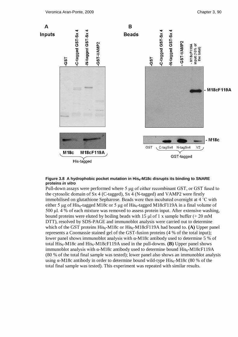
Veronica Aran-Ponte, 2009 Chapter 3, 90
Figure 3.8 A hydrophobic pocket mutation in His6-M18c disrupts its binding to SNARE proteins in vitro
Pull-down assays were performed where 5 μg of either recombinant GST, or GST fused to
the cytosolic domain of Sx 4 (C-tagged), Sx 4 (N-tagged) and VAMP2 were firstly
immobilised on glutathione Sepharose. Beads were then incubated overnight at 4 ˚C with
either 5 μg of His6-tagged M18c or 5 μg of His6-tagged M18cF119A in a final volume of
500 μl. 4 % of each mixture was removed to assess protein input. After extensive washing,
bound proteins were eluted by boiling beads with 15 μl of 1 x sample buffer (+ 20 mM
DTT), resolved by SDS-PAGE and immunoblot analysis were carried out to determine
which of the GST proteins His6-M18c or His6-M18cF119A had bound to. (A) Upper panel
represents a Coomassie stained gel of the GST-fusion proteins (4 % of the total input);
lower panel shows immunoblot analysis with α-M18c antibody used to determine 5 % of
total His6-M18c and His6-M18cF119A used in the pull-downs. (B) Upper panel shows
immunoblot analysis with α-M18c antibody used to determine bound His6-M18cF119A
(80 % of the total final sample was tested); lower panel also shows an immunoblot analysis
using α-M18c antibody in order to determine bound wild-type His6-M18c (80 % of the
total final sample was tested). This experiment was repeated with similar results.

Veronica Aran-Ponte, 2009 Chapter 3, 91
3.4 Discussion
The interaction between syntaxins and SM proteins has been proposed as a potential
regulatory step in the process of SNARE-mediated membrane fusion. Despite all the
research performed in this area, the interaction of SM protein with non-syntaxin members
was an unknown event until Peng and Gallwitz demonstrated that v- SNARE proteins
could also interact with SM proteins in yeast (Peng and Gallwitz, 2004). The results
presented in this chapter have reinforced this finding by demonstrating that the SM protein
M18c, apart from binding Sx 4, also interacts specifically with VAMP2 in vitro (figure
3.1). During the course of this work, an interaction between VAMP2/M18a was also
reported (Shen et al., 2007). Nevertheless, the M18c/VAMP2 interaction has not been
previously identified, providing new insight into the mechanism by which SM functions in
muscle and adipose tissue.
M18c binding to VAMP2 does not require the short N-terminal peptide of VAMP2, since
cytosolic GST-VAMP2 only harbouring the SNARE motif was still able to interact with
His6-M18c (figure 3.2). This result indicates that unlike Sx 4, M18c does not require the
short N-terminal peptide of VAMP2 to interact. Furthermore, data presented in this chapter
also demonstrated that Sx 4 can displace VAMP2 from His6-M18c (figure 3.3 and 3.5B).
In contrast, VAMP2 was unable to displace Sx 4 from His6-M18c (figure 3.4 and 3.5A).
An explanation for this could be that upon Sx 4/M18c binding, a conformational change in
M18c occurs, which in consequence disrupts M18c binding to VAMP2. In fact, the two
binding modes by which Sx 4 interacts with M18c (“open” and “closed”) suggest that
affinity between these two proteins is strong and it could be higher than M18c/VAMP2
affinity. Moreover, the immunoblots performed to demonstrate that M18c binds VAMP2
(figure 3.1), also indicate that the amount of His6-M18c which were recovered on GST-
VAMP2 beads were smaller than that recovered on the GST-Sx 4 beads.
The “bridging hypothesis” by Peng and Gallwitz argues that the binding of specific v- and
t-SNAREs on opposite membranes could involve SM proteins regardless of whether it is
bound to syntaxin or not (Peng and Gallwitz, 2004). Thus, data presented here could serve
as an indicator that the bridging theory could occur since both the v-SNARE VAMP2 and
the t-SNARE Sx 4, which are present in opposite membranes, interact with M18c.
Unfortunately, there was not enough time to perform careful and detailed investigation on
whether M18c could bridge VAMP2 and Sx 4 interaction. Nevertheless, this interaction

Veronica Aran-Ponte, 2009 Chapter 3, 92
might be very transient since Sx 4 displaces VAMP2 from His6-M18c in vitro (figure 3.3
and 3.5B).
The other subject addressed in this chapter was the hydrophobic pocket mode of
interaction between M18c and Sx 4. This N-terminal mode of interaction between
syntaxins and SM proteins has been shown to be conserved from yeast to mammals with
the determination of the structure of both the yeast Sly1p/Sed5p and the mammalian
M18c/Sx 4 N-peptide complex (Bracher and Weissenhorn, 2002; Hu et al., 2007). The
hydrophobic residue L117 in yeast Vps45p was shown to be an important residue
mediating Tlg2p/Vps45p interaction (Carpp et al., 2006). Our alignment studies between
M18c and Vps45p revealed that residue F119 corresponded to residue L117 in Vps45p
(figure 3.6). Therefore, a mutant was generated by changing a phenylalanine to an alanine
in residue F119. This mutation prevented both Sx 4 and VAMP2 binding to M18c (figure
3.8). Unfortunately, it was not possible to perform circular dichroism (CD) to analyse if the
conformation of His6-M18cF119A was affected by the mutation since the high levels of
GroEL co-purified with recombinant His6-M18cF119A could affect the CD measurements.
Thus, a change in the overall structure of the mutant version of M18c could be the reason
for the results obtained, although another group has also confirmed that mutation of F119
to glutamic acid disrupts binding of M18c to Sx 4 (Latham et al., 2006). Structural analysis
have also confirmed that the Sx 4 N-terminal residue L8 is important in mediating Sx 4
interaction with the hydrophobic pocket on the outer face of domain I in M18c and
mutation of this residue (L8) to Lysine disrupts Sx 4 binding to M18c (Hu et al., 2007;
Latham et al., 2006). In yeast, hydrophobic pocket mutations in Sed5p and Vps45p also
prevented interaction with their respective syntaxins (Bracher and Weissenhorn, 2002;
Carpp et al., 2006). In contrast to data obtained in this chapter, Vps45p L117A mutant was
shown to interact with the v-SNARE Snc2p (Carpp et al., 2006). Figure 3.8B shows that
His6-M18c F119A was not able to interact with GST-VAMP2. This result indicates that, if
His6-M18c F119A protein is properly folded, there might be some sort of connection
between the SNARE motif of VAMP2 and the hydrophobic region in the N- terminal of
M18c.
In summary, the data presented in this chapter indicate that the SM protein M18c has two
SNARE partners. These SNARE proteins correspond to the PM SNARE Sx 4 and the
GLUT4 vesicle SNARE VAMP2 (Brandie et al., 2008). However, Sx 4 showed a strong
affinity to M18c, since it can displaces VAMP2 from M18c but VAMP2 cannot do the
same (Brandie et al., 2008). The Sx 4/M18c affinity might involve two modes of

Veronica Aran-Ponte, 2009 Chapter 3, 93
interaction, one being the interaction via the short N-terminal peptide in Sx 4, which inserts
into a hydrophobic pocket in M18c, and one via the closed conformation of Sx 4 which is
predicted to be similar to Sx 1A/M18a crystal structure (D'Andrea-Merrins et al., 2007;
Misura et al., 2000). The His6-M18c F119A mutant reinforced the existence of an N-
terminal mode of interaction since this mutation abolished interaction to Sx 4. Therefore,
this pocket mode of interaction could allow SM proteins not only to bind to monomeric
syntaxins but also to SNARE complexes. All the different modes of interaction between
SM and SNARE proteins might play an active role in the regulation of SNARE complex
formation.

94
Chapter 4
4 Negative regulation of Sx 4/SNAP23/VAMP2-
mediated fusion by Munc18c in vitro

Veronica Aran-Ponte, 2009 Chapter 4, 95
4.1 Introduction
The role of SM proteins in membrane fusion has recently become the centre of attention in
several studies in cell biology. While the SNARE proteins were shown to have a more
defined role, the precise function of SM proteins has not yet been unravelled. However it is
known that SM proteins might regulate positively or negatively SNARE function
depending on the trafficking pathway and organism studied. In chapter 3, the novel finding
that M18c not only binds Sx 4 but also binds VAMP2 was described. In this chapter,
another approach was taken to determine how SM proteins interact with SNAREs.
SNAREs were reconstituted into synthetic liposomes in order to perform in vitro fusion
assays. This technique is an invaluable tool to study SNARE-mediated membrane fusion in
vitro.
The fusion assay performed in this study was first described by Struck and colleagues
(Struck et al., 1981). It is a very useful tool to study the fusogenic properties of SNARE
proteins, which were shown to promote spontaneous fusion of liposomes (Schuette et al.,
2004; Weber et al., 1998; Zhang et al., 2004). The fluorescence resonance energy transfer
(FRET) capability between a donor fluorescent lipid and a nonfluorescent acceptor lipid
can be measured, giving an indication of the levels of liposome fusion. When fusion
occurs, FRET decreases due to an increase in the distance between fluorescent probes.
Details on this technique are described in sections 2.2.3.2, 2.2.3.3, 2.2.3.4 and 4.3.4.
The v-SNARE VAMP2 present in GLUT4 vesicles forms an SDS-resistant complex with
the t-SNAREs Sx 4 and SNAP23 which are present in the PM of muscle cells and
adipocytes (Rea et al., 1998). In vitro fusion studies performed in our lab have confirmed
this finding since these proteins were shown to catalyse fusion when reconstituted in
synthetic liposomes (Brandie et al., 2008). Fusion facilitated by these SNAREs is
subjected to multiple levels of regulation in vivo and the SM protein M18c is thought to
participate in this regulatory process. Studies performed with SM proteins present in other
organisms such as yeast, indicated some clues into the possible role of M18c.
The importance of the yeast SM protein Sec1p in the regulation of post-Golgi secretion
have given further insights into the SM function. Sec1p binds with low affinity to the yeast
syntaxin Sso1p and with higher affinity to both the t-complex formed by Sso1p and Sec9c,
and to the SNARE ternary complex Sso1p/Sec9c/Snc2p (Scott et al., 2004). In vitro fusion
studies using these proteins revealed that Sec1p directly stimulates Sso1p/Sec9c/Snc2p-

Veronica Aran-Ponte, 2009 Chapter 4, 96
mediated membrane fusion in vitro (Scott et al., 2004). In neuronal cells, a similar result
was observed. M18a was shown to be able to strongly stimulate fusion mediated by the t-
SNAREs Sx 1A/SNAP25 with the v-SNARE VAMP2 (Shen et al., 2007). Stimulation
occurred not only via interaction with the t-SNAREs, but also via interaction with v-
SNAREs (Shen et al., 2007).
It is unclear how SM proteins might stimulate some fusion reactions, but some research
groups have identified a small peptide at the N-terminal region of syntaxins as the binding
site for SM proteins (Bracher and Weissenhorn, 2002; Carpp et al., 2006; Dulubova et al.,
2003; Dulubova et al., 2002; Hu et al., 2007; Rickman et al., 2007). The N-terminal region
of Sx 4 will be described in the following section.
4.1.1 The N-terminal domain of Sx 4
As previously described in section 1.2.1, most syntaxins contain an independently folded
N-terminal domain which is an important regulatory region in these proteins. This domain
contains a short N-terminal peptide followed by a three-helix bundle called the Habc
domain similar to Sx 1A (Fernandez et al., 1998). The N-terminal region of Sx 4 was
shown to be necessary for its interaction with M18c (Latham et al., 2006). This SM protein
can interact with Sx 4 alone (Hu et al., 2003), VAMP2 (Brandie et al., 2008) and also with
the binary (Sx 4/SNAP23) and ternary SNARE complexes (Sx 4/SNAP23/VAMP2)
(Latham et al., 2006). It is already establish that the neuronal SM protein M18a binds Sx
1A when the latter is in the closed conformation (Misura et al., 2000; Yang et al., 2000)
and also when Sx 1A is in the open conformation (via the N-terminal peptide region in Sx
1A) (Burkhardt et al., 2008; Khvotchev et al., 2007; Rickman et al., 2007). Sx 4 also uses
its short N-terminal peptide to bind a hydrophobic region in M18c (Hu et al., 2007;
Latham et al., 2006), however it is not certain if Sx 4 also adopts a closed conformation,
such as that reported for Sx 1A. Furthermore, modeling studies based on the crystal
structure of Sx 1A in complex with M18a (Misura et al., 2000), have shown that the
M18a/Sx 1A complex has a high sequence homology to M18c/Sx 4 (D'Andrea-Merrins et
al., 2007). The regions of intramolecular interactions between the Habc domain and the
SNARE motif (H3 domain) of Sx 4 were found to be highly conserved between the two
different syntaxins (D'Andrea-Merrins et al., 2007). This could suggest that Sx 4, like Sx
1A, could adopt a closed conformation. Figure 4.1 shows two different modes of
interaction for Sx 4/M18c. Figure 4.1A shows the hypothetical model of M18c binding Sx
4 in the closed conformation (D'Andrea-Merrins et al., 2007), whereas figure 4.1B shows

Veronica Aran-Ponte, 2009 Chapter 4, 97
the interaction between the N-terminal peptide of Sx 4 and the hydrophobic region (already
described in chapter 3) in the outer face of domain I in M18c.
Figure 4.1 The predicted two modes of interaction between M18c and Sx 4.
(A) Figure A was taken from D‟Andrea-Merrins and colleagues work (D'Andrea-Merrins
et al., 2007). Using a ClustalW alignment, the complex M18c (grey)/Sx4 was modeled
directly into the crystal structure of M18a/Sx 1A (D'Andrea-Merrins et al., 2007). The
helical Habc domains (Ha- pale blue, Hb- gold, Hc- cyan), the SNARE motif (green), and
the linker region which connects the SNARE motif with the Habc domain (orange), are
shown. When Sx 4 is the open conformation, the Habc domain is moved away from the
SNARE motif leaving it free to participate in the SNARE core complex. In vitro, this is
achieved by mutating residues L173A/E174A in the hinge region of Sx 4 (Aran et al.,
2009; D'Andrea-Merrins et al., 2007). I241A and I217A mutations were shown to be also
important, since they reduce affinity for M18c (D'Andrea-Merrins et al., 2007). (B) Using
the Rasmol software, models of M18c (green) interacting with the first 36 amino acids
(grey) from the N-terminus of Sx 4 were obtained based on previous described models (Hu
et al., 2007) and designed by Prof James Milner-White (University of Glasgow). The
model on the top panel shows M18c as an arch shaped molecule binding the 36 amino
acids of the helical Sx 4 short N-terminal region. The lower panel represents the same
figure from a different perspective, showing the M18c hydrophobic region (composed of β
sheets) present on the outer face of domain I, where the short peptide inserts.

Veronica Aran-Ponte, 2009 Chapter 4, 98
In our lab, Dr Fiona Brandie has generated mutant versions of cytosolic Sx 4. These
versions were: Sx 4 lacking the short N-terminal peptide (Sx 4 N∆36); Sx 4 locked in the
open conformation, which was obtained by introducing two point mutations at the hinge
region (L173A, E174A); and Sx 4 in the open conformation but lacking the 36 amino
acids from the N-terminus (Sx 4 open N∆36). These mutants were compared to the wild-
type form of Sx 4 in their ability to bind M18c in vitro. These constructs contained a GST
tag at either the C- or the N- terminal region in order to compare if the position of the
bulky GST moiety could affect M18c/Sx 4 interaction. Indeed, the GST tag affected the
results (Aran et al., 2009). When syntaxin was tagged with GST at the N-terminus, this
affected the pocket-mode of binding and favoured a different interaction not previously
described. This interaction was independent of the binding via the short N-terminal
peptide. As can be seen in figure 4.2A, M18c is able to bind the mutant lacking the short
N-terminal peptide, whereas both open mutants (full-length and N∆36) exhibit a dramatic
reduction in the levels of bound M18c compared to the full-length cytosolic Sx 4.
Nevertheless, figure 4.2B shows that by changing the tag to the C-terminus, the short N-
terminal peptide becomes “free” to interact with M18c via the hydrophobic pocket mode of
interaction. In this case, deletion of the N-terminal peptide in the open and wild-type Sx 4
versions results in a dramatically reduction in binding (Aran et al., 2009). These data
supports the idea that when Sx 4 is tagged at the C-terminus, the cytosolic domain of Sx 4
predominantly adopts an open conformation or transits easily from a closed to an open
form. In contrast, when Sx 4 is tagged at the N-terminus it might adopt a different
conformation which could be related to the closed conformation observed for Sx 1A. In
vitro fusion assays using these mutants are a very useful tool to understand the function of
the N-terminal peptide in the process of SNARE assembly and to define the importance of
M18c in the regulation of Sx 4/SNAP23/VAMP2-mediated membrane fusion.

Veronica Aran-Ponte, 2009 Chapter 4, 99
Figure 4.2 Quantifications of the amount of M18c bound to either N- (A) or C-tagged versions (B) of cytosolic Sx 4 (Figure taken from Dr Fiona Brandie, University of Glasgow)
(A) The N-terminally tagged versions of Sx 4 (GST- Sx 4, GST-Sx 4 L173A/E174A
(OPEN), GST-Sx 4 ∆N36 (ND) and GST-Sx 4 OPEN ND) were incubated with His6-M18c
and the results are shown as a percentage of bound M18c relative to wild-type Sx 4. (B)
The C-terminally tagged Sx 4 versions were incubated with His6-M18c and again, the
results are shown as a percentage of bound M18c relative to wild-type Sx 4. Figure taken
from Dr Fiona Brandie (University of Glasgow).

Veronica Aran-Ponte, 2009 Chapter 4, 100
4.2 Aims of this chapter
The aims of this chapter were to generate full-length Sx 4 constructs containing the
following changes: an N-terminal peptide truncation (N∆36) and open form
(L173A/E174A) also lacking the N-terminal peptide. The original construct containing the
full-length open form of Sx 4 was made by Dr Fiona Brandie (University of Glasgow).
These constructs were generated in order to co-express them with SNAP23 and to test their
ability to fuse in vitro with VAMP2. Previously in our lab, Dr Fiona Brandie has shown
that M18c is able to bind to the extreme N-terminus of Sx 4 in vitro (Aran et al., 2009).
Hence, these mutants when used in the in vitro fusion assay may reveal further details
about the mechanism of action of M18c.
4.3 Results
4.3.1 Expression and purification of t-SNARE complexes
Full-length SNARE complexes were expressed and purified as described in section 2.2.3.1.
Full-length Sx 4 constructs containing the following changes: N-terminal peptide
truncation (N∆36), Sx 4 locked in the OPEN conformation (i.e. containing mutations
L173A/E174A) and Sx 4 OPEN N∆1-36 were generated in this study. Recombinant wild-
type Sx 4 in complex with SNAP23 was obtained from Dr Fiona Brandie. A typical protein
purification of wild-type Sx 4/SNAP23 is shown in figure 4.3.

Veronica Aran-Ponte, 2009 Chapter 4, 101
175
83
62
47.5
32.5
kDa
25
Sx 4
SNAP23
GST
175
83
62
47.5
32.5
kDa
25
Sx 4
SNAP23
GST
175
83
62
47.5
32.5
kDa
25
Sx 4
SNAP23
GST
Figure 4.3 Purified Sx 4/SNAP23 t-SNARE complex from E.coli lysates
Dr Fiona Brandie has expressed Sx 4 and SNAP23-GST in BL21 (DE3) E. coli cells,
purified the complex and samples from the eluted fractions were used in this chapter.
Complete details on how wild-type Sx 4/SNAP23 heterodimer was expressed and purified
are described in section 2.2.3.1. The purity of the proteins was assessed on a 15 % SDS-
PAGE followed by Coomasie Blue staining as outlined in sections 2.2.2.1 and 2.2.2.3. The
figure shows the recovered cleaved complex after 4 h of thrombin cleavage (5 µl).
Positions of the molecular weight markers are shown on the left of the gel.
4.3.1.1 Generation of Sx 4 N∆36 and Sx 4 “OPEN” (L173A/E174A) N∆36 to
co-express with SNAP23
The wild-type and open forms of Sx 4 lacking amino acids 1-36 were cloned into the
vector pQE30. Sx 4 N∆36 and Sx 4 OPEN N∆36 were generated by PCR using pQE30 Sx
4 (full-length) as template for the wild-type Sx 4 N∆36 and pQE30 Sx 4 (full-length
L173A/E174A) as template for Sx 4 OPEN N∆36. Primers used to amplify desired
sequences were:
Sx 4 N∆36 Forward: 5‟ GGT ACC CCG GAC GAC GAG TTC TTC CAG AAG 3‟
Sx 4 N∆36 Reverse: 5‟ AAG CTT TTA TCC AAC GGT TAT GGT GAT GC 3‟

Veronica Aran-Ponte, 2009 Chapter 4, 102
Both versions of Sx 4 were cloned between the KpnI and HindIII sites in pQE30. After
successful cloning, pQE30 Sx 4 N∆36 and pQE30 Sx 4 OPEN N∆36 were co-transformed
with pET41a SNAP23 into E. coli BL21 (DE3) cells. Both plasmids were transformed by
mixing 50 ng of SNAP23 DNA with 100 ng of either pQE30 Sx 4 N∆36 or pQE30 Sx 4
OPEN N∆36 DNA in the same E. coli cells. Cells containing both plasmids were selected
on dual antibiotic plates since SNAP23 is expressed from a kan resistant vector (pET41)
and the different versions of Sx 4 are expressed from amp resistant vectors (pQE30).
Selected colonies were used to start protein purification which is described in detail in
section 2.2.3.1. Both t-SNARE complexes were successfully purified and samples from
each step were analysed by SDS-PAGE, followed by Coomassie blue staining, as shown in
figures 4.4 and 4.5.
Figure 4.4 Purification of Sx 4 N∆36/SNAP23 complex from E.coli
Full-length His6-Sx 4 N∆36 and SNAP23-GST were co-expressed in BL21 (DE3) E.coli
cells. Expression was induced overnight (~16 h) by adding 1 mM IPTG and incubation
was carried out at 25 ˚C. The complex was purified taking advantage of the GST tag
attached to the N-terminus of SNAP23 and this tag was cleaved at the end of the
purification by incubation with thrombin for 4 h at room temperature. The figure shows
several samples taken during the process of protein purification which were run on a 12 %
SDS-PAGE gel and stained with Coomassie Blue as outlined in sections 2.2.2.1 and
2.2.2.3. The positions of the molecular weight markers are shown on the left. The first lane
corresponds to a sample before induction (BI) followed by a sample after induction (I),
sample from the lysate (L), wash 1 (W1), wash 2 (W2), wash 3 (W3) and 3 µl of eluted
fractions (EL1 and EL2).

Veronica Aran-Ponte, 2009 Chapter 4, 103
Figure 4.5 Purification of Sx 4 OPEN N∆36/SNAP23 complex from E.coli
Full-length His6-Sx 4 OPEN N∆36 and SNAP23-GST were co-expressed in BL21 (DE3)
E.coli cells. Expression was induced overnight (~16 h) by adding 1 mM IPTG and
incubation was carried out at 25 ˚C. The complex was purified taking advantage of the
GST tag attached to the N-terminus of SNAP23 and this tag was cleaved at the end of the
purification by incubation with thrombin for 4 h at room temperature. The figure shows
several samples taken during the process of protein purification which were run on a 12 %
SDS-PAGE gel and stained with Coomassie Blue. The positions of the molecular weight
markers are shown on the left. The first lane corresponds to a sample before induction (BI)
followed by a sample after induction (I), sample from the lysate (L), wash 1 (W1), wash 2
(W2) and 5 µl of eluted fraction (EL1). Molecular weight markers are shown on the left.
Lower figure shows more elutions and 2 µl of EL1, EL2 and EL3 were run on a 15 %
SDS-PAGE in order to better resolve the upper band corresponding to Sx 4 OPEN N∆36,
which due to the lack of 36 amino acids, it runs very close to SNAP23.
4.3.1.2 Generation of full-length Sx 4 “OPEN” (L173A/E174A) in complex
with SNAP23
The construct containing full length Sx 4 OPEN (pQE30 Sx 4 L173A/E174A) was made
by Dr Fiona Brandie (University of Glasgow). However, in her work the co-expression of
this protein with SNAP23 was not achieved due to the poor solubility of Sx 4 OPEN.
Therefore, in this study Sx 4 OPEN/SNAP23 expression and purification conditions were

Veronica Aran-Ponte, 2009 Chapter 4, 104
optimised in order to obtain enough quantities of this t-complex to reconstitute into
liposomes and therefore perform in vitro fusion assays. In order to achieve this, SNAP23
and Sx 4 OPEN plasmids were co-transformed into the same BL21 (DE3) E. coli cells by
mixing 50 ng of SNAP23 DNA with 200 ng of Sx 4 OPEN DNA (for wild-type versions
only 100 ng was necessary). Cells containing both plasmids were selected on dual
antibiotic plates (amp/kan) and these colonies were used to start protein purification which
is described in detail in section 2.2.3.1.
The t-SNARE complex Sx 4 OPEN/SNAP23 was purified and samples from each step
were analysed by SDS-PAGE, followed by Coomassie blue staining, as shown in figure
4.6.
Figure 4.6 Purification of Sx 4 OPEN/SNAP23 complex from E.coli
Full-length His6-Sx 4 OPEN and SNAP23-GST were co-expressed in BL21 (DE3) E.coli
cells. Expression was induced overnight (~16 h) by adding 1 mM IPTG and incubation
was carried out at 24 ˚C. The complex was purified taking advantage of the GST tag
attached to the N-terminus of SNAP23 and this tag was cleaved by incubation with
thrombin for 4 h at room temperature. The figure shows several samples taken during the
process of protein purification which were run on a 15 % SDS-PAGE gel and stained with
Coomassie Blue as outlined in sections 2.2.2.1 and 2.2.2.3 respectively. The positions of
the molecular weight markers are shown on the left. The first lane corresponds to a sample
before induction (BI) followed by a sample after induction (I), sample from the lysate (L),
wash 1 (W1), wash 2 (W2), wash 3 (W3) and 3 µl of eluted fractions (EL1 and EL2).
Molecular weight markers are shown on the left. This result is from a typical Sx 4
OPEN/SNAP23 protein purification.
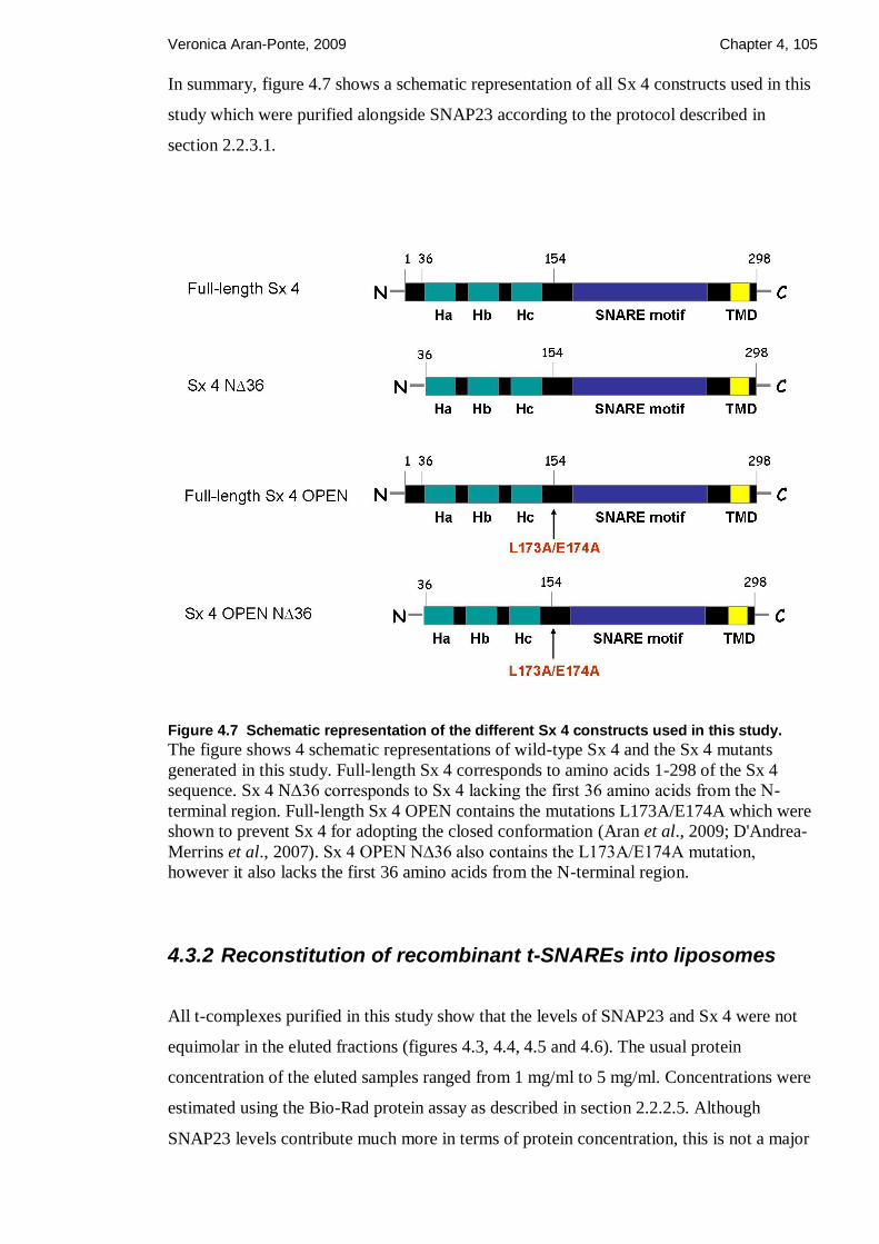
Veronica Aran-Ponte, 2009 Chapter 4, 105
In summary, figure 4.7 shows a schematic representation of all Sx 4 constructs used in this
study which were purified alongside SNAP23 according to the protocol described in
section 2.2.3.1.
Figure 4.7 Schematic representation of the different Sx 4 constructs used in this study.
The figure shows 4 schematic representations of wild-type Sx 4 and the Sx 4 mutants
generated in this study. Full-length Sx 4 corresponds to amino acids 1-298 of the Sx 4
sequence. Sx 4 N∆36 corresponds to Sx 4 lacking the first 36 amino acids from the N-
terminal region. Full-length Sx 4 OPEN contains the mutations L173A/E174A which were
shown to prevent Sx 4 for adopting the closed conformation (Aran et al., 2009; D'Andrea-
Merrins et al., 2007). Sx 4 OPEN N∆36 also contains the L173A/E174A mutation,
however it also lacks the first 36 amino acids from the N-terminal region.
4.3.2 Reconstitution of recombinant t-SNAREs into liposomes
All t-complexes purified in this study show that the levels of SNAP23 and Sx 4 were not
equimolar in the eluted fractions (figures 4.3, 4.4, 4.5 and 4.6). The usual protein
concentration of the eluted samples ranged from 1 mg/ml to 5 mg/ml. Concentrations were
estimated using the Bio-Rad protein assay as described in section 2.2.2.5. Although
SNAP23 levels contribute much more in terms of protein concentration, this is not a major

Veronica Aran-Ponte, 2009 Chapter 4, 106
problem for reconstitution into liposomes since SNAP23 does not contain a trans-
membrane domain. In addition, recombinant SNAP23 purified from E.coli cells does not
possess palmitate groups. This means that SNAP23 needs to be in complex with Sx 4,
which contains a trans-membrane domain, in order to be reconstituted into liposomes.
Therefore, it is very important to have enough recombinant Sx 4 for Sx 4/SNAP23
reconstitution to occur.
In order to reconstitute SNARE proteins into liposomes, first the lipid mixtures were
prepared as outlined in section 2.2.3.2. Briefly, the labelled donor vesicles contained a
quenched mixture of fluorescent phospholipids and the unlabelled acceptor vesicles were
not fluorescent labelled. Lipids were dried under nitrogen and resuspended in a solution
containing either the v-SNAREs (fluorescent donor vesicles) or t-SNAREs (non-
fluorescent acceptor vesicles) in the presence of OG, which helps to incorporate the trans-
membrane domains of SNAREs into the liposome membranes. A complete description on
the SNARE reconstitution technique is outlined in section 2.2.3.3 and a schematic
representation is shown in figure 4.8.

Veronica Aran-Ponte, 2009 Chapter 4, 107
Figure 4.8 Schematic representation of the incorporation of SNARE proteins into liposomes (kindly given by Prof James A. McNew, Rice University)
This schematic model shows how v- and t- SNAREs are reconstituted into vesicles (A)
Dried phospholipids were mixed with protein in the presence of the detergent octyl-β-d
glucopiranoside (OG). The OG monomer and the phospholipid monomer are represented
in blue and orange respectively. After rapid dilution below the critical micellar level of the
detergent by addition of 1.5 ml of buffer A200, solution is placed into a float-a-lyser to
dialyse overnight in order to remove any free OG monomers still present in the solution.
After dialysis, the octylglucoside/phospholipid mixed micelle is formed. (B) The resulting
proteoliposomes can then be isolated by flotation in a Histodenz density gradient. After
ultracentrifugation for 4 h at ~ 372,000 xg, proteoliposomes float to the top layer of the
sample due to their high lipid content. Recovered proteoliposomes can be assessed by
analysing 10 µl on a SDS-PAGE followed by Coomassie Blue staining. Unincorporated
protein remains at the bottom of the tube. Complete details on SNARE reconstitution and
proteoliposome recovery are outlined in sections 2.2.3.3 and 2.2.3.4 respectively.

Veronica Aran-Ponte, 2009 Chapter 4, 108
Purified SNARE proteins shown in figures 4.3, 4.4, 4.5 and 4.6 were successfully
reconstituted into liposomes (figure 4.9). Immunoblot analysis using anti-Sx 4 shows the
Sx 4 levels present in each complex before reconstitution (figure 4.9; left panel). This was
performed in order to reconstitute the 4 different complexes at similar quantities into their
respective liposomes. The resulting different versions of reconstituted Sx 4 in complex
with SNAP23 were resolved by SDS-PAGE and can be seen in figure 4.9 (right panel).
Figure 4.9 Reconstitution of recombinant SNAREs into liposomes
Purified t-SNARE complexes were successfully reconstituted into synthetic liposomes.
Left panel shows an immunoblot analysis using anti-Sx 4 antibody performed to check
levels of recombinant Sx 4 (complexed with SNAP23) before reconstitution. Using the
program Image J, it was possible to calculate the differences in quantities between each
t-SNARE complex. After reconstitution, 10 µl of reconstituted t-SNARE liposomes were
separated on a 15 % SDS-PAGE gel and stained with Coomassie Blue as outlined in
sections 2.2.2.1 and 2.2.2.3 respectively. Positions of the molecular weight markers are
shown. Data from a typical set of reconstitutions is shown.

Veronica Aran-Ponte, 2009 Chapter 4, 109
4.3.3 VAMP2 expression, purification and reconstitution into
liposomes
Full-length C-terminally tagged VAMP2 was expressed as a Hexa-HIS-tagged protein in
BL21 (DE3) cells and purified by Dr Fiona Brandie (University of Glasgow). pET-
VAMP2FL-myc-HIS was a kind gift from Giampietro Schiavo (London, UK).
VAMP2 was successfully reconstituted into liposomes as shown in figure 4.10. Samples
taken before (lane 1) and after (lane 2) reconstitution are shown. Lane 3 represents a
sample of soluble recombinant VAMP2 lacking the trans-membrane domain (VAMP2
∆TMD) which was used as an inhibitor of fusion in the controls for in vitro fusion assays.
Figure 4.10 Purification and reconstitution of VAMP2 into liposomes
Full-length VAMP2 (lane1, 10 µl) purified by Dr Fiona Brandie was reconstituted into
liposomes (lane 2, 10 µl). Cytoplasmic VAMP2 (i.e. lacking the trans-membrane domain-
VAMP2 ∆TMD) which is used as an inhibitor of in vitro fusion, is shown in lane 3 (10 µl).
Samples were separated on a 15 % SDS-PAGE gel and stained with Coomassie blue.
Positions of the molecular weight markers are shown on the left. This is a result of a
typical VAMP2 reconstitution experiment.

Veronica Aran-Ponte, 2009 Chapter 4, 110
4.3.4 In vitro fusion assays using reconstituted liposomes
After the SNARE proteins shown in figures 4.9 and 4.10 were successfully reconstituted
into their appropriate liposomes, a series of different in vitro fusion assays were carried out
using these proteoliposomes. These assays were performed in order to investigate if
mutations in Sx 4 could affect the fusion rates, either positively or negatively.
Figure 4.11 shows a schematic representation of the principle of the in vitro fusion assay.
Briefly, the fusion reactions were set up on a microtitre plate on ice. One well was used to
add the fusion reaction and a second well was used for the fusion control reaction. The
fusion reaction mixtures are set up at approximately 1:1 v-SNARE:t-SNARE molar ratio
which represents the addition of approximately 10 times more volume of t-SNARE
vesicles than v-SNARE in each reaction. Fusion assays containing the wild-type Sx
4/SNAP23, Sx 4 N∆36/SNAP23, Sx 4 OPEN/SNAP23 and Sx 4 OPEN N∆36/SNAP23 t-
SNARE complexes were performed in which 5 µl of fluorescently labelled donor VAMP2
liposomes were mixed with 45 µl of unlabelled acceptor t-complexes. As a control, the t-
SNARE liposomes were pre-incubated for 10 min with 3 µl of VAMP2 lacking the trans-
membrane domain (VAMP2 ∆TMD) which is known to act as a dominant negative
inhibitor of fusion. The plate was incubated for 10 min on ice followed by the addition of 5
µl of VAMP2 liposomes (VAMP2 liposomes are shown in figure 4.10). To the “fusion”
well, 3 µl of buffer A200 (glycerol free) was added instead. NBD fluorescence was then
measured at 2 min intervals for 2 h before addition of 10 µl of 2.5 % dodecylmaltoside,
and measurements proceeded for further 40 min. Raw data was collected and normalised to
maximum detergent signal (see Appendix, figure I). Rate of fusion was then measured as a
percentage of the maximum NBD fluorescence (100 %).

Veronica Aran-Ponte, 2009 Chapter 4, 111
Figure 4.11 Fusion assay model (Prof James A. McNew, Rice University, USA)
The fusion assay model takes advantage of two different labelled lipids, one labelled with
rhodamine (pink spheres) and one labelled with NBD (yellow spheres). A quenched
mixture of these fluorescent labelled lipids is present in the same membrane (donor) which
will contain the reconstituted v-SNARE proteins. The acceptor vesicles (acceptor) are not
labelled and will contain the reconstituted t-SNARE proteins. When both acceptor and
donor vesicles are mixed at 37 ˚C, these vesicles fuse. When NBD and rhodamine are in
close proximity, excitation of NBD leads to an energy transfer to the rhodamine
fluorophore in a process known as fluorescent resonance energy transfer (FRET), which is
dependent on the distance between the 2 fluorophores. Upon fusion, a decrease in
quenching (due to the decrease in proximity of the two fluorophores) occurs, which leads
to an increase in the overall NBD fluorescence which can be measured using a
fluorescence plate reader. Complete details on this technique are outlined in section
2.2.3.6.
Figure 4.12 shows the successful fusion of the four different t-SNARE complexes with
their respective v-SNARE VAMP2 vesicles. Results obtained confirm that, as expected, t-
complex containing wild-type Sx 4 liposomes fuse with VAMP2 liposomes (figure 4.12A).
The removal of the short N-terminal peptide in both open (figure 4.12D) and wild-type
(figure 4.12B) Sx 4 mutants did not inhibit fusion. The t-complex containing the full-
length open version of Sx 4 was also shown to be able to fuse with VAMP2 liposomes
(figure 4.12C). Differences in the rates of fusion observed between all 4 different fusions

Veronica Aran-Ponte, 2009 Chapter 4, 112
might be due to the subtly different levels of t-SNARE complexes reconstituted into
liposomes (figure 4.9).

Veronica Aran-Ponte, 2009 Chapter 4, 113

Veronica Aran-Ponte, 2009 Chapter 4, 114
Figure 4.12 In vitro fusion assays of liposomes containing either full-length Sx 4/SNAP23 (A) or Sx 4 N∆36/SNAP23 (B) or Sx 4 OPEN/SNAP23 (C) or Sx 4 OPEN N∆36/SNAP23 (D) with liposomes containing VAMP2
In all fusion assays A, B, C and D shown, 5 μl of fluorescently labelled donor VAMP2
liposomes were mixed with 45 μl of unlabelled acceptor t-complexes being either wild-
type (A) or Sx 4 N∆36/SNAP23 (B) or Sx 4 OPEN/SNAP23 (C) or Sx 4 OPEN
N∆36/SNAP23 (D). To all 4 fusion assays, 3 μl of buffer A200 (fusion reaction; open
squares) or 3 μl of cytoplasmic VAMP2 (negative control; filled squares) were added. Note
that the soluble VAMP2 was pre-incubated with the t-complex in order to block VAMP2
liposome fusion. Fusions between the two vesicle populations (t- and v-SNAREs) in A, B,
C and D were monitored by first placing the 96 well plate (where the proteoliposomes
were mixed) inside the fluorescence reader. Before starting the fusion measurements, the
plate was incubated inside the reader for 5 min at 37 oC to try to avoid the drop in the
fluorescence intensity observed in all 4 fusion reactions above (see first 10 min). This drop
is the result of the shift from 4 oC to 37
oC at the beginning of each fusion. Fluorescence
was then measured at 2 min intervals for 2 h with the excitation filter set to 485 nm and the
emission filter set at 520 nm. After 2 h, the plate was removed and 10 μl of 2.5 % (w/v) n-
dodecylmaltoside was added to the wells containing liposomes and mixed (this produces a
maximum detergent signal represented by 100 % in the raw graphs of A, B, C and D, see
appendix, figure I) and the fluorescence was recorded for further 40 min at 2 min intervals.
A graphing and analysis programme called KaleidaGraph (Synergy Software) was used in
order to analyse the data obtained from the Fluorescence reader. Raw fluorescence was
normalised to maximum detergent signal (100 %) and plotted against time in minutes (see
appendix, figure I). The levels of fusion were determined as a percentage of the maximum
signal of NBD emission. The detailed protocol is described in section 2.2.3.6 and 2.2.3.7.

Veronica Aran-Ponte, 2009 Chapter 4, 115
These figures show a typical result from the in vitro fusions mediated by the different
versions of Sx 4/SNAP23 withVAMP2, repeated 3 times for all complexes.
4.3.5 Effects of the addition of M18c directly into the in vitro
fusion assay
The role of SM proteins in SNARE-mediated membrane fusion has also been investigated
through the use of in vitro fusion assays, which confirms the valuable tool of these types of
experiments. Previous studies have shown that SM proteins possess a stimulatory effect
over SNARE-mediated liposome fusion. Examples are M18a on Sx 1A/SNAP25/VAMP2
fusion and Sec1p on Sso1p/Sec9p/Snc2p fusion (Scott et al., 2004; Shen et al., 2007).
Since the effect of M18c over Sx 4/SNAP23/VAMP2 fusion has not been previously
analysed, experiments were carried out to investigate the effect of M18c addition to fusion
assays containing the t-SNARE complex Sx 4/SNAP23 and the v-SNARE VAMP2.
4.3.5.1 M18c protein expression and purification
His6-M18c was expressed in M15 E. coli cells that also co-expressed the chaperone protein
GroEL in order to increase M18c solubility. Low levels of IPTG were used (0.2 mM) and
protein expression was induced at 22 ˚C overnight as outlined in section 2.2.4 as these
conditions were empirically determined to give better levels of expression. His6-M18c was
purified using Ni-NTA agarose as described in section 2.2.4.
A typical purification is shown in figure 4.13. M18c was successfully purified and protein
concentration was determined as described in section 2.2.2.5. Recombinant His6-M18c was
used in the in vitro fusion assays described in sections 4.3.5.4 and 4.3.5.5.

Veronica Aran-Ponte, 2009 Chapter 4, 116
Figure 4.13 His6-M18c protein purification
His6-M18c was expressed from vector pQE30 in M15 E.coli cells co-expressing the
chaperone GroEL. Expression was induced by adding 0.2 mM IPTG and incubation was
performed overnight at 22 ˚C. The figure shows several samples taken during the process
of protein purification which were run on a 10 % SDS-PAGE gel and stained with
Coomassie Blue as outlined in sections 2.2.2.1 and 2.2.2.3 respectively. The positions of
the molecular weight markers are shown on the left. The first lane corresponds to a sample
before induction (BI) followed by a sample after induction (I), sample from the lysate (L),
wash 1 (W1), wash 2 (W2) and fraction eluted (8 µl) using 500 mM imidazole (EL).

Veronica Aran-Ponte, 2009 Chapter 4, 117
4.3.5.2 Effects of adding M18c into the fusion assay mediated by wild-type
Sx 4/SNAP23/VAMP2
In order to test how His6-M18c affect wild-type Sx 4/SNAP23/VAMP2-mediated
membrane fusion in vitro, two different experimental protocols were performed in
collaboration with Dr Fiona Brandie (University of Glasgow).
In the first, His6-M18c was added directly to the fusion assays at molar ratios of either 2:1
or 10:1 M18c: t-SNARE. This resulted in a dose-dependent inhibition of liposome fusion
catalysed by wild-type Sx 4/SNAP23 and VAMP2 (Figure 4.14B) (Brandie et al., 2008).
Importantly, when an equivalent amount of M18a, a neuronal SM protein that does not
bind to Sx 4, is added to the same assay, no effect on fusion is observed (unpublished; data
from Prof James A McNew, Rice University). In the second protocol, recombinant His6-
M18c was pre-bound to the t-SNARE complexes prior to reconstitution into liposomes
(equimolar amounts of both proteins were used). The result was that the reconstitution of
pre-bound M18c/Sx4/SNAP23 into liposomes completely abolished fusion when mixed
with VAMP2 liposomes at 37 ˚C (Figure 4.14C).

Veronica Aran-Ponte, 2009 Chapter 4, 118

Veronica Aran-Ponte, 2009 Chapter 4, 119
Figure 4.14 His6-M18c inhibits SNARE-mediated membrane fusion in vitro in a dose dependent manner
(A) Purified t-SNARE complex, consisting of Sx 4/SNAP23 (input, 2 μl), along with
reconstituted t-SNARE complexes (vesicles,10 μl) were separated on a 12 % SDS-PAGE
gel and stained with Coomassie Blue. Purified v-SNARE protein (input, 2 μl) and
reconstituted VAMP2 (vesicles, 10 μl) and the cytoplasmic domain of VAMP2 (VAMP2-
cyto, 2 μl) were separated on a 15 % SDS-PAGE gel and also stained with Coomassie
Blue. These proteins were used for the in vitro fusion assay. (B) Before addition of M18c
to the assays, recombinant His6-M18c was dialysed overnight against glycerol free A200
buffer. M18c was added directly to the fusion assay containing the mixture of t- and v-
SNAREs and incubation occurred at 4 ˚C overnight. Fusion between the two vesicle
populations in the absence (filled circles) and presence of M18c at two different ratios to
Sx 4/SNAP23 (2:1, filled triangles, 10:1, filled squares) was monitored by measuring NBD
fluorescence at 535 nm every 2 min. As a control, fluorescence was monitored in each
population in the presence of 2 μl of VAMP2-cyto (open circles, open triangles and open
squares respectively). The raw Fluorescence was normalised to maximal detergent signal
after addition of n-dodecylmaltoside (DM). Using a program called kaleidagraph, the
percentage of maximal DM fluorescence was converted to “rounds of fusion” or fold lipid
dilution by a calibration curve which is derived from the absorbance of dilutions of the two
head group labeled lipids. (C) In this experiment, a different approach was taken.
Equimolar amounts of M18c (dialysed against A200 buffer) and t-SNARE complex were
pre-mixed overnight at 4 ˚C. As a control, the same amount of t-SNARE was mixed
overnight at 4 ˚C with A200 buffer instead of M18c. OG was added to maintain the
concentration at 1 %. Reconstitution into liposomes was then carried out as outlined in
section 2.2.3.3. Fusion was monitored between vesicles containing t-SNAREs alone (filled
circles) and t-SNAREs pre-mixed with M18c (filled triangles) with v-SNARE vesicles. As
a control, fusion was also monitored between vesicles containing t-SNAREs alone and v-
SNARE vesicles in the presence of VAMP2-cyto (open squares). This figure shows a
typical result obtained (Brandie et al., 2008).
4.3.5.3 Reconstitution of SNARE proteins containing Sx 4 in the wild-type
and mutant forms to use in the assays with M18c
Data from Dr Fiona Brandie (University of Glasgow) has shown that deletion of the N-
terminal 36 residues of Sx 4 significantly reduced M18c binding (Aran et al., 2009) (figure
4.2). Thus, Sx 4 lacking this short N-terminal peptide in the open and wild-type forms were
generated as described in section 4.3.1.1. Since it was observed that M18c negatively
regulates membrane fusion in vitro (figure 4.14), the different Sx 4 constructs were again
reconstituted together with SNAP23 (figure 4.15) and fusion assays were performed again
in the presence and in the absence of His6-M18c. Figure 4.12 shows that these new
constructs, when reconstituted into liposomes, are capable of mediating membrane fusion
together with VAMP2 vesicles.
Figure 4.15 shows the levels of the reconstituted SNARE proteins used in the in vitro
fusion experiments with M18c.

Veronica Aran-Ponte, 2009 Chapter 4, 120
Figure 4.15 Proteoliposomes used for fusion assays with M18c
(A) Approximately similar amounts of purified t-SNARE complexes were successfully
reconstituted into synthetic liposomes. VAMP2 was also reconstituted. After
reconstitution, 10 µl of reconstituted t-SNARE liposomes and VAMP2 liposomes were
separated on a 15 % SDS-PAGE gel and stained with Coomassie Blue as outlined in
sections 2.2.2.1 and 2.2.2.3. (B) Due to the poor levels of the reconstituted t-complexes
containing Sx 4 N∆36/SNAP23 and Sx 4 OPEN N∆36/SNAP23, another reconstitution
was performed as shown. 1 µg of M18c (used in the assays) is also shown. Positions of the
molecular weight markers are shown.
4.3.5.4 Consequences of adding M18c into the fusion assays mediated by
either wild-type Sx 4/SNAP23 or Sx 4 N∆36/SNAP23 with VAMP2
Figure 4.14B shows that M18c inhibits SNARE-mediated membrane fusion in a dose
dependant manner, however it is not known whether this inhibition occurs via the first 36
amino acids present at the N-terminus of Sx 4 or not. Thus, in this section M18c was
directly added to the fusion assays of both wild-type Sx 4/SNAP23/VAMP2 and Sx 4
N∆36/SNAP23/VAMP2. In each set of fusions, measurements were made in the absence
(filled triangles) and in the presence of M18c (empty triangles) at a 4:1 molar ratio to Sx
4/SNAP23. The controls in the presence (open squares) and in the absence of M18c (filled

Veronica Aran-Ponte, 2009 Chapter 4, 121
circles) contained 3 µl of VAMP2 ∆TMD which was pre-incubated with the t-complex
prior to addition of both VAMP2 liposomes and M18c. The difference between sets A and
B is that in set A M18c was added directly to the fusion assay containing the mixture of t-
and v-SNAREs and the mixtures were incubated overnight before starting the fluorescence
measurements. In set B, v- and t-SNARE vesicles were mixed at the same time as set A,
however they were allowed to pre-dock overnight at 4 ˚C before addition of M18c. This
was performed since it was already observed that pre-incubation of liposomes at 4 ˚C
allows SNARE assemblies to accumulate (Shen et al., 2007). After M18c was added,
incubation with the v-and t-SNAREs proceeded for 2 h before starting measurements. In
summary, in set A vesicles were not allowed to pre-dock while in set B, vesicles were
allowed to dock before addition of M18c. This approach is useful to check if M18c
influences SNARE complex formation and to test if deleting the 36 amino acids from Sx 4
abolishes M18c inhibition.
Figure 4.16 shows that M18c inhibits, almost totally (approximately 78 % inhibition), the
fusion between vesicles that were not allowed to form trans-SNARE complexes before
M18c addition (figure 4.16A). The fusion of vesicles that were allowed to pre-dock
overnight, were much less affected (approximately 20 % inhibition) by M18c addition
(figure 4.16B). These results indicate that the formation of the ternary complex formed by
Sx 4/SNAP23/VAMP2 prior to addition of M18c abolishes M18c inhibition.

Veronica Aran-Ponte, 2009 Chapter 4, 122

Veronica Aran-Ponte, 2009 Chapter 4, 123
Figure 4.16 In vitro fusion of wild-type Sx 4/SNAP23 with VAMP2 vesicles (pre-docked or not) in the presence and in the absence of His6-M18c
(A) 5 µl of v- and 45 µl of t- SNARE liposomes were mixed inside a well and incubated at
4 °C with M18c overnight at a molar ratio of 4:1 M18c to Sx 4/SNAP23 (open triangles).
In another well, instead of M18c, an equal volume A200 buffer was added (filled
triangles). To block the formation of trans-SNARE complexes (negative controls), the
cytoplasmic domain of VAMP2 (VAMP2 ∆TMD) was added to the t-complex and
incubated for 10 min before the addition of VAMP2 vesicle in the presence and in the
absence of M18c (open squares and filled circles respectively). (B) In this experiment the
liposomes were NOT pre-incubated overnight with M18c. So, 5 µl of v- and 45 µl of t-
SNARE liposomes were pre-mixed and incubated at 4 °C overnight thus allowing the
trans-SNARE complex to form. In the following day, M18c was added to the pre-
assembled liposomes at a molar ratio of 4:1 M18c to Sx 4/SNAP23 and incubated for 2 h
at 4 °C (open triangles). For the controls, A200 buffer was added instead of M18c (filled
triangles). As negative controls, VAMP2 ∆TMD was added to separate reactions
containing either A200 buffer (filled circles) or M18c (open squares). Fluorescence was
measured at 2 min intervals for 2 h with the excitation filter set to 485 nm and the emission
filter set at 520 nm and it was temperature dependent (37 ˚C). After 2 h, 10 μl of 2.5 %
(w/v) n-dodecylmaltoside was added to the wells containing liposomes and mixed, and the
fluorescence was recorded for further 40 min at 2 min intervals.The raw data normalised to
100 % detergent signal for A and B is shown in the appendix (figure II). Graphs A and B
show levels of fusion as a percentage of the maximum NBD emission plotted against time
in min. This figure shows a typical result obtained after several fusion assays.
In figure 4.17 instead of wild-type Sx 4, a shorter version lacking the 36 amino acids from
the N-terminus was used. Results show that M18c negatively affects the fusion of vesicles
that were not allowed to form trans-SNARE complexes (approximately 63 % inhibition),
in contrast to the vesicles that were allowed to pre-dock overnight at 4 ˚C, which were not
significantly affected (figures 4.17A and 4.17B respectively). These data indicate that the
formation of the ternary complex formed by Sx 4 N∆36/SNAP23/VAMP2 prior to addition
of M18c abolishes M18c inhibition. Deletion of the short N-terminal peptide in Sx 4 does
not seem to influence the result when compared to its wild-type version (figures 4.17 and
4.16 respectively).

Veronica Aran-Ponte, 2009 Chapter 4, 124

Veronica Aran-Ponte, 2009 Chapter 4, 125
Figure 4.17 In vitro fusion of Sx 4 N∆36/SNAP23 with VAMP2 vesicles (pre-docked overnight or not) in the presence and in the absence of His6-M18c
In this fusion assay, the same protocol described in figure 4.16 was followed, but instead of
using reconstituted t-complexes containing wild-type Sx 4, a shorter version lacking the 36
amino acids from the N-terminus was used. Figure 4.15A shows reconstituted Sx 4
N∆36/SNAP23 and VAMP2 used in this assay. The raw data normalised to 100 %
detergent signal for A and B is shown in the appendix (figure III). Graphs A and B show
levels of fusion as a percentage of the maximum NBD emission plotted against time in
minutes. In A and B, filled circles correspond to control with cytoplasmic VAMP2 plus
A200 buffer, open squares represent control with cytoplasmic VAMP2 plus M18c (1:4
molar ratio M18c to Sx 4 N∆36/SNAP23), filled triangles correspond to fusion in the
presence of A200 buffer and finally open triangles correspond to fusion in the presence of
M18c (1:4 molar ratio M18c to Sx 4 N∆36/SNAP23). Percentage of NBD emission
(fusion) was plotted against time using software described in section 2.2.3.7. This figure
shows a typical result obtained.
Another approach was taken to check if M18c inhibition is also time dependent.
Reconstituted Sx 4 N∆36/SNAP23 vesicles were allowed to pre-dock overnight, or not,
before addition of His6-M18c for only 2 h (instead of overnight). Interestingly, results from
figure 4.18A shows that M18c inhibited fusion of vesicles that were not allowed to form
trans-SNARE complexes by approximately 70 %. In contrast, vesicles that were allowed to
pre-dock overnight before 2 h incubation with His6-M18c did not have their fusion rates
altered by M18c addition (figure 4.18B). This result is similar to the result obtained in
figure 4.17, which indicates that the formation of the ternary complex formed by Sx 4
N∆36/SNAP23/VAMP2 prior to addition of M18c abolishes M18c inhibition. There was
essentially no change in the results obtained when comparing M18c incubation overnight
with incubation for only 2 h (figures 4.17A and 4.18A respectively).

Veronica Aran-Ponte, 2009 Chapter 4, 126

Veronica Aran-Ponte, 2009 Chapter 4, 127
Figure 4.18 In vitro fusion of Sx 4 N∆36/SNAP23 liposomes with VAMP2 liposomes (pre-docked or not) in the presence or in the absence of His6-M18c for 2 h
(A) 5 µl of v- and 45 µl of t- SNARE liposomes containing Sx 4 N∆36/SNAP23 shown in
figure 4.15B, were mixed inside a well and incubated at 4 °C with M18c for 2 h at a molar
ratio of 4:1 M18c to Sx 4 N∆36/SNAP23 (open triangles). In another well, instead of
M18c, an equal volume A200 buffer was added (filled triangles). Incubation was continued
at 4 °C before the temperature was elevated to 37 °C for fusion. In order to block the
formation of trans-SNARE complexes, the cytoplasmic domain of VAMP2 (VAMP2
∆TMD) was added to the t-complex and incubated for 10 min before the addition of
VAMP2 vesicles in the presence and in the absence of M18c (negative controls; open
squares and filled circles respectively). (B) In this experiment, 5 µl of v- and 45 µl of t-
SNARE liposomes were pre-mixed and incubated at 4 °C overnight thus allowing the
vesicles to dock. In the following day, M18c was added to the pre-assembled liposomes at
a molar ratio of 4:1 M18c to t-SNARE complex and incubation occurred for 2 h at 4 °C
(open triangles). For the controls, A200 buffer was added instead of M18c (filled
triangles). As negative controls, VAMP2 ∆TMD was added prior to the addition of
VAMP2 vesicles to the reactions containing either A200 buffer (filled circles) or M18c
(open squares). Fluorescence was measured as described in section 2.2.3.6. The raw data
normalised to 100 % detergent signal for A and B is shown in the appendix (figure IV).
Graphs A and B show levels of fusion as a percentage of the maximum NBD emission
plotted against time in minutes. This figure shows a typical result obtained after
performing the experiment twice.
4.3.5.5 Effects of adding M18c into the fusion assays of Sx 4 OPEN/SNAP23
and Sx 4 OPEN N∆36/SNAP23 with VAMP2
In this section, the same approach as in section 4.3.5.4 was taken. The only difference was
the versions of Sx 4 used. Sx 4 locked in the open conformation and Sx 4 open lacking the
36 amino acids from the N-terminus were reconstituted with SNAP23 to form t-SNARE
complexes as shown in figure 4.15. In sets A (figures 4.19, 4.20 and 4.21) vesicles were
not allowed to pre-dock while in sets B (figures 4.19, 4.20 and 4.21), vesicles were allowed
to pre-dock overnight at 4 ˚C before addition of 4:1 molar ratio M18c: t-complex.
Figure 4.19, shows that His6-M18c affects negatively vesicles that were not allowed to
form trans-SNARE complexes (approximately 55 %) rather than vesicles that were
allowed to pre-dock overnight (approximately 24 % inhibition). This result indicates that
the formation of the ternary complex formed by Sx 4 OPEN/SNAP23/VAMP2 prevents
M18c inhibition.
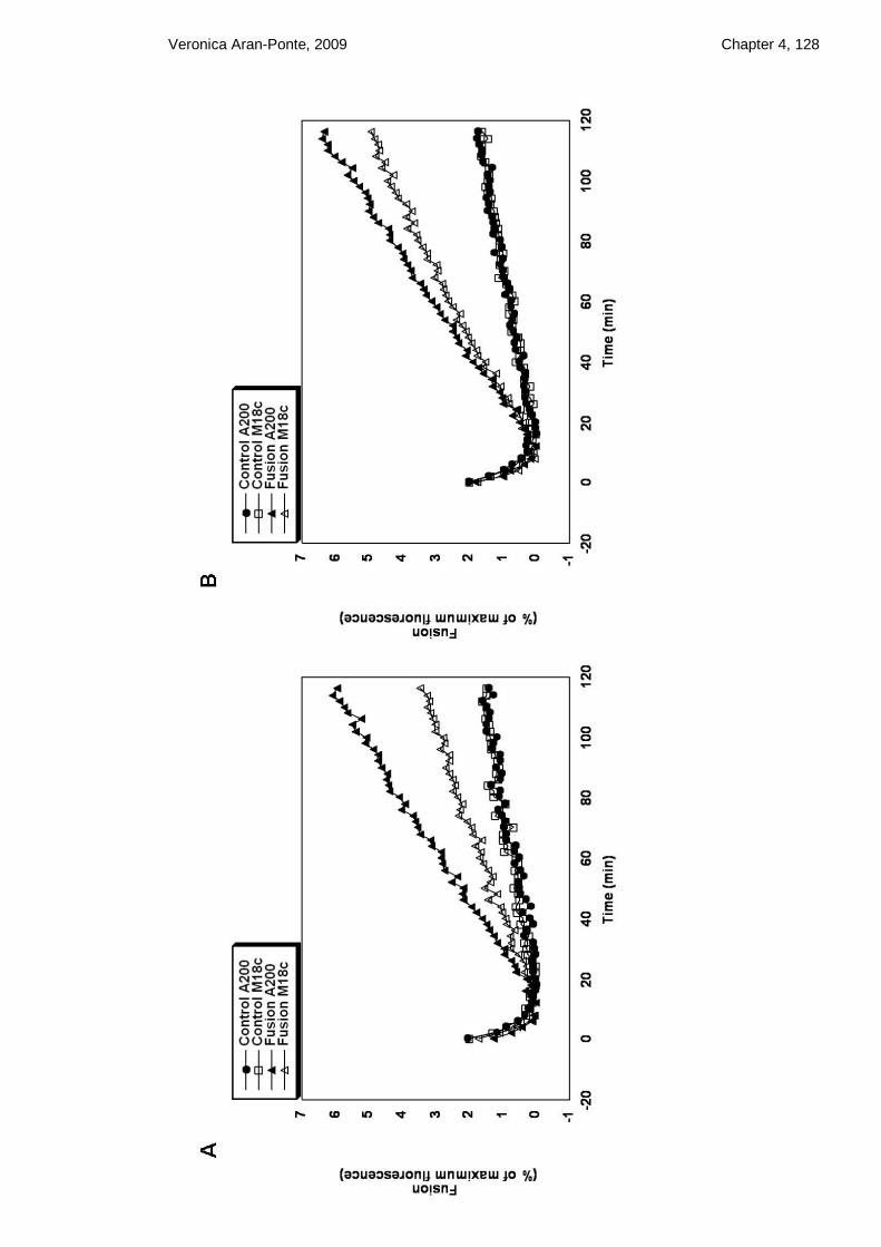
Veronica Aran-Ponte, 2009 Chapter 4, 128

Veronica Aran-Ponte, 2009 Chapter 4, 129
Figure 4.19 In vitro fusion of Sx 4 OPEN/SNAP23 with VAMP2 vesicles (pre-docked overnight or not) in the presence and in the absence of M18c
(A) 5 µl of v- and 45 µl of t- SNARE liposomes containing Sx 4 OPEN instead of wild-
type were mixed inside a well and incubated at 4 °C with M18c overnight at a molar ratio
of 4:1 M18c to Sx 4 OPEN/SNAP23 (open triangles). To another well an equal volume of
buffer A200 was added instead of M18c (filled triangles). To block the formation of trans-
SNARE complexes, the cytoplasmic domain of VAMP2 (VAMP2 ∆TMD) was added to
the t-complex and incubated for 10 min before the addition of VAMP2 vesicle in the
presence and in the absence of M18c (open squares and filled circles respectively). Fusion
was then monitored by placing the 96 well plate in the fluorescence reader set at 37 ˚C as
described in section 2.2.3.6. Top graph correspond to raw fluorescence normalised to
percentage of maximum detergent signal (100 %) against time. Lower graph shows levels
of fusion determined as a percentage of the 100 % signal also against time. (B) In this
experiment, 5 µl of v- and 45 µl of t- SNARE liposomes were pre-mixed and incubated at
4 °C overnight thus allowing the trans-SNARE complex to form before addition of M18c.
In the following day, M18c was added to the pre-assembled liposomes at a molar ratio of
4:1 M18c to Sx 4 OPEN/SNAP23 and incubation occurred for 2 h at 4 °C (open triangles).
For the controls, A200 buffer was added instead of M18c (filled triangles). As negative
controls, VAMP2 ∆TMD was added to separate reactions containing either A200 buffer
(filled circles) or M18c (open squares). Fusion measurements occurred at 37 ˚C as
described in section 2.2.3.6. The raw data corresponds to raw fluorescence normalised to
percentage of maximum detergent signal (100 %) against time (see appendix, figure V).
Graphs A and B show levels of fusion determined as a percentage of the 100 % signal also
against time.
The removal of the 36 amino acids from the N-terminal region of the OPEN Sx 4 protein
did not affect dramatically the results compared to full-length Sx 4 OPEN, the main
difference was in the percentage of inhibition when vesicles were allowed to pre-dock
before M18c addition (24 % for OP and 0 % for OP N∆36). Figure 4.20 shows that M18c
inhibits fusion of vesicles that were not allowed pre-dock by approximately 75 % (figure
4.20A). Rates of fusion of vesicles that were allowed to pre-dock overnight (figure 4.20B),
were not affected by M18c. This result indicates that the formation of the ternary complex
consisting of Sx 4 OPEN N∆36/SNAP23/VAMP2 prior to addition of M18c also abolishes
M18c inhibition. Deletion of the short N-terminal peptide of Sx 4 OPEN did not affect
significantly the results when compared to full length Sx 4 OPEN (figures 4.20 and 4.19
respectively).

Veronica Aran-Ponte, 2009 Chapter 4, 130

Veronica Aran-Ponte, 2009 Chapter 4, 131
Figure 4.20 In vitro fusion of Sx 4 OPEN N∆36/SNAP23 with VAMP2 vesicles (pre-docked overnight and not docked) in the presence and in the absence of M18c
In this fusion assay, the same protocol described in figure 4.19 was followed, however
instead of using reconstituted t-complexes containing OPEN Sx 4, a shorter version
lacking the 36 amino acids from the N-terminus was used instead. Figure 4.15A, shows
reconstituted Sx 4 OPEN N∆36/SNAP23 and VAMP2 used in this assay. (A) and (B) show
levels of fusion as a percentage of the maximum NBD fluorescence emission. In both (A)
and (B), filled circles correspond to control with cytoplasmic VAMP2 plus A200 buffer,
open squares represent control with cytoplasmic VAMP2 plus M18c (1:4 molar ratio M18c
to OPEN Sx 4 N∆36/SNAP23), filled triangles correspond to fusion in the presence of
A200 buffer and finally open triangles correspond to fusion in the presence of M18c (1:4
molar ratio M18c to OPEN Sx 4 N∆36/SNAP23). For fusion reactions, the raw data
normalised to 100 % detergent signal for A and B is shown in the appendix (figure VI).
Graphs A and B show levels of fusion as a percentage of the maximum NBD emission
plotted against time in minutes. This figure shows a typical result obtained after several
fusion assays.
Another approach was taken to confirm the findings obtained in figure 4.20. Reconstituted
Sx 4 OPEN N∆36/SNAP23 from figure 4.15B were allowed to form ternary complexes
with VAMP2 vesicles overnight (figure 4.21, set B) or not (figure 4.21, set A) before the
addition of M18c for only 2 h in both cases. Again, M18c was added at 4:1 molar ratio
M18c: Sx 4 OPEN N∆36/SNAP23. Thus, differently to figure 4.20, M18c was not pre-
incubated overnight with the t- and v-SNARE vesicles. Figure 4.21A, shows that M18c
inhibits fusion of vesicles that were not allowed to pre-dock before M18c addition, since
80 % of inhibition in fusion was observed compared to fusion without M18c. However,
M18c addition to vesicles that were allowed to pre-dock overnight (figure 4.21B), were
just affected slightly by 18 %. Thus, even 2 h incubation with M18c was enough to inhibit
fusion of Sx 4 OPEN N∆36/SNAP23/VAMP2 when these vesicles were not pre-docked.
This result indicates that the formation of the ternary complex formed by Sx 4 OPEN
N∆36/SNAP23/VAMP2 prior to addition of M18c abolishes M18c inhibition, and that
M18c inhibition is not affected by the removal of the 36 amino acids from Sx 4 OPEN.

Veronica Aran-Ponte, 2009 Chapter 4, 132

Veronica Aran-Ponte, 2009 Chapter 4, 133
Figure 4.21 In vitro fusion of Sx 4 OPEN N∆36/SNAP23 with VAMP2 vesicles (pre-docked overnight or not) in the presence and in the absence of M18c for 2 h
(A) 5 µl of v- and 45 µl of t- SNARE liposomes containing Sx 4 OPEN N∆36/SNAP23
shown in figure 4.15B were mixed inside a well and incubated at 4 °C with M18c for 2 h at
a molar ratio of 4:1 M18c to Sx 4 OPEN N∆36/SNAP23 (open triangles). In another well,
instead of M18c, an equal volume A200 buffer was added (filled triangles). Incubation was
continued at 4 °C before the temperature was elevated to 37 °C for fusion at the end of the
2 h. To block the formation of trans-SNARE complexes, the cytoplasmic domain of
VAMP2 (VAMP2 ∆TMD) was added to the t-complex and incubated for 10 min before the
addition of VAMP2 vesicle in the presence and in the absence of M18c (negative controls,
filled circles and open squares). (B) In this experiment, 5 µl of v- and 45 µl of t- SNARE
liposomes were pre-mixed and incubated at 4 °C overnight thus allowing the trans-SNARE
complex to form. In the following day, M18c was added to the pre-assembled liposomes at
a molar ratio of 4:1 M18c to Sx 4 OPEN N∆36/SNAP23 and incubated for further 2 h at 4
°C (open triangles). For the controls, A200 buffer was added instead of M18c (filled
triangles). As negative controls, VAMP2 ∆TMD was added prior to the addition of
VAMP2 vesicles to the reactions containing either A200 buffer (filled circles) or M18c
(open squares). The raw data normalised to 100 % detergent signal for A and B is shown in
the appendix (figure VII). Graphs A and B show levels of fusion as a percentage of the
maximum NBD emission plotted against time in minutes. Details on how the
measurements are made are described in section 2.2.3.7. This figure shows a typical result
obtained after two fusion assays.
Figure 4.22 summarises the results obtained in sections 4.3.5.4 and 4.3.5.5. As can be seen
from the charts, the highest percentages of M18c inhibition came from the experiments
where M18c was added directly together with the SNARE liposomes before fusion
reaction. Lower percentages of inhibition were observed in the fusion assays where the
vesicles were allowed to pre-dock before M18c addition. Furthermore, deletion of the 36
amino acids from both wild-type and open forms of Sx 4 did not alter significantly the
results.

Veronica Aran-Ponte, 2009 Chapter 4, 134
Figure 4.22 Quantifications of the percentages of fusion inhibited by M18c when added at the same time as the SNARE liposomes or added after pre-incubation of SNAREs
The top graph shows the percentages of inhibition by M18c when the different SNARE
complexes were allowed, or not, to pre-dock overnight. The lower graph shows the N-
delete versions of both wild-type Sx 4 and open Sx 4 pre-incubated overnight before M18c
addition or incubated directly with M18c for 2 h before fusion reactions. All percentages
were obtained comparing the maximum fusion rate at 120 min in the absence and in the
presence of M18c. Error bars represent the standard error of the mean of two independent
experiments.

Veronica Aran-Ponte, 2009 Chapter 4, 135
4.4 Discussion
In this chapter a series of experiments were performed to assess the effects of M18c on the
rates of fusion between synthetic liposomes containing Sx 4 (wild-type and mutant
versions) with VAMP2 liposomes. The highly conserved N-terminal domain of exocytic
syntaxins form an autonomously folded 3 helical bundle termed Habc domain, and in the
neuronal Sx 1A this domain was shown to be able to fold back onto the SNARE domain
generating a “closed” conformation unable to form SNARE complexes (Misura et al.,
2000). Mutation of residues L165 and E166 within the hinge region induces this syntaxin
to adopt a more “open” conformation (Dulubova et al., 1999). The introduction of a
L173A/E174A mutation into the same region in Sx 4 sequence also led to a more “open”
form (Aran et al., 2009; D'Andrea-Merrins et al., 2007). Thus, in this chapter, Sx 4
constructs were generated containing the trans-membrane domain in order to reconstitute
them into liposomes. These constructs were in the open, open N∆36, and N∆36 forms.
They were reconstituted into liposomes (together with SNAP23) and compared to the wild-
type form of Sx 4 in the capability to fuse with VAMP2 vesicles. The fusion reactions
were successful as shown in figure 4.12.
Previous studies have shown that SM proteins have the potential to stimulate SNARE-
mediated membrane fusion in vitro in yeast and neuronal cells (Scott et al., 2004; Shen et
al., 2007), but little is known about the effects of M18c on the SNARE proteins involved
in GLUT4 fusion. Since we observed that the short N-terminal peptide comprising 36
amino acids of Sx 4 is important for M18c binding (Aran et al., 2009), the Sx 4 constructs
generated in this study were reconstituted into liposomes and all versions were tested for
fusion with and without M18c.
In these experiments, purified recombinant M18c was added to the liposome fusion assays.
Addition of M18c at molar ratios of either 1:1 or 10:1 M18c:wild-type t-SNARE
complexes, inhibited liposome fusion catalysed by this SNARE complex in a dose-
dependent manner (chapter 4, figure 4.14). Importantly, addition of an equivalent amount
of M18a had no effect on fusion (data not shown, Prof James McNew, Rice University,
Texas). Then, M18c was added at a molar ratio 4:1 M18c: t-complexes (containing the
different versions of Sx 4: wild-type, Sx 4 N∆36, Sx 4 OPEN and Sx 4 OPEN N∆36). The
results showed that deletion of the short N-terminal peptide did not significantly alleviate
M18c negative regulation of fusion. This was unexpected, since this mutant lacks the short
N-terminal region of Sx 4 which has been shown, through crystallographic studies, to

Veronica Aran-Ponte, 2009 Chapter 4, 136
interact with M18c via the pocket mode of binding (Hu et al., 2007). Apart from this, in all
fusion reactions where vesicles were allowed to pre-dock at 4 ˚C, fusion was not
dramatically affected by M18c addition (B in figures 4.16, 4.17, 4.18, 4.19, 4.20, 4.21),
whereas vesicles that were not allowed to pre-dock and were incubated directly with M18c
either overnight or for 2h, were affected (A in figures 4.16, 4.17, 4.18, 4.19, 4.20, 4.21). It
was not possible to reconstitute equimolar amounts of M18c together with the different
versions of Sx 4/SNAP23 (as seen for the wild-type version in figure 4.14C) for technical
reasons.
The negative regulation observed might be happening not only due to the M18c/Sx 4
interaction, but also due to the interaction between M18c and the SNARE motif of
VAMP2 shown in chapter 3. Actually, in neuronal cells, M18a was shown to stimulate
fusion not only via its interaction with Sx 1A, but also via its interaction with VAMP2
(Shen et al., 2007). These data also correlates to the finding that in the absence of M18a, a
total loss of neurotransmitter release occurs, whereas in the absence of M18c an increase in
GLUT4 translocation was observed (Kanda et al., 2005), suggesting that M18c negatively
regulates GLUT4 fusion. These data will be further discussed in chapter 6.
In summary, these results indicate that the formation of the ternary complex formed by Sx
4/SNAP23/VAMP2 might be regulated by M18c at least in vitro, since other factors might
also regulate this process in vivo. M18c negatively regulates Sx 4/SNAP23/VAMP2
mediated membrane fusion in vitro and this apparently involves other interactions apart
from the short N-terminal peptide region in Sx 4.

137
Chapter 5
5 The consequences of Syntaxin 4 and Munc18c
phosphorylation on the regulation of SNARE
complex formation

Veronica Aran-Ponte, 2009 Chapter 5, 138
5.1 Introduction
As shown in chapter 4, when recombinant purified, bacterially expressed SNAP23, Sx 4
and VAMP2 are reconstituted into synthetic proteoliposomes, they can mediate fusion. Our
studies suggested that non-SNARE proteins such as M18c could negatively affect SNARE-
mediated membrane fusion in vitro. Nevertheless, other factors might also regulate
SNARE-mediated membrane fusion in vivo.
In order to maintain normal glucose homeostasis, insulin binds to its heterotetrameric
integral-membrane receptor, which in turn triggers a signalling cascade resulting in the
translocation and fusion of intracellular GLUT4-containing vesicles with the PM (Olson
and Pessin, 1996). This mechanism suggests that factors such as insulin-derived signals
might be involved in the regulation of GLUT4 vesicle fusion with the PM. These signals
could serve as important regulators of the fusion machinery through phosphorylation of
SNARE proteins.
Some studies have reported that SNARE proteins undergo phosphorylation in vitro. For
example, Sx 4 was shown to be susceptible to phosphorylation by the serine/threonine
kinases protein kinase A, casein kinase II, and to a lesser extent by conventional protein
kinase C, in vitro (Foster et al., 1998). In this study, phosphorylation of Sx 4 by PKA (but
not casein kinase II) was shown to affect Sx 4 binding to SNAP23 (Foster et al., 1998).
Another study using the yeast two-hybrid system with the PM t-SNARE Sx 4 as bait, has
identified a novel human kinase termed SNAK (SNARE kinase), which specifically
phosphorylates unassembled SNAP23 (i.e. SNAP23 which are not associated with other
SNAREs) in vivo (Cabaniols et al., 1999). Interestingly, SNAP23 expression and stability
were enhanced upon phosphorylation, therefore increasing the assembly of newly
synthesized SNAP23 with Sx 4 (Cabaniols et al., 1999).
In 2006, Thurmond‟s group showed that M18c becomes tyrosine-phosphorylated upon
stimulation in both islet β-cells and 3T3-L1 adipocytes (Oh and Thurmond, 2006). Also in
2006, Schmelzle and colleagues, by performing mass spectrometric analyses, have
identified tyrosine phosphorylation sites on proteins involved in GLUT4 translocation
(Schmelzle et al., 2006). In this study, 3T3-L1 adipocytes were stimulated with insulin for
0, 5, 15 and 45 min. Interestingly, upon 5 min of insulin stimulation Sx 4 increased in
phosphorylation by >3-fold and surprisingly M18c showed at least a 10-fold increase in
tyrosine phosphorylation. The tyrosine residues that become phosphorylated in response to

Veronica Aran-Ponte, 2009 Chapter 5, 139
insulin are: Y521 on M18c and Y115, Y251 on Sx 4. Interestingly, another group has
reported that M18c becomes phosphorylated on residue Y521 in response to platelet-
derived growth factor (PDGF) stimulation in skeletal muscle, cultured myotubes, and 3T3-
L1 adipocytes (Umahara et al., 2008).
Another potential way in which SM proteins could be regulated is via a protein called
Doc2β (Ke et al., 2007). Doc2 proteins are C2 domain-containing proteins which were first
identified in the brain (Doc2α) (Orita et al., 1995) and later found to be ubiquitously
expressed (Doc2β) (Sakaguchi et al., 1995). Doc2β associates with both M18a in neurons
(Verhage et al., 1997) and M18c in islet β-cells and adipocytes suggesting that Doc
proteins could regulate M18 interaction with syntaxins (Ke et al., 2007). The association of
Doc2β with M18c was shown to be important for glucose-stimulated insulin secretion (Ke
et al., 2007). This data indicates that SM proteins such as M18c might have other
“partners” that help in the regulation of syntaxin function.
It is well established that tyrosine phosphorylation plays an important role in insulin
signalling, however the exact function of tyrosine phosphorylation in SNARE-mediated
membrane fusion remains uncertain. Thus, a more comprehensive study is needed to
improve the understanding of how phosphorylation may affect the machinery that leads to
glucose uptake in muscle and adipocytes.
5.2 Aims of this chapter
SNARE and SM protein phosphorylation might regulate SNARE-mediated GLUT4
translocation and fusion with the PM (see above). M18c and Sx 4 were shown to become
tyrosine phosphorylated in response to insulin, however it is not known if these events are
part of the signalling network leading to GLUT4 translocation, or could affect the binding
of these proteins to other SNAREs. Thus, an approach consisting of mapping studies, site-
directed mutagenesis, pull-down assays and radiolabelling studies were undertaken to
establish whether these phosphorylation events impact on SNARE/SM interactions.

Veronica Aran-Ponte, 2009 Chapter 5, 140
5.3 Results
5.3.1 Sx 4 phosphomimetic mutants
The recombinant SNARE proteins expressed in this study were produced in E.coli, thus
important post-translational modifications that affect mammalian cells could not be
assessed unless they were intentionally engineered to produce modifications in the protein
of interest. Therefore, to examine whether Sx 4 phosphorylation is able to affect its binding
to other proteins, phosphomimetic mutants of tyrosine residues that become
phosphorylated in response to insulin were induced. A change to a glutamic acid was an
ideal choice, since it is bulky (like tyrosine) and will mimic a phosphorylated residue due
to its negative charge.
5.3.1.1 Site-directed mutagenesis and protein purification
Three different mutations were introduced in the Sx 4 sequence using pETDuet-1 Sx 4 as
template. The mutations were Y115E, Y251E and Y115E/Y251E (double-mutant). Site-
directed mutagenesis was performed as outlined in section 2.2.1.2. Primers were designed
taking in consideration melting temperature, length and GC content of the primer.
Successful mutation was confirmed by DNA sequencing. Designed primers were as
follows (letters in bold refer to original codon and underlined refer to mutated codon):
Sx 4 sequence: 5‟ CAG AAG GAG GAA GCT GAT GAG AAT TAC AAC TCA GTC
AAC ACA AGG ATG AAG 3‟
Sx 4 Y115E Forward: 5‟ CAG AAG GAG GAA GCT GAT GAG AAT GAG AAC TCA
GTC AAC ACA AGG ATG AAG 3‟
Sx 4 Y115E Reverse: 5‟ CTT CAT CCT TGT GTT GAC TGA GTT CTC ATT CTC ATC
AGC TTC CTC CTT CTG 3‟
Sx 4 sequence: 5‟ CTG AGC TCG GCC GAC TAC GTG GAA CGT GGG CAA 3‟
Sx 4 Y251E Forward: 5‟ CTG AGC TCG GCC GAC GAG GTG GAA CGT GGG CAA3‟
Sx4 Y251E Reverse: 5‟ TTG CCC ACG TTC CAC CTC GTC GGC CGA GCT CAG 3‟

Veronica Aran-Ponte, 2009 Chapter 5, 141
The Sx 4 double mutant was achieved by first generating one mutation (Y115E) and
afterwards introducing the second mutation (Y251E) in the Sx 4 Y115E DNA template.
The cytosolic domains of Sx 4 Y115E, Y251E and double-mutant were expressed as C-
terminally tagged GST fusion proteins from the pETDuet-1 vector. The vector pETDuet-1-
Sx 4 was a gift from Dr Fiona Brandie (Glasgow University).
Protein expression was performed in BL21 (DE3) cells as outlined in section 2.2.2.4.
Expression was induced using 0.5 mM IPTG, media was incubated overnight at 24 ˚C and
protein purification was achieved as described in section 2.2.2.4. Because these
recombinant proteins were made to be used in pull-down assays, they were eluted using 15
mM of reduced glutathione in 50 mM Tris pH 8.0 in order to keep the GST tag. Proteins
were dialysed overnight against binding buffer (section 2.1), run on a 12 % SDS-PAGE gel
and stained with Coomassie Blue as outlined in sections 2.2.2.1 and 2.2.2.3 respectively.
Results from a typical set of purifications are shown in figure 5.1.
As can be seen in figure 5.1, elutions (EL) from each protein purification contained one
major band of the expected size (indicated with arrows) corresponding to GST-Sx 4
Y115E (A), GST-Sx 4 Y251E (B) and GST-Sx 4 Y115E/251E (C).

Veronica Aran-Ponte, 2009 Chapter 5, 142
Figure 5.1 Purification of Sx 4 phosphomimetic mutants
The cytosolic domains of Sx 4 Y115E (A), Sx 4 Y251E (B) and the double mutant Sx 4
Y115E/ Y251E (C) were expressed in BL21 (DE3) E.coli cells as C-terminally tagged
GST fusion proteins coming from the plasmids pETDuet-1 Sx 4 Y115E, pETDuet-1 Sx 4
Y251E and pETDuet-1 Sx 4 Y115E/Y251E respectively. Proteins were purified as
described in section 2.2.2.4. The lysate (L) was incubated with glutathione sepharose
overnight at 4 ˚C. After several washes (W1, W2, W3), proteins were eluted using 15 mM
of reduced glutathione in 50 mM Tris pH 8.0. Proteins were dialysed overnight against
binding buffer (as outlined in section 2.2.2.6), run on a 12 % SDS-PAGE gel and stained
with Coomassie Blue as outlined in sections 2.2.2.1 and 2.2.2.3. 10 µl Elutions (EL) were
compared to wild-type Sx 4 (wtSx4) sample in A, B and C. Arrows indicate precise
position of Sx 4 proteins in the gel. Positions of molecular weight markers are shown. Data
from a typical set of purifications are presented, repeated many times with similar results.

Veronica Aran-Ponte, 2009 Chapter 5, 143
5.3.1.2 Circular dichroism of purified syntaxin 4 mutants
Mutations can, sometimes, alter the structure of proteins and these alterations can affect
their biological function. To examine whether phosphomimetic mutations affected Sx 4
structure, a comparison between the wild-type and mutant forms of GST-Sx 4 was made
using circular dichroism (CD). CD is a form of spectroscopy which relies on the
differential absorption of left and right circularly polarised light by optically active
elements such as backbone amide bonds, disulphide bonds and aromatic side chains within
the protein molecule (Kelly and Price, 2000). It is extremely useful for examining
structural integrity since the spectra collected allow information on the secondary structure
and, in some cases, the tertiary structure of proteins to be obtained.
CD measurements in the far UV region can give important characteristics of the secondary
structure of molecules. The differential absorption occurs below 240 nm and is due
principally to the peptide bonds of molecules which can adopt various conformations (α-
helix, β-sheets and random coil). CD measurements in the near UV range can be used to
obtain information on the tertiary structure of proteins, corresponding to aromatic amino
acid contributions (phenylalanine, tyrosine and tryptophan side chains) in the range
between 260 to 320 nm. If the aromatic side chains are held rigidly in an asymmetric
environment it can be possible to obtain a unique spectral profile which is a very useful
tool when comparing wild-type and mutant versions of proteins (Kelly et al., 2005).
Figure 5.2 shows the far UV CD spectra comparing the secondary structures of the
cytosolic domains of wild-type GST-Sx 4 with the cytosolic domains of the GST-tagged
phosphomimetic mutants. GST-alone was included for comparison since the proteins were
GST tagged. CD analysis confirmed that with the exception of the Y115E/Y251E double
mutant the overall secondary structure of the Sx 4 mutants are similar which inferred that
the mutations did not significantly affect their secondary structures. In the case of the
double mutant, the spectral intensity was found to be less than the wild-type which may
reflect problems in the reliability of the protein concentration estimation following removal
of the tyrosine residues. A small change in secondary structure may have occurred
following the replacement of Y115 and Y251 with glutamate residues.

Veronica Aran-Ponte, 2009 Chapter 5, 144
Figure 5.2 Far UV CD spectra of the cytosolic domains of wild-type Sx 4, Y115E, Y251E and Y115E/Y251E
Far UV CD spectra were recorded for the cytosolic domains of recombinant GST (blue),
wild type GST- Sx 4 (green), GST-Sx 4 Y115E (pink), GST-Sx 4 Y251E (red) and GST-
Sx 4 Y115E/Y251E (black) from 180 nM to 260 nM. HT[V] stands for high tension (i.e.
the voltage applied to the photomultiplier to keep the current constant). Proteins were
diluted to 0.7 mg/ml in 50 mM K2 HPO4- KH2PO4 pH 7.4. The data shown were obtained
from the average of 8 scans. CD measurements were collected on a CD JASCO J-810
spectropolarimeter by Dr Sharon Kelly (University of Glasgow).
Figure 5.3 shows the near UV CD spectra giving an indication of the overall tertiary
structures of the wild-type GST-Sx 4 and GST-Sx 4 phosphomimetic mutants. As can be
seen in figure 5.3, the overall spectral features of the mutant proteins are similar to the
wild-type, suggesting that the aromatic residues are properly buried and the tertiary
structural integrity for each protein has been maintained. The small difference observed in
the intensity of the peaks is probably due to the removal of tyrosine residues.

Veronica Aran-Ponte, 2009 Chapter 5, 145
Figure 5.3 Near UV CD spectra of wild-type GST-Sx 4 and Sx 4 Y115E, Sx4 Y251E, and Y115E/Y251E
Near UV CD spectra were recorded for the cytosolic domains of recombinant GST (blue),
wild-type GST- Sx 4 (green), GST-Sx 4 Y115E (pink), GST-Sx 4 Y251E (red) and GST-
Sx 4 Y115E/Y251E (black) from 250 nM to 320 nM. Proteins were diluted to 1 mg/ml in
50 mM K2HPO4- KH2PO4 pH 7.4. The data shown was obtained from the average of 8
scans. CD measurements were collected on a CD JASCO J-810 spectropolarimeter by Dr
Sharon Kelly (University of Glasgow).
5.3.1.3 Interaction of Sx 4 phosphomimetic mutants with Munc18c
In order to test whether phosphomimetic mutations could affect Sx 4 binding to M18c,
pull-down experiments were carried out in which set amounts of the phosphomimetic
GST-Sx 4 recombinant proteins were immobilised on glutathione sepharose beads before
incubation with His6-M18c. GST alone was used as a negative control in the pull-downs.
The result of a typical experiment is shown in figure 5.4.

Veronica Aran-Ponte, 2009 Chapter 5, 146
M18c
GST
GST-S4 WT
and mutants
GST WT Sx 4 Y115E Y251E Y115E/Y251E
-M18c
Input
Beads
GST-Sx 4
Figure 5.4 Binding of His6-M18c to GST-Sx 4 wild-type (WT), Sx 4 Y115E, Sx 4 Y251E and Sx 4Y115E/Y251E
Pull-down assay was performed where 5 μg of recombinant GST, GST fused to the
cytosolic domain of wild-type (WT) Sx 4, Sx 4 Y115E, Sx 4 Y251E and Sx 4
Y115E/Y251E were firstly immobilised on glutathione Sepharose for 1.5 h at 4 ˚C. After 3
washes with binding buffer, beads were incubated overnight at 4 ˚C with 5 μg of His6-
M18c in a final volume of 500 μl. 4 % of each mixture was removed to assess protein
input. Bound proteins were eluted, resolved by 12 % SDS-PAGE and immunoblot analysis
was carried out to determine which of the GST proteins His6-M18c had bound to. Upper
panel represents a Coomassie stained gel of the GST-fusion proteins/M18c inputs (4 % of
the total input); lower panel shows immunoblot analysis with α-M18c antibody used to
determine bound M18c. 50 % of the eluted sample was loaded. This experiment was
repeated three times.
As can be seen in figure 5.4, His6-M18c can still efficiently bind phosphomimetic mutants
as well as wild-type GST-Sx 4. In several experiments of this type, we saw no difference in
M18c binding to any phosphomimetic mutant compared to wild-type Sx 4. This result
indicates that post-translational modifications such as phosphorylation of Sx 4 protein do
not affect their interaction with their regulatory protein M18c in vitro.

Veronica Aran-Ponte, 2009 Chapter 5, 147
5.3.1.4 Interaction of Sx 4 phosphomimetic mutants with SNAP23
Since phosphomimetic mutations in Sx 4 were not able to confer considerable changes in
binding to M18c, it was then investigated whether its interaction with the SNARE protein,
SNAP23, was affected. Sx 4 and SNAP23 proteins interact to form a heterodimer at the
PM of muscle and adipocytes and this binding is essential for SNARE complex formation.
Thus, the interaction between Sx 4 phosphomimetic mutants and SNAP23 was investigated
through pull-down studies.
Data obtained from pull-downs, shown in figure 5.5, indicates that phosphomimetic
mutations did not affect significantly Sx 4 binding to SNAP23.
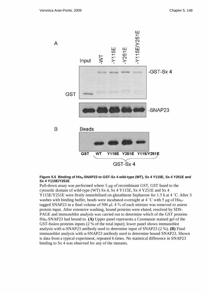
Veronica Aran-Ponte, 2009 Chapter 5, 148
Figure 5.5 Binding of His6-SNAP23 to GST-Sx 4 wild-type (WT), Sx 4 Y115E, Sx 4 Y251E and Sx 4 Y115E/Y251E
Pull-down assay was performed where 5 μg of recombinant GST, GST fused to the
cytosolic domain of wild-type (WT) Sx 4, Sx 4 Y115E, Sx 4 Y251E and Sx 4
Y115E/Y251E were firstly immobilised on glutathione Sepharose for 1.5 h at 4 ˚C. After 3
washes with binding buffer, beads were incubated overnight at 4 ˚C with 5 μg of His6-
tagged SNAP23 in a final volume of 500 μl. 4 % of each mixture was removed to assess
protein input. After extensive washing, bound proteins were eluted, resolved by SDS-
PAGE and immunoblot analysis was carried out to determine which of the GST proteins
His6-SNAP23 had bound to. (A) Upper panel represents a Coomassie stained gel of the
GST-fusion proteins inputs (2 % of the total input); lower panel shows immunoblot
analysis with α-SNAP23 antibody used to determine input of SNAP23 (2 %). (B) Final
immunoblot analysis with α-SNAP23 antibody used to determine bound SNAP23. Shown
is data from a typical experiment, repeated 6 times. No statistical difference in SNAP23
binding to Sx 4 was observed for any of the mutants.

Veronica Aran-Ponte, 2009 Chapter 5, 149
5.3.2 Munc18c phosphomimetic mutant Y521E
It was previously shown that the tyrosine residue at position 521 in M18c is specifically
phosphorylated upon insulin stimulation (Schmelzle et al., 2006). By using amino acid
substitution, the role of tyrosine 521 in binding of Sx 4 was assessed. A glutamate
substitution (Y521E), closely mimicking phosphorylated tyrosine, was obtained via site-
directed mutagenesis.
5.3.2.1 Site-directed mutagenesis and protein purification
The Y521 mutation was introduced into the M18c sequence using pQE30-M18c as
template and mutation was performed as outlined in section 2.2.1.2. Primers were designed
taking in consideration melting temperature, length and GC content of the primer.
Designed primers were as follows (letters in bold refer to original codon and underlined
refer to mutated codon):
M18c wt sequence: 5‟ CAG AAA CCC AGA ACT AAC TAC TTA GAG CTG GAC
CGG AAA 3‟
M18cY521E Forward: 5‟ CAG AAA CCC AGA ACT AAC GAG TTA GAG CTG GAC
CGG AAA 3‟
M18cY521E Reverse: 5‟ TTT CCG GTC CAG CTC TAA CTC GTT AGT TCT GGG
TTT CTG 3‟.
Successful mutation was confirmed by DNA sequencing.
M18cY521E was expressed and purified similarly to wild-type M18c. His6-M18cY521E
was expressed in M15 E. coli cells that also co-expressed the chaperone protein GroEL in
order to increase M18cY521E solubility. Low levels of IPTG were used (0.2 mM) and
protein expression was induced at 22 ˚C overnight as these conditions were empirically
determined to give better levels of expression. His6-M18cY521E was purified using Ni-
NTA agarose as described in section 2.2.4.
A typical purification is shown in figure 5.6.

Veronica Aran-Ponte, 2009 Chapter 5, 150
Figure 5.6 M18cY521E protein purification
His6-M18cY521E was expressed from vector pQE30 in M15 E. coli cells expressing the
chaperone GroEL. Expression was induced by adding 0.2 mM IPTG to the media and
incubation was carried out overnight at 22˚C. The figure shows several samples taken
during the process of protein purification which were run on a 12 % SDS-PAGE gel and
stained with Coomassie Blue as outlined in section 2.2.2.1 and 2.2.2.3 respectively. The
positions of the molecular weight markers are shown on the left. The first lane corresponds
to a sample before induction (BI) followed by a sample after induction (I), sample from the
lysate (L), wash 1 (W1), wash 2 (W2) and fraction eluted (15 µl) using 500 mM imidazole
(EL).
From the data presented in figure 5.6, the elution (EL) obtained from the Ni-TA agarose
contained one major product of the expected size which corresponds to His6-M18cY521E.
The sample purity could be improved if more washes were applied. In order to further
confirm that the elution obtained was really the M18c phosphomimetic mutant, wild-type
and mutant M18c were run side by side on a 12 % SDS-PAGE and subjected to
immunoblot analysis using antibody against M18c.

Veronica Aran-Ponte, 2009 Chapter 5, 151
Figure 5.7 Comparison between wild-type M18c and M18cY521E
Similar quantities (approximately 1 µg each) of recombinant wild-type M18c and M18c
Y521E were resolved on a 12 % SDS-PAGE for subsequent immunoblotting with anti-
Munc18c antibody. Molecular weight markers are shown on the left. Arrow indicates
M18c WT and mutant bands.
As shown in figure 5.7, polyclonal M18c antibody was able to recognise both wild-type
and the mutant M18cY521E and both proteins, as expected, run at a similar molecular
weight.
5.3.2.2 Circular dichroism analysis of purified His6-M18cY521E
SM proteins such as M18c are generally poorly expressed or unstable needing to be co-
expressed with the chaperone GroEL (Scott et al., 2004). The presence of GroEL, a protein
folding chaperone, has been shown to be very useful for improving recombinant proteins
solubility and assisting successful folding (Yasukawa et al., 1995). Truncations in M18c
can affect their structure due to protein instability (Oh and Thurmond, 2006). In order to
assess if the tyrosine mutation to a glutamic acid has affected M18cY521E folding, CD
and intrinsic tryptophan fluorescence spectra were performed to compare both wild-type
and M18cY521E structures. Near UV CD analysis was not possible due to the low protein
concentrations.

Veronica Aran-Ponte, 2009 Chapter 5, 152
Figure 5.8 shows the far UV CD spectra obtained from His6-M18c (blue), and the mutant
His6-M18cY521E (red). As shown, there is no significant change in the overall secondary
structure of the mutant His6- M18c Y521E when compared to wild-type His6- M18c. The
His6-M18c (blue) displayed a poor signal to noise ratio below 195 nm due to traces of
chloride ions in the buffer.
Figure 5.8 Far UV CD spectra of wild-type M18c (blue) and mutant M18cY521E (red)
Far UV CD spectra were recorded for recombinant wild-type His6-M18c (blue), and the
mutant His6-M18cY521E (red) from 180 nM to 260 nM. Proteins were diluted to 0.5
mg/ml in 50 mM K2HPO4- KH2PO4 pH 7.4. Top graph shows molar ellipicity and second
graph shows HT[V] which stands for high tension (i.e. the voltage applied to the
photomultiplier to keep the current constant). The data shown was obtained from the
average of 8 scans. CD measurements were collected on a CD JASCO J-810
spectropolarimeter by Dr Sharon Kelly (University of Glasgow).

Veronica Aran-Ponte, 2009 Chapter 5, 153
Figure 5.9 represents the fluorescence spectra of the wild-type His6-M18c compared with
mutant His6-M18c. Near CD UV analysis were not carried out for His6-M18cY521E since
the removal of tyrosine would affect the spectral contributions corresponding to the
tyrosine side chains and since near UV CD requires protein concentrations of
approximately 1 mg/ml and there was an insufficient concentration of protein for this
analysis. Instead, fluorescence spectroscopy was used. Tryptophan emission fluorescence
following excitation at 295 nm is dominant over the weaker tyrosine and phenylalanine
contributions.
Tryptophan is a very useful intrinsic fluorescent probe which can be used to estimate the
nature of the tryptophan environment when excited. This hydrophobic amino acid favours
being buried in protein hydrophobic cores. Protein mutations can sometimes affect the
tryptophan environment since they could expose the tryptophan to an aqueous or less
hydrophobic environment instead of the protein interior. Thus, tryptophan exposure as a
result of unfolding would result in a peak which would be shifted to longer wavelengths
compared with the wild-type.
The fluorescence spectra of wild-type and mutant forms of M18c allowed the positions of
the tryptophan peaks for each protein to be determined. Figure 5.9 shows that the emission
spectra obtained following excitation at 295nm gave a peak around 345 nm for wild-type
and His6-M18cY521E suggesting that these proteins have a similar conformation in the
region pertaining to the position of the tryptophans. The difference in the levels of
fluorescence might indicate fluorescence quenching due to protein concentration or grade
of purity of protein in this study.
Such data collectively suggest that His6-M18cY521E is correctly folded.

Veronica Aran-Ponte, 2009 Chapter 5, 154
320 340 360 380 400 420 440
0
50
100
150
200
250
300
350
400
450
Wild type
y521e
Flu
ore
scen
ce (
arb
itra
ry u
nit
s)
Wavelength (nm)
Figure 5.9 Tryptophan fluorescence spectra of His6-M18c and His6-M18c Y521E
Emission spectra of His6-M18c and His6-M18cY521E following excitation at 295nm. The
amount of protein used to obtain the fluorescent spectrum were 0.6 mls of 0.2 mg/ml
protein and measurements were performed by Dr Sharon Kelly (University of Glasgow) on
a Perkin-Elmer LS 50B fluorometer.
5.3.2.3 Interaction of His6-M18cY521E with different versions of Sx 4
Glutathione sepharose bound to GST-Sx 4 proteins harbouring the cytosolic domains of the
different version of Sx 4, were used to pull down His6-M18cY521E. C-terminally tagged
GST- Sx 4, GST-Sx 4 N∆36, GST-Sx 4 L173A/E174A, GST-Sx 4 L173A/E174A N∆36
were purified as described in section 2.2.2.4.
The result of a typical experiment is shown in figure 5.10. While wild-type His6-M18c was
able to bind the different version of Sx 4, the mutant His6-M18cY521E did not. Less than
10 % of the total His6-M18cY521E input was shown to bind Sx 4.

Veronica Aran-Ponte, 2009 Chapter 5, 155
After repeating this experiment several times, this data indicates that phosphomimetic
mutation in M18c (Y521E) results in abrogation of M18c/Sx 4 interaction.
Figure 5.10 Pull-down assay of the different versions of cytosolic GST-Sx4 with both wild-type His6- M18c and His6-M18cY521E
Pull-down assay was performed in which 5 μg of either recombinant GST, or GST fused to
the cytosolic domain of WT Sx 4, Sx 4 N∆36, Sx 4 open and Sx 4 open N∆36 were firstly
immobilised on glutathione Sepharose. Beads were then incubated overnight at 4 ˚C with 1
μg of His6-M18cY521E in a final volume of 200 μl. As a positive control, another set of
proteins (GST and GST-Sx 4 wild-type and mutants) were incubated with wild-type 1 μg
His6-M18c. After extensive washing, bound proteins were eluted using 15 µl of 1 x sample
buffer (containing 20 mM DTT), resolved by SDS-PAGE and immunoblot analysis were
carried out to determine which of the GST proteins His6-M18c and His6-M18cY521E had
bound to. Panel (A) shows protein inputs (7.5 %) for GST and GST-tagged Sx 4 proteins
which were purified as described in section 2.2.2.4, were resolved by SDS-PAGE and
visualised by staining with Coomassie Blue (A, top panel). 10 % of the M18c input from
both wild-type and mutant forms were analysed by SDS-PAGE followed by immunoblot
analysis with α-M18c antibody (A, lower panel). Panel (B) shows immunoblot analysis
with α-M18c antibody used to determine bound M18c (top panel) and bound M18cY521E
(lower panel). This experiment was repeated three times.

Veronica Aran-Ponte, 2009 Chapter 5, 156
5.3.2.4 Interaction of His6-M18cY521E with GST-VAMP2
Since no interaction was observed of His6-M18cY521E with cytosolic Sx 4 and we have
recently reported that VAMP2 interacts with M18c (chapter 3), the interaction between
His6-M18cY521E with the cytosolic portion of the v-SNARE VAMP2 was assessed.
Recombinant cytosolic N-terminally tagged GST-VAMP2 was purified as described in
section 2.2.2.4. Glutathione sepharose bound to either recombinant GST or GST- VAMP2
were used to pull down His6-M18cY521E.
Similarly to Sx 4, GST-VAMP2 was unable to pull-down His6-M18cY521E as can be seen
in figure 5.11.

Veronica Aran-Ponte, 2009 Chapter 5, 157
Figure 5.11 GST pull-down of GST-VAMP2 and His6- M18cY521E
Pull-down assay was performed in which 5 μg of either recombinant GST, or GST fused to
the cytosolic domain of VAMP2 were first immobilised on glutathione sepharose. Beads
were then incubated overnight at 4 ˚C with 1 μg of His6-M18cY521E and another set of
GST and GST-VAMP2 were incubated with 1 μg wild-type His6-M18c in a final volume
of 200 μl. After extensive washing, bound proteins were eluted using 15 µl of 1 x sample
buffer (containing 20 mM DTT), resolved by SDS-PAGE and immunoblot analysis were
carried out to determine if His6-M18c and His6-M18cY521E had both bound to GST-
VAMP2. (A) Shows protein inputs (7.5 %) for GST and GST-tagged VAMP2 proteins
which were purified as described in section 2.2.2.4, were resolved by SDS-PAGE and
visualised by staining with Coomassie Blue (A, top panel). 10 % of the M18c input for
both wild-type and mutant forms were analysed by SDS-PAGE followed by immunoblot
analysis with α-M18c antibody (A, lower panels). (B) Shows immunoblot analysis with α-
M18c antibody used to determine bound M18c (top panel) and bound M18cY521E (lower
panel).

Veronica Aran-Ponte, 2009 Chapter 5, 158
5.3.3 The Recombinant Cytoplasmic Insulin Receptor Kinase
(CIRK)
Insulin action results in tyrosine phosphorylation, through the binding of insulin to its
receptor which contains a tyrosine kinase domain. This results in autophosphorylation of
the receptor which will eventually stimulate phosphorylation of the insulin receptor
substrate (IRS) which will carry the signal downstream of the receptor. However,
termination of insulin action requires the dephosphorylation of the insulin receptor and
IRS.
In order to identify which tyrosine phosphatases were responsible for dephosphorylation of
both the insulin receptor and IRS-1, a group of researches generated a recombinant
cytoplasmic domain of the human insulin receptor, which they termed CIRK (Kuhne et al.,
1994). This recombinant receptor consists of amino acids 941-1343 of the β subunit of the
insulin receptor which contains the intrinsic tyrosine kinase domain. CIRK was expressed
and purified from Baculovirus-infected Sf9 cells according to work done by Villalba and
colleagues (Villalba et al., 1989).
The generation of CIRK was a very interesting tool to improve the understanding of the
insulin action via phosphorylation of the insulin receptor and also its consequences among
other proteins including SNAREs. Therefore, studies were carried out to analyse if CIRK
autophosphorylation could result in phosphorylation of other substrates such as Sx 4 and
M18c. CIRK was kindly given to us by Prof Gustav E. Lienhard (Dartmouth University,
USA).
Fig 5.12 shows recombinant CIRK running at approximately 47 kDa on a 12 % SDS-
PAGE.

Veronica Aran-Ponte, 2009 Chapter 5, 159
Figure 5.12 SDS-PAGE analysis of recombinant CIRK
CIRK was autophosphorylated as described in section 2.2.5.1. 20 µl of autophosphorylated
CIRK (7.4 µg) were loaded on a 12 % SDS-PAGE gel followed by Coomassie Blue
staining as outlined in sections 1 and 2. Positions of the broad range molecular weight
markers are shown.
5.3.3.1 Phosphorylation of M18c and SNARE proteins in response to CIRK
In previous studies, protein kinases were shown to induce phosphorylation in some
SNARE proteins and also M18c (see section 5.1). To assess if this was the case for CIRK,
this recombinant kinase was firstly autophosphorylated as described in section 2.2.5.1.
Secondly, autophosphorylated CIRK was incubated with M18c and with the t-SNARE
complex in a buffer containing ATP, MnCl2 and [γ-32
P]ATP. After 30 min incubation at 30
˚C, samples were subjected to SDS-PAGE, and analysed by autoradiography (as outlined
in sections 2.2.5.1 and 2.2.5.2).

Veronica Aran-Ponte, 2009 Chapter 5, 160
Figure 5.13 Autoradiography studies using M18c and Sx 4/SNAP23 complex which were pre-incubated with CIRK
1 µg of M18c and Sx4/SNAP23 were incubated with 1 µl of activated CIRK (370 ng).
CIRK activation (autophosphorylation) was achieved as described in section 2.2.5.1.
Recombinant proteins plus CIRK were incubated in a buffer containing 50 mM HEPES pH
7.5, 4 mM MnCl2, 0.2 mM DTT, 100 µM ATP and 6 µCi [γ-32
P]ATP. Control samples had
no CIRK. Incubation was carried out for 30 min at 30 ˚C. After 30 min, 15 µl of 4 x
sample buffer (containing 20 mM DTT) were added to each tube. Samples were boiled at
65 ˚C for 10 min. Proteins were separated by SDS gel electrophoresis, then the gel was
dried and subjected to autoradiography. Upper panel represents a Coomassie stained gel of
the total M18c and t-complex inputs (25 µl samples, ~ 0.5 µg); lower panel show
autoradiography analysis after 72 h exposure (at room temperature).
As shown in figure 5.13, M18c becomes phosphorylated in response to CIRK (Lane 1,
lower panel). In contrast, the complex formed by Sx 4 and SNAP23 did not become
phosphorylated in response to CIRK (Lane 3, lower panel). As expected, in the absence of
CIRK, none of the proteins became phosphorylated (Lanes 2 and 4, lower panel).
Because Sx 4 and SNAP23 form a heterodimer, this could have masked the sites in Sx 4
that become phosphorylated with activated CIRK. In order to further confirm that Sx 4 was
not able to become phosphorylated, monomeric Sx 4 was incubated in the presence and in
the absence of CIRK. M18c was also incubated with and without CIRK. Because Sx 4 was
GST tagged, an extra control was made by incubating recombinant GST in the presence
and in the absence of CIRK.

Veronica Aran-Ponte, 2009 Chapter 5, 161
The autoradiography analysis presented in figure 5.14 shows that CIRK was able to
phosphorylate both M18c and monomeric Sx 4. However, phosphorylation of M18c
seemed greater than Sx 4, as can be seen by the intensity of the band.
Figure 5.14 M18c and Sx 4 phosphorylation by CIRK
CIRK (370 ng) was incubated with 1µg M18c (lane 1; lower panel), 1 µg GST-Sx 4 (lane
3; lower panel) and with GST alone (lane 5; lower panel). Reactions were carried out as
described in sections 2.2.5.2 and 2.2.5.3. As controls, M18c and GST-Sx 4 were incubated
without CIRK, whereas GST was incubated with and without CIRK (lanes 2, 4, 5 and 6
respectively; lower panel). GST was incubated separately with CIRK because Sx 4 is GST
tagged. Proteins were separated by 12 % SDS gel electrophoresis and the gel was dried and
subjected to autoradiography. Upper panel represents a Coomassie blue stained gel of the
total amounts of His6-M18c (left) and GST-Sx 4 (right) used (~ 0.5 µg samples from each
protein); lower panel show autoradiography analysis after 72 h exposure (at room
temperature).

Veronica Aran-Ponte, 2009 Chapter 5, 162
5.3.3.2 Stoichiometry of protein phosphorylation in response to CIRK
Since Phosphorus-32 were readily introduced into the M18c and Sx 4 molecules, the
stoichiometry of protein phosphorylation could be calculated by measuring the counts per
min (cpm) using a Scintillation counter. Cpm gives a good estimate about the number of
atoms in a given quantity of radioactive material that has decayed in one min. The cpm of
both M18c and Sx 4 were determined by excising the dried gel slices corresponding to
each of the proteins, placing them in vials and measuring cpm using a scintillation counter.
Knowing the molecular weight of the molecule, the amount of protein loaded and the cpm
obtained, it is possible to make an estimate of the percentage of the protein that is
phosphorylated.
Under the conditions used in 5.14, Sx 4 and M18c were shown to be phosphorylated by
less than 1 %. In order to improve M18c phosphorylation levels, changes were made in the
protocol. The time of incubation at 30 ˚C was increased from 30 min to 150 min and the
amount of CIRK was increased 5 times, from 1 µl to 5 µl (from 370 ng to 1.85 µg). Figure
5.15 shows the results of an autoradiography after 48 h exposure. Note the increase in
intensity of the M18c band as the amount of CIRK and the time of incubations were
prolonged.

Veronica Aran-Ponte, 2009 Chapter 5, 163
Figure 5.15 M18c phosphorylation by CIRK using different amounts of CIRK and different times of incubation
1 µg of His6-M18c was incubated with 370 ng CIRK (lanes 1 and 2) for 30 min (lane 1) or
150 min (lane 2), or with 1.85 µg CIRK for 150 min (lane 3), at 30 ˚C. Reaction mixtures
also contained buffer plus radiolabelled ATP as described in section 2.2.5.3. Reactions
were stopped by boiling the samples and 25 µl (~ 0.5 µg of His6-M18c) of each reaction
mixture were processed by SDS-PAGE followed by Coomassie Blue staining and
autoradiography as indicated in section 2.2.5.3. This figure shows an autoradiography
analysis after 48 h exposure (at room temperature).
Figure 5.15 shows that the percentage of M18c phosphorylation is proportional to the
amount of CIRK used and to the time of incubation. After 30 min incubation with 370 ng
CIRK (lane 1), only ~ 1 % of the M18c molecule became phosphorylated, whereas after
150 min, there was an increase of ~ 16 % in the levels of M18c phosphorylation. By
increasing 5 fold the amount of CIRK and keeping the 150 min incubation period (lane 3),
~ 24 % of M18c became phosphorylated. By performing this experiment, it was possible to
optimise the conditions to allow maximum phosphorylation of M18c in response to CIRK
in order to start the in vitro binding assays described in the following sections.

Veronica Aran-Ponte, 2009 Chapter 5, 164
5.3.3.3 Analysis of the interaction between M18c phosphorylated by CIRK
and the different versions of GST-Sx 4
The fact that CIRK was able to phosphorylate M18c in vitro gave rise to the idea of
assessing how this phosphorylation could affect M18c interaction with SNARE proteins
such as Sx 4 or VAMP2.
As previously mentioned in chapter 4, M18c is able to bind Sx 4 lacking the short N-
terminal peptide, which corresponds to the 36 amino acids of the N-terminus, but at a
much lesser extent than wild-type Sx 4 (as outlined in section 4.1.1). M18c is also able to
bind the open version of Sx 4 which contains a mutation in the hinge region of Sx 4
(L173A/E174A) (Aran et al., 2009; D'Andrea-Merrins et al., 2007). Thus, pull-down
experiments were performed in which a set amount of the different versions of C-
terminally tagged Sx 4 were immobilised on glutathione sepharose before overnight
incubation with M18c phosphorylated by CIRK. A C-terminal tagged version of Sx 4 was
used in order to leave the N-terminal part of Sx 4 available to bind M18c. In order to check
if phosphorylated M18c is able to bind the different version of Sx 4 in vitro, a pull-down
assay was performed.
Figure 5.16B shows an overall decrease in the amounts of His6-M18c which are able to
bind the different versions of GST- Sx 4 plus CIRK versus minus CIRK. So, in order to
check if bound His6-M18c were tyrosine phosphorylated, western blot analysis were
carried out using 8 µl of the total 15 µl sample. α-phosphotyrosine antibodies were used to
determine if bound M18c was tyrosine phosphorylated or not. The right side of figure
5.16C shows that autophosphorylated CIRK induces His6-M18c phosphorylation on
tyrosine residues. In contrast, the left hand side on figure 5.16C shows the interesting fact
that His6-M18c (+ CIRK), which was bound to wt Sx 4 and Sx 4 open versions on
glutathione sepharose (figure 5.16B; lower panel), were not tyrosine phosporylated. This
result indicates that M18c phosphorylated by CIRK loses affinity for Sx 4.

Veronica Aran-Ponte, 2009 Chapter 5, 165

Veronica Aran-Ponte, 2009 Chapter 5, 166
Figure 5.16 M18c phosphorylated by CIRK loses affinity for Sx 4 in vitro
Pull-down assay was performed where 1 μg of either recombinant GST, or GST fused to
the cytosolic domain of WT Sx 4, Sx 4 N∆36, Sx 4 open and Sx 4 open N∆36 were firstly
immobilised on glutathione Sepharose. Beads were then incubated overnight at 4 ˚C with 1
μg of His6-M18c (already phosphorylated by CIRK) in a final volume of 300 μl. 6 % of
each mixture was removed to assess protein input (A). After extensive washing, bound
proteins were eluted using 15 µl of 1 x sample buffer (containing 20 mM DTT), resolved
by SDS-PAGE and immunoblot analysis were carried out to determine which of the GST
proteins M18c had bound to. Upper panel represents a Coomassie stained gel of the GST-
fusion proteins (A, left and middle panels) and M18c inputs (1 % of the total input); panel
B shows immunoblot analysis with α-M18c antibody used to determine bound M18c (~ 47
% of the eluted sample was loaded into the gel); panel C shows immunoblot analysis with
α-phosphotyrosine antibody used to determine bound proteins which are tyrosine
phosphorylated (~ 53 % of the eluted sample was loaded into the gel) and also levels of
1 %, 2.5 %, 5 % and 10 % of total phosphorylated M18c input for comparison. This
experiment was repeated 3 times giving similar conclusions.
5.3.3.4 Interaction between Munc18c phosphorylated by CIRK and VAMP2
Results from section 5.3.3.3 show that phosphorylated M18c is not able to interact with
Sx 4, in vitro. In chapter 3, data has demonstrated that M18c is also able to interact with
VAMP2, therefore similar pull-down experiments were carried out to check if CIRK
induced phosphorylation of M18c could also result in a decrease of M18c-VAMP2
affinity. A typical result of an experiment of this type is shown in figure 5.17.

Veronica Aran-Ponte, 2009 Chapter 5, 167
Figure 5.17 Pull-down assay of GST- VAMP2 and M18c phosphorylated by CIRK
Pull-down assay was performed where 1 μg of either recombinant GST, or GST fused to
the cytosolic domain of VAMP2 were immobilised on glutathione Sepharose. Beads were
then incubated overnight at 4 ˚C with 1 μg of His6-M18c, which has been previously
incubated with CIRK, for 150 min and with the control M18c without CIRK. M18c and
VAMP2 were incubated in a final volume of 300 μl. (A) upper panel represents a
Coomassie stained gel of GST and GST-VAMP2 (6 % of total input) and lower panel
represents immunoblot analysis using α-M18c antibody to show 4 % of the total M18c
input in the presence and absence of CIRK; (B) upper panel shows immunoblot analysis
with α-M18c antibody used to determine bound M18c (~ 47 % of the eluted 15 µl sample
was loaded into the gel); lower panel shows immunoblot analysis with α-phosphotyrosine
antibody used to determine bound proteins which are tyrosine phosphorylated (~ 53 % of
the eluted 15 µl sample was loaded into the gel). The right side of the blot shows levels of
total M18c which are phosphorylated in response to CIRK (1 %, 2.5 %, and 5 % of total
phosphorylated M18c input). This experiment was repeated 3 times giving similar
conclusions.
Similarly to section 5.3.3.3, figure 5.17 shows that in the presence of CIRK there is an
overall decrease in the amounts of His6-M18c that are able to bind recombinant cytosolic
GST-VAMP2 compared to the pull-down of His6-M18c which are not phosphorylated by
CIRK (upper panel B). So, in order to check if bound His6-M18c were tyrosine
phosphorylated, western blot analysis were carried out using 8 µl of the total 15 µl eluted
sample. The antibody α-phosphotyrosine was used to determine if M18c which was bound

Veronica Aran-Ponte, 2009 Chapter 5, 168
to VAMP2 was tyrosine phosphorylated or not. The lower panel of figure 5.17B shows that
autophosphorylated CIRK induces His6-M18c phosphorylation on tyrosine residues.
However, His6-M18c which was bound to GST-VAMP2, was not tyrosine phosporylated,
as can be seen in the immunoblot with α-phosphotyrosine (figure 5.17B, lower panel). Less
than 1 % of bound His6-M18c was tyrosine phosphorylated, as judged from the
immunoblot.
5.3.3.5 Mass spectrometry analysis of M18c phosphorylated by CIRK
In order to determine the amino acid residues from M18c that get phosphorylated upon
incubation with CIRK, 1 μg of recombinant M18c was incubated with CIRK as described
in section 5.3.3.1. The sample was then trypsinised and the phosphorylated sites
determined by liquid chromatography – electrospray ionisation – mass spectrometry (LC-
ESI-MS) and electrospray ionisation – mass spectrometry/mass spectrometry (ESI-
MS/MS) using an ABI Qtrap 4000 instrument (all analyses were carried out at the Dundee
University Post-genomics facility). As shown in figure 5.18, incubation of M18c with
CIRK generated two major [M-2H]2–
(doubly charged) quase molecular ions at m/z 551.3
and 615.3. These ions are absent in the M18c sample that was not incubated with CIRK
(figure 5.19), suggesting they represent phosphorylated tryptic peptides. It is worth
pointing out that these analyses were carried in negative ion mode and set up only to
identify tryptic peptides that release 79 mass units (which corresponds to PO3- and suggests
the presence of a phosphopeptide). After identification of candidate phosphorylated
peptides (figure 5.18), the polarity of the mass spectrometer was switched to positive and
the same ions were submitted to ESI-MS/MS in order to determine their amino acid
sequences and locate the phosphorylated residue (s). As shown in the figure XVIII (peptide
1) and figure XIX (peptide 2) of the appendix, fragmentation of both ions generated a
series of intense [M-H]– (singly charged) y- and b-ions, which correspond to the C-
terminus and N-terminus, respectively, and where the number refers to the location in the
peptide sequence. Such fragments made possible the identification of peptide 1 as T N
Yp L E L D R and peptide 2 T N Yp L E L D R K (peptide 2 represents the
miscleavage of peptide 1). In both peptides, Tyr was the only residue found
phosphorylated, which in the entire M18c amino acidic sequence corresponds to Y521 (see
figure X from appendix) and tables I A and B of the appendix (for a summary of the
assignments). In summary, these analyses unequivocally demonstrate that M18c is a
substrate for CIRK phosphorylation. Furthermore, the only amino acid residue that gets
phosphorylated by CIRK appears to be Y521.

Veronica Aran-Ponte, 2009 Chapter 5, 169
-Prec (79.00): Exp 1, 9.966 to 34.106 min from Sample 1 (Munc2) of Munc2.wiff (Nanospray) Max. 1453.0 cps.
500 600 700 800 900 1000 1100 1200 1300 1400 1500 1600 1700 1800 1900 2000m/z, amu
100
200
300
400
500
600
700
800
900
1000
1100
1200
1300
1400
1453
Inte
ns
ity
, c
ps
615.3
551.3
633.3
644.4626.3
562.3
653.4512.3595.3 675.4
801.4783.4 821.5689.4 737.4 1632.9954.5 982.5 1509.81079.6871.5 1683.91406.81248.7 1731.01354.7 1864.01214.7 1907.0
500 520 540 560 580 600 620 640 660 680 700 720 740 760 780 800
m/z, amu
200
400
600
800
1000
1200
1400
1600
1800
2000
2200
Inte
ns
ity
, c
ps
615.3
551.3
633.3625.3
644.4
569.3
562.3
[M-2H]2-
[M-2H]2-
[M-2H + Na]2-
580.3
[M-2H + Na]2-
[M-2H + K]2-
[M-2H + Na + K]2-
500 520 540 560 580 600 620 640 660 680 700 720 740 760 780 800
m/z, amu
200
400
600
800
1000
1200
1400
1600
1800
2000
2200
Inte
ns
ity
, c
ps
615.3
551.3
633.3625.3
644.4
569.3
562.3
[M-2H]2-
[M-2H]2-
[M-2H + Na]2-
580.3
[M-2H + Na]2-
[M-2H + K]2-
[M-2H + Na + K]2-
Pep1
Pep2
Figure 5.18 Negative-ion ESI-MS of phosphorylated M18c
1 µg M18c were incubated with CIRK as outlined in sections 2.2.5.2 and 5.3.3.1. The
reaction was stopped and the protein trypsinised and analysed by LC-ESI-MS in a Qtrap
400 in negative-ion mode. The spectrum shows only the ions detected by the “precursor
ion” scan of 79 mass units, which represents PO3- and suggests the presence of a
phosphopeptide. The inset shows a detail of the two major [M-2H]2–
(doubly charged)
ions (Peptide 1 and peptide 2). The ions representing both peptides, are each one
accompanied by the same peptide but coupled to one or two metal ions (i.e. m/z 562.3,
569.3 and 580.3 (for peptide 1), and m/z 625.3, 633.3 and 644.4 (for peptide 2)).

Veronica Aran-Ponte, 2009 Chapter 5, 170
-Prec (79.00): Exp 1, 9.901 to 34.158 min from Sample 1 (Munc1) of Munc1.wiff (Nanospray) Max. 140.3 cps.
600 700 800 900 1000 1100 1200 1300 1400 1500 1600 1700 1800 1900 2000m/z, amu
5
10
15
20
25
30
35
40
45
50
55
60
65
70
75
80
85
90
95
100
105
110
115
120
125
130
135
140In
ten
sit
y,
cp
s
540.3
568.3
509.3
594.3524.3
644.4573.3556.3
635.3675.4
503.3 607.3666.4
652.4623.3
702.4789.4
692.4629.3
747.4733.4
818.4910.5739.4
716.4
724.4 860.5769.4
927.5
970.51014.6973.5803.4
842.51267.7
999.5900.5 1234.71154.61089.61380.8
1102.6 1164.6 1246.7 1738.01654.9 1786.01080.6 1595.9
1300.7 1980.11343.71208.71167.6 1408.8
1644.91475.8 1703.9
Figure 5.19 Negative-ion ESI-MS of non-phosphorylated M18c
M18c alone was incubated as in figure 5.18 (but without CIRK), trypsinised and the
generated peptides analysed by LC-ESI-MS in a Qtrap 400 in negative-ion mode. The
spectrum shows only the ions detected by the “precursor ion” scan of 79 mass units. As
can be seen, no significant ions were detected.
5.3.4 Phosphorylation of endogenous M18c in response to insulin
The ability of insulin to stimulate tyrosine phosphorylation of endogenous M18c was
investigated by incubating 3T3-L1 adipocytes with and without 150 nM insulin for 5 min
at 37 ˚C. The supernatant fraction coming from these cells were incubated with
phosphotyrosine agarose for 3 h. Beads were washed and bound proteins were subject to
western blot analysis using both anti-M18c and anti-phosphotyrosine antibodies.
The upper panel of figure 5.20 shows that the insulin receptor substrate 1 (IRS1) and the β
subunit of the insulin receptor (IR) become tyrosine phosphorylated only in response to
insulin as expected. This positive control was performed to confirm that phosphorylation

Veronica Aran-Ponte, 2009 Chapter 5, 171
was indeed occurring in 3T3-L1 adipocytes upon insulin stimulation. The lower panel of
figure 5.20 shows that only in the insulin stimulated state M18c becomes phosphorylated.
Figure 5.20 Endogenous M18c undergoes insulin-stimulated tyrosine phosphorylation
Cell lysates derived from 3T3-L1 adipocytes which have been previously incubated for 5
min with 150 nM insulin (I) and without insulin (B) at 37 ˚C, were incubated with anti-
phosphotyrosine agarose for 3 h. At the end of the 3 h, beads were washed 3 times with 1
ml buffer (indicated in section 2.2.6). Bound proteins were eluted by boiling the beads in
50 µl of 1 x sample buffer (containing 20 mM DTT). Bound proteins for both basal (B)
and insulin stimulated samples (I) were resolved on a 12 % SDS-PAGE followed by
immunoblot analysis with antibodies against phosphotyrosine (top panel) and M18c (lower
panel).

Veronica Aran-Ponte, 2009 Chapter 5, 172
5.4 Discussion
The work presented in this chapter aimed to investigate the precise role of phosphorylation
in SNARE complex formation. Proteins such as Sx 4 and M18c, which were not previously
associated directly with the insulin-signalling network, were found to be phosphorylated on
tyrosine residues upon insulin stimulation (Schmelzle et al., 2006). In order to understand
the function of these tyrosine phosphorylation sites, site-directed mutagenesis was
performed to mimic the phosphorylated form of the residue.
Phosphomimetic mutations in Sx 4 (Y115E, Y251E and Y115E/Y251E) were obtained via
site-directed mutagenesis and the conformation of Sx 4 before and after mutations were
analysed by CD. Far UV CD demonstrated that introduction of phosphomimetic mutations
into cytosolic Sx 4 did not cause gross protein misfolding (figure 5.2). The near UV
analysis suggests that the double mutant might be slightly less rigid than the wild-type
(figure 5.3). However, the environment of the aromatic residues did not change
dramatically, suggesting that the overall tertiary structure remained similar to the wild-
type. GST-pull downs were then performed to investigate the effects of Sx 4
phosphomimetic mutations on the interaction with both M18c and its SNARE partner
SNAP23. Interestingly, these mutations did not affect binding to either M18c or SNAP23
as can be seen in figures 5.4 and 5.5 respectively. These results are in agreement with a
previous finding suggesting that Sx 4 residues 118-194 are sufficient for interaction with
M18c since these residues contain the Hc α-helix and flexible linker region which control
closed to open conformational transitions of syntaxins (Jewell et al., 2008).
The role of tyrosine phosphorylation of Y521 in M18c on its interaction with Sx 4 was also
investigated by introducing a phosphomimetic mutation into the murine sequence of His6-
M18c. Recently, studies performed in adipocytes showed that residue Y521 in M18c is the
major PDGF receptor-dependent tyrosine phosphorylation site (Umahara et al., 2008) and
this residue was also shown to become phosphorylated in response to insulin (Schmelzle et
al., 2006; Umahara et al., 2008). In order to mimic the phosphorylated form of the protein,
a phosphomimetic mutant of M18c was generated by changing a tyrosine to the negatively
charged glutamic acid. After the mutations were successfully obtained, GST pull-downs
were carried out to determine whether the mutation could affect binding to different
versions of GST tagged Sx 4 including Sx 4 wild-type, Sx 4 N∆ 36, Sx 4 L173A/E174A
(open Sx 4) and L173A/E174A N∆ 36. Interestingly, His6-M18c mutant was completely
unable to bind Sx 4 as can be seen in figure 5.10. A further pull-down was performed to

Veronica Aran-Ponte, 2009 Chapter 5, 173
determine whether the phosphomutant could still bind VAMP2 in vitro. We found that
His6-M18cY521E was also not able to interact with GST-VAMP2 (figure 5.11). The
disruption of M18c binding to both GST- Sx 4 and GST-VAMP2 were not caused by gross
conformational changes in His6-M18c since the overall structure of His6-M18cY521E
molecule was shown to be preserved by performing CD and fluorescence spectra analysis
(figures 5.8 and 5.9 respectively).
The exact manner by which M18c and Sx 4 interact has been rather a mystery than a clear
relationship. One model of interaction is based upon the structure of the homologous pair
of neuronal proteins Sx 1A/M18a (Misura et al., 2000). In this model, M18a is an arch
shaped molecule that binds Sx 1A and “holds” it in a so-called “closed” conformation that
is incompetent for fusion (Misura et al., 2000). The other model of interaction suggests
that SM proteins bind to the extreme N-terminus of syntaxins. The structure of M18c in
complex with the N-terminal peptide of Sx 4 has been described (Hu et al., 2007). Thus, in
collaboration with Prof James Milner-White (University of Glasgow), a model was
obtained based on previous modelling from M18A/Sx 1A and Sx 4 N-terminal
peptide/M18c (Hu et al., 2007; Misura et al., 2000) in order to verify where these residues
that become tyrosine phosphorylated are in relation to binding sites, and to assess how
their mutations could affect Sx 4/M18c interaction.

Veronica Aran-Ponte, 2009 Chapter 5, 174
Figure 5.21 Ribbon models of Sx 4 in complex with M18c showing the key residues within Sx 4 and M18c which are targets for phosphorylation
Model (A) shows M18c (green) holding Sx 4 (grey) in a closed conformation. Sx 4 Y115
site is highlighted in red and residue Y251 in blue. Model (B) shows the short N-terminal
peptide of Sx 4 (grey) interacting with the outer face of domain I in M18c (green). The part
of the M18c sequence containing the Y521 site was unstructured in the model of Hu et al,
and residues 502 to 526 are absent from their structure. The ends of this region are shown
in yellow. Modelling potential structures of this missing region based on the M18A
structure suggest that residue Y521 may lie close to the pocket into which the sx 4 peptide
inserts. Y521 is shown in red. Models were obtained based on previous described models
and designed by Prof James Milner-White (University of Glasgow) using Rasmol software.
As can be seen in figure 5.21A, residues Y115 and Y251 were confirmed to face away
from the sites of Sx 4/M18c interaction indicating that mutations in these residues are not
likely to affect binding to M18c. This was confirmed in the pull-down experiments
described in sections 5.3.1.3 and 5.3.1.4.
Unfortunately, it is not known exactly where the Y521 site is located in relation to Sx 4
because this region corresponds to an unstructured region (yellow ends in figure 5.21B)
being probably part of a flexible region. Therefore, Y521 could not be definitively added to
the model. Arguably, it is predicted that this residue might be present around the
hydrophobic pocket area present in M18c (described in chapter 1), which would disrupt
binding to the open conformation of the Sx 4. Interestingly, both results presented in
sections 5.3.2.3 and 5.3.2.4 show that His6-M18cY521E loses affinity for both GST-Sx 4
and GST-VAMP2. It is uncertain why there is a loss in affinity to GST-VAMP2, however

Veronica Aran-Ponte, 2009 Chapter 5, 175
it is predicted that the Y521E mutation could disrupt binding to Sx 4 especially via its open
conformation. It would be useful to use Surface Plasmon Resonance to measure and
compare the affinities between M18cY521E/Sx 4 and M18cY521E/VAMP2, in isolation.
A second approach was taken to understand how phosphorylated M18c might affect
binding to SNARE proteins. A recombinant cytoplasmic insulin receptor kinase (CIRK)
was used in order to phosphorylate native M18c. CIRK was shown to be able to
phosphorylate M18c and Sx 4 in vitro (figures 5.13, 5.14 and 5.15). M18c phosphorylation
was greater than Sx 4 phosphorylation which agrees with published data (Schmelzle et al.,
2006). When the interaction of His6 -M18c phosphorylated by CIRK with GST-Sx 4 was
analysed, although M18c still bound to the beads, none of the phosphorylated M18c bound
(figure 5.16). Moreover, His6-M18c phosphorylated by CIRK was also unable to interact
with GST-VAMP2 (figure 5.17). In summary, the experiments performed with CIRK and
also with a phosphomimetic version of M18c (Y521E) suggest that phosphorylation affects
M18c binding to both GST-Sx 4 and GST-VAMP2.
After obtaining the results that CIRK indeed phosphorylates M18c in vitro, phosphorylated
and non-phosphorylated forms of M18c were sent to Dundee University in order to
perform mass spectrometry analysis to map which residues in recombinant His6-M18c
become phosphorylated by CIRK. Interestingly, only one residue in M18c becomes
phosphorylated, which was identified as Y521 (figure 5.18), the same residue identified in
previous mass spectrometry analysis of M18c which were phosphorylated in response to
insulin and PDGF in vivo (Schmelzle et al., 2006; Umahara et al., 2008).
All these data gave rise to the hypothesis that phosphorylation of M18c may displace M18c
from Sx 4 and VAMP2 allowing the in-coming vesicles to fuse with the PM. If M18c is
not present bound to either VAMP2 or Sx 4 these proteins will be free to form SNARE
complexes and consequently drive membrane fusion. Interestingly, work performed with
3T3-L1 adipocytes have shown that over-expression of M18c inhibits insulin-stimulated
GLUT4 translocation to the PM (Tamori et al., 1998; Thurmond et al., 1998) and M18c
gene knockout in adipocytes results in an increase of GLUT4-containing vesicles fusion
with the PM without affecting GLUT4 translocation to the cell periphery (Kanda et al.,
2005).
The idea that phosphorylation of M18c on tyrosine residues may regulate membrane fusion
has been supported by other studies. Glucose stimulation of pancreatic β-cells was shown
Veronica Aran-Ponte, 2009 Chapter 5, 176
to stimulate tyrosine phosphorylation of M18c at residue Y219 (Oh and Thurmond, 2006)
and, as mentioned previously, experiments performed in adipocytes suggested that residue
Y521 in M18c is the major PDGF receptor-dependent tyrosine phosphorylation site and it
is also phosphorylated by insulin causing the dissociation of M18c from Sx 4 (Umahara et
al., 2008). These data support the idea that M18c negatively regulates Sx 4, and that their
dissociation is important to allow GLUT4 translocation and fusion with the PM.
The results obtained in this chapter will be further discussed in chapter 6.

177
Chapter 6
6 Discussion

Veronica Aran-Ponte, 2009 Chapter 6, 178
Defining a conserved mechanism by which SM proteins act in the process of SNARE-
mediated membrane fusion has been difficult mostly due to the considerable divergence on
the binding modes between different SM/syntaxin pairs. Although SM proteins possess
high homology between species, a conserved role could not be determined. In this thesis,
attention was mainly focused on the interaction of Sx 4, SNAP23 and VAMP2 with the
SM protein M18c. Although, these proteins are known to mediate the insulin-stimulated
delivery of GLUT4 vesicles to the PM in muscle and adipose tissue, the regulation of this
process is unclear. M18c has been shown to interact strongly with Sx 4 (Tellam et al.,
1997). Studies have shown that this interaction is mediated via the N-terminal region of Sx
4 (Grusovin et al., 2000) and that the N-terminal 29 amino acids of Sx 4 are necessary for
its interaction with M18c (Latham et al., 2006). This binding mode was also observed in
the yeast syntaxins Sed5p and Tlg2p and mammalian Sx 1A, and Sx 16 with their
respective SM proteins (Bracher and Weissenhorn, 2002; Burkhardt et al., 2008; Carpp et
al., 2006; Dulubova et al., 2002; Latham et al., 2006; Yamaguchi et al., 2002). M18c binds
the short N-terminal peptide region in Sx 4 when this syntaxin is present in an “open”
conformation (Hu et al., 2007) and also binds to binary and ternary SNARE complexes
(Latham et al., 2006). A third binding mode is also a possibility which could relate to the
neuronal M18a/ closed Sx 1A complex , where the whole Sx 1A Habc domain interacts
with M18a (Misura et al., 2000). These different modes of interaction might be related to
the regulation of SNARE assembly/disassembly. In neurons, a study using fluorescent
fusions of M18a and Sx 1A (wild-type and open versions) visualised in N2A cells,
reported that the two modes of interaction are spatially distinct, where Sx 1A binds M18a
predominantly in intracellular locations avoiding any unspecific interaction with the Sx 1A
trafficking to the PM (Rickman et al., 2007). And upon arrival at the PM, the syntaxin
would change conformation from closed to open in order to participate in SNARE
complexes, while M18a could still be bound via the short N-terminal peptide of Sx 1A
(Rickman et al., 2007). Thus, the different modes of interaction between SM and syntaxin
proteins might represent different regulatory steps in the SNARE cycle.
In chapter 1, pull-down assays provided evidence that M18c binds directly to the v-
SNARE VAMP2, which has not been previously identified (Brandie et al., 2008). In
addition, competition binding experiments showed that Sx 4 was able to displace VAMP2
from M18c (figure 3.3 and 3.5B), whereas VAMP2 was not able to displace Sx 4 from
M18c, in vitro (figure 3.4 and 3.5A). These results agreed with studies performed in yeast,
where some v-SNAREs were shown to bind SM proteins in vitro (Carpp et al., 2006; Peng
and Gallwitz, 2004). A hypothetical model would be that in the basal state, M18c could

Veronica Aran-Ponte, 2009 Chapter 6, 179
retain Sx 4 in order to inhibit constant SNARE complex formation. Upon stimulation,
M18c could direct VAMP2-containing vesicles to the PM in order to interact with Sx 4.
M18c would them release VAMP2 to allow it to interact with Sx 4, however Sx 4 could
still bind M18c via a different conformation (probably through its short N-terminal
peptide). Because Sx 4 seems to bind M18c into two different conformations (Aran et al.,
2009; D'Andrea-Merrins et al., 2007) and VAMP2 via its SNARE motif only, this could
explain why VAMP2 is displaced easier from M18c than Sx 4. Other possibility is that
both proteins (Sx 4 and VAMP2) can bind to the same region in M18c, thus explaining
why one (probably with the most affinity) can displace the other. Unfortunately, there was
not enough time to perform careful and detailed investigation on whether M18c could
bridge VAMP2 and Sx 4 interaction as proposed by the “bridging hypothesis” (Peng and
Gallwitz, 2004). Although the observation that Sx 4 binding disrupts the M18c/VAMP2
interaction does not agree with this hypothesis, in cells other factors might work in concert
to allow this possibility. Nevertheless, it would be very useful to use Surface Plasmon
Resonance to measure and compare the affinities between M18c/Sx 4 and M18c/VAMP2,
in isolation.
Membrane fusion within the cells must be regulated precisely in order to maintain the
integrity of the different cellular organelles and the formation of specific SNARE
complexes. In order to understand more about this process, the in vitro fusion assay was an
important tool used in this study. This assay was previously shown to be very useful to
determine that SNARE proteins are necessary and sufficient to drive membrane fusion in
vitro (Weber et al., 1998). Previously in the lab, Dr Fiona Brandie has shown that by
reconstituting Sx 4/SNAP23 and VAMP2 (all produced as recombinant proteins in E. coli)
into two separate population of synthetic liposomes, fusion occurs (Brandie et al., 2008).
This result was further confirmed in chapter 4, where these proteins were also shown to be
sufficient to drive membrane fusion in vitro (chapter 4, fig 4.12A). Although, in vitro,
fusion was shown to occur depending only on the t-and v-SNAREs complex formation, in
vivo other factors are important for the regulation SNARE-mediated membrane fusion.
Thus, the effects of the SM protein M18c on SNARE-mediated membrane fusion catalysed
by Sx 4/SNAP23 and VAMP2 were investigated. We found that M18c inhibited liposome
fusion catalysed by this SNARE complex in a dose-dependent manner (chapter 4, figure
4.14B). In order to define how M18c causes inhibition of fusion, several approaches were
taken including the generation of different Sx 4 constructs (all containing the trans-
membrane domain). They were Sx 4 N∆36, Sx 4 OPEN and Sx 4 OPEN N∆36. Fusion was
successful for all different versions (Figure 4.12). The fusion rates between the different

Veronica Aran-Ponte, 2009 Chapter 6, 180
SNARE complexes could not be precisely compared since the amount of reconstituted
proteins vary. Then, the different complexes were either allowed to pre-incubate at 4 ˚C
before M18c addition or they were incubated directly with M18c without pre-incubation of
the vesicles. By performing these experiments, M18c was shown to inhibit SNARE-
mediated membrane fusion in vitro when pre-incubated with the v-and t- SNAREs prior to
fusion (by 55-80 %; figure 4.22). However, M18c did not inhibit significantly SNARE-
mediated membrane fusion in vitro, when the v- and t-SNARE liposomes were allowed to
pre-dock before M18c addition (inhibition varied between 0 to 24 %; figure 4.22). Note
that these sets of fusion experiments, in the presence and in the absence of M18c, were
performed twice to confirm the results and the percentages reported represent the means of
the two experiments, which exhibited good agreement. Furthermore, it was not possible to
reconstitute equimolar amounts of the different t-complexes together with M18c (such as
in figure 4.14C) due to the low levels of the purified complexes.
The involvement of the Sx 4 N-peptide in the inhibitory activity of M18c was tested in
these in vitro fusion experiments. Surprisingly, even when the N-terminal 36 residues of
Sx 4 were removed (in both wt and open forms), the inhibitory effect of M18c was not
abolished (figures 4.17, 4.18, 4.20 and 4.21). In fact, regulation of SNARE complex
formation was shown to be dependent on the association of M18c with both the C- and N-
terminus of Sx 4 (D'Andrea-Merrins et al., 2007), thus M18c could still be able to bind
other regions on Sx 4 sequence resulting in inhibition of fusion. This was further
confirmed in another study where significant binding between M18c and a truncated
version of Sx 4 (missing the first 40 amino acids) was observed (ter Beest et al., 2005).
Furthermore, in the neuronal Sx 1A, the Habc domain was shown to act as an inhibitor of
fusion and deletion of this region accelerated the fusion rates mediated by neuronal
SNAREs (Fix et al., 2004). A similar event could be happening to Sx 4, thus the removal
of the whole N-terminal domain of Sx 4 (i.e. the Habc domain and the short N-terminal
peptide) would be a good control to test whether M18c inhibits fusion via the Sx 4 N-
terminal region.
Another explanation for M18c negative regulation could come from the fact that upon
SNARE complex formation, the binding sites on Sx 4 that could be exposed to interact
with M18c are making contacts with Sx 4 cognate SNARE proteins, thus inhibiting M18c
binding and consequently allowing complex formation. However, when M18c is added
together with the Sx 4/SNAP23/VAMP2 liposomes, the high affinity interaction between
M18c/Sx 4 could inhibit SNARE complex formation. Also, Prof James McNew has

Veronica Aran-Ponte, 2009 Chapter 6, 181
observed that the position of the tag on the SM protein might influence the fusion process
positively or negatively (Rodkey et al., 2008). Since the M18c construct used in these
experiments was N-terminally tagged, a new construct could be generated containing the
His tag at the C-terminus of the protein in order to test it in the in vitro fusion assay using
the Sx 4 constructs originated in this thesis. Furthermore, M18c negative regulation on the
assays performed in chapter 4, could be also related to data presented in chapter 1. M18c
was shown to bind VAMP2 in vitro and this interaction occurred via the SNARE motif on
the v-SNARE. Since the SNARE motif is the region responsible for mediating SNARE
complex assembly (Paumet et al., 2004), M18c when added directly to the in vitro fusion
assay (before pre-docking of the SNAREs), might associate with the SNARE motif in
VAMP2, thus inhibiting SNARE complex formation and consequently fusion.
Interestingly, M18a was shown to stimulate Sx 1A/SNAP25/VAMP2 membrane fusion in
vitro not only via Sx 1A but also via interaction with VAMP2, since mutations in VAMP2
were shown to abolish the stimulatory effect of M18a (Shen et al., 2007). In vitro fusion
studies performed in our lab have shown that VAMP2 was also important. However, Dr
Fiona Brandie observed that inhibition via VAMP2 was less inhibitory than via the t-
SNARE complex by reconstituting either the t-complex or VAMP2 liposomes with M18c
(unpublished). This might be due to the strong interaction formed between Sx 4 and M18c.
The idea that M18c could act as a regulatory protein controlling the process of SNARE
complex assembly could be tested by mixing the SNARE proteins with M18c and
checking if SDS-resistant complexes can be formed over time. This technique has been
used in the past to show that the neuronal SNARE proteins form stable complexes which
are SDS resistant (Hayashi et al., 1994). Recently, the functional effect of the Sx 1A short
N-terminal peptide in controlling SNARE complex assembly was also tested using this
method (Burkhardt et al., 2008). Thus, M18c could be pre-incubated either with Sx 4 alone
or the t-complex or VAMP2 alone (before addition of the other SNAREs) or it could be
mixed together with all the three different SNAREs to observe if SDS-resistant complexes
are formed. These would be in vitro approaches, nevertheless, in vivo, SNARE-mediated
membrane fusion of GLUT4 vesicles are subjected by several levels of control including
cell signalling cascades.
The fusion between GLUT4 vesicles with the PM was shown to be regulated by insulin
(Koumanov et al., 2005). Insulin stimulation leads to activation of the insulin receptor by
increasing its tyrosine kinase activity. The insulin receptor autophosphorylates and causes
phosphorylation of several substrates including some SNARE proteins involved in GLUT4
translocation. At the PM, GLUT4 acts to clear glucose from the blood. Failure in this

Veronica Aran-Ponte, 2009 Chapter 6, 182
process can lead to insulin resistance and Type 2 Diabetes (Saltiel, 2001). In chapter 5, the
role of phosphorylation on the trafficking machinery that leads to GLUT4 externalisation,
was studied. Two tyrosine sites in Sx 4 (Y115, Y251) and one site in M18c (Y521) were
shown to become phosphorylated in response to insulin (Schmelzle et al., 2006). Thus,
phosphomimetic mutants of both proteins were generated in order to perform pull-down
assays to analyse their interactions with their respective SNARE partners. Phosphomimetic
mutations in Sx 4 (Y115E, Y251E and double mutant) did not affect their interactions with
both M18c and SNAP23 (figures 5.4 and 5.5 respectively), whereas the mutation Y521E in
M18c, abolished binding to both Sx 4 and VAMP2 (figures 5.10 and 5.11 respectively).
These results were not due to protein misfolding, since CD analysis demonstrated that all
the mutants were properly folded (see figures 5.2, 5.3 and 5.8, 5.9). Another study
demonstrated that, in adipocytes, insulin and PDGF stimulation causes M18c
phosphorylation also on residue Y521 (Umahara et al., 2008). This was further confirmed
after incubating 3T3-L1 adipocytes for 5 min with insulin, which resulted in M18c tyrosine
phosphorylation, as can be seen in figure 5.18.
In addition, another approach was taken to confirm phosphorylation of M18c. The use of a
recombinant cytoplasmic tyrosine kinase domain of the insulin receptor (CIRK) has proven
that M18c and Sx 4 indeed become phosphorylated on tyrosine residues in vitro, as can be
seen in figures 5.13, 5.14 and 5.15. Mass spectrometry analysis indicated that the residue
in M18c that becomes tyrosine phosphorylated in response to CIRK is Y521 (figure 5.18).
Pull-down assays analysing the interaction of His6 -M18c phosphorylated by CIRK with
both cytosolic GST-Sx 4 and GST-VAMP2 were performed and demonstrated that,
although M18c was still able to bind to the beads, none of the phosphorylated M18c bound
(figure 5.16 and 5.17). Thus, these CIRK experiments confirmed that phosphorylation
affects M18c binding to both GST-Sx 4 and GST-VAMP2, in vitro.
It is uncertain why Sx 4 phosphorylation did not affect its binding to other SNAREs. An
option could be that Sx 4 phosphorylation could stabilise t-SNARE complex formation
promoting fusion, whereas phosphorylation of M18c seems to inhibit binding to both
VAMP2 and Sx 4, thus inhibiting M18c negative effect on fusion. This agrees with studies
which suggested that M18c needs to be dissociated from Sx 4 in order to allow GLUT4
fusion with the PM in response to insulin (Araki et al., 1997; Kanda et al., 2005;
Thurmond et al., 2000; Umahara et al., 2008). Therefore, phosphorylation could be an
important post-translational modification in the process of SNARE-mediated membrane
fusion. It could influence the activity and conformation of M18c in vivo. There is a

Veronica Aran-Ponte, 2009 Chapter 6, 183
possibility that M18c is not completely dissociated from Sx 4 upon phosphorylation in
vivo, however phosphorylation might change the conformation by which these proteins
interact. It would be interesting to test if the mutant M18cY521E would still be able to
affect negatively the in vitro fusion assay catalysed by Sx 4/SNAP23/VAMP2. However, it
was not possible to perform this experiment due to the poor expression of M18cY521E and
the lack of time to optimise the conditions to purify high levels of the protein.
Others have expressed the M18c mutant Y521A in adipocytes (Umahara et al., 2008).
However, in these experiments both the mutant and the wild-type form of M18c inhibited
GLUT4 translocation to a similar degree (Umahara et al., 2008). However, these
experiments were performed in cells expressing endogenous M18c, which could generate
competition between mutant and wild-type forms. Thus, a phospho-resistant (Y521A) and
a phosphomimetic mutant (Y521E) should be examined separately in adipocytes.
Expressing wild-type M18c, M18cY521A or Y521E at levels approaching those of
endogenous M18c in 3T3-L1 adipocytes, in which endogenous M18c is depleted using
siRNA, will be a good experiment to assess the effects of phosphorylation on GLUT4
trafficking in vivo. These experiments are now being performed in collaboration with Prof
Jeffrey E. Pessin (Albert Einstein College of Medicine, New York, USA).
In summary, a reasonable model for the Sx 4/M18c interaction would be that when
monomeric Sx 4 is in the basal state, M18c is able to inhibit complex formation by binding
to the closed conformation of this syntaxin. Then, upon stimulus, a conformational change
in Sx 4 could occur. Due to the different modes of binding between these two proteins,
M18c would be able to re-associate with Sx 4 when the latter is in an open conformation,
thus allowing SNARE complex formation. In this state, the possibility that M18c could
also remain bound to the SNARE complexes should not be excluded. The trigger factor for
this conformational change in the process of GLUT4 translocation, could be attributed to
the hormone insulin, which would affect protein-protein interactions via phosphorylation
of several substrates including M18c and SNARE proteins. Nevertheless, it is important to
mention that other Sx 4 interacting proteins, such as synip and tomosyn (Min et al., 1999;
Widberg et al., 2003), might also influence SNARE complex assembly. In addition, the
interaction of M18c with its novel partner Doc2β could also be important, since this
association was shown to release M18c inhibition over Sx 4 to facilitate exocytosis (Ke et
al., 2007). Understanding how GLUT4 trafficking events are controlled at the molecular
level will bring new tools for the treatment of the global epidemic of insulin-resistance and
Type 2 diabetes.

Veronica Aran-Ponte, 2009 Chapter 6, 184
The findings presented in this thesis provided new insights into the mechanism by which
SM functions in muscle and adipose tissue. However, which other factors might influence
and regulate the fusion facilitated by Sx 4/SNAP23 and VAMP2 needs further
investigation.
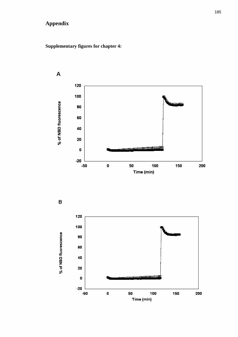
185
Appendix
Supplementary figures for chapter 4:

186

187
Figure I (chapter 4, figure 4.12) Raw fluorescence data from in vitro fusion assays of liposomes containing either full-length Sx 4/SNAP23 (A) or Sx 4 N∆36/SNAP23 (B) or Sx 4 OPEN/SNAP23 (C) or Sx 4 OPEN N∆36/SNAP23 (D) with liposomes containing VAMP2
These are supplement figures related to figure 4.12 (chapter 4). A graphing and analysis
programme called KaleidaGraph (Synergy Software) was used in order to analyse the data
obtained from the Fluorescence reader after fusion assays where 5 μl of fluorescently
labelled donor VAMP2 liposomes were mixed with 45 μl of unlabelled acceptor t-
complexes being either wild-type (A) or Sx 4 N∆36/SNAP23 (B) or Sx 4 OPEN/SNAP23
(C) or Sx 4 OPEN N∆36/SNAP23 (D). Raw fluorescence was normalised to maximum
detergent signal (100 %) and plotted against time in minutes (graphs A, B, C and D).

188

189
Figure II (chapter 4, figure 4.16) Raw fluorescence data from in vitro fusion of wild-type Sx 4/SNAP23 with VAMP2 vesicles (pre-docked and not docked) in the presence and in the absence of His6-M18c
This fusion assay was described as outlined in figure 4.16. Raw data is shown normalised
to 100 % detergent signal for A and B. (A) The liposomes were pre-incubated overnight
with M18c. (B) The liposomes were NOT pre-incubated overnight with M18c, instead they
were pre-docked overnight and incubated with M18c for 2 h before fusion at 37 ˚C.

190

191
Figure III (chapter 4, figure 4.17) Raw fluorescence data from in vitro fusion of Sx 4 N∆36/SNAP23 with VAMP2 vesicles (pre-docked overnight or not) in the presence and in the absence of His6-M18c
This fusion assay was described as outlined in figure 4.17. Raw data is shown normalised
to 100 % detergent signal for A and B. (A) The liposomes were pre-incubated overnight
with M18c. (B) The liposomes were NOT pre-incubated overnight with M18c, instead they
were pre-docked overnight and incubated with M18c for 2 h before fusion took place.

192

193
Figure IV (chapter 4, figure 4.18) Raw fluorescence data from in vitro fusion of Sx 4 N∆36/SNAP23 liposomes with VAMP2 liposomes (pre-docked or not) in the presence or in the absence of His6-M18c for only 2 h
This fusion assay was described as outlined in figure 4.18. Raw data is shown normalised
to 100 % detergent signal for A and B. (A) v- and t-liposomes were pre-incubated for 2 h
with M18c. (B) v- and t-liposomes were allowed to pre-dock before incubation with M18c
for 2 h.

194

195
Figure V (chapter 4, figure 4.19) Raw fluorescence data from in vitro fusion of Sx 4 OPEN/SNAP23 with VAMP2 vesicles (pre-docked overnight and not docked) in the presence and in the absence of M18c
This fusion assay was described as outlined in figure 4.19. Raw data is shown normalised
to 100 % detergent signal for A and B. (A) v- and t-liposomes were pre-incubated
overnight with M18c. (B) v- and t-liposomes were NOT pre-incubated overnight with
M18c, instead they were pre-docked overnight and incubated with M18c for 2 h before
fusion took place.

196

197
Figure VI (chapter 4, figure 4.20) Raw fluorescence data in vitro fusion of Sx 4 OPEN N∆36/SNAP23 with VAMP2 vesicles (pre-docked overnight and not docked) in the presence and in the absence of M18c
This fusion assay was described as outlined in figure 4.16. Raw data is shown normalised
to 100 % detergent signal for A and B. (A) v- and t- liposomes were pre-incubated
overnight with M18c. (B) v- and t- liposomes were NOT pre-incubated overnight with
M18c, instead they were pre-docked overnight and incubated with M18c for 2 h before
fusion.

198

199
Figure VII (chapter 4, figure 4.21) Raw fluorescent data from in vitro fusion of Sx 4 OPEN N∆36/SNAP23 with VAMP2 vesicles (pre-docked overnight or not) in the presence and in the absence of M18c for 2 h
This fusion assay was described as outlined in figure 4.21. Raw data is shown normalised
to 100 % detergent signal for A and B. (A) The liposomes were pre-incubated for 2 h with
M18c. (B) The liposomes were allowed to pre-doc before incubation with M18c for 2 h.
Supplementary figures for chapter 5:
+EPI (552.26) Charge (+2) CE (32.613) FT (292.53): Exp 3, 18.323 min from Sample 1 (Munc2) of Munc2.wiff (Nanospray) Max. 8.2e5 cps.
100 150 200 250 300 350 400 450 500 550 600 650 700 750 800 850 900 950 1000 1050 1100 1150 1200m/z, amu
5.0e4
1.0e5
1.5e5
2.0e5
2.5e5
3.0e5
3.5e5
4.0e5
4.5e5
5.0e5
5.5e5
6.0e5
6.5e5
7.0e5
7.5e5
8.0e5
8.2e5
Inte
ns
ity
, c
ps
888.1
532.3
645.4
403.3
459.4
552.5
216.21013.2
175.3 194.3129.1
251.2
501.7 1002.1113.2 358.2 527.3290.3
985.1
898.1441.1199.1 514.7543.8424.0
158.2329.4
377.2 414.4171.2213.1 808.4572.3
313.3 476.0 953.3 1066.2701.0458.4 496.3386.6
188.3 609.6583.1324.4450.2136.2 627.1 683.3 814.1239.2 852.4
779.2306.2 871.1
b2
b3
b4b5 b6
y1
y2
y3
y4y5
y6
y7
[M + 2H]2+
Figure VIII Positive-ion ESI-MS/MS of Peptide 1 (residues Thr 519- Arg 526)
Several fragment (b and y) ions were observed as indicated. The b3 and y6 fragment ions
indicate that Y521 is phosphorylated. The sequence of phosphorylated peptide 1 is shown
in figure X.

200
+EPI (616.32) Charge (+2) CE (35.8162) FT (300): Exp 3, 15.553 min from Sample 1 (Munc2) of Munc2.wiff (Nanospray) Max. 1.3e6 cps.
150 200 250 300 350 400 450 500 550 600 650 700 750 800 850 900 950 1000 1050 1100 1150 1200m/z, amu
5.00e4
1.00e5
1.50e5
2.00e5
2.50e5
3.00e5
3.50e5
4.00e5
4.50e5
5.00e5
5.50e5
6.00e5
6.50e5
7.00e5
7.50e5
8.00e5
8.50e5
9.00e5
9.50e5
1.00e6
1.05e6
1.10e6
1.15e6
1.20e6
1.25e6
1.30e6
1.34e6In
ten
sit
y,
cp
s616.2
459.1
303.5
660.2 773.3
531.6
418.3
1016.2
216.2 607.9566.0358.2
642.2513.4 584.5289.6 701.2
594.6 755.3 1141.0403.6129.4 441.4 661.4 918.4 998.2888.1199.2 454.1 555.1379.2329.4 936.1290.3 1085.3683.4 742.1576.5 869.0783.0 1103.4
b2
b3
b8
b6Y1
(147.1)
y2
y3
y4y5 y6
y7
[M + 2H]2+
Y8(1130.5)
(814.1)
Figure IX Positive-ion ESI-MS/MS spectrum of the phosphorylated peptide 2 (residues Thr 519-Lys 527) of M18c
Several fragment (b and y) ions were observed as indicated. The b3 and y7 fragment ions
indicate that Y521 is phosphorylated. The sequence of phosphorylated peptide 2 is shown
in figure X.

201
Figure X Sequences of the phosphorylated tryptic peptides 1 and 2 from M18c

202
Table I Summary of the peptides identified after fragmentation of peptide 1 9A) (m/z 551.3) and peptide 2 (B) (m/z 615.3).

203
References
Advani, R.J., Yang, B., Prekeris, R., Lee, K.C., Klumperman, J. and Scheller, R.H. (1999)
VAMP-7 mediates vesicular transport from endosomes to lysosomes. J Cell Biol,
146, 765-776.
Aledo, J.C., Lavoie, L., Volchuk, A., Keller, S.R., Klip, A. and Hundal, H.S. (1997)
Identification and characterization of two distinct intracellular GLUT4 pools in rat
skeletal muscle: evidence for an endosomal and an insulin-sensitive GLUT4
compartment. Biochem J, 325 (Pt 3), 727-732.
Alessi, D.R., James, S.R., Downes, C.P., Holmes, A.B., Gaffney, P.R., Reese, C.B. and
Cohen, P. (1997) Characterization of a 3-phosphoinositide-dependent protein
kinase which phosphorylates and activates protein kinase Balpha. Curr Biol, 7,
261-269.
Antonin, W., Fasshauer, D., Becker, S., Jahn, R. and Schneider, T.R. (2002) Crystal
structure of the endosomal SNARE complex reveals common structural principles
of all SNAREs. Nat Struct Biol, 9, 107-111.
Araki, S., Tamori, Y., Kawanishi, M., Shinoda, H., Masugi, J., Mori, H., Niki, T.,
Okazawa, H., Kubota, T. and Kasuga, M. (1997) Inhibition of the binding of
SNAP-23 to syntaxin 4 by Munc18c. Biochem Biophys Res Commun, 234, 257-
262.
Aran, V., Brandie, F.M., Boyd, A.R., Kantidakis, T., Rideout, E.J., Kelly, S.M., Gould,
G.W. and Bryant, N. (2009) Characterisation of two distinct binding modes
between Syntaxin 4 and Munc18c. Biochem J.
Baba, T., Sakisaka, T., Mochida, S. and Takai, Y. (2005) PKA-catalyzed phosphorylation
of tomosyn and its implication in Ca2+-dependent exocytosis of neurotransmitter. J
Cell Biol, 170, 1113-1125.
Banerji, M.A. (2002) Impaired beta-cell and alpha-cell function in African-American
children with type 2 diabetes mellitus--"Flatbush diabetes". J Pediatr Endocrinol
Metab, 15 Suppl 1, 493-501.
Baumann, C.A., Ribon, V., Kanzaki, M., Thurmond, D.C., Mora, S., Shigematsu, S.,
Bickel, P.E., Pessin, J.E. and Saltiel, A.R. (2000) CAP defines a second signalling
pathway required for insulin-stimulated glucose transport. Nature, 407, 202-207.
Beckers, C.J., Block, M.R., Glick, B.S., Rothman, J.E. and Balch, W.E. (1989) Vesicular
transport between the endoplasmic reticulum and the Golgi stack requires the
NEM-sensitive fusion protein. Nature, 339, 397-398.
Bell, G.I., Murray, J.C., Nakamura, Y., Kayano, T., Eddy, R.L., Fan, Y.S., Byers, M.G.
and Shows, T.B. (1989) Polymorphic human insulin-responsive glucose-transporter
gene on chromosome 17p13. Diabetes, 38, 1072-1075.
Bennett, M.K., Calakos, N. and Scheller, R.H. (1992) Syntaxin: a synaptic protein
implicated in docking of synaptic vesicles at presynaptic active zones. Science, 257,
255-259.
Bennett, M.K., Garcia-Arraras, J.E., Elferink, L.A., Peterson, K., Fleming, A.M., Hazuka,
C.D. and Scheller, R.H. (1993) The syntaxin family of vesicular transport
receptors. Cell, 74, 863-873.
Birnbaum, M.J. (1989) Identification of a novel gene encoding an insulin-responsive
glucose transporter protein. Cell, 57, 305-315.
Bock, J.B., Lin, R.C. and Scheller, R.H. (1996) A new syntaxin family member implicated
in targeting of intracellular transport vesicles. J Biol Chem, 271, 17961-17965.
Bock, J.B., Matern, H.T., Peden, A.A. and Scheller, R.H. (2001) A genomic perspective on
membrane compartment organization. Nature, 409, 839-841.
Bracher, A. and Weissenhorn, W. (2002) Structural basis for the Golgi membrane
recruitment of Sly1p by Sed5p. Embo J, 21, 6114-6124.

204
Brandie, F.M., Aran, V., Verma, A., McNew, J.A., Bryant, N.J. and Gould, G.W. (2008)
Negative regulation of syntaxin4/SNAP-23/VAMP2-mediated membrane fusion by
Munc18c in vitro. PLoS ONE, 3, e4074.
Brenner, S. (1974) The genetics of Caenorhabditis elegans. Genetics, 77, 71-94.
Bryant, N.J., Govers, R. and James, D.E. (2002) Regulated transport of the glucose
transporter GLUT4. Nat Rev Mol Cell Biol, 3, 267-277.
Bryant, N.J. and James, D.E. (2003) The Sec1p/Munc18 (SM) protein, Vps45p, cycles on
and off membranes during vesicle transport. J Cell Biol, 161, 691-696.
Burkhardt, P., Hattendorf, D.A., Weis, W.I. and Fasshauer, D. (2008) Munc18a controls
SNARE assembly through its interaction with the syntaxin N-peptide. Embo J, 27,
923-933.
Cabaniols, J.P., Ravichandran, V. and Roche, P.A. (1999) Phosphorylation of SNAP-23 by
the novel kinase SNAK regulates t-SNARE complex assembly. Mol Biol Cell, 10,
4033-4041.
Cain, C.C., Trimble, W.S. and Lienhard, G.E. (1992) Members of the VAMP family of
synaptic vesicle proteins are components of glucose transporter-containing vesicles
from rat adipocytes. J Biol Chem, 267, 11681-11684.
Carpp, L.N., Ciufo, L.F., Shanks, S.G., Boyd, A. and Bryant, N.J. (2006) The
Sec1p/Munc18 protein Vps45p binds its cognate SNARE proteins via two distinct
modes. J Cell Biol, 173, 927-936.
Charron, M.J., Brosius, F.C., 3rd, Alper, S.L. and Lodish, H.F. (1989) A glucose transport
protein expressed predominately in insulin-responsive tissues. Proc Natl Acad Sci
U S A, 86, 2535-2539.
Cheatham, B., Vlahos, C.J., Cheatham, L., Wang, L., Blenis, J. and Kahn, C.R. (1994)
Phosphatidylinositol 3-kinase activation is required for insulin stimulation of pp70
S6 kinase, DNA synthesis, and glucose transporter translocation. Mol Cell Biol, 14,
4902-4911.
Cheatham, B., Volchuk, A., Kahn, C.R., Wang, L., Rhodes, C.J. and Klip, A. (1996)
Insulin-stimulated translocation of GLUT4 glucose transporters requires SNARE-
complex proteins. Proc Natl Acad Sci U S A, 93, 15169-15173.
Chen, Y.A. and Scheller, R.H. (2001) SNARE-mediated membrane fusion. Nat Rev Mol
Cell Biol, 2, 98-106.
Chernomordik, L.V. and Kozlov, M.M. (2005) Membrane hemifusion: crossing a chasm in
two leaps. Cell, 123, 375-382.
Cheviet, S., Bezzi, P., Ivarsson, R., Renstrom, E., Viertl, D., Kasas, S., Catsicas, S. and
Regazzi, R. (2006) Tomosyn-1 is involved in a post-docking event required for
pancreatic beta-cell exocytosis. J Cell Sci, 119, 2912-2920.
Chiang, S.H., Baumann, C.A., Kanzaki, M., Thurmond, D.C., Watson, R.T., Neudauer,
C.L., Macara, I.G., Pessin, J.E. and Saltiel, A.R. (2001) Insulin-stimulated GLUT4
translocation requires the CAP-dependent activation of TC10. Nature, 410, 944-
948.
Clary, D.O., Griff, I.C. and Rothman, J.E. (1990) SNAPs, a family of NSF attachment
proteins involved in intracellular membrane fusion in animals and yeast. Cell, 61,
709-721.
Cowles, C.R., Emr, S.D. and Horazdovsky, B.F. (1994) Mutations in the VPS45 gene, a
SEC1 homologue, result in vacuolar protein sorting defects and accumulation of
membrane vesicles. J Cell Sci, 107 (Pt 12), 3449-3459.
Cushman, S.W. and Wardzala, L.J. (1980) Potential mechanism of insulin action on
glucose transport in the isolated rat adipose cell. Apparent translocation of
intracellular transport systems to the plasma membrane. J Biol Chem, 255, 4758-
4762.

205
D'Andrea-Merrins, M., Chang, L., Lam, A.D., Ernst, S.A. and Stuenkel, E.L. (2007)
Munc18c interaction with syntaxin 4 monomers and SNARE complex
intermediates in GLUT4 vesicle trafficking. J Biol Chem, 282, 16553-16566.
Diaz, R., Mayorga, L.S., Weidman, P.J., Rothman, J.E. and Stahl, P.D. (1989) Vesicle
fusion following receptor-mediated endocytosis requires a protein active in Golgi
transport. Nature, 339, 398-400.
Dirac-Svejstrup, A.B., Sumizawa, T. and Pfeffer, S.R. (1997) Identification of a GDI
displacement factor that releases endosomal Rab GTPases from Rab-GDI. Embo J,
16, 465-472.
Dulubova, I., Khvotchev, M., Liu, S., Huryeva, I., Sudhof, T.C. and Rizo, J. (2007)
Munc18-1 binds directly to the neuronal SNARE complex. Proc Natl Acad Sci U S
A, 104, 2697-2702.
Dulubova, I., Sugita, S., Hill, S., Hosaka, M., Fernandez, I., Sudhof, T.C. and Rizo, J.
(1999) A conformational switch in syntaxin during exocytosis: role of munc18.
Embo J, 18, 4372-4382.
Dulubova, I., Yamaguchi, T., Arac, D., Li, H., Huryeva, I., Min, S.W., Rizo, J. and Sudhof,
T.C. (2003) Convergence and divergence in the mechanism of SNARE binding by
Sec1/Munc18-like proteins. Proc Natl Acad Sci U S A, 100, 32-37.
Dulubova, I., Yamaguchi, T., Gao, Y., Min, S.W., Huryeva, I., Sudhof, T.C. and Rizo, J.
(2002) How Tlg2p/syntaxin 16 'snares' Vps45. Embo J, 21, 3620-3631.
Dybbs, M., Ngai, J. and Kaplan, J.M. (2005) Using microarrays to facilitate positional
cloning: identification of tomosyn as an inhibitor of neurosecretion. PLoS Genet, 1,
6-16.
Ewart, M.A., Clarke, M., Kane, S., Chamberlain, L.H. and Gould, G.W. (2005) Evidence
for a role of the exocyst in insulin-stimulated Glut4 trafficking in 3T3-L1
adipocytes. J Biol Chem, 280, 3812-3816.
Fasshauer, D., Otto, H., Eliason, W.K., Jahn, R. and Brunger, A.T. (1997) Structural
changes are associated with soluble N-ethylmaleimide-sensitive fusion protein
attachment protein receptor complex formation. J Biol Chem, 272, 28036-28041.
Fasshauer, D., Sutton, R.B., Brunger, A.T. and Jahn, R. (1998) Conserved structural
features of the synaptic fusion complex: SNARE proteins reclassified as Q- and R-
SNAREs. Proc Natl Acad Sci U S A, 95, 15781-15786.
Fernandez, I., Ubach, J., Dulubova, I., Zhang, X., Sudhof, T.C. and Rizo, J. (1998) Three-
dimensional structure of an evolutionarily conserved N-terminal domain of
syntaxin 1A. Cell, 94, 841-849.
Fiebig, K.M., Rice, L.M., Pollock, E. and Brunger, A.T. (1999) Folding intermediates of
SNARE complex assembly. Nat Struct Biol, 6, 117-123.
Fix, M., Melia, T.J., Jaiswal, J.K., Rappoport, J.Z., You, D., Sollner, T.H., Rothman, J.E.
and Simon, S.M. (2004) Imaging single membrane fusion events mediated by
SNARE proteins. Proc Natl Acad Sci U S A, 101, 7311-7316.
Foster, L.J., Yeung, B., Mohtashami, M., Ross, K., Trimble, W.S. and Klip, A. (1998)
Binary interactions of the SNARE proteins syntaxin-4, SNAP23, and VAMP-2 and
their regulation by phosphorylation. Biochemistry, 37, 11089-11096.
Fujita, Y., Sasaki, T., Fukui, K., Kotani, H., Kimura, T., Hata, Y., Sudhof, T.C., Scheller,
R.H. and Takai, Y. (1996) Phosphorylation of Munc-18/n-Sec1/rbSec1 by protein
kinase C: its implication in regulating the interaction of Munc-18/n-Sec1/rbSec1
with syntaxin. J Biol Chem, 271, 7265-7268.
Fujita, Y., Shirataki, H., Sakisaka, T., Asakura, T., Ohya, T., Kotani, H., Yokoyama, S.,
Nishioka, H., Matsuura, Y., Mizoguchi, A., Scheller, R.H. and Takai, Y. (1998)
Tomosyn: a syntaxin-1-binding protein that forms a novel complex in the
neurotransmitter release process. Neuron, 20, 905-915.
Fukuda, M. (2008) Membrane traffic in the secretory pathway: Regulation of secretory
vesicle traffic by Rab small GTPases. Cell Mol Life Sci, 65, 2801-2813.

206
Gagescu, R. (2000) SNARE hypothesis 2000. Nat Rev Mol Cell Biol, 1, 5.
Gerber, S.H., Rah, J.C., Min, S.W., Liu, X., de Wit, H., Dulubova, I., Meyer, A.C., Rizo,
J., Arancillo, M., Hammer, R.E., Verhage, M., Rosenmund, C. and Sudhof, T.C.
(2008) Conformational switch of syntaxin-1 controls synaptic vesicle fusion.
Science, 321, 1507-1510.
Glick, B.S. and Rothman, J.E. (1987) Possible role for fatty acyl-coenzyme A in
intracellular protein transport. Nature, 326, 309-312.
Gould, G.W., Brant, A.M., Kahn, B.B., Shepherd, P.R., McCoid, S.C. and Gibbs, E.M.
(1992) Expression of the brain-type glucose transporter is restricted to brain and
neuronal cells in mice. Diabetologia, 35, 304-309.
Grusovin, J., Stoichevska, V., Gough, K.H., Nunan, K., Ward, C.W. and Macaulay, S.L.
(2000) Definition of a minimal munc18c domain that interacts with syntaxin 4.
Biochem J, 350 Pt 3, 741-746.
Gual, P., Le Marchand-Brustel, Y. and Tanti, J.F. (2005) Positive and negative regulation
of insulin signaling through IRS-1 phosphorylation. Biochimie, 87, 99-109.
Harrison, S.D., Broadie, K., van de Goor, J. and Rubin, G.M. (1994) Mutations in the
Drosophila Rop gene suggest a function in general secretion and synaptic
transmission. Neuron, 13, 555-566.
Hashiramoto, M. and James, D.E. (2000) Characterization of insulin-responsive GLUT4
storage vesicles isolated from 3T3-L1 adipocytes. Mol Cell Biol, 20, 416-427.
Hata, Y., Slaughter, C.A. and Sudhof, T.C. (1993) Synaptic vesicle fusion complex
contains unc-18 homologue bound to syntaxin. Nature, 366, 347-351.
Hayashi, T., McMahon, H., Yamasaki, S., Binz, T., Hata, Y., Sudhof, T.C. and Niemann,
H. (1994) Synaptic vesicle membrane fusion complex: action of clostridial
neurotoxins on assembly. Embo J, 13, 5051-5061.
Hayashi, T., Yamasaki, S., Nauenburg, S., Binz, T. and Niemann, H. (1995) Disassembly
of the reconstituted synaptic vesicle membrane fusion complex in vitro. Embo J,
14, 2317-2325.
Hirshman, M.F., Goodyear, L.J., Wardzala, L.J., Horton, E.D. and Horton, E.S. (1990)
Identification of an intracellular pool of glucose transporters from basal and
insulin-stimulated rat skeletal muscle. J Biol Chem, 265, 987-991.
Hohl, T.M., Parlati, F., Wimmer, C., Rothman, J.E., Sollner, T.H. and Engelhardt, H.
(1998) Arrangement of subunits in 20 S particles consisting of NSF, SNAPs, and
SNARE complexes. Mol Cell, 2, 539-548.
Holt, M., Varoqueaux, F., Wiederhold, K., Takamori, S., Urlaub, H., Fasshauer, D. and
Jahn, R. (2006) Identification of SNAP-47, a novel Qbc-SNARE with ubiquitous
expression. J Biol Chem, 281, 17076-17083.
Hong, W. (2005) SNAREs and traffic. Biochim Biophys Acta, 1744, 493-517.
Hu, S.H., Gee, C.L., Latham, C.F., Rowlinson, S.W., Rova, U., Jones, A., Halliday, J.A.,
Bryant, N.J., James, D.E. and Martin, J.L. (2003) Recombinant expression of
Munc18c in a baculovirus system and interaction with syntaxin4. Protein Expr
Purif, 31, 305-310.
Hu, S.H., Latham, C.F., Gee, C.L., James, D.E. and Martin, J.L. (2007) Structure of the
Munc18c/Syntaxin4 N-peptide complex defines universal features of the N-peptide
binding mode of Sec1/Munc18 proteins. Proc Natl Acad Sci U S A, 104, 8773-
8778.
Hua, Y. and Scheller, R.H. (2001) Three SNARE complexes cooperate to mediate
membrane fusion. Proc Natl Acad Sci U S A, 98, 8065-8070.
Inoue, M., Chang, L., Hwang, J., Chiang, S.H. and Saltiel, A.R. (2003) The exocyst
complex is required for targeting of Glut4 to the plasma membrane by insulin.
Nature, 422, 629-633.
207
Ishikura, S., Bilan, P.J. and Klip, A. (2007) Rabs 8A and 14 are targets of the insulin-
regulated Rab-GAP AS160 regulating GLUT4 traffic in muscle cells. Biochem
Biophys Res Commun, 353, 1074-1079.
James, D.E., Brown, R., Navarro, J. and Pilch, P.F. (1988) Insulin-regulatable tissues
express a unique insulin-sensitive glucose transport protein. Nature, 333, 183-185.
James, D.E., Strube, M. and Mueckler, M. (1989) Molecular cloning and characterization
of an insulin-regulatable glucose transporter. Nature, 338, 83-87.
Jewell, J.L., Oh, E., Bennett, S.M., Meroueh, S.O. and Thurmond, D.C. (2008) The
tyrosine phosphorylation of Munc18c induces a switch in binding specificity from
Syntaxin 4 to Doc2beta. J Biol Chem.
Joost, H.G. and Thorens, B. (2001) The extended GLUT-family of sugar/polyol transport
facilitators: nomenclature, sequence characteristics, and potential function of its
novel members (review). Mol Membr Biol, 18, 247-256.
Kaestner, K.H., Christy, R.J., McLenithan, J.C., Braiterman, L.T., Cornelius, P., Pekala,
P.H. and Lane, M.D. (1989) Sequence, tissue distribution, and differential
expression of mRNA for a putative insulin-responsive glucose transporter in mouse
3T3-L1 adipocytes. Proc Natl Acad Sci U S A, 86, 3150-3154.
Kahn, S.E., Hull, R.L. and Utzschneider, K.M. (2006) Mechanisms linking obesity to
insulin resistance and type 2 diabetes. Nature, 444, 840-846.
Kanda, H., Tamori, Y., Shinoda, H., Yoshikawa, M., Sakaue, M., Udagawa, J., Otani, H.,
Tashiro, F., Miyazaki, J. and Kasuga, M. (2005) Adipocytes from Munc18c-null
mice show increased sensitivity to insulin-stimulated GLUT4 externalization. J
Clin Invest, 115, 291-301.
Kandror, K.V. and Pilch, P.F. (1996) Compartmentalization of protein traffic in insulin-
sensitive cells. Am J Physiol, 271, E1-14.
Kane, S., Sano, H., Liu, S.C., Asara, J.M., Lane, W.S., Garner, C.C. and Lienhard, G.E.
(2002) A method to identify serine kinase substrates. Akt phosphorylates a novel
adipocyte protein with a Rab GTPase-activating protein (GAP) domain. J Biol
Chem, 277, 22115-22118.
Katz, E.B., Stenbit, A.E., Hatton, K., DePinho, R. and Charron, M.J. (1995) Cardiac and
adipose tissue abnormalities but not diabetes in mice deficient in GLUT4. Nature,
377, 151-155.
Kawanishi, M., Tamori, Y., Okazawa, H., Araki, S., Shinoda, H. and Kasuga, M. (2000)
Role of SNAP23 in insulin-induced translocation of GLUT4 in 3T3-L1 adipocytes.
Mediation of complex formation between syntaxin4 and VAMP2. J Biol Chem,
275, 8240-8247.
Kayano, T., Fukumoto, H., Eddy, R.L., Fan, Y.S., Byers, M.G., Shows, T.B. and Bell, G.I.
(1988) Evidence for a family of human glucose transporter-like proteins. Sequence
and gene localization of a protein expressed in fetal skeletal muscle and other
tissues. J Biol Chem, 263, 15245-15248.
Ke, B., Oh, E. and Thurmond, D.C. (2007) Doc2beta is a novel Munc18c-interacting
partner and positive effector of syntaxin 4-mediated exocytosis. J Biol Chem, 282,
21786-21797.
Kee, Y., Yoo, J.S., Hazuka, C.D., Peterson, K.E., Hsu, S.C. and Scheller, R.H. (1997)
Subunit structure of the mammalian exocyst complex. Proc Natl Acad Sci U S A,
94, 14438-14443.
Kelly, S.M., Jess, T.J. and Price, N.C. (2005) How to study proteins by circular dichroism.
Biochim Biophys Acta, 1751, 119-139.
Kelly, S.M. and Price, N.C. (2000) The use of circular dichroism in the investigation of
protein structure and function. Curr Protein Pept Sci, 1, 349-384.
Khan, A.H., Thurmond, D.C., Yang, C., Ceresa, B.P., Sigmund, C.D. and Pessin, J.E.
(2001) Munc18c regulates insulin-stimulated glut4 translocation to the transverse
tubules in skeletal muscle. J Biol Chem, 276, 4063-4069.

208
Khvotchev, M., Dulubova, I., Sun, J., Dai, H., Rizo, J. and Sudhof, T.C. (2007) Dual
modes of Munc18-1/SNARE interactions are coupled by functionally critical
binding to syntaxin-1 N terminus. J Neurosci, 27, 12147-12155.
Klip, A., Ramlal, T., Bilan, P.J., Cartee, G.D., Gulve, E.A. and Holloszy, J.O. (1990)
Recruitment of GLUT-4 glucose transporters by insulin in diabetic rat skeletal
muscle. Biochem Biophys Res Commun, 172, 728-736.
Kohn, A.D., Barthel, A., Kovacina, K.S., Boge, A., Wallach, B., Summers, S.A.,
Birnbaum, M.J., Scott, P.H., Lawrence, J.C., Jr. and Roth, R.A. (1998)
Construction and characterization of a conditionally active version of the
serine/threonine kinase Akt. J Biol Chem, 273, 11937-11943.
Kohn, A.D., Summers, S.A., Birnbaum, M.J. and Roth, R.A. (1996) Expression of a
constitutively active Akt Ser/Thr kinase in 3T3-L1 adipocytes stimulates glucose
uptake and glucose transporter 4 translocation. J Biol Chem, 271, 31372-31378.
Koumanov, F., Jin, B., Yang, J. and Holman, G.D. (2005) Insulin signaling meets vesicle
traffic of GLUT4 at a plasma-membrane-activated fusion step. Cell Metab, 2, 179-
189.
Kuhne, M.R., Zhao, Z., Rowles, J., Lavan, B.E., Shen, S.H., Fischer, E.H. and Lienhard,
G.E. (1994) Dephosphorylation of insulin receptor substrate 1 by the tyrosine
phosphatase PTP2C. J Biol Chem, 269, 15833-15837.
Laage, R., Rohde, J., Brosig, B. and Langosch, D. (2000) A conserved membrane-spanning
amino acid motif drives homomeric and supports heteromeric assembly of
presynaptic SNARE proteins. J Biol Chem, 275, 17481-17487.
Lane, S.R. and Liu, Y. (1997) Characterization of the palmitoylation domain of SNAP-25.
J Neurochem, 69, 1864-1869.
Larance, M., Ramm, G., Stockli, J., van Dam, E.M., Winata, S., Wasinger, V., Simpson,
F., Graham, M., Junutula, J.R., Guilhaus, M. and James, D.E. (2005)
Characterization of the role of the Rab GTPase-activating protein AS160 in insulin-
regulated GLUT4 trafficking. J Biol Chem, 280, 37803-37813.
Latham, C.F., Lopez, J.A., Hu, S.H., Gee, C.L., Westbury, E., Blair, D.H., Armishaw, C.J.,
Alewood, P.F., Bryant, N.J., James, D.E. and Martin, J.L. (2006) Molecular
dissection of the Munc18c/syntaxin4 interaction: implications for regulation of
membrane trafficking. Traffic, 7, 1408-1419.
Leibiger, I.B. and Berggren, P.O. (2008) Insulin signaling in the pancreatic beta-cell. Annu
Rev Nutr, 28, 233-251.
Leysens, N., Murray, J.C. and Bell, G.I. (1989) A KpnI polymorphism for the human
insulin-responsive glucose transporter gene (GLUT4) on chromosome 17. Nucleic
Acids Res, 17, 3621.
Li, D., Randhawa, V.K., Patel, N., Hayashi, M. and Klip, A. (2001) Hyperosmolarity
reduces GLUT4 endocytosis and increases its exocytosis from a VAMP2-
independent pool in l6 muscle cells. J Biol Chem, 276, 22883-22891.
Lin, R.C. and Scheller, R.H. (2000) Mechanisms of synaptic vesicle exocytosis. Annu Rev
Cell Dev Biol, 16, 19-49.
Littleton, J.T., Chapman, E.R., Kreber, R., Garment, M.B., Carlson, S.D. and Ganetzky, B.
(1998) Temperature-sensitive paralytic mutations demonstrate that synaptic
exocytosis requires SNARE complex assembly and disassembly. Neuron, 21, 401-
413.
Livingstone, C., James, D.E., Rice, J.E., Hanpeter, D. and Gould, G.W. (1996)
Compartment ablation analysis of the insulin-responsive glucose transporter
(GLUT4) in 3T3-L1 adipocytes. Biochem J, 315 (Pt 2), 487-495.
Malhotra, V., Orci, L., Glick, B.S., Block, M.R. and Rothman, J.E. (1988) Role of an N-
ethylmaleimide-sensitive transport component in promoting fusion of transport
vesicles with cisternae of the Golgi stack. Cell, 54, 221-227.

209
Martin, L.B., Shewan, A., Millar, C.A., Gould, G.W. and James, D.E. (1998) Vesicle-
associated membrane protein 2 plays a specific role in the insulin-dependent
trafficking of the facilitative glucose transporter GLUT4 in 3T3-L1 adipocytes. J
Biol Chem, 273, 1444-1452.
Martin, S., Tellam, J., Livingstone, C., Slot, J.W., Gould, G.W. and James, D.E. (1996)
The glucose transporter (GLUT-4) and vesicle-associated membrane protein-2
(VAMP-2) are segregated from recycling endosomes in insulin-sensitive cells. J
Cell Biol, 134, 625-635.
Marz, K.E., Lauer, J.M. and Hanson, P.I. (2003) Defining the SNARE complex binding
surface of alpha-SNAP: implications for SNARE complex disassembly. J Biol
Chem, 278, 27000-27008.
McNew, J.A., Parlati, F., Fukuda, R., Johnston, R.J., Paz, K., Paumet, F., Sollner, T.H. and
Rothman, J.E. (2000) Compartmental specificity of cellular membrane fusion
encoded in SNARE proteins. Nature, 407, 153-159.
Miinea, C.P., Sano, H., Kane, S., Sano, E., Fukuda, M., Peranen, J., Lane, W.S. and
Lienhard, G.E. (2005) AS160, the Akt substrate regulating GLUT4 translocation,
has a functional Rab GTPase-activating protein domain. Biochem J, 391, 87-93.
Min, J., Okada, S., Kanzaki, M., Elmendorf, J.S., Coker, K.J., Ceresa, B.P., Syu, L.J.,
Noda, Y., Saltiel, A.R. and Pessin, J.E. (1999) Synip: a novel insulin-regulated
syntaxin 4-binding protein mediating GLUT4 translocation in adipocytes. Mol Cell,
3, 751-760.
Misura, K.M., Scheller, R.H. and Weis, W.I. (2000) Three-dimensional structure of the
neuronal-Sec1-syntaxin 1a complex. Nature, 404, 355-362.
Mitra, P., Zheng, X. and Czech, M.P. (2004) RNAi-based analysis of CAP, Cbl, and CrkII
function in the regulation of GLUT4 by insulin. J Biol Chem, 279, 37431-37435.
Mueckler, M. (1994) Facilitative glucose transporters. Eur J Biochem, 219, 713-725.
Nagamatsu, S., Kornhauser, J.M., Burant, C.F., Seino, S., Mayo, K.E. and Bell, G.I. (1992)
Glucose transporter expression in brain. cDNA sequence of mouse GLUT3, the
brain facilitative glucose transporter isoform, and identification of sites of
expression by in situ hybridization. J Biol Chem, 267, 467-472.
Novick, P., Medkova, M., Dong, G., Hutagalung, A., Reinisch, K. and Grosshans, B.
(2006) Interactions between Rabs, tethers, SNAREs and their regulators in
exocytosis. Biochem Soc Trans, 34, 683-686.
Novick, P. and Schekman, R. (1979) Secretion and cell-surface growth are blocked in a
temperature-sensitive mutant of Saccharomyces cerevisiae. Proc Natl Acad Sci U S
A, 76, 1858-1862.
Novick, P. and Zerial, M. (1997) The diversity of Rab proteins in vesicle transport. Curr
Opin Cell Biol, 9, 496-504.
Oh, E., Spurlin, B.A., Pessin, J.E. and Thurmond, D.C. (2005) Munc18c heterozygous
knockout mice display increased susceptibility for severe glucose intolerance.
Diabetes, 54, 638-647.
Oh, E. and Thurmond, D.C. (2006) The stimulus-induced tyrosine phosphorylation of
Munc18c facilitates vesicle exocytosis. J Biol Chem, 281, 17624-17634.
Okada, S., Ohshima, K., Uehara, Y., Shimizu, H., Hashimoto, K., Yamada, M. and Mori,
M. (2007) Synip phosphorylation is required for insulin-stimulated Glut4
translocation. Biochem Biophys Res Commun, 356, 102-106.
Okada, T., Kawano, Y., Sakakibara, T., Hazeki, O. and Ui, M. (1994) Essential role of
phosphatidylinositol 3-kinase in insulin-induced glucose transport and antilipolysis
in rat adipocytes. Studies with a selective inhibitor wortmannin. J Biol Chem, 269,
3568-3573.
Olson, A.L. and Pessin, J.E. (1996) Structure, function, and regulation of the mammalian
facilitative glucose transporter gene family. Annu Rev Nutr, 16, 235-256.

210
Orita, S., Sasaki, T., Naito, A., Komuro, R., Ohtsuka, T., Maeda, M., Suzuki, H., Igarashi,
H. and Takai, Y. (1995) Doc2: a novel brain protein having two repeated C2-like
domains. Biochem Biophys Res Commun, 206, 439-448.
Oyler, G.A., Higgins, G.A., Hart, R.A., Battenberg, E., Billingsley, M., Bloom, F.E. and
Wilson, M.C. (1989) The identification of a novel synaptosomal-associated protein,
SNAP-25, differentially expressed by neuronal subpopulations. J Cell Biol, 109,
3039-3052.
Palade, G. (1975) Intracellular aspects of the process of protein synthesis. Science, 189,
347-358.
Paumet, F., Rahimian, V. and Rothman, J.E. (2004) The specificity of SNARE-dependent
fusion is encoded in the SNARE motif. Proc Natl Acad Sci U S A, 101, 3376-3380.
Peng, R. and Gallwitz, D. (2002) Sly1 protein bound to Golgi syntaxin Sed5p allows
assembly and contributes to specificity of SNARE fusion complexes. J Cell Biol,
157, 645-655.
Peng, R. and Gallwitz, D. (2004) Multiple SNARE interactions of an SM protein:
Sed5p/Sly1p binding is dispensable for transport. Embo J, 23, 3939-3949.
Pevsner, J., Hsu, S.C., Braun, J.E., Calakos, N., Ting, A.E., Bennett, M.K. and Scheller,
R.H. (1994a) Specificity and regulation of a synaptic vesicle docking complex.
Neuron, 13, 353-361.
Pevsner, J., Hsu, S.C. and Scheller, R.H. (1994b) n-Sec1: a neural-specific syntaxin-
binding protein. Proc Natl Acad Sci U S A, 91, 1445-1449.
Pfeffer, S. and Aivazian, D. (2004) Targeting Rab GTPases to distinct membrane
compartments. Nat Rev Mol Cell Biol, 5, 886-896.
Pfeffer, S.R., Dirac-Svejstrup, A.B. and Soldati, T. (1995) Rab GDP dissociation inhibitor:
putting rab GTPases in the right place. J Biol Chem, 270, 17057-17059.
Pobbati, A.V., Razeto, A., Boddener, M., Becker, S. and Fasshauer, D. (2004) Structural
basis for the inhibitory role of tomosyn in exocytosis. J Biol Chem, 279, 47192-
47200.
Poirier, M.A., Hao, J.C., Malkus, P.N., Chan, C., Moore, M.F., King, D.S. and Bennett,
M.K. (1998a) Protease resistance of syntaxin.SNAP-25.VAMP complexes.
Implications for assembly and structure. J Biol Chem, 273, 11370-11377.
Poirier, M.A., Xiao, W., Macosko, J.C., Chan, C., Shin, Y.K. and Bennett, M.K. (1998b)
The synaptic SNARE complex is a parallel four-stranded helical bundle. Nat Struct
Biol, 5, 765-769.
Ramm, G., Slot, J.W., James, D.E. and Stoorvogel, W. (2000) Insulin recruits GLUT4
from specialized VAMP2-carrying vesicles as well as from the dynamic
endosomal/trans-Golgi network in rat adipocytes. Mol Biol Cell, 11, 4079-4091.
Ravichandran, V., Chawla, A. and Roche, P.A. (1996) Identification of a novel syntaxin-
and synaptobrevin/VAMP-binding protein, SNAP-23, expressed in non-neuronal
tissues. J Biol Chem, 271, 13300-13303.
Rea, S., Martin, L.B., McIntosh, S., Macaulay, S.L., Ramsdale, T., Baldini, G. and James,
D.E. (1998) Syndet, an adipocyte target SNARE involved in the insulin-induced
translocation of GLUT4 to the cell surface. J Biol Chem, 273, 18784-18792.
Ribon, V., Printen, J.A., Hoffman, N.G., Kay, B.K. and Saltiel, A.R. (1998) A novel,
multifuntional c-Cbl binding protein in insulin receptor signaling in 3T3-L1
adipocytes. Mol Cell Biol, 18, 872-879.
Ribon, V. and Saltiel, A.R. (1997) Insulin stimulates tyrosine phosphorylation of the proto-
oncogene product of c-Cbl in 3T3-L1 adipocytes. Biochem J, 324 (Pt 3), 839-845.
Rickman, C., Hu, K., Carroll, J. and Davletov, B. (2005) Self-assembly of SNARE fusion
proteins into star-shaped oligomers. Biochem J, 388, 75-79.
Rickman, C., Medine, C.N., Bergmann, A. and Duncan, R.R. (2007) Functionally and
spatially distinct modes of munc18-syntaxin 1 interaction. J Biol Chem, 282,
12097-12103.

211
Rodkey, T.L., Liu, S., Barry, M. and McNew, J.A. (2008) Munc18a scaffolds SNARE
assembly to promote membrane fusion. Mol Biol Cell, 19, 5422-5434.
Rodnick, K.J., Slot, J.W., Studelska, D.R., Hanpeter, D.E., Robinson, L.J., Geuze, H.J. and
James, D.E. (1992) Immunocytochemical and biochemical studies of GLUT4 in rat
skeletal muscle. J Biol Chem, 267, 6278-6285.
Saito, T., Okada, S., Yamada, E., Ohshima, K., Shimizu, H., Shimomura, K., Sato, M.,
Pessin, J.E. and Mori, M. (2003) Syntaxin 4 and Synip (syntaxin 4 interacting
protein) regulate insulin secretion in the pancreatic beta HC-9 cell. J Biol Chem,
278, 36718-36725.
Sakaguchi, G., Orita, S., Maeda, M., Igarashi, H. and Takai, Y. (1995) Molecular cloning
of an isoform of Doc2 having two C2-like domains. Biochem Biophys Res
Commun, 217, 1053-1061.
Saltiel, A.R. (2001) New perspectives into the molecular pathogenesis and treatment of
type 2 diabetes. Cell, 104, 517-529.
Salzberg, A., Cohen, N., Halachmi, N., Kimchie, Z. and Lev, Z. (1993) The Drosophila
Ras2 and Rop gene pair: a dual homology with a yeast Ras-like gene and a
suppressor of its loss-of-function phenotype. Development, 117, 1309-1319.
Sano, H., Eguez, L., Teruel, M.N., Fukuda, M., Chuang, T.D., Chavez, J.A., Lienhard,
G.E. and McGraw, T.E. (2007) Rab10, a target of the AS160 Rab GAP, is required
for insulin-stimulated translocation of GLUT4 to the adipocyte plasma membrane.
Cell Metab, 5, 293-303.
Sano, H., Kane, S., Sano, E. and Lienhard, G.E. (2005) Synip phosphorylation does not
regulate insulin-stimulated GLUT4 translocation. Biochem Biophys Res Commun,
332, 880-884.
Sano, H., Kane, S., Sano, E., Miinea, C.P., Asara, J.M., Lane, W.S., Garner, C.W. and
Lienhard, G.E. (2003) Insulin-stimulated phosphorylation of a Rab GTPase-
activating protein regulates GLUT4 translocation. J Biol Chem, 278, 14599-14602.
Satoh, S., Nishimura, H., Clark, A.E., Kozka, I.J., Vannucci, S.J., Simpson, I.A., Quon,
M.J., Cushman, S.W. and Holman, G.D. (1993) Use of bismannose photolabel to
elucidate insulin-regulated GLUT4 subcellular trafficking kinetics in rat adipose
cells. Evidence that exocytosis is a critical site of hormone action. J Biol Chem,
268, 17820-17829.
Schmelzle, K., Kane, S., Gridley, S., Lienhard, G.E. and White, F.M. (2006) Temporal
dynamics of tyrosine phosphorylation in insulin signaling. Diabetes, 55, 2171-
2179.
Schuette, C.G., Hatsuzawa, K., Margittai, M., Stein, A., Riedel, D., Kuster, P., Konig, M.,
Seidel, C. and Jahn, R. (2004) Determinants of liposome fusion mediated by
synaptic SNARE proteins. Proc Natl Acad Sci U S A, 101, 2858-2863.
Scott, B.L., Van Komen, J.S., Irshad, H., Liu, S., Wilson, K.A. and McNew, J.A. (2004)
Sec1p directly stimulates SNARE-mediated membrane fusion in vitro. J Cell Biol,
167, 75-85.
Shen, J., Tareste, D.C., Paumet, F., Rothman, J.E. and Melia, T.J. (2007) Selective
activation of cognate SNAREpins by Sec1/Munc18 proteins. Cell, 128, 183-195.
Shisheva, A., Chinni, S.R. and DeMarco, C. (1999) General role of GDP dissociation
inhibitor 2 in membrane release of Rab proteins: modulations of its functional
interactions by in vitro and in vivo structural modifications. Biochemistry, 38,
11711-11721.
Sieber, J.J., Willig, K.I., Heintzmann, R., Hell, S.W. and Lang, T. (2006) The SNARE
motif is essential for the formation of syntaxin clusters in the plasma membrane.
Biophys J, 90, 2843-2851.
Slot, J.W., Geuze, H.J., Gigengack, S., James, D.E. and Lienhard, G.E. (1991a)
Translocation of the glucose transporter GLUT4 in cardiac myocytes of the rat.
Proc Natl Acad Sci U S A, 88, 7815-7819.
212
Slot, J.W., Geuze, H.J., Gigengack, S., Lienhard, G.E. and James, D.E. (1991b) Immuno-
localization of the insulin regulatable glucose transporter in brown adipose tissue of
the rat. J Cell Biol, 113, 123-135.
Sollner, T., Bennett, M.K., Whiteheart, S.W., Scheller, R.H. and Rothman, J.E. (1993a) A
protein assembly-disassembly pathway in vitro that may correspond to sequential
steps of synaptic vesicle docking, activation, and fusion. Cell, 75, 409-418.
Sollner, T., Whiteheart, S.W., Brunner, M., Erdjument-Bromage, H., Geromanos, S.,
Tempst, P. and Rothman, J.E. (1993b) SNAP receptors implicated in vesicle
targeting and fusion. Nature, 362, 318-324.
Spurlin, B.A., Thomas, R.M., Nevins, A.K., Kim, H.J., Kim, Y.J., Noh, H.L., Shulman,
G.I., Kim, J.K. and Thurmond, D.C. (2003) Insulin resistance in tetracycline-
repressible Munc18c transgenic mice. Diabetes, 52, 1910-1917.
St-Denis, J.F., Cabaniols, J.P., Cushman, S.W. and Roche, P.A. (1999) SNAP-23
participates in SNARE complex assembly in rat adipose cells. Biochem J, 338 (Pt
3), 709-715.
Steegmaier, M., Yang, B., Yoo, J.S., Huang, B., Shen, M., Yu, S., Luo, Y. and Scheller,
R.H. (1998) Three novel proteins of the syntaxin/SNAP-25 family. J Biol Chem,
273, 34171-34179.
Stenbit, A.E., Tsao, T.S., Li, J., Burcelin, R., Geenen, D.L., Factor, S.M., Houseknecht, K.,
Katz, E.B. and Charron, M.J. (1997) GLUT4 heterozygous knockout mice develop
muscle insulin resistance and diabetes. Nat Med, 3, 1096-1101.
Stern, M.P. (2000) Strategies and prospects for finding insulin resistance genes. J Clin
Invest, 106, 323-327.
Struck, D.K., Hoekstra, D. and Pagano, R.E. (1981) Use of resonance energy transfer to
monitor membrane fusion. Biochemistry, 20, 4093-4099.
Sutton, R.B., Fasshauer, D., Jahn, R. and Brunger, A.T. (1998) Crystal structure of a
SNARE complex involved in synaptic exocytosis at 2.4 A resolution. Nature, 395,
347-353.
Suzuki, K. and Kono, T. (1980) Evidence that insulin causes translocation of glucose
transport activity to the plasma membrane from an intracellular storage site. Proc
Natl Acad Sci U S A, 77, 2542-2545.
Tamori, Y., Kawanishi, M., Niki, T., Shinoda, H., Araki, S., Okazawa, H. and Kasuga, M.
(1998) Inhibition of insulin-induced GLUT4 translocation by Munc18c through
interaction with syntaxin4 in 3T3-L1 adipocytes. J Biol Chem, 273, 19740-19746.
Tellam, J.T., Macaulay, S.L., McIntosh, S., Hewish, D.R., Ward, C.W. and James, D.E.
(1997) Characterization of Munc-18c and syntaxin-4 in 3T3-L1 adipocytes.
Putative role in insulin-dependent movement of GLUT-4. J Biol Chem, 272, 6179-
6186.
Tellam, J.T., McIntosh, S. and James, D.E. (1995) Molecular identification of two novel
Munc-18 isoforms expressed in non-neuronal tissues. J Biol Chem, 270, 5857-
5863.
Teng, F.Y., Wang, Y. and Tang, B.L. (2001) The syntaxins. Genome Biol, 2,
REVIEWS3012.
ter Beest, M.B., Chapin, S.J., Avrahami, D. and Mostov, K.E. (2005) The role of syntaxins
in the specificity of vesicle targeting in polarized epithelial cells. Mol Biol Cell, 16,
5784-5792.
TerBush, D.R., Maurice, T., Roth, D. and Novick, P. (1996) The Exocyst is a multiprotein
complex required for exocytosis in Saccharomyces cerevisiae. Embo J, 15, 6483-
6494.
TerBush, D.R. and Novick, P. (1995) Sec6, Sec8, and Sec15 are components of a
multisubunit complex which localizes to small bud tips in Saccharomyces
cerevisiae. J Cell Biol, 130, 299-312.

213
Thorens, B. (1992) Molecular and cellular physiology of GLUT-2, a high-Km facilitated
diffusion glucose transporter. Int Rev Cytol, 137, 209-238.
Thurmond, D.C., Ceresa, B.P., Okada, S., Elmendorf, J.S., Coker, K. and Pessin, J.E.
(1998) Regulation of insulin-stimulated GLUT4 translocation by Munc18c in
3T3L1 adipocytes. J Biol Chem, 273, 33876-33883.
Thurmond, D.C., Kanzaki, M., Khan, A.H. and Pessin, J.E. (2000) Munc18c function is
required for insulin-stimulated plasma membrane fusion of GLUT4 and insulin-
responsive amino peptidase storage vesicles. Mol Cell Biol, 20, 379-388.
Tian, W., Ma, C., Liu, Y. and Xu, T. (2008) An efficient co-expression and purification
system for the complex of Stx4 and C-terminal domain of Synip. Biochem Biophys
Res Commun, 371, 366-370.
Trimble, W.S., Cowan, D.M. and Scheller, R.H. (1988) VAMP-1: a synaptic vesicle-
associated integral membrane protein. Proc Natl Acad Sci U S A, 85, 4538-4542.
Umahara, M., Okada, S., Yamada, E., Saito, T., Ohshima, K., Hashimoto, K., Yamada, M.,
Shimizu, H., Pessin, J.E. and Mori, M. (2008) Tyrosine phosphorylation of
Munc18c regulates platelet-derived growth factor-stimulated glucose transporter 4
translocation in 3T3L1 adipocytes. Endocrinology, 149, 40-49.
van Vliet, C., Thomas, E.C., Merino-Trigo, A., Teasdale, R.D. and Gleeson, P.A. (2003)
Intracellular sorting and transport of proteins. Prog Biophys Mol Biol, 83, 1-45.
Verhage, M., de Vries, K.J., Roshol, H., Burbach, J.P., Gispen, W.H. and Sudhof, T.C.
(1997) DOC2 proteins in rat brain: complementary distribution and proposed
function as vesicular adapter proteins in early stages of secretion. Neuron, 18, 453-
461.
Verhage, M., Maia, A.S., Plomp, J.J., Brussaard, A.B., Heeroma, J.H., Vermeer, H.,
Toonen, R.F., Hammer, R.E., van den Berg, T.K., Missler, M., Geuze, H.J. and
Sudhof, T.C. (2000) Synaptic assembly of the brain in the absence of
neurotransmitter secretion. Science, 287, 864-869.
Villalba, M., Wente, S.R., Russell, D.S., Ahn, J.C., Reichelderfer, C.F. and Rosen, O.M.
(1989) Another version of the human insulin receptor kinase domain: expression,
purification, and characterization. Proc Natl Acad Sci U S A, 86, 7848-7852.
Vogel, K. and Roche, P.A. (1999) SNAP-23 and SNAP-25 are palmitoylated in vivo.
Biochem Biophys Res Commun, 258, 407-410.
Volchuk, A., Sargeant, R., Sumitani, S., Liu, Z., He, L. and Klip, A. (1995) Cellubrevin is
a resident protein of insulin-sensitive GLUT4 glucose transporter vesicles in 3T3-
L1 adipocytes. J Biol Chem, 270, 8233-8240.
Volchuk, A., Wang, Q., Ewart, H.S., Liu, Z., He, L., Bennett, M.K. and Klip, A. (1996)
Syntaxin 4 in 3T3-L1 adipocytes: regulation by insulin and participation in insulin-
dependent glucose transport. Mol Biol Cell, 7, 1075-1082.
Vollenweider, P., Martin, S.S., Haruta, T., Morris, A.J., Nelson, J.G., Cormont, M., Le
Marchand-Brustel, Y., Rose, D.W. and Olefsky, J.M. (1997) The small guanosine
triphosphate-binding protein Rab4 is involved in insulin-induced GLUT4
translocation and actin filament rearrangement in 3T3-L1 cells. Endocrinology,
138, 4941-4949.
Watanabe, T., Smith, M.M., Robinson, F.W. and Kono, T. (1984) Insulin action on glucose
transport in cardiac muscle. J Biol Chem, 259, 13117-13122.
Watson, R.T. and Pessin, J.E. (2001) Subcellular compartmentalization and trafficking of
the insulin-responsive glucose transporter, GLUT4. Exp Cell Res, 271, 75-83.
Watson, R.T., Shigematsu, S., Chiang, S.H., Mora, S., Kanzaki, M., Macara, I.G., Saltiel,
A.R. and Pessin, J.E. (2001) Lipid raft microdomain compartmentalization of TC10
is required for insulin signaling and GLUT4 translocation. J Cell Biol, 154, 829-
840.

214
Weber, T., Zemelman, B.V., McNew, J.A., Westermann, B., Gmachl, M., Parlati, F.,
Sollner, T.H. and Rothman, J.E. (1998) SNAREpins: minimal machinery for
membrane fusion. Cell, 92, 759-772.
Widberg, C.H., Bryant, N.J., Girotti, M., Rea, S. and James, D.E. (2003) Tomosyn
interacts with the t-SNAREs syntaxin4 and SNAP23 and plays a role in insulin-
stimulated GLUT4 translocation. J Biol Chem, 278, 35093-35101.
Williams, D. and Pessin, J.E. (2008) Mapping of R-SNARE function at distinct
intracellular GLUT4 trafficking steps in adipocytes. J Cell Biol, 180, 375-387.
Wimmer, C., Hohl, T.M., Hughes, C.A., Muller, S.A., Sollner, T.H., Engel, A. and
Rothman, J.E. (2001) Molecular mass, stoichiometry, and assembly of 20 S
particles. J Biol Chem, 276, 29091-29097.
Wong, S.H., Xu, Y., Zhang, T. and Hong, W. (1998) Syntaxin 7, a novel syntaxin member
associated with the early endosomal compartment. J Biol Chem, 273, 375-380.
Xu, Y., Zhang, F., Su, Z., McNew, J.A. and Shin, Y.K. (2005) Hemifusion in SNARE-
mediated membrane fusion. Nat Struct Mol Biol, 12, 417-422.
Yamada, E., Okada, S., Saito, T., Ohshima, K., Sato, M., Tsuchiya, T., Uehara, Y.,
Shimizu, H. and Mori, M. (2005) Akt2 phosphorylates Synip to regulate docking
and fusion of GLUT4-containing vesicles. J Cell Biol, 168, 921-928.
Yamaguchi, T., Dulubova, I., Min, S.W., Chen, X., Rizo, J. and Sudhof, T.C. (2002) Sly1
binds to Golgi and ER syntaxins via a conserved N-terminal peptide motif. Dev
Cell, 2, 295-305.
Yang, B., Steegmaier, M., Gonzalez, L.C., Jr. and Scheller, R.H. (2000) nSec1 binds a
closed conformation of syntaxin1A. J Cell Biol, 148, 247-252.
Yang, C., Coker, K.J., Kim, J.K., Mora, S., Thurmond, D.C., Davis, A.C., Yang, B.,
Williamson, R.A., Shulman, G.I. and Pessin, J.E. (2001) Syntaxin 4 heterozygous
knockout mice develop muscle insulin resistance. J Clin Invest, 107, 1311-1318.
Yasukawa, T., Kanei-Ishii, C., Maekawa, T., Fujimoto, J., Yamamoto, T. and Ishii, S.
(1995) Increase of solubility of foreign proteins in Escherichia coli by coproduction
of the bacterial thioredoxin. J Biol Chem, 270, 25328-25331.
Yokoyama, S., Shirataki, H., Sakisaka, T. and Takai, Y. (1999) Three splicing variants of
tomosyn and identification of their syntaxin-binding region. Biochem Biophys Res
Commun, 256, 218-222.
Zeigerer, A., McBrayer, M.K. and McGraw, T.E. (2004) Insulin stimulation of GLUT4
exocytosis, but not its inhibition of endocytosis, is dependent on RabGAP AS160.
Mol Biol Cell, 15, 4406-4415.
Zeng, Q., Subramaniam, V.N., Wong, S.H., Tang, B.L., Parton, R.G., Rea, S., James, D.E.
and Hong, W. (1998) A novel synaptobrevin/VAMP homologous protein
(VAMP5) is increased during in vitro myogenesis and present in the plasma
membrane. Mol Biol Cell, 9, 2423-2437.
Zerial, M. and McBride, H. (2001) Rab proteins as membrane organizers. Nat Rev Mol
Cell Biol, 2, 107-117.
Zhang, F., Chen, Y., Su, Z. and Shin, Y.K. (2004) SNARE assembly and membrane
fusion, a kinetic analysis. J Biol Chem, 279, 38668-38672.
Zimmet, P., Alberti, K.G. and Shaw, J. (2001) Global and societal implications of the
diabetes epidemic. Nature, 414, 782-787.

215
Publications
Aran, V., Brandie, F.M., Boyd, A.R., Kantidakis, T., Rideout, E.J., Kelly, S.M., Gould,
G.W. and Bryant, N. (2009) Characterisation of two distinct binding modes
between Syntaxin 4 and Munc18c. Biochem J.
Brandie, F.M., Aran, V., Verma, A., McNew, J.A., Bryant, N.J. and Gould, G.W. (2008)
Negative regulation of syntaxin4/SNAP-23/VAMP2-mediated membrane fusion by
Munc18c in vitro. PLoS ONE, 3, e4074.