Embed Size (px)
Citation preview

Nagoya J. Med. Sci. 54.00 - 00, 1992 Invited Review Article
REGULATORY ROLE OF HEAT SHOCKPROTEIN-SPECIFIC T CELLS
IN HOST DEFENSE
YASUNOBU YOSHIKAI
Laboratory of Germfree Life, Research Institute for Disease Mechanism and Control,Nagoya University School of Medicine, Nagoya 466, Japan.
ABSTRACT
During infection with L.monocytogenes, a facultative intracellular bacteria, TcR y/b T cells specific for65 kd hsp precede TcR a/BT cells specific for the listerial antigens in appearance. The y/ b T cells providea first line of defense against the infection by recognizing exogenous and endogenous 65 kd hsp on infectedcells and producing cytokines such as yIFN. The hsp-specific T cells respond quickly to antigenically diversepathogens before antigen-specific T cells expand clonally, and they playa role in covering the gap betweenthe phagocytic system and highly evolved immune response. 65 kd hsp-specific T cells play important rolesnot only in host defense mechanism against infection with various pathogens but also in induction of autoimmune disease. Both 65 kd hsp-specific yb T cells and 65 kd hsp-specific aB T cells abrogate the unresponsiveness of the self-reactive aB T cells and/or B cells by producing IL-2 and contribute to induction ofautoimmune disease.
Key Words: yb T cells, aB T cells, 65 kd HSP, Listeria monocytogenes, Autoimmune disease
INTRODUCTION
Heat shock proteins (hsp), polypeptides phylogenically conserved from prokaryotes to eukaryotes, are frequently the targets of humoral and cell-mediated immune responses during infection and autoimmune diseases. loS) Among the various kinds of hsp, the 65 kd (kilo dalton)mycobacterial hsp has been identified as a target of T cells capable of causing autoimmune diseases in a rat model of adjuvant-induced arthritis,6) in the synovial infiltrates of rheumatoid arthritis patients,7) and in leprosy and tuberculosis.S) Although most of the hsp-specific T cells express T cell receptor (TcR) a/~, there have been several lines of evidence that at least a substantial fraction of T cells bearing TcR y/ b are specialized to recognize mycobacterial antigensincluding 65 kd hsp,912)
In the present study, we focus on the roles of hsp-specific T cells in host defense against infection with a facultative intracellular bacteria, Listeria monocytogenes, and in induction ofautoimmune diseases in mice. We have found that TcR y/b-bearing T cells capable of recognizing 65 kd hsp contribute to the first line of the host defense against primary infection withL.monocytogenes through production of y interferon (IFN) and that hsp-specific T cells, regardless of their TcR being a/~ or y/b, play an important role in the induction of autoimmune diseases in aged nude mice and newborn-thymectomized (NTX) mice.
11
12
Yasunobu Yoshikai
THE ROLE OF HSP-SPECIFIC y/O T CELLS IN HOST-DEFENSEAGAINST INFECTION WITH L.MONOCYTOGENES
Murine host response to L.monocytogenes has been a useful model for studying protectiveimmunity against facultative intracelluar pathogens. 13) The Listeria-immune T cells and their related cytokines represent the major mediators that confer protection against the disease. Recently, at least a substantial fraction of y / 6 T eels are reported to recognize common epitopes of65 kd hsp derived from various bacteria and mammalian cells.9,12) To elucidate a potential roleof the hsp-reactive y / 6 T cells in host defense against Listeria, we examined the kinetics, Vrepertoire, specificity and functions of y / 6 T cells during an intraperitoneal infection withL. monocytogenes.
The number of Listeria in the organs peaked on day 3 and gradually decreased by day S afterinfection, CD3+CD4-CDS- cells bearing TcR y/6 in the peritoneal cavity, increased in proportion on day 3 and thereafter decreased by day S after infection, whereas CD3+CD4+CDS- cellsor CD3+CD4-CDS+ cells bearing TcR a/Bincreased by day S after infection (Fig. 1). These results suggested that the y / 6 T cells precede the a/BT cells in appearance during listerial infection, PCR analysis with TcR Vy and V6 primers revealed that the early-appearing y/6 T
Day 1 Day 3 Day 8
(a)
.. '
43.0%
/\
on.
41,6%
0 ••
.. ',.''0 '
••0 ".0 ••• ... I •
I 9,9%II
J\.I1IIIII1~'""-
.0'
------------CD3------------
,.',.',.',.',..I.'10',.'10·
...J..'!~'~~l• · .. ,J+'),~'I~"~::.' J
", I .
~ - - ~;;- - - - - - - - - - -iI ,
~t{;{tri;~~i
III
- ~ - - -i:;- - - - - - - - - -I'I,'
,1'". .1
I, '.r,
(bl
,.'
'<t' ,. 'ClU
: .. '
---,-------------CD8-------------
Fig. I. Three-color FACS analysis of CD3, CD4 and CDS expression in the nonadherent population of PEC obtained from mice after i.p, inoculation with L.monocytogenes, Nonadherent population from the PEC onday 1, 3 and S after i.p, injection of 1 X 10.1 L.monocytogenes was stained with F1TC-anti-CD3 and PEanti-CD4 mAb and biotin-CDS mAb, then with DuoCHROME-conjugated streptavidin,(a) Relative cell number of CD3+ was presented on day 1,3 and 8 after the infection.(b) The profile of PE-CD4 and DuoCHROME-CDS was displayed after gating on the CD3+ cells usingforward light scalier to exclude dead cells and red blood cells,

13
HSP-SPECIFIC T CELLS IN HOST DEFENSE
V-gene segment Usage Analysis of 'YO--T Cells
No..",.IPEC
v· ,o .. v· ,"
.)to --at· . ('..·pl'".
J,,2 . ,
".l~"•.. l:W
•
Fig. 2. V-gene segment usage analysis of yl>-T cells. RNA wcre prepared from peptone-induced PEC from normal mice or Listeria-infected mice. RNA were primed either with 10 pmol y chain C region primer or I>chain C region primer in 20 ~d reaction mixture for reverse transcription. The PCR was performed on aprogram temp. control system PC-500 (ASTEC, Japan). The 5' primers are as follows: Vyl/2ACACAGCTATACATTGGTAC, Vy2 CGGCAAAAAACAAATCAACAG, Vy4TGTCCTTGCAACCCCfACCC, Vy5 TGTGCACfGGTACCAACTGA, Vy6 GGAATT-CAAAAGAAAACATTGTCT, Vy7 AAGCTAGAGGGGTCCfCTGC, Vol ATTCAGAAGGCAACAATGAAAG, V02 AGTTCCCTGCAGATCCAAGC, Vb3 TTCCTGGCfATTGCCfCfGAC,VM CCGCTTCTCTGTGAACTTCC, Vb5 CAGATCCTTCCAGTTCATCC, V06 TCAAGTCCATCAGCCTTGTC, VI>7 CGCAGAGCfGCAGTGTAACf. Onc tenth of each y and O-PCR productswere electrophoresed on 1.5% agarose gel and transferred to Gene Screen Plus. The southern blots of yand b-PCR products were hybridized with np-labelJed y or b chain Jbl or J02 probe.
Medium
PPD
HSP
HKL
*
o 5000 10000 15000CPM
Fig. 3. Proliferation of yo·T cells stimulated with various antigens. In vitro stimulation of peritoneal yb-T cells inListeria-infected mice with mycobacterial antigens. PEC on day 6 were passed through nylon-wool andboth CD4 and CD8 cells were depleted by treatment with anti-CD4 mAb, anti-CD8 mAb and complement. Proliferative responses to PPD, HSP or HKL were measured by 12-h 'H-thymidine incorporation incells (3 x lOS/well) cultured for 72 h in the presence of irradiated (3000R) syngeneic spleen cells (3 x1O'/well). The counts per minute (c.p.m.) for 'H-thymidinc incorporation were measured in triplicatedcultures. The results are mean ± SO.*p <0.005 when compared with the medium-alone control

14
Yasunobu Yoshikai
cells preferentially used V yl/Vb6, which is reported to be often used by 65 kd hsp-specific y/6 T cells (Fig.2).14) To determine the possible ligands for the early-appearing y/[) T cells duringlisteriosis, a y/ 6 T cell-enriched population in the PEC was cultured with heat-killed Listeria,PPO or 65 kd hsp. A y/ [) T cell-enriched population was obtained by treatment of PEC passedthrough nylon-wool, with anti-C04 mAb, anti-C08 mAb and complement. As shown in Fig.3,the y/ 6 T cells significantly responded to PPO and 65 kd hsp in the presence of syngeneic irradiated spleen cells, while the y/ [) T cells did not proliferate in response to heat-killed Listeria.The cytokine production was examined in the supernatants of 3-day culture of y/ [) T cells withPPO/65 kd hsp. A significant level of yIFN was detected in the supernatants of the culture ofy/ 6 T cells with 65 kd hsp, whereas IL-2 activity in the supernatants was hardly detected (Table1). These results suggest that the early-appearing y/6 T cells during Iisterial infection are specialized to recognize 65 kd hsp and produce yIFN.
Table J. Cytokine Production of Listeria-primed y&-T Cells Stimulated with
Various Antigens
Antigen y-INF IL-2
PPD 1680 Vlml undetectable"
HSP 940 undetectable
HKL 33 undetectable
Medium 66 undetectable
To further investigate the protective role of y/ 6 T cells in listerial infection, TcR a/ ~ T celldepleted mice or TcR y/ 6 T cell-depleted mice were prepared by in vivo treatment with antiTcR a/~ mAb or anti-TcR y/6 mAb, respectively. We confirmed with FCM analysis that thenumber of TcR a/ ~ T cells or TcR y/ [) T cells remained at an undetectable level in the peripheral lymphoid tissues by day 8 after an intraperitoneal injection of 100 ~g purified anti-TcR a/~
mAb or anti-TcR y/6 mAb. A sublethal dose of viable Listeria was injected into the peritonealcavity of mice three days after treatment with anti-TcR a/~ mAb or anti-TcR y/[) mAb, andthe kinetics of bacterial growth were examined at various intervals after infection. As shown inFigAa, the number of Listeria in the spleen of TcR a/~ T cell-depleted mice on day 3 after infection was much the same as that of untreated mice at this stage, while a significantly increasednumber of Listeria were detected in TcR y/ [) T cell-depleted mice as compared with those inTcR a/~ T cell-depleted mice and untreated mice at this stage after infection. The number ofbacteria in the untreated mice decreased to an undetectable level by day 8 after infection. Onthe other hand, an appreciable number of bacteria remained in TcR a/~ mAb-treated mice onday 8 after infection in spite of the presence of TcR y/ 6 T cells. On the other hand, Listeriawere completely eliminated in TcR y/6 T cell-depleted mice at this stage (FigAb). These resultssuggest that the early- appearing y/ 6 T cells may participate in host defense at the early stage ofIisterial infection. The notable findings in the present study are as follows:1) The y/ [) T cells precede the a/ ~ T cells in appearance in the peritoneal cavity during an in-
traperitoneal infection with Listeria.2) The early-appearing y/[) T cells display a limited Vy/V[) gene usage such as Vyl/Vb6.3) The y/ [) T cells are specialized to recognize 65 kd hsp but not heat-killed Listeria.4) The early-appearing y/6 T cells produce yIFN in response to 65 kd hsp.5) The y/ 6 T cell-depleted mice show impaired protection at the early stage after listerial in
fection.

15
HSP-SPECIFIC T CELLS IN HOST DEFENSE
--0-- Control·····0···· H57-597
(anti-TeR a~ )treated
2510 15 20
Days after infection5
'£"i' './ '.
.. ·····£··············f.................: j
A)lOR
10 7
r::Q)
~10 6
0.'en--~U.
10 5Ul'O.;:Q)
10 4-fIl-I
10 3
102
0
5 10 15Days after infection
B)10 8
10 7
r::Q)Q)
0. 10 6en--~u.U
10 5
l'O.;:Q)- 10 4fIl:.:i
J03
10 2
0
...................ControlUC7-13D5
(anti-TcR yo)treated
Fig. 4. Effects of in vivo administration of anti-TCR a~ (H57-597) (A) or anti-TCR yO (B) on recovery of bacteria from spleens. Female C3H mice were inoculated intraperitoneally with 3 x 10³ of L.monocytogeneson day 0 and with 200 lAg of anti-TCR a~ mAb or 200 lAg of anti-TCR yO mAb on day 3. The number ofListeria recovered from spleens of infected mice on the indicated days were determined by colony formation assay on TSA. Values are means ± SD for a group of five mice.
16
Yasunobu Yoshikai
Listeriosis is caused by the gram-positive rod, L.monocytogenes, which is one of the intracelluar bacteria as well as Mycobacterium tuberculosis. The protective mechanisms against listeriosis are largely divided into two phases. 15-17) The early response, which occurs during the first48 h, is attributed to resident macrophages and early influx of bone marrow-derived phagocytesin the liver and spleen. The late response, beginning at about four days, is characterized by theproliferation of Iisterial antigen-dependent T cells which further enhance bacterial killing invivo. The early-appearing y/6 T cells may playa role in covering the gap between the phagocytic systme and the highly evolved type of immune responses mediated by a/0 T cells in hostdefense against listerial infection.
Hsp are polypeptides phylogenically highly conserved between eukaryotes and prokaryotes. I ,2) Under a variety of stress conditions such as heat shock, nutrient deprivation, and oxigen radicals, eukaryotic cells have been shown to produce stress proteins to preserve cellularfunctions.;;) Recently, Rajasekar et al. have reported that a subset of murine y/6 T cells canreact to antigens on self cells in which a heat-shock response is induced. 12) Born et al. haveshown that the PPO-specific y/ 6 T cell hybridomas derived from murine newborn thymus canrespond to the equivalent portion of the autologous homolog to 65 kd mycobacterial hSp.~·lX)
Thus, it is possible that the early-appearing y/ 6 T cells during listeriosis may recognize endogenous hsp of autologous cells infected with Listeria as well as exogenous hsp derived fromviable Listeria.
Our results with y/ () T cells during listeriosis indicate that at least a subset of y/ () T cells provides a first line of defense against infection, by recognizing 65 kd hsp and producing cytokinessuch as yIFN. Recently, we have found that the y/6 T cells precede a/0 T cells in appearanceduring infection with Bacilllus Calmette Guerin (BCG).l~) It may be generalized that y/6 T cellsmay appear first in the infected site and the early-appearing y/ 6 T cells may serve as a first lineof defense against at least some infectious agents. Our finding is an important clue not only inelucidating the possible ligands for y/ () T cells but also in understanding the functional role ofy/ 6 T cells in host defense mechanisms.
THE ROLE OF HSP-SPECIFIC y/6 T CELLS AND a/~ T CELLSIN INDUCTION OF AUTOIMMUNE DISEASES
Although T cells proliferate and differentiate primarily in the thymus, several studies with athymic nude mice and neonatally thymectomized (NTX) mice have revealed that an extrathymicpathway exists in T cell development. 20-2;;) Aged nude mice and NTX mice are reported to develop autoimmune diseases often. 26.27) To elucidate the mechanism of induction of the autoimune diseases in these mice, we have investigated the fate of self-reactive T cells and theresponsiveness of T cells differentiating outside the thymus to 65 kd hsp. 2X.2~)
To confirm that T cells develop along an extrathymic pathway, we conducted FCM analysisfor TcR/C03 on LN cells of aged BALB/c nude mice and BALB/c NTX mice. An appreciable number of C03+ cells were detected in the LN cells of both aged nude mice and aged NTXmice (6 to 9 months old). Expression of TcR on the T cells in these mice was analyzed bydouble staining with anti-Thy1.2 mAb and anti-TcR a/0 mAb or anti-TcR y/b mAb. Asshown in Fig.5a, and appreciable nubmer of TcR y/ () T cells were detected in the aged nudemice, while most of the T cells in the NTX mice expressed TcR a/0 on their surface (Fig.5b).To examine the TcR repertoire of the TcR a/0 T cells differentiating outside the thymus innude mice and NTX mice, the LN cells were double stained with anti-Thy1.2 mAb and variousanti-V0 mAbs including anti-V03, anti-V06, anti-V08 and anti-V011 mAb. Consistent with

17
HSP-SPECIFIC T CELLS IN HOST DEFENSE
BALB I c Aged nu I nu
0.7%
/f
\1-- , .. , 1 ,,,,,"- (~(-f11 "'T-_or" '''''1-,,'11,1\,
rio
98.5%
0.2%
.J. Llf,""
13.9%
a
Fluorescence intensity

18
Yasunobu Yoshikai
ull
BALB I c
96.6%
NTX
98.9%
0.2%
,.t.----r-rn~"Tlll
10.5%
20.7% 1
11.8%
b
4.1 %
Fig. 5. Cell surface expression of TcR a/~, TcR y/6 or V~s on Thyl + cells in the LN cells from euthymic controlBALB/c mice, aged nude mice (A) or NTX mice (B). The whole LN cell was stained with FITC-antiV~3, anti- VfJ6, anti- V08.1 +8.2, anti-V~ II mAb or anti-TCR a~ mAb, anti-TCR y6 mAb plus FITCami-hamster IgG and PE-anti-Thy1.2 mAb.

19
HSP-SPECIFIC T CELLS IN HOST DEFENSE
the earlier findings,311.31) euthymic BALB/c control mice deleted V~3- or V~lI-bearingT cellscapable of recognizing Mls2a plus MHC class 2IA/lE, or MTV(mammary tumor virus)-relatedantigens plus IE, respectively, in a mature T cell pool as a result of intrathymic clonal deletion. 32.33) On the other hand, an appreciable number of V~ II-bearing T cells were present in theLN of both the nude mice and NTX mice. Although the V~3-bearingT cells were eliminated inthe LN of NTX mice, V~3-bearing T cells were present in the LN of nude mice (Fig.5a,b).These results suggest that T cells differentiating along an extrathymic pathway have not undergone negative selection and that timing of clonal deletion may be different in each self-reactiveT cell population. 34
)
To investigate the functional aspect of the self-reactive T cells in nude mice and NTX mice,we tried to activate the V~3- or V~ II-bearing T cells by SEA, a superantigen that specificallystimulates T cells bearing V~3 or V~11 irrespective of the a chain expressed by these T cells.3:;)V~3-and V~ II-bearing T cells in the nude mice and V~ II-bearing T cells in the NTX miceproliferated significantly in response to SEA in the presence of exogenous IL-2 (data notshown). No V~3-bearingT cells proliferated in response to SEA because of tolerance-induceddeletion of self-reactive T cells. Thus, the self-reactive T cells in the nude mice and NTX micecan normally respond to stimulation via their TcR and be functional.
It is well known that NTX mice develop multiple organ-localized autoimmune diseases. 27 )
Nude mice are also known often to develop autoimmune diseases. 27 ) To confirm whether agednude mice and NTX mice spontaneously develop autoimmune diseases, we investigated the
Titer of anti-DNA antibody in serum
BALB Ie
NTX
a
nu 1+ (8w)
1000 2000 3000
nu I nu (8w)
nu I nlj ( 28-36w )
b 10 000 20 000
Reciprocal tiler
30 000
Fig. 6. Titers of anti-DNA antibody in the serum of BALB/c control mice, NTX mice (A) or aged nude mice(B). Anti-DNA titers in the serum were measured by ELISA. Each column and horizontal bar representthe mean of three individual mice ± SD.
20
Yasunobu Yoshikai
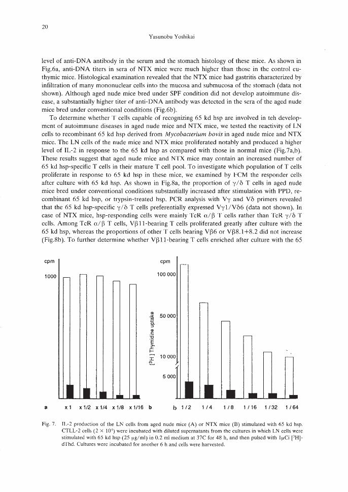
level of anti-DNA antibody in the serum and the stomach histology of these mice. As shown inFig.6a, anti-DNA titers in sera of NTX mice were much higher than those in the control euthymic mice. Histological examination revealed that the NTX mice had gastritis characterized byinfiltration of many mononuclear cells into the mucosa and submucosa of the stomach (data notshown). Although aged nude mice bred under SPF condition did not develop autoimmune disease, a substantially higher titer of anti-DNA antibody was detected in the sera of the aged nudemice bred under conventional conditions (Fig.6b).
To determine whether T cells capable of recognizing 65 kd hsp are involved in teh development of autoimmune diseases in aged nude mice and NTX mice, we tested the reactivity of LNcells to recombinant 65 kd hsp derived from Mycobacterium bovis in aged nude mice and NTXmice. The LN cells of the nude mice and NTX mice proliferated notably and produced a higherlevel of IL-2 in response to the 65 kd hsp as compared with those in normal mice (Fig.7a,b).These results suggest that aged nude mice and NTX mice may contain an increased number of65 kd hsp-specific T cells in their mature T cell pool. To investigate which population of T cellsproliferate in response to 65 kd hsp in these mice, we examined by FCM the responder cellsafter culture with 65 kd hsp. As shown in Fig.8a, the proportion of y/ 6 T cells in aged nudemice bred under conventional conditions substantially increased after stimulation with PPD, recombinant 65 kd hsp, or trypsin-treated hsp. PCR analysis with Vy and V6 primers revealedthat the 65 kd hsp-specific y/6 T cells preferentially expressed Vyl/VM (data not shown). Incase of NTX mice, hsp-responding cells were mainly TcR a/ ~ T cells rather than TcR y/ 6 Tcells. Among TcR a/ ~ T cells, V~ ll-bearing T cells proliferated greatly after culture with the65 kd hsp, whereas the proportions of other T cells bearing V~6 or V~8.1+8.2 did not increase(Fig.8b). To further determine whether V~ ll-bearing T cells enriched after culture with the 65
1000 .--
cpm
8
cpm
-
CI>50000:,t;
<1Ja.:>Q>c'0'E>-
.<:f-
I 10000c::o...
5000
~L~lJI •• ••xl x 1/2 x 1/4 x 1/8 x 1/16 b b 1/2 1 /4 1 /8 1 /16 1/32 1/64
Fig. 7. IL-2 production of the LN cells from aged nude mice (A) or NTX mice (B) stimulated with 65 kd hsp.CfLL-2 cells (2 x 10⁴
) were incubated with diluted supernatants from the cultures in which LN cells werestimulated with 65 kd hsp (25 f.lg/ml) in 0.2 ml medium at 37C for 48 h, and then pulsed with If.lCi [.1H]dThd. Cultures werc incubated for another 6 h and cells were harvested.

21
HSP-SPECIFIC T CELLS IN HOST DEFENSE
0.0%
Before culture
BALB / c
1,~W~~"-""--I"""'--~"""6""'" ~~
Aged nu / nu
!fi ;After culture PPD 2.8% 42.5%
~ mn
~ ...... .t • I I .OJ 16' ..
hsp 'I ;\ 14.1 %
:~ ,,~..~
Tryp-hsp7.3%
Ii. I •
(. )
a
'1/\ 0.1 %
:~\-'.-.,.~-Fig. 8. (A) Responsiveness of normal BALB/c (a) or aged nude mice (b) y/b T cells to PPD or mycobacterial
hsp. LN cells from aged nude mice were cultured with PPD (30 ~g/ml), recombinant 65 kd of M.bovis(25~g/ml) or trypsin-digested (Try-hsp) hsp (25~g/ml) for 3 days. Cultured cells were stained with antiTcR y/6 mAb and with anti-Thy-1.2. Thy-1.2- cells were gated out and shown are histograms ofTcR y/6 expression of Thy-1.2+ cells.

22
Yasunobu Yoshikai
Before culture
1.P
After culture with hsp
3.4
b
12.6
16.3
24.4
Fig. 8. (8) Activation of VIHI-bearing T cells in NTX mice by 65 kd hsp. The LN cells (l x IOt'/ml) fromNTX mice were cultured with the 65 kd hsp at 25 ~lg/ml for three days. Cultured cells were stained withanti-Vp mAb, anti-VB6 mAb, anti-VB8.1 +8.2 mAb, or anti-VI)11 mAb plus FlTC-anti-hamster or antirat IgG and PE-anti-Thy-1.2 mAb, FITC-anti-CD8 mAb, PE-anti-CD4 mAb.

23
HSP-SPECIFIC T CELLS IN HOST DEFENSE
kd hsp were really in an activated state, the size of the V[311-bearing T cells was assessed bytheir forward light scatter profile. Approximately half of the VB II-bearing T cells were blastoidcells, whereas VB8.1+8. 2-bearing T cells were small. (data not shown). Thus, VB II-bearing Tcel1s may proliferate substantially after culture with 65 kd hsp.
A notable finding in the present sutdy was that the aged nude mice and NTX mice that developed autoimmune diseases contained a large number of 65 kd hsp-specific T cells in theirperipheral lymphoid tissues. In aged nude mice, the hsp-responding T cells are mainly VyllVM-bearing T cells, whereas self-reactive VBll-bearing T cells in NTX mice increased in number after culture with the 65 kd hsp. Both T cel1s seem to produce a high level of IL-2 in response to 65 kd hsp. Hsp are polypeptides phylogenetical1y conserved between prokaryotes andeukaryotes. 2) We speculate that the 65 kd hsp-specific T cells may be activated by hsp expressedby bacteria and autologous cells stressed with various pathogens, resulting in the development ofautoimmune diseases in such animals.
Several lines of evidence have shown that tolerance to self-antigens is mainly due to intrathymic clonal deletion of self-reactive T cells.1IU1 ) Our results reveal that a substantial number ofself-reactive T cells such as VB II-bearing T cells were present in the LN of nude mice and NTXmice. These self-reactive T cells can respond normally to signals via the TcR in the presence ofexogenous IL-2. Our results also revealed that the self-reactive VB 11-T cells in NTX mice proliferate substantially after culture with 65 kd hsp. These T cells expressed a high level of IL-2Raeven before stimulation. 28 ,29) Therefore it is possible that hsp-reactive T cells other than VB 11bearing T cells may respond to the 65 kd hsp and subsequently produce a stimulus such as IL-2for proliferation of the self-reactive VB II-bearing T cells. In case of aged nude mice, hspresponding cells are found mainly in y/b T cells expressing VyllVM. At present, we can notdetect any increase in the number of particular T cells including y I 6 T cells other than VB 11bearing T cells in the NTX mice. Recently we have found that an autoreactive T cel1line specificfor self MHC class II recognized 65 kg hsp.3h) Therefore, it is alternatively possible that VB 11bearing T cells in NTX mice can directly recognize the 65 kd hsp in the context of a self MHCmolecule. The reactivity of VB II-bearing T cel1s to the 65 kd hsp should be determined at aclonal level to clarity this possibility.
The self-reactive T cel1s are rendered tolerant mainly by clonal energy and clonal suppressionvia suppressor cells. 37,38) although recent studies reveal that clonal deletion occurs extrathymically.39.41) It has been reported that autoimmune diseases in NTX mice were clearly preventedby the reconstitution of these mice with T cells from syngeneic, normal animals. A deficit ofsuppressor T cells may be a cause of autoimmune disease in NTX mice.42) The high reactivity ofT cells to the 65 kd hsp in NTX mice may result from depletion of a specific T cell subpopulation responsible for checking and controlling the hsp-specific T cells which is normally presnetin the mature T cell pool in euthymic mice. An increase in the number of 65 kd hsp-specific Tcel1s was evident in athymic nude mice bred under conventional conditions. Environmental antigens such as those of intestinal microflora may also play an important role in the expansion ofthe hsp-specific T cells in aged nude and NTX mice.
CONCLUDING REMARKS
Our results presented here suggest that 65 kd hsp-specific T cel1s play an importnat role notonly in induction of autoimmune diseases but also in the host defense mechanism against infection by various pathogens. During infection with L.monocytogenes, TcR y/6 T cells specific for65 kd hsp precede TcR alB T cells specific for the listerial antigen in appearance. The y/6 T

24
Yasunobu Yoshikai
~\.i
cytokines
IL-2
------~~~ autoimmunity
Ag unresponsiveness Ag responsiveness
Fig. 9. Model of roles of hsp-specific T cells in host defense against bacteiral infection and induction of autoimmune disease.
cells seem to provide a first line of defense against the infection by recognizing exogenous andendogenous 65 kd hsp on infected cells and producing cytokines such as yIFN. Thus, the hspspecfic T cells may respond quickly to antigenically diverse pathogens before antigen-specific Tcells expand clonally, and they may playa role in covering the gap between the phagocytic system and the highly evolved type of immune responses (Fig.9).4J.4.l)
Athymic mice such as nude mice and NTX mice contain an appreciable number of self-reactive T cells that have not undergone inrathymic clonal deletion. The self-reactive T cells expressIL2 receptor and respond normally to signals delivered through the TcR in the presence ofexogenous IL2. Notably, a substantially higher level of hsp reactivity was observed in the LNcells in such animals, especially those bred under conventional conditions. In aged nude mice,y/ b T cells greatly proliferated and produced IL2 in response to 65 kd hsp. On the other hand,the self-reactive V~ll-bearing T cells notably proliferated and a high level of IL-2 was detectedafter culture with 65 kd hsp in the NTX mice. Both groups of animals spontaneously developedautoimmune diseases. Taking all the previous results into consideration, we speculate that 65 kdhsp-specific T cells may abrogate the unresponsiveness of the self-reactive T cells and/or B cellsby producting IL-2 and that they may play an important role in the induction to autoimmunediseases (Fig.9).

25
HSP-SPECIFIC T CELLS IN HOST DEFENSE
REFERENCES
1) Craig, E.A.: The heat shock resonse. CRe. Crit. Rev. Biochem., 18,239-280 (1985).2) Jindal, S., Dudani, A.K., Harley, e.B., Singh, B. and Gupta, RS.: Primary structure of a human mitochon
drial protein homologous to the bacterial and plant chaperonins and to the 65-kilodalton mycobacterialantigen. Mol. Cell BioI., 9, 2279-2283 (1989).
3) Kaufman, S.H.E., Schoel, B., Wand-WurUenberger, A., Steinhof, U., Munk, M.E. and Koga, T.: T-cells,stress proteins and pathogenesis of mycobacterial infection. Curro Topics. Microbiol. Immunol., 155,125-141 (1990).
4) Lindquist, S.: The heat-shock response. Ann. Rev. Biochem., 55, 1151-1191 (1986).5) Young, R.A., and Elliot, T.J.: Stress proteins, infection and immune surveillance. Cell, 59, 5-8 (1989).6) van Eden. W., Thole, J.E.R., van der Zee, R., Noordzij, A., van Embden, J.D.A., Hensen, EJ. and Choen,
I.R: Cloning of the mycobacterial epitope recognized by T lymphocytes in adjuvant arthritis. Nature, 331,171-173 (1988).
7) Holoshitz, J. Drucker, I., Yaretzky, A., van Eden, W., Klaiman A., Lapidoz, Z., Frenkel, A. and Cohen, I.R:T lymphocytes of rheumatoid arthritis patients show reactivity to a fraction of mycobacteria cross-reactivewith cartilago. Lancet, 2, 305-309 (1986).
8) Young, D., Lathigra, R, Hendrix, R., Sweetser, D. and Young, R.A.: Stress proteins are immune target in leprosy and tuberculosis. Proc. Natl. Acad. Sci. USA, 85, 4267-4270 (1988).
9) Bron, W., Hall, L., Dallas, A., Boymel, J., Shinnick, T., Young, D., Brennan, P. and 0 Brien, R: Recognition of a peptide antigen by heat-shock reactive y/o T lymphocytes. Science, 241, 67-69 (1990).
10) Haregewoin, A., Soman, G., Hom, Re. and Finburg, R.W.: Human y/o+ T cells respond to mycobacterialheat-shock protoein. Nature, 340, 309-312 (1989).
11) Holoshitz, J., Koning, F., Coligan, J.E., DeBruyn, J., and Strober, S.: Isolation of CD4-CD8- mycobacteriareactive T lymphocyte clone from rheumatoid arthritis synovial fluid. Nature, 339,226-229 (1989).
12) Rejasekar, R, Sim, G.K. and Augustin, A.: Self heat shock and y/o T-cell reactivity. Proc. Natl. A cad. Sci.USA, 87,1767-1771 (1990).
13) Kaufman, S.H.E.: Immunity against intracelluar bacteria: Biological effector functions and antigen specificityofT lymphocytes. Curro Topics. Microbiol. Immunol., 138, 142-176 (1988).
14) Happ, M.P., Kubo, RT., Palmer, E., Born, W.K. and 0 Brien, R.L.: Limited receptor repertoire in a mycobacteria-reactive subset of y/o T lymphocytes. Nature, 342, 696-698 (1989).
15) Kaufman, S.H.E. and Hahn, H.: Biological functions of T cell lines with specificity for the intracelluar bacterium Listeria monocytogenes in vitro and in vivo. J. Exp. Med., 155, 1754-1765 (1982).
16) Mackaness, G.B.: The monocyte in celluar immunology. Semn. Heamtol., 7, 172-184 (1970).17) Mitsuyama, M., Takeya, K., Nomoto, K. and Shimotori, S.: Three phase of phagocyte contribution to resist
ance against Listeria monocytogenes. J. Gen. Microbiol., 106, 165-171 (1978).18) 0 Brien, RL. Happ, M.P., Dallas, A., Palmer, E., Kubo, R and Born, W.K.: Stimulation of a major subset
of lymphocytes expressing T cell receptor y/o by an antigen derived from Mycobacterium tuberculosis, Cell,57,667-674 (1989).
19) Inoue, T., Yoshikai, Y., Matsuzaki, G. and Nomoto, K.: Early appearing y/O-bearing T cells during infectionwith Bacillus Calmette Guerin. J. Immunol., 146,2754-2762 (1991).
20) Fry, AM., Jones, L.A., Kruisbeek, A.M. and Matis, L.A.: Thymic requirement for clonal deletion during Tcell development. Science, 246, 1044-1047 (1989).
21) Hodes, RJ., Sharrow, S.O., and Solomon, A.: Failure ofT cell receptor VI3 negative selection in an athymicenvironment, Science, 246, 1041-1043 (1989).
22) Hunig, T.: T-cell function and specificity in athymic mice. Immunol. Today, 4, 84-87 (1983).23) Kishihara, K., Yoshikai, Y., Matsuzaki, G., Mak, T.W. and Nomoto, K.: Fucntional a and 13 chain T cell re
ceptor messages can be detected in old but not in young athymic mice. Eur. J. Immunol., 17, 477-482(1987).
24) Smith, H., Chen, I.M., Kubo, R. and Tung, K.S.K.: Neonatal thymectomy results in a repertoire enriched inT cells depleted in adult thymus. Science, 245, 749-752 (1989).
25) Yoshikai, Y., Reis, M. and Mak, T.W.: Athymic mice express a higher level of functional y chain but greatlyreduced level of y and 0 chain T cell receptor messages. Nature, 324,482-485 (1986).
26) Monier, J.e. and Sepetjian, M.: Annu. Immunol. (Inst Pasteur), 126C, 63-75 (1975).27) Taguchi, O. and Nishizuka, Y.: Self tolerance and localized autoimmunity. J. Exp. Med., 165, 146-156
(1987).

26
Yasunobu Yoshikai
28) Iwasaki, A., Yoshikai, Y., Yuuki, H., Takimoto, H. and Nomoto, K.: Self-reactive T cells are activated by the65 kDa mycobacterial heat-shock protein in neonatally thymectomized mice. Eur. J. Immunol., 21, 597-603(1991).
29) Yuuki, H., Yoshikai, Y., Kishihara, K., Matsuzaki, G., Iwasaki, A., Takimoto, H. and Nomoto, K.: Clonalagergy in self-reactive ul ~ T cells is abrogated by heat shock protein-reactive y/ & T cells in aged athymicnude mice. Eur. J. Immunol., 20,1475-1482 (1990).
30) Blackman, M., Kappler, J. and Marrack, P.: The role of the T cell receptor in positive and negative selectionof developing T cells. Science, 248, 1335-1341 (1990).
31) Ramsdell, F. and Fowlkes, B.1.: Clonal deletion versus clonal agergy: the role of the thymus in inducing selftolerance. Science, 248,1342-1348 (1990).
32) Dyson, P.1., Knight, A.M., Fairchild, S., Simpson, E. and Tomonari, K.: Genes encoding ligands for deletionof V~l1 T cells cosegregate with mammary tumor virus genomes. Nature, 349, 531-532 (1991).
33) Frankel, W.N., Rudy, C, Coffin, J.M. and Huber, B.T.: Linkage of Mis genes to endogenous mammarytumor viruses of inbred mice. Nature, 349, 526-528 (1991).
34) Matsuzaki, G., Yoshikai, Y, Ogimoto, M., Yuuki, Y, Kishihara, K., Ohga, S. and Nomoto, K.: T cells receptor V~ repertoire at early stage of T cell development in adult thymus. J. Immunol., 145,46-51 (1990).
35) Takimoto, H., Yoshikai, Y., Kishihara, K., Matsuzaki, G., Kuga H., Ohtani, T. and Nomoto, K.: Stimulationof all T cells bearing V~ 1, V~3, V~ 11 and V~ 12 by staphlococcal enterotoxin A. Eur. J. Immunol., 20,617-622 (1990).
36) Matsuzaki, G., Yoshikai, Y., Harada, M. and Nomoto K.: Autoreactive T cells from normal mice recognizemycobacterial 65 kd heat-shock protein from Mycobacterium bovis. Int. Immunol., 3, 215-220 (1991).
37) Nossal, G.1.V.: Cellular mechanism of immunological tolerance. Ann. Rev. Immunol., 1, 33-62 (1983).38) Schwartz, R.H.: A cell culture model for T lymphocyte clonal anergy. Science, 248, 1349-1356 (1990).39) Jones, L.A., Chin, T., Longo, D.L. and Kruisbeek, A.M.: Peripheral clonal elimination of functional T cells.
Science, 250, 1726-1728 (1990).40) Kawabe, Y, and Ochi, A.: Selective anergy of V~8+, CD4+ T cells in staphylococcus enterotoxin B-primed
mice. J. Exp. Med., 172, 1065-1075 (1990).41) Rellahan, B.L., Jones, L.A., Kruisbeek, A.M., Fry, A.M. and Matis, L.A.: In vivo induction of anergy in pe
ripheral V~8+ T cells by staphylococcal enterotoxin B. J. Exp. Med., 172, 1091-1099 (1990).42) Sakaguchi, S., Takahashi, T. and Nishizuka, Y: Study on cellular events in post-thymectomy autoimmune
oophoritis in mice II Requirement of Lyt-1 cells in normal female mice for the prevention of oophoritis. J.Exp. Med. 156, 1577-1586 (1982).
43) Ohga, S., Yoshikai, Y., Takeda, Y., Hiromatsu, K. and Nomoto, K.: Sequential appearance of y/&- andu/~-bearingT cells in the peritoneal cavity during an i.p. infection with Listeria monocytgenes. Eur. J. Immunol., 20, 533-538 (1990).
44) Hiromatsu, K., Yoshikai, Y, Ohga, S., Muramori, K., Matsuzaki, G. and Nomot, K.: A protective role of y&T cells in listerial infection. J. Exp. Med., 75,49-56 (1992).

![Heat Shock Protein HSP101 Affects the Release of … · Heat Shock Protein HSP101 Affects the Release of Ribosomal Protein mRNAs for Recovery after Heat Shock1[OPEN] Rémy Merret2*,](https://img.pdfslide.net/doc/110x75/5bbb60e709d3f241268cd182/heat-shock-protein-hsp101-affects-the-release-of-heat-shock-protein-hsp101-affects.jpg)