Embed Size (px)
Citation preview
J Wood Sci (2000) 46:1-7 �9 The Japan Wood Research Society 2000
Kei'ichi Baba �9 Takayuki Asada �9 Takahisa Hayashi
Relation between developmental changes on anatomical structure and on protein pattern in differentiating xylem of tension wood
Received: September 24, 1998 / Accepted: March 30, 1999
Abstract Tension wood was induced in Eucalyptus camaldulensis L. by fixing the stem at an angle. Proteins in the differentiating tissue of tension wood were compared to those of normal wood on sodium dodecyl sulfate polyacry- lamide gel electrophoresis. An obvious difference was found in the salt-soluble fraction of 14 days after inclination. At least five bands (19, 22, 37, 41, and 55 kDa) were specific in the differentiating tissue of tension wood. These proteins were bound to the cell wall, plasma membrane, or both by their electric charge; they were undetectable until 14 days after inclination. Mature tension wood was observed in the tissue at 14 days. Thus, all differentiating tissue at 14 days was produced after inclination. On the other hand, the dif- ferentiating zone at 7 days contained the same tissue, as in tension wood estimated by the vessel number and diameter during the early phase; and the tissue was indistinguishable from normal wood during the late phase. The proteins found here were related to the phenomenon occurring in the late stage of xylem differentiation.
Key words Cell wall �9 Eucalyptus �9 Tension wood �9 Pro- tein �9 Xylem
Introduction
Tension wood is a reaction wood formed in the upper side of a leaning stem of angiosperm. ~4 Reaction wood causes the stem to bend upward by its mechanical action. Tension wood formation is mainly stimulated by a change of gravity direction. 34 However, many black boxes still exist in the
K. Baba (9) �9 T. Hayashi Wood Research Institute, Kyoto University, Uji, Kyoto 611-0011, Japan Tel. +81-774-38-3633; Fax +81(774)-38-3635 e-mail: [email protected]
T. Asada Forest Research Institute, Kameyama Research Center, Oji Paper Co. Ltd., Kameyama 519-02, Japan
cascade from the stimulus to tension wood formation. Phy- tohormones were investigated if they comprised a step of the cascade. First, it was hypothesized that a relatively low level of auxin would form tension wood, 6 but this did not accord with certain results. 7's In an early study, gibberellic acid (GA) was thought to have no effect, 9 but recently applied GA3 was found to induce tension wood. ~~ Ethylene was abundant in differentiating tension wood. 1~ The rela- tion between hormones and tension wood is still unclear, and other approaches are needed to clarify the mechanisms of tension wood formation.
The reason biochemical approaches have not progressed in this field are the chemical compositions of tension wood reported so far. Their differences from those in normal wood are quantitative. No one has reported any compound specific to tension wood, either present or absent. This fact suggests that we have not identified any enzyme that can reveal the mechanism. Moreover, mature xylem of tension wood has been well investigated by every technique, whereas immature tension wood has been studied by only an anatomical approach. In other words, the changes in cell wall compositions are well known, but nothing is known about cytoplasmic chemicals relating to tension wood for- mation. According to anatomical studies on differentiating tissues in tension wood, the angle of microtubule orienta- tion was found to be smaller than that of normal wood, 1~6 but no other structural change in the cytoplasmic compo- nents has been observed. 12'~3 In the present study, we com- pared the proteins of differentiating xylem of tension and normal wood on sodium dodecyl sulfate polyacrylamide gel electrophoresis (SDS-PAGE). The developmental changes were determined by anatomical techniques, and the relation between the anatomical and proteinaceous changes are discussed.
Eucalyptus camaldulensis L. was used for this study. Ten- sion wood of this species, which has been characterized chemically and anatomically, ~7 contains many typical char- acteristics. It consists of 1.8 times cellulose and 0.45 times lignin of normal wood. A G-layer occurs in the fibers and is classified as type S~ + G. The microfibrillar angle of the fibers is 3.5 ~ , whereas that of normal wood is 22.5 ~ .
Materials and methods
Plant materials
The main stems of 2-year-old E. carnaldulensis were in- clined 17 one by one at 14 days (August 19), 7 days (August 26), 4 days (August 4), and 1 day (September 1) before harvest on Sep tember 2. Each inclined stem and a vert ical stem were cut at the same date. Immature xylem on the upper side of the inclined stem was scraped immedia te ly with a knife b lade after bark peeling. These scrapings were placed in liquid ni t rogen (LN2) and s tored at - 8 0 ~ until use. The same tissue from all a round the s tem vertically was harvested in the same manner.
Blocks (approximate ly 2 • 2 • 2 cm) were cut from the stem before and after harvest, fixed in formalin/acetic acid/ e thanol (5:5:90) ( F F A ) and s tored until use.
Frac t ionat ion of p ro te in and S D S - P A G E
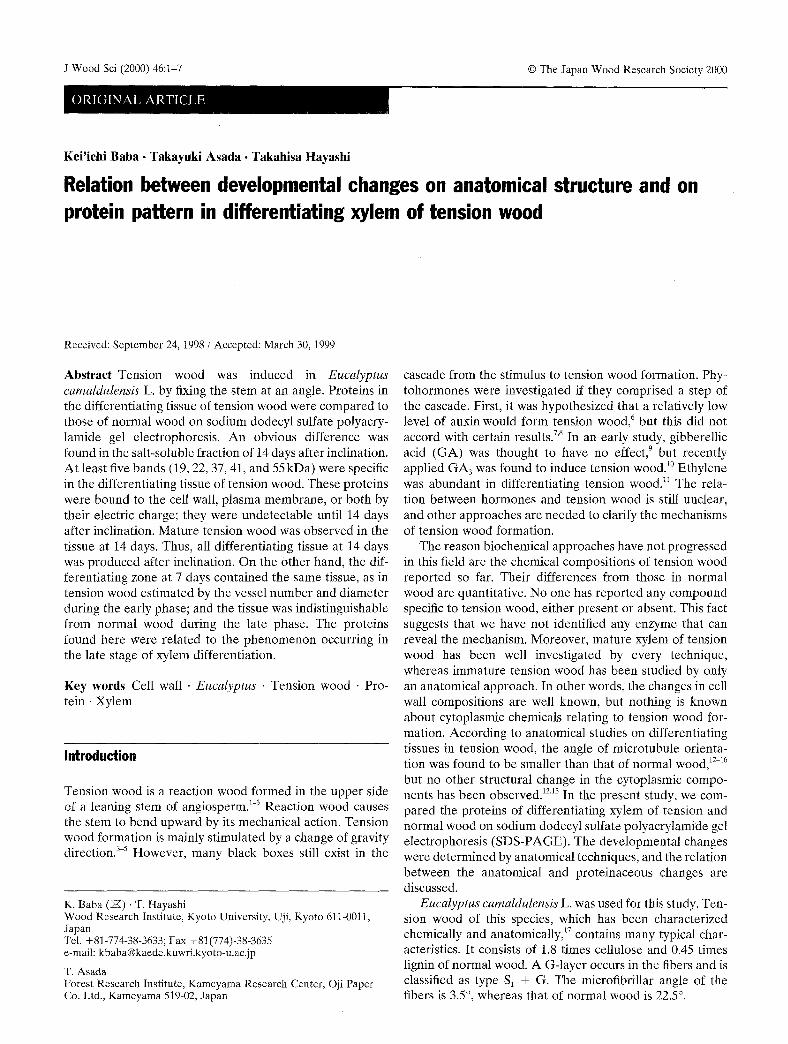
Proteins were f ract ionated through an extract ion (Fig. 1). The tissue (500-750 mg) s tored at - 8 0 ~ was ground in LN2
with a mor ta r and pestle. I t was suspended in t 0 0 m M Tris- HCI buffer (pH 6.8) containing l mM di thiothrei tol (DTT) and protease inhibitor mixture TM as 200rag F.W./ml at 0~ and centr ifuged at 3000g for 10min at 4~ to separate buffer-soluble [BS] and buffer- insoluble (BI)f rac t ions . The BS fraction was recentr i fuged at 100000g for 20min at 4~ The supernatant was concentra ted by ultrafi l trat ion using Centricon-10 (Amicon) as fraction 1. The precipi ta te was collected and used as fraction 2. The BI fraction was washed three times with the same solution as descr ibed above. Then it was suspended in Tr is-HCl-buffered 1M NaC1 and centri- fuged at 10000g for 10min at 4~ The supernatant was concentra ted by ultrafiltration. Tris-HC1 was then added to the concentrate and reultrafi l tered: it was used as fraction 3. The precipi ta te was washed with Tr is -HCl-buffered 1M NaC1 three t imes and Tris-HC1 once for use as fraction 4. Al l the fractions (1, 2 . 3 . 4 ) were suspended in 4% SDS in Tris-HC1. and the pro te in levels were measured with Micro B C A (Pierce). Urea and ~-mercap toe thanot were then added at 8M and 5%. respectively, in final concentra t ion and boiled for 5min. The prote in of each fraction was e lec t rophoresed in a 5 % - 2 0 % gradient gel ( A T T O ) in equal amounts using the discontinuous buffer system of Laemmli . 19 The proteins were visualized with silver staining.
Differentiating Tissue of Xylem
- Extraction with Tris-HCI buffer
- Centrifugation at 3,000"g
sup. Buffer Soluble
- Centrifugation
at lO0,O00"g
I ppt. Buffer Insoluble
Extraction with 1 M NaCI
- - Centrifugation
at lO,O00.g
I I sup. ppt. sup. ppt.
I I I I Frac. 1 Frac. 2 Frac. 3 Frac. 4
Fig. 1. Summary of fractionation scheme of proteins from differentiat- ing xylem tissue. Sup.i supernatant; ppt., precipitate; Frac., fraction
Microscopy
Cross sections 30~tm thick were made from the blocks s tored in F A A . The sections were stained with safranin, dehydra ted with e thanol series, subst i tuted by xyiene, and mounted on glass slides with Bioleit (Oken). The sec- tions were observed under normal light, polarized, and epifluorescence microscopes with blue excitat ion (410rim). Some sections were reacted with phtoroglucinol a n d ob- served by light microscopy.
Results and discussion
Dete rmina t ion of the differentiat ing stage of the harvested tissue
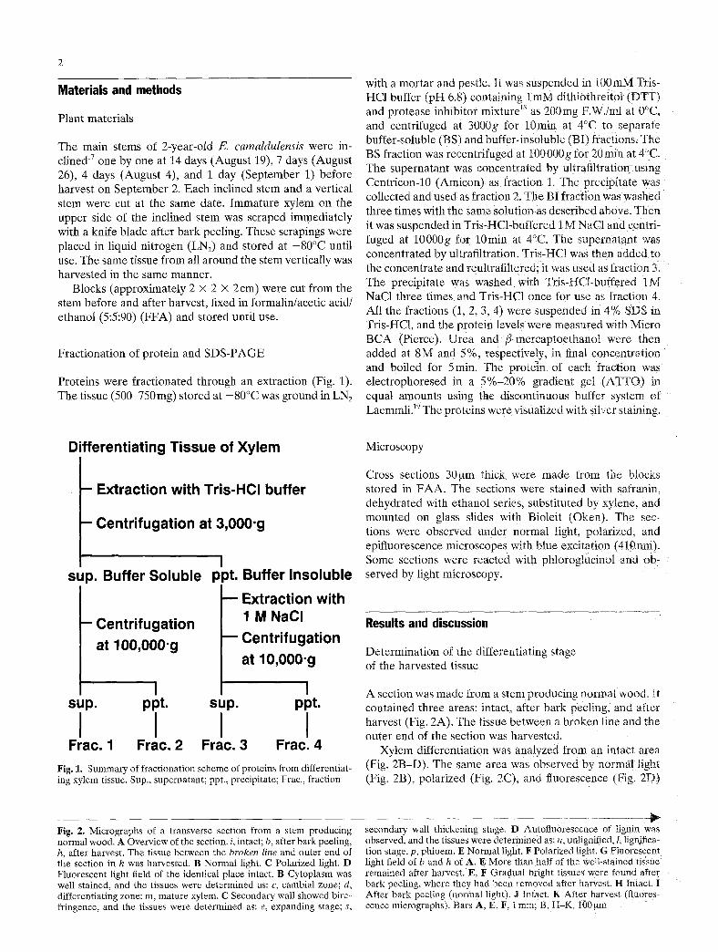
A section was made from a stem producing normal wood. It contained three areas: intact, after ba rk peeling, and after harvest (Fig. 2A). The tissue be tween a b roken line and the outer end of the section was harvested.
Xylem differentiat ion was analyzed from an intact area (Fig. 2B-D) : The same area was observed by normal light (Fig. 2B), polar ized (Fig. 2C), and fluorescence (Fig. 2D)
Fig. 2. Micrographs of a transverse section from a stem producing normal wood. A Overview of the section, i, intact; b, after bark peeling, h, after harvest. The tissue between the broken line and outer end of the section in h was harvested. B Normal light. C Polarized light. D Fluorescent light field of the identical place intact. B Cytoplasm was well stained, and the tissues were determined as: c, cambial zone; d, differentiating zone: m, mature xylem. C Secondary wall showed bire- fringence, and the tissues were determined as: e, expanding stage; s,
secondary wall thickening stage. D Autoflu0rescence of iignin was observed, and the tissues were determined as: u, unlignified, l, lignifiea- tion stage, p, phloem. E Normal light. F Polarized light. G Fluorescent light field of b and h of A. E More than half of the we!l-stained tissuel remained after harvest. E, F Gradual bright tissues were found after bark peeling, where they had been removed after harvest. H Intactl I After bark peeling (normal light)~ J Intact. K After harvest (fluores- cence micrographs). Bars A, E, F; i ram; B, H-K, I00 ~tm
lure
4
microscopy. Differentiating tissue was darker than the ma- ture xylem in normal light because cytoplasm stained well. The cambial zone was also seen in the same field because cambial cells are small and have dense cytoplasm. The sec- ondary wall shows bright birefringence in a polarized field (Fig. 2C). Because dark tissue had primary wall only, the area next to the cambial zone was in the expansion stage. The tissue that had increased brightness in this field was at the stage of secondary wall thickening. Using fluorescence microscopy (Fig. 2D), the unlignified tissue was dark and the tissue of increased brightness was at the lignification stage. The tissue originating in the cambial zone differenti- ated through the stages of expansion, secondary wall thick- ening, and lignification, finally resulting mature wood.
The areas after bark peeling and after harvest were also observed in these three field (Fig. 2E-G). After bark peel- ing the tissue near the cambium cracked, and the outside was removed (Fig. 2E, left). The outer end of such tissue (rectangle in Fig. 2E) was magnified (Fig. 2I) and compared to intact tissue (Fig. 2H). The cells in the cambial zone were small and dense and made well-regulated radial files. Be- cause the cells around the outer end had these morphologi- cal properties, the cambial zone remained on the stem after bark peeling. The outer end after harvest, seen by fluores- cence microscopy (rectangle in Fig. 2G), was magnified (Fig. 2K). Compared to the intact area (Fig. 2J), the stem after harvest did not contain a gradual area of au- tofluorescence. According to these results, the harvested tissue contained a cambial zone and the differentiating stages of expansion, secondary wall thickening, and an early phase of lignification.
Comparing the proteins of normal and tension wood formation
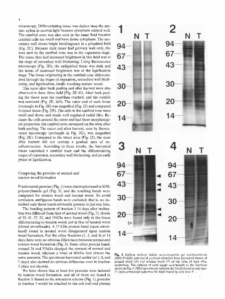
Fractionated proteins (Fig. 1) were electrophoresed in SDS- polyacrylamide gel (Fig. 3), and the resulting bands were compared for tension wood and normal wood. To avoid confusion, ambiguous bands were excluded; that is, we de- scribed only those bands obviously present in just one lane.
The banding pattern of fraction 3 14 days after inclina- tion was different from that of normal wood (Fig. 3). Bands of 55, 41, 37, 22, and 19kDa were found only in the tissue differentiating to tension wood, not in that of normal wood (closed arrowheads). A 17-kDa protein band (open arrow- head) found in normal wood disappeared upon tension wood formation. For the other fractions (1, 2, and 4) at 14 days there were no obvious differences between normal and tension wood formation (Fig. 3). Some other protein bands around 28 and 33kDa changed the amount of normal and tension wood, whereas a band at 80kDa had almost the same amounts. The specimens harvested earlier (at 1, 4, and 7 days) also showed no obvious difference even in fraction 3 (data not shown).
We have shown that at least five proteins were induced by tension wood formation, and all of them are found in fraction 3. Based on the extraction scheme (Fig. 1), proteins in fraction 3 would be attached to the cell wall and plasma
Fig. 3. Sodium dodecyt sulfate potyacrylamide gel eiectrophoresis (SDS-PAGE) patterns of proteins extracted from harvested tissues of normal wood (N) and tension wood (T) of the s~em 14 days after inclination. The number of each image corresponds to the fractions shown in Fig. 4. Filled arrowheads indicate the bands found in only lane T. Open arrowhead indicates the band found in only tane N
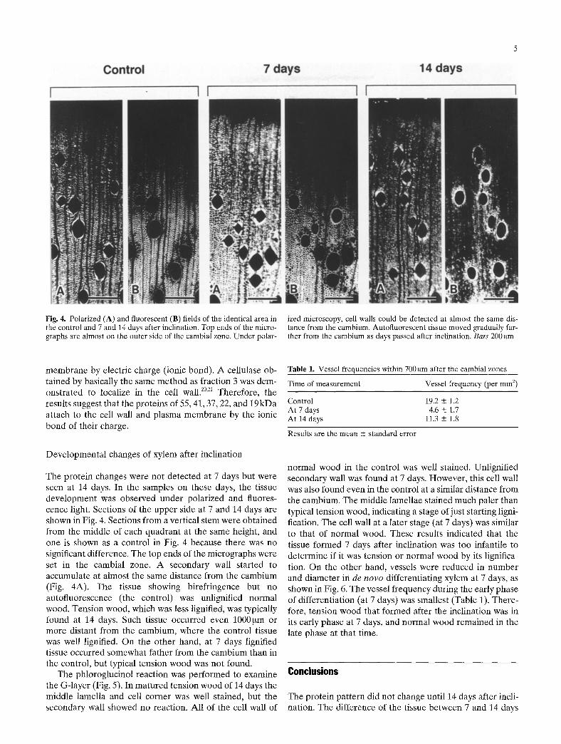
Fig. 4. Polarized (A) and fluorescent (B) fields of the identical area in the control and 7 and 14 days after inclination. Top ends of the micro- graphs are almost on the outer side of the cambial zone. Under polar-
ized microscopy, cell walls could be detected at almost the same dis- tance from the cambium. Autoftuorescent tissue moved gradually far- ther from the cambium as days passed after inclination. Bars 200 gm
membrane by electric charge (ionic bond). A cellulase ob- tained by basically the same method as fraction 3 was dem- onstrated to localize in the cell wall. z~ Therefore, the results suggest that the proteins of 55, 41, 37, 22, and 19kDa attach to the cell wall and plasma membrane by the ionic bond of their charge.
Developmental changes of xylem after inclination
The protein changes were not detected at 7 days but were seen at 14 days. In the samples on these days, the tissue development was observed under polarized and fluores- cence light. Sections of the upper side at 7 and 14 days are shown in Fig. 4. Sections from a vertical stem were obtained from the middle of each quadrant at the same height, and one is shown as a control in Fig. 4 because there was no significant difference, The top ends of the micrographs were set in the cambial zone. A secondary wall started to accumulate at almost the same distance from the cambium (Fig. 4A). The tissue showing birefringence but no autofluorescence (the control) was unlignified normal wood. Tension wood, which was less lignified, was typically found at 14 days. Such tissue occurred even 1000~tm or more distant from the cambium, where the control tissue was well lignified. On the other hand, at 7 days lignified tissue occurred somewhat father from the cambium than in the control, but typical tension wood was not found.
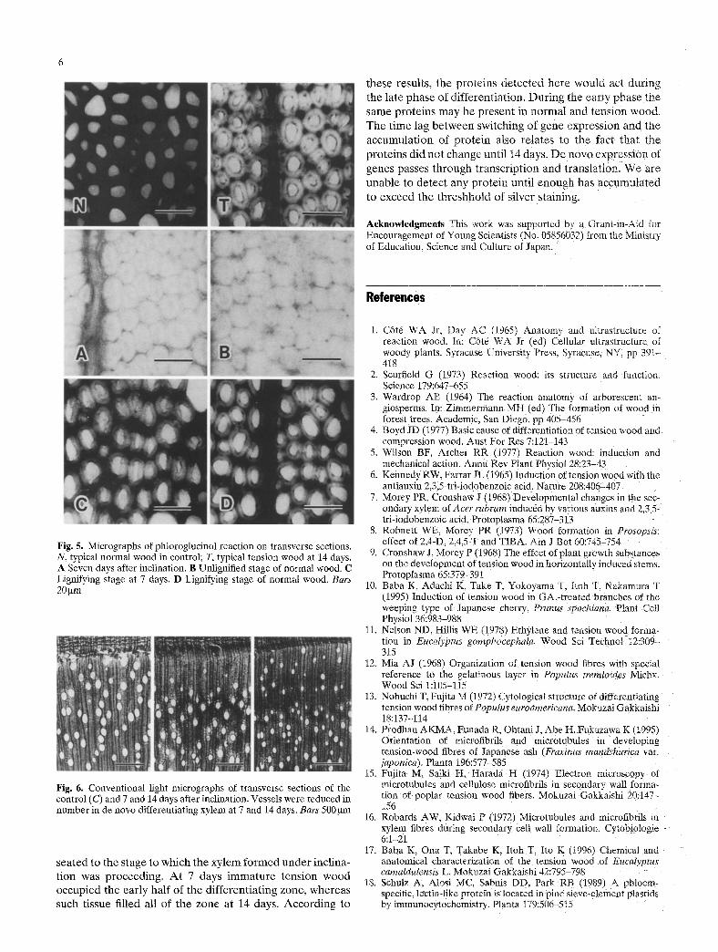
The phloroglucinol reaction was performed to examine the G-layer (Fig. 5). In matured tension wood of 14 days the middle lamella and cell corner was well stained, but the secondary wall showed no reaction. All of the cell wall of
Table 1. Vessel frequencies within 700 gm after the cambial zones
Time of measurement Vessel frequency (per mm 2)
Control 19.2 • 1.2 At 7 days 4.6 • 1.7 At 14 days 11.3 -- 1.8
Results are the mean • standard error
normal wood in the control was well stained. Unlignified secondary wall was found at 7 days. However, this cell wall was also found even in the control at a similar distance from the cambium. The middle lamellae stained much paler than typical tension wood, indicating a stage of just starting ligni- fication. The cell wall at a later stage (at 7 days) was similar to that of normal wood. These results indicated that the tissue formed 7 days after inclination was too infantile to determine if it was tension or normal wood by its lignifica- tion. On the other hand, vessels were reduced in number and diameter in de n o v o differentiating xylem at 7 days, as shown in Fig. 6. The vessel frequency during the early phase of differentiation (at 7 days) was smallest (Table 1). There- fore, tension wood that formed after the inclination was in its early phase at 7 days, and normal wood remained in the late phase at that time.
Conclusions
The protein pattern did not change until 14 days after incli- nation. The difference of the tissue between 7 and 14 days
these results, the p ro te ins d e t e c t e d he re wou ld act dur ing the late phase of d i f ferent ia t ion , D u r i n g the ear ly phase the
same prote ins m a y be p re sen t in normal and tens ion wood . T h e t ime lag b e t w e e n switching of gene express ion and the
accumula t ion of p ro t e in also re la tes to the fact tha t the
p ro te ins did no t change unt i l 14 days. D e n o v o express ion of
genes passes t h rough t ranscr ip t ion and translat ion. W e are
unab le to de tec t any p ro t e in unti l e n o u g h has accumula t ed
to exceed the t h r e shho ld of s i lver staining,
Acknowledgments This work was supported by a Grant-in-Aid for Encouragement of Young Scientists ~ No. 05856032) from the Ministry of Education. Science and Culture of Japan.
References
Fig. 5. Micrographs of phtoroglucinol reaction on transverse sections. N, typical normal wood in control; T, typical tension wood at 14 days. A Seven days after inclination. B Unlignified stage of normal wood. C Lignifying stage at 7 days. D Lignifying stage of normal wood. Bars 20 ~m
Hg. 6. Conventional light micrographs of transverse sections of the control (C) and 7 and 14 days after inclination. Vessels were reduced in number in de novo differentiating xylem at 7 and 14 days. Bars 500 btm
sea ted to the s tage to which the xy l em f o r m e d u n d e r incl ina- t ion was p roceed ing . A t 7 days i m m a t u r e t ens ion w o o d occup ied the ear ly half of the d i f fe ren t ia t ing zone , whe reas such tissue filled all of the zone at 14 days. A c c o r d i n g to
1. C6t6 WA Jr. Day AC (1965) Anatomy and ultras~ructure of reaction wood. In: C6t6 WA Jr (ed) Cellular ultrastrucmre of woody plants. Syracuse University Press. Syracuse. NY. pp 391- 418
2. Scurfield G (1973/ Reaction wood: its structure and function. Science 179:647-655
3. Wardrop AB (1964) The reaction anatomy of arboresce~t an- giosperms. In: Zimrnermann MH (ed) The formation of wood in forest trees. Academic, San Diego, pp 405-456
4. Boyd JD (1977) Basic cause of differentiation of tension wood and compression wood. Aust For Res 7:121-143
5. Wilson BF. Archer RR (1977) Reaction wood: induction and mechanical action. Annu Rev Plant Physiol 28:23-43
6. KennedyRW. Farrar JL(1965) Induction oftension wood with the antiauxin 2,5.5-tri-iodobenzoic acid. Nature 208:406-407
7. Morey PR. Cronshaw J (1968) Developmental changes in the sec- ondary xylem of Acer rubrum induced by various auxins and 2.3.5- tri-iodobenzoic acido Protoplasma 65:287-313
8. Robnett WE. Morey PR (1973) Wood formation in Prosopsis: effect of 2.4-D. 2.4.5-T and TIBA. Am J Bot 60:745-754
9. Cronshaw J. Morey P (1968) The effect of plant growth substances on the development of tension wood in horizontally induced stems. Protoplasma 65:379-39i
10. Baba K. Adachi K. Take T. Yokoyama T. Itoh T. Nakamura T (1995) Induction of tension wood in GA3-treated branches of the weeping type of Japanese cherry, Prunus spachiana. Plant Cell Physiol 36:983-988
11. Nelson ND. Hillis WE (1978) Ethylene and tension wood forma- tion in Eucalyptus gomphocephala. Wood Sci Technol 12=309 315
12. Mia AJ (1968~ Organization of tension wood fibres with special reference to the gelatinous layer in Populus tremloides Michx. Wood Sci 1:105-115
13. Nobuchi T. Fujita M (1972) Cytological structure of differentiating tension wood fibres of Poputus euroamericana. Mokuzai Gakkaishi 18:137-114
14. Prodhan AKMA. Funada R. Ohtani J. Abe H. Fukazawa K (1995) Orientation of microfibrils and microtubules in developing tension-wood fibres of Japanese ash (Fraxinus mandshurica vat. japomca). Planta 196:577-585
15. Fujita M, Saiki H. Harada H (19741 Electron microscopy of microtubules and cellulose microfibrils in secondary wall forma- tion of poplar tension wood fibers. Mokuzai Gakkaishi 20:14% 156
16. Robards AW. Kidwai P (1972) Microtubules and microfibrils in xylem fibres during secondary cell wall formation. Cytobiologie 6:1-21
17. Baba K. Ona T, Takabe K. Itoh T. Ito K (1996) Chemical and anatomical characterization of the tension wood of Eucalyptus camaldulensis L. Mokuzai Gakkaishi 42:795-798
18. Schulz A. Alosi MC. Sabnis DD. Park RB (1989) A phloem- specific, lectin-like protein is located in pine sieve-element plastids by immunocytochemistry. Planta 179:506-5!5

19. Laemmli UK (1970) Cleavage of structural proteins during the assembly of the head of bacteriophage T4. Nature 227:680-685
20. Bal AK, Verma DPS, Byrne H, Maclachlan GA (1976) Subcellular localization of cellulases in auxin-treated pea. J Cell Bio169:97-105
7
21. Byrne H, Christou NV, Verma DPS, Maclachlan GA (1975) Purification and characterization of two cellulases from auxin treated pea epicotyl. J Biol Chem 250:1012-1018