Embed Size (px)
Citation preview
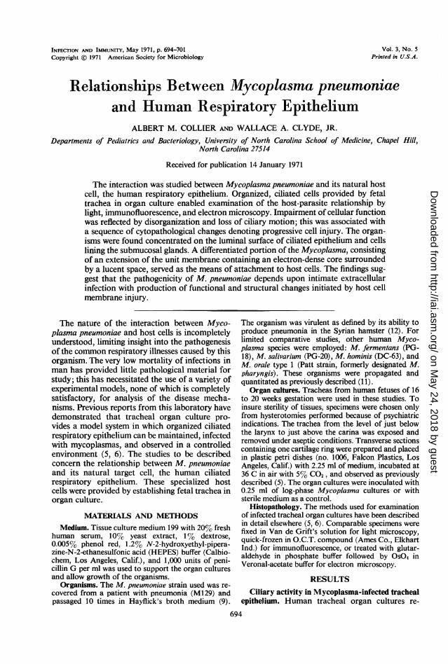
INFECTION AND IMMUNITY, May 1971, p. 694-701 Vol. 3, No. 5Copyright @ 1971 American Society for Microbiology Printed in U.S.A.
Relationships Between Mycoplasma pneumoniaeand Human Respiratory Epithelium
ALBERT M. COLLIER AND WALLACE A. CLYDE, JR.
Departments of Pediatrics and Bacteriology, University of North Carolina School of Medicine, Chapel Hill,North Carolina 27514
Received for publication 14 January 1971
The interaction was studied between Mycoplasma pneumoniae and its natural hostcell, the human respiratory epithelium. Organized, ciliated cells provided by fetaltrachea in organ culture enabled examination of the host-parasite relationship bylight, immunofluorescence, and electron microscopy. Impairment of cellular functionwas reflected by disorganization and loss of ciliary motion; this was associated witha sequence of cytopathological changes denoting progressive cell injury. The organ-isms were found concentrated on the luminal surface of ciliated epithelium and cellslining the submucosal glands. A differentiated portion of the Mycoplasma, consistingof an extension of the unit membrane containing an electron-dense core surroundedby a lucent space, served as the means of attachment to host cells. The findings sug-gest that the pathogenicity of M. pneumoniae depends upon intimate extracellularinfection with production of functional and structural changes initiated by host cellmembrane injury.
The nature of the interaction between Myco-plasma pneumoniae and host cells is incompletelyunderstood, limiting insight into the pathogenesisof the common respiratory illnesses caused by thisorganism. The very low mortality of infections inman has provided little pathological material forstudy; this has necessitated the use of a variety ofexperimental models, none of which is completelysatisfactory, for analysis of the disease mecha-nisms. Previous reports from this laboratory havedemonstrated that tracheal organ culture pro-vides a model system in which organized ciliatedrespiratory epithelium can be maintained, infectedwith mycoplasmas, and observed in a controlledenvironment (5, 6). The studies to be describedconcern the relationship between M. pneumoniaeand its natural target cell, the human ciliatedrespiratory epithelium. These specialized hostcells were provided by establishing fetal trachea inorgan culture.
MATERIALS AND METHODSMedium. Tissue culture medium 199 with 20% fresh
human serum, 10% yeast extract, 1% dextrose,0.005% phenol red, 1.2% N-2-hydroxyethyl-pipera-zine-N-2-ethanesulfonic acid (HEPES) buffer (Calbio-chem, Los Angeles, Calif.), and 1,000 units of peni-cillin G per ml was used to support the organ culturesand allow growth of the organisms.
Organisms. The M. pneumoniae strain used was re-covered from a patient with pneumonia (M129) andpassaged 10 times in Hayflick's broth medium (9).
The organism was virulent as defined by its ability toproduce pneumonia in the Syrian hamster (12). Forlimited comparative studies, other human Myco-plasma species were employed: M. fermentans (PG-18), M. salivarium (PG-20), M. hominis (DC-63), andM. orale type 1 (Patt strain, formerly designated M.pharyngis). These organisms were propagated andquantitated as previously described (11).
Organ cultures. Tracheas from human fetuses of 16to 20 weeks gestation were used in these studies. Toinsure sterility of tissues, specimens were chosen onlyfrom hysterotomies performed because of psychiatricindications. The trachea from the level of just belowthe larynx to just above the carina was exposed andremoved under aseptic conditions. Transverse sectionscontaining one cartilage ring were prepared and placedin plastic petri dishes (no. 1006, Falcon Plastics, LosAngeles, Calif.) with 2.25 ml of medium, incubated at36 C in air with 5% C02, and observed as previouslydescribed (5). The organ cultures were inoculated with0.25 ml of log-phase Mycoplasma cultures or withsterile medium as a control.
Histopathology. The methods used for examinationof infected tracheal organ cultures have been describedin detail elsewhere (5, 6). Comparable specimens werefixed in Van de Grift's solution for light microscopy,quick-frozen in O.C.T. compound (Ames Co., ElkhartInd.) for immunofluorescence, or treated with glutar-aldehyde in phosphate buffer followed by OS04 inVeronal-acetate buffer for electron microscopy.
RESULTSCiliary activity in Mycoplasma-infected tracheal
epithelium. Human tracheal organ cultures re-
694
on May 24, 2018 by guest
http://iai.asm.org/
Dow
nloaded from
M. PNEUMO0NIAE AND HUMAN HOST CELLS
mained viable, as indicated by continuation oforganized ciliary activity, as long as 4 weeks whenthe medium was changed every 4 days. Toexamine the effects of Mycoplasma infection,experiments were performed in which a series ofseparate organ cultures prepared with sectionsfrom the same trachea were inoculated withseveral human Mycoplasma species. Ciliary mo-tion was observed daily, and organisms werequantitated at the beginning and at the end of theobservation period (Table 1). Ciliary activity inthe cultures inoculated with M. pneumoniaeslowed after 3 days and was absent on day 4. Noapparent effect was seen in the organ culturesinoculated with M. hominis, M. salivarium, M.orale (type 1), M. fermentans, or in sterile controlcultures. In other experiments, no detectablechanges resulted from infection of tracheal sec-
tions with an avirulent M. pneumoniae strain (12),inoculation with heat-killed organisms, or addi-tion of sterile culture filtrates to the system. Thesefindings provide evidence that virulent M. pneu-
moniae is unique among the mycoplasmas testedin its ability to interfere with an important func-tion of human respiratory epithelium.
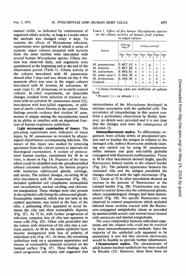
Light microscopic examination of tissues. Thepreceding experiments were indicative of tissueinjury by M. pneumoniae as revealed by physio-logical disturbances accompanying infection. Thenature of this injury was studied by removingspecimens from the culture system at intervals forhistological examination. A section of uninocu-lated human trachea, maintained for 48 hr invitro, is shown in Fig. IA. Features of the tissuewhich could be identified were the pseudostratifiedciliated columnar epithelium, the lamina propriawith numerous submucosal glands, cartilage,and serosa. The earliest changes, occurring 48 hrafter inoculation with M. pneumoniae (Fig. iB),included epithelial cell cytoplasmic eosinophiliaand vacuolization, nuclear swelling, and chroma-tin margination. These changes were also presentin the epithelial cells lining the submucosal glands.Eosinophilic material, which was not seen in thecontrol specimens, was noted at the base of thecilia; a palisading effect appeared to occur be-tween the cilia and these eosinophilic deposits(Fig. 1C). At 72 hr, with further progression ofinfection, complete loss of cilia was apparent inmany cells (Fig. 1D). Other cells protruded intothe lumen, with distortion of the cilia into a sun-
burst pattern. At 96 hr, the entire epithelial layerbecame disorganized with loss of polarity ofindividual cells (Fig. IE). At times, the remainingepithelium took on a squamous appearance andmasses of eosinophilic material occurred on theluminal surface (Fig. IF). These findings indi-cated progressive cell injury and suggested that
TABLE 1. Effect offive humant Mycoplasnia specieson the ciliary activity of humani fetal trachea
in organ culture
CFU/ml(C1lg,Oa Ciliary activitybSpecies
Day Day Day Day Day Day Day0 5 1 2 3 4 5
M. pneumoiiiae. 5.40 7.15 + + i - -M. hominis... 5.88 7.28 + + + + +M. salivarium. .. 6.88 6.81 + + + + +M. orale type L. 5.40 6.38 + + + + +M. fermentans... 6.18 6.38 + + + + +Control.........± + + + +
a Colony-forming units per milliliter of culturefluid.
b Present (+) or absent (-).
microcolonies of the Mycoplasma developed inintimate association with the epithelial cells. Theoccurrence of cytopathology in this system con-firms a preliminary observation by Butler; how-ever, no details were provided and it is not clearthat the changes seen were the consequence ofinfection (2).
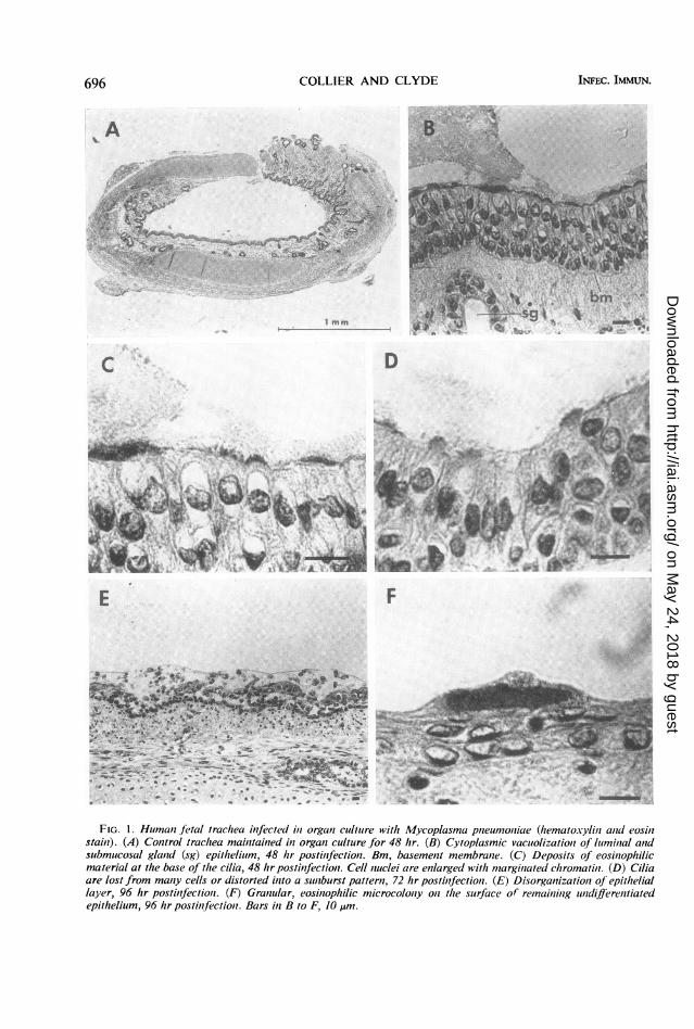
Immunofluorescent studies. To differentiate or-ganisms from cellular debris or precipitated pro-tein and to localize the antigen in relation to thedamaged cells, indirect fluorescent-antibody stain-ing was carried out by using M. pneumoniaerabbit antisera and goat anti-rabbit globulinconjugated with fluorescein isothiocyanate. Tissueat 48 hr after inoculation showed bright, specificfluorescence limited mainly to the ciliated border(Fig. 2A). The palisade appearance between theunstained cilia and the antigen paralleled thechanges observed with the light microscope (Fig.1C). Tissue at 72 hr after inoculation showed anincrease in the amount of fluorescence at theluminal border (Fig. 2B). Fluorescence was alsonoted to extend down into the submucosal glands,where cytopathological changes had been demon-strated (Fig. iB). No specific fluorescence wasobserved in control preparations which includedinfected tissue sections reacted with the fluores-cein-conjugated antiglobulin (alone or precededby normal rabbit serum) and normal tissue treatedwith antiserum and labeled antiglobulin.The exact relationship between individual orga-
nisms and the ciliated cells could not be resolvedby these immunofluorescence methods. Since themajority of the epithelial cells appeared to beparasitized, it was felt that electron microscopywould be feasible to provide further information.
Ultrastructural studies. The ultrastructure ofadult human tracheal epithelium has been studiedby Rhodin (15). However, there have been no
VOL. 3, 1971 695
on May 24, 2018 by guest
http://iai.asm.org/
Dow
nloaded from
COLLIER AND CLYDE
V -0.
N' m
1mm
D
C.
jiSil|
t%E.P..
F
O N
-_ s.~,-_t'
. s
#f . s i S
t us:}c SSs. . - cllw
z 4 <*.-ig0>..s:: w--< :ES.. * t k an--
F s-
.-.v
.-
*
FIG. 1. Humani fetal trachea infected in organi cultare with Mycoplasma pneun1oniae (hematoxylin and eosihlstain). (A) Control trachea maintained in organl cultutre for 48 hr. (B) Cytoplasmic vacuolizationz of luminal an2dsubmucosal gland (sg) epithelium, 48 hr postinfectioni. Bm, basement membrane. (C) Deposits of eosinophilicmaterial at the base of the cilia, 48 hr postinfection. Cell nuclei are enlarged with margilnated chromatini. (D) Ciliaare lost from many cells or distorted inzto a snlburst pattern, 72 hr postinfectiont. (E) Disorganizatioll of epitheliallayer, 96 hr postinfectioni. (F) Granzular, eosinophilic microcolony onz the surface of remainling indifferentiatedepithelium, 96 hr postinfectioni. Bars in B to F, 10 ,um.
INFEC. IMMUN.696
A
... ... -.11-;jj-.4:W2,*1A' .! .,06.4.1.
I .:%: --4.
4
on May 24, 2018 by guest
http://iai.asm.org/
Dow
nloaded from
M. PNEUMONIAE AND HUMAN HOST CELLS
FIG. 2. Human fetal trachea infected in organ culture with Mycoplasma pneumoniae and reacted with M. pneu-moniae antiserum (indirect immunofluorescent technique). (A) Specific fluorescence limited to ciliated epithelium(ce) 48 hr postinfection. Bm, basement membrane. (B) Increased fluorescence at the ciliated epithelial border andextending into the submucosal gland (sg), 72 hr postinfection. Bar, 20 ,um.
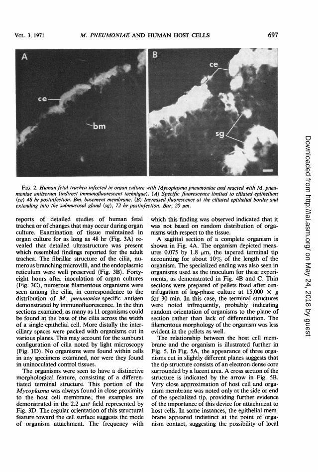
reports of detailed studies of human fetaltrachea or of changes that may occur during organculture. Examination of tissue maintained inorgan culture for as long as 48 hr (Fig. 3A) re-vealed that detailed ultrastructure was presentwhich resembled findings reported for the adulttrachea. The fibrillar structure of the cilia, nu-merous branching microvilli, and the endoplasmicreticulum were well preserved (Fig. 3B). Forty-eight hours after inoculation of organ cultures(Fig. 3C), numerous filamentous organisms wereseen among the cilia, in correspondence to thedistribution of M. pneumoniae-specific antigendemonstrated by immunofluorescence. In the thinsections examined, as many as 11 organisms couldbe found at the base of the cilia across the widthof a single epithelial cell. More distally the inter-ciliary spaces were packed with organisms cut invarious planes. This may account for the sunburstconfiguration of cilia noted by light microscopy(Fig. ID). No organisms were found within cellsin any specimens examined, nor were they foundin uninoculated control tissues.The organisms were seen to have a distinctive
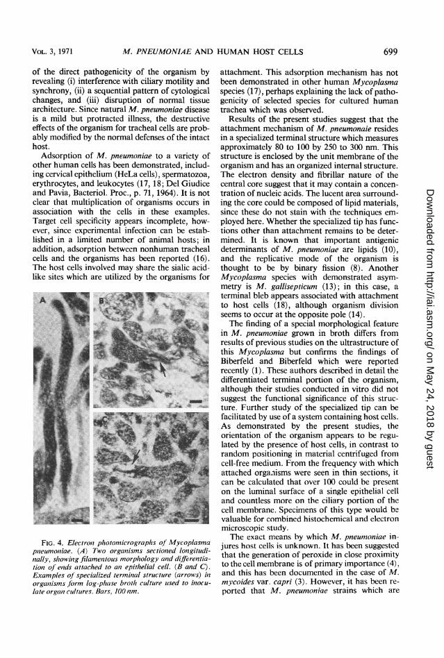
morphological feature, consisting of a differen-tiated terminal structure. This portion of theMycoplcsma was always found in close proximityto the host cell membrane; five examples aredemonstrated in the 2.2 ,Am2 field represented byFig. 3D. The regular orientation of this structuralfeature toward the cell surface suggests the modeof organism attachment. The frequency with
which this finding was observed indicated that itwas not based on random distribution of orga-nisms with respect to the tissue.A sagittal section of a complete organism is
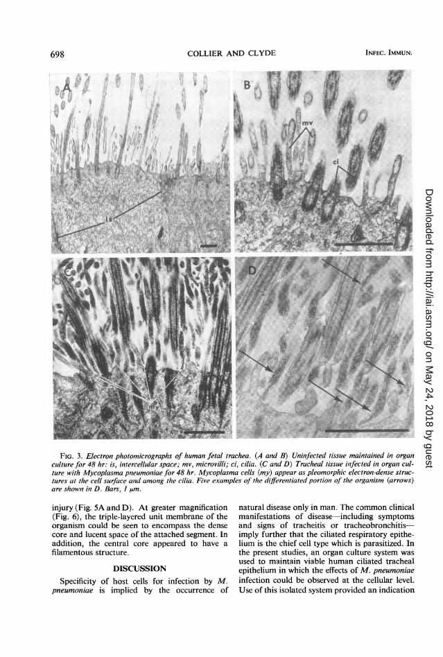
shown in Fig. 4A. The organism depicted meas-ures 0.075 by 1.8 ,um, the tapered terminal tipaccounting for about 10% of the length of theorganism. The specialized ending was also seen inorganisms used as the inoculum for these experi-ments, as demonstrated in Fig. 4B and C. Thinsections were prepared of pellets fixed after cen-trifugation of log-phase culture at 15,000 X gfor 30 min. In this case, the terminal structureswere noted infrequently, probably indicatingrandom orientation of organisms to the plane ofsection rather than lack of differentiation. Thefilamentous morphology of the organism was lessevident in the pellets as well.The relationship between the host cell mem-
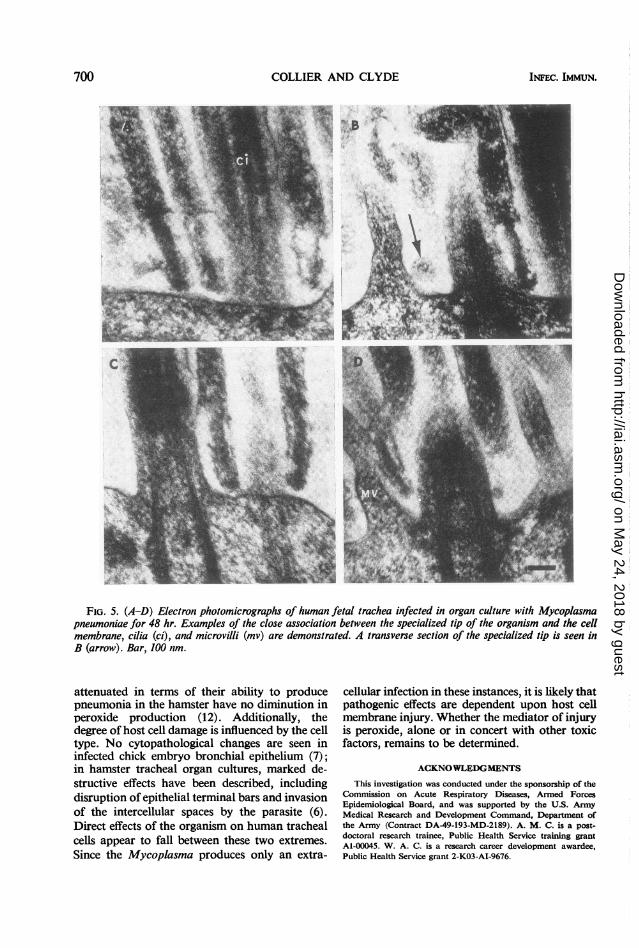
brane and the organism is illustrated further inFig. 5. In Fig. 5A, the appearance of three orga-nisms cut in slightly different planes suggests thatthe tip structure consists of an electron-dense coresurrounded by a lucent area. A cross section of thestructure is indicated by the arrow in Fig. 5B.Very close approximation of host cell and orga-nism membrane was noted only at the side or endof the specialized tip, providing further evidenceof the importance of this device for attachment tohost cells. In some instances, the epithelial mem-brane appeared indistinct at the point of orga-nism contact, suggesting the possibility of local
697VOL. 3, 1971
on May 24, 2018 by guest
http://iai.asm.org/
Dow
nloaded from
COLLIER AND CLYDE
Asr
Yi:~~~~~~~~~~~~~~~~~~1.
/'
; *
4, .
FIG. 3. Electron photomicrographs of human fetal trachea. (A and B) Uninfected tissue maintained in organculture for 48 hr: is, intercellular space; mv, microvilli; ci, cilia. (C and D) Tracheal tissue infected in organ cul-ture with Mycoplasma pnzeumoniae for 48 hr. Mycoplasma cells (my) appear as pleomorphic electron-dense struc-tures at the cell surface an1d among the cilia. Five examples of the differentiated portioln of the organism (arrows)are shown in D. Bars, I ,um.
injury (Fig. 5A and D). At greater magnification natural disease only in man. The common clinical(Fig. 6), the triple-layered unit membrane of the manifestations of disease-including symptomsorganism could be seen to encompass the dense and signs of tracheitis or tracheobronchitis-core and lucent space of the attached segment. In imply further that the ciliated respiratory epithe-addition, the central core appeared to have a lium is the chief cell type which is parasitized. Infilamentous structure. the present studies, an organ culture system was
used to maintain viable human ciliated trachealDISCUSSION epithelium in which the effects of M. pneumoniae
Specificity of host cells for infection by M. infection could be observed at the cellular level.pneumoniae is implied by the occurrence of Use of this isolated system provided an indication
698 INFEC. IMMUN.
on May 24, 2018 by guest
http://iai.asm.org/
Dow
nloaded from
M. PNEUMONIAE AND HUMAN HOST CELLS
of the direct pathogenicity of the organism byrevealing (i) interference with ciliary motility andsynchrony, (ii) a sequential pattern of cytologicalchanges, and (iii) disruption of normal tissuearchitecture. Since natural M. pneumoniae diseaseis a mild but protracted illness, the destructiveeffects of the organism for tracheal cells are prob-ably modified by the normal defenses of the intacthost.
Adsorption of M. pneumoniae to a variety ofother human cells has been demonstrated, includ-ing cervical epithelium (HeLa cells), spermatozoa,erythrocytes, and leukocytes (17, 18; Del Giudiceand Pavia, Bacteriol. Proc., p. 71, 1964). It is notclear that multiplication of organisms occurs inassociation with the cells in these examples.Target cell specificity appears incomplete, how-ever, since experimental infection can be estab-lished in a limited number of animal hosts; inaddition, adsorption between nonhuman trachealcells and the organisms has been reported (16).The host cells involved may share the sialic acid-like sites which are utilized by the organisms for
FIG. 4. Electron photomicrographs of Mycoplasmapneumoniae. (A) Two organisms sectioned loiigitudi-nially, showing filamentous morphology and differentia-tioi of ends attached to ani epithelial cell. (B and C).Examples of specialized terminal structure (arrows) inorganisms Jbrm log-phase broth culture used to inocu-late organ cultures. Bars, 100 nm.
attachment. This adsorption mechanism has notbeen demonstrated in other human Mycoplasmaspecies (17), perhaps explaining the lack of patho-genicity of selected species for cultured humantrachea which was observed.
Results of the present studies suggest that theattachment mechanism of M. pneumonaie residesin a specialized terminal structure which measuresapproximately 80 to 100 by 250 to 300 nm. Thisstructure is enclosed by the unit membrane of theorganism and has an organized internal structure.The electron density and fibrillar nature of thecentral core suggest that it may contain a concen-tration of nucleic acids. The lucent area surround-ing the core could be composed of lipid materials,since these do not stain with the techniques em-ployed here. Whether the specialized tip has func-tions other than attachment remains to be deter-mined. It is known that important antigenicdeterminants of M. pneumoniae are lipids (10),and the replicative mode of the organism isthought to be by binary fission (8). AnotherMycoplasma species with demonstrated asym-metry is M. gallisepticum (13); in this case, aterminal bleb appears associated with attachmentto host cells (18), although organism divisionseems to occur at the opposite pole (14).The finding of a special morphological feature
in M. pneumoniae grown in broth differs fromresults of previous studies on the ultrastructure ofthis Mycoplasma but confirms the findings ofBiberfeld and Biberfeld which were reportedrecently (1). These authors described in detail thedifferentiated terminal portion of the organism,although their studies conducted in vitro did notsuggest the functional significance of this struc-ture. Further study of the specialized tip can befacilitated by use of a system containing host cells.As demonstrated by the present studies, theorientation of the organism appears to be regu-lated by the presence of host cells, in contrast torandom positioning in material centrifuged fromcell-free medium. From the frequency with whichattached orgaalisms were seen in thin sections, itcan be calculated that over 100 could be presenton the luminal surface of a single epithelial celland countless more on the ciliary portion of thecell membrane. Specimens of this type would beivaluable for combined histochemical and electronmicroscopic study.The exact means by which M. pneumoniae in-
jures host cells is unknown. It has been suggestedthat the generation of peroxide in close proximityto the cell membrane is of primary importance (4),and this has been documented in the case of M.mycoides var. capri (3). However, it has been re-ported that M. pneumoniae strains which are
VOL. 3, 1971 699
on May 24, 2018 by guest
http://iai.asm.org/
Dow
nloaded from
COLLIER AND CLYDE INFEC. IMMUN.
FIG. 5. (A-D) Electron photomicrographs of human fetal trachea infected in organ culture with Mycoplasmapneumoniae for 48 hr. Examples of the close association between the specialized tip of the organism and the cellmembrane, cilia (ci), and microvilli (mv) are demonstrated. A transverse section of the specialized tip is seen inB (arrow). Bar, 100 nm.
attenuated in terms of their ability to producepneumonia in the hamster have no diminution inperoxide production (12). Additionally, thedegree of host cell damage is influenced by the celltype. No cytopathological changes are seen ininfected chick embryo bronchial epithelium (7);in hamster tracheal organ cultures, marked de-structive effects have been described, includingdisruption of epithelial terminal bars and invasionof the intercellular spaces by the parasite (6).Direct effects of the organism on human trachealcells appear to fall between these two extremes.Since the Mycoplasma produces only an extra-
cellular infection in these instances, it is likely thatpathogenic effects are dependent upon host cellmembrane injury. Whether the mediator of injuryis peroxide, alone or in concert with other toxicfactors, remains to be determined.
ACKNOWLEDGMENTS
This investigation was conducted under the sponsorship of theCommission on Acute Respiratory Diseases, Armed ForcesEpidemiological Board, and was supported by the U.S. ArmyMedical Research and Development Command, Department ofthe Army (Contract DA-49-193-MD-2189). A. M. C. is a post-doctoral research trainee, Public Health Service training grantAl-00045. W. A. C. is a research career development awardee,Public Health Service grant 2-K03-AI-9676.
700
on May 24, 2018 by guest
http://iai.asm.org/
Dow
nloaded from
M. PNEUMONIAE AND HUMAN HOST CELLS 701
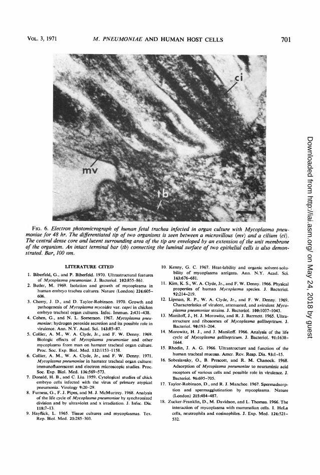
FIG. 6. Electron photomicrograph of human fetal trachea infected in organt culture with Mycoplasma pnzeu-moniiae for 48 hr. The differentiated tip of two organisms is seen between a microvillous (mv) and a cilium (ci).The central dense core and lucent surrounding area of thle tip are enveloped by an extensionz of thle unzit membraneof the organism. An intact terminal bar (tb) connecting the luminal surface of two epithlelial cells is also demont-strated. Bar, 100 nim.
LITERATURE CITED
1. Biberfeld, G., and P. Biberfeld. 1970. Ultrastructural featuresof Mycoplasma pneumoniae. J. Bacteriol. 102:855-861.
2. Butler, M. 1969. Isolation and growth of mycoplasma inhuman embryo trachea cultures. Nature (London) 224:605-606.
3. Cherry, J. D., and D. Taylor-Robinson. 1970. Growth andpathogenesis of Mycoplasma mycoides var. capri in chickenembryo tracheal organ cultures. Infec. Immun. 2:431-438.
4. Cohen, G., and N. L. Somerson. 1967. Mycoplasma pneu-moniae: hydrogen peroxide secretion and its possible role invirulence. Ann. N.Y. Acad. Sci. 143:85-87.
5. Collier, A. M., W. A. Clyde, Jr., and F. W. Denny. 1969.Biologic effects of Mycoplasma pneumoniae and othermycoplasma from man on hamster tracheal organ culture.Proc. Soc. Exp. Biol. Med. 132:1153-1158.
6. Collier, A. M., W. A. Clyde, Jr., and F. W. Denny. 1971.Mycoplasma pneumoniae in hamster tracheal organ culture:immunofluorescent and electron microscopic studies. Proc.Soc. Exp. Biol. Med. 136:569-573.
7. Donald, H. B., and C. Liu. 1959. Cytological studies of chickembryo cells infected with the virus of primary atypicalpneumonia. Virology 9:20-29.
8. Furness, G., F. J. Pipes, and M. J. McMurtrey. 1968. Analysisof the life cycle of Mycoplasma pneumoniae by synchronizeddivision and by ultraviolet and x irradiation. J. Infec. Dis.118:7-13.
9. Hayflick, L. 1965. Tissue cultures and mycoplasmas. Tex.Rep. Biol. Med. 23:285-303.
10. Kenny, G. C. 1967. Heat-lability and organic solvent-solu-bility of mycoplasma antigens. Ann. N.Y. Acad. Sci.143:676-681.
11. Kim, K. S., W. A. Clyde, Jr., and F. W. Denny. 1966. Physicalproperties of human Mycoplasma species. J. Bacteriol.92:214-219.
12. Lipman, R. P., W. A. Clyde, Jr., and F. W. Denny. 1969.Characteristics of virulent, attenuated, and avirulent Myco-plasma pneumoniae strains. J. Bacteriol. 100:1037-1043.
13. Maniloff, J., H. J. Morowitz, and R. J. Barrnett. 1965. Ultra-structure and ribosomes of Mjcoplasma gallisepticum. J.Bacteriol. 90:193-204.
14. Morowitz, H. J., and J. Maniloff. 1966. Analysis of the lifecycle of Mycoplasma gallisepticum. J. Bacteriol. 91:1638-1644.
15. Rhodin, J. A. G. 1966. Ultrastructure and function of thehuman tracheal mucosa. Amer. Rev. Resp. Dis. 93:1-15.
16. Sobeslavsky, O., B. Prescott, and R. M. Chanock. 1968.Adsorption of Mycoplasma pnieumoniae to neuraminic acidreceptors of various cells and possible role in virulence. J.Bacteriol. 96:695-705.
17. Taylor-Robinson, D., and R. J. Manchee. 1967. Spermadsorp-tion and spermagglutination by mycoplasma. Nature(London) 215:484-487.
18. Zucker-Franklin, D., M. Davidson, and L. Thomas. 1966. Theinteraction of mycoplasma with mammalian cells. I. HeLacells, neutrophils and eosinophiles. J. Exp. Med. 124:521-532.
VOL. 3, 1971
on May 24, 2018 by guest
http://iai.asm.org/
Dow
nloaded from