Embed Size (px)
Citation preview
Western Kentucky UniversityTopSCHOLAR®Honors College Capstone Experience/ThesisProjects Honors College at WKU
5-11-2015
Relative Reaction Rates of the Amino AcidsCysteine, Methionine, and Histidine with Analogsof the Anti-Cancer Drug CisplatinCynthia A. TopeWestern Kentucky University, [email protected]
Follow this and additional works at: http://digitalcommons.wku.edu/stu_hon_theses
Part of the Medicinal-Pharmaceutical Chemistry Commons
This Thesis is brought to you for free and open access by TopSCHOLAR®. It has been accepted for inclusion in Honors College Capstone Experience/Thesis Projects by an authorized administrator of TopSCHOLAR®. For more information, please contact [email protected].
Recommended CitationTope, Cynthia A., "Relative Reaction Rates of the Amino Acids Cysteine, Methionine, and Histidine with Analogs of the Anti-CancerDrug Cisplatin" (2015). Honors College Capstone Experience/Thesis Projects. Paper 571.http://digitalcommons.wku.edu/stu_hon_theses/571

RELATIVE REACTION RATES OF THE AMINO ACIDS CYSTEINE,
METHIONINE, AND HISTIDINE WITH ANALOGS OF THE ANTI-CANCER DRUG
CISPLATIN
A Capstone Experience/Thesis Project
Presented in Partial Fulfillment of the Requirements for
the Degree Bachelor of Science with
Honors College Graduate Distinction at Western Kentucky University
By:
Cynthia A. Tope
*****
Western Kentucky University
2015
CE/T Committee: Approved by:
Professor Kevin Williams, Advisor _________________________
Professor Darwin Dahl Advisor
Professor Lee Ann Smith Department of Chemistry

Copyright:
Cynthia A. Tope
2015

i
ABSTRACT
We are studying the reaction of analogs of the anticancer drug cisplatin with
amino acids that differ in size and shape. The reaction of cisplatin with proteins likely
precedes reaction with DNA in the body, forming a variety of products that may be toxic
to the human body. The size and shape of the platinum(II) complexes often affects the
rate of reaction with proteins, more so than with DNA. In this study, triamine cisplatin
analogs are reacted with the amino acids cysteine, methionine, and histidine
simultaneously. These reactions are monitored by NMR spectroscopy. The effect of the
bulk of the ligand and the pH under which the reaction occurs was explored. It is seen
that the bulkier [Pt(Me5dien)(NO3)]+ complex prefers to coordinate with N-
Acetylcysteine than L-methionine or L-histidine. When the pH was raised from 4 to 7,
the coordination to the platinum complex and N-AcCys occurred at a much faster rate.
Keywords: Cisplatin, Cysteine, Methionine, Histidine, Anticancer, Nuclear Magnetic
Resonance, Chemistry, Medicinal-Pharmaceutical Chemistry

ii
Dedicated to everyone that has helped me throughout my undergraduate career: friends,
family, professors, and the Gatton Academy.
iii
ACKNOWLEDGEMENTS
This project would not have been possible without the help and support of
everyone I have met during my undergraduate career at Western Kentucky University. I
would first like to thank my project advisor, Dr. Kevin Williams, for supporting me and
helping me through all of life challenges, from coursework to making personal life
decisions. Our conversations that started off about chemistry and ending with a rant about
football are some of my favorite. I would also like to thank the many professors in the
Chemistry Department that have encouraged me to pursue a future career in this field: Dr.
Dahl, Dr. Maddox, Dr. Pesterfield, and Dr. Nee. I would not have made it this far without
all of your support and guidance.
I would like to thank my many friends both within the Chemistry Department and
in my fraternity for helping me get through some of the more difficult times in my years
at the university. Sometimes all we need is to rant about life. And lastly, I would like to
thank the Gatton Academy, for giving me the chance to challenge myself and letting me
have the opportunity to pursue whatever dreams I have.
iv
VITA
June 25, 1993…………………………………..Born – Cincinnati, OH
2011…………………………………………….Gatton Academy of Math and Science
2011-2015………………………………………Research assistant, Dr. Kevin Williams
2014……………………….…………………….Chemistry Ambassador,
American Chemical Society
2014……………………………………………..Harlaxton College
FIELDS OF STUDY
Major Field: Chemistry (ACS-certified)
Minor Field 1: Spanish
Minor Field 2: History
v
TABLE OF CONTENTS
ABSTRACT………………………………………………………………………………ii
ACKNOWLEDGEMENTS………………………………………………………………iii
VITA……………………………………………………………………………………...iv
LIST OF FIGURES………………………………………………………………………vi
CHAPTERS:
1. INTRODUCTION………………………………………………………………...1
2. EXPERIMENTAL METHODS………………………………………………….10
3. RESULTS………………………………………………………………………..12
4. DISCUSSION…………………………………………………………………....21
BIBLIOGRAPHY………………………………………………………………………..25
vi
LIST OF FIGURES
FIGURE PAGE
1 Structure of Cisplatin …………………………………………………...1
2 Aquation of Cisplatin …………………………………………………...3
3 DNA Adduct Formation of Cisplatin…….……………………………..4
4 Structures of Amino Acids……………………………………………...5
5 Structures of Cisplatin Derivatives……………………………………...7
6 Deprotonation of N-Acetylcysteine……………………………………..7
7 1H NMR of Cisplatin Derivative……………………………………….12
8 1H NMR Signals of Amino Acids ……………………………………..13
9 1H NMR Spectra of L-His during pH 4 reaction ……………………...14
10 1H NMR Spectra of pH 4 reaction …………………………………….15
11 L-Met Chirality Signals ……………………………………………….17
12 1H NMR Spectra of L-His during pH 7 reaction ……………………...18
13 1H NMR Spectra of pH 7 reaction …………………………………….19
14 Comparison of pH 4 and 7 reactions …………………………………..20
1
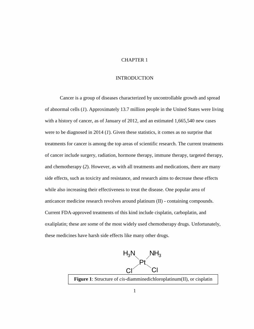
Figure 1: Structure of cis-diamminedichloroplatinum(II), or cisplatin
CHAPTER 1
INTRODUCTION
Cancer is a group of diseases characterized by uncontrollable growth and spread
of abnormal cells (1). Approximately 13.7 million people in the United States were living
with a history of cancer, as of January of 2012, and an estimated 1,665,540 new cases
were to be diagnosed in 2014 (1). Given these statistics, it comes as no surprise that
treatments for cancer is among the top areas of scientific research. The current treatments
of cancer include surgery, radiation, hormone therapy, immune therapy, targeted therapy,
and chemotherapy (2). However, as with all treatments and medications, there are many
side effects, such as toxicity and resistance, and research aims to decrease these effects
while also increasing their effectiveness to treat the disease. One popular area of
anticancer medicine research revolves around platinum (II) - containing compounds.
Current FDA-approved treatments of this kind include cisplatin, carboplatin, and
oxaliplatin; these are some of the most widely used chemotherapy drugs. Unfortunately,
these medicines have harsh side effects like many other drugs.
H3N NH3
Pt
2
The anti-cancer activity of the platinum(II) compounds was first discovered, by
accident, by the Rosenburg group at Michigan State University in 1965. Rosenburg saw
that the electrolysis of platinum electrodes created a platinum complex that inhibited the
cell division of Escherichia coli (E. coli) bacteria while the cell growth was not bothered
(3). The platinum complex generated was found to be cis-diamminedichloroplatinum(II),
or cisplatin. It was later tested on sarcomas in rats and it was found that the complex was
effective in reducing the mass of the tumors (4). This finding led to other conformation
testing in various cancer cell lines and led to its approval for clinical use by the FDA in
1978 (5).
Cisplatin is most effective on testicular and ovarian cancers, two forms of cancer
that yielded a high fatality rate prior to the discovery of the anticancer activity of the
platinum(II) complexes. Prior to 1970, testicular cancer killed over 90% of those
diagnosed (5). After the introduction of cisplatin to the treatment regimen in 1978, over
80% of patients survived the disease (5). Although the drug is very successful in treating
these cancers it also has many harsh side effects. These include, but are not limited to,
nephrotoxicity, ototoxicity, neurotoxicity, vomiting, and seizures (2). It is apparent that
cisplatin causes programmed cell death throughout the body, not just in cancer cells. Due
to the severe side effects of the drug, studies have been conducted to improve the
structure and functionality of the platinum compound to reduce these ill-effects and
improve its anticancer activity. In order to determine what causes the harsh side effects, it
is necessary to understand how the drug works in the body.
3
Cisplatin, administered intravenously, in its dichloro form is highly stable and
unreactive in areas of high chlorine concentrations, such as blood plasma where the
concentration is greater than 100 mM (6). The dichloro form then enters the cell, recently
proposed to be mediated through the copper transporter CTR1, into the relatively low
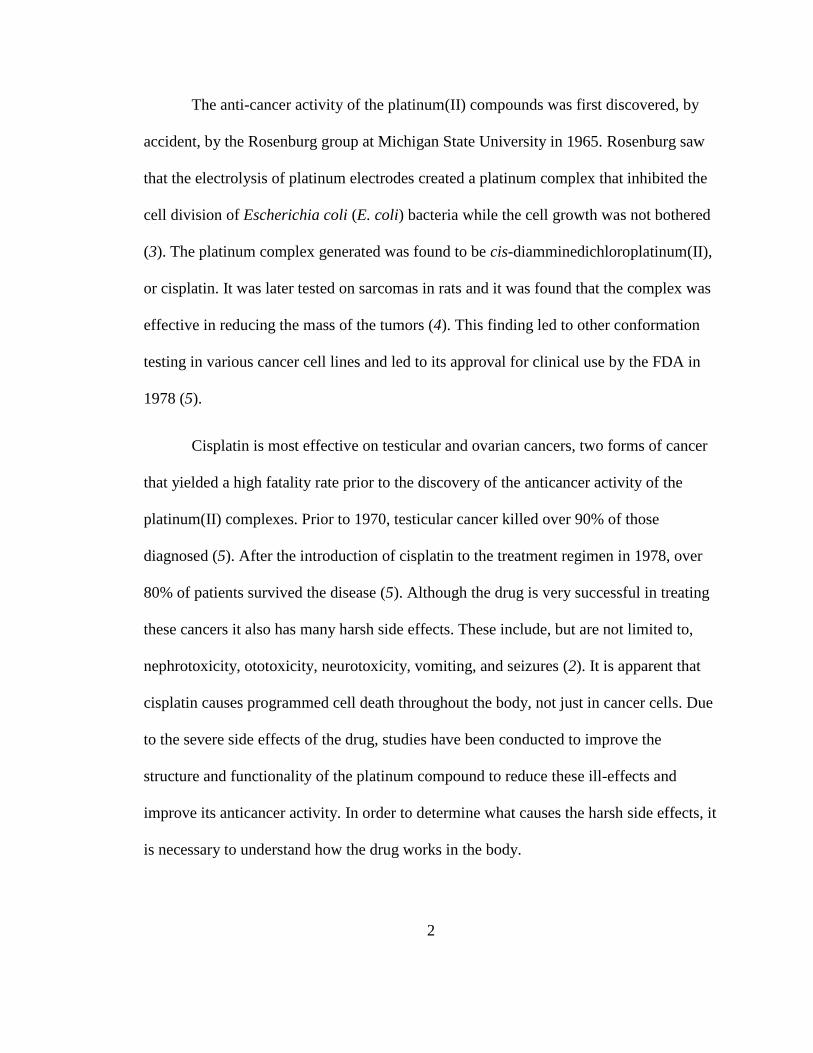
chloride concentrated environment of cytoplasm (7). At this point in cisplatin’s journey,
the chlorines are displaced via aquation (shown in Figure 2), yielding a highly reactive
electrophile whose ionic charge might prevent it from exiting the cell (6). This reactive
species can bind covalently to a variety of macromolecules, where DNA is the most
important, but not only, target of cisplatin.
The anticancer activity of cisplatin is attributed to adducts the active aquated form
makes with the nucleobases of DNA. Cisplatin has a preference to bind at the N7 position
of the guanine residue on the double helix (2). The 1,2 – intrastrand crosslink formed
distorts and unwinds the duplex helix of the DNA, which blocks DNA replication and
transcription, resulting in programmed cell death, or apotosis (6). There are three other
types of adducts that can be formed between DNA and cisplatin: monoadduct, interstrand
crosslink, and protein-DNA crosslink (8). Typically a monoadduct is initially formed and
Figure 2: Shows the aquation of cisplatin once inside the cell.

4
the complex then goes on to form the other types of adducts shown in Figure 3 (8). The
interstrand crosslink and protein-DNA crosslink are less common but could contribute to
cytotoxicity as they also impede DNA replication and transcription (8).
However, cisplatin does not only form adducts with DNA but also with proteins
and other macromolecules. It is these adducts that are thought to be the source of the
drug’s toxicity and resistance (9). While the formation of the Pt-DNA adducts requires
the aquation of the platinum compound, the Pt-protein adducts can occur in the dichloro
form, resulting in a preference of platinum(II) for proteins and related macromolecules
(10). It has been discovered that 24 hours after the cisplatin is administered to the patient,
65-98% of the compound has formed adducts with proteins (9). It is therefore important
to understand the reaction of platinum compounds with DNA and proteins and what
factors might affect the reaction with each.
In previous studies, cisplatin and the other platinum(II) compounds have shown a
preference for sulfur containing groups (9). This preference leads to cisplatin forming
Figure 3: Demonstrates the four Pt-DNA adducts that can be formed:
Intrastrand crosslink, interstrand crosslink, monoadduct, and protein-DNA
crosslink
5
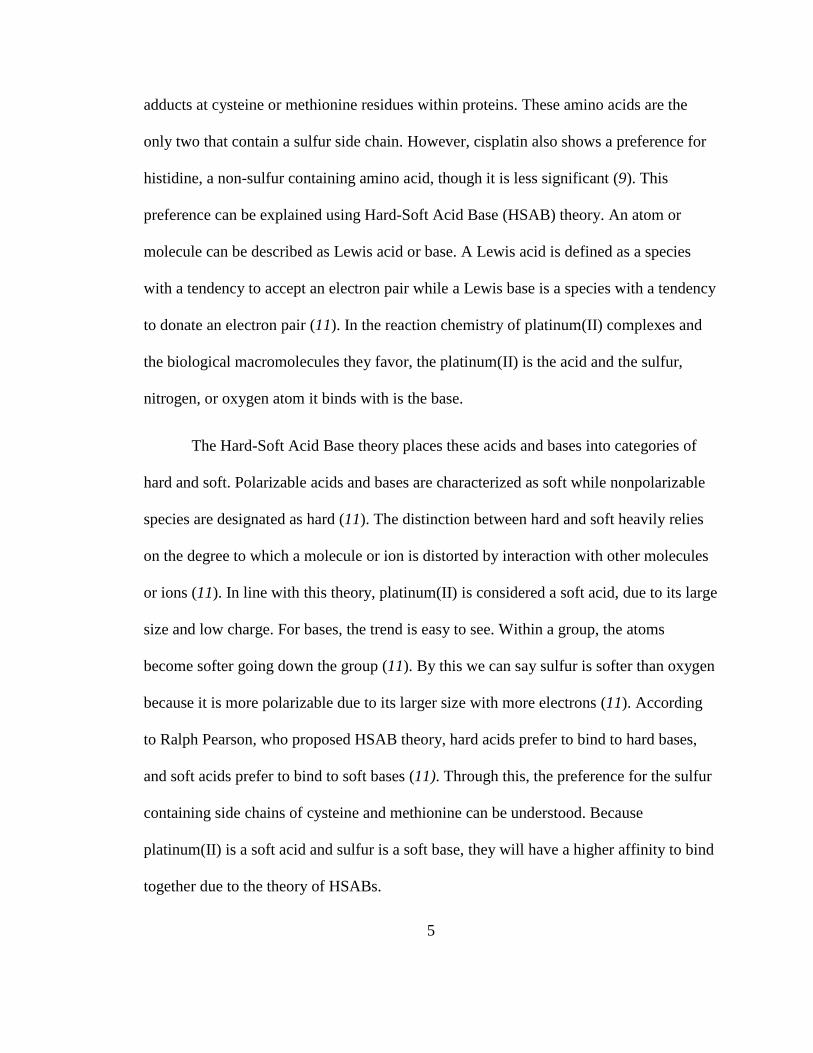
adducts at cysteine or methionine residues within proteins. These amino acids are the
only two that contain a sulfur side chain. However, cisplatin also shows a preference for
histidine, a non-sulfur containing amino acid, though it is less significant (9). This
preference can be explained using Hard-Soft Acid Base (HSAB) theory. An atom or
molecule can be described as Lewis acid or base. A Lewis acid is defined as a species
with a tendency to accept an electron pair while a Lewis base is a species with a tendency
to donate an electron pair (11). In the reaction chemistry of platinum(II) complexes and
the biological macromolecules they favor, the platinum(II) is the acid and the sulfur,
nitrogen, or oxygen atom it binds with is the base.
The Hard-Soft Acid Base theory places these acids and bases into categories of
hard and soft. Polarizable acids and bases are characterized as soft while nonpolarizable
species are designated as hard (11). The distinction between hard and soft heavily relies
on the degree to which a molecule or ion is distorted by interaction with other molecules
or ions (11). In line with this theory, platinum(II) is considered a soft acid, due to its large
size and low charge. For bases, the trend is easy to see. Within a group, the atoms
become softer going down the group (11). By this we can say sulfur is softer than oxygen
because it is more polarizable due to its larger size with more electrons (11). According
to Ralph Pearson, who proposed HSAB theory, hard acids prefer to bind to hard bases,
and soft acids prefer to bind to soft bases (11). Through this, the preference for the sulfur
containing side chains of cysteine and methionine can be understood. Because
platinum(II) is a soft acid and sulfur is a soft base, they will have a higher affinity to bind
together due to the theory of HSABs.
6
Figure 4: Structures of the amino acids: a. N-AcetylCysteine b. L-Methionine
c. L-Histidine
In order to decrease the toxicity of cisplatin and increase its activity, the ligand
sphere around the central atom has been changed (2). This manipulation of the ligand has
led to the creation of the second and third generation platinum(II) chemotherapy drugs,
carboplatin [cis-diammine-1,1-cyclobutanedicarboxylatoplatinum (II)] and oxaliplatin
respectively (2). Both of these complexes show an equivalent or better activity than
cisplatin and less toxicity than the first platinum(II) chemotherapy drug (2). This project
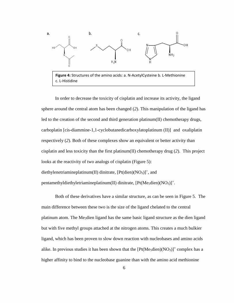
looks at the reactivity of two analogs of cisplatin (Figure 5):
diethylenetriamineplatinum(II) dinitrate, [Pt(dien)(NO3)]+, and
pentamethyldiethyletriamineplatinum(II) dinitrate, [Pt(Me5dien)(NO3)]+.
Both of these derivatives have a similar structure, as can be seen in Figure 5. The
main difference between these two is the size of the ligand chelated to the central
platinum atom. The Me5dien ligand has the same basic ligand structure as the dien ligand
but with five methyl groups attached at the nitrogen atoms. This creates a much bulkier
ligand, which has been proven to slow down reaction with nucleobases and amino acids
alike. In previous studies it has been shown that the [Pt(Me5dien)(NO3)]+ complex has a
higher affinity to bind to the nucleobase guanine than with the amino acid methionine
7
whereas [Pt(dien)(NO3)]+ showed the opposite trend (12). Knowing this, it is important to
also determine which amino acid is favored by the Me5dien platinum complex. This
project will demonstrate the reaction of the two derivatives of cisplatin shown in Figure 5
with a competition of the amino acids cysteine, methionine, and histidine in order to
determine which amino acid the complexes prefer, if there is a preference.
In addition to exploring the affinity of the complexes for different amino acids,
this project will also determine the effect of pH upon the reaction. Previous studies have
examined the reaction of platinum(II) compounds with DNA nucleobases and amino
acids at the pH of 4 (9, 13). At this pH, the amino acids cysteine, methionine, and
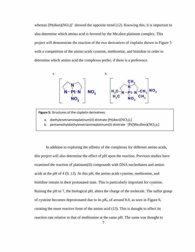
histidine remain in their protonated state. This is particularly important for cysteine.
Raising the pH to 7, the biological pH, alters the charge of the molecule. The sulfur group
of cysteine becomes deprotonated due to its pKa of around 8.0, as seen in Figure 6,
creating the more reactive form of the amino acid (13). This is thought to affect its
reaction rate relative to that of methionine at the same pH. The same was thought to
Figure 5: Structures of the cisplatin derivatives:
a. diethylenetriamineplatinum(II) dinitrate [Pt(dien)(NO3)2]
b. pentamethyldiethylenetriamineplatinum(II) dinitrate [Pt(Me5dien)(NO3)2]
8
occur for the histidine amino acid, as it also deprotonates at pH 7. However, previous
studies have shown that the change in pH has little bearing on the reactivity of histidine
(9).
In addition to the change in amino acid structure, the platinum complexes form
dimers at a higher pH. These dimers have an altered reactivity from the platinum
structure at pH 4. This project explores the effect of higher pH on the reaction of the
triamine compounds with the three amino acids. The relative rates of reaction between
cysteine, methionine, and histidine are examined at the pHs of 4 and 7 to determine if the
change in the cysteine amino acid affects the rate of reaction.
The experiments involved in this project were monitored using Nuclear Magnetic
Resonance, or NMR, spectroscopy. NMR spectroscopy is a technique used to determine
the structure of a molecule or compound. This instrumentation places a sample into a
static magnetic field which is then exposed to a secondary oscillating magnetic field. In
this project 1H NMR spectroscopy is essential to determine the formation of products
during the reaction. In 1H NMR, a peak is shown on the spectrum based on the
environment around each unique hydrogen atom in the sample. The atoms surrounding a
hydrogen atom affect the shift of the peaks down the spectrum. Spectra taken over a
Figure 6: The deprotonation of N-Acetylcysteine from
a.) pH 4 to b.) pH 8.
9
period of time can be used to determine the formation of products and their relative
abundance in the sample. Over time, peaks that are not characteristic of the reactants are
labeled as product peaks. This demonstrates the change in the chemical structure of the
reactants as the reaction moves toward the products. Following the development of these
product peaks can show which product is favored and the rate of the reaction.

10
CHAPTER 2
EXPERIMENTAL METHODS
Synthesis of Pt(Me5dien)(NO3)2. In order to synthesize [Pt(Me5dien)I]2 (Pt2I6),
which is needed before forming the nitrate and adapted from a previous method in
Romeo et al., 0.5 g of potassium tetrachloroplatinate was dissolved in 5 mL of deionized
water in an amber vial (13). 82 g of potassium iodide was added and the mixture was
stirred for 5 minutes at 50°C. Once the 5 minutes had passed, 250 µL of
pentamethyldiethylenetriamine was added. The solution was stirred at 50°C for one hour,
after which the green precipitate was separated via gravitational filtration. This process
yielded 506.6 mg of the iodide, which was allowed to dry over 2 nights to reduce
moisture left in the product.
300 mg of the dried Pt(Me5dien)I2 was added to 20 mL of deionized water in an
amber vial. 98.5 mg of silver nitrate was added to the vial and the mixture was stirred
overnight. The product was filtered out of the solution using a syringe filter and collected
in a round bottom flask. The remaining product was then isolated using a rotary
evaporator yielding 97.8 mg of Pt(Me5dien)(NO3)2. A small sample of the product was
dissolved in 1 mL of deuterium oxide and the identity was confirmed by 1H NMR using a
500 MHz JEOL Eclipse instrument.
11
Preparation of Pt(Me5dien)(NO3)2 and N-AcCys, L-Met, L-His Solutions.
Both solutions were made at 20 mmol concentrations. For the platinum complex solution,
9.8 mg of Pt(Me5dien)(NO3)2 was dissolved in 1.0 mL of deuterium oxide. The pH of the
solution was adjusted to either 4 or 7, depending on the reaction, using deuterated nitric
acid and sodium deuteroxide solutions. The amino acid solution was made by mixing 3.1
mg of L-His, 3.3 mg of N-AcCys, and 3.0 mg L-Met in a 4 mL vial and dissolved in 1
mL deuterium oxide. The pH was adjusted in the same manner as the platinum solution.
Reaction of Pt(Me5dien)(NO3)2 and N-AcCys, L-Met, L-His at pH 4. The
platinum solution and the amino acid solution were both adjusted to pH 4 using the
procedure detailed above. 300 µL of each solution was added to an NMR tube and mixed
together. Kinetics of the reaction was monitored using a 500 MHz JEOL Eclipse
instrument over a period of 12 hours, taking an acquisition scan every 15 min yielding 48
data points.
Reaction of Pt(Me5dien)(NO3)2 and N-AcCys, L-Met, L-His at pH 7. The
platinum solution and the amino acid solution were both adjusted to pH 7 using the
procedure detailed above. 300µL of each solution was added to an NMR tube and mixed
together. Kinetics of the reaction was monitored using a 500 MHz JEOL Eclipse
instrument over a period of 12 hours, taking an acquisition scan every 15 min again
yielding 48 data points.
12
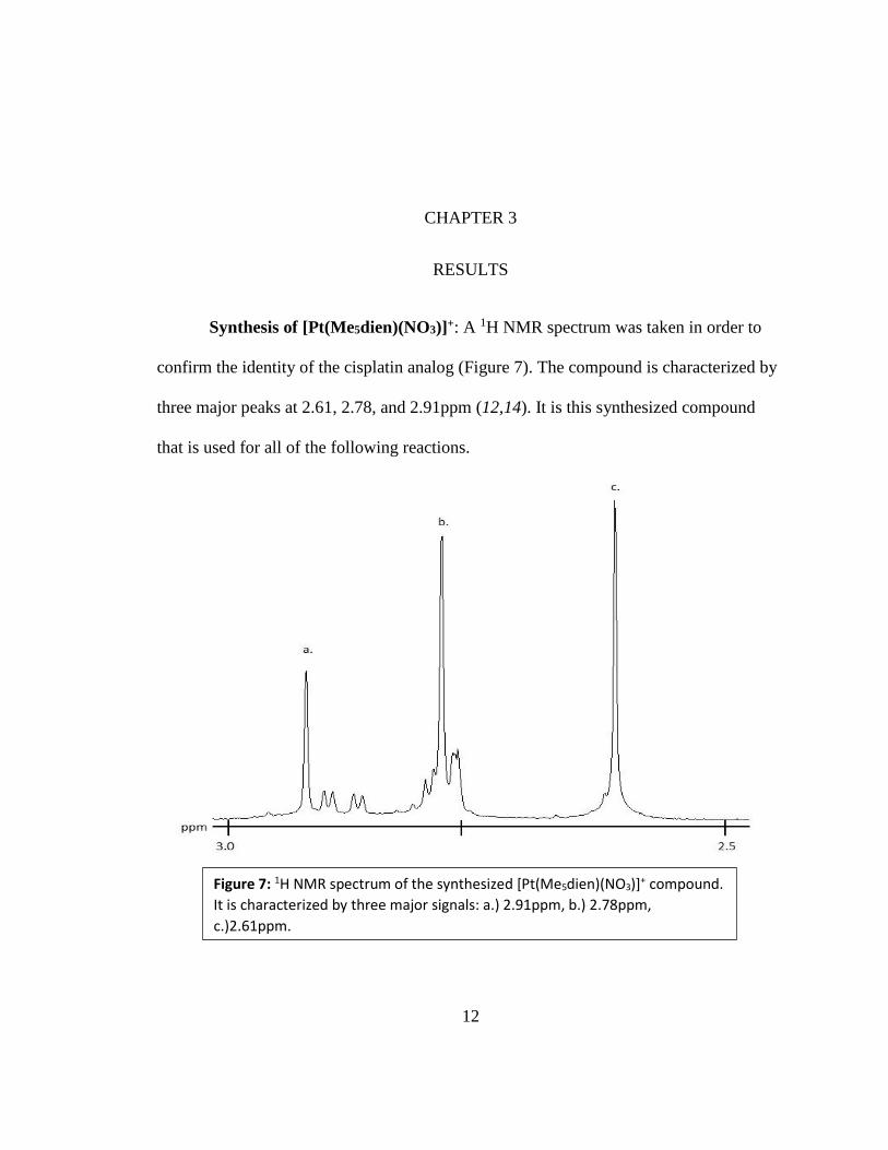
Figure 7: 1H NMR spectrum of the synthesized [Pt(Me5dien)(NO3)]+ compound.
It is characterized by three major signals: a.) 2.91ppm, b.) 2.78ppm,
c.)2.61ppm.
CHAPTER 3
RESULTS
Synthesis of [Pt(Me5dien)(NO3)]+: A 1H NMR spectrum was taken in order to
confirm the identity of the cisplatin analog (Figure 7). The compound is characterized by
three major peaks at 2.61, 2.78, and 2.91ppm (12,14). It is this synthesized compound
that is used for all of the following reactions.
13
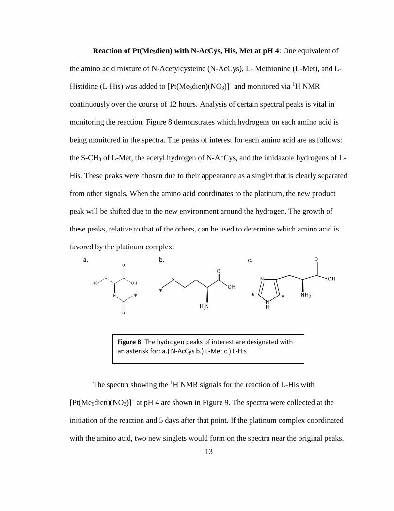
Figure 8: The hydrogen peaks of interest are designated with
an asterisk for: a.) N-AcCys b.) L-Met c.) L-His
Reaction of Pt(Me5dien) with N-AcCys, His, Met at pH 4: One equivalent of
the amino acid mixture of N-Acetylcysteine (N-AcCys), L- Methionine (L-Met), and L-
Histidine (L-His) was added to [Pt(Me5dien)(NO3)]+ and monitored via 1H NMR
continuously over the course of 12 hours. Analysis of certain spectral peaks is vital in
monitoring the reaction. Figure 8 demonstrates which hydrogens on each amino acid is
being monitored in the spectra. The peaks of interest for each amino acid are as follows:
the S-CH3 of L-Met, the acetyl hydrogen of N-AcCys, and the imidazole hydrogens of L-
His. These peaks were chosen due to their appearance as a singlet that is clearly separated
from other signals. When the amino acid coordinates to the platinum, the new product
peak will be shifted due to the new environment around the hydrogen. The growth of
these peaks, relative to that of the others, can be used to determine which amino acid is
favored by the platinum complex.
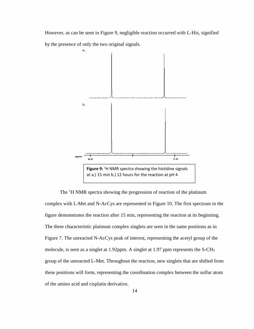
The spectra showing the 1H NMR signals for the reaction of L-His with
[Pt(Me5dien)(NO3)]+ at pH 4 are shown in Figure 9. The spectra were collected at the
initiation of the reaction and 5 days after that point. If the platinum complex coordinated
with the amino acid, two new singlets would form on the spectra near the original peaks.
14
Figure 9: 1H NMR spectra showing the histidine signals
at a.) 15 min b.) 12 hours for the reaction at pH 4
However, as can be seen in Figure 9, negligible reaction occurred with L-His, signified
by the presence of only the two original signals.
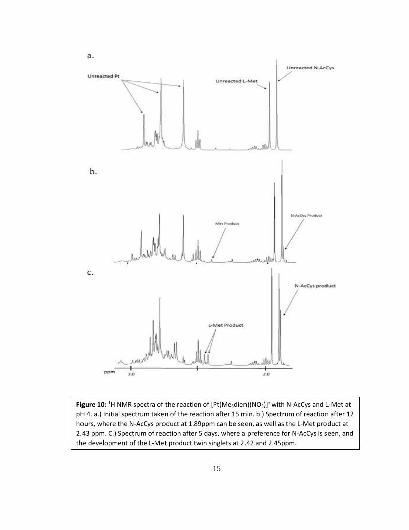
The 1H NMR spectra showing the progression of reaction of the platinum
complex with L-Met and N-AcCys are represented in Figure 10. The first spectrum in the
figure demonstrates the reaction after 15 min, representing the reaction at its beginning.
The three characteristic platinum complex singlets are seen in the same positions as in
Figure 7. The unreacted N-AcCys peak of interest, representing the acetyl group of the
molecule, is seen as a singlet at 1.92ppm. A singlet at 1.97 ppm represents the S-CH3
group of the unreacted L-Met. Throughout the reaction, new singlets that are shifted from
these positions will form, representing the coordination complex between the sulfur atom
of the amino acid and cisplatin derivative.
15
Figure 10: 1H NMR spectra of the reaction of [Pt(Me5dien)(NO3)]+ with N-AcCys and L-Met at
pH 4. a.) Initial spectrum taken of the reaction after 15 min. b.) Spectrum of reaction after 12
hours, where the N-AcCys product at 1.89ppm can be seen, as well as the L-Met product at
2.43 ppm. C.) Spectrum of reaction after 5 days, where a preference for N-AcCys is seen, and
the development of the L-Met product twin singlets at 2.42 and 2.45ppm.

16
In the second spectrum (b) of Figure 10, the state of the reaction after 12 hours
can be seen. Two new resonances appear on the spectrum, each representing the
coordination of one of the amino acids with the platinum complex. A new singlet at 1.89
ppm represents the coordination of N-AcCys with Pt(Me5dien). Due to the new
environment on the N-AcCys molecule, the singlet representing the acetyl group was
shifted upfield, or to the right, from the original, unreacted peak. Its relatively small
height compared to the unreacted singlet shows that little reaction has occurred by this
time in the reaction. A second singlet appears around 2.43 ppm, which represents the
coordination at the sulfur atom of L-Met. This peak is shifted more significantly upfield,
or to the left, of the original peak of the unreacted L-Met than the N-AcCys product was
shifted due to the proximity of the platinum complex to the hydrogen of interest. On N-
AcCys, the platinum coordinates to a position that is far from the hydrogen of interest
while on L-Met, the platinum coordinates to the atom that the hydrogen is bonded to.
This proximity causes a much greater shift of the S-CH3 signal. This L-Met product
singlet is much smaller in relation to the unreacted peak than the N-AcCys product is to
its counterpart, signifying that N-AcCys is preferred by the platinum complex.
The reaction was revisited 5 days after its initiation in order to determine if one
amino acid was significantly preferred, as both product peaks were small after 12 hours.
This spectrum is shown in Figure 10 (c). In this spectrum, the N-AcCys product peak has
grown significantly more in comparison to the L-Met product peak. The most significant
change in the L-Met product is the formation of two singlets instead of just one singlet as
seen at 12 hours into the reaction. This formation is due to the slow interchange in the

17
isomerization of the complex. The L-Met sulfur, after the coordination of the platinum,
becomes chiral. These two singlets (Figure 11) represent the diastereomers that are a
result of this chirality. This phenomenon has been seen in a previous study between
methionine and platinum (II) complexes (15).
Reaction of Pt(Me5dien) with N-AcCys, His, Met at pH 7: As seen in the
reaction at pH 4, there was negligible reaction with L-His 12 hours after the initiation of
reaction. L-His is deprotonated at pH 6, creating a more reactive form of the amino acid.
The spectra taken at the beginning of reaction and after 12 hours is shown in Figure 12.
There is no development of new resonances, signifying that no L-His product was
formed. This shows that even the deprotonated, more reactive form of L-His is not
preferred by the platinum complex in relation to the sulfur-containing L-Met and N-
AcCys.
Figure 11: The twin singlets between 2.40 ppm and
2.46 ppm represent the chirality about the S-atom of
the L-Met product. Taken from the spectrum of 5 days
after reaction initiation.

18
‘
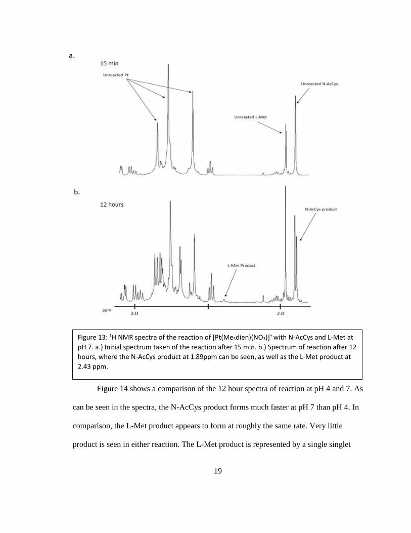
The 1H NMR spectra taken of the reaction at pH 7 is shown in Figure 13. The
initiation of reaction is shown in the first spectrum of the figure, as with the reaction at
pH 4. However, unlike the previous reaction, the sample was not revisited several days
later. Figure 13a shows the three characteristic signals for the platinum complex as well
as the unreacted amino acid peaks seen in the pH 4 spectrum. Figure 13b represents the
reaction after 12 hours. In this spectrum, a significant N-AcCys product singlet can be
seen at 1.89 ppm. The height of the product is close to the same height as that of the
unreacted N-AcCys. This signifies that there is roughly the same amount of unreacted N-
AcCys as product. There is also a small L-Met product peak around 2.43 ppm. In
comparison to the N-AcCys product, the L-Met product is barely formed, suggesting that
N-AcCys is highly preferred at pH 7.
Figure 12: 1H NMR spectra showing the histidine signals
at a.) 15 min b.) 12 hours for the reaction at pH 7
19
Figure 13: 1H NMR spectra of the reaction of [Pt(Me5dien)(NO3)]+ with N-AcCys and L-Met at
pH 7. a.) Initial spectrum taken of the reaction after 15 min. b.) Spectrum of reaction after 12
hours, where the N-AcCys product at 1.89ppm can be seen, as well as the L-Met product at
2.43 ppm.
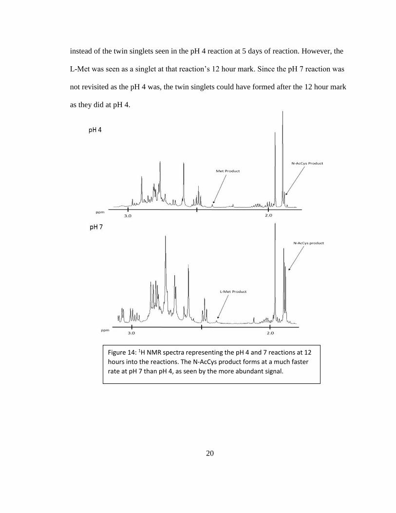
Figure 14 shows a comparison of the 12 hour spectra of reaction at pH 4 and 7. As
can be seen in the spectra, the N-AcCys product forms much faster at pH 7 than pH 4. In
comparison, the L-Met product appears to form at roughly the same rate. Very little
product is seen in either reaction. The L-Met product is represented by a single singlet
20
Figure 14: 1H NMR spectra representing the pH 4 and 7 reactions at 12
hours into the reactions. The N-AcCys product forms at a much faster
rate at pH 7 than pH 4, as seen by the more abundant signal.
instead of the twin singlets seen in the pH 4 reaction at 5 days of reaction. However, the
L-Met was seen as a singlet at that reaction’s 12 hour mark. Since the pH 7 reaction was
not revisited as the pH 4 was, the twin singlets could have formed after the 12 hour mark
as they did at pH 4.

21
CHAPTER 4
DISCUSSION
It has been shown in previous experiments that the cisplatin derivative of
[Pt(Me5dien)(NO3)]+ has a higher affinity for the nucleobase guanine than the amino acid
methionine (12). This project expands on this finding, looking at the cisplatin derivative’s
preference for a certain amino acid among the main targets of cysteine, methionine, and
histidine. The effect of pH was also examined for the competition reaction, conducting
the experiment at both pH 4 and pH 7. The pH within the body is 7, so it is important to
understand how these platinum(II) compounds react with amino acids at this pH.
Histidine has a pKa of 6, meaning it is fully deprotonated at pH 7, while N-acetylcysteine
has a pKa of 8, where it will have some of the sample deprotonated. The deprotonated
forms of these amino acids are more reactive and the effect of this increased reactivity is
examined.
At both pH values, the reaction of L-His and the platinum complex
[Pt(Me5dien)(NO3)]+ is negligible. In the spectra shown in Figures 8 and 12, there was no
development of singlets that would represent an L-His product, even after several days of
reaction. Although the L-His is deprotonated and therefore more reactive, the platinum
(II) compound prefers the sulfur-containing amino acids methionine and cysteine. The
platinum coordinates at a nitrogen group on the L-His which is not as soft of a base as
22
sulfur, which may explain the non-reactivity with histidine when in the presence of the
sulfur groups of cysteine and methionine.
The reaction of [Pt(Me5dien)(NO3)]+ with N-AcCys and L-Met at pH 4 was
monitored continuously over 12 hours and revisited after 5 days. After just 12 hours of
reaction, there was a preference for N-AcCys, as seen in the more significant growth of
its product signal compared to that of L-Met. However, both signals were small in
comparison to the unreacted amino acid peaks, signifying that little reaction had occurred
up to that point. Because of the small amount of reaction that had occurred by 12 hours
into the reaction, it was revisited 5 days later. The spectrum taken at this point, shown in
Figure 13, shows a much higher growth in the N-AcCys product signal than in the L-Met
product. Based on this spectrum, it can be concluded that [Pt(Me5dien)(NO3)]+ has a
much higher affinity for N-AcCys than L-Met.
Increasing the pH from 4 to 7 had a significant effect on the reactivity of N-
AcCys with [Pt(Me5dien)(NO3)]+. After only 12 hours of reaction at pH 7, a higher ratio
of N-AcCys product to unreacted N-AcCys can be seen than after 5 days of reaction at
pH 4. This suggests that the higher pH allows for some of the cysteine to be
deprotonated, creating the more reactive thiolate form of the amino acid. Given that the
pKa of cysteine is 8, raising the pH from 4 to 7 deprotonated a portion of the N-AcCys in
the sample. In comparison to the protonated L-Met, the thiolate is much more reactive,
coordinating much faster to the platinum complex while L-Met had roughly the same rate
of reactivity. This finding is particularly important as the pH within the cell is also 7. This
gives us a better idea as to where the platinum complex is likely to coordinate in the

23
body. Previous studies have shown that the [Pt(Me5dien)(NO3)]+ compound has a higher
affinity for guanine than for methionine (12). However, since the cisplatin derivative
shows a higher affinity for cysteine over methionine, the exploration into the compound’s
preference for cysteine or guanine will be important to determine the platinum complex’s
affinity for the amino acid or DNA nucleobase.
In the pH 4 reaction with the [Pt(Me5dien)(NO3)]+ complex, two singlets were
seen for the L-Met product. This is likely due to the slow interconversion between the
two isomers possible. When the platinum coordinates, the sulfur atom becomes chiral,
giving two possibilities of isomers. At 12 hours into the reaction, only one singlet can be
seen, suggesting that one isomer is faster to form than the other. When the pH 4 reaction
was revisited after 5 days, the twin singlets could be seen, showing that both isomers
eventually form, though one forms faster. It is possible that this also occurs at pH 7
though only one singlet could be seen at the 12 hour mark in the reaction. If this reaction
was revisited after several days, as was the pH 4 reaction, this phenomenon might have
been seen. However, there was significant reaction with N-AcCys at 12 hours, allowing
us to clearly see which amino acid was preferred.
Platinum (II) compounds have long been used in conjunction with other drugs in
the fight as against cancer. Because these compounds have high levels of toxicity and
side effects, it is important to understand how these drugs work inside the body so they
might be improved upon. Since platinum (II) has a high affinity for sulfur, common
targets of these drugs are the sulfur-containing amino acids of cysteine and methionine.
The coordination of cisplatin with amino acid residues of proteins is thought to be a
24
source of toxicity of the drug. Greater affinity for cysteine over other amino acids with
bulkier platinum (II) compounds may lead to new methods of targeting and reacting
within the cell. The results of this study into the amino acid preference of two triamine
derivatives of cisplatin contributes to ongoing research of cancer treatments and gives
insight into new areas of research with these compounds.
25
BIBLIOGRAPHY
(1) Cancer Facts & Figures 2014. Page 1.
http://www.cancer.org/acs/groups/content/@research/documents/webcontent/acspc-
042151.pdf (Accessed March 6, 2015)
(2) Kung, A., D. B. Strickmann, et al. (2001). "Comparison of the binding behavior
of oxaliplatin, cisplatin and analogues to 5'-GMP in the presence of sulfur-containing
molecules by means of capillary electrophoresis and electrospray mass spectrometry."
Journal of Inorganic Biochemistry 86: 691-698.
(3) Rosenberg, B.; Van Camp, L.; Krigas, T. (1965). "Inhibition of cell division in
Escherichia coli by electrolysis products from a platinum electrode". Nature 205 (4972):
698–699. doi:10.1038/205698a0. PMID 14287410.
(4) Rosenberg, B.; Vancamp, L.;Trosko, J.E.; Mansour, V.H. (1969). "Platinum
Compounds: a New Class of Potent Antitumour Agents". Nature 222 (5191): 385–386.
(5) The "Accidental" Cure—Platinum-based Treatment for Cancer: The Discovery of
Cisplatin. http://www.cancer.gov/aboutnci/servingpeople/cancer-research-
progress/discovery/cisplatin (Accessed April 4, 2015)
(6) Howell, D. F. a. S. B. (2000). How does Cisplatin Kill Cells? Platinum-based
Drugs in Cancer Therapy. N. Farrell, Humana Press: 354p.
(7) Howell, S. B., R. Safaei, et al. (2010). "Copper Transporters and the Cellular
Pharmacology of the Platinum-Containing Cancer Drugs." Molecular Pharmacology
77(6): 887-894.
(8) Rabik C, Dolan M. Molecular Mechanisms of Resistance and Toxicity Associated
with Platinating Agents, Cancer Treat Rev., 2007 February, 33(1): 9-23 PMCID:
PMC1855222
(9) Sandlin, R. D., C. J. Whelan, et al. (2012). "Effects of amine ligand bulk and
hydrogen bonding on the rate of reaction of platinum(II) diamine complexes with key
nucleotide and amino acid residues." Inorganica Chimica Acta 391: 135-140.
(10) Berners-Price, S. J. and T. G. Appleton (2000). The Chemistry of Cisplatin in
Aqueous Solution. Platinum-Based Drugs in Cancer Therapy. L. R. Kelland and N.
Farrell. Totowa, NJ, Humana Press, Inc.: 3-35.

26
(11) Miessler, G.L., P.J.Fischer, et al. Inorganic Chemistry, 5th ed.; Pearson: Boston,
2014. pp 201-204.
(12) Sandlin, R. D., M. P. Starling, et al. (2010). "A bulky platinum(II) triamine
complex that reacts faster with guanosine 5'-monophosphate than with N-
acetylmethionine." Journal of Inorganic Biochemistry 104(2): 214-219.
(13) Romeo, R.; Minniti, D.; Lanza, S.; Tobe, M. L. Inorg. Chim. Acta. 1977, 22, 87-
91
(14) R. Cini, F.P. Intini, L. Maresca, C. Pacifico, G. Natile, Eur. J. Inorg. Chem.
(1998) 1305–1312
(15) Williams, K. M., R. P. Dudgeon, et al. (2011). “Reaction of platinum(II)diamine
and triamine complexes with selenomethionine.” Inorganica Chimica Acta 368(1): 187-
193.