Embed Size (px)
Citation preview
1
Removal of melanoidins by potential manganese peroxidase producing bacteria isolated from molasses
effluent
Samir Mahgoub1,2*, K. Tsioptsias2, E. Likotrafiti2 and P. Samaras2
1Microbiology Department, Faculty of Agriculture, Zagazig University, Zagazig 44511, Egypt
2Laboratory of Water and Wastewater Technology, Department of Food Technology, Technological Educational Institute
of Thessaloniki, 57400 Sindos, Thessaloniki, Greece
*Corresponding author: email: [email protected]
Running title: Removal of melanoidins by bacteria isolated from molasses effluent

2
Abstract
The possibility and efficiency of manganese peroxidase (MP) producing bacteria for the removal of melanoidins
from synthetic and real melanoidins solutions were evaluated in this study. Samples collected from bench scale
bioreactor treating melanoidins wastewater (90% v/v) were studied for screening and isolation of bacteria capable of
decolorizing melanoidins.
Treatment efficiency in terms of color removal, COD reduction, pH change and OD590 was investigated under
different synthetic melanoidins solutions e.g. sucrose-glutamic acid (SGA), sucrose-aspartic acid (SAA), glucose-
aspartic acid (GAA) and glucose-glutamic acid (GGA) Maillard products as well as real melanoidins.
Thirteen potential MP producing bacteria were selected for higher SGA-MP, SAA-MP, GAA-MP and GGA-MP
tolerance (7000 mg/l) in modified GPYM Agar (glucose 1.0%, peptone 0.1%, yeast extract 0.1%, K2HPO4 0.1 %
and MgSO4 0.05%) and King broth Agar (glycerol 1.0%, peptone 0.1%, yeast extract 0.1%, K2HPO4 0.1 % and
MgSO4 0.05%). Three of these isolates and Lactobacillus kefir strain showed maximum OD590 (1.38- 1.47 and 1.28-
1.35) of GAA-MP (5000 and 15000 mg/l) in the medium at pH (7.4 ± 0.1), shaking speed (140 rpm) and
temperature (30±2 °C) after 5 days incubation, respectively. However, in real melanoidin solutions (10 % v/v),
theses strains showed low OD590 (0.43- 0.66) after 5 days incubation, these strains showed maximum
decolourisation capacity (8-12 %) of GAA-MP (5000 &15000 mg/l) and real melanoidins (10% v/v) in the medium
after 5 days incubation. Lactob. kefir and the isolates showed maximum reduction of COD ranged from 52 to 79 %
of GAA-MP (5000 & 15000 mg/l) and real melanoidins in the medium after 5 days incubation.
Lactob.kefir and three bacterial isolates showed highest reduction of COD in synthetic and real melanoidins after 5
days of incubation. Yeast extract could be used as bio-stimulator for these isolates during decolourisation of real
melanoidins.
Keywords: Melanoidins, Molasses, Decolourisation, microorganisms

3
Introduction
Molasses distillery wastewater colorants are mainly polyphenols, melanoidin, alkaline degradation products of
hexoses, and caramels. A lot of efforts have been made to remove the colorants including biological methods
employing different fungi, bacteria &algae, enzymatic treatment, chemical oxidation, coagulation/precipitation,
oxidation and membrane filtration [1]. These technologies could also be applied to remove the colorants as a final
treatment step after the anaerobic digestion [2]. Melanoidins are found at concentrations of 2.0 % in effluents
discharged from molasses processing industry. Untreated distillery effluent or spent wash are well known to cause
pollution in the natural streams by increase in organic load, depletion of oxygen content, discoloration and
destruction of aquatic life [1]. This effluent retains very dark brown color even after anaerobic treatment due to
presence various water soluble, recalcitrant and coloring compounds mainly melanoidins [3]. Melanoidins are
generated in sugarcane molasses as complex polymer due to non-enzymatic reaction of sugar and amino acid
produced browning reactions called Maillard reaction [4].These compounds are highly resistant to microbial attack,
conventional biological processes such as activated sludge treatment are inefficient to decolorize melanoidin-
containing wastewaters, such as molasses wastewaters from distilleries and fermentation industries [5]. However,
microbial decolorization of anaerobically treated effluent reduced the toxic effect which indicated that there is
necessity for microbial degradation at secondary or tertiary stage prior to its disposal for environmental safety
[3].Thus, the microbial decolorization can be exploited to develop a cost effective, eco-friendly biotechnology
package for the treatment of distillery effluent [6].The decolorization of four types of synthetic melanoidins i.e.,
glucose–glutamic-acid (GGA), glucose–aspartic-acid (GAA), sucrose–glutamic acid (SGA), and sucrose–aspartic-
acid (SAA), were investigated using three different isolates, viz. Bacillus thuringiensis, Bacillus brevis and Bacillus
sp. [7].The degree of decolorization of the melanoidins separately by each isolate was in the 1–31% range. The
results also indicated that the GAA polymer was the most recalcitrant among the melanoidins tested. The
degradation and decolourisation of melanoidin by bacteria are mediated due to prevalence of manganese peroxidase
(MnP) as decolourising enzyme [8] and play an important role in degradation of several dyes [9]. Lactobacillus
plantarum could rapidly decolorize molasses melanoidin to 60.91% and 15.88% color from treated palm oil mill
effluent [10]. Lactob. hilgardii [11] and Bacillus sp [12] had melanoidins pigment removal ability. The
decolorization activity of acetogenic bacteria has been reported by Sirianuntapiboon and Prasertsong [13] and
showed high decolorization yield in both synthetic melanoidin and molasses wastewater solutions. Pseudomonas
putida produces hydrogen peroxide which is a strong decolorizing agent. Since the organism cannot use spent wash
as a source of carbon, 1% w/v glucose supplement was provided along with 12.5% spent wash. Aeromonas sp.
utilizes the carbonaceous compounds present in spent wash as the sole carbon source, thereby eventually reducing
the effluent COD by 66% in a 24 h period. P. putida also resulted in 44% COD removal accompanied by 60% color
reduction. In another study on predigested distillery effluent with A. formicans, 57% COD reduction and 55%
decrease in color was observed after 72 h [14].Therefore, the development of effective and cost-competitive
techniques for the decolorization is of importance. Thus the aim of this study was to investigate the synthetic and

4
real melanoidins adsorption mechanism by Lactobacillus kefir and three bacterial isolates from indigenous sources
(bench scale bioreactor treating melanoidins wastewater (90% v/v).
2. Materials and methods
2.1. Media and strain used
All the culture media used in this study were of biological grade. Four kinds of media were used i.e. GPYM broth
consisting of (w/v): 1% glucose, 0.05% peptone, 0.1% K2HPO4 and 0.05% MgSO4, modified GPYM broth amended
with synthetic melanoidins solution, King’s B’ broth media consisting of (w/v): 0.15% K2HPO4, O.15% MgSO4,
2.0% Peptone, 1.0% Glycerol and King’s B’ broth amended with synthetic melanoidins solution. Solid media was
prepared by adding 1.5% agar in the above-mentioned composition. Lactobacillus kefir strain used in this study was
kindly obtained from Dr. Eleni Likotrafiti, Laboratory of Food Microbiology, Department of Food Technology,
Technological Educational Institute of Thessaloniki, Greece.
2.2. Preparation of synthetic melanoidins
The synthetic melanoidins solution used in this study was prepared by refluxing the equimolar (1M) solution of
sucrose (Panreac Quimica SAU, Barcelona, Spain), aspartic acid (Panreac Quimica SAU, Barcelona, Spain) and
0.5M sodium carbonate at 100 ◦C for 7 h [7] whereas; natural melanoidins solution was prepared from distillery
effluent. Four types of melanoidins i.e., sucrose-aspartic-acid (SAA), sucrose-glutamic acid (SGA), glucose-
aspartic-acid (GAA) and glucose-glutamic-acid (GGA) were synthesized SAA was synthesized using sucrose,
aspartic acid and sodium carbonate, SGA from sucrose, glutamic acid and sodium carbonate, GAA from glucose,
aspartic acid and sodium carbonate and GGA was prepared utilizing glucose, glutamic acid and sodium carbonate
under above conditions and then after adjustment of the reaction mixture to pH 7.4 with 1N NaOH. These solutions
contained mg/l COD from 267000 to 418000 mg/l.
2.3. Isolation of potential manganese peroxidase producing bacteria
Samples collected from bench scale bioreactor treating melanoidins wastewater (90% v/v) were studied for
screening and isolation of bacteria capable of decolorizing melanoidins. About 5 g of melanoidins wastewater was
transferred to a conical flask having capacity 250 ml containing sterile synthetic melanoidins solution (3000 mg/l) in
modified GPYM medium (95 ml) at pH 7.0 and 0.1% phenol red (w/v) [15]. The flasks were incubated at 30±2 ◦C
in a rotary shaking incubator at 140 rpm for 6 days. When decolourisation was observed in samples, an aliquot
(100µl) was spread onto GPYM or King’s B agar plates amended with synthetic melanoidins solution (3000 mg/l)
and incubated at 30±2 ◦C for 48 h. The morphologically different colonies growing on plates were further purified
onto GPYM agar plates amended with synthetic melanoidins (3000 mg/l). The purity of each bacterial culture was
checked under the microscope. For short term storage, these strains were maintained on GPYM agar slants at 4 °C
and for long term storage GPYM broth glycerol stocks were prepared and kept at -80 °C.

5
2.4. Screening of potential isolates for manganese peroxidase activity
The bacterial isolates and Lactob kefir strain were screened on the basis of growth and peroxidase activity on
modified GPYM agar plates amended with different concentrations of synthetic melanoidin solution GAA (3000,
4000, 5000, 6000 and 7000 mg/l) and 0.1% phenol red (w/v). The bacterial isolates and strain showed optimum
growth and exhibited peroxidase activity by changing the deep red colour of dye to light yellow. Three bacterial
strains B1, B2 and B3 showed exhibiting peroxidase activity (e.g. capability to degrade melanoidin compounds).
These isolates and Lactob. kefir were used in subsequent experiments at 5000 and 15000 mg/l synthetic GAA
melanoidin in GYPM at 30±2 ◦C as well as at 10% v/v real melanoidins in yeast extracts ( 0.1% w/v).
2.5. Inoculum preparation and decolourisation studies
The inoculum was prepared in two steps. In the first stage, a pure culture was transferred to a 50 ml modified GPYM
broth consisting of (w/v): 1.0% glucose, 0.05% peptone, 0.1% K2HPO4 and 0.05% MgSO4 · 7H2O, incubated for 24
h in shaking flasks ( 140 rpm) under aerobic condition at 30 ±2 °C. Subsequently, it was transferred into shaking
flasks containing synthetic melanoidin (3000 mg/l) mended GPYM broth. In the first experiment, the
decolourisation experiments were carried out by the addition of bacterial inoculum ( OD590 0.05- 0.08) in 250 ml
flasks containing 50 ml modified GPYM broth containing GAA with concentration of 5000 or 15000 mg/l under
shaking flask conditions (140 rpm) at pH 7.4 and 30 ±2 °C for 5 days. A separate set of uninoculated flasks was
maintained in parallel as control. In the second experiment Lactob. kefir and three bacterial isolates were assessed
for their decolorization potential in natural melanoidins pigment (10% (v/ v)) amended with yeast extract ( 0.1%
w/v) under shaking flask conditions (140 rpm) at pH 7.4 and 30 ±2 °C for 5 days. The addition of bacterial inoculum
OD590 was 0.05- 0.06 in 250 ml flasks containing 50 ml real melanoidins (10% v/v) and 90% sterilized water
containing yeast extract ( 0.1% w/v) under shaking flask conditions (140 rpm) at pH 7.4 and 30 ±2 °C. A separate
set of uninoculated flasks was maintained in parallel as control. Samples were withdrawn from culture media at
regular 24 h intervals for decolourisation measurements. The supernatant was taken after centrifugation (10000g for
10 min) for determination of color and chemical oxygen demand (COD) [16]. Melanoidins removal or adsorption
activity was determined as a decrease of optical density in the absorbance at 400 to 800 nm against the control. The
PH was determined in supernatant. The cell pellet obtained on centrifugation was resuspended in 5ml distilled water
and its absorbance was measured at OD590 nm and reported as growth of the strains. Experiments were repeated
three times to reduce experimental error.
3. Results and discussion
3.1. Bacterial isolation and screening for manganese peroxidase activity
Thirteen morphologically distinct bacterial strains (B1 to B13) isolates were identified to be 6 Gram negative and 10
Gram-positive. These bacterial cultures and Lactob. kefir strain were screened on the basis of growth and peroxidase
activity shown in GPYM agar plates amended with different concentrations of synthetic melanoidins (3000, 4000,
5000, 6000 and 7000 mg/l) with 0.1 %( w/v) phenol red (Table 1). Results revealed that all the strains showed

6
growth at all concentrations (3000, 4000, 5000, 6000 and 7000 mg/l v/v). The isolates and strain showed fast growth
and peroxidase activity up to 5000 mg/l GAA in terms of change in color from deep orange to light yellow within 48
h incubation periods at 30 °C. However, out of thirteen isolated bacterial strains, B1, B2 and B3 only showed fast
growth and peroxidase activity at all concentrations of GAA. On the other hand, bacterial strains B4 to B13 showed
only moderate growth at 6000 and 7000 mg/l of synthetic melanoidins, but no peroxidase activity even after 120 h.
The same observation has been also reported by Yadav et al., [17]. Phenol red-amended GPYM agar plates used as
indicator of peroxidase activity shifted its color from deep orange to light yellow and this decolorization of phenol
red has been used as an indicator of peroxidase activity [18]. This change in color of phenol red-amended GPYM
agar medium resulted in the oxidation of glucose, leading to the generation of H2O2 and media acidification (i.e.
lowering of pH) because during the melanoidin degradation, peroxidase activity required H2O2, which was produced
during the glucose oxidation by sugar oxidase enzymes [19].
3.2. Decolourisation studies of SAA, GAA and real melanoidins
The change of pH values during the decolorization of SAA, GAA and natural melanoidin at 32±2 ᵒC is shown in
Fig. 1. The pH 7.2± 0.2 noted favorable for SAA, GAA and real melanoidins decolourisation after 24 h incubation at
32±2 ᵒC. While further, after 48 to 120 h the pH values were decreased up to 5 in case of GAA and were increased
and reached the maximum 8.2 in case of SAA and real melanoidins. The acidic and alkaline pH in melanoidins
solution showed reduction in melanoidins decolourisation. This result concluded that this could be as a result of
maximum melanoidins solubility at neutral pH. These results explained that the increase or decrease in pH to
alkaline or acidic adversely affected the growth and decolourisation ability of the strains. Also,the strains might be
loss of MnP enzyme activity at an alkaline or acidic pH values. Thus, it can be concluded that the environmental
factors like pH, temperature, aeration and nutrients play a vital role in microbial degradation process of industrial
wastes because the activity of enzymes is greatly influenced by them various environmental factors.
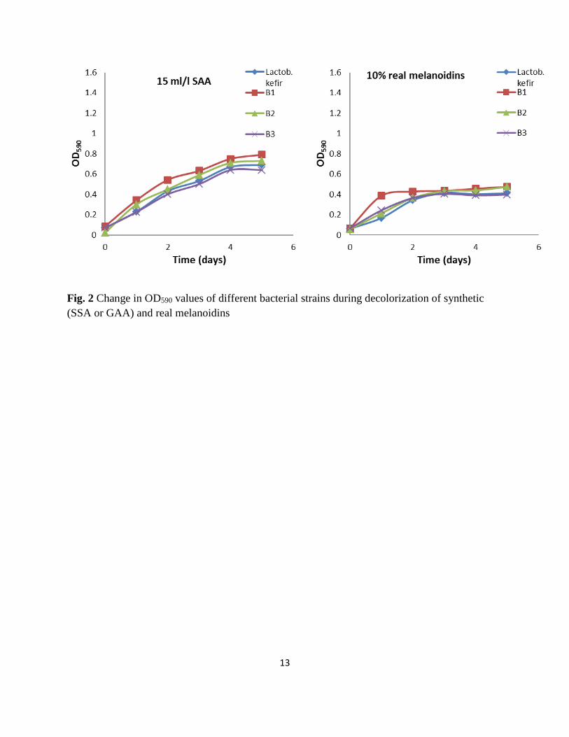
The optical density (OD) for bacterial growth at 590 nm was observed during the degradation of SAA (5 ml/l) and
real melanoidins (%10 v/v) and is presented in Fig.2. The results revealed that a marked increase in OD590 for all
bacterial growth and reached the maximum growth after 2 days of incubation. Three of these isolates and
Lactobacillus kefir strain showed maximum OD590 (1.38- 1.47 and 1.28- 1.35) of GAA-MP (5000 and 15000 mg/l)
in the medium at pH (7.4 ± 0.1), shaking speed (140 rpm) and temperature (30±2 °C) after 5 days incubation,
respectively. However, in real melanoidin solutions (10 % v/v), theses strains showed low OD590 (0.43- 0.66) after 5
days incubation. This increased in the bacterial growth was in parallel with the degradation of SAA and real
melanoidins. Moreover, the bacterial culture was found more effective for the degradation of melanoidins after 1
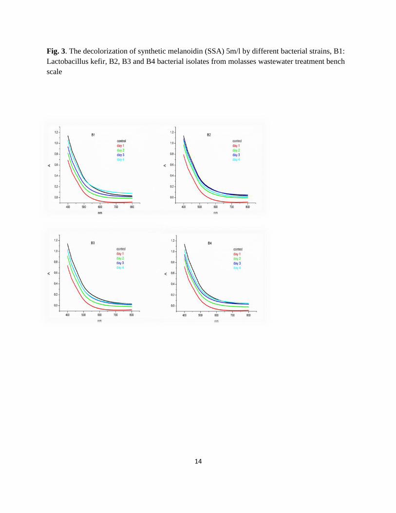
day of incubation, as it decolorized 12 and 8 % SAA (15 ml/l) and natural melanoidins (Fig. 3) respectively. The
same trend was observed in all bacterial cultures. It was noticed that the decolorization of melanoidins
decolorization by Lactob. kifer strains and three bacterial isolates might be due to the cell growth associated.
Lactob. plantarum SF5.6 grew rapidly within one day while the other strains took 2–3 days [10]. Also, the
decolorization of four types of synthetic melanoidins i.e., glucose–glutamic-acid (GGA), glucose–aspartic-acid
7
(GAA), sucrose–glutamic acid (SGA), and sucrose–aspartic-acid (SAA), were investigated using three different
isolates, B. thuringiensis, B. brevis and Bacillus sp. [7].The degree of decolorization of the melanoidins separately
by each isolate was in the 1–31% range. After 2 days of incubation with the bacterial culture, there was no decreased
in decolorazation. It was noticed that with the increase in time duration, decolorization activity of the three isolate
B1, B2 and B3 was decreased up to 5th day (Fig. 3). This was explained by the fact that the pH affected the
metabolic value (microbial acceptability) by generating intermediate hydrolysis products from the compounds
present in the SAA and real melanoidens. The biodegradability was further enhanced by combined SAA with
GPYM broth or real melanoidens with yeast extracts resulting in 34 to 66 % COD reduction after 2 days as
compared to the control (Fig. 4). Lactob. kefir and the isolates showed maximum reduction of COD ranged from 52
to 79 % of SAA (5000 & 15000 mg/l) and real melanoidins in the medium after 5 days incubation. Aeromonas sp.
utilizes the carbonaceous compounds present in spent wash as the sole carbon source, thereby eventually reducing
the effluent COD by 66% in a 24 h period. P. putida also resulted in 44% COD removal accompanied by 60% color
reduction. In another study on predigested distillery effluent with A. formicans, 57% COD reduction and 55%
decrease in color was observed after 72 h [14]. The enhancement in biodegradability was attributed to the source of
bacterial isolation which isolated from molasses wastewater treatment. The decolorization of melanoidin by the
bacterial isolates and Lactob. kifer was shown to be closely related to the composition of medium and the
concentration of melanoidins.
Conclusion
Lactob.kefir and three bacterial isolates showed highest reduction of COD in synthetic and real melanoidins after 5
days of incubation. Yeast extract could be used as bio-stimulator for these isolates during decolourisation of real
melanoidins. These isolates could be useful for decolourisation of industrial wastewater containing high
concentration of melanoidins.
Acknowledgment
I would like to show my gratitude to the funding agencies, Ministry of Egyptian Higher Education and Zagazig
University, Egypt, which provided a support fellowship for my postdoctoral research. The experimental study was
supported and provided by the Laboratory of Water and Wastewater Technology, Department of Food Technology,
Technological Educational Institute of Thessaloniki, 57400 Sindos, Thessaloniki, Greece.
References
[1] Satyawali, Y., Balakrishnan, M.: Wastewater treatment in molasses-based alcohol distilleries for COD and color
removal: a review J. Environ. Manage., 86 (3), 481–497(2008)
[2] Arimi , M.M., Zhang, Y. , Götz, G., Kiriamiti, K. , Geißen, S.: Antimicrobial colorants in molasses distillery
wastewater and their removal technologies. Int. Biodeterioration & Biodegradation 87 34-43(2014)
8
[3] Santal, A.R., Singh, N.P. and Saharan, B.S.: Biodegradation and detoxification of melanoidin from distillery
effluent using an aerobic bacterial strain SAG5 of Alcaligenes faecalis. J. Hazard Mater, 15;193:319-24
(2011)
[4] Adams, A., Tehrani, K.A., Kersiene, M., Venskutonis, R., Kimpe, N.D.: Characterization of model melanoidins
by the thermal degradation profile. J. Agric. Food Chem. 5, 4338–4343(2003)
[5] Sirianuntapiboon, S., Somchai, P., Sihanonth, P., Atthasampunna, P., Ohmomo, S.: Microbial decolorization of
molasses waste water by Mycelia Sterilia D-90. Agric. Biol. Chem. 52, 393–398 (1988)
[6] Pant, D., Adholeya, A.: Biological approaches for treatment of distillery wastewater: a review. Bioresour.
Technol. 98 (12), 2321–2334(2007)
[7] Kumar, P., Chandra, R.: Decolourisation and detoxification of synthetic molasses melanoidins by individual
and mixed cultures of Bacillus spp. Bioresour Technol ;7:2096–102(2006)
[8] Bharagava, R. N., Chandra, R., Rai, V.: Isolation and characterization of aerobic bacteria capable of the
degradation of synthetic and natural melanoidins from distillery effluent. World J. Microbiol. Biotechnol.,
25(5): 737–744(2009)
[9] Gill, P.K., Arora, D.S., Chander, M.: Bidecolourization of azo and triphenylmethane dyes by Dichomitus
squalens and Phlebia spp. J. Ind. Microbiol. Biotechnol., 28, 201–203(2002)
[10] Limkhuansuwan, V., Chaiprasert, P.: Decolorization of molasses melanoidins and palm oil mill effluent
phenolic compounds by fermentative lactic acid bacteria. J. Environ. Sci., 22(8), 1209–1217(2010)
[11] Ohmomo, S., Yoshikawa, H., Nozaki, K., Nakajima, T., Daengsubha, W., Nakamura, I.: Continuous
decolorization of molasses wastewater using immobilized Lactobacillus hilgardii cells. Agric. Biol. Chem.
52, 2437–2441(1998)
[12] Nakajima, K.T., Shimomura, M., Nomura, N., Chanpornpong, T., Takahara, T.: Decolorization of molasses
wastewater by Bacillus sp. under thermophilic and anaerobic conditions. J. Biosci. Bioeng. 87, 119–
121(1999)
[13] Sirianuntapiboon, S., Prasertsong, K.: Treatment of molasses wastewater by acetogenic bacteria BP103 in
sequencing batch reactor (SBR) system. Bioresource Technology, 99, 1806–1815(2008)
[14] Jain, N., Prajapati, S.K., Minocha, A.K., Verma, C.L.: Batch studies on the degradation of spentwash from
distilleries. Ind. J. Environ. Prot. 21 (2), 122–126(2000)
9
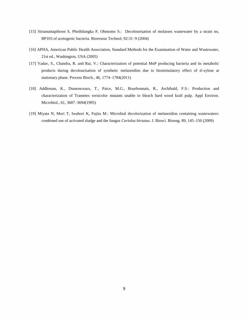
[15] Sirianuntapiboon S, Phothilangka P, Ohmomo S.: Decolourisation of molasses wastewater by a strain no,
BP103 of acetogenic bacteria. Bioresour Technol; 92:31–9 (2004)
[16] APHA, American Public Health Association, Standard Methods for the Examination of Water and Wastewater,
21st ed., Washington, USA (2005)
[17] Yadav, S., Chandra, R. and Rai, V.: Characterization of potential MnP producing bacteria and its metabolic
products during decolourisation of synthetic melanoidins due to biostimulatory effect of d-xylose at
stationary phase. Process Bioch., 46, 1774–1784(2011)
[18] Addleman, K., Dumonceaux, T., Paice, M.G., Bourbonnais, R., Archibald, F.S.: Production and
characterization of Trametes versicolor mutants unable to bleach hard wood kraft pulp. Appl Environ.
Microbiol., 61, 3687–3694(1995)
[19] Miyata N, Mori T, Iwahori K, Fujita M.: Microbial decolorization of melanoidins containing wastewaters:
combined use of activated sludge and the fungus Coriolus hirsutus. J. Biosci. Bioeng. 89, 145–150 (2009)
10
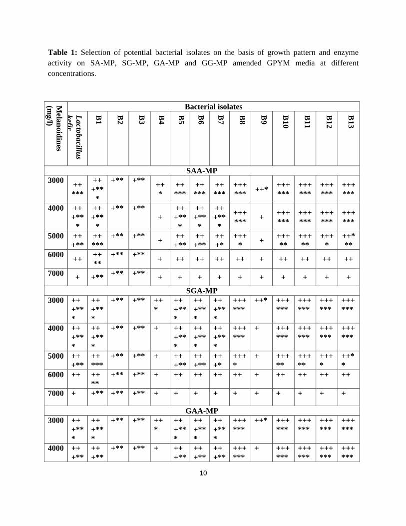
Table 1: Selection of potential bacterial isolates on the basis of growth pattern and enzyme
activity on SA-MP, SG-MP, GA-MP and GG-MP amended GPYM media at different
concentrations.
Mela
noid
ines
(mg/l)
Bacterial isolates
Lacto
bacillu
s
kefir
B1
B2
B3
B4
B5
B6
B7
B8
B9
B10
B11
B12
B13
SAA-MP
3000 ++
***
++
+**
*
+** +** ++
*
++
***
++
***
++
***
+++
*** ++*
+++
***
+++
***
+++
***
+++
***
4000 ++
+**
*
++
+**
*
+** +** +
++
+**
*
++
+**
*
++
+**
*
+++
*** +
+++
***
+++
***
+++
***
+++
***
5000 ++
+**
++
***
+** +** +
++
+**
++
+**
++
+*
+++
* +
+++
**
+++
**
+++
*
++*
**
6000 ++
++
**
+** +** + ++ ++ ++ ++ + ++ ++ ++ ++
7000 + +**
+** +** + + + + + + + + + +
SGA-MP
3000 ++
+**
*
++
+**
*
+** +** ++
*
++
+**
*
++
+**
*
++
+**
*
+++
***
++* +++
***
+++
***
+++
***
+++
***
4000 ++
+**
*
++
+**
*
+** +** + ++
+**
*
++
+**
*
++
+**
*
+++
***
+ +++
***
+++
***
+++
***
+++
***
5000 ++
+**
++
***
+** +** + ++
+**
++
+**
++
+*
+++
*
+ +++
**
+++
**
+++
*
++*
*
6000 ++ ++
**
+** +** + ++ ++ ++ ++ + ++ ++ ++ ++
7000 + +** +** +** + + + + + + + + + +
GAA-MP
3000 ++
+**
*
++
+**
*
+** +** ++
*
++
+**
*
++
+**
*
++
+**
*
+++
***
++* +++
***
+++
***
+++
***
+++
***
4000 ++
+**
++
+**
+** +** + ++
+**
++
+**
++
+**
+++
***
+ +++
***
+++
***
+++
***
+++
***

11
SA-MP=Sucrose-aspartic acid, SG-MP= Sucrose-glutamic acid, GA-MP= Glucose-aspartic acid
and GG-MP= Glucose-glutamic acid Maillard product; += growth; *= enzyme activity
* * * * *
5000 ++
+**
++
***
+** +** + ++
+**
++
+**
++
+*
+++
*
+ +++
**
+++
**
+++
*
++*
**
6000 ++ ++
**
+** +** + ++ ++ ++ ++ + ++ ++ ++ +
7000 + +** +** +** + + + + + + + + + +
GGA-MP
3000 ++
+**
*
++
+**
*
+** +** ++
*
++
+**
*
++
+**
*
++
+**
*
+++
***
++* +++
***
+++
***
+++
***
+++
***
4000 ++
+**
*
++
***
+** +** + ++
+**
*
++
+**
*
++
+**
*
+++
***
+ +++
***
+++
***
+++
***
+++
***
5000 ++
+**
++
**
+** +** + ++
+**
++
+**
++
+*
+++
*
+ +++
**
+++
**
+++
*
++*
**
6000 ++ +**
+** +** + ++ ++ ++ ++ + ++ ++ ++ ++
7000 + +** +* +* + + + + + + + + + +

12
Fig. 1 Change in pH values of synthetic (SSA or GAA) and real melanoidins by different
bacterial strains during their growth for decolorization
13
Fig. 2 Change in OD590 values of different bacterial strains during decolorization of synthetic
(SSA or GAA) and real melanoidins
14
Fig. 3. The decolorization of synthetic melanoidin (SSA) 5m/l by different bacterial strains, B1:
Lactobacillus kefir, B2, B3 and B4 bacterial isolates from molasses wastewater treatment bench
scale

15
Fig. 4 Change of COD mg/l in synthetic and real melanoidins by different bacterial strains