Embed Size (px)
Citation preview
Renal Dysfunction in End-Stage Liver Disease and
Post–Liver TransplantMarcelo S. Sampaio, MD, PhDa, Paul Martin, MD, FRCP, FRCPIb,Suphamai Bunnapradist, MDa,*
KEYWORDS
� Renal failure � Liver-kidney transplant � Liver transplant
KEY POINTS
� Renal dysfunction is common in ESLD patients.
� A 24-hour urine collection or cystatin C are better alternatives to estimate GFR rather thanserum creatinine.
� NGAL may be a better marker to differentiate intrinsic renal injury from HRS.
INTRODUCTION
Acute kidney injury (AKI) is associated with poor outcomes and increased mortality inthe setting of liver disease.1–3 The prevalence of renal dysfunction in orthotropic livertransplant (OLT) recipients has ranged between 17% and 95% depending on thestudy.4,5 The etiologies of renal dysfunction in patients with end-stage liver disease(ESLD) differ from the posttransplant period. This article discusses renal injury in thepatient with cirrhosis and in the ESLD patient, the evaluation of kidney function in pre-transplant liver candidates, renal-replacement therapy (RRT) as a treatment of kidneyinjury or as a bridge to liver transplant, indications for OLT versus a combined liver-kidney transplant (CLKT), and kidney injury in the post–liver transplant period.Fede and colleagues6 reported a seven-fold increase in mortality, with 50% of death
in the first month in patients with cirrhosis who developed renal failure. Prognosis alsovaries with the cause of renal dysfunction. Three-month probability of survival was73% for parenchymal nephropathy, 46% for hypovolemia-associated renal failure,30% for renal injury associated with infection, and 15% for hepatorenal syndrome
The authors have nothing to disclose.a Division of Nephrology, Department of Medicine, Kidney and Pancreas Transplant Program,David Geffen School of Medicine at UCLA, 1015 Gayley Avenue, Suite 220, Los Angeles, CA90024, USA; b Division of Hepatology, Miller School of Medicine, University of Miami, 1500NW 12 Avenue, Jackson Medical Tower E-1101, Miami, FL 33136, USA* Corresponding author. 1015 Gayley Avenue, Suite 220, Los Angeles, CA 90024.E-mail address: [email protected]
Clin Liver Dis 18 (2014) 543–560http://dx.doi.org/10.1016/j.cld.2014.05.003 liver.theclinics.com1089-3261/14/$ – see front matter � 2014 Elsevier Inc. All rights reserved.
Sampaio et al544
(HRS) in a single-center study from Spain.7 Management of AKI complicatingadvanced liver disease involves mitigation of risk factors, hemodynamic support,and RRT, whereas evaluating the need for liver transplant in a cirrhotic patient withprolonged renal insufficiency or in dialysis a CLKT may be indicated. Although suc-cessful liver transplantation reverses renal dysfunction from HRS, posttransplant kid-ney dysfunction may occur. Calcineurin inhibitor (CNI) nephrotoxicity is observed inmost patients maintained on calcineurin and may lead to renal dysfunction with renalfailure after a nonrenal solid organ transplant.8 Eighteen percent of liver transplant re-cipients develop significant chronic kidney disease (CKD) at 5-years posttransplant.4
Reducing risk factors for renal dysfunction, the early identification of AKI, and theappropriate management of AKI and CKD may improve the long-term outcomes ofpatients with liver disease.
PATIENTS WITH CIRRHOSIS AND ESLDRevised Definition of Renal Dysfunction in Patients with Liver Disease
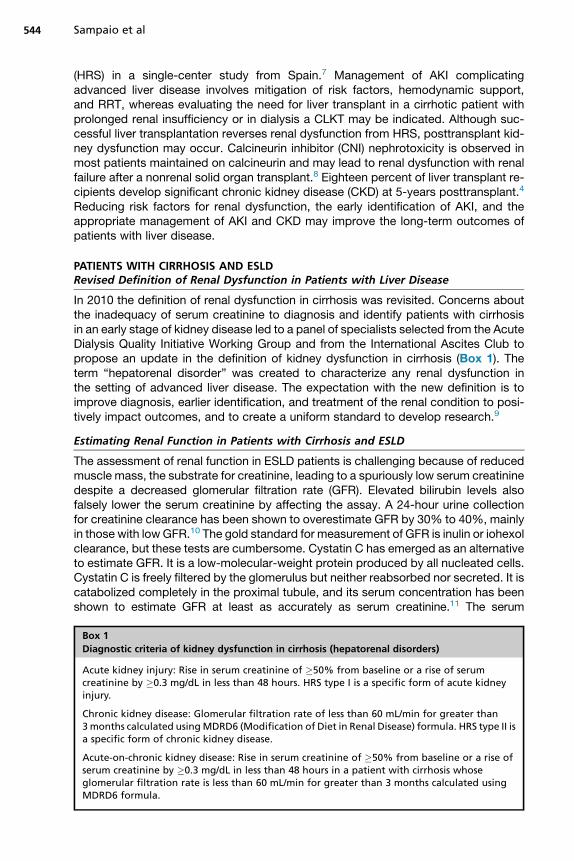
In 2010 the definition of renal dysfunction in cirrhosis was revisited. Concerns aboutthe inadequacy of serum creatinine to diagnosis and identify patients with cirrhosisin an early stage of kidney disease led to a panel of specialists selected from the AcuteDialysis Quality Initiative Working Group and from the International Ascites Club topropose an update in the definition of kidney dysfunction in cirrhosis (Box 1). Theterm “hepatorenal disorder” was created to characterize any renal dysfunction inthe setting of advanced liver disease. The expectation with the new definition is toimprove diagnosis, earlier identification, and treatment of the renal condition to posi-tively impact outcomes, and to create a uniform standard to develop research.9
Estimating Renal Function in Patients with Cirrhosis and ESLD
The assessment of renal function in ESLD patients is challenging because of reducedmuscle mass, the substrate for creatinine, leading to a spuriously low serum creatininedespite a decreased glomerular filtration rate (GFR). Elevated bilirubin levels alsofalsely lower the serum creatinine by affecting the assay. A 24-hour urine collectionfor creatinine clearance has been shown to overestimate GFR by 30% to 40%, mainlyin those with lowGFR.10 The gold standard for measurement of GFR is inulin or iohexolclearance, but these tests are cumbersome. Cystatin C has emerged as an alternativeto estimate GFR. It is a low-molecular-weight protein produced by all nucleated cells.Cystatin C is freely filtered by the glomerulus but neither reabsorbed nor secreted. It iscatabolized completely in the proximal tubule, and its serum concentration has beenshown to estimate GFR at least as accurately as serum creatinine.11 The serum
Box 1
Diagnostic criteria of kidney dysfunction in cirrhosis (hepatorenal disorders)
Acute kidney injury: Rise in serum creatinine of �50% from baseline or a rise of serumcreatinine by �0.3 mg/dL in less than 48 hours. HRS type I is a specific form of acute kidneyinjury.
Chronic kidney disease: Glomerular filtration rate of less than 60 mL/min for greater than3months calculated usingMDRD6 (Modification of Diet in Renal Disease) formula. HRS type II isa specific form of chronic kidney disease.
Acute-on-chronic kidney disease: Rise in serum creatinine of �50% from baseline or a rise ofserum creatinine by �0.3 mg/dL in less than 48 hours in a patient with cirrhosis whoseglomerular filtration rate is less than 60 mL/min for greater than 3 months calculated usingMDRD6 formula.
Renal Dysfunction in Liver Disease 545
concentration of cystatin C is independent of muscle mass and not affected by serumbilirubin levels.12 In a recent study, Souza and colleagues13 have shown that cystatin C–based equations are more accurate than the plasma creatinine-based equations to pre-dict GFR in candidates for liver transplant with cirrhosis. Similar studies in ESLD patientsto assess the accuracy of cystatin C in predicting estimated glomerular filtration rate(eGFR) are still needed. The inability to accurately estimate GFR also contributes to un-toward effects from medications. Many medications are renally excreted and requiredose adjustment based on eGFR, and they may be administered at inappropriatelyhigh doses in patients with cirrhosis because of the inaccuracy of serum creatinine.
TYPES OF RENAL DYSFUNCTIONPrerenal Azotemia
Prerenal azotemia results from renal hypoperfusion and is the most common cause ofacute renal dysfunction in ESLD patients.14 Patients with cirrhosis have multiple po-tential risk factors that can contribute to prerenal azotemia, including hypovolemiafrom diuretic use, diarrhea from lactulose, and gastrointestinal hemorrhage. Patientswith cirrhosis have a progressive rightward shift of the renal vasculature autoregulationcurve with progression of the disease, with renal blood flow more sensitive to adecrease in systemic blood pressure.15 Medications that affect the hemodynamicsof renal vasculature, such as nonsteroidal anti-inflammatory drugs and angiotensin-converting enzyme inhibitors, can induce or contribute to prerenal azotemia. Arterialblood pressure is an independent predictor of survival in patients with cirrhosis.16
Caution should be taken when prescribing angiotensin-converting inhibitors andb-blockers to avoid hypotension. One-year survival was 70% and 40% with meanarterial blood pressure of greater than 82 mm Hg and lower, respectively.17
The HRS
The HRS is functional prerenal azotemia unique to patients with liver failure. HRS, typi-cally seen only in the presence of ascites, can be precipitated by spontaneous bacte-rial peritonitis and other infections, overvigorous diuresis, and large-volumeparacentesis (in general >4 L) without albumin infusion. The pathophysiology ofHRS is complex including splanchnic vasodilation caused by portal hypertension.The shear stress on splanchnic vessels increases production of vasodilators, includingnitric oxide.9 Bacterial translocation from the gut to mesenteric lymph nodes may alsoplay a role in the physiopathology of splanchnic vasodilatation by increasing localinflammation and cytokines production18 and increased mesenteric angiogenesis.With the development of portosystemic shunts the “splanchnic vasodilators” mayalso lead to systemic vasodilatation. Vasodilatation leads to decreased effective circu-latory volume, and overactivation of the renin-angiotensin-aldosterone system andsympathetic nervous system. The compensatory vasoconstrictive effects of therenin-angiotensin-aldosterone system and adrenergic hormones can cause renalischemia and HRS.19 In addition, decreased effective circulatory volume increases so-dium and water retention by the kidneys worsening ascites and edema. The two typesof HRS are differentiated based on their time course. Type I HRS is defined as adoubling of the initial serum creatinine to a level greater than 2.5 mg/dL in less thana 2-week period, whereas type II HRS is characterized by ascites that is resistant todiuretics with moderate renal dysfunction that is slowly progressive.20 Developmentof HRS suggests a poor prognosis with median survival time of 2 weeks in type IHRS and 6 months in type II HRS not responsive to treatment.21 The diagnosis ofHRS is one of exclusion, and true intravascular volume depletion must be ruled out
Sampaio et al546
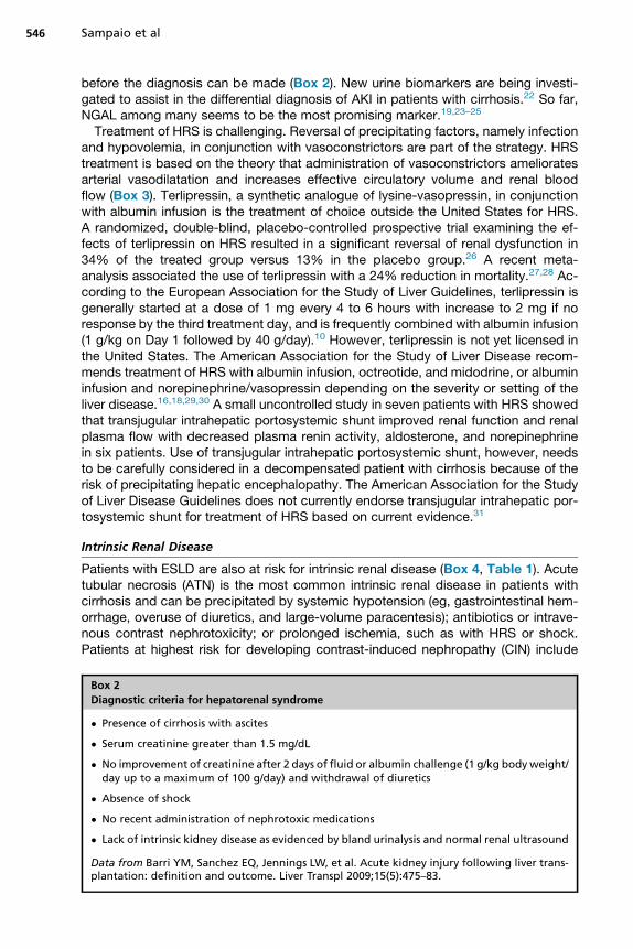
before the diagnosis can be made (Box 2). New urine biomarkers are being investi-gated to assist in the differential diagnosis of AKI in patients with cirrhosis.22 So far,NGAL among many seems to be the most promising marker.19,23–25
Treatment of HRS is challenging. Reversal of precipitating factors, namely infectionand hypovolemia, in conjunction with vasoconstrictors are part of the strategy. HRStreatment is based on the theory that administration of vasoconstrictors amelioratesarterial vasodilatation and increases effective circulatory volume and renal bloodflow (Box 3). Terlipressin, a synthetic analogue of lysine-vasopressin, in conjunctionwith albumin infusion is the treatment of choice outside the United States for HRS.A randomized, double-blind, placebo-controlled prospective trial examining the ef-fects of terlipressin on HRS resulted in a significant reversal of renal dysfunction in34% of the treated group versus 13% in the placebo group.26 A recent meta-analysis associated the use of terlipressin with a 24% reduction in mortality.27,28 Ac-cording to the European Association for the Study of Liver Guidelines, terlipressin isgenerally started at a dose of 1 mg every 4 to 6 hours with increase to 2 mg if noresponse by the third treatment day, and is frequently combined with albumin infusion(1 g/kg on Day 1 followed by 40 g/day).10 However, terlipressin is not yet licensed inthe United States. The American Association for the Study of Liver Disease recom-mends treatment of HRS with albumin infusion, octreotide, and midodrine, or albumininfusion and norepinephrine/vasopressin depending on the severity or setting of theliver disease.16,18,29,30 A small uncontrolled study in seven patients with HRS showedthat transjugular intrahepatic portosystemic shunt improved renal function and renalplasma flow with decreased plasma renin activity, aldosterone, and norepinephrinein six patients. Use of transjugular intrahepatic portosystemic shunt, however, needsto be carefully considered in a decompensated patient with cirrhosis because of therisk of precipitating hepatic encephalopathy. The American Association for the Studyof Liver Disease Guidelines does not currently endorse transjugular intrahepatic por-tosystemic shunt for treatment of HRS based on current evidence.31
Intrinsic Renal Disease
Patients with ESLD are also at risk for intrinsic renal disease (Box 4, Table 1). Acutetubular necrosis (ATN) is the most common intrinsic renal disease in patients withcirrhosis and can be precipitated by systemic hypotension (eg, gastrointestinal hem-orrhage, overuse of diuretics, and large-volume paracentesis); antibiotics or intrave-nous contrast nephrotoxicity; or prolonged ischemia, such as with HRS or shock.Patients at highest risk for developing contrast-induced nephropathy (CIN) include
Box 2
Diagnostic criteria for hepatorenal syndrome
� Presence of cirrhosis with ascites
� Serum creatinine greater than 1.5 mg/dL
� No improvement of creatinine after 2 days of fluid or albumin challenge (1 g/kg bodyweight/day up to a maximum of 100 g/day) and withdrawal of diuretics
� Absence of shock
� No recent administration of nephrotoxic medications
� Lack of intrinsic kidney disease as evidenced by bland urinalysis and normal renal ultrasound
Data from Barri YM, Sanchez EQ, Jennings LW, et al. Acute kidney injury following liver trans-plantation: definition and outcome. Liver Transpl 2009;15(5):475–83.
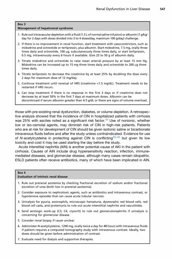
Box 3
Management of hepatorenal syndrome
1. Rule out intravasculardepletionwitha fluid (1.5 L ofnormal saline infusion) or albumin (1g/kg/day for 2 days with doses divided into 3 to 4 doses/day, maximum 100 g/day) challenge.
2. If there is no improvement in renal function, start treatment with vasoconstrictors, such asmidodrine and octreotide or terlipressin, plus albumin. Start midodrine, 7.5 mg, orally threetimes daily and octreotide, 100 mg, subcutaneously three times daily, or start terlipressin,0.5 mg, intravenously every 6 hours if available. Give 25 to 50 g of albumin daily.
3. Titrate midodrine and octreotide to raise mean arterial pressure by at least 15 mm Hg.Midodrine can be increased up to 15 mg three times daily and octreotide to 200 mg threetimes daily.
4. Titrate terlipressin to decrease the creatinine by at least 25% by doubling the dose every2 days for maximum dose of 12 mg/day.
5. Continue treatment until reversal of HRS (creatinine <1.5 mg/dL). Treatment needs to berestarted if HRS recurs.
6. Can stop treatment if there is no response in the first 3 days or if creatinine does notdecrease by at least 50% in the first 7 days at maximum doses. Albumin can bediscontinued if serum albumin greater than 4.5 g/dL or there are signs of volume overload.
Renal Dysfunction in Liver Disease 547
those with pre-existing renal dysfunction, diabetes, or volume depletion. A retrospec-tive analysis showed that the incidence of CIN in hospitalized patients with cirrhosiswas 25% with ascites noted as a significant risk factor.32 Use of nonionic, whetherlow or iso-osmolal agents, may diminish risk of CIN in high-risk patients. Patientswho are at risk for development of CIN should be given isotonic saline or bicarbonateintravenous fluids before and after the study unless contraindicated. Evidence for useof N-acetylcysteine in protecting against CIN is conflicting33–35 but given its lowtoxicity and cost it may be used starting the day before the study.Acute interstitial nephritis (AIN) is another potential cause of AKI in the patient with
cirrhosis. Causes of AIN include drug hypersensitivity reaction, infection, immune-mediated diseases, and glomerular disease, although many cases remain idiopathic.ESLD patients often receive antibiotics, many of which have been implicated in AIN.
Box 4
Evaluation of intrinsic renal disease
1. Rule out prerenal azotemia by checking fractional excretion of sodium and/or fractionalexcretion of urea (both low in prerenal azotemia).
2. Consider exposure to nephrotoxic agents, such as antibiotics and intravenous contrast, orhypotensive episodes that can cause acute tubular necrosis.
3. Urinalysis for pyuria, eosinophils, microscopic hematuria, dysmorphic red blood cells, redblood cell casts, and proteinuria to rule out acute interstitial nephritis and vasculitides.
4. Send serologic work-up (C3, C4, cryocrit) to rule out glomerulonephritis if urinalysis isconcerning for glomerular disease.
5. Consider renal biopsy if cause unclear.
6. Administer N-acetylcysteine, 1200mg, orally twice a day for 48 hours with intravenous fluidsif patient requires a computed tomography study with intravenous contrast. Ideally, twodoses should be given before administration of contrast.
7. Evaluate need for dialysis and supportive therapies.

Table 1Diagnostic criteria and management of intrinsic renal disease
Renal Disease Diagnostic Criteria Risk Factors Treatment
Acute tubular necrosis Bland urinalysis, fractional excretion ofsodium >1%
Hypotension, nephrotoxic medications,intravenous contrast, prolongedischemia caused by hepatorenalsyndrome or shock
Supportive care to treat underlying cause
Acute interstitial nephritis Classic triad of rash, fever, andeosinophilia; sterile pyuria,eosinophiliuria
b-Lactam antibiotics, cephalosporins,sulfonamides, nonsteroidal anti-inflammatory drugs, infection
Discontinue offending agent
IgA nephropathy Microscopic hematuria, proteinuriaoccasionally
Cirrhosis, particularly alcoholic Supportive care
Membranous nephropathy Proteinuria, hypocomplementemia,supepithelial deposits on kidney biopsy
Hepatitis B and C infections Antiviral medications (lamivudine,interferon-a)
Membranoproliferativeglomerulonephritis/cryoglobulinemia
Proteinuria, dysmorphic red cells onurinalysis, hypocomplementemia,positive cryocrit, subendothelialdeposits on kidney deposit withimmune complex deposition
Hepatitis B and C infections Antiviral medications (interferon-a,ribavirin)
Sampaio
etal
548
Renal Dysfunction in Liver Disease 549
The most common drugs implicated in causing AIN include penicillins, cephalospo-rins, sulfonamides, and nonsteroidal anti-inflammatory drugs.36 Proton pump inhibi-tors may also be associated with AIN.37–41 The classic triad of rash, fever, andeosinophilia is seen in less than 30% of patients and the most common presentationis of sterile pyuria in the setting of renal injury. The main treatment consists of discon-tinuation of the suspected drug. Also, some authors advocate the early use of gluco-corticoids, although there are no randomized controlled studies to prove theirbenefit.42–44
IgA nephropathy has been associated with cirrhosis, particularly alcoholic, but itsrole in contributing to renal dysfunction is unclear.45 An observational study of renalbiopsies obtained at time of liver transplantation in 30 patients with hepatitis C–relatedcirrhosis suggested the incidence of IgA nephropathy was 23%.46 Impaired transportof IgA immune complexes from blood to bile by the Kupffer cells in the liver is thoughtto be the cause of increased IgA deposition in the kidneys.47 Treatment options remainlimited, even in primary IgA nephropathy.Glomerulonephritis can complicate chronic hepatitis B and C infection. Patients
with hepatitis C virus (HCV) may develop mixed cryoglobulinemia with associatedmembranoproliferative glomerulonephritis (MPGN).48 The incidence of MPGN wasfound to be 40% in HCV patients who underwent intraoperative renal biopsy attime of liver transplantation.46 Cryoglobulinemic vasculitis is seen in less than 10%of HCV patients, and one-third of those with cryoglobulinemia have MPGN. MostHCV patients with cryoglobulinemia do not exhibit clinical manifestations, but approx-imately one-third have arthralgias, purpura, and asthenia.49 Serologic markersinclude low C3 and C4 levels with elevated rheumatoid factor and cryoglobulins.50
Treatment of hepatitis C with antiviral therapy, including pegylated interferon-a andribavirin, and achievement of virologic response has been shown to improve cryoglo-bulinemia and lessen renal dysfunction.51,52 Excellent results with combined andinterferon-free regimens based on the use of sofusbuvir, simeprevir, and other oralagents for the treatment of hepatitis C were recently published expanding optionsto treat hepatitis C.53–59 Rituximab has also been reported to decrease cryoglobulins,rheumatoid factor, and proteinuria in six patients with hepatitis C–associatedcryoglobulinemia.60
Hepatitis B virus (HBV) can be associated with membranous nephropathy, MPGN,and polyarteritis nodosa. Previous studies have shown that hepatitis B envelope anti-gen is present in the subepithelial deposits seen in HBV-associated membranous ne-phropathy (HBVMN).61 Spontaneous remission of nephrotic syndrome has beenreported in 30% to 60% of patients, and seroconversion to anti–hepatitis B envelopeantigen is associated with remission of proteinuria. Treatment with interferon-a yieldedreduction in proteinuria and HBV DNA levels in 8 of 15 patients treated. All of the treat-ment responders were noted to have HBVMN, whereas the nonresponders mostly hadMPGN.62 Lamivudine has also been used to treat HBVMN. A case control trial of 10patients treated with lamivudine compared with 12 control patients found that thetreatment group had reduction in proteinuria and resolution of HBV DNA.63 In the con-trol group, 42% subsequently developed severe renal dysfunction requiring dialysis,whereas none in the treatment group required renal replacement after 3 years offollow-up. Newer analogs of nucleoside, entecavir, and tenofovir, more potent antiviralagents than lamivudine and adefovir, are now licensed to treat HBV infection.64,65
Although there are no randomized clinical studies examining treatment of hepatitisB–associated glomerulonephritis, antiviral therapy is clearly indicated in patientswith replicating HBV and glomerulonephritis.
Sampaio et al550
ROLE OF RRT
RRT, such as hemodialysis (HD), is often required in sicker OLT candidates as theybecome more overtly decompensated. For patients who required RRT preoperatively,35%survived to liver transplant or discharge,whereas 65%diedwhilewaiting for trans-plant in the hospital, but the 1-year mortality after transplant in those that started RRTwas 30% compared with 9.7% for patients who did not need RRT before transplant.3
Indications for RRT in ESLD patients are essentially similar to other patients (eg, acide-mia, electrolytederangements, volumeoverload, anduremia refractory tomedicalman-agement). However, there are some key differences in ESLD patients. Respiratoryalkalosis typically develops in patients with cirrhosis and can cause a compensatorymetabolic acidosis, which may be mistaken for acidemia unless a blood gas analysisis performed. Uremic encephalopathy may be difficult to differentiate from hepatic en-cephalopathy. The hemodynamic stability of ESLD patients also impacts RRT becausepatients frequently become hypotensive during treatment. Continuous RRT, or contin-uous venovenous HD,may be required if patients have severe hypotension, hyponatre-mia, or cerebral edema. The amount of fluid removed and electrolyte corrections thatare achieved with continuous RRT occur over a 24-hour period versus a 3-hour periodwith single-pass HD, so hemodynamically unstable patients tolerate continuous RRTbetter. However, if rapid adjustments in fluid status or electrolytes are needed, suchas in severe acidemia or hyperkalemia, single-pass HD is the preferred modality. Therole of alternativemethods of RRT, such asmolecular readsorbent recirculation system(MARS [Gambro, Sweden] system) or fractioned plasma separation and adsorption(PROMETHEUS [Fresenius, Germany] system) is still unclear. They may be importantas a bridge for the liver transplant, but studies are needed to create evidence-basedarguments for routine recommendation.18,27
EVALUATION FOR CLKT
Since the introduction of the model of ESLD system in 2002, the rate of CLKT hastripled from 134 patients in 2001 to 399 patients in 2006, reflecting the importanceof creatinine in this model of organ allocation.66 Compared with isolated liver trans-plant recipients, CLKT recipients with end-stage renal disease (ESRD) had higherposttransplant survival at 1 year, but CLKT recipients without ESRD fared no betterthan liver transplant alone recipients.67 There is concern that CLKT may be recom-mended for liver transplant candidates with potentially reversible renal failure (eg,HRS and ATN). A renal biopsy to assess chronicity of renal dysfunction is the goldstandard but is daunting in a patient with cirrhosis and coagulopathy. Intraoperativerenal biopsy has been proposed as a tool to evaluate necessity of CLKT, but thereis currently no consensus or criteria to determine which patients to biopsy.68 Renal ul-trasound to assess kidney size and echogenicity of the cortex can help determine thepotential reversibility of renal dysfunction. Generally, if dialysis has been required formore than a few weeks before liver transplant, renal recovery is much less likely afterOLT, and CLKT needs to be considered. A consensus panel of experts was convenedin 2007 to discuss indications for CLKT, establish a registry, and recommend standardlisting criteria. Recommendations for CLKT were revised in a new summit and werepublished in 2012 (Box 5).10,69 An algorithm has been recommended for evaluationand selection of CLKT patients (Box 6).70
KIDNEY INJURY IN THE POST–LIVER TRANSPLANT PERIOD
Postoperatively, multiple risk factors can cause renal dysfunction in liver transplant re-cipients. Management of postoperative renal dysfunction is summarized in Box 7.
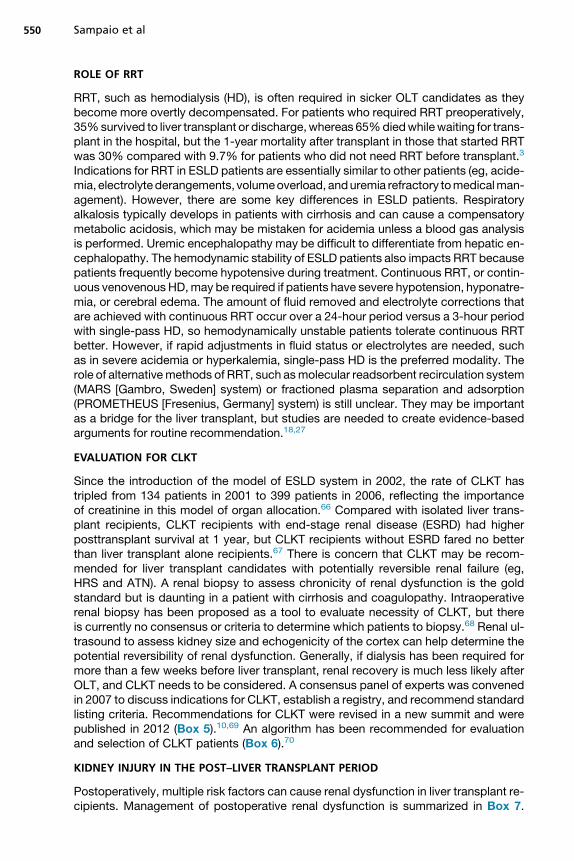
Box 5
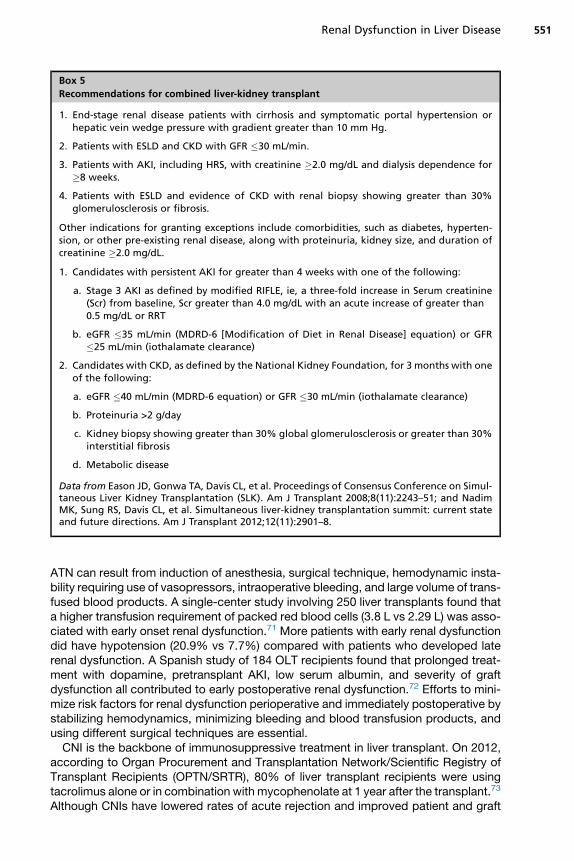
Recommendations for combined liver-kidney transplant
1. End-stage renal disease patients with cirrhosis and symptomatic portal hypertension orhepatic vein wedge pressure with gradient greater than 10 mm Hg.
2. Patients with ESLD and CKD with GFR �30 mL/min.
3. Patients with AKI, including HRS, with creatinine �2.0 mg/dL and dialysis dependence for�8 weeks.
4. Patients with ESLD and evidence of CKD with renal biopsy showing greater than 30%glomerulosclerosis or fibrosis.
Other indications for granting exceptions include comorbidities, such as diabetes, hyperten-sion, or other pre-existing renal disease, along with proteinuria, kidney size, and duration ofcreatinine �2.0 mg/dL.
1. Candidates with persistent AKI for greater than 4 weeks with one of the following:
a. Stage 3 AKI as defined by modified RIFLE, ie, a three-fold increase in Serum creatinine(Scr) from baseline, Scr greater than 4.0 mg/dL with an acute increase of greater than0.5 mg/dL or RRT
b. eGFR �35 mL/min (MDRD-6 [Modification of Diet in Renal Disease] equation) or GFR�25 mL/min (iothalamate clearance)
2. Candidates with CKD, as defined by the National Kidney Foundation, for 3 months with oneof the following:
a. eGFR �40 mL/min (MDRD-6 equation) or GFR �30 mL/min (iothalamate clearance)
b. Proteinuria >2 g/day
c. Kidney biopsy showing greater than 30% global glomerulosclerosis or greater than 30%interstitial fibrosis
d. Metabolic disease
Data from Eason JD, Gonwa TA, Davis CL, et al. Proceedings of Consensus Conference on Simul-taneous Liver Kidney Transplantation (SLK). Am J Transplant 2008;8(11):2243–51; and NadimMK, Sung RS, Davis CL, et al. Simultaneous liver-kidney transplantation summit: current stateand future directions. Am J Transplant 2012;12(11):2901–8.
Renal Dysfunction in Liver Disease 551
ATN can result from induction of anesthesia, surgical technique, hemodynamic insta-bility requiring use of vasopressors, intraoperative bleeding, and large volume of trans-fused blood products. A single-center study involving 250 liver transplants found thata higher transfusion requirement of packed red blood cells (3.8 L vs 2.29 L) was asso-ciated with early onset renal dysfunction.71 More patients with early renal dysfunctiondid have hypotension (20.9% vs 7.7%) compared with patients who developed laterenal dysfunction. A Spanish study of 184 OLT recipients found that prolonged treat-ment with dopamine, pretransplant AKI, low serum albumin, and severity of graftdysfunction all contributed to early postoperative renal dysfunction.72 Efforts to mini-mize risk factors for renal dysfunction perioperative and immediately postoperative bystabilizing hemodynamics, minimizing bleeding and blood transfusion products, andusing different surgical techniques are essential.CNI is the backbone of immunosuppressive treatment in liver transplant. On 2012,
according to Organ Procurement and Transplantation Network/Scientific Registry ofTransplant Recipients (OPTN/SRTR), 80% of liver transplant recipients were usingtacrolimus alone or in combination with mycophenolate at 1 year after the transplant.73
Although CNIs have lowered rates of acute rejection and improved patient and graft
Sampaio et al552
outcomes, they are also associated with nephrotoxicity, hyperglycemia, hyperlipid-emia, and hypertension. CNIs can cause AKI by afferent arteriole vasoconstrictionproducing prerenal ischemia, and prolonged ischemia can lead to ATN. Chronic neph-rotoxicity caused by CNIs is reflected in tubular atrophy, interstitial fibrosis, and glo-merulosclerosis on kidney biopsy.74 The use of a CNI-free regimen is challenging,and so far few studies have shown successful switch to another class of drug withcomplete CNI withdrawal, but yet with short follow-up. The trend in liver transplantis to use regimens that minimize the use of CNIs by addition of or substitution withMycophenolate Mofetil (MMF) or mammalian target of rapamycin inhibitors. Manystudies have shown the potential renal benefit for OLT recipients justifying a combined
Box 6
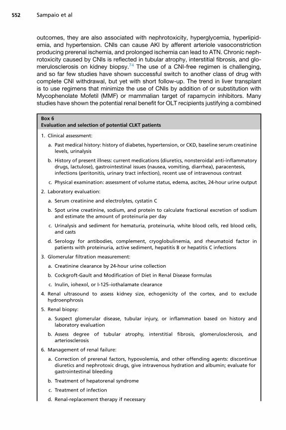
Evaluation and selection of potential CLKT patients
1. Clinical assessment:
a. Past medical history: history of diabetes, hypertension, or CKD, baseline serum creatininelevels, urinalysis
b. History of present illness: current medications (diuretics, nonsteroidal anti-inflammatorydrugs, lactulose), gastrointestinal issues (nausea, vomiting, diarrhea), paracentesis,infections (peritonitis, urinary tract infection), recent use of intravenous contrast
c. Physical examination: assessment of volume status, edema, ascites, 24-hour urine output
2. Laboratory evaluation:
a. Serum creatinine and electrolytes, cystatin C
b. Spot urine creatinine, sodium, and protein to calculate fractional excretion of sodiumand estimate the amount of proteinuria per day
c. Urinalysis and sediment for hematuria, proteinuria, white blood cells, red blood cells,and casts
d. Serology for antibodies, complement, cryoglobulinemia, and rheumatoid factor inpatients with proteinuria, active sediment, hepatitis B or hepatitis C infections
3. Glomerular filtration measurement:
a. Creatinine clearance by 24-hour urine collection
b. Cockgroft-Gault and Modification of Diet in Renal Disease formulas
c. Inulin, iohexol, or I-125–iothalamate clearance
4. Renal ultrasound to assess kidney size, echogenicity of the cortex, and to excludehydroenphrosis
5. Renal biopsy:
a. Suspect glomerular disease, tubular injury, or inflammation based on history andlaboratory evaluation
b. Assess degree of tubular atrophy, interstitial fibrosis, glomerulosclerosis, andarteriosclerosis
6. Management of renal failure:
a. Correction of prerenal factors, hypovolemia, and other offending agents: discontinuediuretics and nephrotoxic drugs, give intravenous hydration and albumin; evaluate forgastrointestinal bleeding
b. Treatment of hepatorenal syndrome
c. Treatment of infection
d. Renal-replacement therapy if necessary
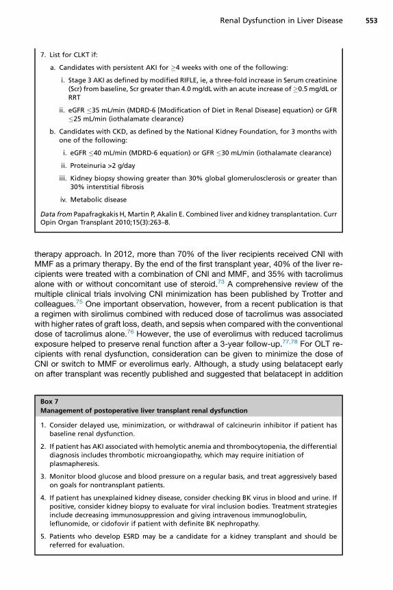
7. List for CLKT if:
a. Candidates with persistent AKI for �4 weeks with one of the following:
i. Stage 3 AKI as defined by modified RIFLE, ie, a three-fold increase in Serum creatinine(Scr) from baseline, Scr greater than 4.0 mg/dL with an acute increase of�0.5 mg/dL orRRT
ii. eGFR �35 mL/min (MDRD-6 [Modification of Diet in Renal Disease] equation) or GFR�25 mL/min (iothalamate clearance)
b. Candidates with CKD, as defined by the National Kidney Foundation, for 3 months withone of the following:
i. eGFR �40 mL/min (MDRD-6 equation) or GFR �30 mL/min (iothalamate clearance)
ii. Proteinuria >2 g/day
iii. Kidney biopsy showing greater than 30% global glomerulosclerosis or greater than30% interstitial fibrosis
iv. Metabolic disease
Data from Papafragkakis H, Martin P, Akalin E. Combined liver and kidney transplantation. CurrOpin Organ Transplant 2010;15(3):263–8.
Renal Dysfunction in Liver Disease 553
therapy approach. In 2012, more than 70% of the liver recipients received CNI withMMF as a primary therapy. By the end of the first transplant year, 40% of the liver re-cipients were treated with a combination of CNI and MMF, and 35% with tacrolimusalone with or without concomitant use of steroid.73 A comprehensive review of themultiple clinical trials involving CNI minimization has been published by Trotter andcolleagues.75 One important observation, however, from a recent publication is thata regimen with sirolimus combined with reduced dose of tacrolimus was associatedwith higher rates of graft loss, death, and sepsis when compared with the conventionaldose of tacrolimus alone.76 However, the use of everolimus with reduced tacrolimusexposure helped to preserve renal function after a 3-year follow-up.77,78 For OLT re-cipients with renal dysfunction, consideration can be given to minimize the dose ofCNI or switch to MMF or everolimus early. Although, a study using belatacept earlyon after transplant was recently published and suggested that belatacept in addition
Box 7
Management of postoperative liver transplant renal dysfunction
1. Consider delayed use, minimization, or withdrawal of calcineurin inhibitor if patient hasbaseline renal dysfunction.
2. If patient has AKI associated with hemolytic anemia and thrombocytopenia, the differentialdiagnosis includes thrombotic microangiopathy, which may require initiation ofplasmapheresis.
3. Monitor blood glucose and blood pressure on a regular basis, and treat aggressively basedon goals for nontransplant patients.
4. If patient has unexplained kidney disease, consider checking BK virus in blood and urine. Ifpositive, consider kidney biopsy to evaluate for viral inclusion bodies. Treatment strategiesinclude decreasing immunosuppression and giving intravenous immunoglobulin,leflunomide, or cidofovir if patient with definite BK nephropathy.
5. Patients who develop ESRD may be a candidate for a kidney transplant and should bereferred for evaluation.
Sampaio et al554
to MMF may be safely used as a bridge to CNI therapy in liver recipients with postop-erative renal dysfunction, the use of belatacept in liver transplant is contraindicateddue to an increase risk of graft loss and death.79
Thrombotic microangiopathy (TMA) of the kidneys has also been associated withCNI use and renal dysfunction in transplant recipients. TMA is a syndrome character-ized by hemolytic anemia, thrombocytopenia, renal dysfunction, fevers, and occasion-ally neurologic deficits. On biopsy, arteriolar thrombi with intimal edema and fibrinoidnecrosis of the vessel wall can be seen.80 Mortality is high, 50% at 3 years in renaltransplant patients with TMA.81 The association between CNI and TMAmay be relatedto direct endothelial injury.82 Kidney biopsies performed in nonrenal transplant recip-ients who had prolonged AKI after transplant or sudden unexplained AKI found a prev-alence of 13% in liver transplant patients.8 Patients with TMA in this cohort also hadthe lowest long-term kidney survival compared with other etiologies of renal dysfunc-tion. Empiric treatment for posttransplant TMA includes therapeutic plasma exchangeand discontinuation or exchange of CNI. A case series of renal transplant patientsshowed that after switching from cyclosporine to tacrolimus, 81% of recipients hadgood graft function 1 year after an episode of TMA.83 In bone marrow transplant recip-ients, however, response rates to plasma exchange have been reported to be lessthan 50%.80
New-onset diabetes mellitus after transplant (NODAT) and hypertension alsocontribute to renal dysfunction in liver transplant recipients. Review of the Organ Pro-curement and Transplant Network/United Network for Organ Sharing databaserevealed that the incidence of NODAT in liver transplant recipients was 26.4%.84 Riskfactors for development of NODAT included older age, African American race, hepatitisC infection, tacrolimususe, steroidsondischarge, andhighbodymass index.Hyperten-sion has also been shown to be associatedwithCNI use.85 Renal insufficiency,NODAT,andposttransplant hypertensionhaveall been identifiedas risk factors formortality afterliver transplant.86 Although there are no formal transplant studies recommending bloodpressure goals, results from large studies summarized in the Joint National Committeeon Prevention, Detection, Evaluation, and Treatment of High Blood Pressure have beenextrapolated for liver transplant recipients.87 Goal blood pressure for patients with renaldysfunction is less than 125/75 and for all other patients is less than 130/80. First-linetherapy includes lifestyle modification, such as weight loss and low-sodium diet. Phar-macologic therapies include initiation of calcium channel blockers initially in patientswithout proteinuria. Use of nondihydropyridine calcium channel blockers, such asverapamil or diltiazem, can increase CNI levels. Recipients with proteinuria should bestarted on angiotensin-converting enzyme inhibitors or angiotensin-receptor blockerswith close monitoring of potassium and renal function. Diabetic nephropathy presentsinitially with microalbuminuria, and efforts to minimize proteinuria with angiotensin-converting enzyme inhibitors or angiotensin-receptor blockers have been shown toretard progression of renal disease in nontransplant patients.Polyomavirus infection, notably BK virus, can occur with immunosuppression
because it remains latent in B lymphocytes and the kidney after primary infection.Its role in nephropathy of renal transplant patients is well established. However, it isunclear if BK virus also causes nephropathy in liver transplant recipients. A study in41 post-OLT patients showed a prevalence of BK viruria in 24.2% of patients, butpresence or absence of viruria was not associated with a decline in eGFR.88 An anal-ysis of nonrenal solid organ transplant patients with unexplained chronic renaldysfunction showed that 15% had BK viruria.89 Although the significance of BK virusand kidney dysfunction in liver transplant recipients is not known at this time, it is apossibility that should be considered if renal disease remains unexplained.

Renal Dysfunction in Liver Disease 555
Hepatitis C may recur after transplant and my affect both liver and kidney graftsurvival.90 Hepatitis C treatment is recommended before the transplant becauseuse of interferon is associated with an increased risk of rejection and profound ane-mia. The development of the antiviral sofosbuvir and the promising combinationsofosbuvir-ledipasvir91 or daclatasvir-sofosbuvir92 may allow treatment of hepatitisC recipients in the posttransplant period with interferon-free regimens and possiblyimprove their outcomes.OLT recipients who subsequent develop ESRD are potentially candidates for
kidney-after-liver transplantation. There was a 330% increase in the number of renaltransplant listings from 1995 to 2008 in ex-OLT recipients.93 In 2008, 124 kidney-after-liver transplants were performed in the United States, which comprised 0.9%of all kidney transplants.94 Kidney transplant does provide a survival benefit comparedwith remaining on dialysis after OLT.93 Overall graft survival in kidney-after-liver recip-ients was less than kidney-alone recipients at 1, 3, and 5 years, but death-censoredgraft survival was similar between the two groups.93
SUMMARY
Renal dysfunction is common in ESLD patients. A 24-hour urine collection or cystatinC are better alternatives to estimate GFR rather than serum creatinine. NGALmay be abetter marker to differentiate intrinsic renal injury from HRS. Preoperatively, the mostcommon causes of renal dysfunction are prerenal azotemia and ATN. Efforts to keeppatients hemodynamically stable and refrain from using nephrotoxic agents are impor-tant. Hepatitis B and C are known to cause glomerulonephritis, whichmay be treatableif recognized early. A CLKT should be considered in patients whose renal function willlikely not recover. After a liver transplant, consideration should be given to reduceexposure to CNI. Future studies are needed to explore potential strategies to reduceor eliminate CNI renal toxicity, such as regimens with everolimus. Aggressive controlof NODAT and blood pressure management may aid in slowing down progression ofCKD. TMA and BK virus are also potential contributors to nephropathy, which is oftenseen in renal and bone marrow transplant recipients. With the introduction of sofosbu-vir and its combination with ledispavir or daclatasvir the treatment of hepatitis C in theposttransplant period looks promising. A kidney-after-liver transplant can be benefi-cial in OLT recipients who develop ESRD.
REFERENCES
1. Gonwa TA, McBride MA, Anderson K, et al. Continued influence of preoperativerenal function on outcome of orthotopic liver transplant (OLTX) in the US: wherewill MELD lead us? Am J Transplant 2006;6(11):2651–9.
2. Fraley DS, Burr R, Bernardini J, et al. Impact of acute renal failure on mortality inend-stage liver disease with or without transplantation. Kidney Int 1998;54(2):518–24.
3. Wong LP, Blackley MP, Andreoni KA, et al. Survival of liver transplant candidateswith acute renal failure receiving renal replacement therapy. Kidney Int 2005;68(1):362–70.
4. Ojo AO, Held PJ, Port FK, et al. Chronic renal failure after transplantation of anonrenal organ. N Engl J Med 2003;349(10):931–40.
5. Barri YM, Sanchez EQ, Jennings LW, et al. Acute kidney injury following livertransplantation: definition and outcome. Liver Transpl 2009;15(5):475–83.
6. Fede G, D’Amico G, Arvaniti V, et al. Renal failure and cirrhosis: a systematic re-view of mortality and prognosis. J Hepatol 2012;56(4):810–8.

Sampaio et al556
7. Martin-Llahi M, Guevara M, Torre A, et al. Prognostic importance of the cause ofrenal failure in patients with cirrhosis. Gastroenterology 2011;140(2):488–96.e4.
8. Schwarz A, Haller H, Schmitt R, et al. Biopsy-diagnosed renal disease in pa-tients after transplantation of other organs and tissues. Am J Transplant 2010;10(9):2017–25.
9. Wong F, Nadim MK, Kellum JA, et al. Working party proposal for a revised clas-sification system of renal dysfunction in patients with cirrhosis. Gut 2011;60(5):702–9.
10. Nadim MK, Sung RS, Davis CL, et al. Simultaneous liver-kidney transplantationsummit: current state and future directions. Am J Transplant 2012;12(11):2901–8.
11. Hojs R, Bevc S, Ekart R, et al. Serum cystatin C-based equation compared toserum creatinine-based equations for estimation of glomerular filtration rate inpatients with chronic kidney disease. Clin Nephrol 2008;70(1):10–7.
12. Orlando R, Mussap M, Plebani M, et al. Diagnostic value of plasma cystatin C asa glomerular filtration marker in decompensated liver cirrhosis. Clin Chem 2002;48(6 Pt 1):850–8.
13. Souza VD, Hadj-Aissa A, Dolomanova O, et al. Creatinine- versus cystatineC-based equations in assessing the renal function of candidates for liver trans-plantation with cirrhosis. Hepatology 2014;59(4):1522–31.
14. Garcia-Tsao G, Parikh CR, Viola A. Acute kidney injury in cirrhosis. Hepatology2008;48(6):2064–77.
15. Stadlbauer V, Wright GA, Banaji M, et al. Relationship between activation of thesympathetic nervous system and renal blood flow autoregulation in cirrhosis.Gastroenterology 2008;134(1):111–9.
16. Runyon BA. Introduction to the revised American Association for the Study ofLiver Diseases Practice Guideline management of adult patients with ascitesdue to cirrhosis 2012. Hepatology 2013;57(4):1651–3.
17. Llach J, Gines P, Arroyo V, et al. Prognostic value of arterial pressure, endoge-nous vasoactive systems, and renal function in cirrhotic patients admitted to thehospital for the treatment of ascites. Gastroenterology 1988;94(2):482–7.
18. Fagundes C, Gines P. Hepatorenal syndrome: a severe, but treatable, cause ofkidney failure in cirrhosis. Am J Kidney Dis 2012;59(6):874–85.
19. Verna EC, Wagener G. Renal interactions in liver dysfunction and failure. CurrOpin Crit Care 2013;19(2):133–41.
20. Salerno F, Gerbes A, Gines P, et al. Diagnosis, prevention and treatment of hep-atorenal syndrome in cirrhosis. Gut 2007;56(9):1310–8.
21. Guevara M, Gines P. Hepatorenal syndrome. Dig Dis 2005;23(1):47–55.22. Belcher JM, Parikh CR, Garcia-Tsao G. Acute kidney injury in patients with
cirrhosis: perils and promise. Clin Gastroenterol Hepatol 2013;11(12):1550–8.23. Belcher JM, Sanyal AJ, Peixoto AJ, et al. Kidney biomarkers and differential
diagnosis of patients with cirrhosis and acute kidney injury. Hepatology 2013.[Epub ahead of print]. http://dx.doi.org/10.1002/hep.26980.
24. Fagundes C, Pepin MN, Guevara M, et al. Urinary neutrophil gelatinase-associated lipocalin as biomarker in the differential diagnosis of impairment ofkidney function in cirrhosis. J Hepatol 2012;57(2):267–73.
25. Verna EC, Brown RS, Farrand E, et al. Urinary neutrophil gelatinase-associatedlipocalin predicts mortality and identifies acute kidney injury in cirrhosis. Dig DisSci 2012;57(9):2362–70.
26. Sanyal AJ, Boyer T, Garcia-Tsao G, et al. A randomized, prospective, double-blind, placebo-controlled trial of terlipressin for type 1 hepatorenal syndrome.Gastroenterology 2008;134(5):1360–8.
Renal Dysfunction in Liver Disease 557
27. Fabrizi F, Aghemo A, Messa P. Hepatorenal syndrome and novel advances in itsmanagement. Kidney Blood Press Res 2013;37(6):588–601.
28. Fabrizi F, Dixit V, Messa P, et al. Terlipressin for hepatorenal syndrome: a meta-analysis of randomized trials. Int J Artif Organs 2009;32(3):133–40.
29. Angeli P, Volpin R, Gerunda G, et al. Reversal of type 1 hepatorenal syndrome withthe administration of midodrine and octreotide. Hepatology 1999;29(6):1690–7.
30. Skagen C, Einstein M, Lucey MR, et al. Combination treatment with octreotide,midodrine, and albumin improves survival in patients with type 1 and type 2hepatorenal syndrome. J Clin Gastroenterol 2009;43(7):680–5.
31. Boyer TD, Haskal ZJ. The role of transjugular intrahepatic portosystemic shunt inthe management of portal hypertension. Hepatology 2005;41(2):386–400.
32. Lodhia N, Kader M, Mayes T, et al. Risk of contrast-induced nephropathy in hos-pitalized patients with cirrhosis. World J Gastroenterol 2009;15(12):1459–64.
33. Alonso A, Lau J, Jaber BL, et al. Prevention of radiocontrast nephropathy withN-acetylcysteine in patients with chronic kidney disease: a meta-analysis of ran-domized, controlled trials. Am J Kidney Dis 2004;43(1):1–9.
34. Isenbarger DW, Kent SM, O’Malley PG. Meta-analysis of randomized clinical tri-als on the usefulness of acetylcysteine for prevention of contrast nephropathy.Am J Cardiol 2003;92(12):1454–8.
35. Marenzi G, Assanelli E, Marana I, et al. N-acetylcysteine and contrast-inducednephropathy in primary angioplasty. N Engl J Med 2006;354(26):2773–82.
36. Michel DM, Kelly CJ. Acute interstitial nephritis. J Am Soc Nephrol 1998;9(3):506–15.
37. Blank ML, Parkin L, Paul C, et al. A nationwide nested case-control study indi-cates an increased risk of acute interstitial nephritis with proton pump inhibitoruse. Kidney Int 2014. http://dx.doi.org/10.1038/ki.2014.74. Advance online pub-lication March 19, 2014 [Epub ahead of print].
38. Berney-Meyer L, Hung N, Slatter T, et al. Omeprazole-induced acute interstitialnephritis: a possible Th1-Th17 mediated injury? Nephrology (Carlton) 2014;19(6):359–65.
39. Harmark L, van der Wiel HE, de Groot MC, et al. Proton pump inhibitor-inducedacute interstitial nephritis. Br J Clin Pharmacol 2007;64(6):819–23.
40. Simpson IJ, Marshall MR, Pilmore H, et al. Proton pump inhibitors and acuteinterstitial nephritis: report and analysis of 15 cases. Nephrology (Carlton)2006;11(5):381–5.
41. Geevasinga N, Coleman PL, Webster AC, et al. Proton pump inhibitors andacute interstitial nephritis. Clin Gastroenterol Hepatol 2006;4(5):597–604.
42. Praga M, Gonzalez E. Acute interstitial nephritis. Kidney Int 2010;77(11):956–61.43. Gonzalez E, Gutierrez E, Galeano C, et al. Early steroid treatment improves the
recovery of renal function in patients with drug-induced acute interstitialnephritis. Kidney Int 2008;73(8):940–6.
44. Appel GB. The treatment of acute interstitial nephritis: more data at last. KidneyInt 2008;73(8):905–7.
45. Pouria S, Feehally J. Glomerular IgA deposition in liver disease. Nephrol DialTransplant 1999;14(10):2279–82.
46. McGuire BM, Julian BA, Bynon JS Jr, et al. Brief communication: glomerulone-phritis in patients with hepatitis C cirrhosis undergoing liver transplantation.Ann Intern Med 2006;144(10):735–41.
47. Amore A, Coppo R, Roccatello D, et al. Experimental IgA nephropathy second-ary to hepatocellular injury induced by dietary deficiencies and heavy alcoholintake. Lab Invest 1994;70(1):68–77.
Sampaio et al558
48. Roccatello D, Fornasieri A, Giachino O, et al. Multicenter study on hepatitis Cvirus-related cryoglobulinemic glomerulonephritis. Am J Kidney Dis 2007;49(1):69–82.
49. Perico N, Cattaneo D, Bikbov B, et al. Hepatitis C infection and chronic renal dis-eases. Clin J Am Soc Nephrol 2009;4(1):207–20.
50. Mackelaite L, Alsauskas ZC, Ranganna K. Renal failure in patients with cirrhosis.Med Clin North Am 2009;93(4):855–69, viii.
51. Alric L, Plaisier E, Thebault S, et al. Influence of antiviral therapy in hepatitis Cvirus-associated cryoglobulinemic MPGN. Am J Kidney Dis 2004;43(4):617–23.
52. Misiani R, Bellavita P, Fenili D, et al. Interferon alfa-2a therapy in cryoglobuline-mia associated with hepatitis C virus. N Engl J Med 1994;330(11):751–6.
53. Asselah T. Sofosbuvir-based interferon-free therapy for patients with HCV infec-tion. J Hepatol 2013;59(6):1342–5.
54. Gane EJ, Stedman CA, Hyland RH, et al. Nucleotide polymerase inhibitor sofos-buvir plus ribavirin for hepatitis C. N Engl J Med 2013;368(1):34–44.
55. Lawitz E, Mangia A, Wyles D, et al. Sofosbuvir for previously untreated chronichepatitis C infection. N Engl J Med 2013;368(20):1878–87.
56. Jacobson IM, Gordon SC, Kowdley KV, et al. Sofosbuvir for hepatitis C genotype2 or 3 in patients without treatment options. N Engl J Med 2013;368(20):1867–77.
57. Hagan LM, Sulkowski MS, Schinazi RF. Cost analysis of sofosbuvir/ribavirinversus sofosbuvir/simeprevir for genotype 1 HCV in interferon ineligible/intol-erant individuals. Hepatology 2014. [Epub ahead of print]. http://dx.doi.org/10.1002/hep.27151.
58. Hussar DA, Jin ZJ. New drugs: simeprevir, sofosbuvir, and dolutegravir sodium.J Am Pharm Assoc (2003) 2014;54(2):202–7.
59. Izumi N, Hayashi N, Kumada H, et al. Once-daily simeprevir with peginterferonand ribavirin for treatment-experienced HCV genotype 1-infected patients inJapan: the CONCERTO-2 and CONCERTO-3 studies. J Gastroenterol 2014;49(5):941–53.
60. Roccatello D, Baldovino S, Rossi D, et al. Long-term effects of anti-CD20 mono-clonal antibody treatment of cryoglobulinaemic glomerulonephritis. Nephrol DialTransplant 2004;19(12):3054–61.
61. Bhimma R, Coovadia HM. Hepatitis B virus-associated nephropathy. Am JNephrol 2004;24(2):198–211.
62. Conjeevaram HS, Hoofnagle JH, Austin HA, et al. Long-term outcome of hepa-titis B virus-related glomerulonephritis after therapy with interferon alfa. Gastro-enterology 1995;109(2):540–6.
63. Tang S, Lai FM, Lui YH, et al. Lamivudine in hepatitis B-associated membranousnephropathy. Kidney Int 2005;68(4):1750–8.
64. Carey I, Harrison PM. Monotherapy versus combination therapy for thetreatment of chronic hepatitis B. Expert Opin Investig Drugs 2009;18(11):1655–66.
65. Pipili CL, Papatheodoridis GV, Cholongitas EC. Treatment of hepatitis B in pa-tients with chronic kidney disease. Kidney Int 2013;84(5):880–5.
66. Tanriover B, Mejia A, Weinstein J, et al. Analysis of kidney function and biopsyresults in liver failure patients with renal dysfunction: a new look to combinedliver kidney allocation in the post-MELD era. Transplantation 2008;86(11):1548–53.
67. Locke JE, Warren DS, Singer AL, et al. Declining outcomes in simultaneous liver-kidney transplantation in the MELD era: ineffective usage of renal allografts.Transplantation 2008;85(7):935–42.
Renal Dysfunction in Liver Disease 559
68. Chopra A, Cantarovich M, Bain VG. Simultaneous liver and kidney transplants:optimizing use of this double resource. Transplantation 2011;91(12):1305–9.
69. Eason JD, Gonwa TA, Davis CL, et al. Proceedings of consensus conference onsimultaneous liver kidney transplantation (SLK). Am J Transplant 2008;8(11):2243–51.
70. Papafragkakis H, Martin P, Akalin E. Combined liver and kidney transplantation.Curr Opin Organ Transplant 2010;15(3):263–8.
71. Lebron Gallardo M, Herrera Gutierrez ME, Seller Perez G, et al. Risk factors forrenal dysfunction in the postoperative course of liver transplant. Liver Transpl2004;10(11):1379–85.
72. Cabezuelo JB, Ramirez P, Rios A, et al. Risk factors of acute renal failure afterliver transplantation. Kidney Int 2006;69(6):1073–80.
73. Kim WR, Smith JM, Skeans MA, et al. OPTN/SRTR 2012 annual data report: liver.Am J Transplant 2014;14(Suppl 1):69–96.
74. Flechner SM, Kobashigawa J, Klintmalm G. Calcineurin inhibitor-sparing regi-mens in solid organ transplantation: focus on improving renal function andnephrotoxicity. Clin Transplant 2008;22(1):1–15.
75. Trotter JF, Grafals M, Alsina AE. Early use of renal-sparing agents in liver trans-plantation: a closer look. Liver Transpl 2013;19(8):826–42.
76. Asrani SK, Wiesner RH, Trotter JF, et al. De novo sirolimus and reduced-dosetacrolimus versus standard-dose tacrolimus after liver transplantation: the2000-2003 phase II prospective randomized trial. Am J Transplant 2014;14(2):356–66.
77. Sterneck M, Kaiser GM, Heyne N, et al. Everolimus and early calcineurin inhib-itor withdrawal: 3-year results from a randomized trial in liver transplantation. AmJ Transplant 2014;14(3):701–10.
78. Saliba F, De Simone P, Nevens F, et al. Renal function at two years in liver trans-plant patients receiving everolimus: results of a randomized, multicenter study.Am J Transplant 2013;13(7):1734–45.
79. LaMattina JC, Jason MP, Hanish SI, et al. Safety of belatacept bridging immuno-suppression in hepatitis C-positive liver transplant recipients with renal dysfunc-tion. Transplantation 2014;97(2):133–7.
80. Batts ED, Lazarus HM. Diagnosis and treatment of transplantation-associatedthrombotic microangiopathy: real progress or are we still waiting? Bone MarrowTransplant 2007;40(8):709–19.
81. Reynolds JC, Agodoa LY, Yuan CM, et al. Thrombotic microangiopathy afterrenal transplantation in the United States. Am J Kidney Dis 2003;42(5):1058–68.
82. Ruggenenti P. Post-transplant hemolytic-uremic syndrome. Kidney Int 2002;62(3):1093–104.
83. Zarifian A, Meleg-Smith S, O’Donovan R, et al. Cyclosporine-associated throm-botic microangiopathy in renal allografts. Kidney Int 1999;55(6):2457–66.
84. Kuo HT, Sampaio MS, Ye X, et al. Risk factors for new-onset diabetes mellitus inadult liver transplant recipients, an analysis of the Organ Procurement andTransplant Network/United Network for Organ Sharing database. Transplanta-tion 2010;89(9):1134–40.
85. Perez MJ, Garcia DM, Taybi BJ, et al. Cardiovascular risk factors after livertransplantation: analysis of related factors. Transplant Proc 2011;43(3):739–41.
86. Watt KD, Pedersen RA, Kremers WK, et al. Evolution of causes and risk factorsfor mortality post-liver transplant: results of the NIDDK long-term follow-up study.Am J Transplant 2010;10(6):1420–7.
Sampaio et al560
87. Guckelberger O. Long-term medical comorbidities and their management: hy-pertension/cardiovascular disease. Liver Transpl 2009;15(Suppl 2):S75–8.
88. Salama M, Boudville N, Speers D, et al. Decline in native kidney function in livertransplant recipients is not associated with BK virus infection. Liver Transpl2008;14(12):1787–92.
89. Barton TD, Blumberg EA, Doyle A, et al. A prospective cross-sectional study ofBK virus infection in non-renal solid organ transplant recipients with chronicrenal dysfunction. Transpl Infect Dis 2006;8(2):102–7.
90. Carbone M, Mutimer D, Neuberger J. Hepatitis C virus and nonliver solid organtransplantation. Transplantation 2013;95(6):779–86.
91. Lawitz E, Poordad FF, Pang PS, et al. Sofosbuvir and ledipasvir fixed-dose com-bination with and without ribavirin in treatment-naive and previously treated pa-tients with genotype 1 hepatitis C virus infection (LONESTAR): an open-label,randomised, phase 2 trial. Lancet 2014;383(9916):515–23.
92. Sulkowski MS, Gardiner DF, Rodriguez-Torres M, et al. Daclatasvir plus sofosbu-vir for previously treated or untreated chronic HCV infection. N Engl J Med 2014;370(3):211–21.
93. Srinivas TR, Stephany BR, Budev M, et al. An emerging population: kidneytransplant candidates who are placed on the waiting list after liver, heart, andlung transplantation. Clin J Am Soc Nephrol 2010;5(10):1881–6.
94. Gonwa TA, McBride MA, Mai ML, et al. Kidney transplantation after previousliver transplantation: analysis of the organ procurement transplant network data-base. Transplantation 2011;92(1):31–5.