Embed Size (px)
Citation preview

2013/2014
Joana Filipa Lopes Figueiredo de Carvalho Paulo
Renal Sympathetic Ablation
New approach of hypertension therapy
março, 2014

Mestrado Integrado em Medicina
Área: Cardiologia
Trabalho efetuado sob a Orientação de:
Prof. Doutor Manuel Joaquim Lopes Vaz da Silva
Revista Portuguesa de Cardiologia
Joana Filipa Lopes Figueiredo de Carvalho Paulo
Renal Sympathetic Ablation
New approach of hypertension therapy
março, 2014


Renal Sympathetic Ablation
New approach of hypertension therapy
Joana Paulo
Faculty of Medicine of the University of Porto (FMUP)
Al. Prof. Hernâni Monteiro 4200-319 Porto, Portugal
e-mail: [email protected] /
Telephone: 00351 963216814
Total de palavras: 5171

i
INDEX
List of Abbreviations -------------------------------------------------------------------------------------------------------- ii
List of Figures ---------------------------------------------------------------------------------------------------------------- iii
List of Tables ----------------------------------------------------------------------------------------------------------------- iii
Abstract ----------------------------------------------------------------------------------------------------------------------- iv
Resumo ------------------------------------------------------------------------------------------------------------------------ v
Introduction ------------------------------------------------------------------------------------------------------------------ 1
Material and Methods ----------------------------------------------------------------------------------------------------- 3
Hypertension Treatment -------------------------------------------------------------------------------------------------- 3
Role of SNS in Hypertensive states -------------------------------------------------------------------------------------- 4
Renal Sympathetic Denervation (RSD) – New treatment for old disease
Historical basis for RSD ------------------------------------------------------------------------------------------ 6
Renal radiofrequency ablation procedure – Pioneering work ---------------------------------------- 7
Recent clinical Evidence
Symplicity system and trials ------------------------------------------------------------------------- 8
Others renal sympathetic denervation trials --------------------------------------------------- 12
Ablation criteria ------------------------------------------------------------------------------------------------------------ 14
Prospective developments ---------------------------------------------------------------------------------------------- 15
Additional effects ---------------------------------------------------------------------------------------------------------- 16
Conclusion ------------------------------------------------------------------------------------------------------------------- 18
Acknowledgments --------------------------------------------------------------------------------------------------------- 19
References------------------------------------------------------------------------------------------------------------------- 20
Annex ------------------------------------------------------------------------------------------------------------------------- 43

ii
LIST OF ABBREVIATIONS
ABPM Ambulatory blood pressure monitoring
BP Blood pressure
CKD Chronic kidney disease
DBP Diastolic blood pressure
ESH/ESC European Society of Hypertension/European Society of Cardiology
GFR Glomerular filtration rate
HF Heart failure
HT Hypertension
HR Heart rate
LV Left ventricular
NA Noradrenaline
RFA Radiofrequency ablation
RH Resistant hypertension
RSD Renal sympathetic denervation
SBP Systolic blood pressure
SNS Sympathetic nervous system

iii
LIST OF FIGURES
Figure 1 ----------------------------------------------------------------------------------------------------------------------- 26
Figure 2 ----------------------------------------------------------------------------------------------------------------------- 27
Figure 3 ----------------------------------------------------------------------------------------------------------------------- 28
Figure 4 ----------------------------------------------------------------------------------------------------------------------- 29
Figure legends -------------------------------------------------------------------------------------------------------------- 30
.
LIST OF TABLES
Table 1 ------------------------------------------------------------------------------------------------------------------------ 31
Table 2 ------------------------------------------------------------------------------------------------------------------------ 32
Table 3 ------------------------------------------------------------------------------------------------------------------------ 34
Table 4 ------------------------------------------------------------------------------------------------------------------------ 35
Table 5 ------------------------------------------------------------------------------------------------------------------------ 37
Table 6 ------------------------------------------------------------------------------------------------------------------------ 38
Table 7 ------------------------------------------------------------------------------------------------------------------------ 41

iv
ABSTRACT
Hypertension is the most common attributable risk factor for stroke and myocardial infarction,
leading to death when inappropriately treated. Despite the wide pharmacological available selection,
blood pressure remains uncontrolled in a significant proportion of patients. Resistant hypertension is
an increasingly common clinical condition, defined by blood pressure ≥140/90 mmHg (or ≥130/80
mmHg in diabetes or renal Insufficiency) regardless concurrent use of 3 or more antihypertensive
drugs from different classes, including one diuretic, all at the optimal doses.
As patients with hypertension are at elevated risk for cardiovascular morbidity and mortality, this
review summarizes the available data, based on bibliographic research using PubMed’s data base, of
new interventional approaches taking into account renal sympathetic activation’s role in
hypertension pathogenesis.
Renal Sympathetic Denervation is a novel catheter percutaneous procedure based on a therapeutic
old concept which intends to ablate the nerves in order to interrupt the central nervous system and
kidneys’ bidirectional connection. So far, trials have demonstrated convincing and safe blood
pressure-lowering effects in majority of treated patients. Moreover, potential additional benefits on
hypertension’s comorbidities, such as left ventricular hypertrophy and renal impairment, have been
identified. Although this current evidence is mainly based on ablation through radiofrequency
energy, several second-generation catheters have been developed aiming at safety and efficacy
improvement.
Therefore, renal sympathetic denervation appears as a future potential hypertension treatment
option, despite being already conducted, under severe controlled conditions, in some countries.
KEYWORDS
Blood pressure; Radiofrequency ablation; Resistant Hypertension; Renal Sympathetic Denervation;
Sympathetic nervous system activity

v
RESUMO
A Hipertensão arterial é um dos principais fatores de risco para Acidente Vascular Cerebral e Enfarte
do Miocárdio, resultando em morte quando não tratada. Apesar da vasta variabilidade
farmacológica, a pressão arterial permanece incontrolada numa significativa percentagem de
doentes. A Hipertensão resistente é uma situação clínica cada vez mais frequente, definida como
pressão arterial ≥140/90 mmHg (ou ≥130/80 mmHg em caso de Diabetes ou Insuficiência Renal)
apesar do uso concomitante de pelo menos 3 anti-hipertensivos de diferentes classes, incluindo um
diurético, todos em doses adequadas.
Visto que os doentes hipertensos apresentam um risco elevado de morbilidade e mortalidade
cardiovasculares, esta revisão reúne a informação disponível, com base em pesquisa bibliográfica
através da PubMed, de novas intervenções baseadas na influência do sistema simpático renal na
patogénese da Hipertensão.
A Desnervação Renal é um novo procedimento percutâneo fundamentado pelo antigo conceito
terapêutico de ablação que interrompe a conexão bidirecional entre o sistema nervoso central e os
rins. Até ao momento, os estudos têm demonstrado efeitos significativos e seguros na diminuição da
pressão arterial na maioria dos indivíduos tratados. Para além disso, potenciais efeitos adicionais nas
co-morbilidades da Hipertensão, como a Hipertrofia Ventricular Esquerda ou Disfunção Renal, têm
sido detetados. Apesar da atual evidência ser baseada sobretudo na ablação por radiofrequência,
vários cateteres têm sido desenvolvidos com o objetivo de melhorar a eficácia e segurança do
procedimento.
Assim, a Desnervação Renal apresenta-se como uma potencial futura opção para o tratamento da
Hipertensão, apesar de já ser utilizada em alguns países, em situações muito específicas.

vi
PALAVRAS-CHAVE
Ablação por radiofrequência; Atividade do sistema nervoso simpático; Desnervação simpática;
Hipertensão resistente; Pressão arterial

1
INTRODUCTION
Hypertension (HT) is a growing worldwide public health problem with an overall prevalence around
30-45% of the general population, with a steep increase with ageing attaining 65% in those over 60
years of age.1 Since 2003 HT is defined as systolic blood pressure (SBP) ≥140 mmHg and/or diastolic
blood pressure (DBP) ≥90 mmHg.2 It is recognized as the leading risk factor for cardiovascular and
cerebrovascular disease, renal and visual impairment and death.3 Cardiovascular risk is linearly
related to blood pressure (BP) levels, doubling with every 20/10 mmHg increase in SBP/DBP,
beginning at 115/75 mmHg. Thereby, it is considered to be responsible for more than 7.5 million
deaths annually in the world.4 A study conducted in 2003 showed that 42.1% of the Portuguese adult
population aged 18-90 years had HT with a prevalence of approximately 79% in patients older than
64 years. Among hypertensive subjects only 11.2% achieved adequate BP control.5
HT can be primary (without apparent cause) or secondary to an identified etiology. The last, seldom
diagnosed, is potentially curable or at least sensible to treatment with cardiovascular risk decrease.1
The most common between these causes are obstructive sleep apnea, chronic renal disease
(parenchymal and vascular) and primary hyperaldosteronism. Cushing’s syndrome,
pheochromocytoma, hyperparathyroidism and aortic coarctation are less frequent causes.3 On the
contrary, in most patients several factors contribute to disease’s development.6 Regardless the
availability of several effective and safe antihypertensive treatment options, BP remains uncontrolled
in a significant percentage of patients (approximately 50%).3,7,8 Table 1 discloses some patient
characteristics associated with resistance to HT treatment.
According to 2013 ESC/ESH recommendations and 2008 American Heart Association, resistant
hypertension (RH) is defined as the inability to reach an effective BP control (<140/90 mmHg and
<140/85 mmHg or <140mmHg in case of diabetes and renal insufficiency, respectively) despite the
association of at least 3 antihypertensive agents with different action mechanisms, one of these

2
being a diuretic, at the maximum tolerated dosage.1,3 Some authors also argue that patients whose
BP is controlled with 4 or more medications are considered to have RH.9
Pseudoresistant HT apparently includes treatment-resistant cases (poorly controlled BP) which are,
however, attributed to other factors.10 Consequently, the RH concept rejects secondary forms,
inefficient BP measurement technique, poor drug and lifestyle approach adherence and inadequate
treatment strategy.11 Moreover, this clinical condition should only be established when the
uncontrolled BP is confirmed to be permanent through 24h-ambulatory BP monitoring (24h-ABPM) 12
with a mean threshold of SBP ≥130 and/or DBP ≥80 mmHg. This monitoring is essential to exclude
pseudoresistance like white coat HT. RH has a multifactorial etiology and its exact prevalence
remains unknown, varying from 5% to 30%.1,11
In the past decades the Sympathetic Nervous System (SNS) has been recognized as a central player in
the cardiovascular homeostasis13 and its enhanced activity has been established as a major
contributor to chronic BP elevation.6 It is also well known that kidneys’ sympathetic innervation plays
a major role in the pathogenesis of HT through modulation of renin’s secretion, glomerular filtration
rate (GFR) and renal absorption of sodium. Based on constantly higher heart rates and absence of
response to intensive diuretic treatment among patients with RH, some authors suggested that
treatment failure may be due to neurologic mechanisms like sympathetic overactivity. This differs
from the traditional assumption that RH is mainly due to persistent hypervolemia.10
Therefore, considering the high prevalence of RH worldwide, the limited efficiency of
pharmacological agents and the significant risk of accelerated cardiovascular mortality11, new
therapeutic procedures targeting the SNS have been developed in order to correct or at least slow
down the pathological mechanism. One of these new and refreshing interventional approaches is the
Renal Sympathetic Denervation (RSD). This review outlines the impact of renal sympathetic activity
on BP regulation, the recent evolution of RSD therapy on RH treatment and its future potential
applications.

3
METHODS
The present review was written based on bibliographic research using PubMed’s data base. The
search was conducted based on MeSH terms using the following combinations: hypertension, blood
pressure, autonomic denervation, catheter ablation, sympathectomy, sympathetic nervous system.
HYPERTENSION TREATMENT
Nowadays, essential HT therapy is based on lifestyle modifications and pharmacological agents. The
former is based on both dietary adjustments with salt restriction (5-6 g per day is recommended)
associated with high consumption of vegetables and fruits and regular exercise practice (at least 30
minutes of moderate dynamic exercise on 5 to 7 days per week is recommended) leading to weight
reduction (of BMI to 25 kg/m2 and of waist circumference to <102 cm in men and <88 cm in women
is recommended, unless contraindicated). Moreover, smoking cessation and moderation of alcohol
consumption (no more than 20-30 g and 10-20 g of ethanol per day in men and women, respectively)
are essential for BP control.
According to 2013 ESH/ESC Guidelines the major advantage of pharmacological therapy is BP-
lowering per se regardless the drug class of first line used, since all medications present similar
effects and particular contraindications. Nevertheless, it must be taken into account both the
adverse side effects and specific conditions in which some drugs have been proved to be more
effective. The most used agents for the initiation and maintenance of therapy, contraindications and
specific conditions to use each class are described on table 2. Combination therapy success has been
attested not only in BP-lowering (possible synergism) but also in therapy compliance (single tablet)
and side effects’ avoidance. Therefore, it is recommended initiation with combined therapy in
patients with high cardiovascular risk or considerably high baseline BP.1

4
The new algorithm purposed by the Eighth Joint National Committee14 for the management of HT is
exposed on figure 1. According to this Committee, BP thresholds for HT definition are maintained.
However, as there are no proven beneficial effects of decreasing BP to levels lower than 140/90
mmHg, the 2014 Guidelines recommend, with grade A level, BP <150/90 mmHg for the general
population aged more than 60 years.
ROLE OF SNS IN HYPERTENSIVE STATES
The sympathetic nervous system (SNS) is a hallmark of primary HT implicated not only in the
development, perpetuation and severity of the disease but also in the pathophysiological
consequences associated (heart and end-stage renal disease) (figure 2).13,15 Nevertheless, only
recently was its differential activation in the various organs established.16
This differential autonomic cardiovascular modulation can be quantified through microneurography
or regional spillover method. The former measures postganglionic sympathetic nerve firing in the
subcutaneous nerves (skin and skeletal muscle vasculature), giving immediate information on the
electrical transmission. The regional noradrenaline (NA) spillover method is considered nowadays the
gold standard.13 This biochemical technique is a clinical index of organ-specific and overall SNS’s
activity since NA efflux into the venous system is proportional to SNS’s firing rate. Consequently,
during constant-rate infusion of radiolabeled NA (which takes into account NA uptake): regional NA
spillover = [(CV - CA) + CAE] x PF, where CV and CA are the plasma concentrations of NA in regional
venous and arterial plasma, respectively, E is the fractional extraction of radiolabeled NA in transit of
blood through the organ, and PF is the organ plasma flow.17
The cardiac baroreflex sensitivity reduction in HT has been recognized in various studies, confirming
the SNS effect on the heart.18 There is also evidence of peripheral vasoconstriction related to SNS
involvement. Additionally, measurement of kidney NA spillover revealed an increased activation of
5
sympathetic outflow with a mean elevation of 2 or 3 times the normal value.13,16 Therefore, the
hyperkinetic circulation profile is early established in HT development with raised resting heart rate
(HR) and cardiac output, increased peripheral and renovascular resistance and elevated plasmatic
levels of NA, independent of disease severity stage.19
The involvement of the renal SNS in the BP control is complex since it is simultaneously the receiver
and originator of sympathetic signals by the efferent and afferent fibers, respectively, both located
within the renal artery.20
The efferent renal sympathetic nerves are regulated by central sympathetic outflow, vagal tone and
renorenal reflexes. These fibers affect renal function not only by inducing antinatriuresis through
elevation of renin secretion via the juxtaglomerular apparatus (β1-adrenoceptors) but also by
enhancing sodium and water reabsorption through activation of the Na+/K+ adenosine
triphosphatase in the renal tubular cells (α1B-adrenoceptors). On the other hand, they also decrease
GFR by inducing direct renal vasoconstriction (α1A-adrenoceptors) which decreases renal blood flow.
However, there seems to be a graded response depending on the effect of the sympathetic signal on
adrenoceptors’ differential activation: low frequency stimulation only affects renin secretion whilst
higher frequencies also influence sodium reabsorption and renal vascular tone.23
The afferent renal sympathetic nerves are stimulated by chemoreceptors in renal interstitium and
mechanoreceptors in the renal wall. The former are sensitive to variations in electrolyte
concentration and plasma osmolality and renal ischemia. The mechanoreceptors are stimulated by
signaling changes in hydrostatic renal pelvic pressure.20 The stimulation of these fibers modulates
autonomic centers in the hypothalamus, the paraventricular nucleus, which increases (directly or
through rostral ventrolateral medulla neurons’ activation) the sympathetic outflow to the kidney and
other organs involved in the cardiovascular control, contributing to the neurogenic elevation of
BP.13,15 Despite central sympathetic system’s predominant role, raised BP is also influenced by
peripheral adrenergic abnormalities as inappropriate neuronal reuptake of NA or peripheral α-

6
adrenoceptors’ downregulation.19 Consequently, through the influence on the regulation of overall
sympathetic tone, afferent fibers have a predominant role in the genesis and maintenance of HT.
Furthermore, these fibers are essential to preserve hydrolytic balance in case of unilateral excretion
disorder since there is a direct communication with the contralateral kidney (renorenal reflex).7
Subsequently, neurogenic primary HT is considered to be responsible for more than 50% of all cases
of high BP.16 Although mechanisms of SNS overdrive are not completely understood, some
hypotheses have been considered such as the impairment of volume-sensitive receptors or arterial
chemoreceptors. Also, the contribution of humoral elements (insulin, angiotensin II) and the
involvement of nutritional or behavioral features are suggested.19
RENAL SYMPATHETIC DENERVATION (RSD)
NEW TREATMENT FOR OLD DISEASE
HISTORICAL BASIS FOR RSD
SNS has been considered a possible therapeutic target in cases of RH since the early 20th century as
its effect on vasoconstriction was already acknowledged. There were many surgical sympathectomy
approaches with different removal extension.24 According to Gulati and White25, the radical lumbo-
dorsal splanchnicectomy was firstly developed in 1938 by Smithwick. Despite being extremely
effective in BP control with reported cardiac size reduction, renal function improvement, decreased
incidence of precordial pain and cerebrovascular events and mortality rate7, these procedures were
associated with intolerable side effects like severe orthostatic hypotension, syncope, paradoxical
excessive sweating, sphincter incontinence and erectile dysfunction. Moreover, the technique’s
significant invasiveness associated with the development of effective and better tolerable oral
sympathetic-blocking drugs led to its abandonment.26

7
Nonetheless, it was an undisputable proof-of-concept of SNS overdrive in RH, confirming BP control’s
achievement through reduction in sympathetic tone. Besides, it revealed that adequate renal
function is independent of intact renal SNS, confirmed by transplantation, since kidneys are still
capable of maintaining electrolyte and volume homeostasis and adrenaline-mediated stress
responses over time despite reduction in the sympathetic input.27
RENAL RADIOFREQUENCY ABLATION (RFA) PROCEDURE – PIONEERING WORK
Recently, RSD has been receiving additional interest as an HT therapeutic option not only because of
sympathetic fibers’ activation role (in particular renal network) in disease’s progression and
complications but also due to significant degree of SNS overdrive demonstrated in RH.19,28 The
development of a new endovascular approach was based on cardiac arrhythmias efficient treatment
with percutaneous radiofrequency ablation (RFA)24, aiming to avoid surgical denervation side effects
and still achieve its success. Therefore, percutaneous renal RFA appears as a selective and minimally
invasive procedure, with limited periprocedural risk and shorter recovery time.
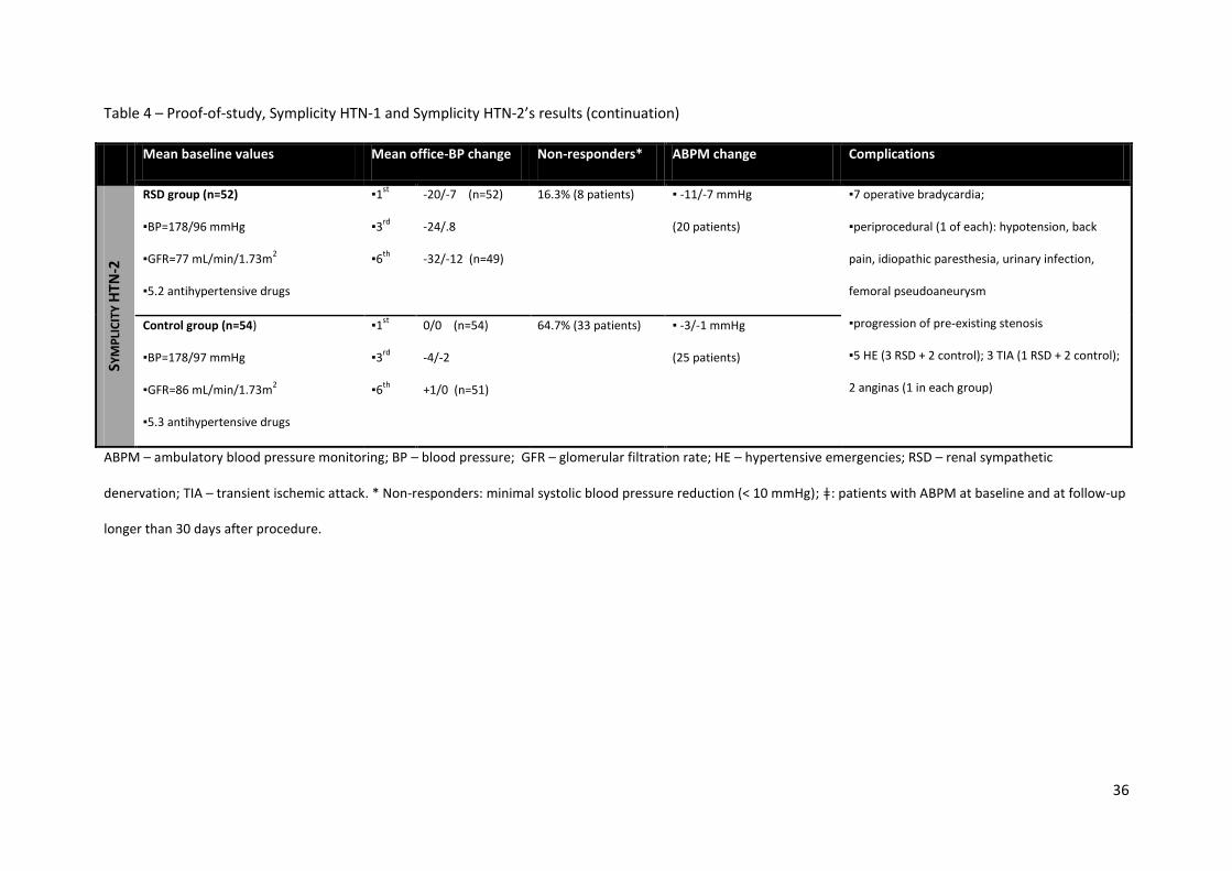
The catheter-based RSD therapy using radiofrequency was firstly outlined by Krum et al in an
international Proof-of-principle study29 which included 50 patients with RH (table 3); 5 were excluded
based on anatomical criteria. This group was followed-up during the trial and had comparable
baseline patient characteristics (table 4). After renal artery angiography and heparin administration
to achieve 250s activated clotting time, the “Symplicity” catheter was placed onto the distal renal
artery wall via femoral artery. The catheter was connected to a radiofrequency generator which
enabled energy delivery to endoluminal surface according to a predetermined algorithm and data on
temperature, length of treatment and impedance, constantly monitored in order to prevent arterial
injury. Radiofrequency energy, lower than that used for cardiac electrophysiological procedures, was
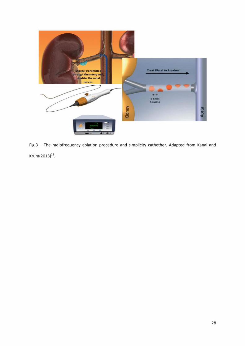
delivered 4 to 6 times in each artery in a helical pattern lasting up to 2 minutes (figure 3).

8
The procedure was initially conducted in a 2-stage way (10 patients had contralateral artery ablation
1 month later). As safety was established, it became a simultaneous bilateral procedure. Both
treatment compliance and maintenance were emphasized to patients and physicians, respectively.
In every visit after the procedure both office SBP and DBP revealed a significant decrease when
compared to baseline (p<0.05). Although the extent of BP reduction is significantly different taking
into account BP measurement method (table 4), office and ABPM decrease are strictly related. RFA
efficiency was sustained even when medical treatment alterations were considered. Regardless the
12-month promising outcomes, 6 patients’ BP decreased less than 10 mmHg (non-responders) which
may point out to either ablation failure or SNS overdrive’s secondary role in some cases of RH.
However, the mean decrease in renal NA spillover rate of 47% in 10 patients 15-30 days after the
procedure associated with the significant reduction of total body NA spillover in 1 patient30 attested
RSD effectiveness in kidney’s both efferent and afferent sympathetic drive reduction.
No significant side effects were detected, both procedure-related renovascular damages (confirmed
by 18 patients’ 14-30 days angiogram and 14 patients’ 6-month magnetic resonance angiogram) and
renal function deterioration (GFR estimated in 25 patients). Only 2 surgical complications were
identified which were immediately resolved: renal artery dissection through stenting and femoral
artery aneurysm with antibiotics and analgesics. Also, the considerable amount of pain during the
procedure (common pathway of sympathetic nerves and C pain fibers) led to a more aggressive
control in the subsequent trials.31
RECENT CLINICAL EVIDENCE
Symplicity system and trials
Despite being the first evidence of RSD’s safety and efficiency, the small number of patients treated
limited Proof-of-principle’s relevance. Therefore, investigators decided to spread the therapeutic
approach to 153 patients, including the patients already treated and increase the follow-up period to

9
36-month (Symplicity HTN-1 Trial).32 After the first 12 months, patients were given the choice of a 24
or 36 months follow-up period, 111 of whom agreed on the second period. The respective baseline
data, BP variations and complications are reported on table 4. BP control (<140 mmHg) was
significantly augmented through the follow-up period, contrarily to the decrease of patients with SBP
>180 mmHg (from 30% at baseline to 5% at 36-month). Although re-innervation was hypothesized,
BP-lowering persisted and even augmented at 36-month when compared with 12-month decrease,
without HR’s significant alteration. One eventual explanation is the afferent renal fibers’ probable
role on central sympathetic overdrive, changing the baroreflex to lower homeostatic regulation
point.30,33 However, antihypertensive drug therapy’s influence remains undetermined since it could
be changed after the 12th month. No significant differences were detected in BP-lowering considering
age, renal function or diabetes status. Despite being overall well preserved, the 10 patients’ 24-
month GFR revealed a decrease of 16 mL/min/1.73m2.34 Nevertheless, according to Sadowski et al
(2011) quoting Bakris and Williams, RSD has a renoprotective effect since GFR decline is lower than it
would be expected considering baseline SBP. The 3 deaths that occurred (1 myocardial infarction, 1
sudden death syndrome and 1 cardiac and respiratory arrest) were considered to be independent
from the ablation procedure. No vascular alteration was detected at 6-month follow-up imaging
(available in 81 patients)34 and stenosis rate was really low. Consequently, the results of the
expanded-cohort were the first to demonstrate not only a sustained BP reduction but also a
preserved safety and maintained renal function 3 years after the procedure.
Nevertheless, Symplicity HTN-1 upholds Proof-of-study’s weaknesses since they lack a control group
(risk of placebo and Hawthorne effect) and both the RH definition (without screening of HT etiology
and no BP measurement method established) and exclusion criteria are inadequate. Moreover,
follow-up numbers remain reduced and the antihypertensive regime adjustments and adherence
were not taken into account. The unblinded analysis might also lead to observer bias. Besides,
selection bias cannot be excluded.7,29,32,33,35

10
Meanwhile, in order to validate the outcomes obtained, the Symplicity HTN-236, an international
randomized clinical trial, recruited 190 patients according to inclusion criteria (table 3). After
recording medication intake and BP values for 2 weeks, 106 of these were randomly distributed in
1:1 ratio to RSD or control groups, both continuing the previous antihypertensive treatment without
adjustments except if medically required.
At baseline, patients’ characteristics, mean BP and antihypertensive therapeutics between the two
groups were identical. The primary endpoint was achieved in 49 of the treated patients (94.2%) and
in 51 of the controls (94.4%), with a significantly different BP reduction between-group (p<0.05).
However, once again these results differ from the 6-month 24h-ABPM (table 4). Six-month BP control
(SBP <140 mmHg) was reached in 39% of RSD group compared to 6% of controls. Six-month SBP
reduction ≥10 mmHg was also significantly different between-group. It is noteworthy to report the
procedure’s complete inefficiency (no BP reduction) in 5 of the treated patients (10%). The
antihypertensive treatment’s decrease was significantly different between groups: 20.4% in RSD and
5.9% in control group. Contrarily, pharmacological intensification was not. Besides, there was no
significant alteration in renal function in both groups during the follow-up period.
Through 6-month imaging tests undertaken in 43 of the 49 patients no difference was detected in
renal vascular anatomy. Although progression of a pre-existent atherosclerotic lesion was observed,
it was placed away from the RSD ablation site. Bradycardia cases were successfully managed as well
as the 5 minor periprocedure complications reported. Major complications during follow-up period
were similar in both groups (table 4). Also, the occurrence of adverse effects was comparable
between groups, without severe procedure or device-related complications in RSD group.
After the 6-month follow-up period, control patients were offered the RSD procedure as long as they
maintained SBP ≥160 mmHg.37 The crossover group (35 patients) had baseline demographic features
and antihypertensive treatments comparable to the initial RSD group. This 12-month follow-up data
exhibited not only persistence of significant BP reduction in the initial RSD group compared to

11
baseline (28/10 mmHg) but also an equivalent significant decrease in the crossover group: a variation
of -24/-8 mmHg, comparable to the 6-month change in the initial interventional group (-32/-8 mmHg,
p=0.15). Besides, therapy regimens’ modifications were not significantly different between the initial
RSD and the crossover groups. GFR remained stable in both groups. Also, safety was confirmed
through only 1 case of artery dissection, 3 hypertensive and 1 hypotensive episodes and no deaths.
Symplicity HTN-2’s results, notwithstanding being on behalf of previous findings, are still
questionable due to similar previous limitations: non-double blinded analysis, incomplete exclusion
criteria (no secondary HT screening) and small sample size and follow-up period, which may mask the
development of complications. Despite being a randomized trial, group baseline characteristics like
sex, diabetes and coronary artery disease rates are different leading to a severity discrepancy into
RSD group. Besides, since baseline 24h-ABPM was not measured, white-coat HT was not excluded.
Although the included patients had to carry out a 2-week antihypertensive therapy, there was no
systematic adherence assessment during follow-up.38-40
RSD future trials need to respond to some of the questions brought up by past trials’ limitations. It is
Symplicity HTN-3’s purpose41, a multicenter randomized single-blinded trial conducted in America,
with wider inclusion and exclusion criteria in order to assess RSD’s both effectiveness and safety in
true RH patients (table 5). Six-month both office-BP and 24h-ABPM are, respectively, primary and
secondary efficiency outcomes, in order to clarify the differences observed in the previous trials. The
6-month incidence of major adverse effects is the primary safety outcome.
Following recruitment phase, in which were enrolled patients with SBP ≥160 mmHg while on stable
treatment with 3 or more different drugs (including one diuretic) for no less than 2 weeks, there was
the screening period with ABPM and therapy adherence registries for at least 2 weeks. Patients with
sustained high SBP and mean 24h-ABPM ≥135 mmHg undertook selective renal angiography to verify

12
the accomplishment of anatomic criteria. 530 patients were posteriorly randomized in a 2:1 ratio to
RSD or sham procedure, both continuing the previous medical treatment.
As a blinded study, patients were unaware of randomization attribution with similar follow-up in
both groups. Moreover, staff measuring BP was also blinded throughout all trial. Before 6-month
evaluation, which includes renal artery duplex imaging, patients recorded ABPM and therapy intake
for 2 weeks. Follow-up period is 3 years for both groups.
The announcement made by Medtronic (symplicity system’s producer) on January the 9th 2014
about Symplicity HTN-3’s results tempered RSD enthusiasm on RH treatment. Despite guaranteeing
primary safety endpoint, the primary efficacy endpoint (a sustained SBP reduction at 6-month) was
not accomplished. (http://newsroom.medtronic.com/phoenix.zhtml?c=251324&p=irol-
newsArticle&id=1889335) According to Bakris, co-principal investigator, even though BP reduction
was not statistically significant, the methodology used is far more rigorous than in previous trials.
Therefore, despite believing European guidelines will be reformulated, he considers procedure’s
dismissal ethically unacceptable. (http://www.medscape.com/viewarticle/819018) Besides, Dr.
Marco Valgimigli, on behalf of the ESC, argues the importance of having the complete data since
treatment’s efficacy is not determined by studies’ primary endpoint success. These concepts are also
supported by Dr. Sanjay Kaul. Other investigators consider Symplicity HTN-3’s results as a possible
failure's consequence of either procedure or Medtronic’s device.
(http://www.medpagetoday.com/Cardiology/Hypertension/43726) However, only through the final
data will definite conclusions be defined.
Others Renal Sympathetic Denervation trials
24h-BP profile has been recognized as organ damage independent prognostic factor. Despite not
established, autonomic dysfunction seems to be the probable underlying mechanism. Taking into

13
account RSD’s effect both on renal and total body sympathetic activity30, Zuern et al hypothesized a
similar outcome regarding 24h-BP variability.42 Inclusion and exclusion criteria and methodology
were comparable to Symplicity’s. Six months after the procedure, the selected patients (n=11)
revealed both an office-SBP decrease equivalent to previous trials (-30.4 mmHg, p=0.007) and a
significant 24h-BP variability reduction (systolic coefficient of variation: from 0.11 to 0.09, p=0.041;
diastolic coefficient of variation: from 0.14 to 0.11, p=0.024), more pronounced than 24h-ABPM
decline (SBP from 149 to 142 mmHg, p=0.086; DBP from 82 to 79 mmHg, p=0.167).
SNS dysfunction is also a predictor of Chronic Kidney Disease’s (CKD) development and progression
as afferent and efferent renal fibers contribute to the sympathetic overactivity vicious cycle. Hence,
15 patients with concomitant RH and moderate to severe CKD underwent renal ablation.43 12-month
results suggest not only RSD’s efficacy and safety since BP was significantly reduced (∆ office-BP=-
33/-19 mmHg) without procedure-related complications, but also maintenance of renal function
(electrolyte and water homeostasis preserved; ∆GFR with p>0.05). Moreover, as all patients had
baseline and follow-up 24h-ABPM, a significant effect on nocturnal BP was detected after only 3
months (∆ABPM night-time=-14/-8 mmHg, p=0.03/0.02), through physiologic dipping pattern
reestablishment (p=0.01).
As BP reduction has enormous cardiovascular benefits (stroke, myocardial infarction, heart failure
and death) RSD was recently performed in 20 cases of long-standing mild RH (even with 3 or more
antihypertensive drugs, SBP=[140-160] mmHg) in order to determine procedure’s efficacy and
safety.44 Six-month outcomes demonstrated office-SBP as well as mean systolic-ABPM significant
decrease despite more modest than in Symplicity’s patients, respectively, -13.1 mmHg (p<0.01) and -
11.3 mmHg (p<0.01), associated with the absence of renal stenosis and preserved renal function
(p=0.5). Therefore, further evidence is required to establish RSD as second-line therapy in mild RH.

14
In a multicenter national Portuguese registry, 78 patients were submitted to RSD, between July-2011
and November-2012, using both Symplicity (n=75) and EnligHTN (n=3) catheters. Among the included
patients mean office-BP was 176.5/94.7 mmHg, most having HT for more than 10 years. Despite the
fact that 2 cases of significant stenosis at the final angiogram and 2 pseudoaneurysms were detected,
RSD’s efficacy was proven by the 74% response rate in 23 patients with more than 6-month follow-
up.45 Already in September 2011 the procedure’s feasibility was confirmed by a Portuguese two-case
report.46
ABLATION CRITERIA
Nowadays, based on clinical trial’s evidence, RSD via Symplicity catheter has been already approved
in Australia47, Europe48 and Canada49 exclusively for RH treatment in patients with preserved renal
function (GFR ≥45 mL/min/1.73m2). Therefore, only patients with raised office-SBP (≥160 mmHg or
≥150 mmHg in type 2 diabetes patients) despite the combination of at least 3 antihypertensive drugs
(including one diuretic) and without contributing factors, like increasing BP substances or
inappropriate lifestyles, are recommended to this procedure. Secondary HT causes must be excluded
as well as non-compliance to antihypertensive treatment. Moreover, confirmation of high BP
through 24h-ABPM is required in order to exclude white-coat HT. Although medical therapy may be
optimized with association of mineralocorticoid receptor antagonist, prolonged treatment with this
drug is restricted due to its adverse effects. An imaging test of the renal system should be realized
before the procedure since it is recommended in renal arteries ≥ 4mm in diameter and ≥ 20 mm in
length before any major branch bifurcation (figure 4).
As there is unsatisfactory clinical evidence, RSD is not recommended in cases of significant renal
artery abnormalities (hemodynamically or anatomically), past renal interventions (angioplasty or
stents), unstable clinical conditions (e.g. acute coronary event), pulmonary arterial HT, chronic

15
oxygen need, pregnancy, preeclampsia or children. Besides, this therapeutic approach should only be
conducted in specialized centers fully equipped not only for diagnosis and procedure execution but
also for complications management. It also requires infrastructure for a complete vigilance with
constant follow-up assessments.50
PROSPECTIVE DEVELOPMENTS
Notwithstanding all the current questions, RSD through endovascular catheter has proved its
influence in BP-lowering. Therefore, this therapeutic potential has led to an exponential
development of new technological concepts (table 6) in order to exploit not only safety and
efficiency with reduction of procedural time but also patient’s comfort through pain reduction.24,51
Regarding RFA techniques, the most widely used is the Symplicity system. However, there are others
systems using different catheters like EnligHTN multielectrode, Vessix Vascular V2 or OneShot
already on trials that aim at reducing both BP and procedural time.21,52 RSD has been proved effective
through ThermoCool, an off-the-shelf saline-irrigated RFA catheter, which is able to significantly
decrease 24h-ABPM (-21/-11 mmHg, p=0.003/0.005) and SNS metabolites’ levels. As proved on
cardiac electrophysiology ablation, this catheter is capable of minimizing the surface damage and
increasing lesions’ depth since it actively cools the electrode.53
Furthermore, this procedural improvement includes ultrasound as a more precise, quicker and less
damaging ablative technique through both invasive and noninvasive procedures, also under
development. Ultrasound energy consists in high-frequency sound waves being able to increase
temperature at depth. The PARADISE catheter emits uniform circumferential ultrasound energy
through the cylindrical transducer positioned inside the water balloon. This ultrasound ablation
presented similar results to RFA’s, decreasing not only non-target tissue’s damage but also
procedure’s time.54
16
Another strategy is tissue-directed microinfusion of neurotoxins, such as guanethidine or vincristine,
into the vessel wall and perivascular area, leading to a chemical sympathectomy without the adverse
side effects of systemic delivery.21,52
However, all these approaches require long-term clinical data in order to correctly establish the most
beneficial.24
ADDITIONAL EFFECTS
As SNS disorder is a systemic condition implicated in chronic diseases’ development, RSD is expected
to have further physiologic benefits. Beyond HT, SNS dysfunction has been established in heart
failure’s progression with β-blockers therapy as a survival prolonger.55 In a recent hypertensive heart
disease trial56 46 patients underwent RSD while 18 attended as control group. The RSD group
exhibited not only marked reduction of left ventricular (LV) mass (from 112.4 g/m2 at baseline to 94.9
g/m2 at 6 months, p<0.001) but also significant improvement of both systolic and diastolic function at
6-month echocardiography evaluation, compared with baseline and controls’ values (table 7). These
results were associated with significant BP decrease. However, non-responders patients (BP
reduction <10 mmHg) showed equally a significant decrease in LV mass and diastolic dysfunction
which may emphasize RSD’s influence on LV hypertrophy despite the lack of BP response.
An extension of Symplicity HTN-257, aiming at establishing RSD’s effect on physical activity with a 3:1
randomization pattern, revealed BP reduction during exercise (from 226/104 mmHg at baseline to
205/99 mmHg at 3 months; p<0.0001 for SBP, p=0.033 for DBP) without chronotropic function
compromise (HR at peak exercise decreased 3 bpm, p=0.141). Also, BP after exercise (limited by
symptoms’ development) and HR recovery improved in the 37 treated patients, -29/-8 mmHg
(p<0.002) and +4 bpm (p=0.009), respectively.

17
Furthermore, RSD has been proved effective in HR reduction58 directly related to baseline HR values
(p<0.05 when baseline HR >60 bpm). Six months after RSD, no correlation was established between
its effect on HR or other electrocardiographic parameters and on BP (r=-0.102, p=0.369). On the
contrary, PR prolongation, which confirms systemic sympathetic activity’s inhibition, is correlated
with a more pronounced HR decrease. These breakthroughs are noteworthy since raised HR plays a
major role in the pathogenesis of coronary disease, myocardial infarction, chronic heart failure and
HT.
Insulin’s role on SNS’s activation through direct central stimulation is already acknowledged.
However, it has also been proved that SNS influences metabolic disarray.19 This potential effect on
metabolic disorders was not only proved by fasting glucose, insulin and C-peptide levels’ significant
reduction, -9.4 mg/dL (p=0.039), -11.6 μIU/mL (p=0.006) and -2.3 ng/mL (p=0.002) respectively, but
also by the significant increase in insulin sensitivity (ISQUICKI=+0.04, p=0.001) in 37 patients 3 months
after RSD, compared to unchangeable levels in control group.59 This data confirms the bidirectional
relationship between SNS overactivity and insulin resistance. Nevertheless, RSD’s metabolic effects
were not correlated with BP variation. Patients with obstructive sleep apnea and RH (n=10) revealed
a metabolic control with improvement of hemoglobin A1C levels (from 6.1% to 5.6%, p<0.05)
associated with BP (-34/-13 mmHg, p<0.01) and apnea-hypoapnea index reduction (from 16.3 to 4.5
events per hour, p=0.059) 6 months after RSD.60
Despite not being assertive, these indications are sufficient to promote future investigation of RSD’s
additional effects.
18
CONCLUSION
Gathering all the current evidence, RSD presents itself as a feasible, safe and clinically relevant
procedure, able to significantly decrease BP levels in RH patients without important complications
associated. Moreover, this technique has been proved effective in HT’s associated complications with
potential cardiac function improvement, HR decrease and metabolic control enhancement.
Since it is a recent interventional approach, most evidence assessing this procedure in humans comes
from trials examining the efficacy and safety of the Symplicity Catheter System. Nevertheless, it has
been submitted to great development with several endovascular catheters presenting different
energy sources.
However, enthusiasm for this possibly revolutionary procedure must be tempered since data is still
restricted due to trial’s limitations which cast doubts about long-term durability or unpredictable
effects development. Also, the discrepancy between office-BP and ABPM measurements raises the
possibility of BP reduction being attributed to nonprocedural-related effects.
Aside these apprehensions, RSD is already considered the last resort treatment in some countries
exclusively for patients with RH who have exhausted all other available medical management
options.

19
ACKNOWLEDGMENTS
I am indebted to my mentor, Manuel Vaz da Silva MD, PhD, for his wise advices, insightful critics,
patience and commitment. To him, an outstanding Physician, an extraordinary Human Being and one
of a kind as a Professor, I owe my deepest gratitude.

20
REFERENCES
1. Mancia G, Fagard R, Narkiewicz K, et al. Task Force for the management of arterial
hypertension of the European Society of Hyperte sion, Task Force for the management of arterial
hypertension of the European Society of Cardiology. 2013 ESH/ESC Guidelines for the Management
of Arterial Hypertension. Blood Press. 2013;22(4):193-278.
2. Mancia G, Agabiti E, Cifkova R, et al. European Society of Hypertension-European Society of
Cardiology Guidelines. 2003 European Society of Hypertension-European Society of Cardiology
guidelines for the management of arterial hypertension. J Hypertens. 2003;21(6):1011-53.
3. Calhoun DA, Jones D, Textor S, et al. Resistant hypertension: diagnosis, evaluation, and
treatment. A scientific statement from the American Heart Association Professional Education
Committee of the Council for High Blood Pressure Research. Hypertension. 2008;51(6):1403-19.
4. World Health Organization - Global Health Observatory. Raised blood pressure - Situation
and trends 2013 [Last access on 8/12/2013]. Available from:
http://www.who.int/gho/ncd/risk_factors/blood_pressure_prevalence_text/en/index.html.
5. Macedo ME, Lima MJ, Silva AO, Alcântara P, Ramalhinho V, Carmona J. Prevalence,
awareness, treatment and control of hypertension in Portugal: the PAP study. J Hypertens.
2005;23(9):1661-6.
6. Chobanian AV, Bakris GL, Black HR, et al. Seventh report of the Joint National Committee on
Prevention, Detection, Evaluation, and Treatment of High Blood Pressure. Hypertension.
2003;42(6):1206-52.
7. Bertog SC, Sobotka PA, Sievert H. Renal denervation for hypertension. JACC Cardiovasc
Interv. 2012;5(3):249-58.
8. Go AS, Mozaffarian D, Roger VL, et al. Heart disease and stroke statistics--2013 update: a
report from the American Heart Association. Circulation. 2013;127(1):e6-e245.

21
9. Kaplan NM, Calhoun DA. Treatment of resistant hypertension. In: UpToDate, Basow DS (Ed),
UpToDate, Waltham, MA. (Accessed on 27/12/2013.).
10. Kaplan NM, Calhoun DA. Definition, risk factors, and evaluation of resistant hypertension. In:
UpToDate, Basow DS (Ed), UpToDate, Waltham, MA. (Accessed on November 25, 2013.).
11. Sarafidis PA, Bakris GL. Resistant hypertension: an overview of evaluation and treatment. J
Am College Cardiol. 2008;52(22):1749-57.
12. Pathak A, Girerd X, Azizi M, et al. Expert consensus: Renal denervation for the treatment of
hypertension. Diagn Interv Imaging. 2012;93(5):386-94.
13. Esler M. The 2009 Carl Ludwig Lecture: Pathophysiology of the human sympathetic nervous
system in cardiovascular diseases: the transition from mechanisms to medical management. J Appl
Physiol. 2010;108(2):227-37.
14. James PA, Oparil S, Carter BL, et al. 2014 Evidence-Based Guideline for the Management of
High Blood Pressure in Adults: Report From the Panel Members Appointed to the Eighth Joint
National Committee (JNC 8). JAMA. 2013.
15. Kumagai H, Oshima N, Matsuura T, et al. Importance of rostral ventrolateral medulla neurons
in determining efferent sympathetic nerve activity and blood pressure. Hypertens Res.
2012;35(2):132-41.
16. DiBona GF, Esler M. Translational medicine: the antihypertensive effect of renal denervation.
Am J Physiol Regul Integr Comp Physiol. 2010;298(2):R245-53.
17. Esler M, Jennings G, Korner P, et al. Assessment of human sympathetic nervous system
activity from measurements of norepinephrine turnover. Hypertension. 1988;11(1):3-20.
18. Parati G, Di Rienzo M, Bertinieri G, et al. Evaluation of the baroreceptor-heart rate reflex by
24-hour intra-arterial blood pressure monitoring in humans. Hypertension. 1988;12(2):214-22.
19. Grassi G. Assessment of sympathetic cardiovascular drive in human hypertension:
achievements and perspectives. Hypertension. 2009;54(4):690-7.

22
20. Krum H, Sobotka P, Mahfoud F, et al. Device-based antihypertensive therapy: therapeutic
modulation of the autonomic nervous system. Circulation. 2011;123(2):209-15.
21. Bunte MC, Infante de Oliveira E, Shishehbor MH. Endovascular treatment of resistant and
uncontrolled hypertension: therapies on the horizon. JACC Cardiovasc Interv. 2013;6(1):1-9.
22. Kanai T, Krum H. New Treatment for Old Disease: Management of Resistant Hypertension by
Percutaneous Renal Sympathetic Denervation. Revi Esp Cardiol. 2013;66(9):734-40.
23. DiBona GF. Physiology in perspective: The Wisdom of the Body. Neural control of the kidney.
Am J Physiol Regul Integr Comp Physiol. 2005;289(3):R633-41.
24. Prochnau D, Figulla HR, Surber R. Renal denervation in the treatment of drug-resistant
hypertension: current knowledge and future perspectives. Expert Rev Med Devices. 2013;10(2):247-
56.
25. Gulati V, White WB. Review of the state of renal nerve ablation for patients with severe and
resistant hypertension. J Am Soc Hypertens. 2013;7(6):484-93.
26. Gewirtz JR, Bisognano JD. Catheter-based renal sympathetic denervation: a targeted
approach to resistant hypertension. Cardiology journal. 2011;18(1):97-102.
27. Schlaich MP, Hering D, Sobotka PA, et al. Renal denervation in human hypertension:
mechanisms, current findings, and future prospects. Curr Hypertens Rep. 2012;14(3):247-53.
28. Briasoulis A, Bakris GL. Timing and efficacy of alternative methods of sympathetic blockade.
Curr Hypertens Rep. 2012;14(5):455-61.
29. Krum H, Schlaich M, Whitbourn R, et al. Catheter-based renal sympathetic denervation for
resistant hypertension: a multicentre safety and proof-of-principle cohort study. Lancet.
2009;373(9671):1275-81.
30. Schlaich MP, Sobotka PA, Krum H, et al. Renal sympathetic-nerve ablation for uncontrolled
hypertension. N Engl J Med. 2009;361(9):932-4.
31. Mahfoud F, Himmel F, Ukena C, et al. Treatment strategies for resistant arterial
hypertension. Dtsch Arztebl Int. 2011;108(43):725-31.
23
32. Krum H, Schlaich MP, Sobotka PA, et al. Percutaneous renal denervation in patients with
treatment-resistant hypertension: final 3-year report of the Symplicity HTN-1 study. Lancet.
2014;383(9917):622-9.
33. Johns EJ. Resistant hypertension and renal denervation: 3 years on. Lancet.
2014;383(9917):583-4.
34. Sadowski J, Bartus K, Kapelak B, et al. Catheter-based renal sympathetic denervation for
resistant hypertension: durability of blood pressure reduction out to 24 months. Hypertension.
2011;57(5):911-7.
35. Doumas M, Faselis C, Papademetriou V. Renal sympathetic denervation and systemic
hypertension. Am J Cardiol. 2010;105(4):570-6.
36. Esler MD, Krum H, Sobotka PA, et al. Renal sympathetic denervation in patients with
treatment-resistant hypertension (The Symplicity HTN-2 Trial): a randomised controlled trial. Lancet.
2010;376(9756):1903-9.
37. Esler MD, Krum H, Schlaich M, et al. Renal sympathetic denervation for treatment of drug-
resistant hypertension: one-year results from the Symplicity HTN-2 randomized, controlled trial.
Circulation. 2012;126(25):2976-82.
38. Persu A, Renkin J, Thijs L, et al. Renal denervation: ultima ratio or standard in treatment-
resistant hypertension. Hypertension. 2012;60(3):596-606.
39. Uder M, Schmid A, Titze S, et al. Renal artery denervation for the treatment of hypertension:
opening up new horizons. Cardiovasc Intervent Radiol. 2011;34(3):442-4.
40. Doumas M, Douma S. Renal sympathetic denervation: the jury is still out. Lancet.
2010;376(9756):1878-80.
41. Kandzari DE, Bhatt DL, Sobotka PA, et al. Catheter-based renal denervation for resistant
hypertension: rationale and design of the SYMPLICITY HTN-3 Trial. Clin Cardiol. 2012;35(9):528-35.
42. Zuern CS, Rizas KD, Eick C, et al. Effects of Renal Sympathetic Denervation on 24-hour Blood
Pressure Variability. Front Physiol. 2012;3:134.

24
43. Hering D, Mahfoud F, Walton AS, et al. Renal denervation in moderate to severe CKD. J Am
Soc Nephrol. 2012;23(7):1250-7.
44. Kaltenbach B, Franke J, Bertog SC, et al. Renal sympathetic denervation as second-line
therapy in mild resistant hypertension: a pilot study. Catheter Cardiovasc Interv. 2013;81(2):335-9.
45. Araújo Gonçalves P, Infante De Oliveira E, Cyrne De Carvalho H, et al. Renal denervation for
resistant hypertension: initial results of the Portuguese National registry [abstract]. EuroIntervention.
2013.
46. Araujo Goncalves P, Sousa Almeida M, Branco P, et al. Renal sympathetic denervation for
treatment of resistant hypertension. Rev Port Cardiol. 2012;31(10):671-5.
47. Weber T, Zweiker R, Watschinger B, et al. Clinical application of interventional renal
sympathetic denervation: recommendations of the Austrian Society of Hypertension 2012. Wien Klin
Wochenschr. 2012;124(21-22):789-98.
48. Mahfoud F, Luscher TF, Andersson B, et al. Expert consensus document from the European
Society of Cardiology on catheter-based renal denervation. Eur Heart J. 2013;34(28):2149-57.
49. Khan NA, Herman RJ, Quinn RR, et al. Renal denervation therapy for the treatment of
resistant hypertension: a position statement by the canadian hypertension education program. Can J
Cardiol. 2014;30(1):16-21.
50. Schlaich MP, Schmieder RE, Bakris G, et al. International expert consensus statement:
percutaneous transluminal renal denervation for the treatment of resistant hypertension. J Am Coll
Cardiol. 2013;62(22):2031-45.
51. Granada JF, Buszman PP. Renal denervation therapies for refractory hypertension. Curr
Cardiol Rep. 2012;14(5):619-25.
52. Mafeld S, Vasdev N, Haslam P. Renal denervation for treatment-resistant hypertension. Ther
Adv Cardiovasc Dis. 2012;6(6):245-58.

25
53. Ahmed H, Neuzil P, Skoda J, et al. Renal sympathetic denervation using an irrigated
radiofrequency ablation catheter for the management of drug-resistant hypertension. JACC
Cardiovascular interventions. 2012;5(7):758-65.
54. Mabin T, Sapoval M, Cabane V, et al. First experience with endovascular ultrasound renal
denervation for the treatment of resistant hypertension. EuroIntervention. 2012;8(1):57-61.
55. Parati G, Esler M. The human sympathetic nervous system: its relevance in hypertension and
heart failure. Eur Heart J. 2012;33(9):1058-66.
56. Brandt MC, Mahfoud F, Reda S, et al. Renal sympathetic denervation reduces left ventricular
hypertrophy and improves cardiac function in patients with resistant hypertension. J Am Coll Cardiol.
2012;59(10):901-9.
57. Ukena C, Mahfoud F, Kindermann I, et al. Cardiorespiratory response to exercise after renal
sympathetic denervation in patients with resistant hypertension. J Am Coll Cardiol.
2011;58(11):1176-82.
58. Ukena C, Mahfoud F, Spies A, et al. Effects of renal sympathetic denervation on heart rate
and atrioventricular conduction in patients with resistant hypertension. Int J Cardiol.
2013;167(6):2846-51.
59. Mahfoud F, Schlaich M, Kindermann I, et al. Effect of renal sympathetic denervation on
glucose metabolism in patients with resistant hypertension: a pilot study. Circulation.
2011;123(18):1940-6.
60. Witkowski A, Prejbisz A, Florczak E, et al. Effects of renal sympathetic denervation on blood
pressure, sleep apnea course, and glycemic control in patients with resistant hypertension and sleep
apnea. Hypertension. 2011;58(4):559-65.

26
Fig. 1 - Algorithm for hypertension management. Adapted from James, Oparil and Carter (2013).14 β-blockers are not recommended for HT’s initial
treatment since not only compared with the 4 recommended classes the results were similar in some studies but also stroke’s incidence was higher
compared with ARB. *No diabetes or CKD. ǂ with or without diabetes. ACEI – angiotensin converting enzyme inhibitors; ARB – angiotensin receptor
blockers; BP – blood pressure; CCB – calcium channel blockers; CKD – chronic kidney disease; DBP – diastolic blood pressure; SBP – systolic blood pressure.
Adult aged ≥18 years with hypertension
LIFESTYLE INTERVENTIONS (continue throughout
management)
Set BP goal and initiate medication based on age,
diabetes and CKD
General population *
Diabetes or CKD present
≥60 years
<60 years
All ages CKD ǂ
SBP <150 mmHg DBP <90 mmHg
SBP <140 mmHg DBP <90 mmHg
SBP <140 mmHg DBP <90 mmHg
SBP <140 mmHg DBP <90 mmHg
All races ACEI or ARB, alone or
in combination with other drug class
THIAZIDE-DIURETIC, ACEI, ARB or CCB,
alone or in combination
THIAZIDE-DIURETIC or CCB, alone or in
combination
Bla
ck
No
nb
lack
Select a drug treatment titration strategy
1. First drug maximization before second addition
2. Second drug addiction before first drug’s dose maximization
3. Initiation with 2 drugs, separately or as fixed-dose combination
BP goal achieved?
No
Continue current treatment and
monitoring
All ages diabetes, no CKD
GOAL
Medication and lifestyle adherence reinforcement Addition and titration of drug class not
previously selected, avoiding ACEI and ARB’s combination.
Yes

27
Fig.2 – Sympathetic nervous system role in Hypertension. Adapted from Bunte, Infante-Oliveira and
Shishehbor (2013)21; Kanai and Krum (2013)22. GFR – glomerular filtration rate.
28
Fig.3 – The radiofrequency ablation procedure and simplicity cathether. Adapted from Kanai and
Krum(2013)22.

29
Fig.4 – Recommended pathway to determine patients’ eligibility for Renal Sympathetic Denervation
procedure. Adapted from Schlaich, Schmieder, Bakris et al(2013)50; Mahfoud, Luscher, Andersson et
al(2013)48. ABPM – ambulatory blood pressure monitoring; BP – blood pressure; CKD – chronic
kidney disease; DM – diabetes mellitus; GFR – glomerular filtration rate; HT- hypertension; NSAID’s –
non-steroidal anti-inflammatory drugs; OSA – obstructive sleep apnea; RSD – renal sympathetic
denervation; SBP – systolic blood pressure.

30
FIGURE LEGENDS
Fig. 1 - Algorithm for hypertension management. Adapted from James, Oparil and Carter (2013).14 β-
blockers are not recommended for HT’s initial treatment since not only compared with the 4
recommended classes the results were similar in some studies but also stroke’s incidence was higher
compared with ARB. *No diabetes or CKD. ǂ with or without diabetes. ACEI – angiotensin converting
enzyme inhibitors; ARB – angiotensin receptor blockers; BP – blood pressure; CCB – calcium channel
blockers; CKD – chronic kidney disease; DBP – diastolic blood pressure; SBP – systolic blood pressure.
Fig.2 – Sympathetic nervous system role in Hypertension. Adapted from Bunte, Infante-Oliveira and
Shishehbor (2013)21; Kanai and Krum (2013)22. GFR – glomerular filtration rate.
Fig.3 – The radiofrequency ablation procedure and simplicity cathether. Adapted from Kanai and
Krum(2013)22.
Fig.4 – Recommended pathway to determine patients’ eligibility for Renal Sympathetic Denervation
procedure. Adapted from Schlaich, Schmieder, Bakris et al(2013)50; Mahfoud, Luscher, Andersson et
al(2013)48. ABPM – ambulatory blood pressure monitoring; BP – blood pressure; CKD – chronic
kidney disease; DM – diabetes mellitus; GFR – glomerular filtration rate; HT- hypertension; NSAID’s –
non-steroidal anti-inflammatory drugs; OSA – obstructive sleep apnea; RSD – renal sympathetic
denervation; SBP – systolic blood pressure.

31
Table 1 – Patients characteristics associated with hypertension treatment-resistance
RISK FACTORS
excessive sodium intake
excessive alcohol consumption
physical inactivity
female sex
African race
advanced age
higher baseline BP (specially systolic)
chronic renal disease
obesity
diabetes
presence of left ventricular hypertrophy
medications that increase BP or decrease antihypertensive agents effect
inadequate therapy
HT secondary causes
Adapted from Calhoun et al (2008)3 and UpToDate (2013). BP – blood pressure; HT – hypertension.

32
Table 2 – Pharmacological treatment of Hypertension
ANTIHYPERTENSIVES † COMPELLING CONTRAINDICATIONS POSSIBLE CONTRAINDICATIONS CONDITIONS IN WHICH ARE PREFERRED
AC
E IN
HIB
ITO
RS
Captopril, Enalapril, Fosinopril,
Imidapril, Lisinopril, Perindopril,
Quinapril, Ramipril, Trandolapril,
Zofenopril
Pregnancy, bilateral renal artery
stenosis angioneurotic oedema,
hyperkalaemia,
Women with child bearing
potential
LVH
Microalbuminuria
Renal dysfunction
Previous MI
HF
Metabolic syndrome
Diabetes mellitus
Asymptomatic atherosclerosis ǂ
Peripheral artery disease ǂ
AR
B
Telmisartan, Irbesartan,
Candersartan, Losartan,
Valsartan, Eprosartan
Pregnancy, hyperkalaemia,
bilateral renal artery stenosis
Women with child bearing
potential
Β-B
LOC
KER
S
Bisoprolol, Nebivolol, Carvedilol,
Celiprolol, Atenolol, Metoprolol
Asthma, A–V block (grade 2 or 3) Metabolic syndrome, glucose
intolerance, athletes and
physically active patients, COPD
Previous MI
Angina pectoris
HF
AF (ventricular rate control)
Pregnancy
CA
LCIU
M C
HA
NN
EL
BLO
CK
ERS
NONDIHYDROPYRIDINES
Verapamil, Diltiazem
A–V block (grade 2 or 3,
trifascicular block), severe LV
dysfunction, HF
LVH
ISH (elderly)
Angina pectoris
Black population
Pregnancy
Peripheral artery disease
Metabolic syndrome
Asymptomatic atherosclerosis
AF (ventricular rate control) * DIHYDROPYRIDINES
Amlodipine,
Tachyarrhythmia
HF

33
Table 2 – Pharmacological treatment of Hypertension (continuation)
ANTIHYPERTENSIVES † COMPELLING CONTRAINDICATIONS POSSIBLE CONTRAINDICATIONS CONDITIONS IN WHICH ARE PREFERRED
DIU
RET
ICS
THIAZIDE-TYPE
Chlorthalidone,
Indapamide,
Hydrochlorothiazide
Gout HF
ISH (elderly)
Black population
MINERALOCORTICOID RECEPTOR
ANTAGONISTS
Spironolactone,
Eplenerone
Acute or severe renal failure
(eGFR <30 mL/min), hyperkalaemia
Metabolic syndrome, glucose
intolerance, pregnancy,
hypercalcemia, hypokalaemia
HF
Adapted from ESC/ESH Guidelines (2013)1 and JNC8 Guidelines (2014)
14. ACE – angiotensin-converting enzyme; AF – atrial fibrillation; ARB – angiotensin receptor blockers;
A-V – atrio-ventricular; COPD – chronic obstructive pulmonary disease; eGFR – estimated glomerular filtration rate; HF – heart failure; ISH – Isolated systolic hypertension;
LV – left ventricular; LVH – left ventricular hypertrophy; MI – myocardial infarction. † - Only some examples of several classes; ǂ - not angiotensin receptor blockers; * - not
dihydropyridines.

34
Table 3 – Proof-of-Principle, Symplicity HTN-1 and Symplicity HTN-2’s study characteristics
Type of study Inclusion Criteria Exclusion criteria Outcomes Follow-up
PR
OO
F –
OF
–PR
INC
IPLE
Observational
study
▪SBP ≥ 160 mmHg with ≥ 3
antihypertensive drugs (1 diuretic)
▪confirmed intolerance to medications
▪GFR ≥ 45 mL/min/1.73m2
▪≥ 18 years
▪not pregnant
▪secondary HT cause excluded
▪type 1 diabetes
▪hemodynamically significant valvular disease
▪implanted pacemakers
▪renovascular abnormalities (renal artery
stenosis, previous renal stent or angioplasty,
dual renal or polar arteries)
▪on treatment with: clonidine, monoxidine,
rilmenidine, warfarin
Primary:
▪BP reduction and procedural safety
Secondary:
▪NA release and renal function
12 months
SYM
PLI
CIT
Y H
TN-1
Observational
study
Primary
▪durability of BP-lowering effects
▪ late adverse vascular or renal effects
36 months
SYM
PLI
CIT
Y H
TN-2
Randomized
control trial
(1:1 ratio)
▪SBP ≥ 160 or ≥ 150 mmHg in type 2
diabetics with ≥ 3 antihypertensive drugs
▪2-week twice daily BP measurement and
medication recording
▪GFR ≥ 45 mL/min/1.73m2
▪18-85 years
▪type 1 diabetes
▪substantial stenotic valvular heart disease
▪renovascular abnormalities (major stenosis,
previous intervention, precluding anatomy)
▪contraindications to MRI
▪history of MI, unstable angina or
cerebrovascular accident in the last 6 months
▪pregnancy
Primary:
▪ office-SBP difference between-group
Secondary:
▪procedural safety (acute and chronic)
▪composite CV endpoint
▪SBP reduction ≥ 10mmHg
▪24h-ABPM and home-based BP change
6 months
ABPM – ambulatory blood pressure monitoring; BP – blood pressure; CV – cardiovascular; GFR – glomerular filtration rate; HT – Hypertension; MI – myocardial infarction;
MRI – magnetic resonance imaging; NA – noradrenaline; SBP – systolic blood pressure.

35
Table 4 – Proof-of-study, Symplicity HTN-1 and Symplicity HTN-2’s results
Mean baseline values Mean office-BP change Non-responders* ABPM change Complications
PR
OO
F -O
F-P
RIN
CIP
LE
RSD group (n=45)
▪BP=177/101 mmHg
▪GFR=81 mL/min/1.73m2
▪4.7 antihypertensive drugs
▪1st
▪3rd
▪6th
▪9th
▪12th
-14/-10 (n=41)
-21/-10 (n=39)
-22/-11 (n=26)
-24/-11 (n=20)
-27/-17 (n=9)
13% (6 patients) ▪ -11 mmHg (9 responders)
▪ 10 mmHg (3 non-
responders)
ǂ
▪1 renal artery dissection (not procedure related)
▪1 femoral pseudoaneurysm
Control group (n=5)
▪BP=173/98 mmHg
▪GFR=95 mL/min/1.73m2
▪4.6 antihypertensive drugs
▪1st
▪3rd
▪6th
▪9th
+3/-2 (n=5)
+2/+3 (n=5)
+14/+9 (n=5)
+26/+17 (n=2)
SYM
PLI
CIT
Y H
TN-1
▪BP=175/98 mmHg
▪GFR=85 mL/min/1.73m2
▪5.1 antihypertensive drugs
▪1st
▪6th
▪12th
▪24th
▪36th
-19/-9 (n=141)
-22/-10 (n=144)
-27/-14 (n=132)
-29/-14 (n=105)
-32/-14 (n=88)
7% (6 patients) ▪8 operative bradycardia
▪1 renal artery dissection (not procedure related)
▪3 femoral pseudoaneurysms
▪renal stenosis: 2 progressions of pre-existing
stenosis; 2 new cases (1 of 80% needed stent)
▪2 hypotensive events (unrelated to RSD); 2
orthostatic hypotension episodes (same patient);
13 hypertensive episodes; 1 acute tubular necrosis
36
Table 4 – Proof-of-study, Symplicity HTN-1 and Symplicity HTN-2’s results (continuation)
Mean baseline values Mean office-BP change Non-responders* ABPM change Complications
SYM
PLI
CIT
Y H
TN-2
RSD group (n=52)
▪BP=178/96 mmHg
▪GFR=77 mL/min/1.73m2
▪5.2 antihypertensive drugs
▪1st
▪3rd
▪6th
-20/-7 (n=52)
-24/.8
-32/-12 (n=49)
16.3% (8 patients) ▪ -11/-7 mmHg
(20 patients)
▪7 operative bradycardia;
▪periprocedural (1 of each): hypotension, back
pain, idiopathic paresthesia, urinary infection,
femoral pseudoaneurysm
▪progression of pre-existing stenosis
▪5 HE (3 RSD + 2 control); 3 TIA (1 RSD + 2 control);
2 anginas (1 in each group)
Control group (n=54)
▪BP=178/97 mmHg
▪GFR=86 mL/min/1.73m2
▪5.3 antihypertensive drugs
▪1st
▪3rd
▪6th
0/0 (n=54)
-4/-2
+1/0 (n=51)
64.7% (33 patients) ▪ -3/-1 mmHg
(25 patients)
ABPM – ambulatory blood pressure monitoring; BP – blood pressure; GFR – glomerular filtration rate; HE – hypertensive emergencies; RSD – renal sympathetic
denervation; TIA – transient ischemic attack. * Non-responders: minimal systolic blood pressure reduction (< 10 mmHg); ǂ: patients with ABPM at baseline and at follow-up
longer than 30 days after procedure.

37
Table 5 – Symplicity HTN-3
INCLUSION CRITERIA EXCLUSION CRITERIA EFFECTIVENESS ENDPOINTS SAFETY ENDPOINTS
▪18-80 years
▪office-SBP ≥160 mmHg
(initial and confirmatory
screening)
▪stable medication
regimen for at least 2
weeks before initial
screening and no
changes planned for 6
months
▪written informed
consent
▪renovascular ineligibilities (diameter <4mm or length <20mm; multiple
renal arteries; stenosis>50% or aneurysm; previous interventions)
▪GFR <45 mL/min/1.73m2
▪average ABPM <135 mmHg
▪pregnancy, nursing
▪chronic oxygen support or mechanical ventilation beyond night
▪primary pulmonary HT, type 1 DM, pheochromocytoma, cushing’s disease,
hyperthyroidism, hyperparathyroidism, coarctation of aorta, severe cardiac
valve stenosis
▪MI, unstable angina, syncope or cerebrovascular accident (prior 6 months)
▪planned surgery or CV intervention in the next 6 months
▪history of dependency, inability to comprehend instructions, unable to
comply with trial’s requirements
Primary
▪office-SBP change
Secondary
▪average 24h-ABPM change
▪incidence of: SBP reduction
≥10, 15, 20 mmHg; SBP
control (<140 or 130 mmHg
in DM and RD); medication
changes; home-BP change
▪12, 18, 24 and 36-month BP
change
Primary
▪major adverse events (MAE)
incidence (composite of many
events)
▪new renal artery stenosis >70%
(6-month angiography)
Secondary:
▪each component of MAE
▪chronic safety
▪change in renal function
ABPM – ambulatory blood pressure monitoring; CV – cardiovascular; DM – Diabetes Mellitus; GFR – glomerular filtration rate; HT – Hypertension; MI – myocardial
infarction; RD – renal disease; SBP – systolic blood pressure.

38
Table 6 – Overview of Renal Denervation systems
PRODUCT NAME AND SPONSOR DESIGN OPERATING MODE ? CLINICAL TRIAL
RADIOFREQUENCY ABLATION
Symplicity catheter
Medtronic Inc.
Single-electrode catheter Multiple rotations through spiral
pattern
Symplicity HTN 1-3; Renal Nerve
Ablation in CKD patients; RDN in
patients with RH and OSA
EnligHTN catheter
St. Jude Inc.
Multi-electrode catheter Simultaneous energy delivery to 4
sites along arterial surface
ARSENAL
Vessix V2 catheter
Vessix Vascular Inc.
Balloon-mounted catheter Low-pressure balloon with superficial
bipolar electrodes
REDUCE-HTN
OneShot catheter
Maya Medical Inc.
Irrigated balloon-mounted
catheter
Energy delivery by spiral electrode
with cooling irrigation holes
RAPID

39
Table 6 – Overview of Renal Denervation systems (continuation)
PRODUCT NAME AND SPONSOR DESIGN OPERATING MODE ? CLINICAL TRIAL
RADIOFREQUENCY ABLATION
ThermoCool cryoablative
catheter †
Biosense Webster Inc.
Irrigated catheter Constant preset energy delivery with
maintenance of lower temperatures
SWAN HT; SAVE; RELIEF
Chilli II cryoablative catheter †
Boston Scientific Inc.
Irrigated catheter SAVE
ULTRASONIC ABLATION
PARADISE catheter
ReCor Medical Inc.
US balloon catheter Circumferential energy by inflatable
balloon with cooled fluid flow
REALISE
TIVUS catheter
Cardiosonic Ltd.
US autoregulating balloon
catheter
High-intensity, non-focused, self-
regulating ablation
ǂ

40
Table 6 – Overview of Renal Denervation systems (continuation)
PRODUCT NAME AND SPONSOR DESIGN OPERATING MODE ? CLINICAL TRIAL
ULTRASONIC ABLATION
Kona medical US system
Kona Medical Inc.
Low intensity external US
ablation catheter
Non-invasive low-intensity focused
energy with imaging modality
ǂ
TISSUE-DIRECTED PHARMACOLOGICAL ABLATION
Bullfrog micro-infusion
catheter
Mercator MedSystems Inc.
Microneedle-equipped
balloon catheter
Perivascular direct delivery of
neurotoxins
ǂ
Adapted from Bunte, Oliveira-Infante and Shishehbor (2013).21
ARSENAL – Safety and Efficacy Study of Renal Artery Ablation in Resistant Hypertension Patient trial; CKD –
chronic kidney disease; HTN – Hypertension; OSA – obstructive sleep apnea; PARADISE – ReCor Percutaneous Renal Denervation System catheter; RAPID – Rapid Renal
Sympathetic Denervation for Resistant Hypertension trial; RDN – Renal Denervation; REALISE – Renal Denervation by Ultrasound Transcatheter Emission trial; REDUCE-HTN
– Treatment of Resistant Hypertension Using a Radiofrequency Percutaneous Transluminal Angioplasty Catheter; RELIEF – Renal Sympathetic Denervation for the
Management of Chronic Hypertension trial; RH – resistant hypertension; SAVE – Impact of Renal Sympathetic Denervation on Chronic Hypertension study; SWAN HT –
Renal Sympathetic Modification in Patients With Essential Hypertension study; SYMPLICITY HTN-1 – SYMPLICITY I: One-Year Results Following Sympathetic Renal
Denervation in Refractory Hypertension trial; SYMPLICITY HTN-2 – Renal Sympathetic Denervation in Patients With Treatment-Resistant Hypertension trial; SYMPLICITY
HTN-3 – Renal Denervation in Patients With Uncontrolled Hypertension trial; TIVUS – therapeutic intravascular ultrasound; † - unknown mechanism; ǂ - under development

41
Table 7 – RSD’s effect on left ventricular hypertrophy and cardiac function
HEMODYNAMIC LV HYPERTROPHY SYSTOLIC FUNCTION DIASTOLIC FUNCTION
SBP/DBP
(mmHg)
HRR
(bpm)
LV
mass/BSA
(g/m2)
IV septum
thickness
(mm)
LV end-
systolic
volume (mL)
LV ejection
fraction (%)
Mitral E-wave
deceleration
(ms)
Isovolumic
relaxation
time (ms)
Diastolic
relaxation
velocity * (cm/s)
LV filling
pressure
ǂ
LA size
(mm)
RSD
gro
up
(n
=48
)
Bas
elin
e 180.7/95.8 66.5 112.4 14.1 32.8 63.1 227.2 109.1 8.1 9.9 45.2
6 m
on
th
152.9/87.0
(p<0.001)
60.9 94.9
(p<0.001)
12.5
(p=0.009)
25.6 (p=0.001)
70.1
(p=0.001)
185.2
(p=0.013)
85.6
(p=0.006)
9.9
(p=0.001)
7.4
(p=0.001)
42.5
(p<0.001)
Co
ntr
ols
(n
=18
)
Bas
elin
e 184.5/98.2 66.3 114.8 14.2 31.1 64.3 236.0 119.4 6.6 10.9 43.7
6 m
on
th 182.8/99.8 64.3 118.7 14.2 31.8 62.9 233.4 11.6 6.3 12.1 46.0
p for
trend †
0.0396/0.041 0.047 0.004 0.032 0.015 0.048 0.008 <0.001 0.023 0.001 0.021
Adapted from Brandt et al(2012)56
. BSA – body surface area; DBP – diastolic blood pressure; HRR – heart rate at rest; IV – interventricular; LA – left atrium; LV – left
ventricular; RSD – renal sympathetic denervation; SBP – systolic blood pressure.* of the lateral mitral annulus; ǂ ratio of mitral inflow velocity to annular relaxation
velocity); † differential efficacy between RSD and control group.

42
ANEXOS
Normas de publicação da Revista Portuguesa de Cardiologia
A Revista Portuguesa de Cardiologia, órgão oficial da Sociedade Portuguesa de Cardiologia, é uma
publicação científica internacional destinada ao estudo das doenças cardiovasculares. Publica artigos
em português na sua edição em papel e em português e inglês na sua edição online, sobre todas as
áreas da Medicina Cardiovascular. Se os artigos são publicados apenas em inglês, esta versão surgirá
simultaneamente em papel e online. Inclui regularmente artigos originais sobre investigação clínica
ou básica, revisões temáticas, casos clínicos, imagens em cardiologia, comentários editoriais e cartas
ao editor. Para consultar as edições online deverá aceder através do link www.revportcardiol.org.
Todos os artigos são avaliados antes de serem aceites para publicação por peritos designados pelos
Editores (peer review). A submissão de um artigo à Revista Portuguesa de Cardiologia implica que
este nunca tenha sido publicado e que não esteja a ser avaliado para publicação noutra revista.
Os trabalhos submetidos para publicação são propriedade da Revista Portuguesa de Cardiologia e a
sua reprodução total ou parcial deverá ser convenientemente autorizada. Todos os autores deverão
enviar a Declaração de Originalidade, conferindo esses direitos à RPC, na altura em que os artigos são
aceites para publicação.
Envio de manuscritos
Os manuscritos para a Revista Portuguesa de Cardiologia são enviados através do link
http://www.ees.elsevier.com/repc. Para enviar um manuscrito, é apenas necessário aceder ao
referido link e seguir todas as instruções que surgem.

43
Responsabilidades Éticas
Os autores dos artigos aceitam a responsabilidade definida pelo Comité Internacional dos Editores
das Revistas Médicas (consultar www.icmje.org).
Os trabalhos submetidos para publicação na Revista Portuguesa de Cardiologia devem respeitar as
recomendações internacionais sobre investigação clínica (Declaração de Helsínquia da Associação
Médica Mundial, revista recentemente) e com animais de laboratório (Sociedade Americana de
Fisiologia). Os estudos aleatorizados deverão seguir as normas CONSORT.
Informação sobre autorizações
A publicação de fotografias ou de dados dos doentes não devem identificar os mesmos. Em todos os
casos, os autores devem apresentar o consentimento escrito por parte do doente que autorize a sua
publicação, reprodução e divulgação em papel e na Revista Portuguesa de Cardiologia. Do mesmo
modo os autores são responsáveis por obter as respetivas autorizações para reproduzir na Revista
Portuguesa de Cardiologia todo o material (texto, tabelas ou figuras) previamente publicado. Estas
autorizações devem ser solicitadas ao autor e à editora que publicou o referido material.
Conflito de interesses
Cada um dos autores deverá indicar no seu artigo se existe ou não qualquer tipo de Conflito de
Interesses.
Declaração de originalidade
O autor deverá enviar uma declaração de originalidade. Ver anexo I

44
Proteção de dados
Os dados de carácter pessoal que se solicitam vão ser tratados num ficheiro automatizado da
Sociedade Portuguesa de Cardiologia (SPC) com a finalidade de gerir a publicação do seu artigo na
Revista Portuguesa de Cardiologia (RPC). Salvo indique o contrário ao enviar o artigo, fica
expressamente autorizado que os dados referentes ao seu nome, apelidos, local de trabalho e
correio eletrónico sejam publicados na RPC, bem como no portal da SPC (www.spc.pt) e no portal
online www.revportcardiol.org, com o intuito de dar a conhecer a autoria do artigo e de possibilitar
que os leitores possam comunicar com os autores.
INSTRUÇÕES AOS AUTORES
Todos os manuscritos deverão ser apresentados de acordo com as normas de publicação. Pressupõe-
se que o primeiro autor é o responsável pelo cumprimento das normas e que os restantes autores
conhecem, participam e estão de acordo com o conteúdo do manuscrito.
NOTA IMPORTANTE! Para que se possa iniciar o processo de avaliação, o documento com o corpo do
artigo deverá incluir todos os elementos que fazem parte do artigo: Títulos em português e em
inglês; autores; proveniência; palavras-chave e keywords; Resumos em português e em inglês; Corpo
do artigo, incluindo as tabelas; bibliografia; legendas das figuras e das tabelas.
1. Artigos Originais
Apresentação do documento:
• Com espaço duplo, margens de 2,5 cm e páginas numeradas.
• Não deverão exceder 5.000 palavras, contadas desde a primeira à última página, excluindo as
tabelas.

45
• Consta de dois documentos: primeira página e manuscrito
• O manuscrito deve seguir sempre a mesma ordem: a) resumo estruturado em português e
palavras-chave; b) resumo estruturado em inglês e palavras-chave; c) quadro de abreviaturas em
português e em inglês; d) texto; e) bibliografia; f) legendas das figuras; g) tabelas (opcional) e h)
figuras (opcional)-
Primeira página
Título completo (menos de 150 caracteres) em português e em inglês.
Nome e apelido dos autores pela ordem seguinte: nome próprio, seguido do apelido (pode conter
dois nomes)
Proveniência (Serviço, Instituição, cidade, país) e financiamento caso haja.
Endereço completo do autor a quem deve ser dirigida a correspondência, fax e endereço eletrónico.
Faz-se referência ao número total de palavras do manuscrito (excluindo as tabelas).
Resumo estruturado
O resumo, com um máximo de 250 palavras, está dividido em quatro partes: a) Introdução e
objetivos; b) Métodos; c) Resultados e d) Conclusões. Deverá ser elucidativo e não inclui referências
bibliográficas nem abreviaturas (exceto as referentes a unidades de medida).
Inclui no final três a dez palavras-chave em português e em inglês. Deverão ser preferencialmente
selecionadas a partir da lista publicada na Revista Portuguesa de Cardiologia, oriundas do Medical
Subject Headings (MeSH) da National Libray of Medicine, disponível em:
www.nlm.nihgov/mesh/meshhome.html.
O resumo e as palavras-chave em inglês devem ser apresentados da mesma forma.
46
Texto
Deverá conter as seguintes partes devidamente assinaladas: a) Introdução; b) Métodos; c)
Resultados; d) Discussão e e) Conclusões. Poderá utilizar subdivisões adequadamente para organizar
cada uma das secções.
As abreviaturas das unidades de medida são as recomendadas pela RPC (ver Anexo II).
Os agradecimentos situam-se no final do texto.
Bibliografia
As referências bibliográficas deverão ser citadas por ordem numérica no formato ‘superscript’, de
acordo com a ordem de entrada no texto. As referências bibliográficas não incluem comunicações
pessoais, manuscritos ou qualquer dado não publicado. Todavia podem estar incluídos, entre
parêntesis, ao longo do texto. São citados abstracts com menos de dois anos de publicação,
identificando-os com [abstract] colocado depois do título. As revistas médicas são referenciadas com
as abreviaturas utilizadas pelo Index Medicus: List of Journals Indexed, tal como se publicam no
número de Janeiro de cada ano. Disponível em:
http://www.ncbi.nlm.nih.gov/entrez/citmatch_help.html#JournalLists. O estilo e a pontuação das
referências deverão seguir o modelo Vancouver 3.
Revista médica: Lista de todos os autores. Se o número de autores for superior a três, incluem-se
os três primeiros, seguidos da abreviatura latina et al. Exemplo:
17. Sousa PJ, Gonçalves PA, Marques H et al. Radiação na AngioTC cardíaca; preditores de maior
dose utilizada e sua redução ao longo do tempo. Rev Port cardiol, 2010; 29:1655-65
Capítulo em livro: Autores, título do capítulo, editores, título do livro, cidade, editora e páginas.
Exemplo:
23. Nabel EG, Nabel GJ. Gene therapy for cardiovascular disease. En: Haber E, editor. Molecular
cardiovascular medicine. New York: Scientific American 1995. P79-96.

47
Livro: Cite as páginas específicas. Exemplo:
30. Cohn PF. Silent myocardial ischemia and infarction. 3rd ed. New York: Mansel Dekker; 1993. P.
33.
Material electrónico: Artigo de revista em formato eletrónico. Exemplo:
Abood S. Quality improvement initiative in nursing homes: the ANA acts it an advisory role. Am J
Nurs. [serie na internet.] 2002 Jun citado 12 Ago 2002:102(6): [aprox. 3] p. Disponível em:
http://www.nursingworld.org/AJN/2002/june/Wawatch.htm
A Bibliografia será enviada como texto regular, nunca como nota de rodapé. Não se aceitam códigos
específicos dos programas de gestão bibliográfica.
Figuras
As figuras correspondentes a gráficos e desenhos são enviadas no formato TIFF ou JPEG de
preferência, com uma resolução nunca inferior a 300 dpi e utilizando o negro para linhas e texto. São
alvo de numeração árabe de acordo com a ordem de entrada no texto.
• A grafia, símbolos, letras, etc, deverão ser enviados num tamanho que, ao ser reduzido, os
mantenha claramente legíveis. Os detalhes especiais deverão ser assinalados com setas
contrastantes com a figura.
• As legendas das figuras devem ser incluídas numa folha aparte. No final devem ser identificadas as
abreviaturas empregues por ordem alfabética.
• As figuras não podem incluir dados que dêem a conhecer a proveniência do trabalho ou a
identidade do paciente. As fotografias das pessoas devem ser feitas de maneira que estas não sejam
identificadas ou incluir-se-á o consentimento por parte da pessoa fotografada.
Tabelas
São identificadas com numeração árabe de acordo com a ordem de entrada no texto. Cada tabela
será escrita a espaço duplo numa folha aparte.

48
• Incluem um título na parte superior e na parte inferior são referidas as abreviaturas por ordem
alfabética.
• O seu conteúdo é auto-explicativo e os dados que incluem não figuram no texto nem nas figuras.
2. Artigos de Revisão
Nº máximo de palavras do artigo sem contar com o resumo e quadros- 5.000
Nº máximo de palavras do Resumo - 250
Nº máximo de Figuras - 10
Nº máximo de quadros - 10
Nº máximo de ref. bibliográficas - 100
3. Cartas ao Editor
Devem ser enviadas sob esta rubrica e referem-se a artigos publicados na Revista. Serão somente
consideradas as cartas recebidas no prazo de oito semanas após a publicação do artigo em questão.
• Com espaço duplo, com margens de 2,5 cm.
• O título (em português e em inglês), os autores (máximo quatro), proveniência, endereço e figuras
devem ser especificados de acordo com as normas anteriormente referidas para os artigos originais.
• Não podem exceder as 800 palavras.
• Podem incluir um número máximo de duas figuras. As tabelas estão excluídas.
4. Casos Clínicos
Devem ser enviados sob esta rubrica.
• A espaço duplo com margens de 2,5 cm.

49
• O título (em português e em inglês) não deve exceder 10 palavras
Os autores (máximo oito) proveniência, endereço e figuras serão especificados de acordo com as
normas anteriormente referidas para os artigos originais. O texto explicativo não pode exceder 3.000
palavras e contem informação de maior relevância. Todos os símbolos que possam constar nas
imagens serão adequadamente explicados no texto. Contêm um número máximo de 4 figuras e pode
ser enviado material suplementar, como por exemplo vídeoclips.
5. Imagens em Cardiologia
• A espaço duplo com margens de 2,5 cm.
• O título (em português e em inglês) não deve exceder oito palavras
• Os autores (máximo seis), proveniência, endereço e figuras serão especificados de acordo com as
normas anteriormente referidas para os artigos originais.
• O texto explicativo não pode exceder as 250 palavras e contem informação de maior relevância,
sem referências bibliográficas. Todos os símbolos que possam constar nas imagens serão adequada-
mente explicados no texto.
• Contêm um número máximo de quatro figuras.
Normas de publicação da revista portuguesa de cardiologia
6. Material adicional na WEB
A Revista Portuguesa de Cardiologia aceita o envio de material eletrónico adicional para apoiar e
melhorar a apresentação da sua investigação científica. Contudo, unicamente se considerará para
publicação o material eletrónico adicional diretamente relacionado com o conteúdo do artigo e a sua
aceitação final dependerá do critério do Editor. O material adicional aceite não será traduzido e
publicar-se-á eletronicamente no formato da sua receção.

50
Para assegurar que o material tenha o formato apropriado recomendamos o seguinte:
FORMATO EXTENSÃO DETALHES
TEXTO Word .doc ou docx Tamanho máximo 300 Kb
IMAGEM TIFF .tif Tamanho máximo 10 Mb
AUDIO MP3 .mp3 Tamanho máximo 10 Mb
VÍDEO WMV .wmv Tamanho máximo 30 Mb