Embed Size (px)
Citation preview
Renal tumors Renal tumors Dr. Abdelaty Shawky Dr. Gehan MohamedDr. Abdelaty Shawky Dr. Gehan Mohamed
2. Papillary RCC2. Papillary RCC
* Clinical Features:
•Comprises about 10% to 15% of all RCCs.
•More likely to be bilateral or multiple than other
RCCs
•Significantly better outcome than that of the
clear cell type.
* Gross Pathology:
•Solitary, well-circumscribed cortical mass.
•Necrosis and hemorrhage are common.
•More likely to be bilateral or multifocal than
other RCCs.
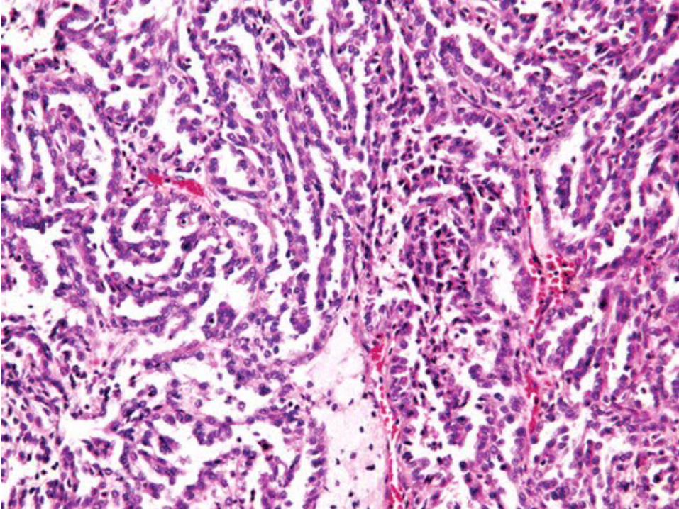
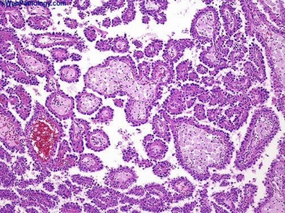
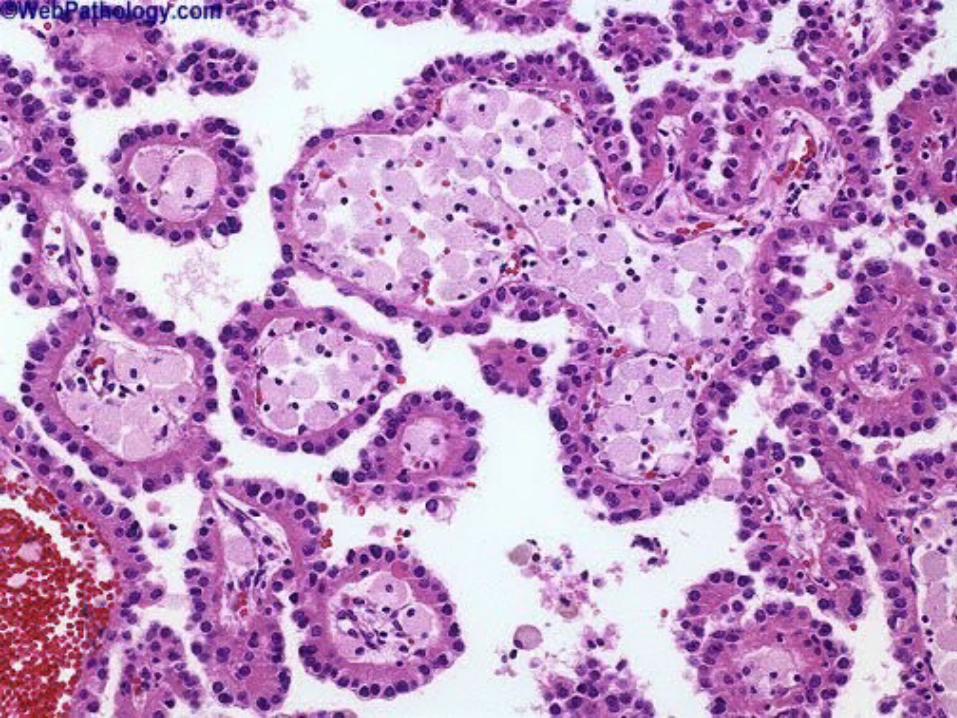
* Histopathology:
•Papillae and tubulopapillary structures with
fibrovascular cores.
•Foamy histiocytes expanding the papillary cores
and Psammoma bodies are characteristic.
3. Chromophobe RCC3. Chromophobe RCC
* Clinical Features:
•About 5% of RCCs
•Significantly better prognosis than clear cell
RCC.
* Gross Pathology:
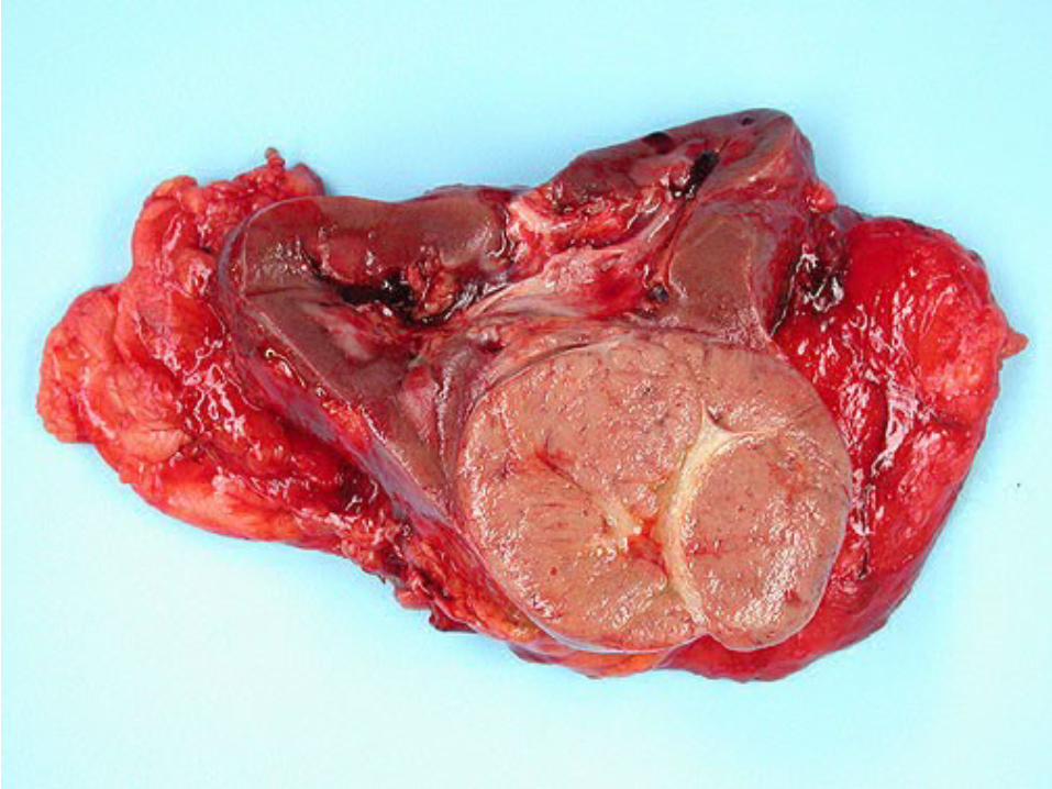
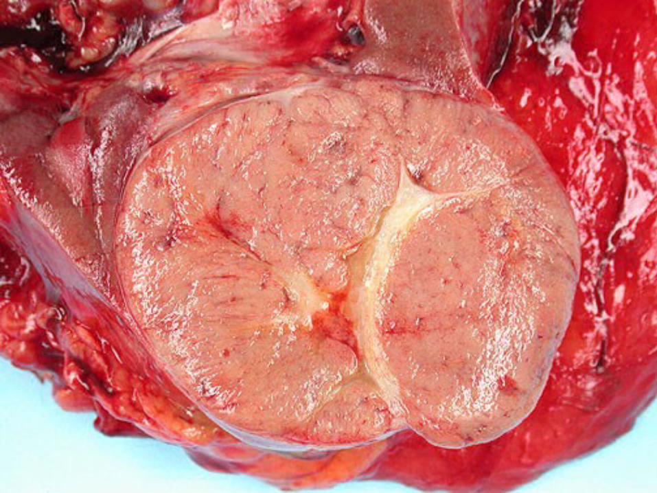
•Solitary, spherical, well-circumscribed mass.
•Homogeneous, tan or light-brown cut surface.
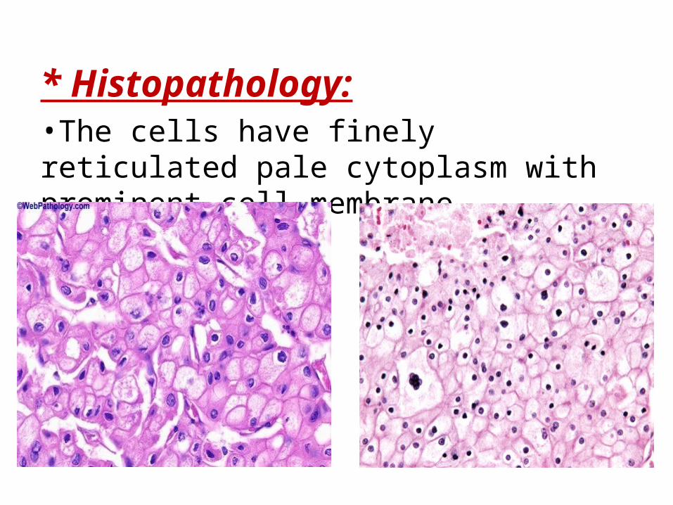
* Histopathology:•The cells have finely reticulated pale cytoplasm with prominent cell membrane
4. Collecting duct carcinoma4. Collecting duct carcinoma
* Clinical Features:
•Rare, comprising about 0.1% of RCCs.
•Flank mass, pain, and hematuria.
•One third have metastasis at presentation.
* Gross Pathology:
•Medullary location.
•Light-gray, white cut surface with invasive
borders.
•Necrosis, hemorrhage, and cystic changes may
be present.
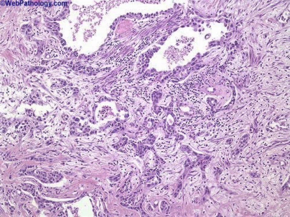
* Histopathology:•Highly infiltrative border
•Tubular and tubulopapillary structures
surrounded by Inflamed desmoplastic stroma.
•The cells show high-grade atypia.
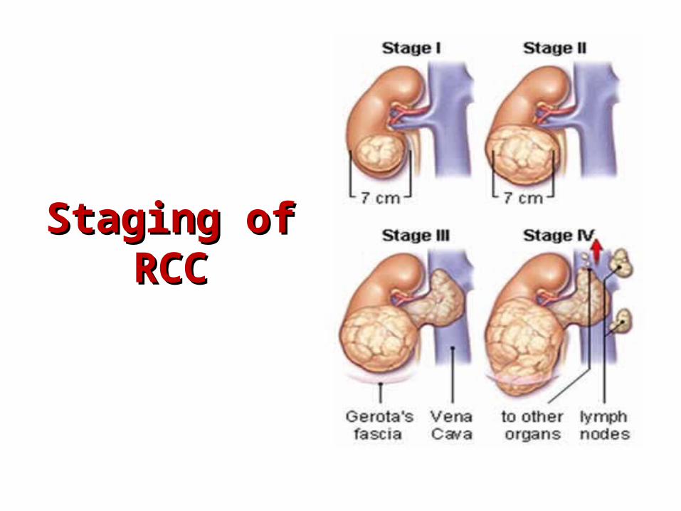
Staging of RCCStaging of RCC
Wilms tumor (nephroblastoma)Wilms tumor (nephroblastoma)
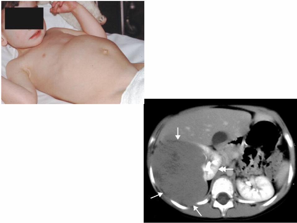
* Clinical Features:•Common solid tumor of childhood; 90% found before the age of 6 years with peak incidence at the ages 2 to 5 years.•Rarely found in adults or neonates.•Patients usually present with an abdominal mass or abdominal tenderness; may present with hematuria, hypertension, or rarely peritoneal symptoms if spontaneous rupture has occurred•Treatment includes surgical resection, chemotherapy and radiation
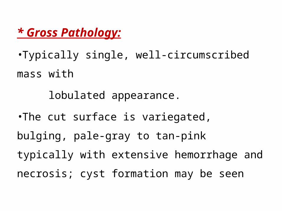
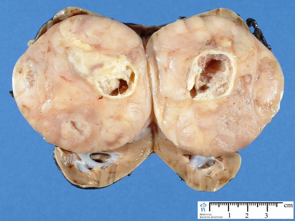
* Gross Pathology:
•Typically single, well-circumscribed mass with
lobulated appearance.
•The cut surface is variegated, bulging, pale-gray
to tan-pink typically with extensive hemorrhage
and necrosis; cyst formation may be seen
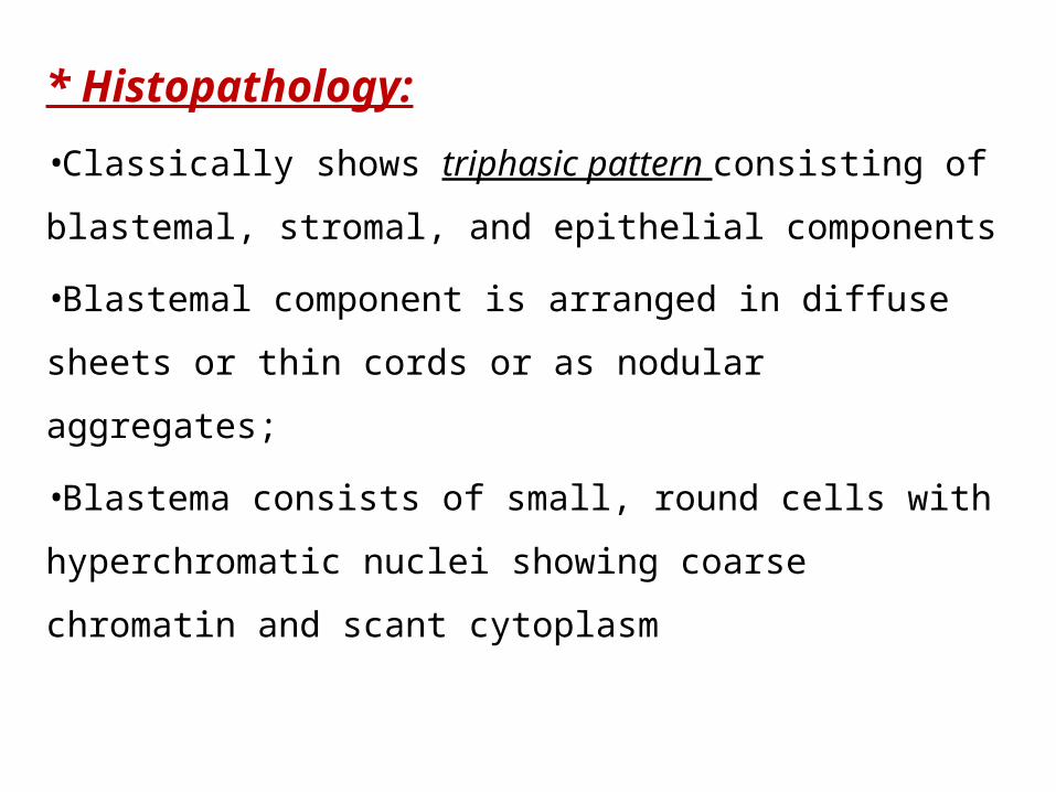
* Histopathology:
•Classically shows triphasic pattern consisting of
blastemal, stromal, and epithelial components
•Blastemal component is arranged in diffuse sheets or
thin cords or as nodular aggregates;
•Blastema consists of small, round cells with
hyperchromatic nuclei showing coarse chromatin and
scant cytoplasm
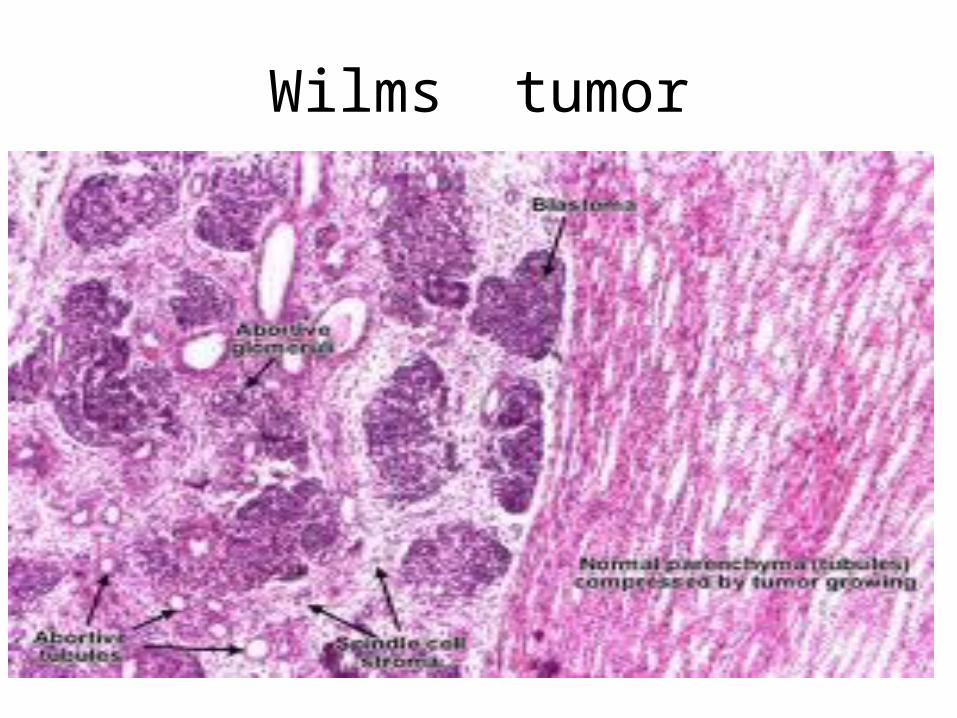
Wilms tumor
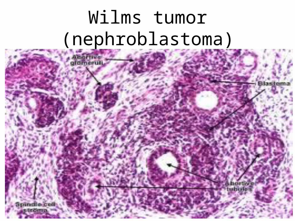
Wilms tumor (nephroblastoma)

References:Robbins and Cotran’s:
Pathologic Basis of Disease. Seventh edition.