Embed Size (px)
Citation preview
W
M
3
6
0h
The American Journal of Surgery (2013) 205, 85–101
Review Article
Repair of incisional hernias with biological prosthesis: asystematic review of current evidence
Charles F. Bellows, M.D.a,*, Alison Smith, B.S.a, Jennifer Malsbury, D.O.a,illiam Scott Helton, M.D.b
aDepartment of Surgery, Tulane University, 1430 Tulane Ave., SL-22, New Orleans, LA 70112, USA; bVirginia Mason
edical Center, Seattle, WA, USAAbstractBACKGROUND: No consensus has been reached on the use of bioprosthetics to repair abdominal
wall defects. The purpose of this systematic review was to summarize the outcomes from studiesdescribing this use of various bioprosthetics for incisional hernia repair.
METHODS: Studies published by October 2011 were identified through literature searches usingEMBASE, MEDLINE, and the Cochrane Central Register of Controlled Trials.
RESULTS: A total of 491 articles were scanned, 60 met eligibility criteria. Most studies wereretrospective case studies. The studies ranged considerably in methodologic quality, with a modifiedMethodological Index of Nonrandomized Studies score from 5 to 12. Many repairs were performed incontaminated surgical sites (47.9%). At least one complication was seen in 87% of repairs. Majorcomplications noted were wound infections (16.9%) and seroma (12.0%). With a mean follow-upperiod of 13.6 months the hernia recurrence rate was 15.2%.
CONCLUSIONS: There is an insufficient level of high-quality evidence in the literature on the valueof bioprosthetics for incisional hernia repair. Randomized controlled trials that use standardizedreporting comparing bioprosthetics with synthetic mesh for incisional hernia repair are needed.© 2013 Elsevier Inc. All rights reserved.
KEYWORDS:Bioprosthetic;Systematic review;Incisional hernia
Despite advances in surgical technique and prosthetictechnologies, repair of complex anterior abdominal walldefects, particularly when bacterial contamination is presentor the risk of infection is high, remains a complex andchallenging surgical undertaking. Although permanentprosthetic mesh is considered the gold standard for mini-mizing hernia recurrence, their nonabsorbable characteris-tics may cause potential problems, resulting in erosion into
The authors report no potential conflicts of interest or financial interest.* Corresponding author. Tel.: �1-504-988-2307; fax: �1-504-988-
843.E-mail address: [email protected] received November 19, 2011; revised manuscript February
, 2012
002-9610/$ - see front matter © 2013 Elsevier Inc. All rights reserved.ttp://dx.doi.org/10.1016/j.amjsurg.2012.02.019
the abdominal viscera, bowel fistulae, and chronic pain,which can lead to more complex and costly surgery.1,2
Moreover, placement of synthetic mesh is sometimes im-prudent, especially in high-risk contaminated wounds. Con-sequently, within the past decade, several bioprosthetic ma-terials have been developed to support tissue reconstructionwhile minimizing the potential complications that comefrom foreign material reactions of synthetic mesh and theirpotential to act as a nidus for infection.
Biological mesh was introduced in clinical surgerymore than a decade ago as an alternative to syntheticmesh for abdominal incisional hernia repair. In general,biological prostheses possess the physical and mechani-cal characteristics of a clinically acceptable synthetic
mesh in that they promote tissue in-growth and have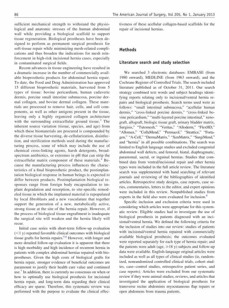
cttdspebssttr
(tmipphpu
86 The American Journal of Surgery, Vol 205, No 1, January 2013
sufficient mechanical strength to withstand the physio-logical and anatomic stresses of the human abdominalwall while providing a biological scaffold to supporttissue regeneration. Biological prostheses have been de-signed to perform as permanent surgical prosthesis forsoft-tissue repair while minimizing mesh-related compli-cations and thus broaden the indications for mesh rein-forcement in high-risk incisional hernia cases, especiallyin contaminated surgical fields.
Recent advances in tissue engineering have resulted ina dramatic increase in the number of commercially avail-able bioprosthetic products for abdominal hernia repair.To date, the Food and Drug Administration has approved15 different bioprosthetic materials, harvested from 5types of tissue: bovine pericardium, human cadavericdermis, porcine small intestine submucosa, porcine der-mal collagen, and bovine dermal collagen. These mate-rials are processed to remove hair, cells, and cell com-ponents, as well as other antigens present in the tissue,leaving only a highly organized collagen architecturewith the surrounding extracellular ground tissue.3 Theinherent source variation (tissue, species, and age) fromwhich these biomaterials are procured is compounded bythe diverse tissue harvesting, de-cellularization, disinfec-tion, and sterilization methods used during the manufac-turing process, some of which may include the use ofchemical cross-linking agents, harsh detergents, broad-spectrum antibiotics, or extremes in pH that can strip theextracellular matrix component of these materials.4 Be-ause the manufacturing process influences the charac-eristics of a final bioprosthetic product, the postimplan-ation biological response in human beings is expected toiffer between products. Postimplantation biological re-ponses range from foreign body encapsulation to im-lant degradation and resorption, to site-specific remod-led tissue in which the implanted material is repopulatedy local fibroblasts and a new vasculature that togetherupport the generation of a new, metabolically active,trong tissue at the site of the hernia repair. However, ifhe process of biological tissue engraftment is inadequatehe surgical site will weaken and the hernia likely willecur.
Initial case series with short-term follow-up evaluation�1 y) reported favorable clinical outcomes with biologicalissue grafts for hernia repairs.5,6 However, with longer andore detailed follow-up evaluation it is apparent that there
s high morbidity and high incidence of recurrent hernia inatients with complex abdominal hernias repaired with bio-rostheses. Given the high costs of biological grafts forernia repair, stronger evidence of beneficial outcomes arearamount to justify their health care value and continuedse.7 In addition, there is currently no consensus on when or
how to optimally use biological prosthesis for incisionalhernia repair, and long-term data regarding their clinicalefficacy are sparse. Therefore, this systematic review was
performed with the purpose to evaluate the clinical effec-tiveness of these acellular collagen-based scaffolds for therepair of incisional hernias.
Methods
Literature search and study selection
We searched 3 electronic databases: EMBASE (from1980 onward), MEDLINE (from 1963 onward), and theCochrane Register of Controlled Trials. The search includedliterature published as of October 31, 2011. Our searchstrategy combined text words and subject headings identi-fying reports relating only to incisional/ventral hernia re-pairs and biological prosthesis. Search terms used were asfollows: “small intestinal submucosa,” “acellular humandermis,” “cross-linked porcine dermis,” “cross-linked bo-vine pericardium,” “multi-layered porcine intestinal,” xeno-graft, allograft, biologic tissue graft, urinary bladder matrix,Surgisis,” “Tutomesh,” “Veritas,” “Alloderm,” “FlexHD,”“Allomax,” “CollaMend,” “Permacol,” “Strattice,” “Forta-gen,” “A-Cell,” “DermaMatrix,” “XenMatrix,” “SurgiMend,”and “hernia” in all possible combinations. The search waslimited to English language studies and excluded congenitalabdominal wall defects, and femoral, hiatal, diaphragmatic,parastomal, sacral, or inguinal hernias. Studies that com-bined data from ventral/incisional repair and other herniatypes were included in the full article review. The literaturesearch was supplemented with hand searching of relevantjournals and reviewing of the bibliographies of identifiedarticles. Retrospective study designs, case reports, case se-ries, commentaries, letters to the editor, and expert opinionswere included in this review. Nonpublished studies fromexperts in the field also were evaluated for inclusion.8
Specific inclusion and exclusion criteria were used inconsidering which articles were appropriate for this system-atic review. Eligible studies had to investigate the use ofbiological prosthesis in patients diagnosed with an inci-sional/ventral hernia. We defined the following criteria forthe inclusion of studies into our review: studies of patientswith incisional/ventral hernia repaired with commerciallyavailable biological prosthesis; the outcomes evaluatedwere reported separately for each type of hernia repair; andthe patients were adult (age, �18 y) subjects and follow-updata were available. English-language original articles wereincluded as well as all types of clinical studies (ie, random-ized, nonrandomized controlled clinical trials, cohort stud-ies, case–control studies, retrospective patient series, andcase reports). Articles were excluded from our systematicreview if they were animal studies, reviews, and articles thatinvestigated the application of biological prosthesis fortransverse rectus abdominis myocutaneous flap repairs or
open abdomens from trauma patients.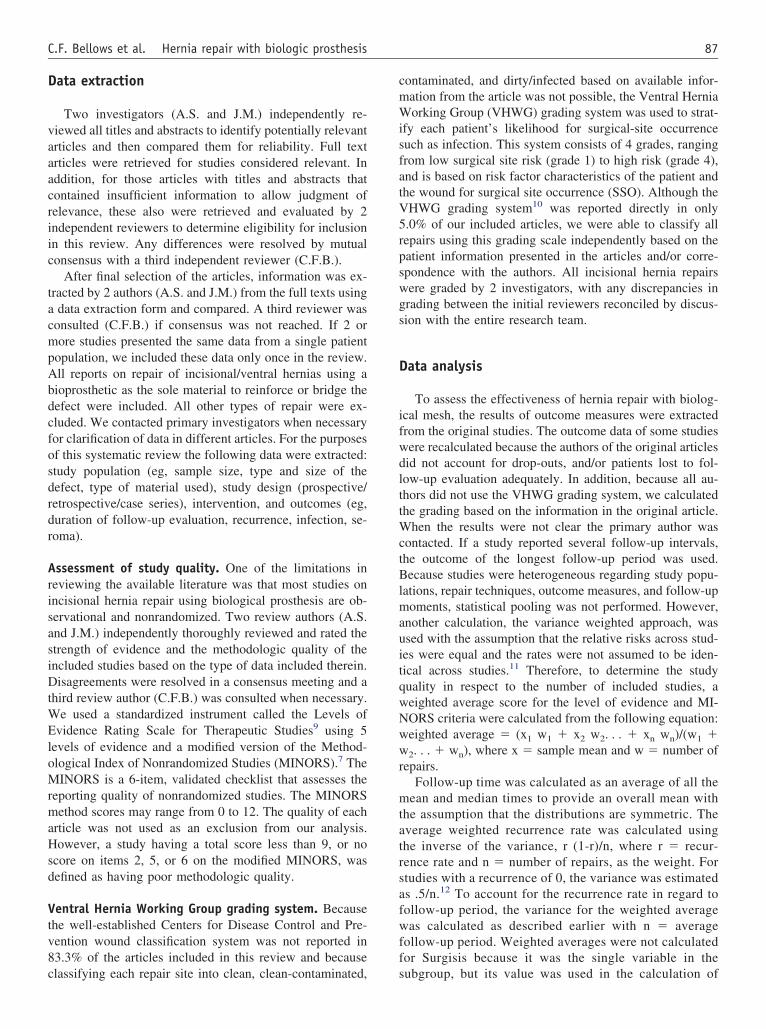
lo
wr
mtatrsa
87C.F. Bellows et al. Hernia repair with biologic prosthesis
Data extraction
Two investigators (A.S. and J.M.) independently re-viewed all titles and abstracts to identify potentially relevantarticles and then compared them for reliability. Full textarticles were retrieved for studies considered relevant. Inaddition, for those articles with titles and abstracts thatcontained insufficient information to allow judgment ofrelevance, these also were retrieved and evaluated by 2independent reviewers to determine eligibility for inclusionin this review. Any differences were resolved by mutualconsensus with a third independent reviewer (C.F.B.).
After final selection of the articles, information was ex-tracted by 2 authors (A.S. and J.M.) from the full texts usinga data extraction form and compared. A third reviewer wasconsulted (C.F.B.) if consensus was not reached. If 2 ormore studies presented the same data from a single patientpopulation, we included these data only once in the review.All reports on repair of incisional/ventral hernias using abioprosthetic as the sole material to reinforce or bridge thedefect were included. All other types of repair were ex-cluded. We contacted primary investigators when necessaryfor clarification of data in different articles. For the purposesof this systematic review the following data were extracted:study population (eg, sample size, type and size of thedefect, type of material used), study design (prospective/retrospective/case series), intervention, and outcomes (eg,duration of follow-up evaluation, recurrence, infection, se-roma).
Assessment of study quality. One of the limitations inreviewing the available literature was that most studies onincisional hernia repair using biological prosthesis are ob-servational and nonrandomized. Two review authors (A.S.and J.M.) independently thoroughly reviewed and rated thestrength of evidence and the methodologic quality of theincluded studies based on the type of data included therein.Disagreements were resolved in a consensus meeting and athird review author (C.F.B.) was consulted when necessary.We used a standardized instrument called the Levels ofEvidence Rating Scale for Therapeutic Studies9 using 5evels of evidence and a modified version of the Method-logical Index of Nonrandomized Studies (MINORS).7 The
MINORS is a 6-item, validated checklist that assesses thereporting quality of nonrandomized studies. The MINORSmethod scores may range from 0 to 12. The quality of eacharticle was not used as an exclusion from our analysis.However, a study having a total score less than 9, or noscore on items 2, 5, or 6 on the modified MINORS, wasdefined as having poor methodologic quality.
Ventral Hernia Working Group grading system. Becausethe well-established Centers for Disease Control and Pre-vention wound classification system was not reported in83.3% of the articles included in this review and because
classifying each repair site into clean, clean-contaminated,contaminated, and dirty/infected based on available infor-mation from the article was not possible, the Ventral HerniaWorking Group (VHWG) grading system was used to strat-ify each patient’s likelihood for surgical-site occurrencesuch as infection. This system consists of 4 grades, rangingfrom low surgical site risk (grade 1) to high risk (grade 4),and is based on risk factor characteristics of the patient andthe wound for surgical site occurrence (SSO). Although theVHWG grading system10 was reported directly in only5.0% of our included articles, we were able to classify allrepairs using this grading scale independently based on thepatient information presented in the articles and/or corre-spondence with the authors. All incisional hernia repairswere graded by 2 investigators, with any discrepancies ingrading between the initial reviewers reconciled by discus-sion with the entire research team.
Data analysis
To assess the effectiveness of hernia repair with biolog-ical mesh, the results of outcome measures were extractedfrom the original studies. The outcome data of some studieswere recalculated because the authors of the original articlesdid not account for drop-outs, and/or patients lost to fol-low-up evaluation adequately. In addition, because all au-thors did not use the VHWG grading system, we calculatedthe grading based on the information in the original article.When the results were not clear the primary author wascontacted. If a study reported several follow-up intervals,the outcome of the longest follow-up period was used.Because studies were heterogeneous regarding study popu-lations, repair techniques, outcome measures, and follow-upmoments, statistical pooling was not performed. However,another calculation, the variance weighted approach, wasused with the assumption that the relative risks across stud-ies were equal and the rates were not assumed to be iden-tical across studies.11 Therefore, to determine the studyquality in respect to the number of included studies, aweighted average score for the level of evidence and MI-NORS criteria were calculated from the following equation:weighted average � (x1 w1 � x2 w2. . . � xn wn)/(w1 �
2. . . � wn), where x � sample mean and w � number ofepairs.
Follow-up time was calculated as an average of all theean and median times to provide an overall mean with
he assumption that the distributions are symmetric. Theverage weighted recurrence rate was calculated usinghe inverse of the variance, r (1-r)/n, where r � recur-ence rate and n � number of repairs, as the weight. Fortudies with a recurrence of 0, the variance was estimateds .5/n.12 To account for the recurrence rate in regard to
follow-up period, the variance for the weighted averagewas calculated as described earlier with n � averagefollow-up period. Weighted averages were not calculatedfor Surgisis because it was the single variable in the
subgroup, but its value was used in the calculation of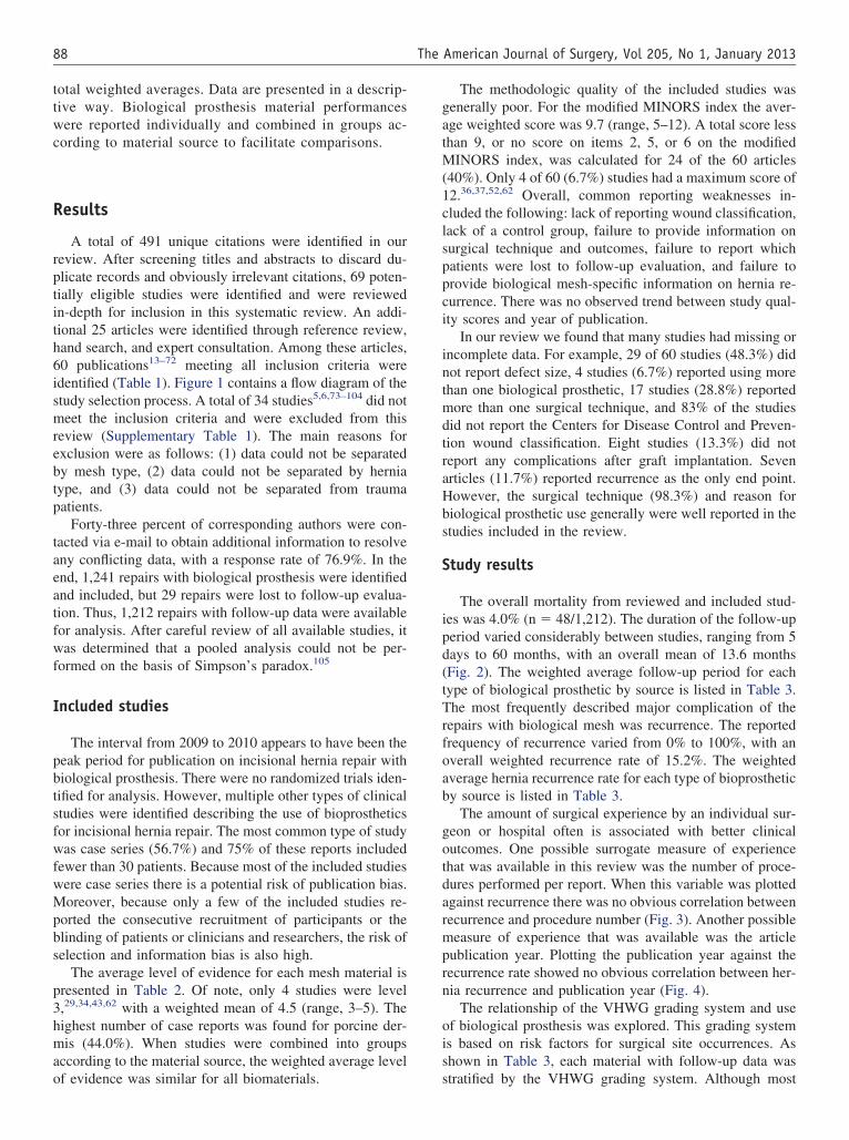
ismrebtp
taeatfwf
hmao
gatM(1
88 The American Journal of Surgery, Vol 205, No 1, January 2013
total weighted averages. Data are presented in a descrip-tive way. Biological prosthesis material performanceswere reported individually and combined in groups ac-cording to material source to facilitate comparisons.
Results
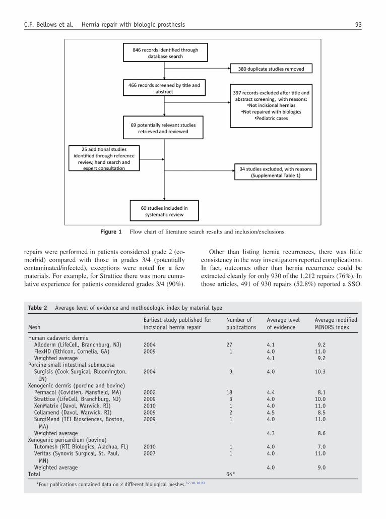
A total of 491 unique citations were identified in ourreview. After screening titles and abstracts to discard du-plicate records and obviously irrelevant citations, 69 poten-tially eligible studies were identified and were reviewedin-depth for inclusion in this systematic review. An addi-tional 25 articles were identified through reference review,hand search, and expert consultation. Among these articles,60 publications13–72 meeting all inclusion criteria weredentified (Table 1). Figure 1 contains a flow diagram of thetudy selection process. A total of 34 studies5,6,73–104 did noteet the inclusion criteria and were excluded from this
eview (Supplementary Table 1). The main reasons forxclusion were as follows: (1) data could not be separatedy mesh type, (2) data could not be separated by herniaype, and (3) data could not be separated from traumaatients.
Forty-three percent of corresponding authors were con-acted via e-mail to obtain additional information to resolveny conflicting data, with a response rate of 76.9%. In thend, 1,241 repairs with biological prosthesis were identifiednd included, but 29 repairs were lost to follow-up evalua-ion. Thus, 1,212 repairs with follow-up data were availableor analysis. After careful review of all available studies, itas determined that a pooled analysis could not be per-
ormed on the basis of Simpson’s paradox.105
Included studies
The interval from 2009 to 2010 appears to have been thepeak period for publication on incisional hernia repair withbiological prosthesis. There were no randomized trials iden-tified for analysis. However, multiple other types of clinicalstudies were identified describing the use of bioprostheticsfor incisional hernia repair. The most common type of studywas case series (56.7%) and 75% of these reports includedfewer than 30 patients. Because most of the included studieswere case series there is a potential risk of publication bias.Moreover, because only a few of the included studies re-ported the consecutive recruitment of participants or theblinding of patients or clinicians and researchers, the risk ofselection and information bias is also high.
The average level of evidence for each mesh material ispresented in Table 2. Of note, only 4 studies were level3,29,34,43,62 with a weighted mean of 4.5 (range, 3–5). Theighest number of case reports was found for porcine der-is (44.0%). When studies were combined into groups
ccording to the material source, the weighted average level
f evidence was similar for all biomaterials.The methodologic quality of the included studies wasenerally poor. For the modified MINORS index the aver-ge weighted score was 9.7 (range, 5–12). A total score lesshan 9, or no score on items 2, 5, or 6 on the modified
INORS index, was calculated for 24 of the 60 articles40%). Only 4 of 60 (6.7%) studies had a maximum score of2.36,37,52,62 Overall, common reporting weaknesses in-
cluded the following: lack of reporting wound classification,lack of a control group, failure to provide information onsurgical technique and outcomes, failure to report whichpatients were lost to follow-up evaluation, and failure toprovide biological mesh-specific information on hernia re-currence. There was no observed trend between study qual-ity scores and year of publication.
In our review we found that many studies had missing orincomplete data. For example, 29 of 60 studies (48.3%) didnot report defect size, 4 studies (6.7%) reported using morethan one biological prosthetic, 17 studies (28.8%) reportedmore than one surgical technique, and 83% of the studiesdid not report the Centers for Disease Control and Preven-tion wound classification. Eight studies (13.3%) did notreport any complications after graft implantation. Sevenarticles (11.7%) reported recurrence as the only end point.However, the surgical technique (98.3%) and reason forbiological prosthetic use generally were well reported in thestudies included in the review.
Study results
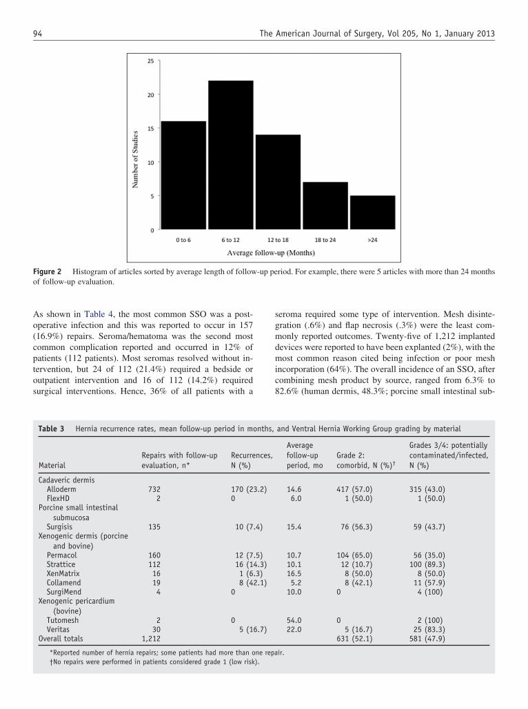
The overall mortality from reviewed and included stud-ies was 4.0% (n � 48/1,212). The duration of the follow-upperiod varied considerably between studies, ranging from 5days to 60 months, with an overall mean of 13.6 months(Fig. 2). The weighted average follow-up period for eachtype of biological prosthetic by source is listed in Table 3.The most frequently described major complication of therepairs with biological mesh was recurrence. The reportedfrequency of recurrence varied from 0% to 100%, with anoverall weighted recurrence rate of 15.2%. The weightedaverage hernia recurrence rate for each type of bioprostheticby source is listed in Table 3.
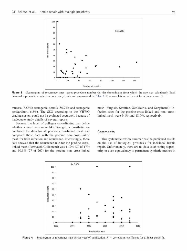
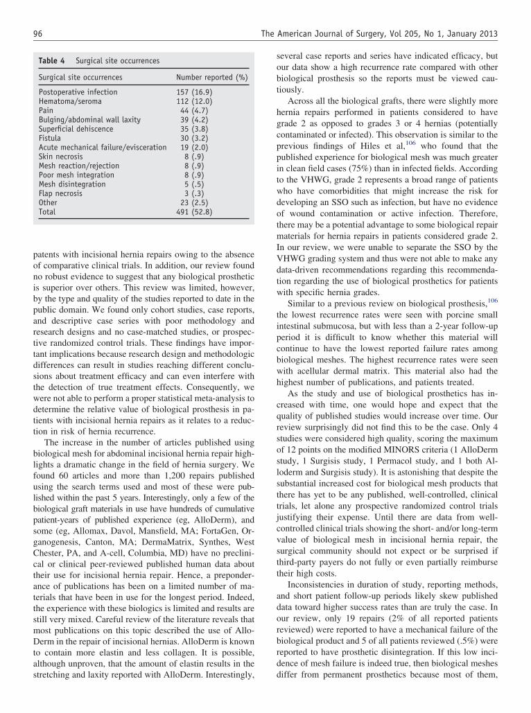
The amount of surgical experience by an individual sur-geon or hospital often is associated with better clinicaloutcomes. One possible surrogate measure of experiencethat was available in this review was the number of proce-dures performed per report. When this variable was plottedagainst recurrence there was no obvious correlation betweenrecurrence and procedure number (Fig. 3). Another possiblemeasure of experience that was available was the articlepublication year. Plotting the publication year against therecurrence rate showed no obvious correlation between her-nia recurrence and publication year (Fig. 4).
The relationship of the VHWG grading system and useof biological prosthesis was explored. This grading systemis based on risk factors for surgical site occurrences. Asshown in Table 3, each material with follow-up data was
stratified by the VHWG grading system. Although most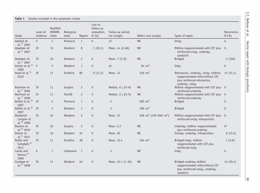
Table 1 Studies included in the systematic review
StudyLevel ofevidence
ModifiedMINORSindex
Biologicalmesh Repairs
Lost tofollow-upevaluation,N (%)
Follow-up period,mo (range) Defect size (range) Types of repair
Recurrence,N (%)
Adedeji etal,13 2002
V 5 Permacol 1 0 12 NR Onlay 0
Alaedeen etal,14 2007
IV 10 Alloderm 8 1 (29.1) Mean, 14 (2–68) NR Midline reapproximated with CST plusreinforced-onlay, underlay,sandwich
0
Alaedeen etal,14 2007
IV 10 Alloderm 2 0 Mean, 7 (5–8) NR Bridged 2 (100)
Asham et al,15
2006V 5 Alloderm 1 0 24 84 cm2 Inlay 0
Awad et al,16
2010IV 11 Strattice 80 9 (11.3) Mean, 12 236 cm2 Retrorectus, underlay, onlay; midline
reapproximated with/without CSTplus reinforced-retrorectus,underlay, onlay
15 (21.1)
Bachman etal,17 2009
IV 11 Surgisis 2 0 Median, 6 (.25–9) NR Midline reapproximated with CST plusreinforced-underlay
0
Bachman etal,17 2009
IV 11 FlexHD 2 0 Median, 6 (.25–9) NR Midline reapproximated with CST plusreinforced-underlay
0
Baillie et al,18
2007IV 5 Permacol 1 0 5 600 cm2 Bridged 0
Baillie et al,18
2007IV 5 Alloderm 1 0 5 300 cm2 Bridged 0
Bluebond-Langner etal,19 2008
IV 10 Alloderm 5 0 Mean, 12 628 cm2 (220–900 cm2) Midline reapproximated with CST plusreinforced-onlay interposition
0
Boutros etal,20 2010
IV 10 Surgisis 3 0 Mean, 6.3 NR Underlay; midline reapproximatedplus reinforced-underlay
0*
Brewer etal,21 2011
IV 10 Alloderm 34 0 Mean, 26 NR Overlay, underlay, interposition 8 (23.5)
Butler andCampbell,22
2011
IV 11 Strattice 38 0 Mean, 12.4 494 cm2 Bridged-inlay; midlinereapproximated with CST plusreinforced-inlay
1 (2.6)
Canda andKaraca,23
2009
V 7 Collamend 1 0 3 NR Inlay 0
Candage etal,24 2008
IV 11 Alloderm 46 0 Mean, 12.1 (1–39) NR Bridged-underlay midlinereapproximated with/without CSTplus reinforced-onlay, underlay,sandwich
14 (30.4)
89C.F.
Bellows
etal.
Hernia
repairw
ithbiologic
prosthesis
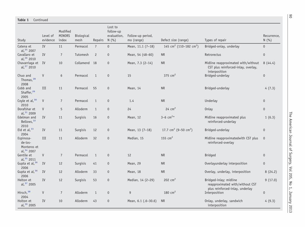
Table 1 Continued
StudyLevel ofevidence
ModifiedMINORSindex
Biologicalmesh Repairs
Lost tofollow-upevaluation,N (%)
Follow-up period,mo (range) Defect size (range) Types of repair
Recurrence,N (%)
Catena etal,25 2007
IV 11 Permacol 7 0 Mean, 11.1 (7–18) 145 cm2 (110–182 cm2) Bridged-onlay, underlay 0
Cavallaro etal,26 2010
IV 7 Tutomesh 2 0 Mean, 54 (48–60) NR Retrorectus 0
Chavarriaga etal,27 2010
IV 10 Collamend 18 0 Mean, 7.3 (2–14) NR Midline reapproximated with/withoutCST plus reinforced-inlay, overlay,interposition
8 (44.4)
Chuo andThomas,28
2008
V 6 Permacol 1 0 15 375 cm2 Bridged-underlay 0
Cobb andShaffer,29
2005
III 11 Permacol 55 0 Mean, 14 NR Bridged-underlay 4 (7.3)
Coyle et al,30
2010V 7 Permacol 1 0 1.4 NR Underlay 0
Dorafshar etal,31 2009
V 5 Alloderm 1 0 24 24 cm2 Onlay 0
Edelman andBellows,32
2010
IV 11 Surgisis 16 0 Mean, 12 3–6 cm2* Midline reapproximated plusreinforced-underlay
1 (6.3)
Eid et al,33
2004IV 11 Surgisis 12 0 Mean, 13 (7–18) 17.7 cm2 (9–50 cm2) Bridged-underlay 0
Espinosa-de-los-Monteros etal,34 2007
III 11 Alloderm 32 0 Median, 15 155 cm2 Midline reapproximatedwith CST plusreinforced-overlay
0
Gentile etal,35 2011
V 7 Permacol 1 0 12 NR Bridged 0
Gupta et al,36
2006IV 12 Surgisis 41 0 Mean, 29 NR Overlayunderlay interposition 0
Gupta et al,36
2006IV 12 Alloderm 33 0 Mean, 18 NR Overlay, underlay, interposition 8 (24.2)
Helton etal,37 2005
IV 12 Surgisis 53 0 Median, 14 (2–29) 202 cm2 Bridged-inlay; midlinereapproximated with/without CSTplus reinforced-inlay, underlay
9 (17.0)
Hirsch,38
2004V 7 Alloderm 1 0 9 180 cm2 Interposition 0
Holton etal,39 2005
IV 10 Alloderm 43 0 Mean, 6.1 (.6–30.6) NR Onlay, underlay, sandwichinterposition
4 (9.3)
90The
American
Journalof
Surgery,Vol
205,No
1,January
2013
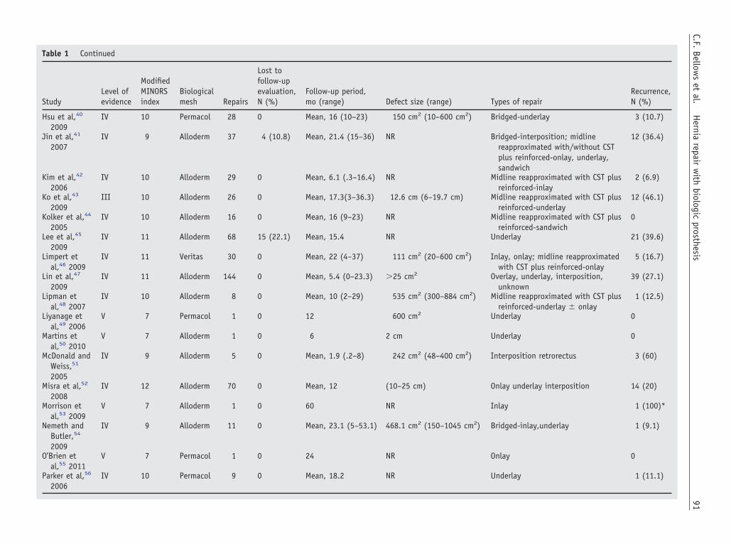
Table 1 Continued
StudyLevel ofevidence
ModifiedMINORSindex
Biologicalmesh Repairs
Lost tofollow-upevaluation,N (%)
Follow-up period,mo (range) Defect size (range) Types of repair
Recurrence,N (%)
Hsu et al,40
2009IV 10 Permacol 28 0 Mean, 16 (10–23) 150 cm2 (10–600 cm2) Bridged-underlay 3 (10.7)
Jin et al,41
2007IV 9 Alloderm 37 4 (10.8) Mean, 21.4 (15–36) NR Bridged-interposition; midline
reapproximated with/without CSTplus reinforced-onlay, underlay,sandwich
12 (36.4)
Kim et al,42
2006IV 10 Alloderm 29 0 Mean, 6.1 (.3–16.4) NR Midline reapproximated with CST plus
reinforced-inlay2 (6.9)
Ko et al,43
2009III 10 Alloderm 26 0 Mean, 17.3(3–36.3) 12.6 cm (6–19.7 cm) Midline reapproximated with CST plus
reinforced-underlay12 (46.1)
Kolker et al,44
2005IV 10 Alloderm 16 0 Mean, 16 (9–23) NR Midline reapproximated with CST plus
reinforced-sandwich0
Lee et al,45
2009IV 11 Alloderm 68 15 (22.1) Mean, 15.4 NR Underlay 21 (39.6)
Limpert etal,46 2009
IV 11 Veritas 30 0 Mean, 22 (4–37) 111 cm2 (20–600 cm2) Inlay, onlay; midline reapproximatedwith CST plus reinforced-onlay
5 (16.7)
Lin et al,47
2009IV 11 Alloderm 144 0 Mean, 5.4 (0–23.3) �25 cm2 Overlay, underlay, interposition,
unknown39 (27.1)
Lipman etal,48 2007
IV 10 Alloderm 8 0 Mean, 10 (2–29) 535 cm2 (300–884 cm2) Midline reapproximated with CST plusreinforced-underlay � onlay
1 (12.5)
Liyanage etal,49 2006
V 7 Permacol 1 0 12 600 cm2 Underlay 0
Martins etal,50 2010
V 7 Alloderm 1 0 6 2 cm Underlay 0
McDonald andWeiss,51
2005
IV 9 Alloderm 5 0 Mean, 1.9 (.2–8) 242 cm2 (48–400 cm2) Interposition retrorectus 3 (60)
Misra et al,52
2008IV 12 Alloderm 70 0 Mean, 12 (10–25 cm) Onlay underlay interposition 14 (20)
Morrison etal,53 2009
V 7 Alloderm 1 0 60 NR Inlay 1 (100)*
Nemeth andButler,54
2009
IV 9 Alloderm 11 0 Mean, 23.1 (5–53.1) 468.1 cm2 (150–1045 cm2) Bridged-inlay,underlay 1 (9.1)
O’Brien etal,55 2011
V 7 Permacol 1 0 24 NR Onlay 0
Parker et al,56
2006IV 10 Permacol 9 0 Mean, 18.2 NR Underlay 1 (11.1)
91C.F.
Bellows
etal.
Hernia
repairw
ithbiologic
prosthesis
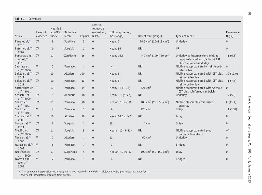
Table 1 Continued
StudyLevel ofevidence
ModifiedMINORSindex
Biologicalmesh Repairs
Lost tofollow-upevaluation,N (%)
Follow-up period,mo (range) Defect size (range) Types of repair
Recurrence,N (%)
Parra et al,57
2010IV 8 Strattice 3 0 Mean, 6 70.5 cm2 (20–113 cm2) Underlay 0
Paton et al,58
2007IV 9 Surgisis 2 0 Mean, 36 NR NR 0
Pomahac andAflaki,59
2010
IV 11 XenMatrix 16 0 Mean, 16.5 440 cm2 (100–750 cm2) Underlay � interposition; midlinereapproximated with/without CSTplus reinforced-underlay
1 (6.3)
Saettele etal,60 2006
V 7 Permacol 1 0 4 NR Midline reapproximated� reinforced-retrorectus
0
Sailes et al,61
2011IV 10 Alloderm 100 0 Mean, 6* NR Midline reapproximated with CST plus
reinforced-onlay19 (19.0)
Sailes et al,61
2011IV 10 Permacol 13 0 Mean, 6* NR Midline reapproximated with CST plus
reinforced-onlay1 (7.7)
Satterwhite etal,62 2011
III 12 Permacol 19 0 Mean, 11 (1–33) 321 cm2 Midline reapproximated with/withoutCST plus reinforced-sandwich
0
Schuster etal,63 2006
IV 9 Alloderm 18 0 Mean, 9.1 (5–27) NR Underlay 9 (50)
Shaikh etal,64 2007
IV 11 Permacol 18 0 Median, 18 (6–36) 180 cm2 (96–850 cm2) Midline closed plus reinforced-underlay
2 (11.1)
Shaikh etal,65 2011
V 7 Permacol 1 0 6 120 cm2 Onlay 1 (100)
Singh et al,66
2008IV 10 Alloderm 10 0 Mean, 10.3 (.1–24) NR Inlay 0
Tong et al,67
2011IV 6 Surgisis 1 0 12 4 cm Onlay 0
Treviño etal,68 2006
IV 11 Surgisis 5 0 Median 10 (3–12) NR Midline reapproximated plusreinforced-sandwich
0*
Tung et al,69
2006V 7 Alloderm 1 0 12 60 cm2 Underlay 0
Walker et al,70
2009V 6 Permacol 1 0 3 NR Bridged 0
Wietfeldt etal,71 2009
IV 11 SurgiMend 4 0 Median, 10 (9–17) 100 cm2 (50–150 cm2) Inlay 0
Wotton andAkoh,72
2009
V 7 Permacol 1 0 4 NR Bridged 0
CST � component separation technique; NR � not reported; sandwich � biological onlay plus biological underlay.*Additional information obtained from author.
92The
American
Journalof
Surgery,Vol
205,No
1,January
2013
searc
93C.F. Bellows et al. Hernia repair with biologic prosthesis
repairs were performed in patients considered grade 2 (co-morbid) compared with those in grades 3/4 (potentiallycontaminated/infected), exceptions were noted for a fewmaterials. For example, for Strattice there was more cumu-lative experience for patients considered grades 3/4 (90%).
Figure 1 Flow chart of literature
Table 2 Average level of evidence and methodologic index by
MeshEarliest study pubincisional hernia
Human cadaveric dermisAlloderm (LifeCell, Branchburg, NJ) 2004FlexHD (Ethicon, Cornelia, GA) 2009Weighted average
Porcine small intestinal submucosaSurgisis (Cook Surgical, Bloomington,
IN)2004
Xenogenic dermis (porcine and bovine)Permacol (Covidien, Mansfield, MA) 2002Strattice (LifeCell, Branchburg, NJ) 2009XenMatrix (Davol, Warwick, RI) 2010Collamend (Davol, Warwick, RI) 2009SurgiMend (TEI Biosciences, Boston,
MA)2009
Weighted averageXenogenic pericardium (bovine)
Tutomesh (RTI Biologics, Alachua, FL) 2010Veritas (Synovis Surgical, St. Paul,
MN)2007
Weighted averageTotal
*Four publications contained data on 2 different biological meshes.
Other than listing hernia recurrences, there was littleconsistency in the way investigators reported complications.In fact, outcomes other than hernia recurrence could beextracted cleanly for only 930 of the 1,212 repairs (76%). Inthose articles, 491 of 930 repairs (52.8%) reported a SSO.
h results and inclusion/exclusions.
rial type
for Number ofpublications
Average levelof evidence
Average modifiedMINORS index
27 4.1 9.21 4.0 11.0
4.1 9.2
9 4.0 10.3
18 4.4 8.13 4.0 10.01 4.0 11.02 4.5 8.51 4.0 11.0
4.3 8.6
1 4.0 7.01 4.0 11.0
4.0 9.064*
61
mate
lishedrepair
17,18,36,
o(cptos
94 The American Journal of Surgery, Vol 205, No 1, January 2013
As shown in Table 4, the most common SSO was a post-perative infection and this was reported to occur in 15716.9%) repairs. Seroma/hematoma was the second mostommon complication reported and occurred in 12% ofatients (112 patients). Most seromas resolved without in-ervention, but 24 of 112 (21.4%) required a bedside orutpatient intervention and 16 of 112 (14.2%) requiredurgical interventions. Hence, 36% of all patients with a
Figure 2 Histogram of articles sorted by average length of followof follow-up evaluation.
Table 3 Hernia recurrence rates, mean follow-up period in m
MaterialRepairs with follow-upevaluation, n*
RecurreN (%)
Cadaveric dermisAlloderm 732 170 (2FlexHD 2 0
Porcine small intestinalsubmucosa
Surgisis 135 10 (7Xenogenic dermis (porcine
and bovine)Permacol 160 12 (7Strattice 112 16 (1XenMatrix 16 1 (6Collamend 19 8 (4SurgiMend 4 0
Xenogenic pericardium(bovine)
Tutomesh 2 0Veritas 30 5 (1
Overall totals 1,212
*Reported number of hernia repairs; some patients had more than o†No repairs were performed in patients considered grade 1 (low risk
seroma required some type of intervention. Mesh disinte-gration (.6%) and flap necrosis (.3%) were the least com-monly reported outcomes. Twenty-five of 1,212 implanteddevices were reported to have been explanted (2%), with themost common reason cited being infection or poor meshincorporation (64%). The overall incidence of an SSO, aftercombining mesh product by source, ranged from 6.3% to82.6% (human dermis, 48.3%; porcine small intestinal sub-
riod. For example, there were 5 articles with more than 24 months
and Ventral Hernia Working Group grading by material
Averagefollow-upperiod, mo
Grade 2:comorbid, N (%)†
Grades 3/4: potentiallycontaminated/infected,N (%)
14.6 417 (57.0) 315 (43.0)6.0 1 (50.0) 1 (50.0)
15.4 76 (56.3) 59 (43.7)
10.7 104 (65.0) 56 (35.0)10.1 12 (10.7) 100 (89.3)16.5 8 (50.0) 8 (50.0)5.2 8 (42.1) 11 (57.9)
10.0 0 4 (100)
54.0 0 2 (100)22.0 5 (16.7) 25 (83.3)
631 (52.1) 581 (47.9)
ir.
-up pe
onths,
nces,
3.2)
.4)
.5)4.3).3)2.1)
6.7)
ne repa).
in Ta
95C.F. Bellows et al. Hernia repair with biologic prosthesis
mucosa, 82.6%; xenogenic dermis, 50.7%; and xenogenicpericardium, 6.3%). The SSO according to the VHWGgrading system could not be evaluated accurately because ofinadequate study details of several reports.
Because the level of collagen cross-linking can definewhether a mesh acts more like biologic or prosthetic wecombined the data for all porcine cross-linked mesh andcompared these data with the porcine non–cross-linkedmesh for both infection and recurrence. Interestingly, thesedata showed that the recurrence rate for the porcine cross-linked mesh (Permacol, Collamend) was 11.2% (20 of 179)and 10.1% (27 of 267) for the porcine non–cross-linked
Figure 3 Scattergram of recurrence rates versus procedure nudiamond represents the rate from one study. Data are summarized
Figure 4 Scattergram of recurrence rate versus year of publi
mesh (Surgisis, Strattice, XenMatrix, and Surgimend). In-fection rates for the porcine cross-linked and non–cross-linked mesh were 9.1% and 18.6%, respectively.
Comments
This systematic review summarizes the published resultson the use of biological prosthesis for incisional herniarepair. Unfortunately, there are no data establishing superi-ority or even equivalency to permanent synthetic meshes in
ie, the denominator from which the rate was calculated). Eachble 3. R � correlation coefficient for a linear curve fit.
mber (
cation. R � correlation coefficient for a linear curve fit.
96 The American Journal of Surgery, Vol 205, No 1, January 2013
patents with incisional hernia repairs owing to the absenceof comparative clinical trials. In addition, our review foundno robust evidence to suggest that any biological prostheticis superior over others. This review was limited, however,by the type and quality of the studies reported to date in thepublic domain. We found only cohort studies, case reports,and descriptive case series with poor methodology andresearch designs and no case-matched studies, or prospec-tive randomized control trials. These findings have impor-tant implications because research design and methodologicdifferences can result in studies reaching different conclu-sions about treatment efficacy and can even interfere withthe detection of true treatment effects. Consequently, wewere not able to perform a proper statistical meta-analysis todetermine the relative value of biological prosthesis in pa-tients with incisional hernia repairs as it relates to a reduc-tion in risk of hernia recurrence.
The increase in the number of articles published usingbiological mesh for abdominal incisional hernia repair high-lights a dramatic change in the field of hernia surgery. Wefound 60 articles and more than 1,200 repairs publishedusing the search terms used and most of these were pub-lished within the past 5 years. Interestingly, only a few of thebiological graft materials in use have hundreds of cumulativepatient-years of published experience (eg, AlloDerm), andsome (eg, Allomax, Davol, Mansfield, MA; FortaGen, Or-ganogenesis, Canton, MA; DermaMatrix, Synthes, WestChester, PA, and A-cell, Columbia, MD) have no preclini-cal or clinical peer-reviewed published human data abouttheir use for incisional hernia repair. Hence, a preponder-ance of publications has been on a limited number of ma-terials that have been in use for the longest period. Indeed,the experience with these biologics is limited and results arestill very mixed. Careful review of the literature reveals thatmost publications on this topic described the use of Allo-Derm in the repair of incisional hernias. AlloDerm is knownto contain more elastin and less collagen. It is possible,although unproven, that the amount of elastin results in the
Table 4 Surgical site occurrences
Surgical site occurrences Number reported (%)
Postoperative infection 157 (16.9)Hematoma/seroma 112 (12.0)Pain 44 (4.7)Bulging/abdominal wall laxity 39 (4.2)Superficial dehiscence 35 (3.8)Fistula 30 (3.2)Acute mechanical failure/evisceration 19 (2.0)Skin necrosis 8 (.9)Mesh reaction/rejection 8 (.9)Poor mesh integration 8 (.9)Mesh disintegration 5 (.5)Flap necrosis 3 (.3)Other 23 (2.5)Total 491 (52.8)
stretching and laxity reported with AlloDerm. Interestingly,
several case reports and series have indicated efficacy, butour data show a high recurrence rate compared with otherbiological prosthesis so the reports must be viewed cau-tiously.
Across all the biological grafts, there were slightly morehernia repairs performed in patients considered to havegrade 2 as opposed to grades 3 or 4 hernias (potentiallycontaminated or infected). This observation is similar to theprevious findings of Hiles et al,106 who found that thepublished experience for biological mesh was much greaterin clean field cases (75%) than in infected fields. Accordingto the VHWG, grade 2 represents a broad range of patientswho have comorbidities that might increase the risk fordeveloping an SSO such as infection, but have no evidenceof wound contamination or active infection. Therefore,there may be a potential advantage to some biological repairmaterials for hernia repairs in patients considered grade 2.In our review, we were unable to separate the SSO by theVHWG grading system and thus were not able to make anydata-driven recommendations regarding this recommenda-tion regarding the use of biological prosthetics for patientswith specific hernia grades.
Similar to a previous review on biological prosthesis,106
the lowest recurrence rates were seen with porcine smallintestinal submucosa, but with less than a 2-year follow-upperiod it is difficult to know whether this material willcontinue to have the lowest reported failure rates amongbiological meshes. The highest recurrence rates were seenwith acellular dermal matrix. This material also had thehighest number of publications, and patients treated.
As the study and use of biological prosthetics has in-creased with time, one would hope and expect that thequality of published studies would increase over time. Ourreview surprisingly did not find this to be the case. Only 4studies were considered high quality, scoring the maximumof 12 points on the modified MINORS criteria (1 AlloDermstudy, 1 Surgisis study, 1 Permacol study, and 1 both Al-loderm and Surgisis study). It is astonishing that despite thesubstantial increased cost for biological mesh products thatthere has yet to be any published, well-controlled, clinicaltrials, let alone any prospective randomized control trialsjustifying their expense. Until there are data from well-controlled clinical trials showing the short- and/or long-termvalue of biological mesh in incisional hernia repair, thesurgical community should not expect or be surprised ifthird-party payers do not fully or even partially reimbursetheir high costs.
Inconsistencies in duration of study, reporting methods,and short patient follow-up periods likely skew publisheddata toward higher success rates than are truly the case. Inour review, only 19 repairs (2% of all reported patientsreviewed) were reported to have a mechanical failure of thebiological product and 5 of all patients reviewed (.5%) werereported to have prosthetic disintegration. If this low inci-dence of mesh failure is indeed true, then biological meshes
differ from permanent prosthetics because most of them,
cdifrbMcir
wroctpws
otoMoaauhtotocSuo
97C.F. Bellows et al. Hernia repair with biologic prosthesis
once infected, have to be removed. It should be noted,however, that this low explant rate is in contrast to a recentreview of complications reported to the Food and DrugAdministration manufacturer and user facility device expe-rience (MAUDE) database by practitioners and patientswho have used or undergone hernia repair with biologicalmesh devices.107 In this article, the most common compli-ations were acute mechanical failure (42%), and meshisintegration (32%). However, because the denominator ofnfected meshes nationwide is unknown, one cannot knowor certain what the real percentage is of infected meshesequiring surgical removal. Nevertheless, the discrepancyetween the anonymous voluntary reporting system used forAUDE and the peer-review medical literature raises con-
erns regarding publication bias in this field about the truencidence of complications in patients undergoing herniaepair with biological mesh.
The available published evidence suggests that, as ahole, the clinical results of biological mesh application in
epair of abdominal wall defects were satisfactory in termsf recurrence (18.3%) and seroma formation (12.0%) inomplex hernia repairs. These results are not dissimilar withhe outcomes after repair of complex incisional hernia re-airs with permanent synthetic mesh.108 However, theound infection rate in our review was higher than in most
eries using synthetic mesh.109,110 This is most likely be-cause about half of the hernia repairs in our review wereperformed in potentially contaminated or contaminated sur-gical fields and the others were performed in patients con-sidered at risk for infection owing to comorbidities.
The use of this review is limited for a number of reasons.First, there was tremendous heterogeneity of the publishedliterature. The selection of patients, severity of hernia, med-ical comorbidities, surgical technique, type of material used,and manner in which the material was implanted were allwidely variable and therefore interpretation of specific co-variates on their individual impact on outcomes is difficultand probably not feasible. Other limitations of this reviewwere that only articles in English were included and there-fore additional outcomes available in the gray literature orpublished in other languages was excluded. Another limi-tation of the review was that most studies did not risk-stratify patients and their outcomes. Despite this weakness,we still were able to classify repairs by the VHWG gradingsystem.
Another limitation of our systematic review may berelated to the high degree of publication bias in this field.The number of publications on the use of biological mate-rials probably underestimates the actual use of these mate-rials throughout the world. Although published studies haveincreased our understanding of some types of complicationsrelated to the use of biological mesh for hernia repair, it ispossible that the utility and limitations of biological meshwhen used for abdominal wall hernia repairs are underre-ported. The public also needs to appreciate that it is likely
that only positive results are being published because pub-lication bias prevents adverse outcomes from being re-ported. This is particularly true when the results of industry-sponsored large prospective clinical trials are neverpresented or published because of adverse effects, or lessthan expected favorable outcomes are observed. One clini-cal trial that bears mentioning is the Laparoscopic Surgisis(LAPSIS) trial.111 This randomized controlled multicenterEuropean study compared open retromuscular (mesh aug-mentation technique) versus laparoscopic repair (meshbridging technique) and the use of a non–cross-linked bio-logical mesh (Surgisis Gold) versus permanent syntheticmesh for clean primary ventral and incisional hernia with adiameter of 4 to 10 cm, in a 2-factorial design. The primaryoutcome was major complication rate (hernia recurrence,prosthetic infection, or reoperation associated with previoushernia surgery) within 3 years after surgery. Because ofserious concerns with a low rate of patient recruitment,incompleteness of the study data, and a higher preliminaryrecurrence rate in the biological mesh group compared withthe synthetic mesh group, the trial was stopped prematurely.Because the trial was terminated and the clinical outcomesof enrollees was not reported, the potential value of definingthe limits and benefits of the particular mesh product beingstudied (Surgisis Gold) are lost to the surgical community.Such publication bias may result in an overestimation oftechnical and clinical success rates, and an underestimationof complications and length of hospital stay.
Despite the presence of publication bias that appears toexist in this field, there are a number of high-quality clinicaltrials registered at http://clinicaltrials.gov that appear to bef sufficient design and statistical power to address many ofhe shortcomings in the current literature regarding the usef biological mesh for incisional hernia repairs (Table 5).any of these studies already have accrued large numbers
f patients and will have several years of follow-up evalu-tion at completion. Public disclosure of the results gener-ted from these clinical trials is essential to advance currentnderstanding of the use of biological meshes for incisionalernia repair. Hence, it is sincerely hoped that the results ofhese trials, whether positive or negative, will be published,r at least presented publicly. A Food and Drug Adminis-ration or third-party payer-sponsored mandatory clinicalutcomes database, in which long-term risk-adjusted out-omes are reported, also could overcome publication bias.uch a data repository dramatically could enhance currentnderstanding of the limits, benefits, and health care valuef biological mesh for incisional hernia repairs.
Conclusions
This systematic review shows that a paucity of high-quality evidence exists in the peer-reviewed medical litera-ture on the use of biological tissue grafts for incisionalhernia repair. Although the rationale for using biological
prosthesis for complex and contaminated incisional hernias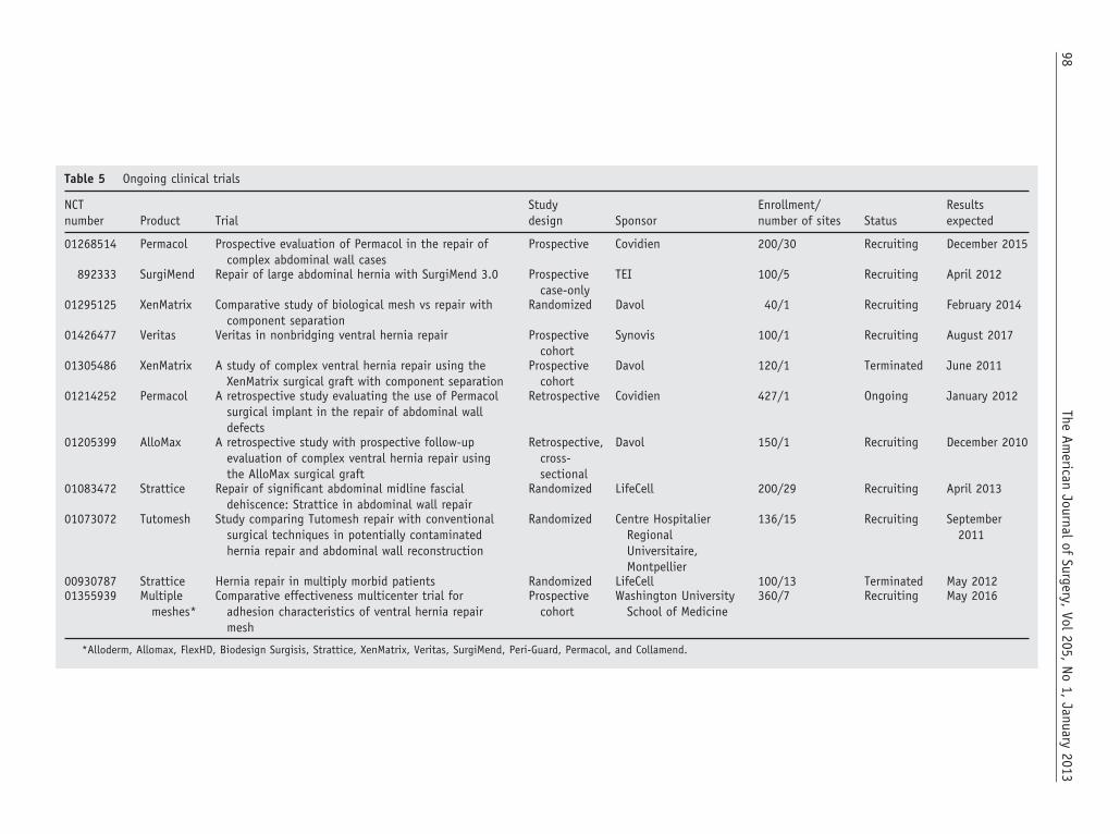
Table 5 Ongoing clinical trials
NCTnumber Product Trial
Studydesign Sponsor
Enrollment/number of sites Status
Resultsexpected
01268514 Permacol Prospective evaluation of Permacol in the repair ofcomplex abdominal wall cases
Prospective Covidien 200/30 Recruiting December 2015
892333 SurgiMend Repair of large abdominal hernia with SurgiMend 3.0 Prospectivecase-only
TEI 100/5 Recruiting April 2012
01295125 XenMatrix Comparative study of biological mesh vs repair withcomponent separation
Randomized Davol 40/1 Recruiting February 2014
01426477 Veritas Veritas in nonbridging ventral hernia repair Prospectivecohort
Synovis 100/1 Recruiting August 2017
01305486 XenMatrix A study of complex ventral hernia repair using theXenMatrix surgical graft with component separation
Prospectivecohort
Davol 120/1 Terminated June 2011
01214252 Permacol A retrospective study evaluating the use of Permacolsurgical implant in the repair of abdominal walldefects
Retrospective Covidien 427/1 Ongoing January 2012
01205399 AlloMax A retrospective study with prospective follow-upevaluation of complex ventral hernia repair usingthe AlloMax surgical graft
Retrospective,cross-sectional
Davol 150/1 Recruiting December 2010
01083472 Strattice Repair of significant abdominal midline fascialdehiscence: Strattice in abdominal wall repair
Randomized LifeCell 200/29 Recruiting April 2013
01073072 Tutomesh Study comparing Tutomesh repair with conventionalsurgical techniques in potentially contaminatedhernia repair and abdominal wall reconstruction
Randomized Centre HospitalierRegionalUniversitaire,Montpellier
136/15 Recruiting September2011
00930787 Strattice Hernia repair in multiply morbid patients Randomized LifeCell 100/13 Terminated May 201201355939 Multiple
meshes*Comparative effectiveness multicenter trial for
adhesion characteristics of ventral hernia repairmesh
Prospectivecohort
Washington UniversitySchool of Medicine
360/7 Recruiting May 2016
*Alloderm, Allomax, FlexHD, Biodesign Surgisis, Strattice, XenMatrix, Veritas, SurgiMend, Peri-Guard, Permacol, and Collamend.
98The
American
Journalof
Surgery,Vol
205,No
1,January
2013

99C.F. Bellows et al. Hernia repair with biologic prosthesis
is related to surgeons’ concerns regarding the potential direconsequences of using permanent mesh in contaminatedfields, there are yet to be any published prospective clinicaltrials justifying their preference over conventional meshmaterials. Until such evidence is forthcoming, the use ofbiological prosthetics in complex incisional hernia repairsshould proceed with caution. There may very well be a solidplace for the use of these materials, but for them to add truevalue to complex hernia repair, better-designed and reportedstudies are necessary to help guide clinical practice.
References
1. Leber GE, Garb JL, Alexander AI, et al. Long-term complicationsassociated with prosthetic repair of incisional hernias. Arch Surg1998;133:378–82.
2. Robinson TN, Clarke JH, Schoen J, et al. Major mesh-related com-plications following hernia repair: events reported to the Food andDrug Administration. Surg Endosc 2005;19:1556–60.
3. Cornwell KG, Landsman A, James KS. Extracellular matrix bioma-terials for soft tissue repair. Clin Podiatr Med Surg 2009;26:507–23.
4. Bellows C, Smith A, Hodde J, et al. Tissue engineering in abdominalwall surgery. Minerva Chir 2011;66:129–43.
5. Diaz JJ Jr, Conquest AM, Ferzoco SJ, et al. Multi-institutional ex-perience using human acellular dermal matrix for ventral herniarepair in a compromised surgical field. Arch Surg 2009;144:209–15.
6. Patton JH Jr, Berry S, Kralovich KA. Use of human acellular dermalmatrix in complex and contaminated abdominal wall reconstructions.Am J Surg 2007;193:360–3.
7. Slater NJ, Hansson BM, Buyne OR, et al. Repair of parastomalhernias with biologic grafts: a systematic review. J Gastrointest Surg2011;15:1252–8.
8. Devillé WL, Buntinx F, Bouter LM, et al. Conducting systematicreviews of diagnostic studies: didactic guidelines. BMC Med ResMethodol 2002;2:9.
9. Zhong T, Janis JE, Ahmad J, et al. Outcomes after abdominal wallreconstruction using acellular dermal matrix: a systematic review. JPlast Reconstr Aesthet Surg 2011;64:1562–71.
10. Ventral Hernia Working Group, Breuing K, Butler CE, et al. Inci-sional ventral hernias: review of the literature and recommendationsregarding the grading and technique of repair. Surgery 2010;148:544–58.
11. Rosenberg L, Joseph L, Barkun A. Surgical Arithmetic: Epidemio-logical Statistical and Outcome-Based Approach to Surgical Practice.Austin, TX; Landes 2000:149–50.
12. Carlson MA, Frantzides CT, Shostrom VK, et al. Minimally invasiveventral herniorrhaphy: an analysis of 6,266 published cases. Hernia2008;12:9–22.
13. Adedeji OA, Bailey CA, Varma JS. Porcine dermal collagen graft inabdominal-wall reconstruction. Br J Plast Surg 2002;55:85–6.
14. Alaedeen DI, Lipman J, Medalie D, et al. The single-staged approachto the surgical management of abdominal wall hernias in contami-nated fields. Hernia 2007;11:41–5.
15. Asham E, Uknis ME, Rastellini C, et al. Acellular dermal matrixprovides a good option for abdominal wall closure following smallbowel transplantation: a case report. Transplant Proc 2006;38:1770–1.
16. Itani KM, Rosen M, Vargo D, et al. Prospective study of single-stagerepair of contaminated hernias using a biologic porcine tissue matrix:The RICH Study. Surgery 2012. [Epub ahead of print].
17. Bachman SL, Ramaswamy A, Ramshaw BJ. Early results of midlinehernia repair using a minimally invasive component separation tech-
nique. Am Surg 2009;75:572–7.18. Baillie DR, Stawicki SP, Eustance N, et al. Use of human and porcinedermal-derived bioprostheses in complex abdominal wall reconstruc-tions: a literature review and case report. Ostomy Wound Manage2007;53:30–7.
19. Bluebond-Langner R, Keifa ES, Mithani S, et al. Recurrent abdom-inal laxity following interpositional human acellular dermal matrix.Ann Plast Surg 2008;60:76–80.
20. Boutros C, Somasundar P, Espat NJ. Early results on the use ofbiomaterials as adjuvant to abdominal wall closure following cytore-duction and hyperthermic intraperitoneal chemotherapy. WorldJ Surg Oncol 2010;8:72.
21. Brewer MB, Rada EM, Milburn ML, et al. Human acellular dermalmatrix for ventral hernia repair reduces morbidity in transplant pa-tients. Hernia 2011;15:141–5.
22. Butler CE, Campbell KT. Minimally invasive component separationwith inlay bioprosthetic mesh (MICSIB) for complex abdominal wallreconstruction. Plast Reconstr Surg 2011;128:698–709.
23. Canda AE, Karaca A. Incisional hernia in action: the use of vacuum-assisted closure and porcine dermal collagen implant. Hernia 2009;13:651–5.
24. Candage R, Jones K, Luchette FA, et al. Use of human acellulardermal matrix for hernia repair: friend or foe? Surgery 2008;144:703–9.
25. Catena F, Ansaloni L, Gazzotti F, et al. Use of porcine dermalcollagen graft (Permacol) for hernia repair in contaminated fields.Hernia 2007;11:57–60.
26. Cavallaro A, Lo Menzo E, Di Vita M, et al. Use of biological meshesfor abdominal wall reconstruction in highly contaminated fields.World J Gastroenterol 2010;16:1928–33.
27. Chavarriaga LF, Lin E, Losken A, et al. Management of complexabdominal wall defects using acellular porcine dermal collagen. AmSurg 2010;76:96–100.
28. Chuo CB, Thomas SS. Absorbable mesh and topical negative pres-sure therapy for closure of abdominal dehiscence with exposedbowel. J Plast Reconstr Aesthet Surg 2008;61:1378–81.
29. Cobb GA, Shaffer J. Cross-linked acellular porcine dermal collagenimplant in laparoscopic ventral hernia repair: case-controlled study ofoperative variables and early complications. Int Surg 2005;90:S24–9.
30. Coyle P, Jaber S, Smith J, et al. Damage control apronectomy fornecrotising fasciitis and strangulated umbilical hernia. Ir J Med Sci2010;179:607–8.
31. Dorafshar AH, Wu C, Zachary LS. Abdominal wall imbrication usingacellular dermal matrix: a novel technique for correcting laxity inpost-bariatric surgery patients. Plast Reconstr Surg 2009;124:270e–2e.
32. Edelman DS, Bellows CF. Umbilical herniorrhaphy reinforced withbiologic mesh. Am Surg 2010;76:1205–9.
33. Eid GM, Mattar SG, Hamad G, et al. Repair of ventral hernias inmorbidly obese patients undergoing laparoscopic gastric bypassshould not be deferred. Surg Endosc 2004;18:207–10.
34. Espinosa-de-los-Monteros A, de la Torre JI, Marrero I, et al. Utili-zation of human cadaveric acellular dermis for abdominal herniareconstruction. Ann Plast Surg 2007;58:264–7.
35. Gentile P, Colicchia GM, Nicoli F, et al. Complex abdominal wallrepair using a porcine dermal matrix. Surg Innov 2011. Epub aheadof print.
36. Gupta A, Zahriya K, Mullens PL, et al. Ventral herniorrhaphy:experience with two different biosynthetic mesh materials, Surgisisand Alloderm. Hernia 2006;10:419–25.
37. Helton WS, Fisichella PM, Berger R, et al. Short-term outcomes withsmall intestinal submucosa for ventral abdominal hernia. Arch Surg2005;140:549–60.
38. Hirsch EF. Repair of an abdominal wall defect after a salvage lapa-rotomy for sepsis. J Am Coll Surg 2004;198:324–8.
39. Holton LH 3rd, Kim D, Silverman RP, et al. Human acellular dermalmatrix for repair of abdominal wall defects: review of clinical expe-rience and experimental data. J Long Term Eff Med Implants 2005;
15:547–58.
100 The American Journal of Surgery, Vol 205, No 1, January 2013
40. Hsu PW, Salgado CJ, Kent K, et al. Evaluation of porcine dermalcollagen (Permacol) used in abdominal wall reconstruction. J PlastReconstr Aesthet Surg 2009;62:1484–9.
41. Jin J, Rosen MJ, Blatnik J, et al. Use of acellular dermal matrix forcomplicated ventral hernia repair: does technique affect outcomes?J Am Coll Surg 2007;205:654–60.
42. Kim H, Bruen K, Vargo D. Acellular dermal matrix in the manage-ment of high-risk abdominal wall defects. Am J Surg 2006;192:705–9.
43. Ko JH, Salvay DM, Paul BC, et al. Soft polypropylene mesh, but notcadaveric dermis, significantly improves outcomes in midline herniarepairs using the components separation technique. Plast ReconstrSurg 2009;124:836–47.
44. Kolker AR, Brown DJ, Redstone JS, et al. Multilayer reconstructionof abdominal wall defects with acellular dermal allograft (AlloDerm)and component separation. Ann Plast Surg 2005;55:36–41.
45. Lee EI, Chike-Obi CJ, Gonzalez P, et al. Abdominal wall repair usinghuman acellular dermal matrix: a follow-up study. Am J Surg 2009;198:650–7.
46. Limpert JN, Desai AR, Kumpf AL, et al. Repair of abdominal walldefects with bovine pericardium. Am J Surg 2009;198:e60–5.
47. Lin HJ, Spoerke N, Deveney C, et al. Reconstruction of complexabdominal wall hernias using acellular human dermal matrix: a singleinstitution experience. Am J Surg 2009;197:599–603.
48. Lipman J, Medalie D, Rosen MJ. Staged repair of massive incisionalhernias with loss of abdominal domain: a novel approach. Am J Surg2008;195:84–8.
49. Liyanage SH, Purohit GS, Frye JN, et al. Anterior abdominal wallreconstruction with a Permacol implant. J Plast Reconstr AesthetSurg 2006;59:553–5.
50. Martins PN, Butt K, El-Sabrout R. Richter’s hernia at a Tenckhoffcatheter exit site. Surg Laparosc Endosc Percutan Tech 2010;20:e136–8.
51. McDonald MD, Weiss CA. Human acellular dermis for recurrenthernia. Contemp Surg 2005;61:276–80.
52. Misra S, Raj PK, Tarr SM, et al. Results of AlloDerm use in abdom-inal hernia repair. Hernia 2008;12:247–50.
53. Morrison JE, Brizendine JB, Yost MJ, et al. Massive ventral herniawith extensive heterotopic ossification: the honeycomb-abdomen.J Trauma 2009;66:1234–7.
54. Nemeth NL, Butler CE. Complex torso reconstruction with humanacellular dermal matrix: long-term clinical follow-up. Plast ReconstrSurg 2009;123:192–6.
55. O’Brien JA, Ignotz R, Montilla R, et al. Long-term histologic andmechanical results of a Permacol abdominal wall explant. Hernia2011;15:211–5.
56. Parker DM, Armstrong PJ, Frizzi JD, et al. Porcine dermal collagen(Permacol) for abdominal wall reconstruction. Curr Surg 2006;63:255–8.
57. Parra MW, Rodas EB, Niravel AA. Laparoscopic repair of potentiallycontaminated abdominal ventral hernias using a xenograft: a caseseries. Hernia 2011;15:575–8.
58. Paton BL, Novitsky YW, Zerey M, et al. Management of infectionsof polytetrafluoroethylene-based mesh. Surg Infect (Larchmt) 2007;8:337–41.
59. Pomahac B, Aflaki P. Use of a non-cross-linked porcine dermalscaffold in abdominal wall reconstruction. Am J Surg 2010;199:22–7.
60. Saettele TM, Bachman SL, Costello CR, et al. Use of porcine dermalcollagen as a prosthetic mesh in a contaminated field for ventralhernia repair: a case report. Hernia 2007;11:279–85.
61. Sailes FC, Walls J, Guelig D, et al. Synthetic and biological mesh incomponent separation: a 10-year single institution review. Ann PlastSurg 2010;64:696–8.
62. Satterwhite TS, Miri S, Chung C, et al. Abdominal wall reconstruc-tion with dual layer cross-linked porcine dermal xenograft: the “porksandwich” herniorrhaphy. J Plast Reconstr Aesthet Surg 2012;65:
333–41.63. Schuster R, Singh J, Safadi BY, et al. The use of acellular dermalmatrix for contaminated abdominal wall defects: wound status pre-dicts success. Am J Surg 2006;192:594–7.
64. Shaikh FM, Giri SK, Durrani S, et al. Experience with porcineacellular dermal collagen implant in one-stage tension-free recon-struction of acute and chronic abdominal wall defects. World J Surg2007;31:1966–72.
65. Shaikh FM, Kennedy TE, Coyle P, et al. Diastasis as a cause ofrecurrence in ventral herniorrhaphy with porcine acellular dermalcollagen implant. Plast Reconstr Surg 2011;127:167e–9e.
66. Singh MK, Rocca JP, Rochon C, et al. Open abdomen managementwith human acellular dermal matrix in liver transplant recipients.Transplant Proc 2008;40:3541–4.
67. Tong P, Ha J, Chandraratna H. Fibrin glue and porcine-derived meshrepair of ruptured umbilical hernia in a patient with ascites. Eur Surg2011;43:55–7.
68. Treviño JM, Franklin ME Jr, Berghoff KR, et al. Preliminary resultsof a two-layered prosthetic repair for recurrent inguinal and ventralhernias combining open and laparoscopic techniques. Hernia 2006;10:253–7.
69. Tung CS, Zighelboim I, Scott B, et al. Human acellular dermal matrixfor closure of a contaminated gynecologic wound. Gynecol Oncol2006;103:354–6.
70. Walker H, Brooker T, Gelman W. Abdominal wall reconstructionfollowing removal of a chronically infected mid-urethral tape. IntUrogynecol J Pelvic Floor Dysfunct 2009;20:1273–5.
71. Wietfeldt ED, Hassan I, Rakinic J. Utilization of bovine acellulardermal matrix for abdominal wall reconstruction: a retrospective caseseries. Ostomy Wound Manage 2009;55:52–6.
72. Wotton FT, Akoh JA. Rejection of Permacol mesh used in abdominalwall repair: a case report. World J Gastroenterol 2009;15:4331–3.
73. Byrnes MC, Irwin E, Carlson D, et al. Repair of high-risk incisionalhernias and traumatic abdominal wall defects with porcine mesh. AmSurg 2011;77:144–50.
74. Diaz JJ Jr, Guy J, Berkes MB, et al. Acellular dermal allograft forventral hernia repair in the compromised surgical field. Am Surg2006;72:1181–7.
75. Guy JS, Miller R, Morris JA Jr, et al. Early one-stage closure inpatients with abdominal compartment syndrome: fascial replacementwith human acellular dermis and bipedicle flaps. Am Surg 2003;69:1025–8.
76. Blatnik J, Jin J, Rosen M. Abdominal hernia repair with bridgingacellular dermal matrix—an expensive hernia sac. Am J Surg 2008;196:47–50.
77. Butler CE, Langstein HN, Kronowitz SJ. Pelvic, abdominal, andchest wall reconstruction with AlloDerm in patients at increased riskfor mesh-related complications. Plast Reconstr Surg 2005;116:1263–75; discussion, 1276–7.
78. Bellows CF, Albo D, Berger DH, et al. Abdominal wall repair usinghuman acellular dermis. Am J Surg 2007;194:192–8.
79. Han JG, Ma SZ, Song JK, et al. Acellular dermal matrix in themanagement of contaminated abdominal wall defects following pri-mary actinomycosis excision. J Plast Reconstr Aesthet Surg 2008;61:1544–5.
80. Tang R, Gu Y, Gong DQ, et al. Immediate repair of major abdominalwall defect after extensive tumor excision in patients with abdominalwall neoplasm: a retrospective review of 27 cases [corrected]. AnnSurg Oncol 2009;16:2895–907.
81. Xiao SC, Zhu SH, Li HY, et al. Repair of complex abdominal walldefects from high-voltage electric injury with two layers of acellulardermal matrix: a case report. J Burn Care Res 2009;30:352–4.
82. Franklin ME Jr, Gonzalez JJ Jr, Glass JL, et al. Laparoscopic ventraland incisional hernia repair: an 11-year experience. Hernia 2004;8:23–7.
83. Williams RF, Martin DF, Mulrooney MT, et al. Intraperitoneal mod-ification of the Rives-Stoppa repair for large incisional hernias. Her-
nia 2008;12:141–5.
101C.F. Bellows et al. Hernia repair with biologic prosthesis
84. Hadeed JG, Walsh MD, Pappas TN, et al. Complex abdominal wallhernias: a new classification system and approach to managementbased on review of 133 consecutive patients. Ann Plast Surg 2011;66:497–503.
85. Snyder CW, Graham LA, Gray SH, et al. Effect of mesh type andposition on subsequent abdominal operations after incisional herniarepair. J Am Coll Surg 2011;212:496–502.
86. Samstein B, Pichardo E, Perez T, et al. High incidence but low rateof repair of incisional hernias after liver transplantation. Liver Trans-plant 2009;15:S226.
87. Moore M, Bax T, MacFarlane M, et al. Outcomes of the fascialcomponent separation technique with synthetic mesh reinforcementfor repair of complex ventral incisional hernias in the morbidly obese.Am J Surg 2008;195:575–9.
88. Jenkins ED, Yom V, Melman L, et al. Prospective evaluation ofadhesion characteristics to intraperitoneal mesh and adhesiolysis-related complications during laparoscopic re-exploration after priorventral hernia repair. Surg Endosc 2010;24:3002–7.
89. Shah BC, Tiwari MM, Goede MR, et al. Not all biologics are equal!Hernia 2011;15:165–71.
90. Ghazi B, Deigni O, Yezhelyev M, et al. Current options in themanagement of complex abdominal wall defects. Ann Plast Surg2011;66:488–92.
91. Newcomb WL, Polhill JL, Chen AY, et al. Staged hernia repairpreceded by gastric bypass for the treatment of morbidly obesepatients with complex ventral hernias. Hernia 2008;12:465–9.
92. Chiara O, Cimbanassi S, Boati S, et al. Surgical management ofabdominal compartment syndrome. Minerva Anestesiol 2011;77:457–62.
93. Olson C, Abdelnaby A, Fayiga Y, et al. Simultaneous stoma closureand massive ventral hernia repair reinforced with biologic tissuematrix. Dis Colon Rectum 2010;53:585.
94. Buinewicz B, Rosen B. Acellular cadaveric dermis (AlloDerm): anew alternative for abdominal hernia repair. Ann Plast Surg 2004;52:188–94.
95. Loganathan A, Ainslie WG, Wedgwood KR. Initial evaluation ofPermacol bioprosthesis for the repair of complex incisional andparastomal hernias. Surgeon 2010;8:202–5.
96. Franklin ME Jr, Treviño JM, Portillo G, et al. The use of porcinesmall intestinal submucosa as a prosthetic material for laparoscopichernia repair in infected and potentially contaminated fields: long-term follow-up. Surg Endosc 2008;22:1941–6.
97. Franklin M, Russek K. Use of porcine small intestine submucosa asa prosthetic material for laparoscopic hernia repair in infected andpotentially contaminated fields: long-term follow-up assessment.Surg Endosc 2011;25:1693–4.
98. Ueno T, Pickett LC, de la Fuente SG, et al. Clinical application of
porcine small intestinal submucosa in the management of infected orpotentially contaminated abdominal defects. J Gastrointest Surg2004;8:109–12.
99. Awad SS, Rao RK, Berger DH, et al. Microbiology of infectedacellular dermal matrix (AlloDerm) in patients requiring complexabdominal closure after emergency surgery. Surg Infect (Larchmt)2009;10:79–84.
100. Ko JH, Wang EC, Salvay DM, et al. Abdominal wall reconstruction:lessons learned from 200 “components separation” procedures. ArchSurg 2009;144:1047–55.
101. Propst JT, Hansen KJ, Yost MJ, et al. Unique recalcitrant herniarepair: enterocutaneous fistula with complex ventral hernia. Am Surg2008;74:1117–9.
102. Scott BG, Welsh FJ, Pham HQ, et al. Early aggressive closure of theopen abdomen. J Trauma 2006;60:17–22.
103. Latifi R, Joseph B, Kulvatunyou N, et al. Enterocutaneous fistulas anda hostile abdomen: reoperative surgical approaches. World J Surg2012;36:516–23.
104. Latifi R, Gustafson M. Abdominal wall reconstruction in patientswith enterocutaneous fistulas. Eur J Trauma Emerg Surg 2011;37:241–50.
105. Hernán MA, Clayton D, Keiding N. The Simpson’s paradox unrav-eled. Int J Epidemiol 2011;40:780–5.
106. Hiles M, Record Ritchie RD, Altizer AM. Are biologic grafts effec-tive for hernia repair?: a systematic review of the literature. SurgInnov 2009;16:26–37.
107. Harth KC, Rosen MJ. Major complications associated with xenograftbiologic mesh implantation in abdominal wall reconstruction. SurgInnov 2009;16:324–9.
108. Burger JW, Luijendijk RW, Hop WC, et al. Long-term follow-up ofa randomized controlled trial of suture versus mesh repair of inci-sional hernia. Ann Surg 2004;240:578–83.
109. Rosen MJ. Polyester-based mesh for ventral hernia repair: is it safe?Am J Surg 2009;197:353–9.
110. Zografos GN, Mitropapas G, Vasiliadis G, et al. Open and laparo-scopic approach in incisional hernia repair with ePTFE prosthesis. JLaparoendosc Adv Surg Tech A 2007;17:277–81.
111. Miserez M, Grass G, Weiss C, et al. Closure of the LAPSIS trial. Br JSurg 2010;97:1598.
Supplementary data
Supplementary data associated with this article can befound, in the online version, at http://dx.doi.org/10.1016/
j.amjsurg.2012.02.019.