Embed Size (px)
Citation preview
Cranfield Forensic Institute 23rd March 2018
Report on the examination of human remains from Burrow/Rat Island, Gosport
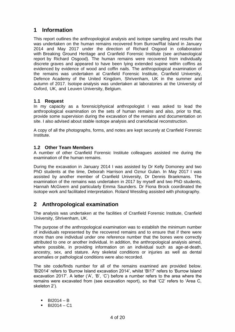
1(c) Harvey Mills/Richard Osgood. Cranfield Forensic Institute staff documenting at the site during the May 2017 excavations.
2 of 20
REPORT ON THE HISTORICAL/ARCHAEOLOGIAL HUMAN
REMAINS OF BURROW/RAT ISLAND, GOSPORT, UK
Report by
Nicholas MÁRQUEZ-GRANT, Cranfield Forensic Institute (CFI)
Name Position Signature Date
Author Nicholas Marquez-
Grant Lecturer in
Anthropology
23/03/18
Dated the 23rd March 2018
Non-technical Summary
The findings detailed within this report are summarised below in a non-technical manner.
A minimum of nine skeletons were recovered in 2014 and 2017 from Burrow Island/Rat Island, Gosport, Portsmouth, UK and these were examined anthropologically.
The anthropological examination was undertaken in order to reconstruct the biological profile. This profile was based on information obtained from the human remains, and where possible provided information on age-at-death, sex, ancestry, stature and any individuating or unique features. With regard to the latter, observations included an assessment of any dental conditions and any previous injuries or diseases which may have affected the individual in life. Any trauma that appeared to have occurred at or around the individual’s time of death was also documented. In addition to the anthropological analysis, samples were submitted for chemical analysis to investigate diet and provenance.
The findings of this examination indicate that all individuals were males, ranging from adolescence to middle adulthood. Stature was estimated and provided a range of estimate and pathological analysis provided a number of clues as to the lives of these individuals. Two individuals presented clay pipe notches. Two skeletons had evidence of cut marks from dissection/autopsy. The chemical (isotope) analysis seems to reflect a number of individuals likely to be from outside Britain.

3 of 20
Table of Contents
1 Information ......................................................................................................... 4 1.1 Request ................................................................................................................... 4 1.2 Other Team Members .............................................................................................. 4
2 Anthropological examination ............................................................................... 4 2.1 Methods: Anthropological Examination .................................................................... 5 2.2 Sampling for isotope analysis ................................................................................... 5
3 Results ................................................................................................................. 5 SKELETON BI2014-B ......................................................................................................... 6 SKELETON BI2014-C1 ....................................................................................................... 6 SKELETON BI2014-C2 ....................................................................................................... 7 SKELETON BI2014-A1 ....................................................................................................... 8 SKELETON BI2014-A2 ....................................................................................................... 8 SKELETON BI2014-A4 ....................................................................................................... 9 SKELETON BI17-A5 ......................................................................................................... 10 SKELETON BI17-A6 ......................................................................................................... 10 SKELETON BI17-A7 ......................................................................................................... 11 SKELETON BI17-A8 ......................................................................................................... 11 DISARTICULATED AND UNSTRATIFIED BONES ................................................................. 12 3.7.1. ISOTOPE RESULTS .......................................................................................... 12
4. Summary and Discussion of the Anthropological Analysis ................................... 13
4 of 20
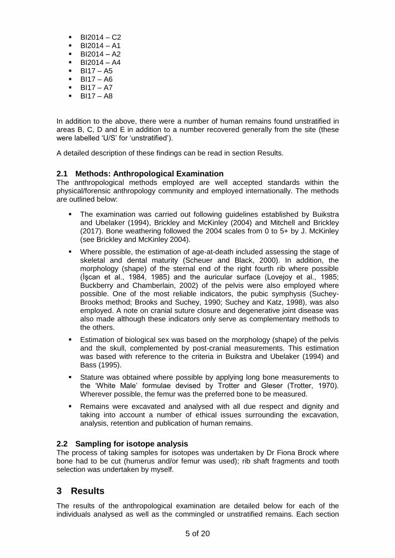
1 Information
This report outlines the anthropological analysis and isotope sampling and results that was undertaken on the human remains recovered from Burrow/Rat Island in January 2014 and May 2017 under the direction of Richard Osgood in collaboration with Breaking Ground Heritage and Cranfield Forensic Institute (see archaeological report by Richard Osgood). The human remains were recovered from individually discrete graves and appeared to have been lying extended supine within coffins as evidenced by evidence of wood and coffin nails. The anthropological examination of the remains was undertaken at Cranfield Forensic Institute, Cranfield University, Defence Academy of the United Kingdom, Shrivenham, UK in the summer and autumn of 2017. Isotope analysis was undertaken at laboratories at the University of Oxford, UK, and Leuven University, Belgium.
1.1 Request In my capacity as a forensic/physical anthropologist I was asked to lead the anthropological examination on the sets of human remains and also, prior to that, provide some supervision during the excavation of the remains and documentation on site. I also advised about stable isotope analysis and craniofacial reconstruction.
A copy of all the photographs, forms, and notes are kept securely at Cranfield Forensic Institute.
1.2 Other Team Members A number of other Cranfield Forensic Institute colleagues assisted me during the examination of the human remains.
During the excavation in January 2014 I was assisted by Dr Kelly Domoney and two PhD students at the time, Deborah Harrison and Oznur Gulan. In May 2017 I was assisted by another member of Cranfield University, Dr Dennis Braekmans. The examination of the remains was undertaken in 2017 by myself and two PhD students, Hannah McGivern and particularly Emma Saunders. Dr Fiona Brock coordinated the isotope work and facilitated interpretation. Roland Wessling assisted with photography.
2 Anthropological examination
The analysis was undertaken at the facilities of Cranfield Forensic Institute, Cranfield University, Shrivenham, UK.
The purpose of the anthropological examination was to establish the minimum number of individuals represented by the recovered remains and to ensure that if there were more than one individual under one reference number that the bones were correctly attributed to one or another individual. In addition, the anthropological analysis aimed, where possible, in providing information on an individual such as age-at-death, ancestry, sex, and stature. Any skeletal conditions or injuries as well as dental anomalies or pathological conditions were also recorded.
The site code/finds number for all of the remains examined are provided below. ‘BI2014’ refers to ‘Burrow Island excavation 2014’, whilst ‘BI17’ refers to ‘Burrow Island excavation 2017’. A letter (‘A’, ‘B’, ‘C’) before a number refers to the area where the remains were excavated from (see excavation report), so that ‘C2’ refers to ‘Area C, skeleton 2’).
BI2014 – B BI2014 – C1
5 of 20
BI2014 – C2 BI2014 – A1 BI2014 – A2 BI2014 – A4 BI17 – A5 BI17 – A6 BI17 – A7 BI17 – A8
In addition to the above, there were a number of human remains found unstratified in areas B, C, D and E in addition to a number recovered generally from the site (these were labelled ‘U/S’ for ‘unstratified’).
A detailed description of these findings can be read in section Results.
2.1 Methods: Anthropological Examination The anthropological methods employed are well accepted standards within the physical/forensic anthropology community and employed internationally. The methods are outlined below:
The examination was carried out following guidelines established by Buikstraand Ubelaker (1994), Brickley and McKinley (2004) and Mitchell and Brickley(2017). Bone weathering followed the 2004 scales from 0 to 5+ by J. McKinley(see Brickley and McKinley 2004).
Where possible, the estimation of age-at-death included assessing the stage ofskeletal and dental maturity (Scheuer and Black, 2000). In addition, themorphology (shape) of the sternal end of the right fourth rib where possible(İşcan et al., 1984, 1985) and the auricular surface (Lovejoy et al., 1985;Buckberry and Chamberlain, 2002) of the pelvis were also employed wherepossible. One of the most reliable indicators, the pubic symphysis (Suchey-Brooks method; Brooks and Suchey, 1990; Suchey and Katz, 1998), was alsoemployed. A note on cranial suture closure and degenerative joint disease wasalso made although these indicators only serve as complementary methods tothe others.
Estimation of biological sex was based on the morphology (shape) of the pelvisand the skull, complemented by post-cranial measurements. This estimationwas based with reference to the criteria in Buikstra and Ubelaker (1994) andBass (1995).
Stature was obtained where possible by applying long bone measurements tothe ‘White Male’ formulae devised by Trotter and Gleser (Trotter, 1970).Wherever possible, the femur was the preferred bone to be measured.
Remains were excavated and analysed with all due respect and dignity andtaking into account a number of ethical issues surrounding the excavation,analysis, retention and publication of human remains.
2.2 Sampling for isotope analysis The process of taking samples for isotopes was undertaken by Dr Fiona Brock where bone had to be cut (humerus and/or femur was used); rib shaft fragments and tooth selection was undertaken by myself.
3 Results
The results of the anthropological examination are detailed below for each of the individuals analysed as well as the commingled or unstratified remains. Each section
6 of 20
gives an overview for each of the skeletons including the completeness, preservation, biological profile, and any unique features as well as palaeopathological conditions.
All the skeletal remains presented varying degrees of preservation, with fragmentation, incomplete bones and weathering present in most skeletons. This preservation and completeness of the skeletal remains limited the information that could be obtained during the anthropological examination.
The detailed anthropology recording forms provide further information including preservation, any other taphonomic modifications to the bone, a detailed skeletal inventory, the specific features and methods employed for the biological profile, the measurements taken, a dental inventory, and any observed anatomical variations, trauma, or pathology.
SKELETON BI2014-B The recovered bone elements from this individual comprised over 75% of the skeleton. Primarily, the foot bones and the left fibula were missing.
Some post-mortem damage was visible across the skeleton. Weathering (erosion of the cortical bone surface) was present overall as Grade 2. The most fragmentation occurred at the ribs. There were areas of dark staining on thoracic vertebrae, the left femur and a number of ribs from the left side.
All the bones appear to be from the same individual. There was no duplication or any inconsistencies with age or sex that may suggest more than one individual.
The individual was male, as assessed primarily from pelvic traits but also from skull traits, supported by the dimensions of a number of bones. The estimated age-at-death of the individual was between 30 and 45 years based primarily on skeletal maturation, the auricular surface. A wider age range was given as 25-45 years. Stature was calculated as 1.73m (range 1.70-1.76m) by measuring the left femur. Ancestry was not assessed.
There was no observable degenerative joint disease nor infectious disease present (apart from dental caries). The only changes were bone growth (osteophytosis) to the vertebral bodies of T2 and T3. There appeared to be osteochondritis (non) dissecans on the right lunate with a lesion of 3.5 mm diameter. The first segment of the sacrum (S1) revealed a depression of anterior half of the body; a similar alteration was observed in the inferior surface of the fifth (L5) lumbar vertebra. A cortical lesions may be observed on the rhomboid fossa of the right clavicle. Cribra orbitalia could not be assessed. With regard to the dentition, dental caries was present on some teeth (9/26) and ante-mortem tooth loss was evident (3/32). Enamel hypoplasia was absent. As unique identifying features are heavy occlusal wear on lower and upper central incisors, a rotated left upper left canine and a partially erupted (impacted?) upper right canine.
Most of the damage present on the skeleton appears to be post-mortem. However, there were three rib fragments that appeared to have a cut which may be associated with autopsy cuts.
No samples for isotope analysis were taken.
SKELETON BI2014-C1 The recovered bone elements from this individual comprised between 50% and 75% of the skeleton. Primarily, the right clavicle was absent, as well as a number of vertebrae, some ribs, hand and foot bones. Part of the pelvic bones (the left and the right pubis) was also missing.
7 of 20
Some post-mortem damage was visible across the skeleton. Long bones were generally well preserved but fragmentation was noted on the ribs, pelvic bones, the right humerus, right radius and some areas of the skull. Weathering (erosion of the cortical bone surface) was present overall as Grades 2-3. There were areas of dark staining on the left tibia.
All the bones appear to be from the same individual; except for an additional right calcaneous (foot bone).
The individual was classified overall as possibly male, according to a number of traits in the pelvis and skull. Some traits however were ambiguous, gracile or more characteristic of female skeletons. The estimated age-at-death of the individual was between 17 and 20 years based primarily on skeletal maturation and the auricular surface. A wider age range was given as 16 and 21 years. Stature was calculated as 1.61m (range 1.58-1.64m) by measuring the left femur. Ancestry was not assessed.
There was no observable degenerative joint disease nor infectious disease present (apart from dental caries). There is pitting observed on the occipital and right parietal bones. Cribra orbitalia was present on the right orbital roof (not observed on the left side). With regard to the dentition, dental caries was present on some teeth (4/23) and ante-mortem tooth loss was evident (1/32). Enamel hypoplasia was present too (12/22 teeth observed). There was a possible periapical cavity in the site of the upper right second premolar. As unique identifying features there was a shovel shaped upper right second incisor.
Most of the damage present on the skeleton appears to be post-mortem.
No samples for isotope analysis were taken.
SKELETON BI2014-C2 The recovered bone elements from this individual comprised between 50% and 75% of the skeleton. Many ribs, the left scapula, most hand and all foot bones were missing. Many long bones were incomplete.
Some post-mortem damage was visible across the skeleton, and in particular the ribs, vertebrae and pelvis. Bone preservation was fair with weathering (erosion of the cortical bone surface) present overall as Grade 3. There was more post-mortem (taphonomic) damage on the left bones of the skeleton compared to the right side. There were areas of dark staining on the frontal bone.
All the bones appear to be from the same individual. There was no duplication or any inconsistencies with age or sex that may suggest more than one individual.
The individual was male, as assessed from pelvic and skull traits, complemented by bone dimensions. The estimated age-at-death of the individual was limited and it was only possible to indicate an individual older than 25 years (or older than 25-30 years). Stature was calculated as 1.75m (range 1.72-1.78m) by measuring the left femur. Ancestry was not assessed.
Observations regarding pathological conditions were limited due to the condition and completeness of the skeleton. Nevertheless no degenerative joint disease was present. There was an area of woven bone present possibly on the frontal bone. There was a healed fracture on the right clavicle. There was no cribra orbitalia. With regard to the dentition, dental caries was present on some teeth (6/26) and ante-mortem tooth loss was evident (3/32). Enamel hypoplasia was also present (10/23 teeth observed). There are slight calculus (tartar) deposits overall. As a unique identifying feature is the wear (activity related) on the left side (lower first and second premolars, upper second incisor and canine) which is consistent with a clay pipe notch. There is also activity
8 of 20
related dental wear on the right lower second and third molars, and areas of enamel chipping in several teeth.
Most of the damage present on the skeleton appears to be post-mortem.
No samples for isotope analysis were taken.
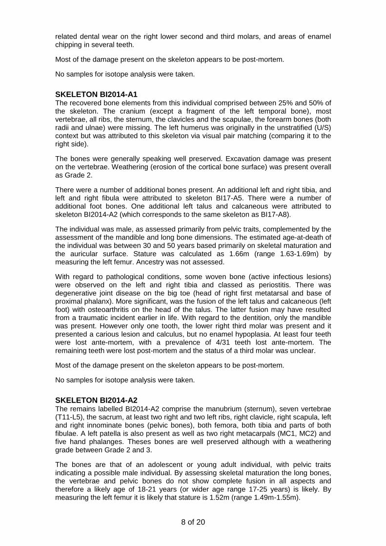
SKELETON BI2014-A1 The recovered bone elements from this individual comprised between 25% and 50% of the skeleton. The cranium (except a fragment of the left temporal bone), most vertebrae, all ribs, the sternum, the clavicles and the scapulae, the forearm bones (both radii and ulnae) were missing. The left humerus was originally in the unstratified (U/S) context but was attributed to this skeleton via visual pair matching (comparing it to the right side).
The bones were generally speaking well preserved. Excavation damage was present on the vertebrae. Weathering (erosion of the cortical bone surface) was present overall as Grade 2.
There were a number of additional bones present. An additional left and right tibia, and left and right fibula were attributed to skeleton BI17-A5. There were a number of additional foot bones. One additional left talus and calcaneous were attributed to skeleton BI2014-A2 (which corresponds to the same skeleton as BI17-A8).
The individual was male, as assessed primarily from pelvic traits, complemented by the assessment of the mandible and long bone dimensions. The estimated age-at-death of the individual was between 30 and 50 years based primarily on skeletal maturation and the auricular surface. Stature was calculated as 1.66m (range 1.63-1.69m) by measuring the left femur. Ancestry was not assessed.
With regard to pathological conditions, some woven bone (active infectious lesions) were observed on the left and right tibia and classed as periostitis. There was degenerative joint disease on the big toe (head of right first metatarsal and base of proximal phalanx). More significant, was the fusion of the left talus and calcaneous (left foot) with osteoarthritis on the head of the talus. The latter fusion may have resulted from a traumatic incident earlier in life. With regard to the dentition, only the mandible was present. However only one tooth, the lower right third molar was present and it presented a carious lesion and calculus, but no enamel hypoplasia. At least four teeth were lost ante-mortem, with a prevalence of 4/31 teeth lost ante-mortem. The remaining teeth were lost post-mortem and the status of a third molar was unclear.
Most of the damage present on the skeleton appears to be post-mortem.
No samples for isotope analysis were taken.
SKELETON BI2014-A2 The remains labelled BI2014-A2 comprise the manubrium (sternum), seven vertebrae (T11-L5), the sacrum, at least two right and two left ribs, right clavicle, right scapula, left and right innominate bones (pelvic bones), both femora, both tibia and parts of both fibulae. A left patella is also present as well as two right metacarpals (MC1, MC2) and five hand phalanges. Theses bones are well preserved although with a weathering grade between Grade 2 and 3.
The bones are that of an adolescent or young adult individual, with pelvic traits indicating a possible male individual. By assessing skeletal maturation the long bones, the vertebrae and pelvic bones do not show complete fusion in all aspects and therefore a likely age of 18-21 years (or wider age range 17-25 years) is likely. By measuring the left femur it is likely that stature is 1.52m (range 1.49m-1.55m).
9 of 20
The right clavicle displays osteophythosis of the lateral (shoulder) end. Both femora present bowing (anterior-posterior) and there is slight bowing (medio-lateral) on the tibiae. Infectious lesions are present in both tibiae, characterised by periostitis which was active (woven bone) prior to death.
The vertebrae, pelvis and lower long bones appear to correspond with Skeleton BI17-A8. A number of foot bones from BI2014-A1 is also attributed to this skeleton. The upper part of the skeleton (e.g. clavicle) would seem to belong to another individual and it is likely that BI2014-A2 includes a mix of bones primarily BI17-A8 but also belonging to BI2014-A1. Please refer to the reports for BI17-A8 for more detail.
One animal (non-human) bone was found amongst the human remains.
No sampling for isotope analysis was undertaken from these remains.
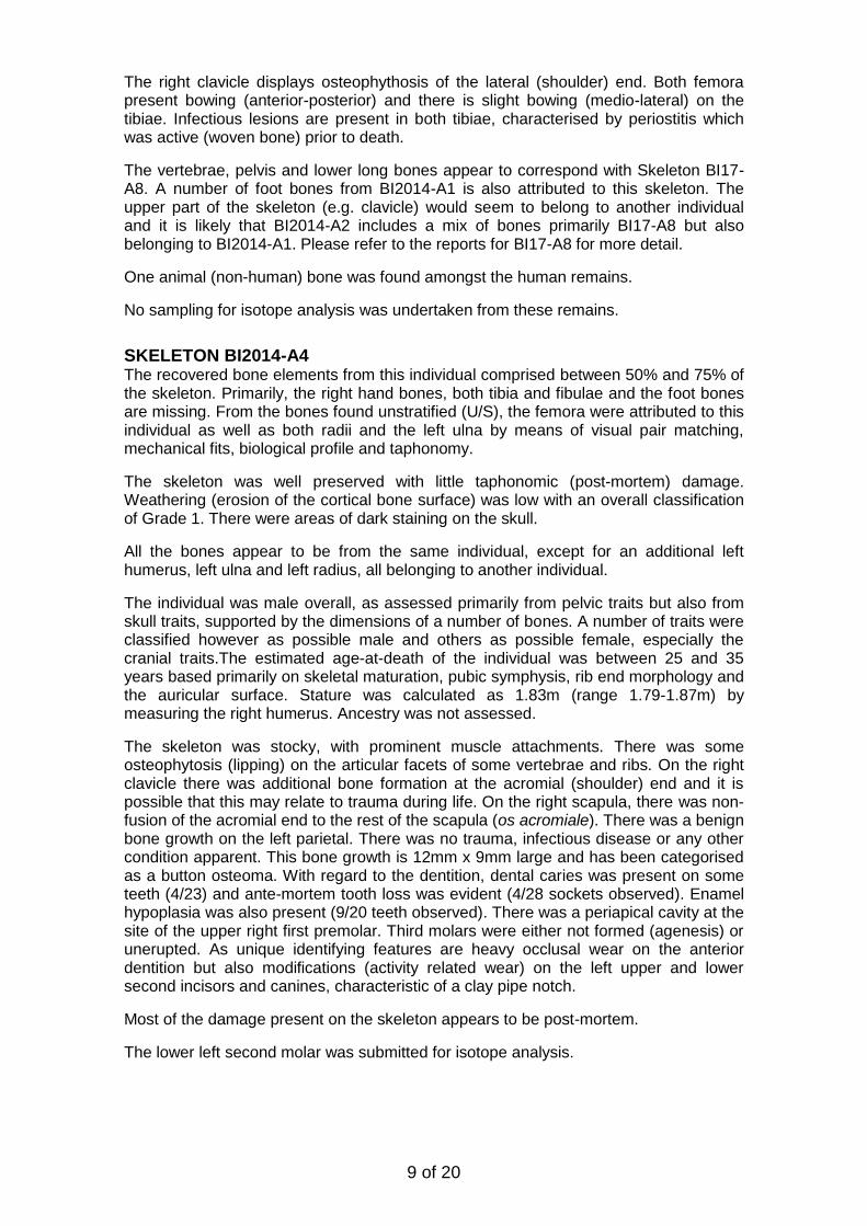
SKELETON BI2014-A4 The recovered bone elements from this individual comprised between 50% and 75% of the skeleton. Primarily, the right hand bones, both tibia and fibulae and the foot bones are missing. From the bones found unstratified (U/S), the femora were attributed to this individual as well as both radii and the left ulna by means of visual pair matching, mechanical fits, biological profile and taphonomy.
The skeleton was well preserved with little taphonomic (post-mortem) damage. Weathering (erosion of the cortical bone surface) was low with an overall classification of Grade 1. There were areas of dark staining on the skull.
All the bones appear to be from the same individual, except for an additional left humerus, left ulna and left radius, all belonging to another individual.
The individual was male overall, as assessed primarily from pelvic traits but also from skull traits, supported by the dimensions of a number of bones. A number of traits were classified however as possible male and others as possible female, especially the cranial traits.The estimated age-at-death of the individual was between 25 and 35 years based primarily on skeletal maturation, pubic symphysis, rib end morphology and the auricular surface. Stature was calculated as 1.83m (range 1.79-1.87m) by measuring the right humerus. Ancestry was not assessed.
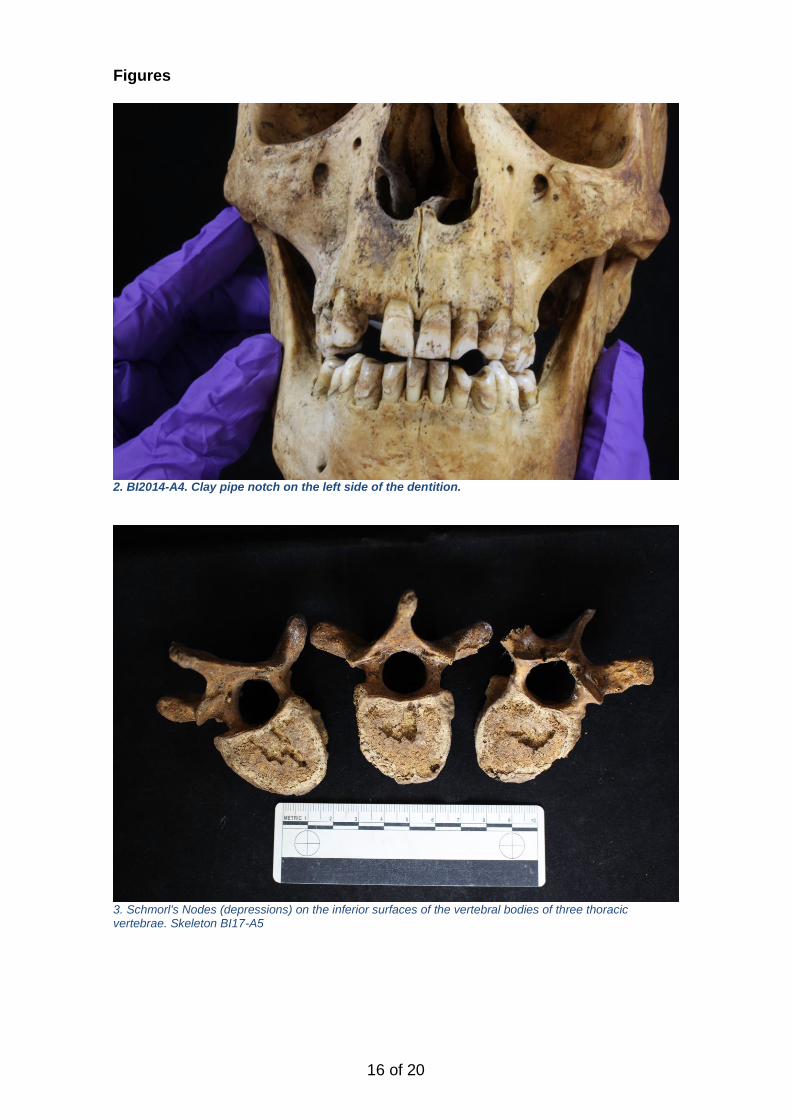
The skeleton was stocky, with prominent muscle attachments. There was some osteophytosis (lipping) on the articular facets of some vertebrae and ribs. On the right clavicle there was additional bone formation at the acromial (shoulder) end and it is possible that this may relate to trauma during life. On the right scapula, there was non-fusion of the acromial end to the rest of the scapula (os acromiale). There was a benign bone growth on the left parietal. There was no trauma, infectious disease or any other condition apparent. This bone growth is 12mm x 9mm large and has been categorised as a button osteoma. With regard to the dentition, dental caries was present on some teeth (4/23) and ante-mortem tooth loss was evident (4/28 sockets observed). Enamel hypoplasia was also present (9/20 teeth observed). There was a periapical cavity at the site of the upper right first premolar. Third molars were either not formed (agenesis) or unerupted. As unique identifying features are heavy occlusal wear on the anterior dentition but also modifications (activity related wear) on the left upper and lower second incisors and canines, characteristic of a clay pipe notch.
Most of the damage present on the skeleton appears to be post-mortem.
The lower left second molar was submitted for isotope analysis.
10 of 20
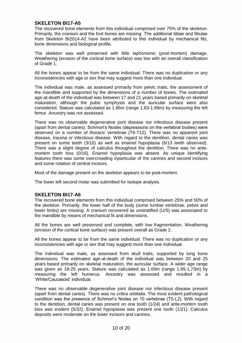
SKELETON BI17-A5 The recovered bone elements from this individual comprised over 75% of the skeleton. Primarily, the cranium and the foot bones are missing. The additional tibiae and fibulae from Skeleton BI2014-A2 have been attributed to this individual by mechanical fits, bone dimensions and biological profile.
The skeleton was well preserved with little taphonomic (post-mortem) damage. Weathering (erosion of the cortical bone surface) was low with an overall classification of Grade 1.
All the bones appear to be from the same individual. There was no duplication or any inconsistencies with age or sex that may suggest more than one individual.
The individual was male, as assessed primarily from pelvic traits, the assessment of the mandible and supported by the dimensions of a number of bones. The estimated age-at-death of the individual was between 17 and 21 years based primarily on skeletal maturation, although the pubic symphysis and the auricular surface were also considered. Stature was calculated as 1.86m (range 1.83-1.89m) by measuring the left femur. Ancestry was not assessed.
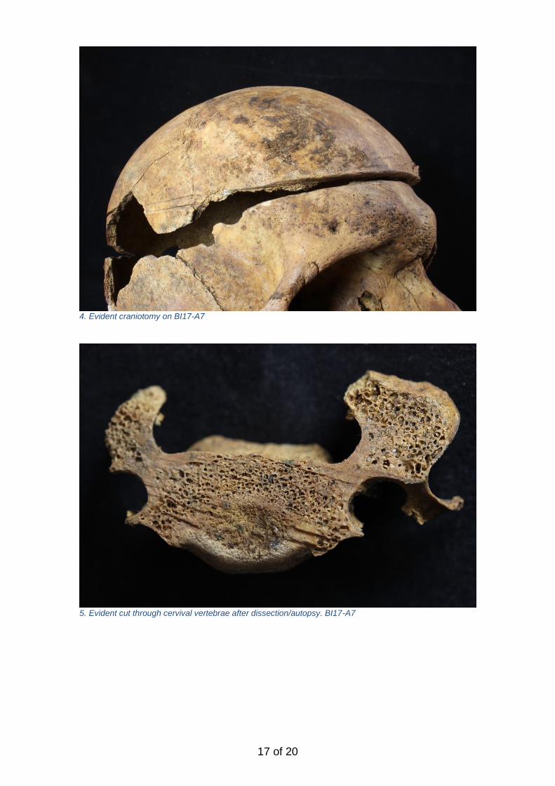
There was no observable degenerative joint disease nor infectious disease present (apart from dental caries). Schmorl’s Nodes (depressions on the vertebral bodies) were observed on a number of thoracic vertebrae (T6-T12). There was no apparent joint disease, trauma or infectious disease. With regard to the dentition, dental caries was present on some teeth (3/16) as well as enamel hypoplasia (6/13 teeth observed). There was a slight degree of calculus throughout the dentition. There was no ante-mortem tooth loss (0/16). Enamel hypoplasia was absent. As unique identifying features there was some overcrowding inparticular of the canines and second incisors and some rotation of central incisors.
Most of the damage present on the skeleton appears to be post-mortem.
The lower left second molar was submitted for isotope analysis.
SKELETON BI17-A6 The recovered bone elements from this individual comprised between 25% and 50% of the skeleton. Primarily, the lower half of the body (some lumbar vertebrae, pelvis and lower limbs) are missing. A cranium recovered as unstratified (U/S) was associated to the mandible by means of mechanical fit and dimensions.
All the bones are well preserved and complete, with low fragmentation. Weathering (erosion of the cortical bone surface) was present overall as Grade 2.
All the bones appear to be from the same individual. There was no duplication or any inconsistencies with age or sex that may suggest more than one individual.
The individual was male, as assessed from skull traits, supported by long bone dimensions. The estimated age-at-death of the individual was between 20 and 25 years based primarily on skeletal maturation, the auricular surface. A wider age range was given as 18-25 years. Stature was calculated as 1.69m (range 1.65-1.73m) by measuring the left humerus. Ancestry was assessed and resulted in a ‘White/Caucasoid’ individual.
There was no observable degenerative joint disease nor infectious disease present (apart from dental caries). There was no cribra orbitalia. The most evident pathological condition was the presence of Schmorl’s Nodes on 10 vertebrae (T5-L2). With regard to the dentition, dental caries was present on one tooth (1/24) and ante-mortem tooth loss was evident (5/32). Enamel hypoplasia was present one tooth (1/21). Calculus deposits were moderate on the lower incisors and canines.
11 of 20
The lower left second molar was submitted for isotope analysis.
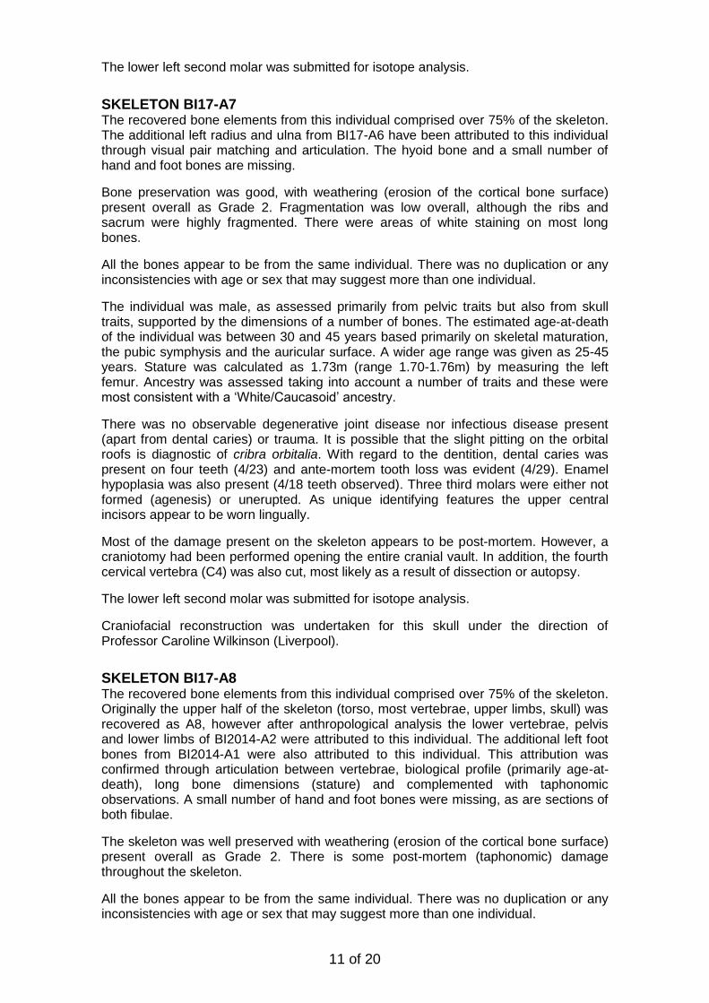
SKELETON BI17-A7 The recovered bone elements from this individual comprised over 75% of the skeleton. The additional left radius and ulna from BI17-A6 have been attributed to this individual through visual pair matching and articulation. The hyoid bone and a small number of hand and foot bones are missing.
Bone preservation was good, with weathering (erosion of the cortical bone surface) present overall as Grade 2. Fragmentation was low overall, although the ribs and sacrum were highly fragmented. There were areas of white staining on most long bones.
All the bones appear to be from the same individual. There was no duplication or any inconsistencies with age or sex that may suggest more than one individual.
The individual was male, as assessed primarily from pelvic traits but also from skull traits, supported by the dimensions of a number of bones. The estimated age-at-death of the individual was between 30 and 45 years based primarily on skeletal maturation, the pubic symphysis and the auricular surface. A wider age range was given as 25-45 years. Stature was calculated as 1.73m (range 1.70-1.76m) by measuring the left femur. Ancestry was assessed taking into account a number of traits and these were most consistent with a ‘White/Caucasoid’ ancestry.
There was no observable degenerative joint disease nor infectious disease present (apart from dental caries) or trauma. It is possible that the slight pitting on the orbital roofs is diagnostic of cribra orbitalia. With regard to the dentition, dental caries was present on four teeth (4/23) and ante-mortem tooth loss was evident (4/29). Enamel hypoplasia was also present (4/18 teeth observed). Three third molars were either not formed (agenesis) or unerupted. As unique identifying features the upper central incisors appear to be worn lingually.
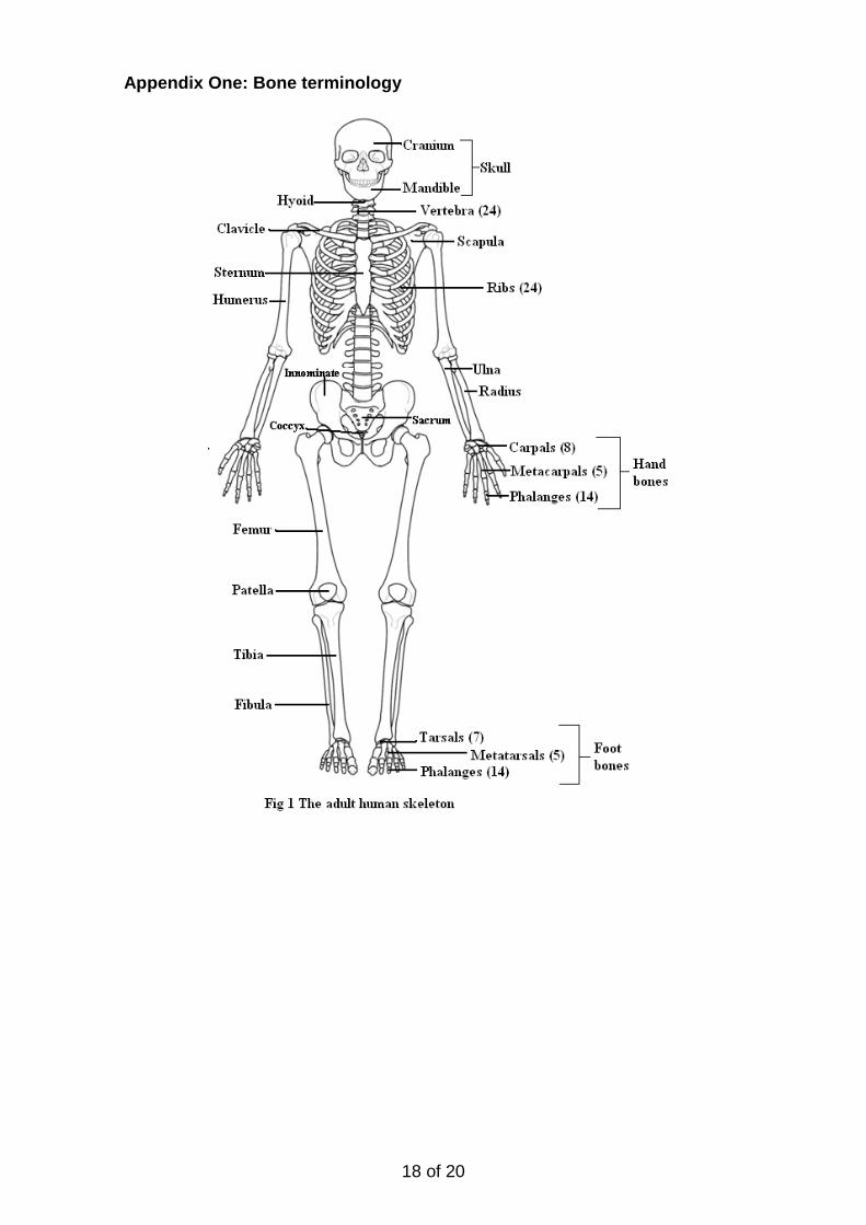
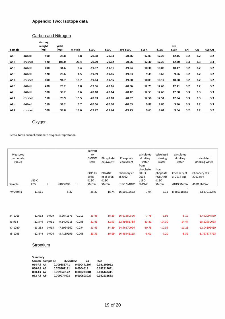
Most of the damage present on the skeleton appears to be post-mortem. However, a craniotomy had been performed opening the entire cranial vault. In addition, the fourth cervical vertebra (C4) was also cut, most likely as a result of dissection or autopsy.
The lower left second molar was submitted for isotope analysis.
Craniofacial reconstruction was undertaken for this skull under the direction of Professor Caroline Wilkinson (Liverpool).
SKELETON BI17-A8 The recovered bone elements from this individual comprised over 75% of the skeleton. Originally the upper half of the skeleton (torso, most vertebrae, upper limbs, skull) was recovered as A8, however after anthropological analysis the lower vertebrae, pelvis and lower limbs of BI2014-A2 were attributed to this individual. The additional left foot bones from BI2014-A1 were also attributed to this individual. This attribution was confirmed through articulation between vertebrae, biological profile (primarily age-at-death), long bone dimensions (stature) and complemented with taphonomic observations. A small number of hand and foot bones were missing, as are sections of both fibulae.
The skeleton was well preserved with weathering (erosion of the cortical bone surface) present overall as Grade 2. There is some post-mortem (taphonomic) damage throughout the skeleton.
All the bones appear to be from the same individual. There was no duplication or any inconsistencies with age or sex that may suggest more than one individual.
12 of 20
The individual was male as assessed from pelvic traits; a number of traits on the skull were ambiguous however. By assessing skeletal maturation the long bones, the vertebrae and pelvic bones do not show complete fusion in all aspects and therefore a likely age of 18-21 years (or wider age range 17-25 years) is likely. Stature was calculated as 1.52m (range 1.49m-1.55m) by measuring the left femur. Ancestry was not assessed.
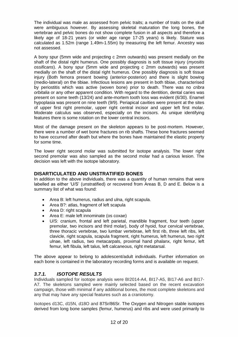
A bony spur (5mm wide and projecting c 2mm outwards) was present medially on the shaft of the distal right humerus. One possibly diagnosis is soft tissue injury (myositis ossificans). A bony spur (5mm wide and projecting c 2mm outwards) was present medially on the shaft of the distal right humerus. One possibly diagnosis is soft tissue injury (Both femora present bowing (anterior-posterior) and there is slight bowing (medio-lateral) on the tibiae. Infectious lesions are present in both tibiae, characterised by periostitis which was active (woven bone) prior to death. There was no cribra orbitalia or any other apparent condition. With regard to the dentition, dental caries was present on some teeth (13/24) and ante-mortem tooth loss was evident (6/30). Enamel hypoplasia was present on nine teeth (9/9). Periapical cavities were present at the sites of upper first right premolar, upper right central incisor and upper left first molar. Moderate calculus was observed, especially on the incisors. As unique identifying features there is some rotation on the lower central incisors.
Most of the damage present on the skeleton appears to be post-mortem. However, there were a number of wet bone fractures on rib shafts. These bone fractures seemed to have occurred after death but where the bones have maintained the elastic property for some time.
The lower right second molar was submitted for isotope analysis. The lower right second premolar was also sampled as the second molar had a carious lesion. The decision was left with the isotope laboratory.
DISARTICULATED AND UNSTRATIFIED BONES In addition to the above individuals, there was a quantity of human remains that were labelled as either ‘U/S’ (unstratified) or recovered from Areas B, D and E. Below is a summary list of what was found:
Area B: left humerus, radius and ulna, right scapula.
Area B?: atlas, fragment of left scapula
Area D: right scapula
Area E: male left innominate (os coxae)
U/S: cranium, frontal and left parietal, mandible fragment, four teeth (upper premolar, two incisors and third molar), body of hyoid, four cervical vertebrae, three thoracic vertebrae, two lumbar vertebrae, left first rib, three left ribs, left clavicle, right scapula, scapula fragment, right humerus, left humerus, two right ulnae, left radius, two metacarpals, proximal hand phalanx, right femur, left femur, left fibula, left talus, left calcaneous, right metatarsal.
The above appear to belong to adolescent/adult individuals. Further information on each bone is contained in the laboratory recording forms and is available on request.
3.7.1. ISOTOPE RESULTS Individuals sampled for isotope analysis were BI2014-A4, BI17-A5, BI17-A6 and BI17-A7. The skeletons sampled were mainly selected based on the recent excavation campaign, those with minimal if any additional bones, the most complete skeletons and any that may have any special features such as a craniotomy.
Isotopes d13C, d15N, d18O and 87Sr/86Sr. The Oxygen and Nitrogen stable isotopes derived from long bone samples (femur, humerus) and ribs and were used primarily to
13 of 20
infer diet; whilst the latter two, Oxygen and Strontium, derived from tooth sampled and were analysed primarily to investigate provenance. Whilst the bone provides an average of the latter 5-10 years of life, the dental samples provides a record of childhood.
The results of these analyses are attached as an Appendix to this report.
Diet
All four individuals have statistically identical d13C values. However, there are 2 distinct pairs in terms of d15N: A4 and A7 are different to A5 and A8, but there is no difference within these pairs. A5 and A8 have typical 'terrestrial' signatures, and have nothing to indicate they had marine or freshwater protein in their diet or in any significant quantities.
Individuals A4 and A7, however, clearly have N values which would likely result from a food chain with more steps in it than the terrestrial food web, and in this case it is more likely to be marine protein.
Provenance
It seems that the different values may indicate that the four individuals came from different backgrounds. From the O results, it seems that there were two individuals from outside UK geography and give more continental European signals. These are individuals A5 and A7. In terms of the Sr data, individuals A4 and A5 had similar, but not identical values; whilst A7 and A8 both had distinctly different values. This supports the data from the d13C, d15N and d18O analysis which, when considered all together, indicates that all 4 individuals may have been born/grown up in different regions to each other.
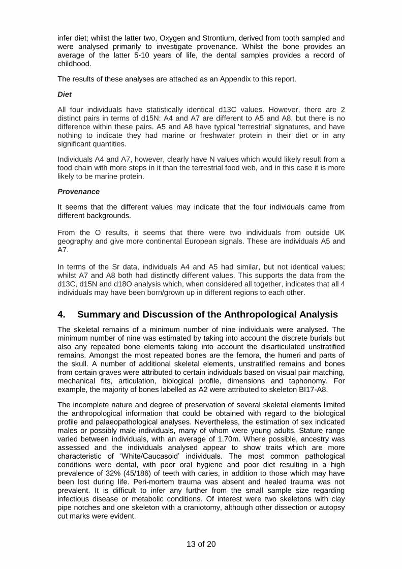
4. Summary and Discussion of the Anthropological Analysis
The skeletal remains of a minimum number of nine individuals were analysed. The minimum number of nine was estimated by taking into account the discrete burials but also any repeated bone elements taking into account the disarticulated unstratified remains. Amongst the most repeated bones are the femora, the humeri and parts of the skull. A number of additional skeletal elements, unstratified remains and bones from certain graves were attributed to certain individuals based on visual pair matching, mechanical fits, articulation, biological profile, dimensions and taphonomy. For example, the majority of bones labelled as A2 were attributed to skeleton BI17-A8.
The incomplete nature and degree of preservation of several skeletal elements limited the anthropological information that could be obtained with regard to the biological profile and palaeopathological analyses. Nevertheless, the estimation of sex indicated males or possibly male individuals, many of whom were young adults. Stature range varied between individuals, with an average of 1.70m. Where possible, ancestry was assessed and the individuals analysed appear to show traits which are more characteristic of ‘White/Caucasoid’ individuals. The most common pathological conditions were dental, with poor oral hygiene and poor diet resulting in a high prevalence of 32% (45/186) of teeth with caries, in addition to those which may have been lost during life. Peri-mortem trauma was absent and healed trauma was not prevalent. It is difficult to infer any further from the small sample size regarding infectious disease or metabolic conditions. Of interest were two skeletons with clay pipe notches and one skeleton with a craniotomy, although other dissection or autopsy cut marks were evident.
14 of 20
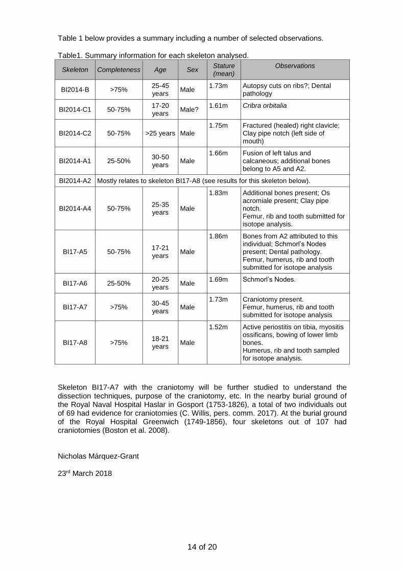
Table 1 below provides a summary including a number of selected observations. Table1. Summary information for each skeleton analysed.
Skeleton Completeness Age Sex Stature (mean)
Observations
BI2014-B >75% 25-45 years
Male 1.73m Autopsy cuts on ribs?; Dental
pathology
BI2014-C1 50-75% 17-20 years
Male? 1.61m Cribra orbitalia
BI2014-C2 50-75% >25 years Male 1.75m Fractured (healed) right clavicle;
Clay pipe notch (left side of mouth)
BI2014-A1 25-50% 30-50 years
Male 1.66m Fusion of left talus and
calcaneous; additional bones belong to A5 and A2.
BI2014-A2 Mostly relates to skeleton BI17-A8 (see results for this skeleton below).
BI2014-A4 50-75% 25-35 years
Male
1.83m Additional bones present; Os acromiale present; Clay pipe notch. Femur, rib and tooth submitted for isotope analysis.
BI17-A5 50-75% 17-21 years
Male
1.86m Bones from A2 attributed to this individual; Schmorl’s Nodes present; Dental pathology. Femur, humerus, rib and tooth submitted for isotope analysis
BI17-A6 25-50% 20-25 years
Male 1.69m Schmorl’s Nodes.
BI17-A7 >75% 30-45 years
Male 1.73m Craniotomy present.
Femur, humerus, rib and tooth submitted for isotope analysis
BI17-A8 >75% 18-21 years
Male
1.52m Active periostitis on tibia, myositis ossificans, bowing of lower limb bones. Humerus, rib and tooth sampled for isotope analysis.
Skeleton BI17-A7 with the craniotomy will be further studied to understand the dissection techniques, purpose of the craniotomy, etc. In the nearby burial ground of the Royal Naval Hospital Haslar in Gosport (1753-1826), a total of two individuals out of 69 had evidence for craniotomies (C. Willis, pers. comm. 2017). At the burial ground of the Royal Hospital Greenwich (1749-1856), four skeletons out of 107 had craniotomies (Boston et al. 2008). Nicholas Márquez-Grant 23rd March 2018
15 of 20
Bibliography Bass, W. M. 1995. Human Osteology: A Laboratory and Field Manual. Missouri
Archaeological Society, Inc.
Boston, C., Witkin, A., Boyle, A. and Wilkinson, D.R.P. 2008. ‘Safe Moor’d in
Greenwich Tier’. A Study of the Skeletons of the Royal Navy Sailors and Marines
Excavated at the Royal Hospital Greenwich. Oxford Archaeology, Oxford.
Brickley, M. and McKinley, J. (editors). 2004. Guidelines to the Standards for
Recording Human Remains. IFA Paper No. 7.
Brooks, S.T. and Suchey, J.M. 1990. Skeletal age determination based on the os
pubis: a comparison of the Acsádi-Nemeskeri and Suchey-Brooks methods.
Human Evolution, 5: 227-238.
Buckberry, J.L. and Chamberlain, A.T. 2002. Age estimation from the auricular surface
of the ilium: a revised method. American Journal of Physical Anthropology, 119:
231-239.
Buikstra, J.E. and Ubelaker, D.H. 1994. Standards for Data Collection from Human
Skeletal Remains. Arkansas Archaeological Survey Research Series No.44.
İşcan, M.Y., Loth, S.R. and R.K. Wright. 1985. “Age estimation from the rib by phase
analysis. White males”. Journal of Forensic Sciences, 29: 1094-1104.
İşcan, M.Y., Loth, S.R. and R.K. Wright. 1985. “Age estimation from the rib by phase
analysis: females”. Journal of Forensic Sciences, Vol. 30, 853-863.
Mitchell, P.D. and Brickley, M. 2017. Updated Guidelines to the Standards for
Recording Human Remains. CiFA/BABAO.
Scheuer, L. and Black, S. 2000. Developmental Juvenile Osteology. Academic Press,
London.
Suchey, J.M. and Katz, D. 1998. Applications of pubic age determination. In K.J.
Reichs (ed), Forensic Osteology: Advances in the Identification of Human
Remains. 2nd edition. Charles C. Thomas, Springfield. Pages: 204-236.
Trotter, M. 1970. Estimation of stature from intact long bones. In T.D. Stewart (1970),
Personal Identification in Mass Disasters. Smithsonian Institution, Washington.
Pages: 71-83.
16 of 20
Figures
2. BI2014-A4. Clay pipe notch on the left side of the dentition.
3. Schmorl's Nodes (depressions) on the inferior surfaces of the vertebral bodies of three thoracic vertebrae. Skeleton BI17-A5
17 of 20
4. Evident craniotomy on BI17-A7
5. Evident cut through cervival vertebrae after dissection/autopsy. BI17-A7
18 of 20
Appendix One: Bone terminology
19 of 20
Appendix Two: Isotope data
Carbon and Nitrogen
Sample
starting weight
(mg) yield (mg) % yield d13C d13C ave d13C d15N d15N
ave d15N CN CN Ave CN
A4F drilled 500 28.8 5.8 -20.38 -20.34 -20.36 12.03 12.26 12.15 3.2 3.2 3.2
A4R crushed 520 106.0 20.4 -20.09 -20.02 -20.06 12.30 12.29 12.30 3.3 3.3 3.3
A5F drilled 490 31.6 6.4 -19.97 -19.91 -19.94 10.30 10.03 10.17 3.2 3.2 3.2
A5H drilled 520 23.6 4.5 -19.99 -19.66 -19.83 9.49 9.63 9.56 3.2 3.2 3.2
A5R crushed 490 91.7 18.7 -19.64 -19.55 -19.60 10.03 10.12 10.08 3.2 3.2 3.2
A7F drilled 490 29.2 6.0 -19.96 -20.16 -20.06 12.73 12.68 12.71 3.2 3.2 3.2
A7H drilled 500 33.2 6.6 -20.10 -20.14 -20.12 12.53 12.66 12.60 3.3 3.3 3.3
A7R crushed 510 78.9 15.5 -20.03 -20.10 -20.07 12.56 12.51 12.54 3.3 3.3 3.3
A8H drilled 510 34.2 6.7 -20.06 -20.00 -20.03 9.87 9.85 9.86 3.3 3.2 3.3
A8R crushed 500 98.0 19.6 -19.72 -19.74 -19.73 9.63 9.64 9.64 3.2 3.2 3.2
Oxygen
Strontium
Summary Sample Sample ID 87Sr/86Sr 2σ RSD 054-A4 A4 0.709353741 0.000441304 0.031106052 056-A5 A5 0.709307191 0.0004613 0.032517641 060-13 A7 0.709648122 0.000233381 0.016443411 062-A8 A8 0.709974403 0.000603927 0.042531633
Dental tooth enamel carbonate oxygen interpretation
Measured carbonate
values
convert to
SMOW scale
Phosphate equivalent
Phosphate equivalent
calculated drinking
water
calculated drinking
water
calculated drinking
water calculated
drinking water
COPLEN 1988
BRYANT et al 1996
Chennery et al 2012
from phophate DAUX 2008
from phophate POLLARD
Chennery et al 2012 eq6
Chennery et al 2012 eq4
Sample d13 C PDV ± d18O PDB ±
d18O SMOW
d18O SMOW d18O SMOW
d18O SMOW
d18O SMOW d18O SMOW d18O SMOW
PWD RM1 -11.511
-5.37
25.37 16.74 16.50615653 -7.94 -7.12 -
8.289318853 -8.687012246
a4-1019 -12.632 0.009 -5.2641376 0.011 25.48 16.85 16.61880526 -7.78 -6.92 -8.12 -8.492097859
a5-938 -12.546 0.011 -9.1406218 0.058 21.49 12.93 12.49381788 -13.81 -14.30 -14.47 -15.62950093
a7-1020 -13.283 0.015 -7.1954362 0.034 23.49 14.89 14.56370024 -10.78 -10.59 -11.28 -12.04801489
a8-1059 -12.844 0.006 -5.4139199 0.008 25.33 16.69 16.45942115 -8.01 -7.20 -8.36 -8.767877763
20 of 20
Report Disclaimer
Whilst Cranfield University has used reasonable endeavours to ensure the accuracy and completeness of the report any information provided or opinions expressed therein it does not give any express or implied warranty as to the fitness for purpose or accuracy of the information contained in the said report. Use of the information contained therein is at the users sole risk and Cranfield University accepts no liability for any loss or damage whether direct or indirect occasioned by the use made of any such information contained in the report by the client or any third party.