Embed Size (px)
Citation preview
198 Korean J Radiol 8(3), June 2007
Reproducibility of Computer-AidedDetection Marks in DigitalMammography
Objective: To evaluate the performance and reproducibility of a computer-aided detection (CAD) system in mediolateral oblique (MLO) digital mammo-grams taken serially, without release of breast compression.
Materials and Methods: A CAD system was applied preoperatively to the full-field digital mammograms of two MLO views taken without release of breast com-pression in 82 patients (age range: 33 83 years; mean age: 49 years) with previ-ously diagnosed breast cancers. The total number of visible lesion components in82 patients was 101: 66 masses and 35 microcalcifications. We analyzed thesensitivity and reproducibility of the CAD marks.
Results: The sensitivity of the CAD system for first MLO views was 71%(47/66) for masses and 80% (28/35) for microcalcifications. The sensitivity of theCAD system for second MLO views was 68% (45/66) for masses and 17% (6/35)for microcalcifications. In 84 ipsilateral serial MLO image sets (two patients hadbilateral cancers), identical images, regardless of the existence of CAD marks,were obtained for 35% (29/84) and identical images with CAD marks wereobtained for 29% (23/78). Identical images, regardless of the existence of CADmarks, for contralateral MLO images were 65% (52/80) and identical images withCAD marks were obtained for 28% (11/39). The reproducibility of CAD marks forthe true positive masses in serial MLO views was 84% (42/50) and that for thetrue positive microcalcifications was 0% (0/34).
Conclusion: The CAD system in digital mammograms showed a high sensitivi-ty for detecting masses and microcalcifications. However, reproducibility of micro-calcification marks was very low in MLO views taken serially without release ofbreast compression. Minute positional change and patient movement can alterthe images and result in a significant effect on the algorithm utilized by the CADfor detecting microcalcifications.
computer-aided detection (CAD) system can assist the radiologist in theearly detection of breast cancer by highlighting suspicious areas seen onmammography (1, 2). Reproducibility is one of the important factors for
determining the quality of a CAD system. Recent CAD studies involving repeatedscanning of film mammograms of breast cancer patients showed 39 53%reproducibility, markedly decreased from the 95 99% claimed by the manufacturersof the CAD systems (3, 4). It is likely that variability of marks is primarily caused bysmall shifts in the film position between sequential digitizations (5).
Recently, the CAD system has been applied to full-field digital mammography andresults for breast cancer detection were reported to be similar to those obtained usingan analog system (6). When CAD is used with full-field digital mammography, it
Seung Ja Kim, MD1
Woo Kyung Moon, MD2
Nariya Cho, MD2
Joo Hee Cha, MD3
Sun Mi Kim, MD2
Jung-Gi Im, MD2
Index terms:Breast neoplasms, diagnosisComputers, diagnostic aidDigital radiography
Korean J Radiol 2007;8:198-205Received December 21, 2005; accepted after revision August 24, 2006.
1Department of Radiology, KonkukUniversity Hospital, Seoul 143-701,Korea; 2Department of Radiology, SeoulNational University College of Medicineand Clinical Research Institute, SeoulNational University Hospital and theInstitute of Radiation Medicine, SeoulNational University Medical ResearchCenter, Seoul 110-744, Korea;3Department of Radiology, BoramaeMunicipal Hospital, Seoul 156-707, Korea
This study is supported by KISTEP,Ministry of Science and Technology,Korea.
Address reprint requests to:Woo Kyung Moon, MD, Department ofRadiology, Seoul National UniversityHospital, 28, Yongon-dong, Chongno-gu,Seoul 100-744, Korea.Tel. (822) 2072-2584Fax. (822) 743-6385e-mail: [email protected]
A
eliminates the need for digitization. Because the CADsystem uses a computer algorithm, the CAD system will be100% reproducible when the same CAD scheme is appliedrepeatedly to the same digital image. However, it will bepossible to take more than one digital image of the samebreast in a repeated exposure and it is likely that CAD willshow variable reproducibility with such repeated digitalimages. To our knowledge, there have been few studies toevaluate the reproducibility of a CAD system combined tofull-field digital mammography.
The purpose of this study was to evaluate the perfor-mance and reproducibility of the CAD system in twomediolateral oblique (MLO) digital mammograms takenserially without release of breast compression.
MATERIALS AND METHODS
Patient SelectionBetween March 2004 and June 2004, 100 patients with
breast cancer underwent full-field digital mammography(Senographe 2000D FFDM, GE Medical Systems, Buc,France) including two serial MLO views without release ofbreast compression and one craniocaudal view (i.e., threeimages per each breast) of the bilateral breasts. Of these100 patients, we selected 82 consecutive patients in whomthe malignant lesions were visible in both the craniocaudalview and the MLO view and the CAD system(ImageChecker M1000-DM, version 3.1; R2 Technology)was applied to these mammograms. The patients had amean age of 49 years (range: 33 83 years). Because twoof the 82 patients had bilateral breast cancer, the totalnumber of diseased breasts was 84. In 68 (81%) of the 84breasts, the tumors were palpable and in 16 (19%) breaststhey were nonpalpable. The mean time interval betweenthe serial MLO views was 18 seconds. The serial MLOmammograms of the bilateral breasts were taken byautomatic exposure control methods. The average glandu-lar dose to the breasts calculated by the exposure of the siximages was about 8.0 9.0 mGy using our mammographysystem, which was considered to be acceptable consideringthe guidelines of the American College of Radiology(ACR) for screening mammography, that is, not to exceed3.0 mGy per view (7). This study was conducted withinstitutional review board approval and informed consentwas obtained from all patients.
Mammographic and Histological FindingsBecause the CAD system identifies the mass component
and the calcification component separately, we counted themass component and the calcification componentseparately for those malignancies that presented with both
a mass and a calcification cluster. On mammograms of the84 diseased breasts of 82 patients, the total number ofvisible lesion components was 101. We found 66 masses(mean size, 24 mm) and 35 microcalcifications (mean size,21 mm) including one mass (n = 50), one microcalcification(n = 20), one mass plus one microcalcification (n = 7), twomasses (n = 2), two microcalcifications (n = 2), two massesand one microcalcification (n = 2), and one mass and twomicrocalcifications (n = 1). The categories of the lesions inthe 84 diseased breasts using the American College ofRadiology’s Breast Imaging Reporting and Data System(ACR BI-RADS) (8) were category 4 (n = 8) and category5 (n = 76). The size of the mass was 10 mm (n = 3), 1120 mm (n = 24), 21 30 mm (n = 27), 31 40 mm (n = 6),and 41 mm (n = 6) and the size of the calcific clusterwas 10 mm (n = 16), 11 20 mm (n = 9), 21 30 mm (n= 5), 31 40 mm (n = 1), and 41 mm (n = 4).
Utilizing the density pattern of the ACR BI-RADS, wedivided the patients into two subgroups: 1) dense breastgroup (BI-RADS 3 and 4 density in 50 patients), and 2)fatty breast group (BI-RADS 1 and 2 density in 32patients).
The final postoperative pathological diagnoses were allmalignant (ductal carcinoma in situ [DCIS] [n = 15] andinvasive carcinoma [n = 69]).
CAD Mark EvaluationWe applied the CAD system to each set of digital
mammograms including the craniocaudal and serial MLOviews of the 82 patients and saved the images with CADmarks at a review workstation before sending them to thePicture Archiving Communication System (PACS). Wethen analyzed the CAD marks of each view by reviewingthe original digital mammograms, the images with CADmarks, ultrasound and the pathology reports. The consen-sus of two breast imaging specialists was used in theinterpretation.
The CAD system marks regions suspicious for a mass ora microcalcification cluster by superimposing a smallasterisk or a triangle, respectively, on the image. If theasterisk was located within a true positive mass, this masswas considered to have been identified correctly by theCAD system. Similarly, if the triangle overlapped any ofthe microcalcification areas, the CAD marks were consid-ered to represent true positive detection. Because allpatients had ultrasound preoperatively, any mass marksthat fell on normal parenchyma identified on ultrasoundwere considered as false positives. When the CAD systemmarked typical benign calcifications or crossing lines, weconsidered them as false positives.
CAD Marks in Digital Mammography
Korean J Radiol 8(3), June 2007 199
CAD Performance Analysis The sensitivity, reproducibility and the false positive
marks per image of the CAD system in these 82 patientswere analyzed. We analyzed sensitivity in the first MLOviews, the second MLO views, craniocaudal views, and ina combination of the first MLO views and craniocaudalviews. The latter was characterized as case-based sensitiv-ity and the remaining as image-based sensitivity (i.e.,sensitivity of each mammographic view).
For case-based sensitivity, a successful mark for thecancer in either the MLO or craniocaudal view was consid-ered a true-positive identification of the cancer for thatcase. Breast parenchymal density was used in theseanalyses in order to show its effect on the sensitivity.
We analyzed the image-based reproducibility and themark-based reproducibility of the CAD system in serialMLO views of ipsilateral diseased breasts and contralateralnormal breasts, respectively, and together. Image-basedreproducibility was defined as the identical images and thevalue was obtained in two different ways: one way byanalyzing images irrespective of the existence of CADmarks and another way by analyzing images with CADmarks. The mark-based reproducibility was defined as anymarks within the same mass or the same microcalcificationin the serial MLO images. For example, as seen in Figures1A and B, the CAD marks on the first MLO view were notidentical with that on the second MLO view. Thus, the twoserial MLO views were not reproducible in terms of image-based reproducibility. The true mass mark on the firstMLO view was identical with that on the second MLOview, so this CAD mark was reproducible in terms ofmark-based reproducibility. We analyzed the reproducibil-ity of true and false positive marks in serial MLO views ofbilateral breasts.
The false positive marks per image (of a mass or amicrocalcification) were obtained in serial MLO views andcraniocaudal views of ipsilateral breasts and contralateral
breasts, respectively and then together. In order to evaluate of the effect of the variables such as
kVp, mAs and breast thickness, we calculated the differ-ences of the exposure parameters between the two MLOviews and presented them as percentages in reproducibleand in non-reproducible serial MLO sets. In all patients, wealso calculated the average glandular dose delivered to thepatient during the exposure of the six images (i.e., twoserial MLO views and one craniocaudal view of bilateralbreasts) by adding the amount of the dose appeared on thedigital images.
Statistical AnalysisTo compare the differences in the sensitivity of the first
MLO views and the second MLO views, the paired t-testwas applied. To compare the differences of sensitivity forlesions and reproducibility of CAD marks in fatty breastand dense breast and the difference in false positivecalcification marks per image in the first MLO views andthe second MLO views, the unpaired t-test was applied.The differences of the exposure parameters between thetwo MLO views were compared for reproducible and non-reproducible serial MLO sets using the unpaired t-tests.
RESULTS
SensitivitiesTable 1 summarizes the sensitivity of the CAD system in
serial MLO views and in craniocaudal views of 82 patients.The case-based sensitivity was 86% (57 of 66) for massesand 100% (35 of 35) for microcalcifications. The case-based sensitivity for masses in the fatty breast group was93% (25 of 27) and that in the dense breast group was82% (32 of 39). This difference was not statistically signifi-cant (p = 0.08). The image-based sensitivity of the CADsystem for masses was 71% (47 of 66) in the first MLOviews and 68% (45 of 66) in the second MLO views. The
Kim et al.
200 Korean J Radiol 8(3), June 2007
Table 1. Detection Performance of the CAD System in Two Serial Mediolateral Oblique and Craniocaudal Views
1st MLO (%) 2nd MLO (%) CC (%) 1st MLO + CC (%)
Mass Ca++ Mass Ca++ Mass Ca++ Mass Ca++
Total 71 80* 68 17* 70 91 86 100(n = 82) (47/66) (28/35) (45/66) (6/35) (46/66) (32/35) (57/66) (35/35)
Fatty 93 75 93 25 81 100 93 100(n = 32) (25/27) (9/12) (25/27) (3/12) (22/27) (12/12) (25/27) (12/12)Dense 56 83* 51 13* 62 87 82 100(n = 50) (22/39) (19/23) (20/39) (3/23) (24/39) (20/23) (32/39) (23/23)
Note. numbers are percentages, with raw data in parentheses.MLO = mediolateral oblique view, CC = craniocaudal view, 1st = first, 2nd = second Ca++ = microcalcification, n = patient number * This difference was statistically significant (p < 0.0001 by two-tailed paired t-test).
These differences were statistically significant ( p = 0.0011 and p = 0.0003 by two-tailed unpaired t-test).
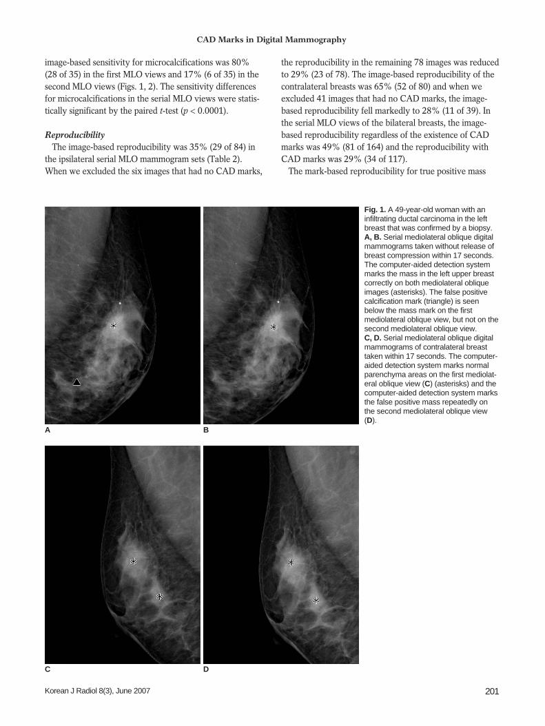
image-based sensitivity for microcalcifications was 80%(28 of 35) in the first MLO views and 17% (6 of 35) in thesecond MLO views (Figs. 1, 2). The sensitivity differencesfor microcalcifications in the serial MLO views were statis-tically significant by the paired t-test (p < 0.0001).
ReproducibilityThe image-based reproducibility was 35% (29 of 84) in
the ipsilateral serial MLO mammogram sets (Table 2).When we excluded the six images that had no CAD marks,
the reproducibility in the remaining 78 images was reducedto 29% (23 of 78). The image-based reproducibility of thecontralateral breasts was 65% (52 of 80) and when weexcluded 41 images that had no CAD marks, the image-based reproducibility fell markedly to 28% (11 of 39). Inthe serial MLO views of the bilateral breasts, the image-based reproducibility regardless of the existence of CADmarks was 49% (81 of 164) and the reproducibility withCAD marks was 29% (34 of 117).
The mark-based reproducibility for true positive mass
CAD Marks in Digital Mammography
Korean J Radiol 8(3), June 2007 201
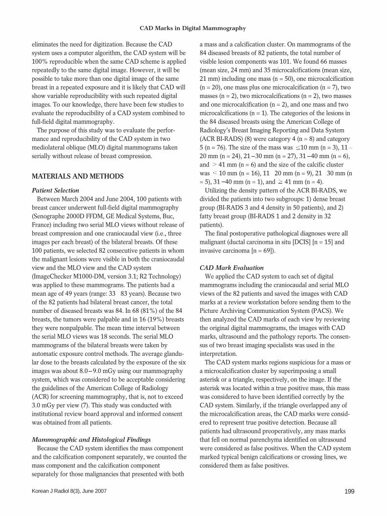
Fig. 1. A 49-year-old woman with aninfiltrating ductal carcinoma in the leftbreast that was confirmed by a biopsy.A, B. Serial mediolateral oblique digitalmammograms taken without release ofbreast compression within 17 seconds.The computer-aided detection systemmarks the mass in the left upper breastcorrectly on both mediolateral obliqueimages (asterisks). The false positivecalcification mark (triangle) is seenbelow the mass mark on the firstmediolateral oblique view, but not on thesecond mediolateral oblique view. C, D. Serial mediolateral oblique digitalmammograms of contralateral breasttaken within 17 seconds. The computer-aided detection system marks normalparenchyma areas on the first mediolat-eral oblique view (C) (asterisks) and thecomputer-aided detection system marksthe false positive mass repeatedly onthe second mediolateral oblique view(D).
A B
C D

marks in the serial MLO views was 84% (42 of 50) andthat for true positive microcalcification marks was 0% (0of 34) (Table 3) (Figs. 1, 2). The 28 calcific clusters thatwere correctly marked on the first MLO views were notmarked on the second MLO views, while six calcificclusters that were correctly marked on the second MLOviews were not marked on the first MLO views. Thereproducibility for true positive mass marks was signifi-cantly higher in the fatty breast group than in the densebreast group (100% vs. 68%, p = 0.0015) (Fig. 3). Foripsilateral breasts, the reproducibility for false positivemass marks was 42% (8 of 19) and that for false positivemicrocalcification marks was 0% (0 of 19) (Fig. 1). Forcontralateral breasts, the reproducibility for false positivemass marks was 44% (16 of 36) and that for false positivemicrocalcification marks was 0% (0 of 18) (Fig. 1). In total,for bilateral breasts, the reproducibility for false positivemass marks was 44% (24 of 55) and that for false positivemicrocalcification marks was 0% (0 of 37). Thereproducibility for false positive mass marks in bilateralbreasts was slightly higher in the fatty breast group than inthe dense breast group, but that difference was not found
to be statistically significant (48% [12 of 25] vs. 40% [12of 30], p = 0.5).
The false positive marks per image on the first MLOviews for bilateral breasts were 0.45, with 0.24 mass marksand 0.21 calcification marks and those for the second MLOviews of bilateral breasts were 0.26, with 0.24 mass marksand 0.02 calcification marks. The false positive calcificationmarks per image were significantly lower in the secondMLO views (p < 0.0001). The false positives per image oncraniocaudal views of bilateral breasts were 0.41, with0.19 mass marks and 0.22 calcification marks.
The changes of the exposure parameters in 81reproducible serial MLO mammogram sets and in 83 non-reproducible serial MLO mammogram sets were 0.2% and0.5% in kVp, 5% and 7% in mAs and 0.5% and 1% inbreast thickness. These differences were not statisticallysignificant (p = 0.2043, 0.2987, 0.1058, respectively). Thetwo radiologists who analyzed cases in our study foundlittle difference in the two serial MLO mammograms.
The total average glandular doses delivered to thepatients during the exposure of two serial MLO views andone craniocaudal view of bilateral breasts ranged from 4.5
Kim et al.
202 Korean J Radiol 8(3), June 2007
Table 2. Image-based Reproducibility of Mediolateral Oblique Mammograms by the CAD System
Ipsilateral Breast (%) Contralateral Breast (%) Bilateral Breasts (%)
Mark (+/ )* Mark (+) Mark (+/ )* Mark (+) Mark (+/ )* Mark (+)
Total 35 29 65 28 49 29(n = 82) (29/84 ) (23/78) (52/80) (11/39) (81/164) (34/117)
Fatty 39 39 65 21 52 34(n = 32) (13/33) (13/33) (20/31) (3/14) (33/64) (16/47)Dense 31 22 65 32 48 26(n = 50) (16/51) (10/45) (32/49) (8/25) (48/100) (18/70)
Note. numbers are percentages, with raw data in parentheses.n = patient number *These columns are percentages of the identical images irrespective of the existence of CAD marks.
These columns are percentages of the identical images with CAD marks.Two patients had bilateral cancers.
Table 3. Reproducibility of CAD Marks in Serial Mediolateral Oblique Views by the CAD System
Ipsilateral Breast (%) Contralateral Breast (%)
TP mass TP ca++ FP mass FP ca++ FP mass FP ca++
Total 84* 0 42* 0 44 0(n = 82) (42/50) (0/34) (8/19) (0/19) (16/36) (0/18)
Fatty 100 0 55 0 43 0(n = 32) (25/25) (0/12) (6/11) (0/10) (6/14) (0/6)Dense 68 0 25 0 45 0(n = 50) (17/25) (0/22) (2/8) (0/9) (10/22) (0/12)
Note. numbers are percentages, with raw data in parentheses.TP = true positive; FP = false positive; ca++ = microcalcification; n = patient number * These differences were statistically significant (*p = 0.0003 and p = 0.0015 by two-tailed unpaired t-test).
mGy to 12.5 mGy, with a mean dose of 8.8 mGy. Thedoses were less than 8 mGy in 22 patients, 8 12 mGy in59 patients, and over 12 mGy in only one patient.
DISCUSSION
In our study, patients with breast cancers underwent twoserial MLO mammograms of the ipsilateral and contralat-eral breasts without release of breast compression toevaluate the reproducibility of the CAD system applied to
full-field digital mammography. There should be onlyminute positional change and patient movement betweenthe two mammograms since the same breast was exposedtwice without release of breast compression. The results ofour study showed that the sensitivity of the CAD systemfor malignant masses was similar in two serial MLO views:71% (47 of 66) and 68% (45 of 66) while the sensitivityfor microcalcifications was quite different: 80% (28 of 35)and 17% (6 of 35). The reproducibility of CAD marks fortrue positive mass in serial MLO views was 84% (42 of 50)
CAD Marks in Digital Mammography
Korean J Radiol 8(3), June 2007 203
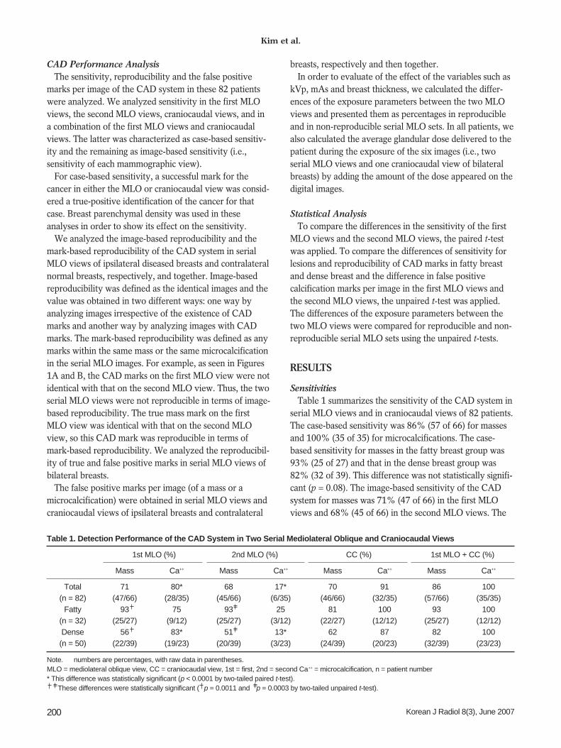
Fig. 2. A 56-year-old woman withmalignant microcalcifications in the leftbreast. A, B. Serial mediolateral oblique digitalmammograms taken without release ofbreast compression within 17 seconds.The computer-aided detection systemmarks the microcalcifications correctlyon the first mediolateral oblique view (A)(triangle). The computer-aided detectionsystem, however, does not mark themicrocalcifications on the secondmediolateral oblique view (B).
A B
Fig. 3. A 61-year-old woman presentedwith a screening-detected breast cancerin the left breast.A, B. Serial mediolateral oblique digitalmammograms taken without release ofbreast compression within 19 seconds.The computer-aided detection systemmarks the mass component and themicrocalcifications component correctlyon the first mediolateral oblique view (A)(asterisk and triangle). The computer-aided detection system, however, doesnot mark the microcalcifications on thesecond mediolateral oblique view (B).
A B
and that for true positive microcalcification was 0% (0 of34). Our results suggest that even a minute positionalchange can cause a significant effect on the algorithm ofthe CAD system for detecting malignant microcalcifica-tions in full-field digital mammography.
In current CAD systems, a binary threshold is typicallyused to generate detection marks. Each marked region hasa computed score that is above a predetermined threshold;lesions with computed scores that are near the thresholdare vulnerable to small changes and may be detected inone image and missed in another image (3). In our study,the sensitivity of the CAD system for detecting microcalci-fications was significantly lower in the second MLO study.A minimal positional change might have affected pixelsthus resulting in the same variability as in repeatedscanning of the same films. In addition, motion artifactsand blurring are also possible causes of low reproducibility,especially in microcalcification with detailed structures. Inour study, it is likely that the false positive microcalcifica-tion marks per image were substantially lower in thesecond MLO views for the same reason (p < 0.0001).
With digital mammography, the CAD system does notrequire a digitizer and allows display of the CAD markersrapidly after the image acquisition. Because the CADsystem uses a computer algorithm, the CAD system is100% reproducible when the same CAD scheme is appliedrepeatedly to the same digital image. This is different froma CAD system using film mammography. Our studyshowed, however, that CAD in full-field digital mammog-raphy could show variable reproducibility with repeatedimages of the same breast.
In our study, the case-based sensitivity of the CADsystem for masses and microcalcifications was 86% (57 of66) and 100% (35 of 35), respectively. This is similar to orbetter than seen in previous studies using film or digitalmammography and a CAD system (3 7, 9 10). It isdifficult to compare directly the detection performance ofthese studies, because different image databases and caseselection criteria were used. The image-based sensitivityfor masses was higher in the fatty breast group than in thedense breast group in serial MLO views (93%, 93% vs.56%, 51%) and these differences were statistically signifi-cant (p = 0.0011, 0.0003). The different sensitivity formasses relating to the parenchymal density is consistentwith the results of the study of Brem et al. (10).
There are several limitations to our study. When wecalculated the differences of the exposure parametersbetween the two MLO views in the reproducible and in thenon-reproducible serial MLO sets, the changes of parame-ters such as kVp, mAs and breast thickness in the non-reproducible serial MLO mammogram sets were slightly
greater than those in the reproducible serial MLOmammogram sets. These parameters may affect thereproducibility of the CAD system. Subtraction of the twoserial images in reproducible and in non-reproducible serialMLO sets could suggest changes in position and thus areason for inconsistency of the CAD system, even thoughthe radiologists in this study found no difference betweenthe two serial mammograms. A method to test thereproducibility of computer-aided detection schemes hasbeen recently described for digitized mammograms (11).We selected cases in which lesions were visible in bothcraniocaudal and MLO views, and thus the overall sensitiv-ity of the CAD system may be overestimated.
In conclusion, the CAD system in full-field digitalmammography showed a high sensitivity for detectingmasses and microcalcifications related to breast cancer.However, the reproducibility of microcalcification CADmarks was very low in two MLO views taken seriallywithout release of breast compression. Minute positionalchange and patient movement between serialmammograms might have a significant effect on thealgorithm of the CAD system in detecting microcalcifica-tions. Reproducibility of CAD system remains animportant issue in full-field digital mammography.
References1. Warren Burhenne LJ, Wood SA, D’Orsi CJ, Feig SA, Kopans
DB, O’Shaughnessy KF, et al. Potential contribution ofcomputer-aided detection to the sensitivity of screeningmammography. Radiology 2000;215:554-562
2. Freer TW, Ulissey MJ. Screening mammography withcomputer-aided detection: prospective study of 12,860 patientsin a community breast center. Radiology 2001;220:781-786
3. Zheng B, Hardesty LA, Poller WR, Sumkin JH, Golla S.Mammography with computer-aided detection: reproducibilityassessment initial experience. Radiology 2003;228:58-62
4. Malich A, Azhari T, Bohm T, Fleck M, Kaiser WA.Reproducibility—an important factor determining the quality ofcomputer-aided detection (CAD) systems. Eur J Radiol2000;36:170-174
5. Taylor CG, Champness J, Reddy M, Taylor P, Potts HW, Given-Wilson R. Reproducibility of prompts in computer-aideddetection (CAD) of breast cancer. Clin Radiol 2003;58:733-738
6. Baum F, Fischer U, Obenauer S, Grabbe E. Computer-aideddetection in direct digital full-field mammography: initial results.Eur Radiol 2002;12:3015-3017
7. American College of Radiology. Mammography quality controlmanual. Reston, VA: American College of Radiology,1999:284-285
8. American College of Radiology. Breast imaging reporting anddata system: BI-RADS atlas, 4th ed. Reston, VA: AmericanCollege of Radiology, 2003
9. Baker JA, Lo JY, Delong DM, Floyd CE. Computer-aideddetection in screening mammography: variability in cues.Radiology 2004;233:411-417
10. Brem RF, Hoffmeister JW, Rapelyea JA, Zisman G,
Kim et al.
204 Korean J Radiol 8(3), June 2007

CAD Marks in Digital Mammography
Korean J Radiol 8(3), June 2007 205
Mohtashemi K, Jindal G, et al. Impact of breast density oncomputer-aided detection for breast cancer. AJR Am JRoentgenol 2005;184:439-444
11. Zheng B, Gur D, Good WF, Hardesty LA. A method to test the
reproducibility and to improve performance of computer-aideddetection schemes for digitized mammograms. Med Phys2004;31:2964-2972


![Stereotactic Core Biopsy Following Screening Mammography ...[1] [2]. The increase in breast cancer awareness and national use of screening mammography has led to early detection of](https://img.pdfslide.net/doc/110x75/6006b884502554211a658446/stereotactic-core-biopsy-following-screening-mammography-1-2-the-increase.jpg)