Embed Size (px)
Citation preview
Aus dem
Department für Veterinärwissenschaften
der Tierärztlichen Fakultät
der Ludwig-Maximilians-Universität München
Arbeit angefertigt unter der Leitung von
Univ.-Prof. Dr. Eckhard Wolf
(Re)producing transgenic pigs for xenotransplantation – selection of
founder animals and establishment of breeding herds
Inaugural Dissertation
zur Erlangung der tiermedizinischen Doktorwürde
der Tierärztlichen Fakultät
der Ludwig-Maximilians-Universität München
von
Andrea Bähr
aus München
München 2011
Gedruckt mit der Genehmigung der Tierärztlichen Fakultät
der Ludwig-Maximilians-Universität München
Dekan: Univ.-Prof. Dr. Braun
Berichterstatter: Univ.-Prof. Dr. Wolf
Korreferent: Univ.-Prof. Dr. Zerbe
Tag der Promotion
30. Juli 2011

Für den Puck
I
TABLE OF CONTENTS
1 INTRODUCTION ......................................................................................1
2 REVIEW OF THE LITERATURE ..........................................................3
2.1 The pig as a model species in biomedical research ................................ 3
2.2 Established pig models .............................................................................. 4
2.2.1 Neurodegenerative diseases ........................................................................ 4
2.2.2 Cardiovascular diseases ............................................................................... 6
2.2.3 Diabetes mellitus ......................................................................................... 7
2.2.4 Cystic Fibrosis ............................................................................................. 9
2.3 Pigs as donor animals in the context of xenotransplantation .............. 11
2.3.1 Hyperacute xenograft rejection ................................................................. 12
2.3.2 Acute humoral xenograft rejection ............................................................ 14
2.3.3 Cell-mediated xenograft rejection ............................................................. 16
2.4 (Re)producing transgenic pigs ............................................................... 17
2.4.1 Pig breeds and pig breeding ...................................................................... 19
2.4.2 Inbreeding depression ............................................................................... 22
2.4.3 Inbreeding in pig livestock and experimental animals .............................. 24
2.5 Selection of breeding material – expression analysis ........................... 28
3 ANIMALS, MATERIALS AND METHODS ........................................33
3.1 Animals ..................................................................................................... 33
3.1.1 GalKO ....................................................................................................... 33
3.1.2 CD46 ......................................................................................................... 33
3.1.3 hTM ........................................................................................................... 34
3.1.4 HLA-E ....................................................................................................... 34
3.1.5 LEA29Y .................................................................................................... 35
3.2 Materials .................................................................................................. 36
3.2.1 Chemicals .................................................................................................. 36
3.2.2 Consumables ............................................................................................. 37
3.2.3 Devices ...................................................................................................... 38
3.2.4 Antibodies, drugs, enzymes, oligonucleotides, standards ......................... 39
Table of contents
II
3.2.4.1 Antibodies ................................................................................................. 39
3.2.4.2 Drugs ......................................................................................................... 39
3.2.4.3 Enzymes .................................................................................................... 39
3.2.4.4 Oligonucleotides ........................................................................................ 40
3.2.4.5 Protein standards ....................................................................................... 42
3.2.5 Buffers, media, solutions ........................................................................... 42
3.2.6 Kits ............................................................................................................ 48
3.2.7 Others ........................................................................................................ 48
3.2.8 Software .................................................................................................... 48
3.3 Methods .................................................................................................... 49
3.3.1 Genomic analysis ...................................................................................... 49
3.3.1.1 Genotyping of founder animals and F1 generation ................................... 49
3.3.1.2 Duplex PCR ............................................................................................... 51
3.3.1.3 Agarose Gel Electrophoresis ..................................................................... 52
3.3.1.4 Southern Blot ............................................................................................. 52
3.3.1.5 Inverse Polymerase Chain Reaction (inverse PCR) .................................. 56
3.3.1.5.1 Genomic DNA isolation ............................................................................ 57
3.3.1.5.2 DNA fragmentation ................................................................................... 57
3.3.1.5.3 Circularisation of DNA fragments ............................................................ 58
3.3.1.5.4 Nested PCR ............................................................................................... 59
3.3.1.5.5 DNA Eluation ............................................................................................ 62
3.3.1.5.6 Ligation ..................................................................................................... 62
3.3.1.5.7 Heat Shock Transformation ...................................................................... 63
3.3.1.5.8 Plasmid preparation ................................................................................... 63
3.3.1.5.9 Restriction digest ....................................................................................... 64
3.3.1.5.10 PEG precipitation ...................................................................................... 64
3.3.1.5.11 Sequencing ................................................................................................ 65
3.3.1.5.12 EtOH precipitation .................................................................................... 66
3.3.2 Expression analysis ................................................................................... 66
3.3.2.1 Protein isolation ......................................................................................... 67
3.3.2.2 Enzyme-Linked Immunosorbent Assay (ELISA) ..................................... 67
Table of contents
III
3.3.2.3 Immunohistochemistry .............................................................................. 69
3.3.3 Calculation of inbreeding coefficient ........................................................ 70
3.3.4 Design of breeding schedules .................................................................... 71
4 RESULTS ..................................................................................................73
4.1 Identification of suitable breeding herds .............................................. 73
4.2 Selection of founder animals .................................................................. 74
4.3 Expression and functional analysis of novel transgenic lines .............. 75
4.3.1 hTM ........................................................................................................... 75
4.3.1.1 Clarification of hTM transgene integration site ........................................ 79
4.3.2 INS-LEA ................................................................................................... 83
4.3.3 CAG-LEA ................................................................................................. 88
4.4 Breeding schedules .................................................................................. 92
4.5 Breeding & analysis of intermediate (F1) generations ........................ 99
4.6 F2 generations and hTM incorporation into breeding herd ............. 108
4.7 Identification of transgene zygosity ..................................................... 109
4.7.1 GalKO duplex PCR ................................................................................. 109
4.7.2 hTM duplex PCR .................................................................................... 111
5 DISCUSSION .........................................................................................113
6 SUMMARY .............................................................................................125
7 ZUSAMMENFASSUNG .......................................................................129
8 INDEX OF FIGURES ............................................................................133
9 INDEX OF TABLES ..............................................................................135
10 REFERENCES .......................................................................................137
11 ACKNOWLEDGEMENTS ...................................................................177

IV

V
INDEX OF ABBREVIATIONS
ADP adenosine diphosphate
aPC activated protein C
APP amyloid precursor protein
BAC bacterial artificial chromosome
bla blasticidin
bp base pair
cDNA complementary DNA
CFTR cystic fibrosis transmembrane
conductance regulator
cm centimetre
CTLA4-Ig cytotoxic T-lypmphocyte antigen 4
immunoglobulin
DOP-PCR degenerate oligonucleotide primed PCR
DNA deoxyribonucleic acid
dNTP deoxynucleotide triphosphate
eNOS endothelial cell nitric oxide synthase
ENU N-ethyl-N-nitrosurea
ET embryo transfer
ELISA enzyme linked immunosorbent assay
F inbreeding coefficient
FACS fluorescence-activated cell-sorting
F1 generation first filial generation
F2 generation second filial generation
Gal α1,3-galactosyl-transferase
GalKO Gal knock-out
gDNA genomic DNA
GIP glucose-dependent insulinotropic
polypeptide
GIPR GIP receptor
GIPRdn dominant negative GIPR
GLP-1 glucagon-like peptide-1
Index of abbreviations
VI
hDAF human decay accelerating factor
HLA-E human leukocyte antigen E
HNF1A hepatocyte nuclear factor 1 alpha
hPBMCs human peripheral blood mononuclear
cells
HRP horseradish peroxidase
hTM human thrombomodulin
INS insulin
Ins2 mouse insulin2
kb kilo base
l litre
LB lysogeny broth
M mole
MDA multiple displacement amplification
mg milligramm
ml milliliter
MODY3 maturity onset diabetes in the young
mM millimolar
mmol millimole
mRNA messenger RNA
MVG Moorversuchsgut
µg microgram
µl microlitre
neo neomycin
ng nanogram
NK cells natural killer cells
nm nanometer
NOD-SCID non obese diabetic-severe combined
immunodeficiency
NT nuclear transfer
PCR polymerase chain reaction
Index of abbreviations
VII
PEP primer extension preamplification
PSEN1 presenilin 1
PSEN2 presenilin 2
RNA ribonucleic acid
RNase ribonuclease
RP retinitis pigmentosa
RT room temperature
RT-PCR reverse transcription PCR
SCNT somatic cell nuclear transfer
SLA swine leukocyte antigen
STZ streptozotocin
TAE tris-acetate buffer
TFPI tissue factor pathway inhibitor
TRAIL tumor necrosis factor-alpha-related
apoptosis-inducing ligand
UV ultraviolet

VIII
1
1 INTRODUCTION
The importance of pigs in translational biomedical research has been on a constant
increase, as their anatomical and physiological suitability as model animals is distinct
(Aigner et al. 2010). Furthermore, pigs are considered a feasible source of
replacement organs or tissues in the context of xenotransplantation (Petersen et al.
2009). But potential donor animals need to be tailored in their genetic properties as an
imperative prerequisite for overcoming detrimental graft rejection processes (Sachs
and Galli 2009). Somatic cell nuclear transfer has evolved into the preferential
transgenic technology for achieving this (Melo et al. 2007). However, even though it
is a successful method for generating novel transgenic pig lines, efficiency in large
scale reproduction of already established lines has been disappointingly low (Palmieri
et al. 2008). A feasible rectification of this issue can be found in the establishment of
breeding herds where transgenic pigs are expanded by means of natural reproduction.
By this, substantial numbers of experimental animals can be generated within a viable
time frame. The conflicting matters of inbreeding and segregation of multiple
transgenes, however, have to be taken into account. Rising inbreeding coefficients
have been connected to lower productivity of breeding stock (Charlesworth and
Charlesworth 1987; Ralls et al. 1988; Lynch 1989; 1991). While homozygosity of
transgene integration sites would rectify the problem of transgene segregation and
limit time requirements, it can only be achieved on the expense of inbreeding.
Reproduction of already established (multiple) transgenic pigs by breeding can
therefore be accomplished if the issues of time, transgene segregation and inbreeding
are weighed against each other and a suitable breeding strategy that accommodates all
of them is identified. When incorporating novel transgenes into already established
breeding herds, selection of transgenic founder animals has to be performed on the
basis of careful evaluation of genomic and expression analyses in order to be able to
fully exploit cumulative effects of transgenes in multiple transgenic animals.
The aim of this work was to identify a suitable breeding strategy for already
established lines of transgenic pigs for xenotransplantation research and select
founders from novel transgenic lines for incorporation into the breeding herd.

2
3
2 REVIEW OF THE LITERATURE
2.1 The pig as a model species in biomedical research
Although the predominant species of animals in biomedical research are still rodents,
the pig becomes an ever more important model animal for numerous applications
(Swindle in Conn 2008), especially in the context of ‘translational’ medicine that
spans the gap between basic research and clinical trials (Wehling 2008).
Rodent models may have a well defined genetic background and suit research in terms
of space requirements and subsequent cost effectiveness (Rand 2010), but generally,
surgery or sample taking as well as instrumentation are more easily accomplished in
animals larger than a mouse (Roberts et al. 2009). This alone does not necessitate the
pig as a model species for biomedical research. However, pigs share many more
anatomical and physiological similarities with humans than mice, rats or other large
domestic animals do. Consequently, the pig models the human situation in various
ways more accurately than other species do.
Pigs are truly omnivorous animals and among all large domestic animals their
gastrointestinal morphology, digestive effectiveness, as well as their energy
metabolism (Aigner et al. 2010; Miller and Ullrey 1987; Spurlock and Gabler 2008)
correspond to that of humans most closely, making swine the suitable candidates for
research in the fields of digestion, nutrition, or metabolic syndrome. Meyer (1996)
reported on the similarities in porcine and human epidermal and dermal structures,
including the unpronounced body hair layer and the size, orientation and distribution
of blood vessels. Wound healing in pigs has been found to resemble that in humans in
many ways (Sullivan et al. 2001). Cardiovascular structures in pigs share numerous
anatomical and physiological characteristics with humans, for example similar sized
heart and blood vessels (Smith et al. 1990), a right side dominant conduction system
as can be found in the majority of humans, and no significant collateral circulation in
the coronary system (Swindle in Conn 2008). For these reasons, the pig is a popular
model for research on myocardial infarction. Porcine and human lungs feature
abundant common characteristics. Rogers et al. (2008) reviewed the available
Review of the literature
4
information on aspects of porcine airways and lung that relate to Cystic Fibrosis, and
could show that porcine and human lungs correspond with respect to volume,
development of lobularity, pleural structure, and vascular supply, amongst other
aspects.
In addition to these physiological and morphological analogies, Wernersson et al.
could demonstrate in 2005 that on the genomic level the pig is also much more similar
to humans than the mouse is. By generating ≈ 3.84 million shotgun sequences (0.66X
coverage) from the pig genome, he found that almost all ultra-conserved elements in
the human genome can also be detected in the pig, putting it in closer evolutionary
relationship to man than rodents.
Compared to other large domestic animals, pigs are superior with respect to their
reproductive performance (Aigner et al. 2010). Relatively early sexual maturity, short
generation intervals, and large litters in combination with year round breeding
constitute desirable traits in animal models.
Disease in pig models may be of spontaneous onset, for example artherosclerosis,
obesity or gastric ulcers (Roberts et al. 2009), or it may be surgically or medicinally
induced (Swindle in Conn 2008). Alternatively, pigs may be genetically modified for
the development of new disease models or for applications in the field of
xenotransplantation.
2.2 Established pig models
2.2.1 Neurodegenerative diseases
Alzheimer’s disease is a neural disorder leading to memory loss, confusion and,
ultimately, to a breakdown of bodily functions and death. While in most cases
Alzheimer’s is a multifactorial disease that occurs sporadically, a familial form of
autosomal dominant inheritance also exists (Kragh et al. 2009). Causative genetic
predispositions include mutations in the amyloid precursor protein (APP) gene, and in
Review of the literature
5
the presenilin 1 (PSEN1) and presenilin 2 (PSEN2) genes. These mutations are
associated with a change in the production of the Aβ fragment of APP which
eventually leads to neuron loss following the formation of neuritic plaques (Hardy et
al. 2002).
Although mice transgenic for a mutated human APP gene form these neuritic plaques,
extensive neuron loss does not occur in them (Takeuchi et al. 2000). Therefore, Kragh
et al. (2009) postulated the generation of an Alzheimer’s disease model in an animal
that is evolutionarily closer to humans to obtain a more homologous model. They
chose to produce a transgenic Göttingen miniature pig via so-called handmade cloning
using a splice variant of human APP with a dominant mutation known to cause
Alzheimer’s disease. They gained seven healthy piglets that showed a strong
expression of the transgenic protein in the brain. Neuropathological impairments are
expected to develop as the pigs become older.
Huntington’s disease affects specific neurons leading to impaired muscle
coordination, cognitive decline and dementia. It is an autosomal dominant disorder
ascribed to a mutation in the huntingtin gene (Graveland et al. 1985).
Mouse models with a targeted modification in their endogenous huntingtin gene that
mimics the genetic situation in human patients fail to exhibit full Huntington
phenotypes with widespread neuronal cell death (Wheeler et al. 1999). In an attempt
to generate an alternative animal model, Matsuyama et al. (2000) identified and
characterised the Huntington’s disease gene homologue in miniature pigs and found
that the disease-relevant segments of the gene and the protein expression profile were
more similar to the human version than that of rodents. Five transgenic founder piglets
derived from DNA microinjection into Göttingen miniature pig embryos and each
with one to three integration sites of a mutated porcine huntingtin gene were generated
by Uchida et al. (2001). Information about expression profiles or developing
phenotypes has not been made available so far.
Retinitis pigmentosa is a group of heritable progressive retinal disorders leading to
vision impairments and ultimately to blindness. A large number of different genes
Review of the literature
6
have been associated with the development of retinitis pigmentosa, including
mutations in the gene for rhodopsin, the visual pigment on the rod photoreceptors
(Petters et al. 1997).
Various rodent models of retinal dystrophies are already available. However,
differences in the number and distribution of photoreceptors, as well as in overall eye
size, between the human and the rodent eye enforce limits on the usefulness of these
models (Gregory-Evans and Weleber 1997). With respect to these particulars the
porcine eye shares more similarities with the human eye suggesting that a pig eye
model with a specific genetic flaw would react in a similar way as humans do
(Gregory-Evans and Weleber 1997). Petters et al. (1997) describe the generation of
transgenic pigs expressing a porcine rhodopsin with a mutation known to cause severe
rod photoreceptor degeneration in man. One founder animal was established by DNA
microinjection of the expression vector, and transmission and segregation of the
transgenes over two generations led to two independent mutant pig lines. These pigs
develop a form of retinal degeneration which closely corresponds to that of humans
with the same mutation in the rhodopsin gene.
2.2.2 Cardiovascular diseases
Pig models in cardiovascular research are based largely on the many shared
anatomical and physiological characteristics between pigs and humans. Most of these
models make use of swine as experimental settings for invasive procedures,
development of medical devices or for surgical or dietary induction of specific
pathological conditions such as myocardial infarction or artherosclerosis (Swindle in
Conn 2008).
However, the establishment of a transgenic pig over-expressing the endothelial cell
nitric oxide synthase (eNOS) (Hao et al. 2006) provides a swine model on the basis of
genetic modification for a better understanding of cardiovascular regulation. eNOS-
derived nitric oxide is said to serve a wide array of important functions in the
cardiovascular system. Vascular tone, vascular smooth muscle cell proliferation or
thrombosis is all influenced by nitric oxide (Huang 2009). Endothelial dysfunction
Review of the literature
7
involving a decrease in nitric oxide availability is a common feature of many
cardiovascular risk factors, as has been confirmed by the phenotypes of numerous
transgenic or knock-out rodent models (Huang et al. 1995; Moroi et al. 1998;
Freedman et al. 1999). But several differences in the cardiovascular system of rodents
and humans have made direct extrapolation of data to humans more feasible from a
pig rather than a rodent model.
Hao et al. (2006) used additive gene transfer and subsequent somatic cell nuclear
transfer to produce four Yucatan miniature pigs transgenic for a recombinant porcine
eNOS protein. Expression of the transgenic eNOS on the vascular endothelium and
distinction from the endogenous protein could be demonstrated. These pigs are to be
used as models in long term studies further clarifying the role of eNOS in the
cardiorespiratory system.
2.2.3 Diabetes mellitus
The term diabetes mellitus stands for a number of metabolic disorders with
multifactorial genetic, immunological and lifestyle aetiology. They all share the
common characteristic of elevated blood glucose levels due to insufficient availability
of insulin in the sufferer. A distinction has to be made between type 1 diabetes, where
the main causative matter is an inability of the pancreatic beta-cells to produce
sufficient amounts of insulin (Tuomi 2005), and type 2 diabetes which results from a
peripheral insulin resistance (Martin et al. 1992) leading to progressive pancreatic
beta-cell dysfunction (Prentki et al. 2006). Other types of diabetes with various causes
are less common.
In the past, numerous swine models with a diabetic pathogenesis that show symptoms
of altered glucose tolerance and insulin resistance, or elevated blood glucose and
insulin levels, had been established by selective breeding of certain strains of Yucatan
(Phillips et al. 1982), Chinese Guizhou (Xi et al. 2004) and Göttingen (Johansen et al.
2001; Larsen et al. 2004) miniature pigs. Other diabetic swine models utilising
Sinclair (Dixon et al. 1999) and Göttingen (Larsen et al. 2003) miniature pigs,
developed the disease following induction by Streptozotocin or Alloxan
Review of the literature
8
administration (Yamamoto et al. 1981).
Umeyama et al. (2009) established the first genetically modified pigs exhibiting the
pathophysiological characteristics of diabetes. These pigs carry a dominant-negative
mutation of the hepatocyte nuclear factor 1 alpha (HNF1A), causing the so-called type
3 of maturity-onset diabetes in the young (MODY3). Previously, this correlation had
already been demonstrated in a comparable mouse model (Watanabe et al. 2007). A
combined method of intracytoplasmic sperm injection-mediated gene transfer and
somatic cell nuclear transfer was used to generate four viable transgenic animals that
showed persistently elevated non-fasting blood glucose levels and abnormal oral
glucose tolerance tests.
Recently, a transgenic pig model expressing a dominant-negative glucose-dependent
insulinotropic polypeptide receptor (GIPRdn) (Renner et al. 2010) has been generated
and is expected to shed light on a feature in the clinical picture of type 2 diabetes that
can be universally found in human patients: an impaired incretin function (Meier et al.
2001). The two incretin hormones glucose-dependent insulinotropic polypeptide (GIP)
and glucagon-like peptide-1 (GLP-1) enhance glucose-induced insulin secretion in
response to the presence of nutrients (Baggio and Drucker 2007). Earlier experiments
of Herbach et al. (2005) indicated that over-expression of a dominant-negative GIP-
receptor in mice leads to a severe diabetic phenotype with early-onset diabetes and a
pronounced reduction and structural alteration in pancreatic islets. However, these
findings contrast the effects of a lack of functional GIP receptor expression described
for other mouse models (Miyawaki et al. 1999). Renner et al. (2010) generated GIPRdn
pigs in order to clarify the role of GIP receptor signalling in the pathogenesis of
impaired pancreatic islet function. Lentiviral vectors were used to generate transgenic
pigs that mimic important aspects of human type 2 diabetes mellitus. These pigs
initially display a reduced GIP action with impaired oral and, subsequently, also
impaired intravenous glucose tolerance tests, and progress to a reduction in pancreatic
beta-cell proliferation and overall beta-cell mass.
Additionally, Aigner et al. (2010) report on the ongoing establishment of a pig model
with disturbed intravenous glucose tolerance and reduced insulin secretion due to a
point mutation in the insulin (INS) gene. The corresponding mutation in the mouse
Review of the literature
9
insulin2 (Ins2) causes a progressive diabetes mellitus with a pronounced reduction in
total pancreatic islet and beta-cell volume (Herbach et al. 2007). Comprehensive data
on the characterisation of this novel pig has not been made available yet.
2.2.4 Cystic Fibrosis
The pathology of cystic fibrosis involves multiple organs, including the pancreas,
intestine, liver, vas deferens and, most commonly, the lung. Persistent airway
inflammation and chronic bacterial infections leading to progressive lung destruction
and pancreatic disease cause most of the morbidity and mortality in cystic fibrosis
patients (Elston et al. 2007; Rogers et al. 2008a).
Cystic fibrosis is a disease of autosomal recessive inheritance of mutations in the gene
encoding the cystic fibrosis transmembrane conductance regulator (Cftr) (Rogers et al.
2008; Meyerholz et al. 2010). More than 1000 different mutations in the Cftr gene are
associated with the manifestation of cystic fibrosis (Welsh et al. 2009). The deletion
of phenylalanine at position 508 (∆F508) in the Cftr gene is the most common one
(Davis et al. 1996), accounting for approximately 70% of cystic fibrosis alleles
(Zielenski et al. 1995). The ∆F508 mutation causes different defects on human Cftr. In
Cftr/∆F508 patients, most of the mutant protein is retained within the endoplasmic
reticulum and its maturation to the plasma membrane is prevented (Dorwart et al.
2004). In addition, chloride channel activity of the remaining processed Cftr is
reduced, and the protein’s stability on the cell surface is impaired (Swiatecka-Urban et
al. 2005). The combination of these deficiencies results in the pathological
manifestation of cystic fibrosis. However, the exact mechanisms underlying these
processes and the extent to which each of the defects is responsible for the
development of cystic fibrosis pathology, is as of now still largely unclear (Meyerholz
et al. 2010a).
A number of genetically engineered mouse models with the ∆F508 mutation in their
Cftr gene, displaying varying phenotypes from almost absent to near 100% mortality
rate before maturity, have been characterised since the early 1990s (Guilbault et al.
2007). None of these mouse models develop the chronic lung inflammations that are
Review of the literature
10
characteristic for the human cystic fibrosis pathology. Neither do any of them exhibit
evidence of pancreatic disease (Colledge et al. 1995; Zeiher et al. 1995). Ostedgaard et
al. (2007) postulate that the severity of the Cftr/∆F508 processing defect is much more
pronounced in humans than in mice, accounting for the obvious compensation
mechanisms in some mutant mouse lines that prevent marked phenotypes. Their data
also suggests that in pigs the effect of a ∆F508 mutation on the posttranslational Cftr
processing is less than in humans but still a lot more profound than in mice.
The pig lung has already served as an excellent model for the normal and diseased
human lung, or the effect of therapeutics, in many ways (Rogers et al. 2008).
Structural and size similarities between porcine and human lungs (Jones et al. 1975)
facilitate extrapolation of porcine data to the human situation. In the context of cystic
fibrosis, particulars of electrolyte transport by airway epithelia are a critical point. In
vitro experiments demonstrated quantitative similarities in epithelial ion transport
between pig and man (Liu et al. 2007). However, even though defects in electrolyte
transport due to a limited availability and reduced activity of the Cftr channels are
seen as a hallmark of cystic fibrosis (Quinton 2007), knowledge about electrolyte
transport in the human cystic fibrosis lung, especially during the neonatal period
before onset of associated lung symptoms, is extremely limited (Rogers et al. 2008).
Other parameters, such as mucociliary clearance defects, also call for detection before
the development of severe lung disease. This, too, has been difficult to achieve in
humans. On that account, the pig might become a useful tool in clarifying the
(patho)mechanisms of the abnormal processes in cystic fibrosis affected lungs, and
align the onset of symptoms to biochemical or morphological changes in the airways.
Rogers et al. (2008b) chose this species to establish two new animal models of cystic
fibrosis because the pig has become increasingly popular in biomedical research, and
its similarities with man in terms of anatomical, histological, biochemical, and
physiological features are distinct.
Pigs with Cftr-null alleles were generated to lack any Cftr function, so a full porcine
cystic fibrosis phenotype could be observed. Adeno-associated virus-mediated gene
targeting and subsequent somatic cell nuclear transfer were employed to insert the
targeting vector into the Cftr gene via homologous recombination. Nine male
Review of the literature
11
heterozygous Cftr-null piglets were gained, which sired numerous heterozygous male
and female offspring. By mating male and female heterozygous animals to each other,
Rogers et al. (2008a) established homozygous Cftr-null pigs. Loss of CFTR chloride
channel activity could be detected in newborn piglets, as well as the development of
meconium ileus, exocrine pancreatic insufficiency, focal biliary cirrhosis and gall
bladder abnormalities. This phenotype mimics that of human newborns with cystic
fibrosis, however the symptoms appear to be accelerated and more severe in these
particular pigs (Meyerholz et al. 2010a). Lung affection could also be demonstrated in
homozygous Cftr-null piglets. Evidence for defects in eradicating bacteria from the
lung was found shortly after birth. Over time, the pigs developed spontaneous lung
disease that was largely similar to that observed in human patients, such as
spontaneous inflammation, airway remodelling, mucus accumulation and chronic
bacterial infection (Stoltz et al. 2010).
Additionally, Rogers et al. (2008b) attempted the establishment of a pig carrying the
∆F508 mutation in its Cftr gene. The techniques applied in the generation of these
pigs were the same as were used for the Cftr-null model, and produced four transgenic
animals. This model might offer a progression towards understanding the mechanisms
of Cftr metabolism. However, so far a phenotypic evaluation of these pigs has not
been made available.
2.3 Pigs as donor animals in the context of xenotransplantation
In xenotransplantation lies a great potential for providing life-saving treatment for
patients with many end-stage diseases leading to organ failure. The gap between
demand and availability of appropriate allogeneic organs for transplantation to treat
these patients is ever increasing as life expectancy rises (Petersen et al. 2009). As a
result, non-human sources of replacement organs and tissues have to be explored and
exploited (Ekser et al. 2009).
Pigs are considered a feasible source because of their physiological and organ size
Review of the literature
12
similarities to humans, in addition to their growth capacity and favourable breeding
characteristics (Petersen et al. 2009; Sachs and Galli 2009). To date, the biggest hurdle
in utilising pigs as donor animals for organs or tissue remains in the immunological
barriers between the porcine and human organism (d’Apice and Cowan 2009;
Sprangers et al. 2008; Yang and Sykes 2007). Immunological rejection of the
xenograft that cannot be controlled by immunosuppressant regimens is the result.
Various strategies for genetically altering pigs in order to overcome these
immunological incompatibilities (Sachs and Galli 2009) have already been, or are
currently being, pursued. Building on the availability of a number of different
techniques for modification of the porcine genome that can be applied without posing
major ethical problems (Sprangers et al. 2008), significant progress in graft survival in
pig to non-human primate transplantation settings has already been achieved (Petersen
et al. 2009). The mechanisms leading to xenograft rejection include several different
immunological processes that have to be addressed by tailored modification of the
donor pig in order to be successful in prolonging graft survival.
2.3.1 Hyperacute xenograft rejection
Hyperacute rejection occurs within seconds to minutes or hours of transplantation
(Rand 2010) and is characterised by an almost immediate loss of graft function and
distinct changes in the physical appearance of the graft (Petersen et al. 2009). It is
mediated primarily by natural antibodies that are directed against carbohydrate
epitopes synthesised by the enzyme α1,3-galactosyl-transferase (Gal) (Klymiuk et al.
2010). Most species, including pigs, express this enzyme. However, humans, apes and
Old World Monkeys do not. Moreover, they possess natural preformed antibodies that
are able to bind these Gal epitopes on porcine vascular endothelium, leading to an
activation of the complement system and coagulation cascade, and the subsequent
destruction of the xenograft (Yang and Sykes 2007).
By generating pigs lacking a functional Gal expression, the antigens become absent
from the donor organs and cannot trigger a reaction by the recipient. Various
approaches in targeting the gene for Gal in pigs have been described (Dai et al. 2002;
Review of the literature
13
Lai et al. 2002; Harrison et al. 2004; Ramsoondar 2003; Sharma et al. 2003) . Phelps
et al. (2003) reported on the establishment of the first Gal knock out (GalKO) pigs
completely deficient of a functional version of the enzyme. These pigs were generated
by two rounds of homologous recombination with a knockout targeting vector and
subsequent somatic cell nuclear transfer. Expression analysis including an in-vivo
immunogenicity test in GalKO mice demonstrated the absence of Gal epitopes on the
porcine cells. Other transgenic pigs lacking functional Gal expression through loss of
heterozygosity mutations have been established since (Kolber-Simonds et al. 2004).
A range of different organs derived from GalKO pigs, from kidneys (Yamada et al.
2005) and lungs (Schroeder et al. 2005) to hearts (Kuwaki et al. 2005; Hisashi et al.
2008; Shimizu et al. 2008), have already been put to test in various xenogenic
transplantation and perfusion settings, demonstrating the prolongation of graft survival
in the absence of Gal epitopes, and the lowered requirements for immunosuppression
(Tseng et al. 2005).
Lowering the incidence of Gal epitopes in porcine tissues by expressing enzymes that
utilise the same substrate as Gal and therefore compete with it (Koike et al. 1997;
Sharma et al. 1996; Costa et al. 1999; Miyagawa et al. 2001), constitute an alternative
approach to combating the effect preformed xenoreactive antibodies have on the
donor organ or tissue. However, since even smallest amounts of Gal epitopes trigger
immunological reactions in the graft recipients, this method seems insufficient in
preventing hyperacute rejection (Yang and Sykes 2007).
The expression of one or more human complement regulators on porcine tissue is a
critical component in creating the optimal donor pig (d’Apice and Cowan 2009). The
cross-species incompatibilities in controlling complement activation mean that porcine
complement regulators are not efficient in directing human complement activation and
vice versa (Miyagawa et al 1988). The expression of human decay accelerating factor
(hDAF), CD59 and/or CD46 on porcine cells have been the most favoured strategies
for overcoming this problem.
Expressing hDAF on porcine tissues has been shown to suppress endothelial
activation and reduce thrombin generation in transplants (Miwa et al. 2010). Different
attempts at generating pig lines transgenic for hDAF have been made by a number of
Review of the literature
14
groups (Cozzi et al. 1994; Langford et al. 1994; McCurry et al. 1995; Lavitrano et al.
2002), each being successful at expressing the protein endothelial cell specifically.
Transgenesis for the complement membrane attack complex inhibitor CD59 has been
used by others in order to protect pig organs from recipient complement (Fodor et al.
1994; McCurry et al. 1995; Diamond et al. 1996).
CD46 is an inhibitory complement receptor which, in the physiological situation,
prevents cells from damage through autologous complement (Liszewski et al. 1996).
Diamond et al. (2001) describe the establishment of a transgenic pig line expressing
high levels of human CD46 in a pattern similar to the endogenous situation in man. In-
vivo experiments indicated the effectiveness of this approach in overcoming
hyperacute rejection. Comparable results were achieved by Zhou et al. (2002), who
subsequently used these pigs to generate a double transgenic line with hDAF. Kidney
transplantation experiments into baboons using organs of CD46 transgenic pigs
established by Loveland et al. (2004) provided further evidence for the effective
protection of transgenic tissues against antibody and complement-mediated lysis or
damage.
In addition, a variety of multiple transgenic pigs expressing two or three genes of the
above have been generated by Byrne et al. (1997), Chen et al. (1999) or Cowan et al.
(2000), among others.
2.3.2 Acute humoral xenograft rejection
If hyperacute rejection is prevented, acute humoral rejection develops (Schuurman et
al. 2003), likely induced by low levels of natural and elicited xenoreactive antibodies
(Yang and Sykes 2007). The exact mechanisms that trigger the ensuing complement
activation and thrombotic microangiopathy are not known yet, however, results of
Shimizu et al. (2006) indicate that incomplete cross-species regulation of complement
activation might contribute to the process. Cowan and d’Apice (2008) point out that
the ultimate killer of most xenografts has proven to be thrombosis. Immune mediated
endothelial injury converts the normally anticoagulant endothelial surface to a
procoagulant state (Bach et al. 1994). This is regarded as the most important aspect in
Review of the literature
15
the development of microvascular thrombosis occurring during xenograft rejection. A
number of factors modulating platelet activation and the coagulation cascade have
been under discussion for being able to prevent the destruction of the xenograft by
coagulopathies.
CD39 is responsible for the inhibition of ADP-induced platelet aggregation. Studies
have shown that transgenic expression of CD39 is able to protect transplants from
thrombosis (Dwyer et al. 2004).
Tissue factor pathway inhibitor (TFPI) targets tissue factor, which is the initiator of
the extrinsic activation of the coagulation cascade. Contrary to earlier findings of
Kopp et al. (1997), Lee et al. (2008) could show that porcine TFPI exercises similar
anticoagulant activity in human coagulation as does human TFPI. Since studies were
performed in-vitro, however, the in-vivo effect still remains to be elucidated.
Human thrombomodulin (hTM) is a membrane protein on endothelial cells. It is an
important factor in the generation of the anticoagulant activated protein C (aPC). By
forming a thrombin/thrombomodulin complex, it enhances the activation level of
protein C 20-fold (Taylor et al. 2001). As had been suggested in in-vitro studies,
porcine thrombomodulin is unable to efficiently bind human thrombin (Siegel et al.
1997; Kopp et al. 1998), resulting in an inadequate activation of human protein C and
subsequently in increased coagulation. Roussel et al. (2008) could show that even
though porcine thrombomodulin binds to human thrombin and reduces the
procoagulant characteristics of thrombin significantly, its cofactor activity in the
thrombin/thrombomodulin complex for protein C activation is only ~10% that of
human thrombomodulin. Incorporating hTM in xenogeneic transplant tissues by
expressing it on endothelia might therefore offer benefits in terms of coagulation
cascade inhibition. As Petersen et al. (2009) report, fibroblasts isolated from hTM
transgenic pigs showed an elevated production of activated protein C in an in-vitro
coactivity assay. So far, possible in-vivo effects have not been published yet.
Review of the literature
16
2.3.3 Cell-mediated xenograft rejection
In allotransplantation, cellular rejection mechanisms are the major issues that have to
be dealt with. This chronic rejection process occurs between days and weeks after
transplantation and is mainly mediated by T- lymphocytes and macrophages (Petersen
et al. 2008). It can be combated by detailed immunosuppressive protocols. However,
incompatibilities between human and porcine cell interactions may limit the
effectiveness of these protocols in cross-species transplantation (Petersen et al. 2009).
Transgenic strategies to overcome this cell-mediated rejection of xenografts build on
the expression of T-cell modulating genes in donor pigs.
Different approaches including the selective inhibition of CD4+-T-cell activation by
proteins such as CTLA4-Ig or LEA29Y (Mirenda et al. 2005; Huurman et al. 2007;
Phelps et al. 2009) have been employed in achieving this. Martin et al. (2005)
established transgenic pigs that express CTLA4-Ig brain-specifically to be used as
neuron donors for potential treatment of neurodegenerative disorders.
A different attempt used human tumor necrosis factor-alpha-related apoptosis-
inducing ligand (TRAIL), which acts as an apoptosis inducing agent (Ursini-Siegel et
al. 2002) and cell cycle inhibitor on human lymphocytes (Song et al. 2000). As Klose
et al. (2005) demonstrated, lymphocytes derived from human TRAIL transgenic pigs
are able to induce apoptosis in a line of immortalised T-lymphocytes in-vitro.
Neutralising TRAIL with antibodies indicated the TRAIL-specificity of the effect.
Natural killer (NK) cells play a diverse role in cellular rejection processes in
xenotransplantation settings. Apart from activating the porcine endothelium upon
direct contact (Goodman et al. 1996), they have also been shown to infiltrate graft
tissue (Khalfoun et al. 2000) and exercise direct and antibody-mediated cytotoxicity
upon porcine cells (Rieben and Seebach 2005). The xenogeneic cytotoxic effect of
human NK cells can be diminished by expression of the human major
histocompatibility complex class I molecule human leukocyte antigen (HLA)-E on
endothelial cells of pig organs, as demonstrated in-vitro by a number of authors
(Matsunami et al. 2002; Rieben and Seebach 2005; Lilienfeld et al. 2007). HLA-E
binds specifically to the inhibitory receptor CD94/NKG2A on activated human NK
Review of the literature
17
cells and thereby prevents reactivity (Forte et al. 2005). To make use of this effect,
Weiss et al. (2009) generated transgenic pigs by pronuclear microinjection of genomic
fragments of HLA-E and human β2-microglobulin into zygotes. These animals
showed a strong, consistent expression of HLA-E in endothelia of multiple organs. In-
vitro NK cell cytotoxicity assays revealed that the capacity of NK cells to lyse HLA-E
expressing target cells was markedly reduced, making a contribution of this transgene
towards ameliorating xenograft survival likely.
Organs or tissue derived from transgenic pigs appear to be the most imminent option
in the pursuit of utilising xenotransplantation in order to ameliorate transplant supply
for our aging population. As many research groups and authors have demonstrated in
the past years, this designated target seems to be drawing closer.
2.4 (Re)producing transgenic pigs
Genetic modification of pigs has been utilised for more than 25 years to refine and
tailor large animal models for applications in translational biomedicine and
xenotransplantation. A number of different techniques for additive gene transfer or
targeted alterations of the porcine genome are available.
First successful attempts in producing transgenic large animals were made with
pronuclear DNA microinjection (Brem et al. 1985; Hammer et al. 1985) whereby the
gene of interest is directly injected into one of the pronuclei of a zygote. This
technique, however, harbours some problems. Apart from the general inefficiency in
producing viable embryos with this method (Nottle et al. 2001; Hofmann et al. 2003),
the unpredictability of the integration site of the transgene leads to great variations in
expression levels (Clark et al. 1994) or even mosaicism in the resulting embryos
(Keefer 2004).
Gene transfer employing the infection capacity of lentiviruses allows integration of
the transgene into very early embryos (Pfeifer 2004). In lentiviral transgenesis the
viral genome is integrated into the host chromosome, providing a prerequisite for
Review of the literature
18
stable transgene expression (Pfeifer and Hofmann 2009; Pfeifer et al. 2010).
Efficiency of this method has proven higher than with pronuclear DNA microinjection
(Hofmann et al. 2003; Whitelaw et al. 2004), however, stable expression of the
transgene may be compromised by epigenetic silencing of promoter regions or coding
sequences through DNA methylation (Hofmann et al. 2006).
Sperm-mediated gene transfer makes use of the ability of spermatozoa to incorporate
exogenous DNA and pass it on to the oocyte during fertilisation (Lavitrano et al.
2006). In pigs this method has shown a comparably high efficiency while at the same
time being very cost-effective (Lavitrano et al. 2003). Incorporation of multiple
transgenes at the same time (Webster et al. 2005) is feasible but success of the
procedure depends greatly on the selection of sperm donors and incubation parameters
(Lavitrano et al. 2003).
Presently, the leading technique in generating transgenic livestock is somatic cell
nuclear transfer, also known as cloning (Melo et al. 2007). Genetically altered cells
originating from foetal, neonatal or adult donors are transferred into enucleated
oocytes and the resulting embryos are subsequently transferred to recipient animals.
Campbell et al. (1996) reported the first live offspring derived from this method in
sheep. Since then, numerous attempts have been successful in applying this technique
of generating transgenic animals to a variety of species, including pigs (Betthauser et
al. 2000; Onishi et al. 2000; Polejaeva et al. 2000; Lagutina et al. 2007; Kurome et al.
2008). The great advantage of somatic cell nuclear transfer compared to other ways of
generating transgenic animals is the option of site specific introduction of transgenes
via homologous recombination (Lai et al. 2002; Rogers et al. 2008). So far, this
method has been the only one that offers such a possibility.
Campbell et al. (2005) point out that judging the overall efficiency of cloning in
producing transgenic offspring is difficult because of the large differences in
experimental protocols, embryo selection and data presentation applied in the various
reports. However, the percentage of live offspring derived from transferred embryos is
generally quoted as below 5% (Palmieri et al. 2008; Aigner et al. 2010) across all
examined species. A number of reasons have been suggested for this limited
efficiency in the production of cloned piglets. Cloning procedure and in-vitro
Review of the literature
19
manipulations might affect the embryos so embryonic signalling to the recipient
becomes too weak (King et al. 2002; Petersen et al. 2008). Reduced intrauterine
transport of the embryos (Schmidt et al. 2010) or breed differences between embryos
and recipients have also been discussed as contributing factors (Estrada et al. 2008;
Kurome et al. 2008; Koo et al. 2009). The major focus, however, has been on a
perceived reduction in epigenetic reprogramming of the embryos attributed to the
cloning procedure. This has been investigated and discussed extensively by numerous
groups and authors (Dean et al. 2001; Humpherys et al. 2001; Khosla et al. 2001;
Young et al. 2001; Jiang et al. 2007; Bonk et al. 2008). They have also linked a
variety of developmental abnormalities to insufficient epigenetic reprogramming of
the somatic donor cell (reviewed in Tian et al. 2009). However, cloned pigs that are
actually born alive and grow to maturity appear to show no difference in reproductive
characteristics compared to wild-type controls. Gestation length, litter size, birth and
weaning weights are similar, as reported in a variety of studies (Martin et al. 2004;
Mir et al. 2005; Williams et al. 2006; Shibata et al. 2006).
The low numbers of transgenic animals that can be generated through somatic cell
nuclear transfer or any of the other methods make these procedures valuable tools for
introducing new transgenes into the porcine genome. However, for routinely
reproducing such animals, these methods are as of now too inefficient and cost
intensive. Reproduction of already established transgenic lines by breeding seems a
feasible alternative.
Cross-generational stability of transgene expression levels has been shown in
transgenic cattle and pigs derived from somatic cell nuclear transfer (Bordignon et al.
2003; Brunetti et al. 2008), leading to the conclusion that continuous generation of
transgenic animals by breeding might be an affordable and reproducible alternative to
in-vitro techniques.
2.4.1 Pig breeds and pig breeding
In contrast to mouse models where one century of standardised breeding experience
already exists and numerous suitable strains have been established and characterised,
Review of the literature
20
systematic breeding of experimental herds for large animal models is only in the early
stages of development. A large proportion of current knowledge about pig breeding
derives from reproduction of conventionally utilised pig breeds in agriculture. The
term breed refers to a group of animals of a defined species that has been selected by
people for their heritable similarity in terms of appearance or productive and
reproductive characteristics (Porter 1993).
In Europe, directed breeding of conventional pig lines for pork or lard production
developed in England during the 18th century, when the increasing industrialisation
led to urbanisation and an enhanced demand of food supplies that could not be derived
from self-subsistence. Pigs from a number of different countries were imported to
England and a crossbreed between Asian and Italian pigs with the existing landraces
turned out the first modern pig breed, the Leicester pig. Small, Middle and Large
(=Yorkshire) White followed during the first half of the 19th century. These breeds
were spread throughout Europe and were used to improve the prevalent landraces
(Nickels 1997). In Germany, a wide variety of new breeds developed until the mid-
20th century. The late 1950s brought a change in the demographics of pig breeds. Pigs
that had been bred for lard production were successively eliminated and breeding
began to centralise on meat production to satisfy the increasing demand during the
economic miracle (v. Lengerken and Wicke in v. Lengerken et al. 2006). German
Landrace became the predominant breed by far. Other breeds such as Hampshire,
Duroc, Angler Saddle Pig or Swabian-Hall were at some point during the 1970s on the
verge of extinction. However, over the course of the 1980s, a few of these breeds were
rediscovered (Nickels 1997). Today, the most common commercial pig breeds in
Germany are German Landrace, German Large White and Pietrain or, comprising the
majority of animals in conventional pig production, hybrids of these breeds (Horst and
Gregor in Kräußlich and Brem 1997). Some of the rediscovered breeds such as
Swabian-Hall, Duroc or Hampshire pigs constitute niche populations in pork
production on the one hand, and an important component in the conservation of old
farm animal breeds on the other hand.
In contrast to pig livestock, miniature pigs have been bred specifically for
experimental use and, later and to a lesser extent, as pets. Miniature pig is the generic
Review of the literature
21
term for growth-restricted pig breeds or lines with varying heights. Generally, pigs
with an adult weight of less than 100 kg are called miniature pigs, even though the
majority are much smaller, most weighing around 15-45 kg when they reach sexual
maturity (Fisher 1993). The goal in miniature pig breeding was to create a
conveniently sized animal for research that would be able to compete with other
experimental animal species and make use of the anatomical and physiological
similarities between pigs and humans but without the disadvantages of commercial
pig breeds. Miniature pigs require less housing space, are easier to handle and are
more cost-effective in terms of feed and experiments due to lower material
requirements, compared to larger pigs (Swindle 2007). In 1949 the Hormel Institute of
the University of Minnesota initiated a project to develop a breed of miniature swine
specifically for use in biomedical research (Bustad 1966). One of the first miniature
pigs, the Minnesota miniature pig, arose from this effort. Today, there are over 50
other breeds of miniature pigs worldwide, but only a few of them are regularly used
for research. The most commonly utilised breeds include the Göttingen, Sinclair,
Yucatan, and Hanford miniature pigs (Swindle in Conn 2008).
The Hanford miniature pig, for example, has served as a model for a number of
neonatal and paediatric diseases (Glauser 1966; Cohen et al. 1990; 1991). Yucatan and
Sinclair pigs have been extensively used in diabetes research (Phillips et al 1982;
Dixon et al. 1999; 2002) and as models of cardiovascular disease (Wissler and
Vesselinovitch 1968; Gal et al. 1990). Sachs et al. (1976) reported on the
establishment of a partially inbred herd of miniature swine homozygous for a specific
swine leukocyte antigen allele. This model has been put to use in various
transplantation settings, such as skin grafting (Leight et al. 1978), bone marrow
transmissions (Pennington et al. 1988) and liver transplantations (Flye et al. 1999). By
sequential brother-sister matings, the co-ancestry, also known as inbreeding, of this
herd has reached levels greater than 90% (Mezrich et al. 2003; Cho et al. 2007).
This so-called inbreeding is defined by the probability that the two alleles at an
autosomal locus of an individual are identical by descent, meaning that the parents of
an individual must have one or more common ancestor for that individual to be termed
inbred. Inbreeding is measured by the inbreeding coefficient (Wright 1922).
Review of the literature
22
Dickerson et al. (1954) showed that in pigs inbreeding in the mother leads to a
reduction in litter size of approximately 0.2 piglets per 10% rise in the inbreeding
coefficient. As Dettmers and Rempel reported in 1968, this correlation became
apparent during the recurrent mass selection for small size in the establishment of the
Minnesota miniature pig breed. Although litter size underlies great variation
depending on the exact breed but also on environmental factors, large, commercial pig
breeds generally farrow more than 10 live piglets per litter (v. Lengerken and Wicke
in v. Lengerken et al. 2006). Contrary to this, miniature pigs produce average litters of
no more than 5-6 piglets (Swindle 2007). Due to the limited population size of
miniature pig breeds, inbreeding becomes inevitable if specific characteristics of the
breed are to be conserved. Other reproductive parameters such as the number of
stillborn piglets per litter, the piglets raised in a litter or the average weight gain until
weaning have been equally shown to negatively correlate with an increase in the
inbreeding coefficient (Bradford et al. 1958). These unwanted effects, in addition to
others, are summarised under the term inbreeding depression.
2.4.2 Inbreeding depression
Various studies have indicated that inbreeding causes a shift in mean phenotypes
towards a reduction in fitness related characters, either taking shape in the form of
distinct abnormalities or, in a less overt manner, in lower fertility, survival and growth
rates of individuals with high inbreeding coefficients (Charlesworth and Charlesworth
1987; Ralls et al. 1988; Lynch 1989; 1991). This is called inbreeding depression. This
phenomenon has been studied extensively in a variety of species, both theoretically
(Charlesworth et al. 1990; Bataillon and Kirkpatrick 2000) and experimentally
(Bradford et al. 1958; Bereskin et al. 1968; Casellas et al. 2009) because it appears to
play an important role in the evolution of mating systems and challenge the viability
of restricted populations (Glemin et al. 2003).
Inbreeding depression is triggered by increased homozygosity of individuals
(Charlesworth and Willis 2009). The two rivalling hypotheses of partial dominance
and overdominance are discussed as explanation for the lower overall fitness of inbred
Review of the literature
23
individuals. Deleterious but normally recessive and rare traits phenotypically manifest
themselves in individuals that are homozygous for these particular alleles. Because the
likelihood for homozygosity at any given gene locus increases with inbreeding,
recessive traits are more prevalent in inbred populations (Charlesworth and
Charlesworth 1999). The inbred line becomes fixed for recessive detrimental alleles,
leading to an overall reduced fitness of the population (Charlesworth and
Charlesworth 1987). This explanation of partial dominance as the origin of inbreeding
depression was first outlined by the maize scientist Davenport (1908).
In the hypothesis of overdominance, inbreeding depression is ascribed to the
superiority of heterozygotes over homozygotes at specific gene loci (Charlesworth and
Charlesworth 1987). Recessive or partially recessive alleles that in homozygosity
result in a phenotype that decreases fitness can survive in a population because their
heterozygous occurrence produces an advantage for the affected individual. For
example, the overdominant sickle-cell allele in humans protects against malaria in
heterozygotes, providing a distinct advantage in malaria-endemic regions. On the
other hand, homozygosity for this partially recessive allele causes sickle-cell anaemia
(Currat et al. 2002), greatly shortening the life expectancy of patients. In an inbred
population, homozygosity for overdominant alleles increases and disadvantageous
phenotypes become prevalent.
The level to which each of these phenomena contribute to inbreeding depression has
been discussed extensively since these theories were formulated in the early 20th
century, with shifting emphasis over the decades. In recent years, molecular
evolutionary studies and fine mapping of genes involved in fitness variation support
the notion that the main cause for inbreeding depression is to be found in an
accumulation of recessive deleterious mutations at many gene loci, attributing a major
part of inbreeding depression to the theory of partial dominance. Charlesworth and
Willis (2009) argue that the higher fitness of heterozygotes rarely derives from single
overdominant loci but only appears to do so because mapping of so-called quantitative
trait loci does as yet not offer a resolution high enough to exactly define the
responsible gene loci. Thus, the overall extent to which overdominant genes
contribute to the phenomenon of inbreeding depression remains unclear, as it has not
Review of the literature
24
been possible so far to identify single responsible genes and draw conclusions upon
the extent of their effects.
It has been proposed that inbreeding can purge deleterious alleles when it is combined
with selection pressure (Boakes et al. 2007; McParland et al. 2009), thereby avoiding
the phenomenon of inbreeding depression. A notable example supporting this
hypothesis is a study of the Chillingham cattle in Northern England. This herd of 93
(www.chillinghamwildcattle.com 2010) animals is genetically almost uniform as it
has experienced no additions to its genetic pool in at least 300 years. Studies of blood
groups and biochemical polymorphisms as well as microsatellite markers known for
their polymorphisms in cattle (Visscher et al. 2001), demonstrated that the
homozygosity of this herd far exceeds that of other cattle breeds or wild species. The
continuing viability of this herd (animal numbers rose from 49 in 2000 to 93 in 2010)
is seen as evidence for purging of deleterious alleles.
Heterosis is often described as the opposite of inbreeding depression (Glodek in
Kräußlich 1994). This effect occurs as a result of outbreeding. Crossing individuals of
two inbred lines or separate populations (for example two breeds) will lead to
offspring that, on average, possesses greater fitness than the parent generation did
(Brem in Kräußlich and Brem 1997). Analogously to inbreeding depression theory,
this effect is attributed to a shift in allele distribution, in the case of heterosis in an
increase in heterozygosity in the offspring generation. Again, as with inbreeding
depression, the two concepts of partial dominance and overdominance have been
employed in elucidating the genetic basis for heterosis (Charlesworth and Willis
2009).
2.4.3 Inbreeding in pig livestock and experimental animals
Animal genetic resources have contracted dramatically over the past decades,
especially in areas of great economic importance and vast commercial impact such as
the pig livestock industry (Welsh et al. 2010). As the number of core swine breeds
employed in meat production has decreased, selection for highly specific productivity
traits within these breeds has intensified at the same time. Meuwissen and Woolliams
Review of the literature
25
(1994) suggest that fitness of offspring may decrease either due to inbreeding
depression or as a negatively correlated response to artificial selection. According to
Bereskin et al. (1968) the so-called margin-of-safety effect appears to provide a
certain amount of resistance against the detrimental effects of homozygosity in the
lower levels of inbreeding in pigs. This, however, does not apply to inbreeding levels
beyond approximately 10%. When Donald (1955) gave an account on data drawn in
Britain on the effect of inbreeding pig lines of the Large White breed, several
productive and reproductive characteristics were shown to be negatively influenced by
inbreeding. Abnormalities in new born piglets, for example, were about twice as
prevalent in the inbred lines as they were in the outbred controls. Similarly, litter
parameters deteriorated with increased inbreeding. Elaborating on this, Bereskin et al.
(1968) differentiated between the effect inbreeding in the sire, dam or litter has on
reproductive performance. While an inbred sire was shown to be of little consequence
for litter size at farrowing, inbreeding in the dam significantly depressed not only litter
size, but also average piglet weight and total litter weight. On the other hand, the
inbreeding in the litters themselves effected an increased influence on weaning traits
rather than on farrowing.
Closed swine breeding company nucleus herds supplying animal breeding stock for
commercial swine producers undergo intense selection for specific traits (Rathje
2000), leading to greater likelihoods of elevated inbreeding levels that hamper
productivity and are subsequently transferred to commercial pork production herds.
For the European swine population, Laval et al. (2000) examined 11 European pig
breeds with respect to their genetic diversity using microsatellite markers. They
observed a strong clustering into individual breeds, each with a common genetic
background, and significantly reduced heterozygosity in the case of the German
Landrace breed as well as in the Swabian-Hall breed. Consanguineous mating,
heightening inbreeding levels, is proposed as the likely cause for these findings. In a
follow-up project, San Cristobal et al. (2006) presented data on a total of 58 European
pig breeds and lines confirming and elaborating on the previously published material.
Similarly, a study of Welsh et al. (2010), who analysed the pedigree of five swine
breeds in the United States, reported that, presently, more than 99% of the surveyed
Review of the literature
26
pigs are inbred. The majority display inbreeding coefficients of less than 10% but a
notable exception was found in Landrace pigs, which feature a mean inbreeding
coefficient of almost 18%. An earlier work of Hubbard et al. (1990) determined the
rates of inbreeding in a number of performance tested breeds in Canada in the 1970s
and 80s. They found increasing numbers of inbred animals in each of the tested herds
as well as rising average inbreeding coefficients over the years.
Today, the intense selection pressure and competition within the swine industry makes
strategies for controlling the accumulation of inbreeding ever more important (Rathje
2000). Blackburn and Welsh (2010) conclude that the actions of breeders are the key
in managing the extent of inbreeding because it is within their scope to determine
which animals to mate to one another in order to reach the best possible equilibrium
between productivity and selection for other traits. Even though the avoidance of
matings between close relatives does not lower the eventual rate of inbreeding within
a herd, it nevertheless delays it (Meuwissen 2009). The frequently occurring shifts in
breeding objectives may also contribute to delaying inbreeding in commercially
exploited pig breeds even further.
Experimental pig herds, on the other hand, are used as resource populations to supply
animals for biomedical research. Initiation of those herds and selection for required
traits usually resulted from comparably small animal numbers. For example, a Sinclair
miniature swine herd susceptible to the development of cutaneous malignant
melanoma (Gomez-Raya et al. 2008) originated from only 10 animals. Similarly, the
founding animals of Göttingen miniature pigs were a group of 16 Minnesota miniature
and Vietnamese potbellied pigs (Simianer and Kohn 2010). The main breeding
objective usually includes a very clearly defined physiological parameter such as body
weight (Dettmers et al. 1965; Panepinto and Phillips 1981; Kohn et al. 2008) that is
being selected for, or a certain genetic property for which populations need to be kept
fairly closed in order to maintain the desired trait. In small populations this often
results in a continuous increase in inbreeding and loss of alleles (Simianer and Kohn
2010) similar to that of closed seed stock herds in commercial pig production.
Reproductive capacity may be further impaired by a genetic and physiological
antagonism between litter size and body weight. In a study analysing the
Review of the literature
27
developmental capacity of cloned embryos, Koo et al. (2009) found that genes related
to implantation and maintenance of pregnancy were significantly down-regulated in
miniature pig fetuses compared to domestic pigs. In earlier experiments Diehl et al.
(1986) had already concluded that the comparably small litters of miniature pigs are
due to lower ovulation rates and an unfavourable uterine environment not wholly
attributable to a depression originating in the 40% inbreeding coefficient of the
examined miniature pig herd.
From an experimental viewpoint, variations in the genetic background of animals
constitute factors that contribute to heterogeneity of experimental results (Sachs in
Swindle 1992). Inbred strains of rodents are defined by at least 20 consecutive
brother-to-sister matings resulting in homozygosity close to 100%. In mice, the
resulting inbreeding depression has been shown to be most evident in intermediate
generations (Issa and Seeland 2001) with inbreeding coefficients of around 40%. Over
time, selection for fitness and reproductive capacity may offset the negative effect an
increasing inbreeding coefficient has. Certain miniature swine herds have been
established as highly inbred lines (Mezrich et al. 2003; Wang et al. 2006). However,
fertility problems make reproduction in these herds a trying venture. A SLA
homozygous miniature pig line first reported on by Sachs et al. (1976) has reached
inbreeding levels greater than 90% (Mezrich et al. 2003; Cho et al. 2007). Data on
their reproductive performance in 1992 already showed average litter sizes that were
reduced to 4.5 piglets per sow (Koketsu et al. 1994). Similarly, an inbred herd of
Westran pigs with an estimated inbreeding coefficient of 98% (O’Connell et al. 2005)
has average litters of only 4 piglets. For domestic pig breeds Soe et al. (2008) also
report on deteriorating reproductive performance in the establishment of a highly
inbred SLA homozygous Duroc pig line. In a review on pigs utilised in
xenotransplantation settings, d’Apice and Cowan (2009) pointed out that most groups
that had generated GalKO pigs found that breeding these animals to homozygosity for
this gene and establishing a GalKO herd proved difficult because the inbreeding
necessary to achieve this resulted in very low fertility.
Consequently, breeding management of experimental pig herds must pursue the
conservation of the desired physiological or genetic characteristics but it must also

Review of the literature
28
include the goal of keeping enough genetic variation within the population in order to
maintain a satisfactory reproduction level (Prather et al. 1997; Wang et al. 2006;
Gomez-Raya et al. 2008; Simianer and Kohn 2010).
2.5 Selection of breeding material – expression analysis
Producing transgenic pigs by any of the earlier mentioned techniques, resulting in
germline stability of the transgene in the genome, offer the opportunity to expand
transgenic pig herds by means of natural reproduction. Additive gene transfer entails
random integration of the transgene, thus leading to the possibility of an integration
site influence on the expression potential of the transgene. In a first step, breeding
schedules for transgenic pigs therefore require the identification of founder animals
with appropriate transgene expression. Taking into account the potential of multiple
integration loci and the resulting segregation during breeding, founder animals ideally
carry only one integration site, necessitating genomic analysis in addition to
expression profiles. In contrast to site-directed mutagenesis, the unpredictability of
integration sites in additive gene transfer effects unknown endogenous genomic
flanking regions of the transgene. Several possibilities exist for clarifying the adjacent
genomic sequence.
Chromosome-walking based polymerase chain reactions (PCR) for DNA fragment
walking adjacent to known sequences are available as different approaches. Inverse
PCR (Ochman et al. 1988; 1990) or ligation-mediated PCR (Dai et al. 2000) are both
based on genomic DNA fragmented by restriction digest. The first uses two specific
primers that bind on the transgene sequence and face apart on the genomic DNA but
subsequently face towards each other after circularisation by ligation. The latter uses
only one transgene-specific primer as well as a primer binding on a synthetic adapter
which is ligated to the genomic DNA fragments after digestion. Both methods have
their limitations, mainly due to the unpredictable length of the genomic fragments and,
especially in the case of the adapter-based method, amplification of unspecific
Review of the literature
29
fragments. The use of nested PCR overcomes these limitations at least partially.
Further methodological improvements have been presented (Ren et al. 2005; Bryda et
al. 2006; Liang et al. 2008), but in all cases the increase in efficiency is paid for by a
significant rise in effort. An alternative approach avoiding the unpredictable length of
genomic DNA fragments has been described by Liu and Whittier (1995). The method
is based on significantly different annealing temperatures of the primers used. In
sequential steps, the high melting and transgene-specific primer is used for linear and
single strand amplification of the transgene plus adjacent region, whereas the
complementary strand is synthesised in a later step using the annealing of degenerate
primers at lower temperatures. Several rounds of this procedure using semi-nested
PCRs shift the proportion of unspecific amplicons to that of defined length towards
the latter, again making the attainment of specificity a laborious process.
Libraries of bacterial artificial chromosomes (BACs) covering the genome of the
founder animals constitute a different approach in clarifying transgene integration
sites. This method relies on the cloning of large genomic DNA fragments into plasmid
vectors and their multiplication in bacteria. By generating probes that recognise the
transgene sequence, it then becomes possible to identify the BACs that carry the
transgenic DNA, sequence them and thereby identify the surrounding genomic DNA
sequence. Construction of porcine BAC libraries is being conducted regularly (Liu et
al. 2010; Suzuki et al. 2000), however, the intense effort this technique involves
makes it feasible only on the small scale. Analysis of large numbers of different
transgenic lines would be extremely time consuming, as the whole procedure has to be
repeated for each individual founder animal.
Whole genome sequencing involves whole genome amplification of limited DNA
quantities and the subsequent sequencing using high-throughput techniques (Coskun
and Alsmadi 2007). Currently, this method is mainly used in preimplantation
diagnostics (Zheng et al. 2011), but has also found application in population genetics
(Pool et al. 2010). Alignment of amplified and sequenced whole genomic DNA with
genome databases can determine transgene integration sites. Different techniques for
amplifying the genomic DNA are being routinely utilised. Primer extension
preamplification (PEP) (Zhang et al. 1992; Dietmaier et al. 1999) and degenerate
Review of the literature
30
oligonucleotide primed PCR (DOP-PCR) (Telenius et al. 1992) both rely on thermal
cycling using Taq polymerase. Multiple displacement amplification (MDA), on the
other hand, is an isothermal method dependent on bacteriophage phi29 DNA
polymerase (Dean et al. 2002). In contrast to the former methods, this polymerase
offers better proofreading activity, making preferential amplification of one allele or
random amplification failure of alleles (allele drop out) less likely. Further
improvements in the accuracy of whole genome amplification are currently being
discussed (reviewed in Zheng et al. 2011). High-throughput sequencing technology is
then necessary to master the huge amounts of data derived from whole genome
amplification. A variety of systems such as pyrosequencing or reversible terminator
technology are already in use (reviewed in Kircher and Kelso 2010), with new
developments constantly increasing efficiency. Initial studies demonstrated the proof
of principle for whole genome sequencing in the determination of ENU-induced
mutations in mice as well as in C. elegans (Flibotte et al. 2010; Arnold et al. 2011).
Applications in integration site determination of transgenes, however, currently find
their limit in the high costs involved in this technique as well as in the availability of
comprehensive sequencing data for the genome of the species in question, or lack
thereof. For the pig, the Swine Genome Sequencing Consortium initiated a whole
genome sequencing project in 2006 (Chen et al. 2007) which has been underway
since. Archibald et al. (2010) report on the preliminary release of an annotated draft
sequence but so far no whole porcine genome sequence has been made available.
Whole genome amplification is therefore currently not an option in clarifying
transgene integration sites in the pig.
Expression analysis focuses either on the RNA or the protein level. While the latter is
more significant for the performance of the transgene, the RNA level is easier to
determine and to quantify. Although RNA is a much less stable poly-ribonucleic acid
compared to DNA, isolation of transcripts have been described decades ago (Chirgwin
et al. 1979, Han et al. 1987). First quantification experiments were performed by
Northern blotting (Alwine et al. 1977; 1979) which was developed analogously to the
DNA analysis described by Southern (Southern 1975). However, a more systematic
and precise quantification has become available by the combination of reverse
Review of the literature
31
transcription and PCR (RT-PCR) (Han et al. 1987, Simpson et al. 1988). By using
viral reverse transcriptase enzymes, complementary DNA (cDNA) is synthesised that
constitutes a suitable template for PCR amplification. Several steps have improved
this method from a semi-quantitative approach that evaluated one single sample per
reaction to a technique that quantifies the sample more precisely, and eventually to an
application addressing the expression levels of the whole transcriptome on one single
chip (Schena et al. 1995). Transcriptome analysis has thus evolved to an automatised
high-throughput technology for expression analysis. However, in contrast to
quantification of expression levels, the localisation of expression is more difficult to
achieve in transcriptome analysis. Even though hybridisation of tissue sections with
labelled probes has long been employed, the technology is still laborious, time-
consuming and error prone (Coghlan et al. 1985). Additionally, biological limitations
have to be taken into account. As the action of a given gene conventionally takes place
at the protein level, the transcription status gives only limited information about the
protein activity. Numerous post-transcriptional regulatory mechanisms, such as
mRNA stability, translational control, intra-cellular protein trafficking or protein
activation/de-activation are essential, too.
From the technological point of view, the protein level is much more complicated to
address than the RNA level. In contrast to RNA, which represents a homogenous pool
of poly-ribonucleic acids with almost identical biochemical features, proteins are more
diverse regarding their solubility, their sensitivity for degradation and their
accessibility for detection. Even more, whereas RT-PCR represents a powerful tool
for detection of even low amounts of particular molecules, such an amplification
technology does not exist for proteins, limiting the sensitivity of protein analysis.
Antibody based detection methods such as Western blotting, immunohistochemistry,
enzyme-linked immunosorbent assay (ELISA) or fluorescence-activated cell-sorting
(FACS) are the most widely employed applications. While Western blotting has
evolved as a routine technology in many laboratories for detection or semi-
quantification of differently sized proteins, or the study of their glyocosylation
patterns (Renart et al. 1979), immunohistochemistry addresses the cellular, or even
sub-cellular localization of specific proteins (reviewed in Ramos-Vara 2005). ELISA
Review of the literature
32
has been established as the most reliable quantification method for particular proteins
from tissue samples (Engvall and Perlmann 1971) whereas FACS was developed for
protein analysis on the surface of cells in liquids (Sugarbaker et al. 1979).
Alternative methods for protein analysis have emerged over the last decade, aiming
for increased sensitivity and a higher throughput. While microarrays address the
analysis of whole proteome samples by an antibody based means (reviewed in Lopez
and Pluskal 2003), sensitivity of protein analysis has been increased by implementing
the principle of mass-spectrometry detection (Sickmann et al. 2002). However, both
approaches, similar to advanced RNA-technologies, require cost-intensive equipment
and profound know-how. Genomic and transcriptome or proteome analyses thus
provide diverse possibilities for examining potential founder animals with respect to
their suitability for breeding transgenic pig lines.
Since continuous reproduction of transgenic pigs by cloning had proven itself as too
inefficient in terms of animal numbers that can reasonably be generated by it, the aim
of this thesis was to identify novel transgenic founder animals by genomic and
expression analysis to subsequently re-establish them by cloning for utilisation in
breeding.
Concurrently, already existing transgenic founder pigs for xenotransplantation were to
be expanded into multitransgenic breeding herds while accounting for the two
conflicting issues of transgene segregation and rising inbreeding coefficients.
33
3 ANIMALS, MATERIALS AND METHODS
3.1 Animals
In this work genetically engineered pigs displaying the following modifications to
their genome were investigated and used for the establishment of transgenic breeding
herds. All animal experiments were carried out in compliance with the German
Animal Protection Law (AZ: 211-2531-58/97 and AZ: 55.2-1-54-2531-54/08,
Regierung von Oberbayern).
3.1.1 GalKO
Original founder animals of the Large White breed were established at Revivicor Inc.,
Blacksburg, VA. As described in detail in Dai et al. (2002) and Phelps et al. (2003),
targeting of the catalytic domain in exon IX of the α1,3-galactosyl-transferase gene by
homologous recombination initiated a loss of gene function. This was achieved by
using a targeting vector consisting of two homologous arms of 4.9 kb and 1.9 kb,
respectively. Targeting efficiency was increased by promoter trapping with an IRES-
neo-polyadenylation cassette. Somatic cell nuclear transfer (SCNT) and embryo
transfer (ET) yielded founder animals.
3.1.2 CD46
CD46 transgenic pigs were established by Loveland et al. (2004). The transgene
vector was assembled from a genomic human CD46 fragment containing exons I and
II as well as a cDNA fragment of exons III-XII and a SV40 polyadenylation site. This
CD46 minigene was used for pronuclear microinjection into fertilised oocytes which
were subsequently transferred to oestrus-synchronised recipients. These CD46
transgenic pigs were mated to homozygous GalKO pigs at Revivicor Inc., Blacksburg,
VA, generating double transgenic founder animals (Hara et al. 2008). Primary cells of
a double transgenic boar were transferred to the Chair of Molecular Animal Breeding
and Biotechnology, LMU Munich. These cells were subsequently used for SCNT
according to the protocol of Kurome et al. (2006) and, following that, ET into oestrus-
Animals, Materials and Methods
34
synchronised German Landrace gilts as described in Besenfelder et al (1997). First
cloned GalKO/CD46 founders were born at the Moorversuchsgut (MVG), Badersfeld
in 2006. Nuclear transfers as well as embryo transfer experiments at the MVG were
performed by Dr. Mayuko Kurome and Dr. Barbara Kessler. Cloned offspring has
since been used for xenotransplantation experiments and for the establishment of the
breeding herd.
3.1.3 hTM
The transgene vectors for human thrombomodulin (hTM) were established at the
Chair for Molecular Animal Breeding and Biotechnology, LMU Munich and have as
of now not been published yet. The DNA constructs consist of a 6 kb porcine
thrombomodulin promoter, the coding sequence of human thrombomodulin, a poly-
adenylation cassette from the bovine growth hormone gene and a neomycin or
blasticidin resistance cassette for positive selection. The hTM-neo construct (Figure
3.1 (A)) was transfected into primary kidney cells from a wild-type German Landrace
boar, whereas the hTM-bla construct (Figure 3.1 (B)) was transfected into primary
kidney cells of a neomycin resistant GalKO/CD46 transgenic boar cloned from the
above described Revivicor cells. Both types of hTM-transgenic cells were then used
for SCNT and subsequent ET into oestrus-synchronised German Landrace gilts as
described above in order to generate hTM and GalKO/CD46/hTM transgenic boars.
Vectors were constructed by Dr. Nikolai Klymiuk and cell culture experiments were
performed by Dr. Annegret Wünsch, both at the MVG.
3.1.4 HLA-E
Human leukocyte antigen (HLA)-E transgenic pigs were established by Weiss et al
(2009) using a 7.7 kb genomic fragment of HLA-E with a HLA-B7 signal sequence
and a 15 kb genomic human β2 microglobulin fragment. Both vectors were
microinjected simultaneously into the pronucleus of zygotes with which ET to
oestrus-synchronised recipient gilts was performed. Transgenic founder animals were
generated at the MVG, Badersfeld in 2002 according to the methods described in this
publication. These pigs have been reproduced at the MVG by breeding since.
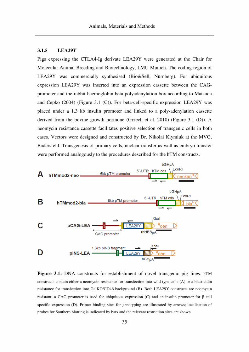
Animals, Materials and Methods
35
3.1.5 LEA29Y
Pigs expressing the CTLA4-Ig derivate LEA29Y were generated at the Chair for
Molecular Animal Breeding and Biotechnology, LMU Munich. The coding region of
LEA29Y was commercially synthesised (Bio&Sell, Nürnberg). For ubiquitous
expression LEA29Y was inserted into an expression cassette between the CAG-
promoter and the rabbit haemoglobin beta polyadenylation box according to Matsuda
and Cepko (2004) (Figure 3.1 (C)). For beta-cell-specific expression LEA29Y was
placed under a 1.3 kb insulin promoter and linked to a poly-adenylation cassette
derived from the bovine growth hormone (Grzech et al. 2010) (Figure 3.1 (D)). A
neomycin resistance cassette facilitates positive selection of transgenic cells in both
cases. Vectors were designed and constructed by Dr. Nikolai Klymiuk at the MVG,
Badersfeld. Transgenesis of primary cells, nuclear transfer as well as embryo transfer
were performed analogously to the procedures described for the hTM constructs.
Figure 3.1: DNA constructs for establishment of novel transgenic pig lines. hTM
constructs contain either a neomycin resistance for transfection into wild-type cells (A) or a blasticidin
resistance for transfection into GalKO/CD46 background (B). Both LEA29Y constructs are neomycin
resistant; a CAG promoter is used for ubiquitous expression (C) and an insulin promoter for β-cell
specific expression (D). Primer binding sites for genotyping are illustrated by arrows; localisation of
probes for Southern blotting is indicated by bars and the relevant restriction sites are shown.
Animals, Materials and Methods
36
3.2 Materials
3.2.1 Chemicals
All chemicals were used in p.a. quality unless noted otherwise.
Acetic acid (glacial) Merck, Darmstadt
Agar-agar Roth, Karlsruhe
Agarose Universal Bio&SELL, Nürnberg
Agarose UltraPureTM Invitrogen, Karlsruhe
Ampicillin Roth, Karlsruhe
Bromophenolblue Roth, Karlsruhe
Chloroform (Trichlormethan) Merck, Darmstadt
D-Mannitol Sigma, Steinheim
DTT (Dithiothreitol) Biomol, Hamburg
EDTA (Ethylenediaminetetraacetic acid) Roth, Karlsruhe
Ethanol Roth, Karlsruhe
Ethidiumbromide (1mg/ml) Merck, Darmstadt
Formaldehyde solution, 37% Roth, Karlsruhe
Formamide Roth, Karlsruhe
Glucose Roth, Karlsruhe
Glycerol Roth, Karlsruhe
Hydrochloric acid, 37% Roth, Karlsruhe
Isoamylalcohol Roth, Karlsruhe
Magnesium chloride Merck, Darmstadt
Paraformaldehyde Merck, Darmstadt
PEG 8000 Roth, Karlsruhe
Peptone/Tryptone Roth, Karlsruhe
Phenol Roth, Karlsruhe
Phosphoric acid Roth, Karlsruhe
PIPES (Piperazin-1,4-bis-(2-ethansulfonic-acid)) Sigma, Steinheim
Potassium acetate Roth, Karlsruhe
Animals, Materials and Methods
37
Potassium chloride Sigma, Steinheim
2-Propanol Roth, Karlsruhe
SDS (Sodiumdodecylsulfate), ultrapure Roth, Karlsruhe
Sodium acetate-trihydrate Merck, Darmstadt
Sodium chloride Roth, Karlsruhe
tri-Sodium citrate-2-hydrtae Roth, Karlsruhe
Sodiumdihydrogenphosphate-1-hydrate Merck, Darmstadt
di-Sodiumhydrogenphosphate-2-hydrate Roth, Karlsruhe
Sodium hydroxide (2N) Roth, Karlsruhe
D(+)-Sucrose Roth, Karlsruhe
Sulfuric acid Sigma, Steinheim
Tris (Tris-(hydroxymethyl)-aminomethan) Roth, Karlsruhe
Tween® 20 Sigma, Steinheim
Yeast extract Roth, Karlsruhe
3.2.2 Consumables
ABgene® PCR plates, 96-well Thermo Scientific, UK
Centrifuge tubes (15ml, 50ml) Falcon®, Becton Dickinson,
Heidelberg
Coated filter paper Roth, Karlsruhe
Culture dishes Roth, Karlsruhe
Developing and fixing solutions for x-rays AGFA-Gevaert N.V.,
Belgium
Hybond-N+ nylon membrane GE Healthcare, Munich
Maxisorp 96-well immuno plates NuncTM, Denmark
Parafilm® M American Can Company,
USA
PCR reaction tubes (0.2ml) Braun, Wertheim
Pipet tips Eppendorf, Hamburg
Pipet tips with filter Axygen Inc., USA
SafeGrip® Latex gloves SLG, Munich
Animals, Materials and Methods
38
Safe-Lock reaction tubes (1.5ml, 2ml) Eppendorf, Hamburg
Saran Barrier food wrap Dow, USA
Self-adhesive plastic covers NuncTM, Denmark
Sephadex G-50 columns Amersham, UK
Super RX Fuji medical x-ray film FujiFilm Corp., Japan
Whatman paper Roth, Karlsruhe
3.2.3 Devices
AccuJet® pro Pipetman Brand, Wertheim
Agarose gel electrophoresis chamber OWL Inc., USA
Centrifuge 5415 D Eppendorf, Hamburg
Centrifuge 5417 R Eppendorf, Hamburg
Chyo scales YMC Co., Japan
Glass tubes 80mm Zefa, Harthausen
Eppendorf HH Mastercycler Gradient Eppendorf, Hamburg
GeneQuant Pro spectrophotometer Amersham, UK
Gel documentation system BioRad, Munich
GFL 3031 shaker Hilab, Düsseldorf
Glass pipets Hirschmann, Eberstadt
Hybrid mini 38 hybridisation oven H. Saur, Reutlingen
37°C incubator Memmert, Schwabach
60°C incubator Memmert, Schwabach
Labofuge M centrifuge Heraeus, Osterode
Microprocessor pH meter WTW, Weilheim
MS1 minishaker IKA Labortechnik, Staufen
Multitip pipet (300µl) Eppendorf, Hamburg
Pipets (1000µl, 200µl, 20µl) Gilson Inc., USA
Polytron homogeniser Kinematica, Switzerland
Power Pac 300 gel electrophoresis unit BioRad, Munich
REAX2 Automatic swivel unit Hilab, Düsseldorf
RH Basic heating plate with magnetic stirrer IKA Labortechnik, Staufen
Animals, Materials and Methods
39
Severin 900 microwave Severin, Sundern
Tecan Sunrise ELISA reader with Magellan software Tecan, Austria
Thermomixer 5436 Eppendorf, Hamburg
UV cross-linker Vilber-Lourmat, France
Varioklav 400 autoclave H+P Labortechnik,
Oberschleißheim
WB6 water bath Firmengruppe Preiss-
Daimler, Medingen
X-ray cassette Rego, Augsburg
3.2.4 Antibodies, drugs, enzymes, oligonucleotides, standards
3.2.4.1 Antibodies
Primary antibodies
Polyclonal rabbit anti-human IgG Dako, Denmark
Secondary antibodies
Polyclonal rabbit anti-human IgG/HRP Dako, Denmark
3.2.4.2 Drugs
Azaperon (Stresnil®) Janssen Pharmaceutica,
Belgium
Ketamine hydrochloride (Ursotamin®) Serumwerk Bernburg,
Bernburg
T61 Intervet, Unterschleissheim
3.2.4.3 Enzymes
BigDye® Terminator v3.1 Applied Biosystems,
Animals, Materials and Methods
40
Weiterstadt
Herculase® II and buffer (5x) Agilent, Böblingen
Klenow fragment exo- (5U/µl) and buffer (10x) Fermentas, St. Leon Roth
Taq Polymerase (5U/µl) and buffer (10x) Agrobiogen,
Hilgertshausen
Proteinase K (20mg/ml) Roth, Karlsruhe
Restriction enzymes and recommended buffers: Fermentas, St. Leon Roth
Alw44I
BamHI
EcorI
HindIII
NcoI
PstI
PvuII
TaqI
XbaI
Ribonuclease A (RNase A) (0.2U/µl) Roche, Mannheim
T4 DNA Ligase (2000U/µl) and buffer (10x) Fermentas, St. Leon Roth
3.2.4.4 Oligonucleotides
Thermo Scientific, UK
CD46ex2F 5-GGC TAC CTG TCT CAG ATG AC-3
CD46ex3R 5-CCA TTT GCA GGG ACT GCT TG-3
GGTAr21 5-GCC ACC TCT TCT CGG ACT TGA TCT C-3
GGTAf22 5-CAT CCA GGC ACA TAC AGC ACA AG-3
hTMf1a 5-CTT CAT GGC ATT TC-3
hTMf1b 5-TGC TAG TCA ATC TT-3
Animals, Materials and Methods
41
hTMf2a 5-GTG ACC ATG GCT TT-3
hTMf2b 5-AGC TCT GAT TCA AC-3
hTMs3f 5-TTG GAA GTT CCT GGG ACA GAG-3
hTMs6f 5-GGT TTC AGG TTC GAT TCC TG-3
hTMf21 5- GGC CAT AAC TGA CAT CCC GTT C-3
hTMf22 5-ACA GTC ACA GAG TTG CCT CTG C-3
hTMf23 5-GAA TCC CAG CCA CAG CTT TGA C-3
hTMf24 5-TGG ATG AAC TCA GAC CAA ACA G-3
hTMf25 5-GAG ATG GAC TGG CCC TCT GCA AG-3
hTMf26 5-GAC AGT GAT GGA CAG CCG ACG GCA G-3
hTMf27 5-CGA CAG GTG CTT CTC GAT CTG-3
hTM43f1 5-CAG TGT TTG TCT TCT TAA CTA TC-§
hTM1101f 5-CGA CGC AGT CCT GCA ACG A-3
hTMr1 5-CAG CTA TCC TGA AC-3
hTMr2 5-AGC GCC TTT ATC CT-3
hTM44r1 5-ATC ACT ATG TCA TTC AAG AGC TC-3
hTM44r4 5-TCG TTT CTG TCC AAA ACC AGA AG-3
hTM1608r 5-CCG GAG TCA CAG TCG GTG CCA A-3
hTM-pA2050r 5-GGG CAA ACA ACA GAT GGC TG-3
hTM-pA2100r 5-AAA GGA CAG TGG GAG TGG CA-3
pTMSr 5-AAG CGC ACC AGC TGA AAG-3
neokanf 5-GAC AAT AGC AGG CAT GCT G-3
neokanR 5-GTG GAT GTG GAA TGT GTG C-3
Animals, Materials and Methods
42
bla241r 5-TTC CGA TCG CGA CGA TAC AAG TCA G-3
bla343r 5-GCT GTC CAT CAC TGT CCT TCA CT-3
PGKr 5-GCT GCT AAA GCG CAT GCT CCA GAC-3
bGHpAf1 5-AGG TGC CAC TCC CAC TGT CTT TTC-3
bGHpAr3 5-GAT GGC TGG CAA CTA GAA GGC AC-3
bGHpAr4 5-GTC GAG GCT GAT CAG CGA GCT C-3
CAGf 5-CTC TGC TAA CCA TGT TCA TG-3
Neof 5-TGA TTC CCA CTT TGT GGT TC-3
NeoPf 5-CAG CTG TGC TCG ACG TTG TC-3
HLAE-f 5-CCC AAG TGA AAT ACC CTG GCA-3
HLAE-r 5-CGA AGA TTC CCT GAC AAT CCC-3
LEAf 5-CCA GCA CCT GAA CTC CTG-3
LEAr 5-GGC TTT GTC TTG GCA TTA TG-3
pACTB954f 5-CGC TCG TGG TCG ACA ACG-3
pACTB1919r 5-CTG GAT GGC CAC GTA CAT G-3
3.2.4.5 Protein standards
Bovine serum albumin (BSA) Fraction V Roth, Karlsruhe
Human serum protein calibrator Dako, Denmark
3.2.5 Buffers, media, solutions
Water, deionised in a Millipore machine (EASYpure® II, pure Aqua, Schnaitsee) and
termed aqua bidest., was used as solvent, unless indicated otherwise.
Animals, Materials and Methods
43
Chloroform-isoamylalcohol (CiA)
96 ml chloroform
4 ml isoamylalcohol
Stored at 4°C protected from light
DNA loading buffer (10x)
10% glycerol in aqua bidest.
1 spatula tip of bromophenolblue
Add 0.5 M NaOH until colours turns blue
Stored aliquoted at 4°C
dNTP-mix
2 mM dATP, dCTP, dGTP, dTTP
in aqua bidest.
Stored aliquoted at -20°C
ELISA coating buffer
0.35 g NaH2PO4, H2O
1.34 g Na2HPO4, 2H2O
8.47 g NaCl
Ad 1000 ml aqua bidest.
Adjust pH to 7.2 with 5M NaOH
Stored at 4°C
ELISA dilution and washing buffer
0.35 g NaH2PO4, H2O
1.34 g Na2HPO4, 2H2O
29.22 g NaCl
Ad 1000 ml aqua bidest.
1 ml Tween 20
Adjust pH to 7.2 with 5M NaOH
Animals, Materials and Methods
44
Stored at 4°C
ELISA stop solution
0.5 M Sulphuric acid
Stored at room temperature
LB medium
5 g yeast extract
10 g peptone/tryptone
5 g NaCl
Ad 1000 ml aqua bidest.
pH 7.0 (adjust with 5 M NaOH)
autoclave
Medium was stored at room temperature.
LB-agar plates
1000 ml LB pH 7.0
15 g agar-agar
Autoclave
Cool to 50°C
Add 1 ml ampicillin (500 mg/ml)
Pour into sterile 10 cm culture dishes
After setting, plates were stored at 4°C.
4% Paraformaldehyde in 0.1 M Phosphate Buffer
1. Prepare 0.2 M Phosphate Buffer (PB), pH 7.4:
21.8 g Na2HPO4
6.4 g NaH2PO4
Ad 1000 ml aqua bidest.
Adjust pH with 5 M NaOH
Animals, Materials and Methods
45
2. Prepare 0.1 M PB pH 7.4:
500 ml 0.2M PB
Ad 1000 ml aqua bidest.
40 g paraformaldehyde was added to 1000 ml 0.1 M PB, pH 7.4 and heated to 60-
65ºC while stirring under the hood. A few drops of 1 N NaOH cleared the solution.
After cooling the solution was filtered and stored at 4°C.
PEG-MgCl2
40% (w/v) PEG 8000
30 mM MgCl2
Stored at room temperature
Phenol-chloroform-isoamylalcohol (PCiA)
25 ml phenol
25 ml CiA
Stored at 4°C protected from light
Plasmid A
50 mM glucose
25 mM Tris/HCl pH 8.0
10 mM EDTA/NaOH pH 8.0
Stored at room temperature
Plasmid B
0.1 M NaOH
0.5% (w/v) SDS
Prepare freshly
Plasmid C
3 M KOAc
pH 4.8 with 9 M HOAc
Animals, Materials and Methods
46
autoclave
Stored at room temperature
Sequencing buffer (5x)
17.5 ml 1 M Tris/HCl (pH 9.0)
125 µl 1 M MgCl2
Ad 50 ml aqua bidest.
Stored aliquoted at -20°C
Sodium-phosphate Buffer 1M
4 ml 85% H3PO4
89 g Na2HPO4, 2H2O
Ad 1000 ml aqua bidest.
Southern Blot Church buffer
1% BSA
1 mM EDTA, pH 8.0
500 mM Sodium-phosphate Buffer pH 7.2
7% SDS
Southern Blot depurination solution
0.3M HCl
Southern Blot high stringency buffer
1 mM EDTA, pH 8.0
40 mM Sodium-phosphate Buffer pH 7.2
1% SDS
Southern Blot low stringency buffer
0.5% BSA
1 mM EDTA
Animals, Materials and Methods
47
40 mM Sodium-phosphate Buffer pH 7.2
5% SDS
Southern Blot neutralisation solution
0.5 M Tris
1.5 M NaCl
pH 7.5
Southern Blot 20x SSC
0.3 M Na-Cit
3 M NaCl
Southern Blot strand break solution
0.5 M NaOH
1.5 M NaCl
STE
10 mM Tris/HCl pH 8.0
0.1 M NaCl
1 mM EDTA/NaOH pH 8.0
Stored at room temperature
TAE buffer (50x)
242 g Tris
100 ml 0.5M EDTA (pH8.0)
57 ml AcOH
Ad 1000 ml aqua bidest
Buffer was stored at room temperature and diluted to single concentration with aqua
bidest. for use.
Animals, Materials and Methods
48
10 mM Tris/HCl pH 8.0
10 mM Tris
Adjust pH to 8.0 with HCl
Stored at room temperature
3.2.6 Kits
CloneJETTM PCR Cloning Kit Fermentas, St. Leon Rot
NexttecTM Genomic DNA Isolation Kit Nextecc, Leverkusen
Qiaex® II Gel Extraction Kit Qiagen, Hilden
Roti®-Quant Roth, Karlsruhe
3.2.7 Others
α32P-dCTP Perkin-Elmer, Netherlands
dNTPs (dATP, dCTP, dGTP, dTTP) Fermentas, St. Leon Roth
Gene RulerTM (1 kb DNA ladder) Fermentas, St. Leon Roth
DNA molecular weight standard
6 x DNA loading dye Fermentas, St. Leon Roth
Random primers (3µg/µl) Invitrogen, Karlsruhe
TMB Solution (HRP substrate) Uptima, France
TOP 10 chemically competent E.coli Invitrogen, Karlsruhe
3.2.8 Software
BioEdit Sequence Alignment Editor
FinchTV Version 1.3.1, Geospiza Inc.
Macromedia Freehand MX
Animals, Materials and Methods
49
3.3 Methods
3.3.1 Genomic analysis
3.3.1.1 Genotyping of founder animals and F1 generation
In order to identify transgenic pigs, genotyping PCR was performed on tissue samples
of piglets. For this, ear punches were obtained from three-day-old piglets and stored at
-20°C until further processing.
For genotyping of founder animals, genomic DNA was isolated in 1.5 ml reaction
tubes from approximately 100 mg of shredded tissue using the following protocol. All
steps were carried out at room temperature unless indicated otherwise.
Composition of lysis buffer
160 mM Sucrose
80 mM EDTA pH 8.0
100 mM Tris/HCl pH 8.0
0.5% SDS
Add 400 µl lysis buffer and 30 µl Proteinase K (10 mg/ml) to tissue, mix
Incubate at 60°C overnight
Centrifuge 5 min, 16,100 g
Transfer supernatant to fresh tubes
Add 400 µl sodium chloride 4.5 M, mix
Add 300 µl phenol-chloroform-isoamylalcohol (PCiA)
Pivot in automatic swivel unit 15 min
Centrifuge 10 min, 16,100 g
Transfer aqueous phase to fresh tubes
Repeat PCiA extraction twice: add 300 µl PCiA, pivot 10 min, centrifuge 2.5 min
Add 550 µl 2-propanol to precipitate DNA
Transfer DNA successively into 2 tubes with 1 ml EtOH 70% each
Incubate in EtOH 70% overnight
Animals, Materials and Methods
50
Discard EtOH 70%
Air dry DNA pellet 10 min
Resolve DNA in 55 µl 10 mM Tris/HCl
Mix repeatedly for 1 hour
DNA concentration was measured at 260 nm using a GeneQuant Pro
spectrophotometer. For genotyping PCR analysis, isolated gDNA was diluted with 10
mM Tris/HCl to a concentration of 100 ng/µl.
Genomic DNA of F1 generation pigs was isolated using NexttecTM Genomic DNA
Isolation Kit (Nextecc Biotechnologie GmbH, Leverkusen) according to the
manufacturer’s protocol.
In both cases genotyping PCR was performed in 0.2 ml reaction tubes in a total
volume of 25 µl as follows:
Composition of reaction mix
2.5 µl 10x PCR-Buffer
2.5 µl MgCl2 (25mM)
2.5 µl dNTPs (2mM)
0.4 µl forward + 0.4 µl reverse primer (10 µM)
0.2 µl Taq polymerase (5 U/µl)
1 µl DNA-template
15.5 µl aqua bidest.
Cycler protocol
2 min 95°C denaturation
30 sec 95°C denaturation
30 sec xx°C annealing at PCR specific temperature
1 min 72°C elongation
GO TO Step 2 34x
10 min 72°C final elongation
Animals, Materials and Methods
51
15 min 4°C termination of reaction
Transgene specific primers were used to identify the respective transgenes. In
addition, a house-keeping gene, the ubiquitously expressed β-actin, was amplified in
parallel to verify integrity of isolated genomic DNA. For details of genotyping
primers used and their respective annealing temperatures, see Table 3.1.
3.3.1.2 Duplex PCR
For discrimination of homozygous from heterozygous transgenic offspring, duplex
PCRs were established for the GalKO and the hTM locus. One transgene specific and
two wild-type sequence specific primers were employed in each duplex PCR. Details
of the primers used are given in Table 3.1.
PCRs were performed in 0.2 ml reaction tubes in a total volume of 25 µl as follows:
Composition of reaction mix
2.5 µl 10x PCR-Buffer
2.5 µl MgCl2 (25 mM)
2.5 µl dNTPs (2 mM)
0.4 µl first + 0.4 µl second + 0.4 µl third primer (10 µM)
0.2 µl Taq polymerase (5 U/µl)
1 µl DNA-template
15.5 µl aqua bidest.
Thermocycling for duplex PCR was performed according to the cycler protocol
detailed above for initial genotyping PCR.
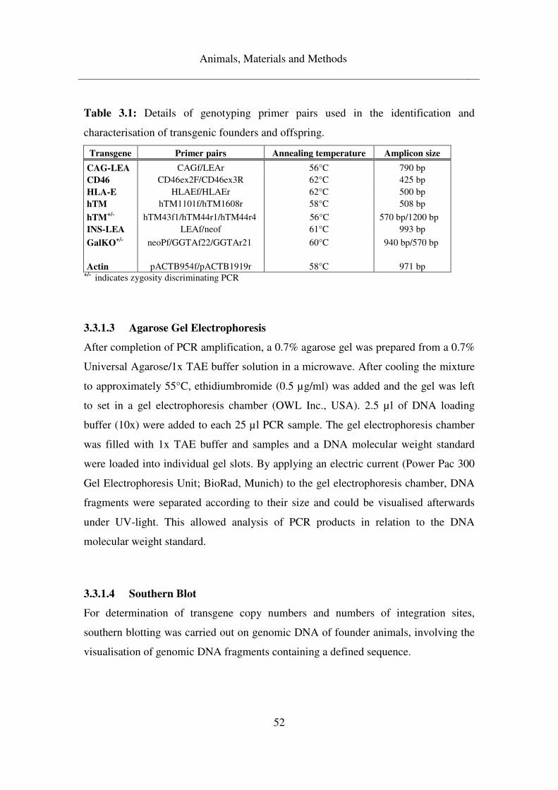
Animals, Materials and Methods
52
Table 3.1: Details of genotyping primer pairs used in the identification and
characterisation of transgenic founders and offspring.
Transgene Primer pairs Annealing temperature Amplicon size
CAG-LEA CAGf/LEAr 56°C 790 bp CD46 CD46ex2F/CD46ex3R 62°C 425 bp HLA-E HLAEf/HLAEr 62°C 500 bp hTM hTM1101f/hTM1608r 58°C 508 bp
hTM+/-
hTM43f1/hTM44r1/hTM44r4 56°C 570 bp/1200 bp INS-LEA LEAf/neof 61°C 993 bp
GalKO+/-
neoPf/GGTAf22/GGTAr21 60°C 940 bp/570 bp Actin pACTB954f/pACTB1919r 58°C 971 bp
+/- indicates zygosity discriminating PCR
3.3.1.3 Agarose Gel Electrophoresis
After completion of PCR amplification, a 0.7% agarose gel was prepared from a 0.7%
Universal Agarose/1x TAE buffer solution in a microwave. After cooling the mixture
to approximately 55°C, ethidiumbromide (0.5 µg/ml) was added and the gel was left
to set in a gel electrophoresis chamber (OWL Inc., USA). 2.5 µl of DNA loading
buffer (10x) were added to each 25 µl PCR sample. The gel electrophoresis chamber
was filled with 1x TAE buffer and samples and a DNA molecular weight standard
were loaded into individual gel slots. By applying an electric current (Power Pac 300
Gel Electrophoresis Unit; BioRad, Munich) to the gel electrophoresis chamber, DNA
fragments were separated according to their size and could be visualised afterwards
under UV-light. This allowed analysis of PCR products in relation to the DNA
molecular weight standard.
3.3.1.4 Southern Blot
For determination of transgene copy numbers and numbers of integration sites,
southern blotting was carried out on genomic DNA of founder animals, involving the
visualisation of genomic DNA fragments containing a defined sequence.
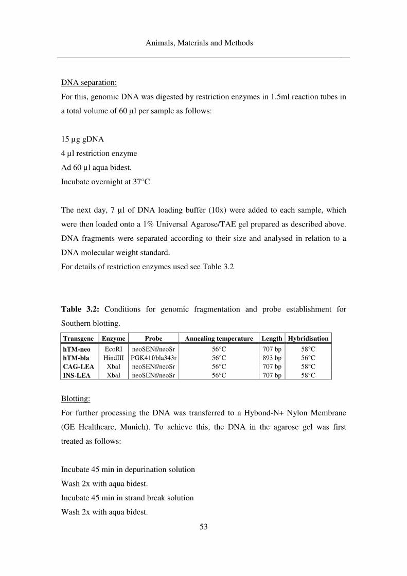
Animals, Materials and Methods
53
DNA separation:
For this, genomic DNA was digested by restriction enzymes in 1.5ml reaction tubes in
a total volume of 60 µl per sample as follows:
15 µg gDNA
4 µl restriction enzyme
Ad 60 µl aqua bidest.
Incubate overnight at 37°C
The next day, 7 µl of DNA loading buffer (10x) were added to each sample, which
were then loaded onto a 1% Universal Agarose/TAE gel prepared as described above.
DNA fragments were separated according to their size and analysed in relation to a
DNA molecular weight standard.
For details of restriction enzymes used see Table 3.2
Table 3.2: Conditions for genomic fragmentation and probe establishment for
Southern blotting.
Transgene Enzyme Probe Annealing temperature Length Hybridisation
hTM-neo EcoRI neoSENf/neoSr 56°C 707 bp 58°C hTM-bla HindIII PGK41f/bla343r 56°C 893 bp 56°C CAG-LEA XbaI neoSENf/neoSr 56°C 707 bp 58°C INS-LEA XbaI neoSENf/neoSr 56°C 707 bp 58°C
Blotting:
For further processing the DNA was transferred to a Hybond-N+ Nylon Membrane
(GE Healthcare, Munich). To achieve this, the DNA in the agarose gel was first
treated as follows:
Incubate 45 min in depurination solution
Wash 2x with aqua bidest.
Incubate 45 min in strand break solution
Wash 2x with aqua bidest.
Animals, Materials and Methods
54
Incubate 20 min in neutralisation solution
Wash 2x with aqua bidest.
Incubate 20 min in 5x SSC
Subsequently, the DNA was blotted onto the membrane in a semi-dry manner. For
this, the gel was placed upside down onto cling film (Saran Barrier Food Wrap; Dow,
USA). Two layers of Whatman paper (Roth, Karlsruhe) and multiple layers of
absorbent tissue were placed on top. To facilitate capillary transfer of the DNA onto
the membrane, additional weight was applied on top and transfer was carried out over
24 hours.
Then DNA was crosslinked to the membrane under 0.120 J/cm2 UV light (Vilber-
Lourmat, France) and stored at room temperature.
Hybridisation:
DNA fragments containing the transgene were detected by probes specific for the
neomycin or blasticidin resistance cassette in the transgene construct. For this, the
probes were amplified by PCR from plasmids containing the vectors.
PCR was carried out according to a standard protocol as described for genotyping
PCRs above. For details of primers and annealing temperatures used in the
establishment of probes for the different transgenes, see Table 3.2.
The PCR products were separated on 1% Universal Agarose/TAE gels prepared as
described above, eluated from the gel using QiaexII Gel Extraction Kit (Qiagen,
Hilden), and DNA concentration of eluates was determined in relation to a DNA
molecular weight standard on 1% Universal Agarose/TAE gels.
Prior to hybridisation the membranes were placed in 80 mm glass tubes (Zefa,
Harthausen), pre-wetted with 5x SSC and incubated in 30 ml prehybridisation buffer
(Church Buffer) under permanent rotation at 58°C in an incubator (H. Saur,
Reutlingen) for 1 hour.
The probes were then radio-labelled with α32P-dCTP (Perkin-Elmer, Netherlands)
Animals, Materials and Methods
55
according to the following protocol:
7 µl Klenow 10x Bu
50-100 ng DNA probe
10 µl Random primer (3 µg/µl)
Ad 50 µl aqua bidest.
Probe DNA was denaturated at 97°C for 10 minutes. After incubation on ice for 2
minutes, 20 µl of hybridisation mix was added to each sample.
Hybridisation mix:
3 µl C-Mix (0.33 mM of each dATP, dGTP, dTTP)
5 µl α 32P-dCTP (3000 Ci/mmol)
1 µl Klenow exo-
Ad 20 µl aqua bidest.
Probe labelling was performed at 37°C for one hour. Unincorporated nucleotides were
separated from the labelled probes by centrifugation through Sephadex G-50 columns
(Amersham, UK). The purified and labelled probes were denaturated at 97°C for 5
minutes and cooled on ice. Meanwhile, the prehybridisation buffer was exchanged for
fresh Church Buffer in which the labelled probes were diluted.
Hybridisation of the probes to the membrane was carried out overnight at 58°C under
permanent rotation.
The next day, the hybridisation solution was discarded and the membrane was washed
twice with low-stringency buffer at room temperature and afterwards twice with high-
stringency buffer at 58°C. Then the membrane was placed on a coated filter paper
(Roth, Karlsruhe), wrapped in cling film, and exposed to x-ray film (FujiFilm Corp.,
Japan) in an x-ray cassette (Rego, Augsburg) for at least one day at -80°C.
The exposed x-ray films were developed using developing and fixing solution
(AGFA-Gevaert, Belgium) according to the manufacturer’s instructions and
subsequently analysed.

Animals, Materials and Methods
56
3.3.1.5 Inverse Polymerase Chain Reaction (inverse PCR)
Inverse PCR was performed in order to clarify the unknown genomic DNA flanking
regions of the transgene at the insertion site. Figure 3.2 gives an overview of inverse
PCR procedure.
All steps were carried out at room temperature unless indicated otherwise.
Unknown flanking
region
Unknown flanking
region
gDNA
Restriction Digest
PstI
Transgene
Ligation
PstI
(nested) PCR
Sequencing
Transgene
Res. cassette
PstI
Unknown flanking
region
Transgene
Primer-f Primer-r
Primer-r Primer-f
Primer-r Primer-f
Primer-r Primer-f
PstI
PstI
PstI PstI
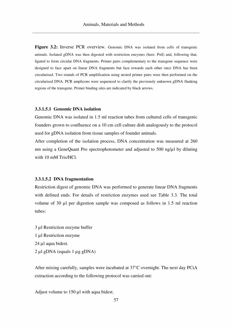
Animals, Materials and Methods
57
Figure 3.2: Inverse PCR overview. Genomic DNA was isolated from cells of transgenic
animals. Isolated gDNA was then digested with restriction enzymes (here: PstI) and, following that,
ligated to form circular DNA fragments. Primer pairs complementary to the transgene sequence were
designed to face apart on linear DNA fragments but face towards each other once DNA has been
circularised. Two rounds of PCR amplification using nested primer pairs were then performed on the
circularised DNA. PCR amplicons were sequenced to clarify the previously unknown gDNA flanking
regions of the transgene. Primer binding sites are indicated by black arrows.
3.3.1.5.1 Genomic DNA isolation
Genomic DNA was isolated in 1.5 ml reaction tubes from cultured cells of transgenic
founders grown to confluence on a 10 cm cell culture dish analogously to the protocol
used for gDNA isolation from tissue samples of founder animals.
After completion of the isolation process, DNA concentration was measured at 260
nm using a GeneQuant Pro spectrophotometer and adjusted to 500 ng/µl by diluting
with 10 mM Tris/HCl.
3.3.1.5.2 DNA fragmentation
Restriction digest of genomic DNA was performed to generate linear DNA fragments
with defined ends. For details of restriction enzymes used see Table 3.3. The total
volume of 30 µl per digestion sample was composed as follows in 1.5 ml reaction
tubes:
3 µl Restriction enzyme buffer
1 µl Restriction enzyme
24 µl aqua bidest.
2 µl gDNA (equals 1 µg gDNA)
After mixing carefully, samples were incubated at 37°C overnight. The next day PCiA
extraction according to the following protocol was carried out:
Adjust volume to 150 µl with aqua bidest.
Animals, Materials and Methods
58
Add 100 µl PCiA
Extract by pivoting for 1 min
Centrifuge 2.5 min, 16,100g
Transfer aqueous phase to new tube
Add 15 µl 3M NaOAc
Add 400 µl EtOH 100%
Store at -80°C 30 – 60 min
Centrifuge 30 min, 16,100g, 4°C
Wash in EtOH 70% overnight
Centrifuge 2.5 min, 16,100g
Discard supernatant
Air dry DNA pellet 6 min
Resolve in 30 µl 10 mM Tris/HCl
10 µl of resolved, digested gDNA was mixed with 12.5 µl aqua bidest. and 2.5 µl
DNA loading buffer (10x) and loaded onto a 1% agarose gel to verify digestion
success. The gel was prepared from a 1% Universal Agarose/TAE buffer solution as
described above.
The remaining 20 µl of digested gDNA were diluted with 100 µl 10 mM Tris/HCl and
used for further processing.
3.3.1.5.3 Circularisation of DNA fragments
Digested and diluted gDNA was ligated overnight at room temperature in a total
volume of 40 µl in 1.5 ml reaction tubes to circularise DNA. Each reaction mix was
composed as follows:
4 µl 10x ligation buffer
4 µl T4 DNA ligase
12 µl aqua bidest.
20 µl digested gDNA
Termination of the reaction by 15 min 65°C
Animals, Materials and Methods
59
3.3.1.5.4 Nested PCR
For amplification of DNA sequences on circularised fragments, (nested) PCR
amplification was performed in a total volume of 25 µl in 0.2 ml reaction tubes. For
general principle of nested PCR see Figure 3.3.
Composition of reaction mix
5.0 µl 5x Herculase-Buffer
2.5 µl dNTPs
0.35 µl forward + 0.35 µl reverse Primer
0.2 µl Herculase II
1 µl DNA-template
15.6 µl aqua bidest.
Cycler-protocol
2 min 95°C denaturation
30 sec 95°C denaturation
30 sec 58°C annealing
3 min 72°C elongation
GO TO Step 2 39x
10 min 72°C final elongation
15 min 4°C termination of reaction
After completion of PCR amplification, samples were used as DNA templates for
nested PCR amplification. For this, each sample was used undiluted and in a 1:100
dilution with aqua bidest. Reaction mix and cycler protocol were the same as in the
first amplification. Primers used were nested.
For an overview of restriction enzymes and primer pairs used for the different
transgenes in the inverse PCR process, see Table 3.3
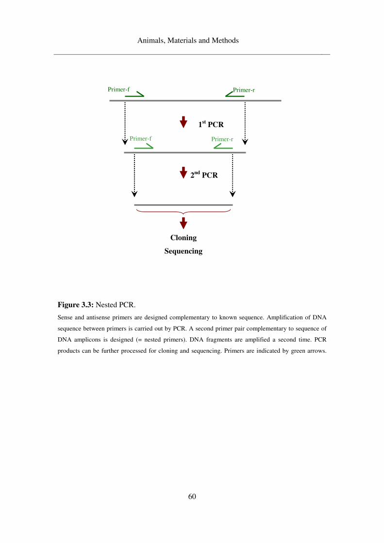
Animals, Materials and Methods
60
Figure 3.3: Nested PCR.
Sense and antisense primers are designed complementary to known sequence. Amplification of DNA
sequence between primers is carried out by PCR. A second primer pair complementary to sequence of
DNA amplicons is designed (= nested primers). DNA fragments are amplified a second time. PCR
products can be further processed for cloning and sequencing. Primers are indicated by green arrows.
1st PCR
Primer-f Primer-r
2nd
PCR
Cloning
Sequencing
Primer-f Primer-r
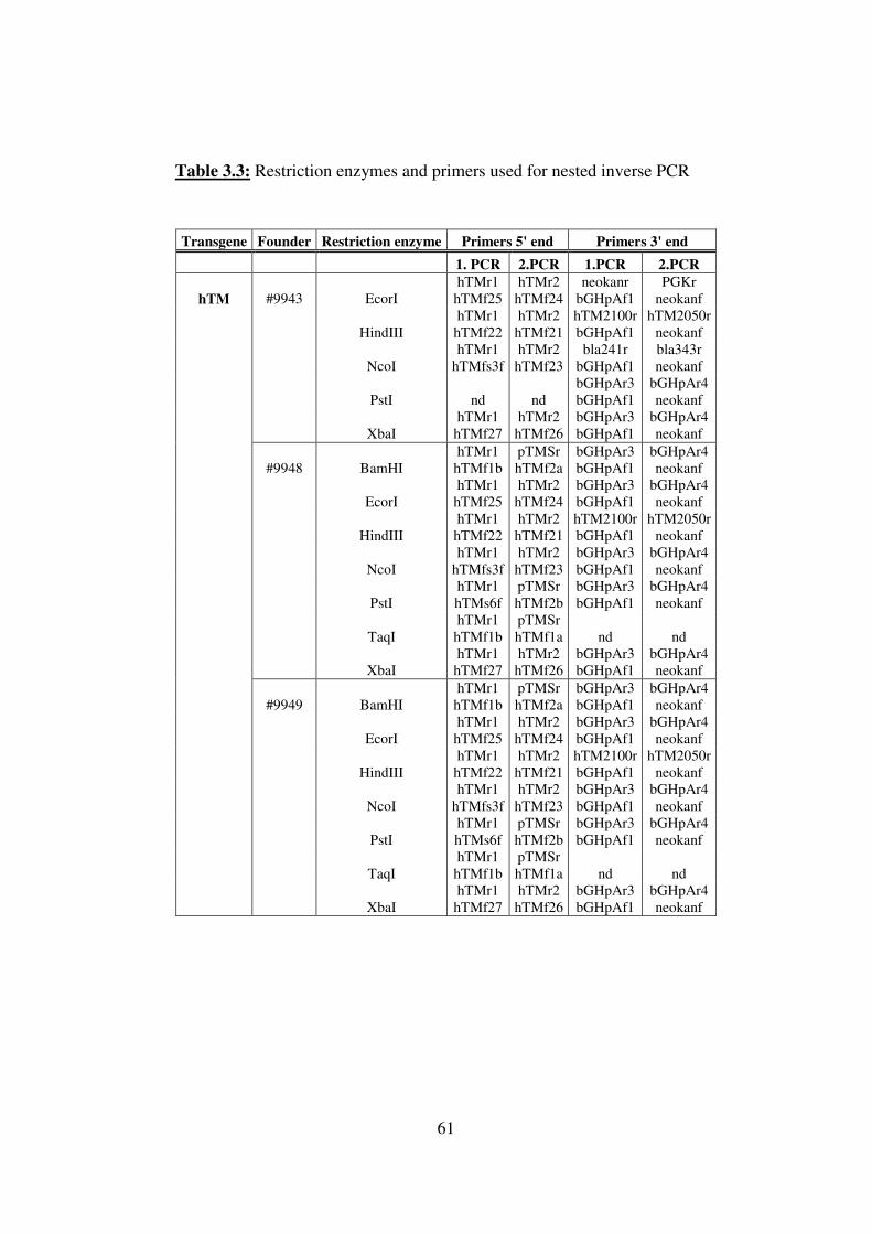
61
Table 3.3: Restriction enzymes and primers used for nested inverse PCR
Transgene Founder Restriction enzyme Primers 5' end Primers 3' end
1. PCR 2.PCR 1.PCR 2.PCR
hTM #9943 EcorI hTMr1
hTMf25 hTMr2
hTMf24 neokanr
bGHpAf1 PGKr
neokanf
HindIII hTMr1
hTMf22 hTMr2
hTMf21 hTM2100r bGHpAf1
hTM2050r neokanf
NcoI hTMr1
hTMfs3f hTMr2
hTMf23 bla241r
bGHpAf1 bla343r neokanf
PstI nd nd bGHpAr3 bGHpAf1
bGHpAr4 neokanf
XbaI hTMr1
hTMf27 hTMr2
hTMf26 bGHpAr3 bGHpAf1
bGHpAr4 neokanf
#9948 BamHI hTMr1
hTMf1b pTMSr hTMf2a
bGHpAr3 bGHpAf1
bGHpAr4 neokanf
EcorI hTMr1
hTMf25 hTMr2
hTMf24 bGHpAr3 bGHpAf1
bGHpAr4 neokanf
HindIII hTMr1
hTMf22 hTMr2
hTMf21 hTM2100r bGHpAf1
hTM2050r neokanf
NcoI hTMr1
hTMfs3f hTMr2
hTMf23 bGHpAr3 bGHpAf1
bGHpAr4 neokanf
PstI hTMr1 hTMs6f
pTMSr hTMf2b
bGHpAr3 bGHpAf1
bGHpAr4 neokanf
TaqI hTMr1
hTMf1b pTMSr hTMf1a nd nd
XbaI hTMr1
hTMf27 hTMr2
hTMf26 bGHpAr3 bGHpAf1
bGHpAr4 neokanf
#9949 BamHI hTMr1
hTMf1b pTMSr hTMf2a
bGHpAr3 bGHpAf1
bGHpAr4 neokanf
EcorI hTMr1
hTMf25 hTMr2
hTMf24 bGHpAr3 bGHpAf1
bGHpAr4 neokanf
HindIII hTMr1
hTMf22 hTMr2
hTMf21 hTM2100r bGHpAf1
hTM2050r neokanf
NcoI hTMr1
hTMfs3f hTMr2
hTMf23 bGHpAr3 bGHpAf1
bGHpAr4 neokanf
PstI hTMr1 hTMs6f
pTMSr hTMf2b
bGHpAr3 bGHpAf1
bGHpAr4 neokanf
TaqI hTMr1
hTMf1b pTMSr hTMf1a nd nd
XbaI hTMr1
hTMf27 hTMr2
hTMf26 bGHpAr3 bGHpAf1
bGHpAr4 neokanf
Animals, Materials and Methods
62
3.3.1.5.5 DNA Eluation
After preparation of a 1% UltraPureTMAgarose/TAE buffer gel analogously to the
method described above, 2.5 µl DNA loading dye (10x) was added to each of the
samples from the nested PCR, which were then loaded onto the gel. DNA separation
was performed as detailed above. After visualisation, appropriate DNA strands were
excised from the gel and extracted using a QiaexII Gel Extraction Kit (Qiagen,
Hilden) following the manufacturer’s protocol. Then
2 µl DNA eluate
2 µl DNA loading buffer (10x)
15 µl aqua bidest.
were loaded onto a 1% Universal Agarose/TAE buffer gel prepared as described
above. A DNA molecular weight standard allowed determination of DNA
concentration in eluate.
3.3.1.5.6 Ligation
Eluated DNA fragments were inserted into pJet plasmid cloning vector (Fermentas,
St. Leon Roth) in a ligation reaction as follows:
2 µl 10x ligation buffer
1 µl T4 DNA ligase
1 µl pJet (5ng/µl)
15-30 ng DNA eluate
Ad 20 µl aqua bidest.
Ligation was carried out overnight at room temperature. Ligase was deactivated by 15
min 65°C.
Animals, Materials and Methods
63
3.3.1.5.7 Heat Shock Transformation
Plasmid vectors containing the DNA inserts were transformed into E.coli TOP10
competent cells according to the following protocol:
Thaw up competent cells carefully on ice
Add 5 µl of ligation
Mix carefully, avoid re-pipetting
20 min on ice
45 sec at 42°C
2 min on ice
Add 1 ml LB-Medium
45 min 37°C
Centrifuge 5 min, 2,300g
Resuspend pellet in 100 – 200 µl of the supernatant
Plate on LB-Amp (50 µg/ml) agar dishes
Cultivate overnight at 37°C
The next day, individual colonies were picked from the agar dishes and inoculated
individually into 2.5 ml LB-Amp (50 µg/ml) medium each. Cultivation was carried
out at 37°C overnight whilst shaking.
3.3.1.5.8 Plasmid preparation
Preparation of plasmids from the overnight liquid cultures was carried out as follows:
Centrifuge overnight cultures 10 min, 1,300g, discard supernatant
Resuspend pellet in 750 µl STE and transfer to 1.5 ml tubes
Centrifuge 5 min, 4,500g, discard supernatant
Resuspend pellet in 200 µl Plasmid A
Add 400 µl Plasmid B, 5 – 7 x mix (don´t vortex), 5 min on ice
Add 300 µl Plasmid C, 5 – 7 x mix (don´t vortex), 3 min on ice
Centrifuge 10 min, 16,100g
Animals, Materials and Methods
64
Transfer supernatant to new tube
Add 4 µl RNase A (10 mg/ml)
Incubate 45 min 37°C
Add 300 µl PCiA, 1 min shake
Centrifuge 2.5 min, 16,100g
Transfer aqueous phase into new tube
Add 650 µl 2-propanol, invert
Centrifuge 10 min, 16,100g, discard supernatant
Wash pellet in 700 µl EtOH 70%
Centrifuge 2.5 min, 16,100g, discard supernatant
Air dry pellet 6 min
Resolve pellet in 55 µl 10 mM Tris/HCl
DNA concentration was measured in a GeneQuant Pro spectrophotometer at 260 nm,
and approximately 2 µg were used for qualitative analysis by restriction digestion.
3.3.1.5.9 Restriction digest
2 µl 10x restriction enzyme buffer
0.2 µl restriction enzyme
2 µg Plasmid
Ad 20 µl aqua bidest.
After overnight incubation at 37°C samples were loaded onto a 1% Universal
Agarose/TAE buffer gel prepared as described above to determine ligation and
transformation success. Samples displaying appropriate restriction digestion patterns
were chosen for further processing.
3.3.1.5.10 PEG precipitation
Precipitation of DNA for sequencing was carried out as follows:
Animals, Materials and Methods
65
20 µl plasmid preparation
20 µl PEG-MgCl2
20 µl aqua bidest.
Equilibrate 10 min
Centrifuge 20 min 15,700g
Wash in 100 µl EtOH 70% overnight
Centrifuge 2.5 min, 16,100g
Air dry 6 min
Resolve in 20 µl 10 mM Tris/HCl
DNA concentration was measured at 260 nm in a GeneQuant Pro spectrophotometer.
Subsequently, samples were diluted with 10 mM Tris/HCl to a DNA concentration of
30 ng/µl to serve as template for sequencing.
3.3.1.5.11 Sequencing
Sequencing of samples was performed in 0.2 ml reaction tubes in a total reaction
volume of 10 µl. Each sample was sequenced twice, once with a pJet forward primer
and another time with a pJet reverse primer.
Composition of each reaction mix
4 µl 5x sequencing buffer
1 µl Big Dye
1 µl primer (10 µM)
2 µl template
2 µl aqua bidest.
Cycler-protocol
1 min 95°C denaturation
5 sec 95°C denaturation
10 sec 50°C annealing
Animals, Materials and Methods
66
4 min 60°C elongation
GO TO Step 2 39x
15 min 4° termination of reaction
3.3.1.5.12 EtOH precipitation
After sequencing amplification, DNA was precipitated from the reaction mix as
follows:
Add 2.5 µl 125 mM EDTA into PCR-lid, spin down
Add 30 µl EtOH 100%, transfer to 1.5 ml tube
Incubate on ice 5 min
Centrifuge 30 min, 15,700g, 4°C
Wash pellet in 50 µl EtOH 70%
Centrifuge 2.5 min, 15,700
Air dry pellet 6 min
Resolve DNA in 30 µl aqua bidest. and transfer to sequencing plate (Abgene® PCR
Plates; Thermo Scientific, UK)
Capillary electrophoretic separation of DNA samples was carried out at the German
Mouse Clinic, Helmholtz Centre, Neuherberg.
Nucleotide sequences were bioinformatically analysed using FinchTV Version 1.3.1,
Geospiza Inc. and BioEdit Sequence Alignment Editor.
3.3.2 Expression analysis
For transgene expression analysis tissue was either obtained in the form of ear
punches or as an organ spectrum.
Ear punches of approximately 0.5 cm2 were taken from three-day-old manually
Animals, Materials and Methods
67
fixated piglets. Tissue was either shock frozen in liquid nitrogen and stored at -80°C
or transferred to 15 ml reaction tubes filled with 4% PFA.
Pigs sampled for organ spectrum analysis were anaesthetised with Ketamine (2 ml/10
kg) and Azaperon (0.5 ml/10 kg) i.m. Following that the animals received a vein
catheter and were then euthanized with T61 (1 ml/10 kg) i.v. Organs were explanted
postmortally without delay and samples of approximately 1 cm3 were either shock
frozen in liquid nitrogen and stored at -80°C or transferred to 4% PFA.
3.3.2.1 Protein isolation
For protein isolation frozen tissue was pulverised using a mortar and pestle and
approximately 100 mg were homogenised in a sterile 2 ml reaction tube with 750 µl
homogenising buffer of the following composition:
1.25 ml 1 M Mannitol
0.2 ml 2 M Sucrose
0.1 ml 0.5 M EDTA (pH 8.0)
0.5 ml 0.1M PIPES/Tris (pH 6.7)
Ad 10 ml aqua bidest.
After 30 min centrifugation at 4,100g, 4°C, supernatant was transferred to fresh
reaction tubes and protein concentration was measured using bovine serum albumin
(BSA) as standard protein. For this, 500 mg BSA was dissolved in 5 ml homogenising
buffer of the above composition and dilutions of 25 µg/ml, 20 µg/ml, 15 µg/ml, 10
µg/ml, 5 µg/ml and 1 µg/ml were prepared.
Standards and samples were measured in a GeneQuant Pro spectrophotometer at 595
nm using Roti®-Quant according to the manufacturer’s instructions.
3.3.2.2 Enzyme-Linked Immunosorbent Assay (ELISA)
In order to perform qualitative and quantitative evaluation of transgenic protein,
ELISAs were carried out in 96 well immuno plates (Maxisorp; NuncTM, Denmark).
Animals, Materials and Methods
68
Experiments were performed for CAG-LEA transgene expression analysis.
Antibodies used were directed against the human IgG fragment of the transgene
construct and reaction was measured against a human serum protein calibrator (Dako,
Denmark) in dilutions of 1:11,900, 1:59,500, 1:297,500, 1:1,487,500 and 1:7,437,500.
For reaction development chromogenic substrate for the horseradish peroxidase
system was used. The following protocol was employed in carrying out the
experiments:
Dilute primary antibody (polyclonal rabbit anti-human IgG; Dako, Denmark) 1:570 in
coating buffer
Add 100 µl to each well
Cover with self-adhesive plastic covers and incubate overnight at 4°C
Discard well contents
Wash 3x with 200 µl washing buffer per well
Dilute standard and samples to appropriate concentrations in washing/dilution buffer
Add 100 µl to each well
Cover and incubate 2 hours at room temperature
Discard well contents
Wash 3x with 200 µl washing buffer per well
Dilute secondary antibody (polyclonal rabbit anti-human IgG/HRP; Dako, Denmark)
1:4800 in washing/dilution buffer
Add 100 µl to each well
Cover and incubate 1 hour at room temperature
Discard well contents
Wash 3x with 200 µl washing buffer per well
Add 100 µl 3,3’,5,5’-tetramethylbenzidine (TMB) substrate (Uptima, France) to each
well
Cover and incubate 7 min at room temperature
Add 100 µl Stop solution
Colour development was read immediately by measuring extinction at 490 nm and
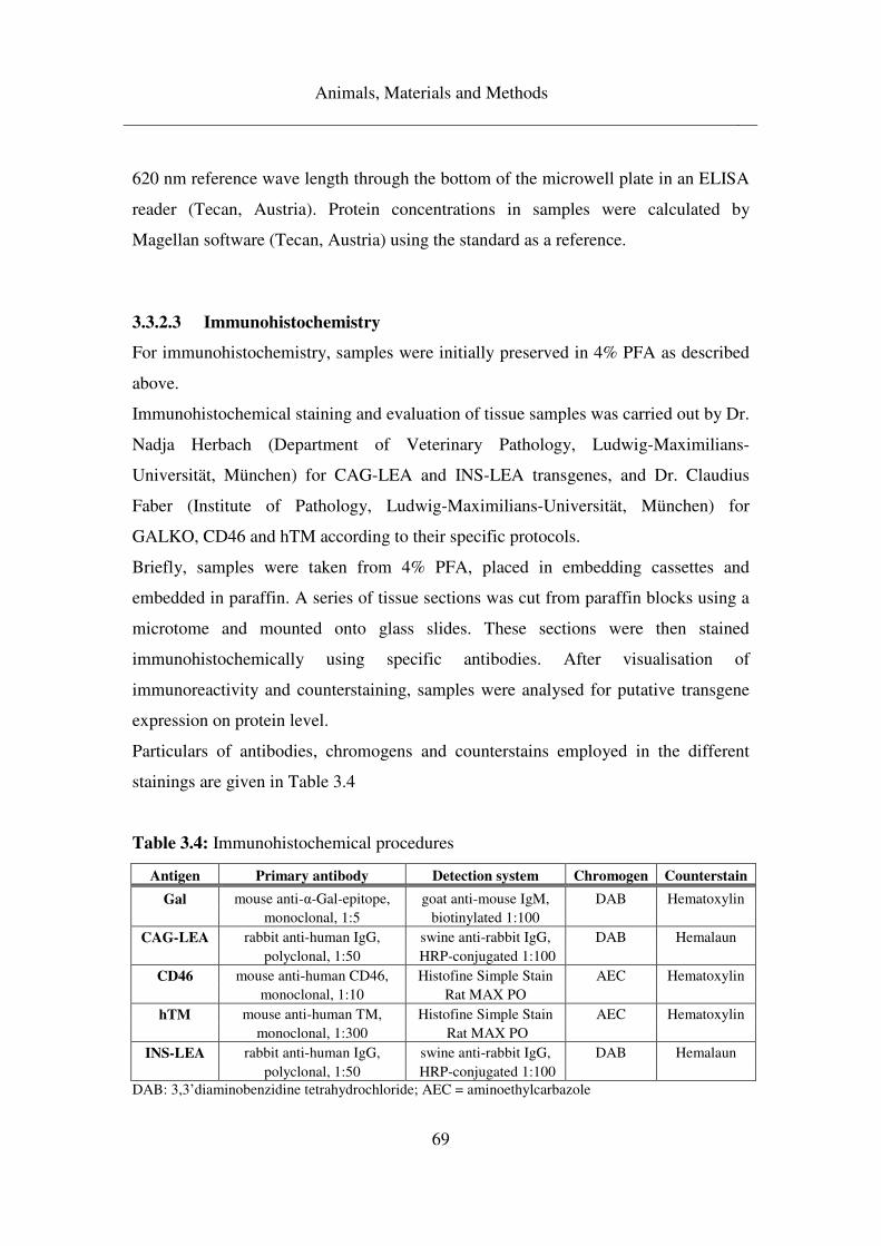
Animals, Materials and Methods
69
620 nm reference wave length through the bottom of the microwell plate in an ELISA
reader (Tecan, Austria). Protein concentrations in samples were calculated by
Magellan software (Tecan, Austria) using the standard as a reference.
3.3.2.3 Immunohistochemistry
For immunohistochemistry, samples were initially preserved in 4% PFA as described
above.
Immunohistochemical staining and evaluation of tissue samples was carried out by Dr.
Nadja Herbach (Department of Veterinary Pathology, Ludwig-Maximilians-
Universität, München) for CAG-LEA and INS-LEA transgenes, and Dr. Claudius
Faber (Institute of Pathology, Ludwig-Maximilians-Universität, München) for
GALKO, CD46 and hTM according to their specific protocols.
Briefly, samples were taken from 4% PFA, placed in embedding cassettes and
embedded in paraffin. A series of tissue sections was cut from paraffin blocks using a
microtome and mounted onto glass slides. These sections were then stained
immunohistochemically using specific antibodies. After visualisation of
immunoreactivity and counterstaining, samples were analysed for putative transgene
expression on protein level.
Particulars of antibodies, chromogens and counterstains employed in the different
stainings are given in Table 3.4
Table 3.4: Immunohistochemical procedures
Antigen Primary antibody Detection system Chromogen Counterstain
Gal mouse anti-α-Gal-epitope, goat anti-mouse IgM, DAB Hematoxylin monoclonal, 1:5 biotinylated 1:100
CAG-LEA rabbit anti-human IgG, swine anti-rabbit IgG, DAB Hemalaun polyclonal, 1:50 HRP-conjugated 1:100
CD46 mouse anti-human CD46, Histofine Simple Stain AEC Hematoxylin monoclonal, 1:10 Rat MAX PO
hTM mouse anti-human TM, Histofine Simple Stain AEC Hematoxylin monoclonal, 1:300 Rat MAX PO
INS-LEA rabbit anti-human IgG, swine anti-rabbit IgG, DAB Hemalaun polyclonal, 1:50 HRP-conjugated 1:100
DAB: 3,3’diaminobenzidine tetrahydrochloride; AEC = aminoethylcarbazole
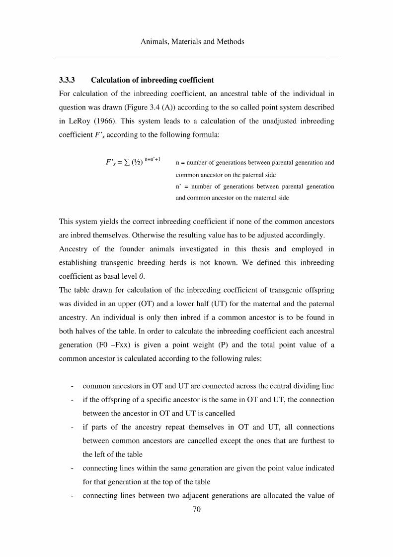
Animals, Materials and Methods
70
3.3.3 Calculation of inbreeding coefficient
For calculation of the inbreeding coefficient, an ancestral table of the individual in
question was drawn (Figure 3.4 (A)) according to the so called point system described
in LeRoy (1966). This system leads to a calculation of the unadjusted inbreeding
coefficient F’x according to the following formula:
F’x = ∑ (½) n+n’+1 n = number of generations between parental generation and
common ancestor on the paternal side
n’ = number of generations between parental generation
and common ancestor on the maternal side
This system yields the correct inbreeding coefficient if none of the common ancestors
are inbred themselves. Otherwise the resulting value has to be adjusted accordingly.
Ancestry of the founder animals investigated in this thesis and employed in
establishing transgenic breeding herds is not known. We defined this inbreeding
coefficient as basal level 0.
The table drawn for calculation of the inbreeding coefficient of transgenic offspring
was divided in an upper (OT) and a lower half (UT) for the maternal and the paternal
ancestry. An individual is only then inbred if a common ancestor is to be found in
both halves of the table. In order to calculate the inbreeding coefficient each ancestral
generation (F0 –Fxx) is given a point weight (P) and the total point value of a
common ancestor is calculated according to the following rules:
- common ancestors in OT and UT are connected across the central dividing line
- if the offspring of a specific ancestor is the same in OT and UT, the connection
between the ancestor in OT and UT is cancelled
- if parts of the ancestry repeat themselves in OT and UT, all connections
between common ancestors are cancelled except the ones that are furthest to
the left of the table
- connecting lines within the same generation are given the point value indicated
for that generation at the top of the table
- connecting lines between two adjacent generations are allocated the value of
Animals, Materials and Methods
71
the generation on the right times two
- if a connecting line skips a generation the value of the generation to the right is
timed by four (or six if two generations are skipped)
- all point values are added up and the inbreeding coefficient is taken by
inserting the total calculated point value into Figure 3.4 (B)
Figure 3.4: Example of tables for calculation of inbreeding coefficient of individual
‘x’. (A) Ancestral table for individual ‘x’; connecting lines of common ancestors on maternal and
paternal side have been drawn; (B) Table for reading of unadjusted inbreeding coefficient F’x
Tables adapted from LeRoy (1966)
3.3.4 Design of breeding schedules
Breeding schedules were designed using Macromedia Freehand MX software while
taking into account the calculated inbreeding coefficients of potential offspring and
segregation patterns of transgenes according to Mendelian rules of inheritance.

72
73
4 RESULTS
4.1 Identification of suitable breeding herds
In order to satisfy demand for a regular supply of donor animals for
xenotransplantation experiments, breeding herds of pigs displaying
xenotransplantation-relevant transgene expression were to be established. Planned
experiments included donor animals with GalKO/CD46/HLA-E expression for blood
perfusion studies utilising the fore limbs, hearts and kidneys of these animals and
GalKO/CD46/hTM transgenic pigs for pig-to-baboon heart transplantation and pig-to-
cynomolgus monkey kidney transplantation. Up until now donor animals for these
studies had been derived from recloning experiments of the original founders with
proven transgene expression. This strategy, however, had been too inefficient and
cost-intensive to ensure regular supply of donor animals in sufficient quantities.
Creating a feasible breeding strategy that satisfies the demand for transgenic offspring
provided a suitable alternative. All currently performed experiments are covered by
the DFG Transregio Research Unit “Xenotransplantation” (FOR 535) which provided
the basis for calculation of required numbers of donor animals per year. An outline of
the number of experiments with each of the different transgene combinations is given
in Table 4.1. This compilation served as a ground for estimating the size of the
breeding herds according to aspired numbers of litters per year.
In these experiments the eventual goal was to utilise donor animals transgenic for
GalKO/CD46 and either HLA-E or hTM. However, double instead of triple transgenic
animals were to be used during a transition period where breeding had not produced
the full set of transgenics yet.
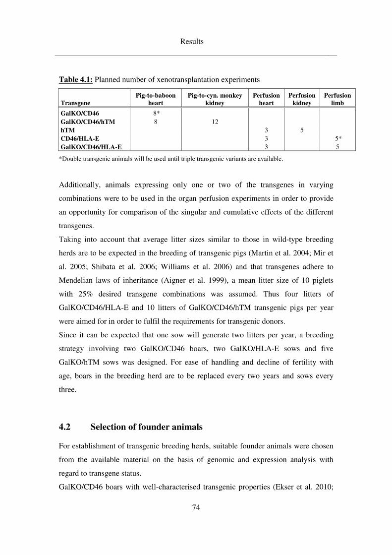
Results
74
Table 4.1: Planned number of xenotransplantation experiments
Transgene
Pig-to-baboon
heart
Pig-to-cyn. monkey
kidney
Perfusion
heart
Perfusion
kidney
Perfusion
limb
GalKO/CD46 8* GalKO/CD46/hTM 8 12 hTM 3 5 CD46/HLA-E 3 5* GalKO/CD46/HLA-E 3 5
*Double transgenic animals will be used until triple transgenic variants are available.
Additionally, animals expressing only one or two of the transgenes in varying
combinations were to be used in the organ perfusion experiments in order to provide
an opportunity for comparison of the singular and cumulative effects of the different
transgenes.
Taking into account that average litter sizes similar to those in wild-type breeding
herds are to be expected in the breeding of transgenic pigs (Martin et al. 2004; Mir et
al. 2005; Shibata et al. 2006; Williams et al. 2006) and that transgenes adhere to
Mendelian laws of inheritance (Aigner et al. 1999), a mean litter size of 10 piglets
with 25% desired transgene combinations was assumed. Thus four litters of
GalKO/CD46/HLA-E and 10 litters of GalKO/CD46/hTM transgenic pigs per year
were aimed for in order to fulfil the requirements for transgenic donors.
Since it can be expected that one sow will generate two litters per year, a breeding
strategy involving two GalKO/CD46 boars, two GalKO/HLA-E sows and five
GalKO/hTM sows was designed. For ease of handling and decline of fertility with
age, boars in the breeding herd are to be replaced every two years and sows every
three.
4.2 Selection of founder animals
For establishment of transgenic breeding herds, suitable founder animals were chosen
from the available material on the basis of genomic and expression analysis with
regard to transgene status.
GalKO/CD46 boars with well-characterised transgenic properties (Ekser et al. 2010;
Results
75
Lin et al. 2010) that had been obtained from Revivicor Inc. were re-established at the
MVG, Badersfeld. From these efforts, two GalKO/CD46 transgenic boars were gained
in June (#9872) and August (#9896) 2009 which were raised until sexual maturity and
are now being utilised in breeding.
HLA-E transgenic pigs had been established at the MVG, Badersfeld and were
characterised for their transgene expression as reported by Weiss et al (2009).
Breeding of these initial founders generated three HLA-E transgenic sows in June
2008 (#9713) and June 2009 (#9864, #9869). These three animals are now being
employed in establishing a GalKO/CD46/HLA-E breeding herd.
4.3 Expression and functional analysis of novel transgenic lines
Additionally, potential founder pigs for new transgenic lines from litters derived from
nuclear and embryo transfer have been characterised for their transgenic properties on
the basis of genomic and expression analysis. The conclusions drawn from these
analyses provided the foundation for the choice of founder animals which were to be
re-cloned and utilised in the breeding of pigs carrying the novel transgenes.
4.3.1 hTM
hTM transgenic founder animals were derived from two different strategies. In one
case the hTM-neo vector was used to establish single transgenic pigs. Three litters
generated by NT and ET and born in August and December 2008 provided a total of
eight piglets. Genotyping PCR on genomic DNA isolated from ear punches of three
days old piglets determined seven of them as hTM transgenic (Figure 4.1 (A)).
Immunohistochemical staining of tissue sections demonstrated strong transgene
expression in all examined organs of animal #9781, while the litter mates #9780 and
#9782 did not show consistent expression across all organs (Table 4.2).
In order to receive information about the transgene copy number and the number of
integration sites in the genome, Southern blotting on isolated genomic DNA was
conducted. Taking into account the restriction sites within the transgene vector and the
Results
76
feasible array of transgene copies in the genome this revealed the integration of only a
few copies of the transgene at one single site in founder #9781, while it can be
assumed that the examined litter mates display a more complicated transgene
integration pattern (Figure 4.2). Boar #9781 was therefore chosen for re-cloning.
Table 4.3 outlines the NTs and ETs performed in re-establishing boar #9781, which
led to the birth of boar #9943 in December 2009. This animal has been utilised as a
founder in the establishment of the GalKO/CD46/hTM breeding herd.
At the same time, animal #9744 has undergone re-cloning attempts (Table 4.3). This
boar had also been genotyped positive for the hTM-neo vector (Figure 4.1 (A)) and
southern blotting indicated a single integration site of one copy of the transgene
(Figure 4.2). Transgene expression capacity was verified by immunohistochemical
staining of tissue sections, where this particular animal showed strong hTM
expression in all examined organs (Table 4.2).
Additionally, the hTM-bla vector was established on the GalKO/CD46 background
obtained from Revivicor Inc. From this effort, four potential founder pigs were gained
in January 2010 of which three were analysed positive for the hTM-bla transgene in a
genotyping PCR (Figure 4.1 (B)). Immunohistochemical staining of tissue sections
demonstrated strong hTM expression across all examined organs in animals #9947
and #9948. Animal #9949 showed equivalent expression in ear and heart tissue,
however, staining of other organs was markedly weaker than in the two litter mates
(Table 4.2). Boar #9948 was therefore chosen as suitable founder animal for
GalKO/CD46/hTM breeding. Re-cloning efforts for this animal are summarised in
Table 4.4. Boar #1103, a clone of #9948 born in December 2010, is currently being
raised for breeding.
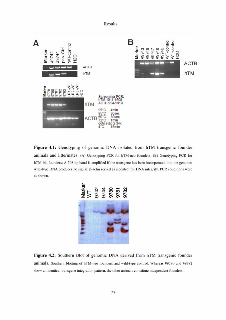
Results
77
Figure 4.1: Genotyping of genomic DNA isolated from hTM transgenic founder
animals and littermates. (A) Genotyping PCR for hTM-neo founders; (B) Genotyping PCR for
hTM-bla founders; A 508 bp band is amplified if the transgene has been incorporated into the genome;
wild-type DNA produces no signal; β-actin served as a control for DNA integrity. PCR conditions were
as shown.
Figure 4.2: Southern Blot of genomic DNA derived from hTM transgenic founder
animals. Southern blotting of hTM-neo founders and wild-type control. Whereas #9780 and #9782
show an identical transgene integration pattern, the other animals constitute independent founders.
Mark
er
WT
9742
9744
9780
9781
9782
Mark
er
WT
9742
9744
9780
9781
9782
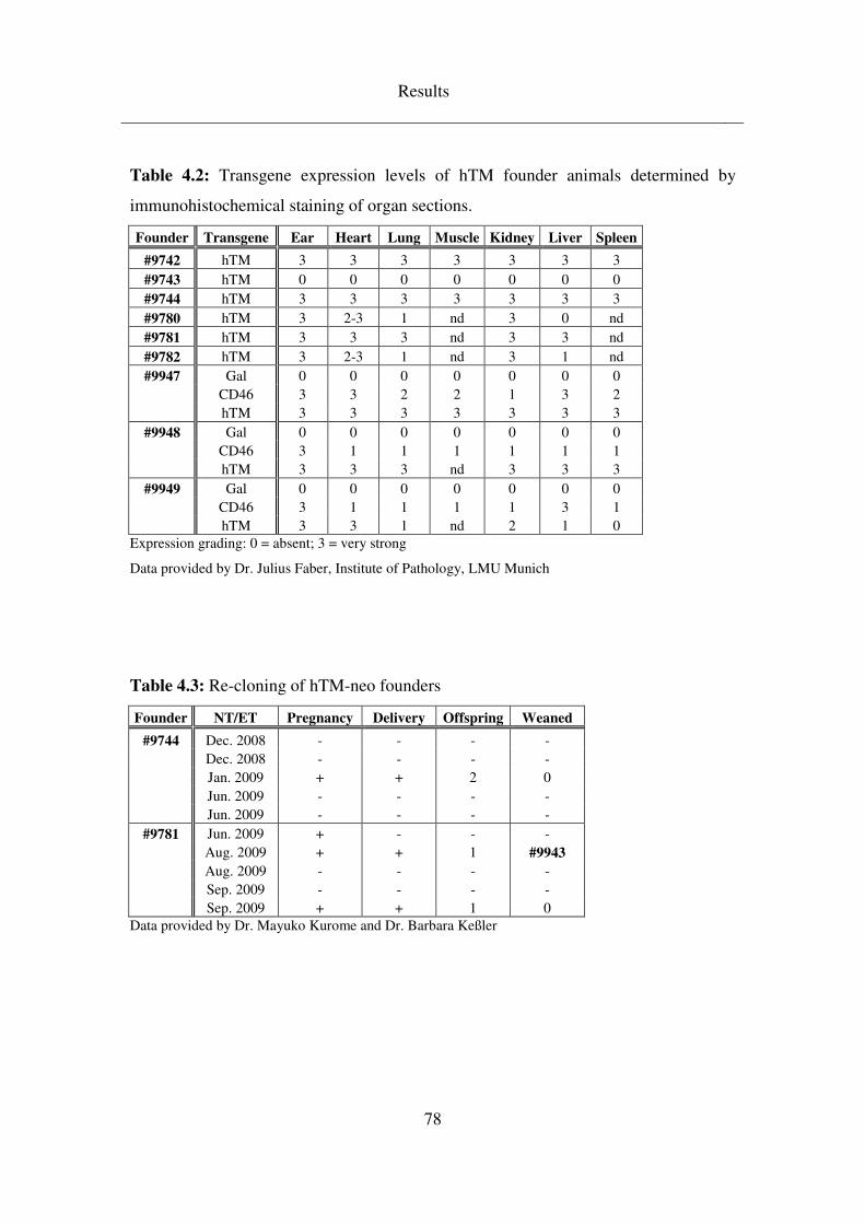
Results
78
Table 4.2: Transgene expression levels of hTM founder animals determined by
immunohistochemical staining of organ sections.
Founder Transgene Ear Heart Lung Muscle Kidney Liver Spleen
#9742 hTM 3 3 3 3 3 3 3
#9743 hTM 0 0 0 0 0 0 0
#9744 hTM 3 3 3 3 3 3 3
#9780 hTM 3 2-3 1 nd 3 0 nd
#9781 hTM 3 3 3 nd 3 3 nd
#9782 hTM 3 2-3 1 nd 3 1 nd
#9947 Gal 0 0 0 0 0 0 0 CD46 3 3 2 2 1 3 2 hTM 3 3 3 3 3 3 3
#9948 Gal 0 0 0 0 0 0 0 CD46 3 1 1 1 1 1 1 hTM 3 3 3 nd 3 3 3
#9949 Gal 0 0 0 0 0 0 0 CD46 3 1 1 1 1 3 1 hTM 3 3 1 nd 2 1 0
Expression grading: 0 = absent; 3 = very strong
Data provided by Dr. Julius Faber, Institute of Pathology, LMU Munich
Table 4.3: Re-cloning of hTM-neo founders
Founder NT/ET Pregnancy Delivery Offspring Weaned
#9744 Dec. 2008 - - - - Dec. 2008 - - - - Jan. 2009 + + 2 0 Jun. 2009 - - - - Jun. 2009 - - - -
#9781 Jun. 2009 + - - - Aug. 2009 + + 1 #9943
Aug. 2009 - - - - Sep. 2009 - - - - Sep. 2009 + + 1 0
Data provided by Dr. Mayuko Kurome and Dr. Barbara Keßler
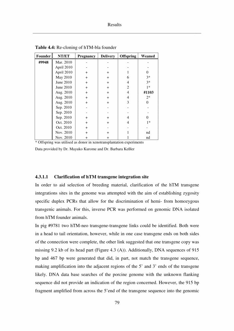
Results
79
Table 4.4: Re-cloning of hTM-bla founder
Founder NT/ET Pregnancy Delivery Offspring Weaned
#9948 Mar. 2010 - - - - April 2010 - - - - April 2010 + + 1 0 May 2010 + + 6 3* June 2010 + + 4 3* June 2010 + + 2 1* Aug. 2010 + + 4 #1103
Aug. 2010 + + 4 2* Aug. 2010 + + 3 0 Sep. 2010 - - - - Sep. 2010 - - - - Sep. 2010 + + 4 0 Oct. 2010 + + 4 1* Oct. 2010 + - - - Nov. 2010 + + 1 nd Nov. 2010 + + 1 nd
* Offspring was utilised as donor in xenotransplantation experiments
Data provided by Dr. Mayuko Kurome and Dr. Barbara Keßler
4.3.1.1 Clarification of hTM transgene integration site
In order to aid selection of breeding material, clarification of the hTM transgene
integrations sites in the genome was attempted with the aim of establishing zygosity
specific duplex PCRs that allow for the discrimination of hemi- from homozygous
transgenic animals. For this, inverse PCR was performed on genomic DNA isolated
from hTM founder animals.
In pig #9781 two hTM-neo transgene-transgene links could be identified. Both were
in a head to tail orientation, however, while in one case transgene ends on both sides
of the connection were complete, the other link suggested that one transgene copy was
missing 9.2 kb of its head part (Figure 4.3 (A)). Additionally, DNA sequences of 915
bp and 467 bp were generated that did, in part, not match the transgene sequence,
making amplification into the adjacent regions of the 5’ and 3’ ends of the transgene
likely. DNA data base searches of the porcine genome with the unknown flanking
sequence did not provide an indication of the region concerned. However, the 915 bp
fragment amplified from across the 5’end of the transgene sequence into the genomic
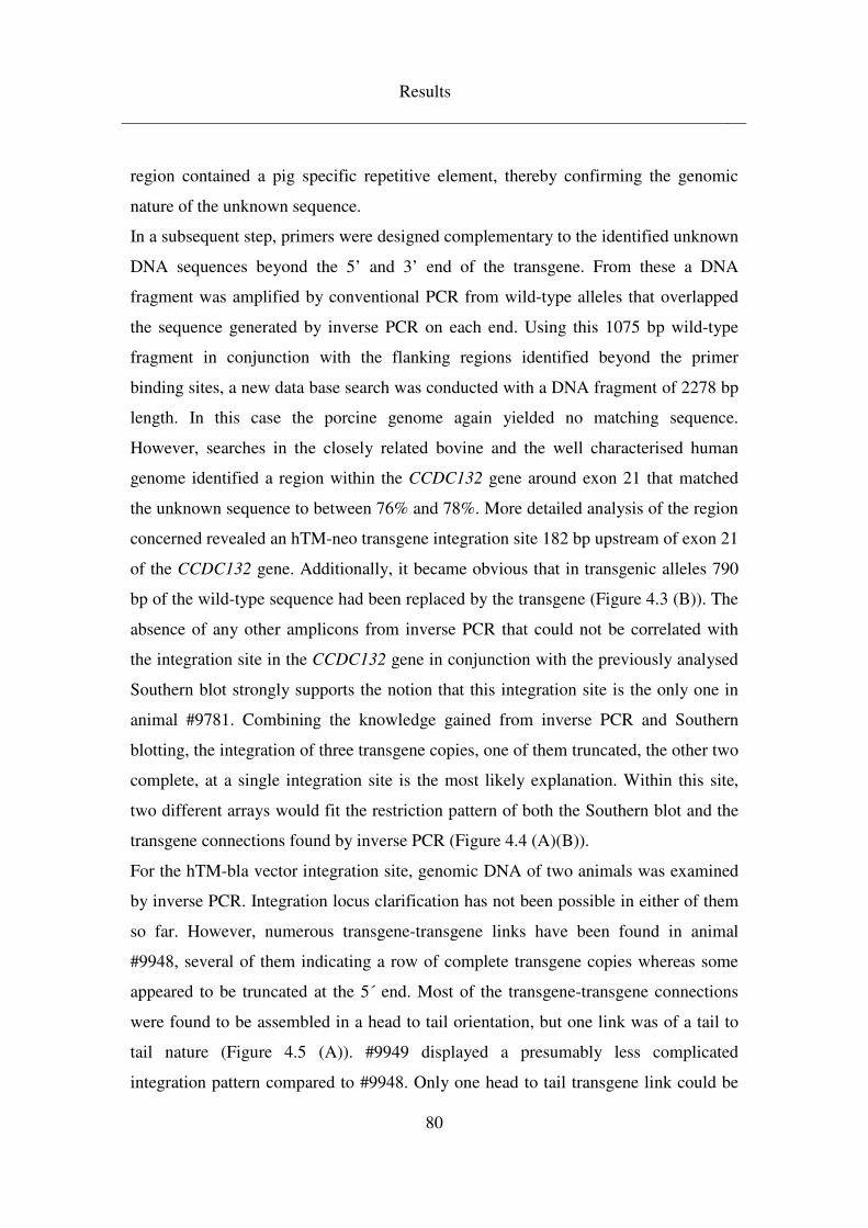
Results
80
region contained a pig specific repetitive element, thereby confirming the genomic
nature of the unknown sequence.
In a subsequent step, primers were designed complementary to the identified unknown
DNA sequences beyond the 5’ and 3’ end of the transgene. From these a DNA
fragment was amplified by conventional PCR from wild-type alleles that overlapped
the sequence generated by inverse PCR on each end. Using this 1075 bp wild-type
fragment in conjunction with the flanking regions identified beyond the primer
binding sites, a new data base search was conducted with a DNA fragment of 2278 bp
length. In this case the porcine genome again yielded no matching sequence.
However, searches in the closely related bovine and the well characterised human
genome identified a region within the CCDC132 gene around exon 21 that matched
the unknown sequence to between 76% and 78%. More detailed analysis of the region
concerned revealed an hTM-neo transgene integration site 182 bp upstream of exon 21
of the CCDC132 gene. Additionally, it became obvious that in transgenic alleles 790
bp of the wild-type sequence had been replaced by the transgene (Figure 4.3 (B)). The
absence of any other amplicons from inverse PCR that could not be correlated with
the integration site in the CCDC132 gene in conjunction with the previously analysed
Southern blot strongly supports the notion that this integration site is the only one in
animal #9781. Combining the knowledge gained from inverse PCR and Southern
blotting, the integration of three transgene copies, one of them truncated, the other two
complete, at a single integration site is the most likely explanation. Within this site,
two different arrays would fit the restriction pattern of both the Southern blot and the
transgene connections found by inverse PCR (Figure 4.4 (A)(B)).
For the hTM-bla vector integration site, genomic DNA of two animals was examined
by inverse PCR. Integration locus clarification has not been possible in either of them
so far. However, numerous transgene-transgene links have been found in animal
#9948, several of them indicating a row of complete transgene copies whereas some
appeared to be truncated at the 5´ end. Most of the transgene-transgene connections
were found to be assembled in a head to tail orientation, but one link was of a tail to
tail nature (Figure 4.5 (A)). #9949 displayed a presumably less complicated
integration pattern compared to #9948. Only one head to tail transgene link could be
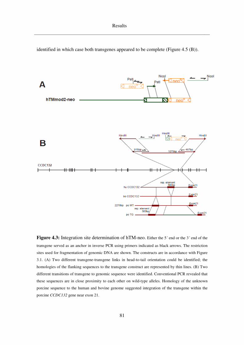
Results
81
identified in which case both transgenes appeared to be complete (Figure 4.5 (B)).
Figure 4.3: Integration site determination of hTM-neo. Either the 5´ end or the 3´ end of the
transgene served as an anchor in inverse PCR using primers indicated as black arrows. The restriction
sites used for fragmentation of genomic DNA are shown. The constructs are in accordance with Figure
3.1. (A) Two different transgene-transgene links in head-to-tail orientation could be identified; the
homologies of the flanking sequences to the transgene construct are represented by thin lines. (B) Two
different transitions of transgene to genomic sequence were identified. Conventional PCR revealed that
these sequences are in close proximity to each other on wild-type alleles. Homology of the unknown
porcine sequence to the human and bovine genome suggested integration of the transgene within the
porcine CCDC132 gene near exon 21.
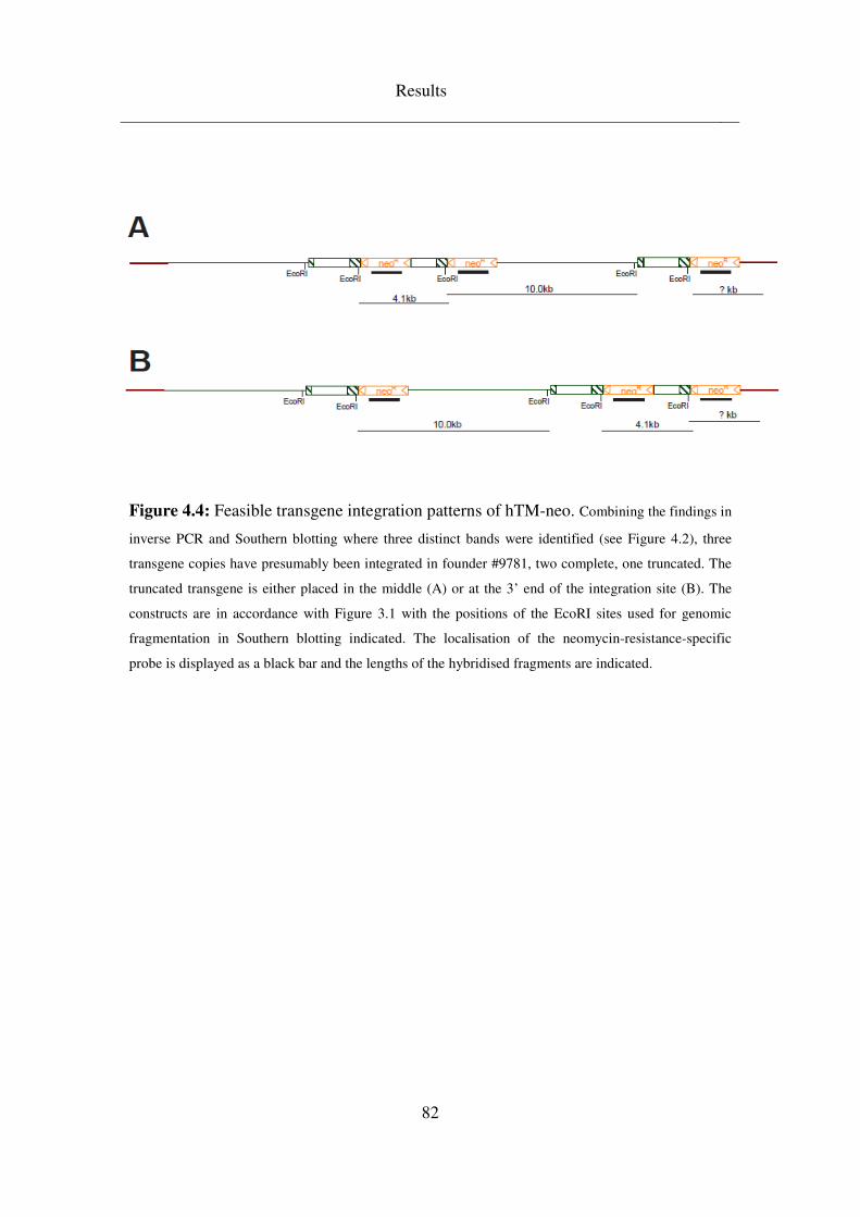
Results
82
Figure 4.4: Feasible transgene integration patterns of hTM-neo. Combining the findings in
inverse PCR and Southern blotting where three distinct bands were identified (see Figure 4.2), three
transgene copies have presumably been integrated in founder #9781, two complete, one truncated. The
truncated transgene is either placed in the middle (A) or at the 3’ end of the integration site (B). The
constructs are in accordance with Figure 3.1 with the positions of the EcoRI sites used for genomic
fragmentation in Southern blotting indicated. The localisation of the neomycin-resistance-specific
probe is displayed as a black bar and the lengths of the hybridised fragments are indicated.
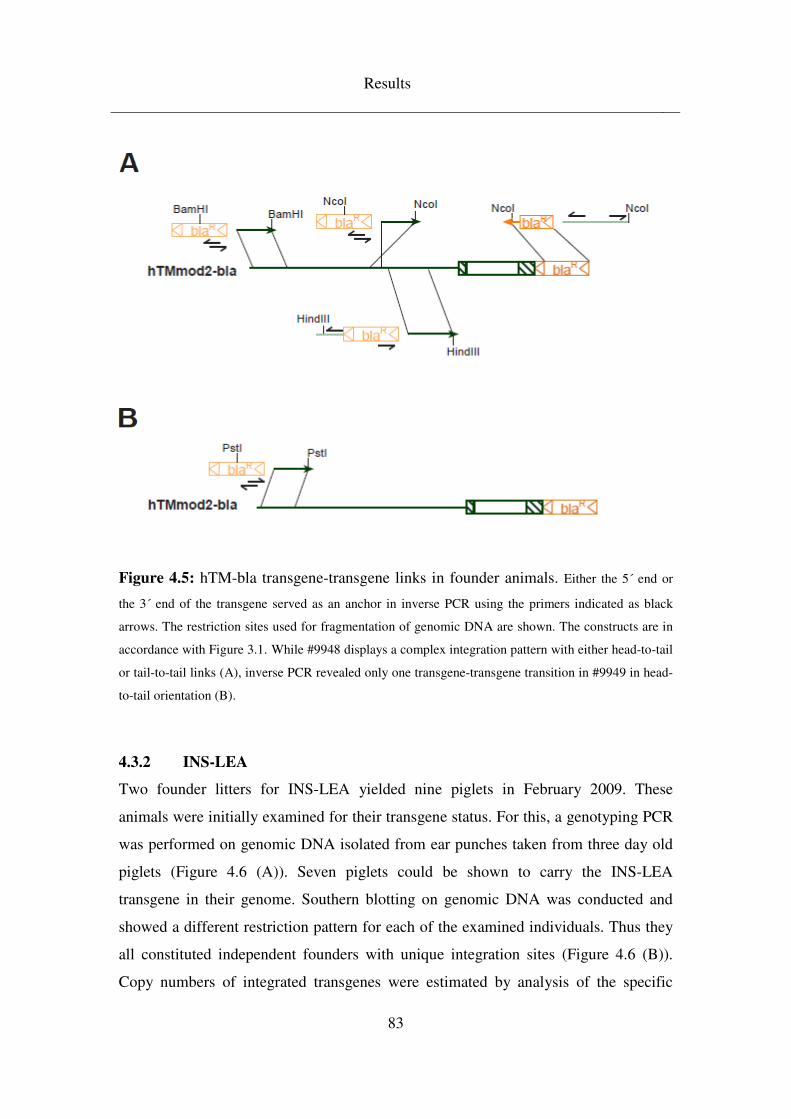
Results
83
Figure 4.5: hTM-bla transgene-transgene links in founder animals. Either the 5´ end or
the 3´ end of the transgene served as an anchor in inverse PCR using the primers indicated as black
arrows. The restriction sites used for fragmentation of genomic DNA are shown. The constructs are in
accordance with Figure 3.1. While #9948 displays a complex integration pattern with either head-to-tail
or tail-to-tail links (A), inverse PCR revealed only one transgene-transgene transition in #9949 in head-
to-tail orientation (B).
4.3.2 INS-LEA
Two founder litters for INS-LEA yielded nine piglets in February 2009. These
animals were initially examined for their transgene status. For this, a genotyping PCR
was performed on genomic DNA isolated from ear punches taken from three day old
piglets (Figure 4.6 (A)). Seven piglets could be shown to carry the INS-LEA
transgene in their genome. Southern blotting on genomic DNA was conducted and
showed a different restriction pattern for each of the examined individuals. Thus they
all constituted independent founders with unique integration sites (Figure 4.6 (B)).
Copy numbers of integrated transgenes were estimated by analysis of the specific
Results
84
restriction pattern and comparison of signal intensity to wild-type DNA mixed with
stoichiometric equivalents of the transgene vector construct. From this, #9816 and
#9818 were found to contain only one copy of the transgene, while the others
contained more.
Animals #9813, #9814, #9816 and #9818 survived to an age of three months and were
then sacrificed for organ spectrum expression analysis. Figure 4.7 shows
immunohistochemical stainings of pancreas and kidney sections of these animals.
Because the INS-LEA construct contains a porcine insulin promoter which controls
the transgene, expression should be confined to pancreas islets, where this particular
promoter is active. Founders #9813 and #9814 showed strong transgene expression
islet specifically (Figure 4.7 (A), (B)), while pancreas sections of #9816 and #9818
stained only weakly for the transgene product (Figure 4.7 (D)). As expected,
expression was absent from all kidney sections (Figure 4.7 (C)). In integration site
analysis, Southern blot patterns led to the assumption that both well expressing pigs
#9813 and #9814 contained four copies of the transgene at only one integration site,
making them equivalent in their suitability as founders for an INS-LEA transgenic
line. Both animals were therefore chosen to be re-cloned. Table 4.5 provides an
overview of NTs and ETs conducted in re-cloning efforts for these two founder pigs.
So far, boars #1044 and #1050, both re-cloned #9814, have been born in September
2010 and will be raised to be utilised for breeding.
In-vivo studies determined the functional effect of pancreas islet specific LEA29Y
expression. For this, islet like clusters were isolated from pancreata of neonatal INS-
LEA transgenic piglets, cultured for several days and subsequently transplanted under
the kidney capsule of NOD-SCID mice that suffered from a streptozotocin induced
diabetes mellitus. After regaining normogylcaemia, hPBMCs were administered to
these mice in order to restore their immune system in a humanised way. While mice
transplanted with wild-type islets showed steadily increasing blood glucose levels,
mice with LEA29Y transgenic islets stayed within normal ranges (Figure 4.8).
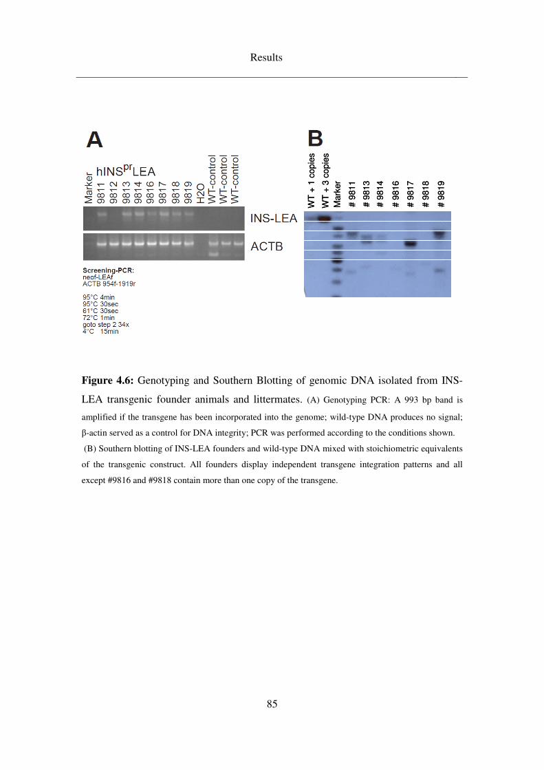
Results
85
Figure 4.6: Genotyping and Southern Blotting of genomic DNA isolated from INS-
LEA transgenic founder animals and littermates. (A) Genotyping PCR: A 993 bp band is
amplified if the transgene has been incorporated into the genome; wild-type DNA produces no signal;
β-actin served as a control for DNA integrity; PCR was performed according to the conditions shown.
(B) Southern blotting of INS-LEA founders and wild-type DNA mixed with stoichiometric equivalents
of the transgenic construct. All founders display independent transgene integration patterns and all
except #9816 and #9818 contain more than one copy of the transgene.
WT
+ 1
co
pie
s
WT
+ 3
co
pie
s
Mark
er
# 9
811
# 9
813
# 9
814
# 9
816
# 9
817
# 9
818
# 9
819
WT
+ 1
co
pie
s
WT
+ 3
co
pie
s
Mark
er
# 9
811
# 9
813
# 9
814
# 9
816
# 9
817
# 9
818
# 9
819
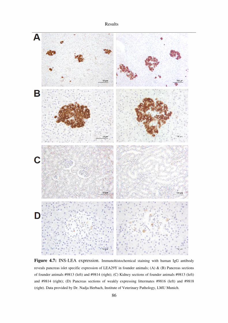
Results
86
Figure 4.7: INS-LEA expression. Immunohistochemical staining with human IgG antibody
reveals pancreas islet specific expression of LEA29Y in founder animals; (A) & (B) Pancreas sections
of founder animals #9813 (left) and #9814 (right); (C) Kidney sections of founder animals #9813 (left)
and #9814 (right); (D) Pancreas sections of weakly expressing littermates #9816 (left) and #9818
(right). Data provided by Dr. Nadja Herbach, Institute of Veterinary Pathology, LMU Munich.
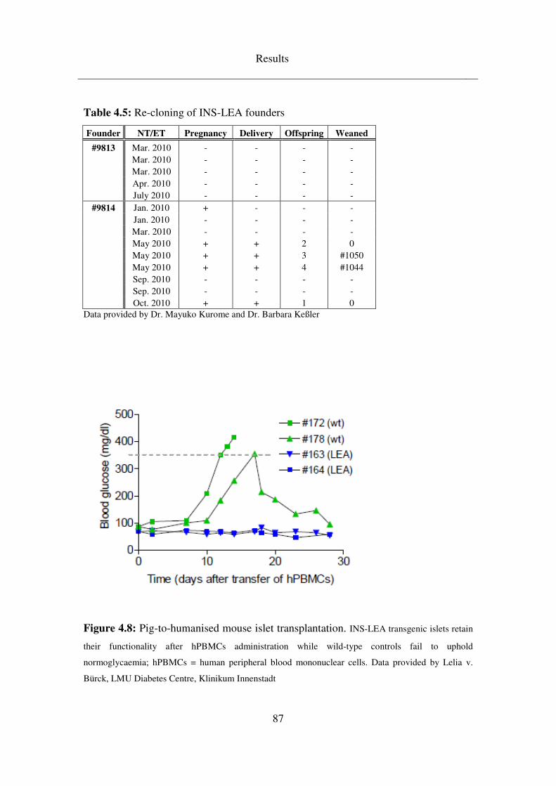
Results
87
Table 4.5: Re-cloning of INS-LEA founders
Founder NT/ET Pregnancy Delivery Offspring Weaned
#9813 Mar. 2010 - - - - Mar. 2010 - - - - Mar. 2010 - - - - Apr. 2010 - - - - July 2010 - - - -
#9814 Jan. 2010 + - - - Jan. 2010 - - - - Mar. 2010 - - - - May 2010 + + 2 0 May 2010 + + 3 #1050 May 2010 + + 4 #1044 Sep. 2010 - - - - Sep. 2010 - - - - Oct. 2010 + + 1 0
Data provided by Dr. Mayuko Kurome and Dr. Barbara Keßler
Figure 4.8: Pig-to-humanised mouse islet transplantation. INS-LEA transgenic islets retain
their functionality after hPBMCs administration while wild-type controls fail to uphold
normoglycaemia; hPBMCs = human peripheral blood mononuclear cells. Data provided by Lelia v.
Bürck, LMU Diabetes Centre, Klinikum Innenstadt
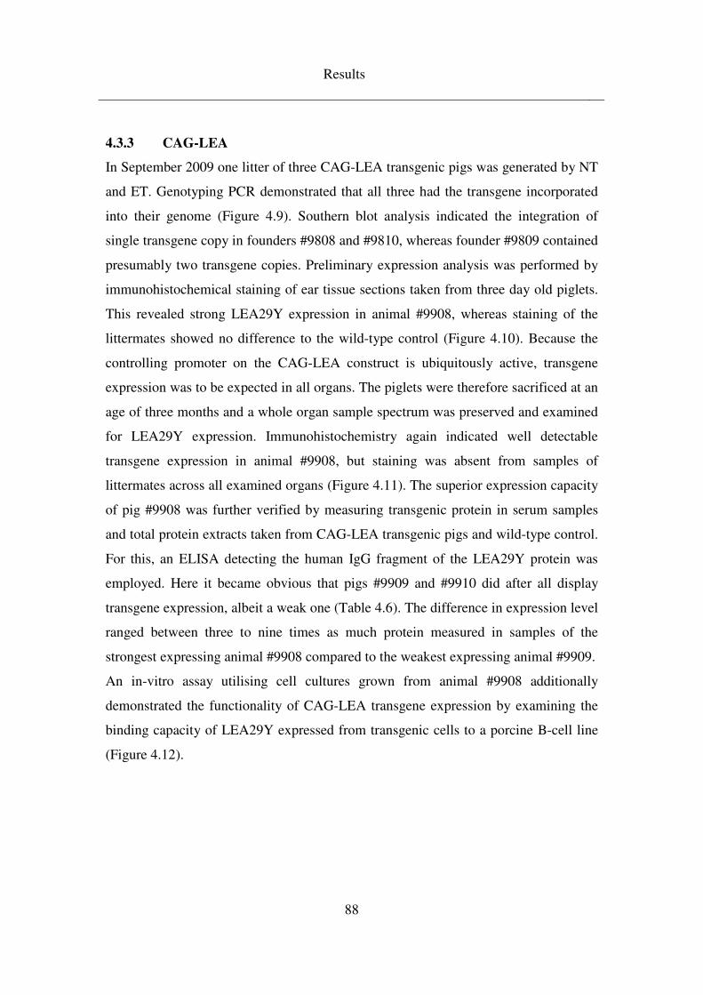
Results
88
4.3.3 CAG-LEA
In September 2009 one litter of three CAG-LEA transgenic pigs was generated by NT
and ET. Genotyping PCR demonstrated that all three had the transgene incorporated
into their genome (Figure 4.9). Southern blot analysis indicated the integration of
single transgene copy in founders #9808 and #9810, whereas founder #9809 contained
presumably two transgene copies. Preliminary expression analysis was performed by
immunohistochemical staining of ear tissue sections taken from three day old piglets.
This revealed strong LEA29Y expression in animal #9908, whereas staining of the
littermates showed no difference to the wild-type control (Figure 4.10). Because the
controlling promoter on the CAG-LEA construct is ubiquitously active, transgene
expression was to be expected in all organs. The piglets were therefore sacrificed at an
age of three months and a whole organ sample spectrum was preserved and examined
for LEA29Y expression. Immunohistochemistry again indicated well detectable
transgene expression in animal #9908, but staining was absent from samples of
littermates across all examined organs (Figure 4.11). The superior expression capacity
of pig #9908 was further verified by measuring transgenic protein in serum samples
and total protein extracts taken from CAG-LEA transgenic pigs and wild-type control.
For this, an ELISA detecting the human IgG fragment of the LEA29Y protein was
employed. Here it became obvious that pigs #9909 and #9910 did after all display
transgene expression, albeit a weak one (Table 4.6). The difference in expression level
ranged between three to nine times as much protein measured in samples of the
strongest expressing animal #9908 compared to the weakest expressing animal #9909.
An in-vitro assay utilising cell cultures grown from animal #9908 additionally
demonstrated the functionality of CAG-LEA transgene expression by examining the
binding capacity of LEA29Y expressed from transgenic cells to a porcine B-cell line
(Figure 4.12).
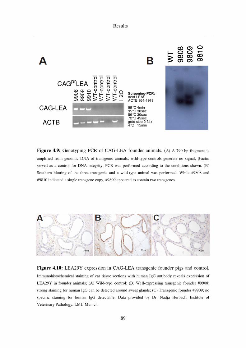
Results
89
Figure 4.9: Genotyping PCR of CAG-LEA founder animals. (A) A 790 bp fragment is
amplified from genomic DNA of transgenic animals; wild-type controls generate no signal; β-actin
served as a control for DNA integrity. PCR was performed according to the conditions shown. (B)
Southern blotting of the three transgenic and a wild-type animal was performed. While #9808 and
#9810 indicated a single transgene copy, #9809 appeared to contain two transgenes.
Figure 4.10: LEA29Y expression in CAG-LEA transgenic founder pigs and control.
Immunohistochemical staining of ear tissue sections with human IgG antibody reveals expression of
LEA29Y in founder animals; (A) Wild-type control; (B) Well-expressing transgenic founder #9908;
strong staining for human IgG can be detected around sweat glands; (C) Transgenic founder #9909; no
specific staining for human IgG detectable. Data provided by Dr. Nadja Herbach, Institute of
Veterinary Pathology, LMU Munich
50µm50µm50µm50µm50µm50µm
CAGprLEA
CAG-LEA
ACTB
Screening-PCR:neof-LEAfACTB 954-1919
95°C 4min95°C 30sec56°C 30sec72°C 45secgoto step 2 34x4°C 15min
WT
9808
9809
9810
WT
9808
9809
9810
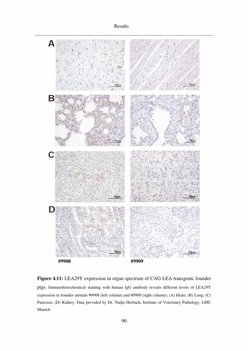
Results
90
#9908 #9909
Figure 4.11: LEA29Y expression in organ spectrum of CAG-LEA transgenic founder
pigs. Immunohistochemical staining with human IgG antibody reveals different levels of LEA29Y
expression in founder animals #9908 (left column) and #9909 (right column); (A) Heart; (B) Lung; (C)
Pancreas; (D) Kidney. Data provided by Dr. Nadja Herbach, Institute of Veterinary Pathology, LMU
Munich.
50µm50µm 50µm50µm
50µm50µm 50µm50µm
50µm50µm50µm 50µm50µm50µm
50µm50µm50µm 50µm50µm50µm
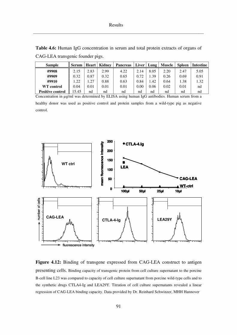
Results
91
Table 4.6: Human IgG concentration in serum and total protein extracts of organs of
CAG-LEA transgenic founder pigs.
Sample Serum Heart Kidney Pancreas Liver Lung Muscle Spleen Intestine
#9908 2.15 2.83 2.99 4.22 2.14 8.05 2.20 2.47 5.05 #9909 0.32 0.87 0.32 0.65 0.72 1.39 0.26 0.69 0.91 #9910 1.22 1.27 0.88 0.63 0.84 1.42 0.64 1.38 1.32
WT control 0.04 0.01 0.01 0.01 0.00 0.06 0.02 0.01 nd Positive control 15.45 nd nd nd nd nd nd nd nd
Concentration in µg/ml was determined by ELISA using human IgG antibodies. Human serum from a
healthy donor was used as positive control and protein samples from a wild-type pig as negative
control.
CTLA-4-IgCAG-LEA
LEA29Y
WT ctrl
0
50
100
150
200
250
100µl 50µl 25µl 10µl
CTLA-4.Ig
LEA
WT-ctrl
CAG-LEA
mean
flu
ore
sce
nce
inte
nsit
y
fluorescence intensity
num
ber
of
ce
lls
CTLA-4-IgCAG-LEA
LEA29Y
WT ctrl
0
50
100
150
200
250
100µl 50µl 25µl 10µl
CTLA-4.Ig
LEA
WT-ctrl
CAG-LEA
mean
flu
ore
sce
nce
inte
nsit
y
CTLA-4-IgCAG-LEA
LEA29Y
WT ctrl
0
50
100
150
200
250
100µl 50µl 25µl 10µl
CTLA-4.Ig
LEA
WT-ctrl
CAG-LEA
mean
flu
ore
sce
nce
inte
nsit
y
0
50
100
150
200
250
100µl 50µl 25µl 10µl
CTLA-4.Ig
LEA
WT-ctrl
CAG-LEA
mean
flu
ore
sce
nce
inte
nsit
y
fluorescence intensity
num
ber
of
ce
lls
Figure 4.12: Binding of transgene expressed from CAG-LEA construct to antigen
presenting cells. Binding capacity of transgenic protein from cell culture supernatant to the porcine
B-cell line L23 was compared to capacity of cell culture supernatant from porcine wild-type cells and to
the synthetic drugs CTLA4-Ig and LEA29Y. Titration of cell culture supernatants revealed a linear
regression of CAG-LEA binding capacity. Data provided by Dr. Reinhard Schwinzer, MHH Hannover
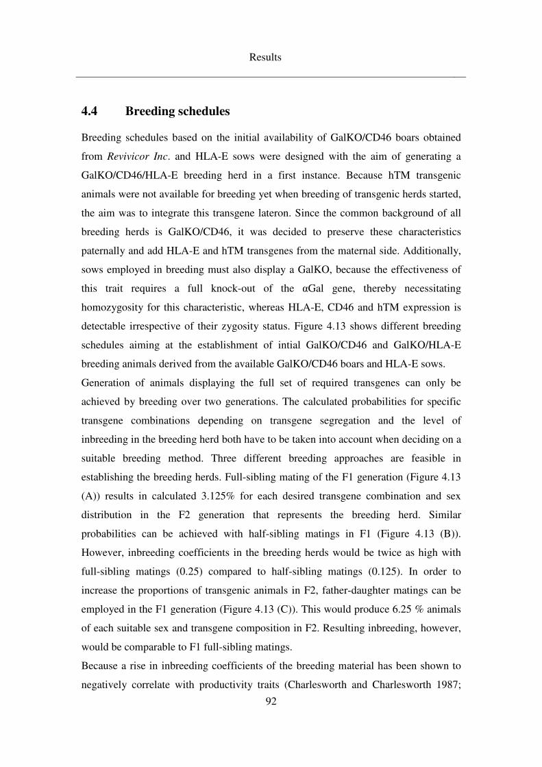
Results
92
4.4 Breeding schedules
Breeding schedules based on the initial availability of GalKO/CD46 boars obtained
from Revivicor Inc. and HLA-E sows were designed with the aim of generating a
GalKO/CD46/HLA-E breeding herd in a first instance. Because hTM transgenic
animals were not available for breeding yet when breeding of transgenic herds started,
the aim was to integrate this transgene lateron. Since the common background of all
breeding herds is GalKO/CD46, it was decided to preserve these characteristics
paternally and add HLA-E and hTM transgenes from the maternal side. Additionally,
sows employed in breeding must also display a GalKO, because the effectiveness of
this trait requires a full knock-out of the αGal gene, thereby necessitating
homozygosity for this characteristic, whereas HLA-E, CD46 and hTM expression is
detectable irrespective of their zygosity status. Figure 4.13 shows different breeding
schedules aiming at the establishment of intial GalKO/CD46 and GalKO/HLA-E
breeding animals derived from the available GalKO/CD46 boars and HLA-E sows.
Generation of animals displaying the full set of required transgenes can only be
achieved by breeding over two generations. The calculated probabilities for specific
transgene combinations depending on transgene segregation and the level of
inbreeding in the breeding herd both have to be taken into account when deciding on a
suitable breeding method. Three different breeding approaches are feasible in
establishing the breeding herds. Full-sibling mating of the F1 generation (Figure 4.13
(A)) results in calculated 3.125% for each desired transgene combination and sex
distribution in the F2 generation that represents the breeding herd. Similar
probabilities can be achieved with half-sibling matings in F1 (Figure 4.13 (B)).
However, inbreeding coefficients in the breeding herds would be twice as high with
full-sibling matings (0.25) compared to half-sibling matings (0.125). In order to
increase the proportions of transgenic animals in F2, father-daughter matings can be
employed in the F1 generation (Figure 4.13 (C)). This would produce 6.25 % animals
of each suitable sex and transgene composition in F2. Resulting inbreeding, however,
would be comparable to F1 full-sibling matings.
Because a rise in inbreeding coefficients of the breeding material has been shown to
negatively correlate with productivity traits (Charlesworth and Charlesworth 1987;
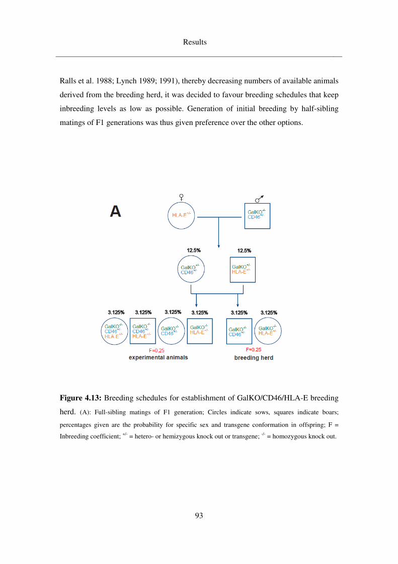
Results
93
Ralls et al. 1988; Lynch 1989; 1991), thereby decreasing numbers of available animals
derived from the breeding herd, it was decided to favour breeding schedules that keep
inbreeding levels as low as possible. Generation of initial breeding by half-sibling
matings of F1 generations was thus given preference over the other options.
Figure 4.13: Breeding schedules for establishment of GalKO/CD46/HLA-E breeding
herd. (A): Full-sibling matings of F1 generation; Circles indicate sows, squares indicate boars;
percentages given are the probability for specific sex and transgene conformation in offspring; F =
Inbreeding coefficient; +/- = hetero- or hemizygous knock out or transgene; -/- = homozygous knock out.
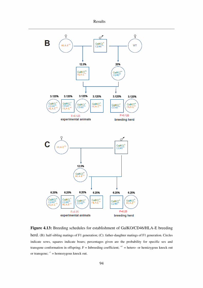
Results
94
Figure 4.13: Breeding schedules for establishment of GalKO/CD46/HLA-E breeding
herd. (B): half-sibling matings of F1 generation; (C): father-daughter matings of F1 generation. Circles
indicate sows, squares indicate boars; percentages given are the probability for specific sex and
transgene conformation in offspring; F = Inbreeding coefficient; +/- = hetero- or hemizygous knock out
or transgene; -/- = homozygous knock out.
Results
95
Integration of the hTM transgene into the breeding herd can be achieved by two
different strategies. In one case, hTM is derived from boars generated by transfection
of the hTM vector construct into GalKO/CD46 cells from Revivicor Inc. In this case
the genetic background of the resulting GalKO/CD46/hTM boars is identical to the
GalKO/CD46 boars employed in the establishment of the initial breeding herds.
Figure 4.14 (A) shows a breeding schedule built on this option. Inbreeding
coefficients in breeding material derived from mating a GalKO/CD46/hTM boar to an
F1 sow of the half-sibling breeding schedule in Figure 4.13 will result in an
inbreeding coefficient of the offspring of 0.25. Additionally, probabilities for a
GalKO/hTM sow that could be utilised within the breeding herd calculate to only
3.125%.
However, since the hTM vector construct had also been transfected into a wild-type
cell line, a boar transgenic only for hTM was also available for furthering the breeding
herd. In this schedule, shown in Figure 4.14 (B), the establishment of GalKO/hTM
sows takes one generation longer than in the other case. But probabilities for the
correct transgene combination are doubled and, even more importantly, the inbreeding
coefficient of the resulting individuals had been calculated at only 0.062. Therefore,
preference was given to introducing the hTM transgene into the breeding herd via the
more time-consuming route.
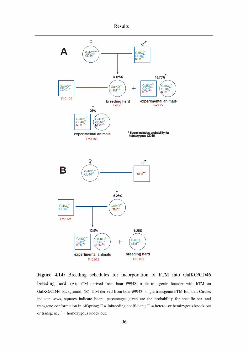
Results
96
Figure 4.14: Breeding schedules for incorporation of hTM into GalKO/CD46
breeding herd. (A): hTM derived from boar #9948, triple transgenic founder with hTM on
GalKO/CD46 background; (B) hTM derived from boar #9943, single transgenic hTM founder. Circles
indicate sows, squares indicate boars; percentages given are the probability for specific sex and
transgene conformation in offspring; F = Inbreeding coefficient; +/- = hetero- or hemizygous knock out
or transgene; -/- = homozygous knock out.
Results
97
In addition, simultaneous outbreeding of the breeding herd with wild-type animals is
supposed to keep inbreeding at levels as low as feasible. As can be seen in Figure
4.15, outbreeding of GalKO/CD46 boars over two or three generations with wild-type
sows prior to backcrossing offspring into breeding herd will lower inbreeding levels
within the breeding herd itself significantly to 0.062 or 0.031 for GalKO/CD46/HLA-
E combinations and 0.047 or 0.023 for GalKO/hTM, respectively if half-sibling
matings are chosen for the establishment of breeding herds and hTM is derived from
the single transgenic hTM boar.
Since GalKO/CD46 had been chosen as the common background for all transgene
combinations, outbreeding is being performed with respect to these transgenes,
building on the original founder boars available and introducing wild-type blood on
the maternal side of the breeding schedule.
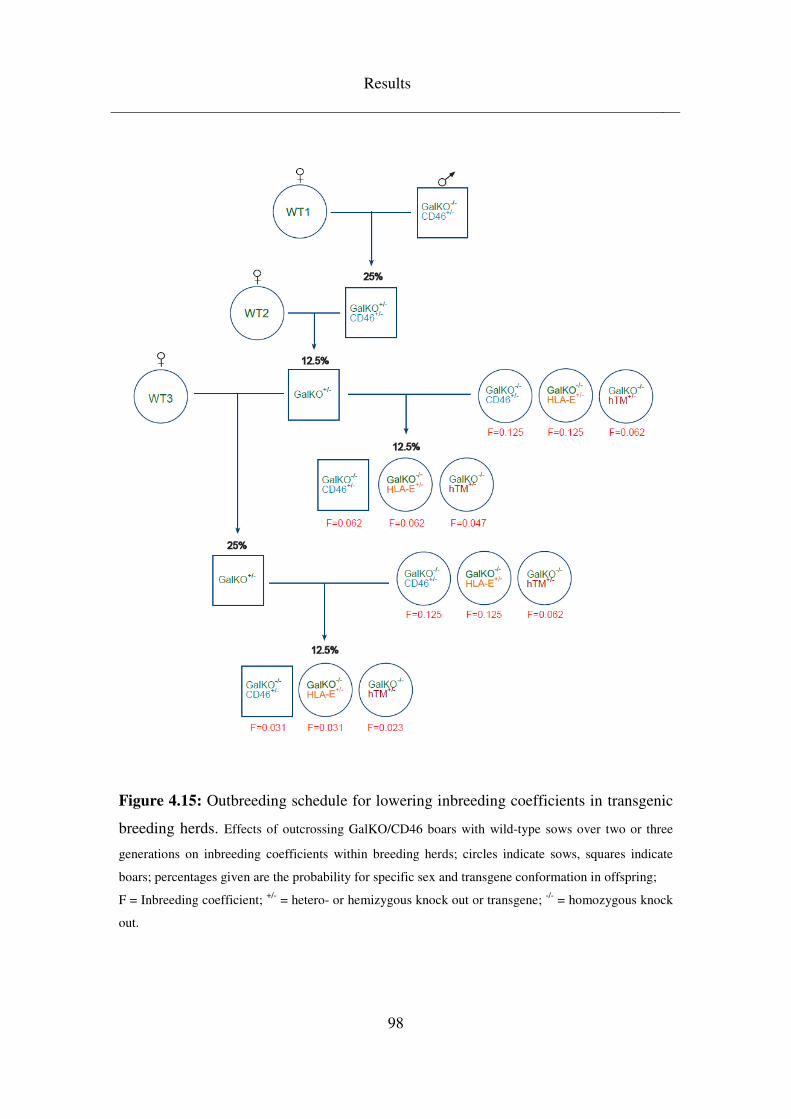
Results
98
Figure 4.15: Outbreeding schedule for lowering inbreeding coefficients in transgenic
breeding herds. Effects of outcrossing GalKO/CD46 boars with wild-type sows over two or three
generations on inbreeding coefficients within breeding herds; circles indicate sows, squares indicate
boars; percentages given are the probability for specific sex and transgene conformation in offspring;
F = Inbreeding coefficient; +/- = hetero- or hemizygous knock out or transgene; -/- = homozygous knock
out.
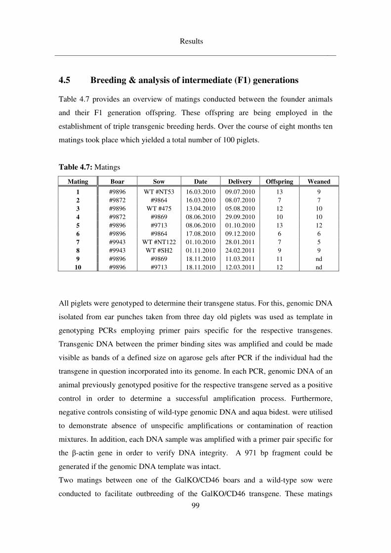
Results
99
4.5 Breeding & analysis of intermediate (F1) generations
Table 4.7 provides an overview of matings conducted between the founder animals
and their F1 generation offspring. These offspring are being employed in the
establishment of triple transgenic breeding herds. Over the course of eight months ten
matings took place which yielded a total number of 100 piglets.
Table 4.7: Matings
Mating Boar Sow Date Delivery Offspring Weaned
1 #9896 WT #NT53 16.03.2010 09.07.2010 13 9 2 #9872 #9864 16.03.2010 08.07.2010 7 7 3 #9896 WT #475 13.04.2010 05.08.2010 12 10 4 #9872 #9869 08.06.2010 29.09.2010 10 10 5 #9896 #9713 08.06.2010 01.10.2010 13 12 6 #9896 #9864 17.08.2010 09.12.2010 6 6 7 #9943 WT #NT122 01.10.2010 28.01.2011 7 5 8 #9943 WT #SH2 01.11.2010 24.02.2011 9 9 9 #9896 #9869 18.11.2010 11.03.2011 11 nd
10 #9896 #9713 18.11.2010 12.03.2011 12 nd
All piglets were genotyped to determine their transgene status. For this, genomic DNA
isolated from ear punches taken from three day old piglets was used as template in
genotyping PCRs employing primer pairs specific for the respective transgenes.
Transgenic DNA between the primer binding sites was amplified and could be made
visible as bands of a defined size on agarose gels after PCR if the individual had the
transgene in question incorporated into its genome. In each PCR, genomic DNA of an
animal previously genotyped positive for the respective transgene served as a positive
control in order to determine a successful amplification process. Furthermore,
negative controls consisting of wild-type genomic DNA and aqua bidest. were utilised
to demonstrate absence of unspecific amplifications or contamination of reaction
mixtures. In addition, each DNA sample was amplified with a primer pair specific for
the β-actin gene in order to verify DNA integrity. A 971 bp fragment could be
generated if the genomic DNA template was intact.
Two matings between one of the GalKO/CD46 boars and a wild-type sow were
conducted to facilitate outbreeding of the GalKO/CD46 transgene. These matings
Results
100
produced a total of 25 offspring, of which 13 piglets could be shown to have inherited
the CD46 transgene from the father. All offspring were heterozygous for the GalKO
since the boar was homozygous for this trait (Figure 4.16 and 4.17).
The initial matings for generating the core breeding herd for GalKO/CD46/HLA-E
constituted six matings of GalKO/CD46 boars to HLA-E sows which yielded 59
piglets. Of these piglets, 26 carried the CD46 and 22 the HLA-E transgene. Nine of
the CD46 and the HLA-E transgenic piglets had inherited both transgenes. Again, as
the boars were homozygous GalKO, all piglets were heterozygous for it (Figure 4.16,
4.18 and 4.19).
Immunohistochemical stainings of ear tissue sections of representatively selected
animals demonstrated the absence of Gal epitopes in founder boar #9896 but
heterozygous GalKO offspring displayed Gal epitope staining (Figure 4.20).
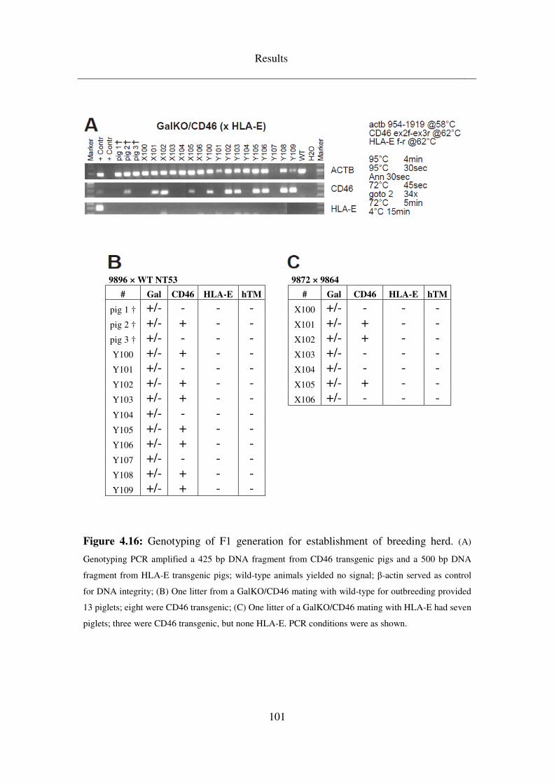
Results
101
9896 × WT NT53 9872 × 9864
# Gal CD46 HLA-E hTM # Gal CD46 HLA-E hTM
pig 1 † +/- - - - X100 +/- - - - pig 2 † +/- + - - X101 +/- + - - pig 3 † +/- - - - X102 +/- + - - Y100 +/- + - - X103 +/- - - - Y101 +/- - - - X104 +/- - - - Y102 +/- + - - X105 +/- + - - Y103 +/- + - - X106 +/- - - - Y104 +/- - - -
Y105 +/- + - -
Y106 +/- + - -
Y107 +/- - - -
Y108 +/- + - -
Y109 +/- + - -
Figure 4.16: Genotyping of F1 generation for establishment of breeding herd. (A)
Genotyping PCR amplified a 425 bp DNA fragment from CD46 transgenic pigs and a 500 bp DNA
fragment from HLA-E transgenic pigs; wild-type animals yielded no signal; β-actin served as control
for DNA integrity; (B) One litter from a GalKO/CD46 mating with wild-type for outbreeding provided
13 piglets; eight were CD46 transgenic; (C) One litter of a GalKO/CD46 mating with HLA-E had seven
piglets; three were CD46 transgenic, but none HLA-E. PCR conditions were as shown.
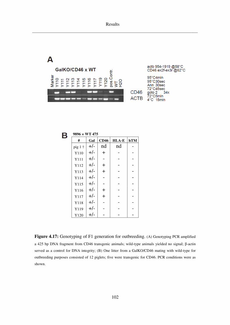
Results
102
9896 × WT 475
# Gal CD46 HLA-E hTM
pig 1 † +/- nd nd - Y110 +/- + - - Y111 +/- - - - Y112 +/- + - - Y113 +/- + - - Y114 +/- - - - Y115 +/- - - - Y116 +/- + - - Y117 +/- + - - Y118 +/- - - - Y119 +/- - - - Y120 +/- - - -
Figure 4.17: Genotyping of F1 generation for outbreeding. (A) Genotyping PCR amplified
a 425 bp DNA fragment from CD46 transgenic animals; wild-type animals yielded no signal; β-actin
served as a control for DNA integrity; (B) One litter from a GalKO/CD46 mating with wild-type for
outbreeding purposes consisted of 12 piglets; five were transgenic for CD46. PCR conditions were as
shown.
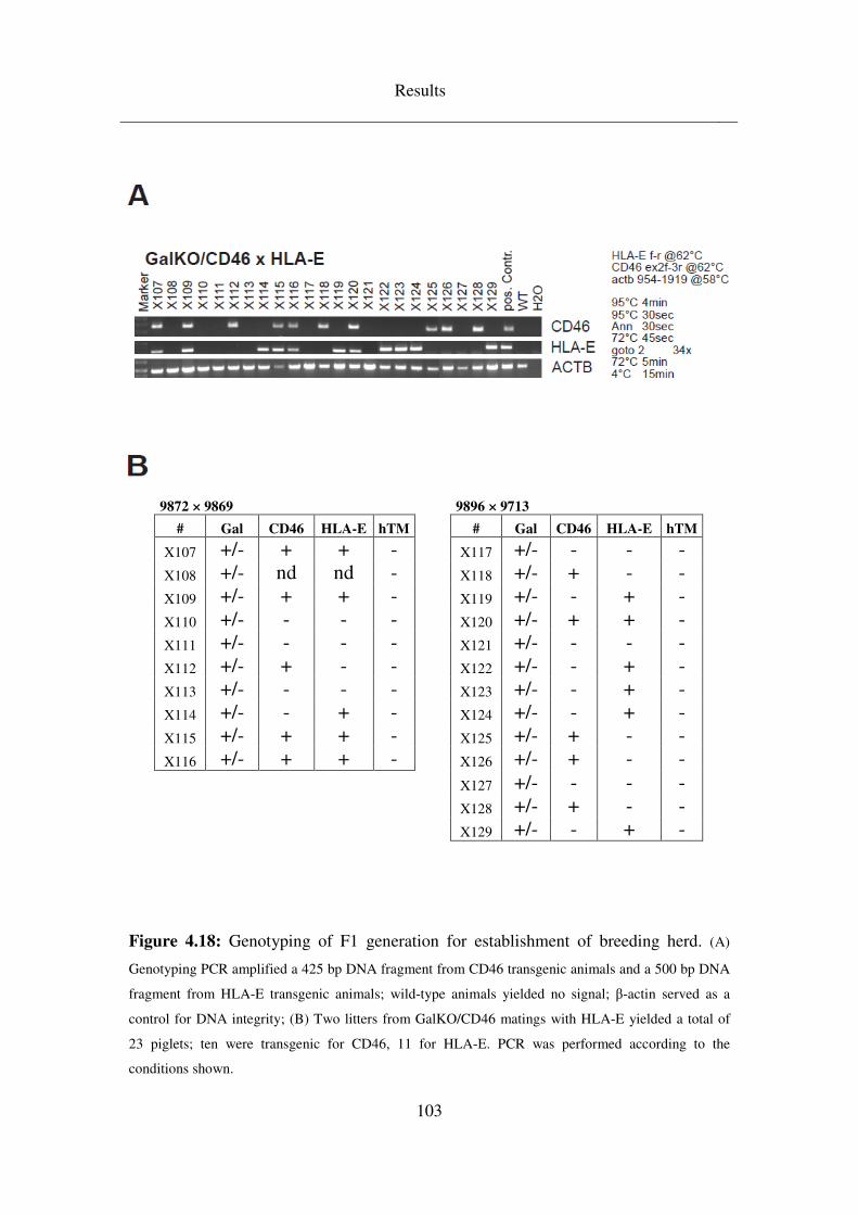
Results
103
9872 × 9869 9896 × 9713
# Gal CD46 HLA-E hTM # Gal CD46 HLA-E hTM
X107 +/- + + - X117 +/- - - - X108 +/- nd nd - X118 +/- + - - X109 +/- + + - X119 +/- - + - X110 +/- - - - X120 +/- + + - X111 +/- - - - X121 +/- - - - X112 +/- + - - X122 +/- - + - X113 +/- - - - X123 +/- - + - X114 +/- - + - X124 +/- - + - X115 +/- + + - X125 +/- + - - X116 +/- + + - X126 +/- + - -
X127 +/- - - - X128 +/- + - - X129 +/- - + -
Figure 4.18: Genotyping of F1 generation for establishment of breeding herd. (A)
Genotyping PCR amplified a 425 bp DNA fragment from CD46 transgenic animals and a 500 bp DNA
fragment from HLA-E transgenic animals; wild-type animals yielded no signal; β-actin served as a
control for DNA integrity; (B) Two litters from GalKO/CD46 matings with HLA-E yielded a total of
23 piglets; ten were transgenic for CD46, 11 for HLA-E. PCR was performed according to the
conditions shown.
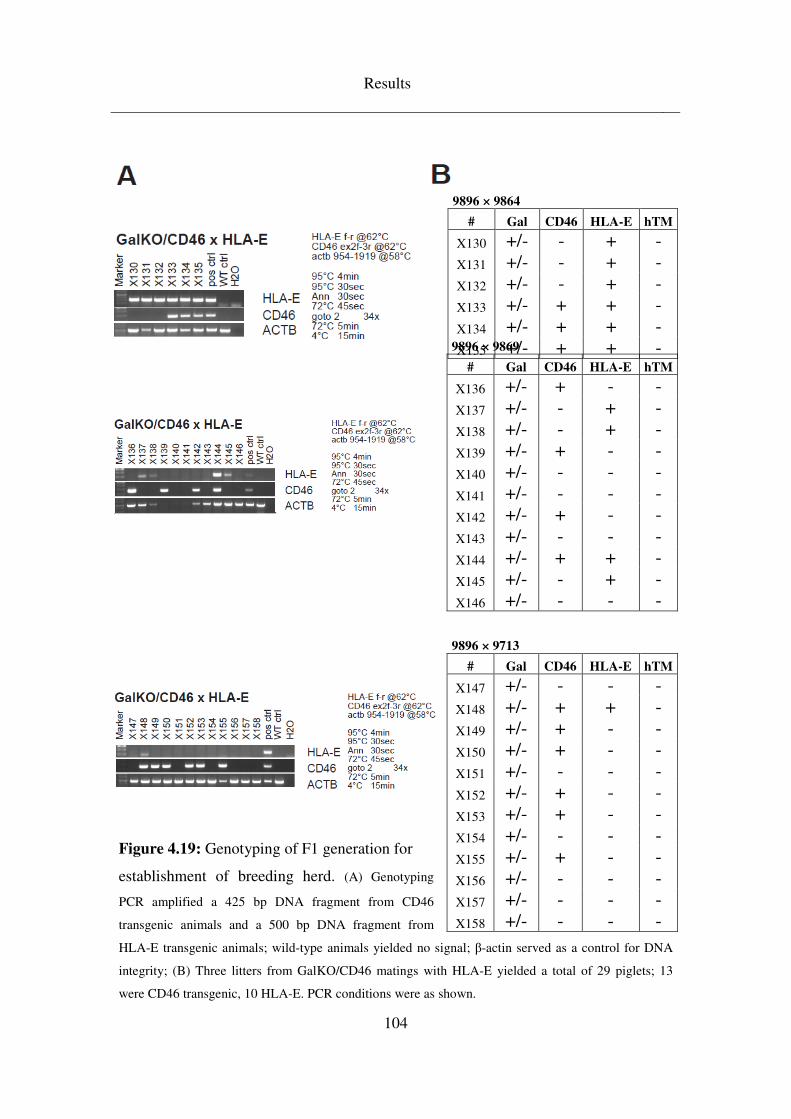
Results
104
Figure 4.19: Genotyping of F1 generation for
establishment of breeding herd. (A) Genotyping
PCR amplified a 425 bp DNA fragment from CD46
transgenic animals and a 500 bp DNA fragment from
HLA-E transgenic animals; wild-type animals yielded no signal; β-actin served as a control for DNA
integrity; (B) Three litters from GalKO/CD46 matings with HLA-E yielded a total of 29 piglets; 13
were CD46 transgenic, 10 HLA-E. PCR conditions were as shown.
9896 × 9864
# Gal CD46 HLA-E hTM
X130 +/- - + - X131 +/- - + - X132 +/- - + - X133 +/- + + - X134 +/- + + - X135 +/- + + - 9896 × 9869
# Gal CD46 HLA-E hTM
X136 +/- + - - X137 +/- - + - X138 +/- - + - X139 +/- + - - X140 +/- - - - X141 +/- - - - X142 +/- + - - X143 +/- - - - X144 +/- + + - X145 +/- - + - X146 +/- - - -
9896 × 9713
# Gal CD46 HLA-E hTM
X147 +/- - - - X148 +/- + + - X149 +/- + - - X150 +/- + - - X151 +/- - - - X152 +/- + - - X153 +/- + - - X154 +/- - - - X155 +/- + - - X156 +/- - - - X157 +/- - - - X158 +/- - - -
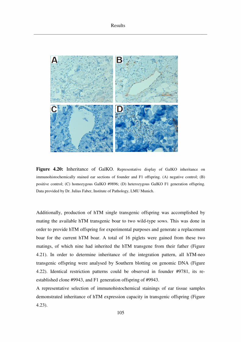
Results
105
Figure 4.20: Inheritance of GalKO. Representative display of GalKO inheritance on
immunohistochemically stained ear sections of founder and F1 offspring. (A) negative control; (B)
positive control; (C) homozygous GalKO #9896; (D) heterozygous GalKO F1 generation offspring.
Data provided by Dr. Julius Faber, Institute of Pathology, LMU Munich.
Additionally, production of hTM single transgenic offspring was accomplished by
mating the available hTM transgenic boar to two wild-type sows. This was done in
order to provide hTM offspring for experimental purposes and generate a replacement
boar for the current hTM boar. A total of 16 piglets were gained from these two
matings, of which nine had inherited the hTM transgene from their father (Figure
4.21). In order to determine inheritance of the integration pattern, all hTM-neo
transgenic offspring were analysed by Southern blotting on genomic DNA (Figure
4.22). Identical restriction patterns could be observed in founder #9781, its re-
established clone #9943, and F1 generation offspring of #9943.
A representative selection of immunohistochemical stainings of ear tissue samples
demonstrated inheritance of hTM expression capacity in transgenic offspring (Figure
4.23).
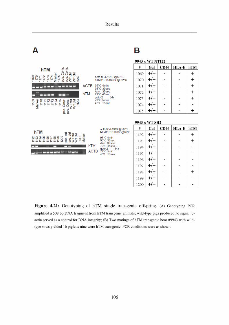
Results
106
9943 × WT SH2
# Gal CD46 HLA-E hTM
1192 +/+ - - + 1193 +/+ - - + 1194 +/+ - - - 1195 +/+ - - - 1196 +/+ - - - 1197 +/+ - - - 1198 +/+ - - + 1199 +/+ - - - 1200 +/+ - - -
Figure 4.21: Genotyping of hTM single transgenic offspring. (A) Genotyping PCR
amplified a 508 bp DNA fragment from hTM transgenic animals; wild-type pigs produced no signal; β-
actin served as a control for DNA integrity; (B) Two matings of hTM transgenic boar #9943 with wild-
type sows yielded 16 piglets; nine were hTM transgenic. PCR conditions were as shown.
9943 × WT NT122
# Gal CD46 HLA-E hTM
1069 +/+ - - + 1070 +/+ - - + 1071 +/+ - - + 1072 +/+ - - + 1073 +/+ - - + 1074 +/+ - - - 1075 +/+ - - +
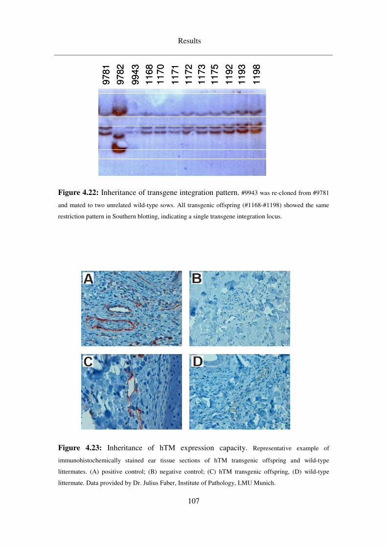
Results
107
Figure 4.22: Inheritance of transgene integration pattern. #9943 was re-cloned from #9781
and mated to two unrelated wild-type sows. All transgenic offspring (#1168-#1198) showed the same
restriction pattern in Southern blotting, indicating a single transgene integration locus.
Figure 4.23: Inheritance of hTM expression capacity. Representative example of
immunohistochemically stained ear tissue sections of hTM transgenic offspring and wild-type
littermates. (A) positive control; (B) negative control; (C) hTM transgenic offspring, (D) wild-type
littermate. Data provided by Dr. Julius Faber, Institute of Pathology, LMU Munich.
9781
9782
9943
1168
1170
1171
1172
1173
1175
1192
1193
1198
9781
9782
9943
1168
1170
1171
1172
1173
1175
1192
1193
1198
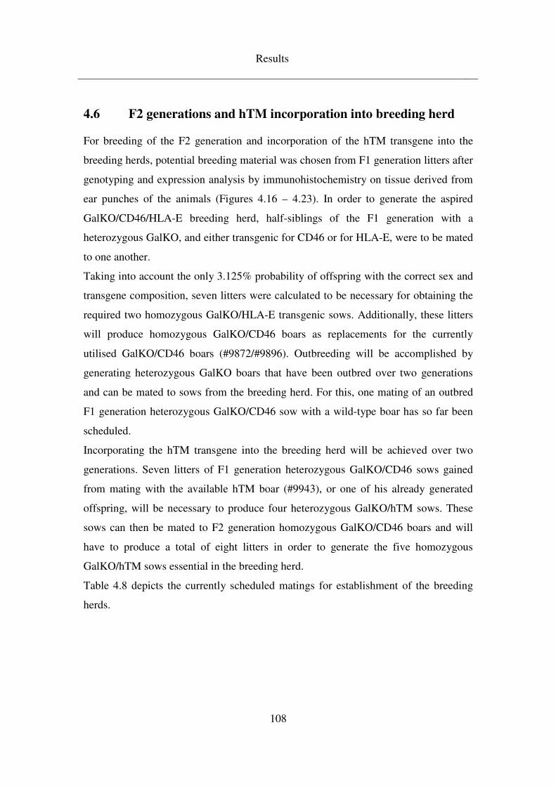
Results
108
4.6 F2 generations and hTM incorporation into breeding herd
For breeding of the F2 generation and incorporation of the hTM transgene into the
breeding herds, potential breeding material was chosen from F1 generation litters after
genotyping and expression analysis by immunohistochemistry on tissue derived from
ear punches of the animals (Figures 4.16 – 4.23). In order to generate the aspired
GalKO/CD46/HLA-E breeding herd, half-siblings of the F1 generation with a
heterozygous GalKO, and either transgenic for CD46 or for HLA-E, were to be mated
to one another.
Taking into account the only 3.125% probability of offspring with the correct sex and
transgene composition, seven litters were calculated to be necessary for obtaining the
required two homozygous GalKO/HLA-E transgenic sows. Additionally, these litters
will produce homozygous GalKO/CD46 boars as replacements for the currently
utilised GalKO/CD46 boars (#9872/#9896). Outbreeding will be accomplished by
generating heterozygous GalKO boars that have been outbred over two generations
and can be mated to sows from the breeding herd. For this, one mating of an outbred
F1 generation heterozygous GalKO/CD46 sow with a wild-type boar has so far been
scheduled.
Incorporating the hTM transgene into the breeding herd will be achieved over two
generations. Seven litters of F1 generation heterozygous GalKO/CD46 sows gained
from mating with the available hTM boar (#9943), or one of his already generated
offspring, will be necessary to produce four heterozygous GalKO/hTM sows. These
sows can then be mated to F2 generation homozygous GalKO/CD46 boars and will
have to produce a total of eight litters in order to generate the five homozygous
GalKO/hTM sows essential in the breeding herd.
Table 4.8 depicts the currently scheduled matings for establishment of the breeding
herds.
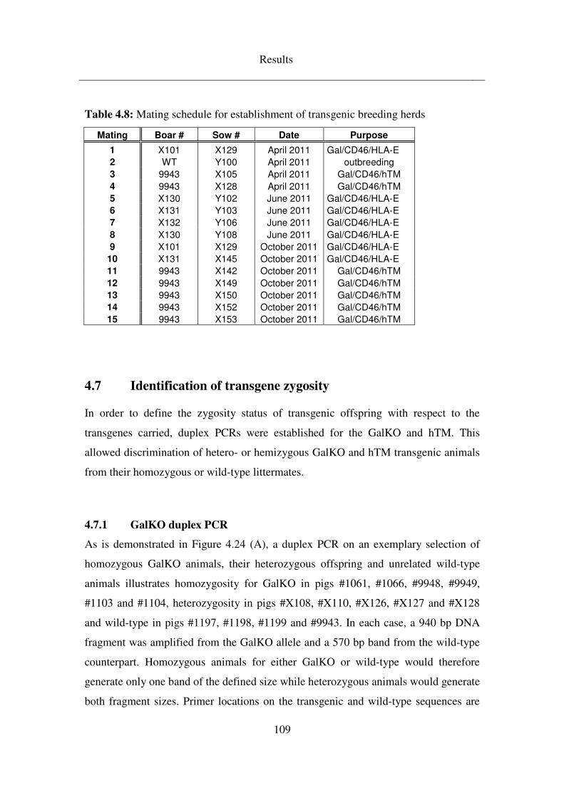
Results
109
Table 4.8: Mating schedule for establishment of transgenic breeding herds
Mating Boar # Sow # Date Purpose
1 X101 X129 April 2011 Gal/CD46/HLA-E
2 WT Y100 April 2011 outbreeding
3 9943 X105 April 2011 Gal/CD46/hTM
4 9943 X128 April 2011 Gal/CD46/hTM
5 X130 Y102 June 2011 Gal/CD46/HLA-E
6 X131 Y103 June 2011 Gal/CD46/HLA-E
7 X132 Y106 June 2011 Gal/CD46/HLA-E
8 X130 Y108 June 2011 Gal/CD46/HLA-E
9 X101 X129 October 2011 Gal/CD46/HLA-E
10 X131 X145 October 2011 Gal/CD46/HLA-E
11 9943 X142 October 2011 Gal/CD46/hTM
12 9943 X149 October 2011 Gal/CD46/hTM
13 9943 X150 October 2011 Gal/CD46/hTM
14 9943 X152 October 2011 Gal/CD46/hTM
15 9943 X153 October 2011 Gal/CD46/hTM
4.7 Identification of transgene zygosity
In order to define the zygosity status of transgenic offspring with respect to the
transgenes carried, duplex PCRs were established for the GalKO and hTM. This
allowed discrimination of hetero- or hemizygous GalKO and hTM transgenic animals
from their homozygous or wild-type littermates.
4.7.1 GalKO duplex PCR
As is demonstrated in Figure 4.24 (A), a duplex PCR on an exemplary selection of
homozygous GalKO animals, their heterozygous offspring and unrelated wild-type
animals illustrates homozygosity for GalKO in pigs #1061, #1066, #9948, #9949,
#1103 and #1104, heterozygosity in pigs #X108, #X110, #X126, #X127 and #X128
and wild-type in pigs #1197, #1198, #1199 and #9943. In each case, a 940 bp DNA
fragment was amplified from the GalKO allele and a 570 bp band from the wild-type
counterpart. Homozygous animals for either GalKO or wild-type would therefore
generate only one band of the defined size while heterozygous animals would generate
both fragment sizes. Primer locations on the transgenic and wild-type sequences are
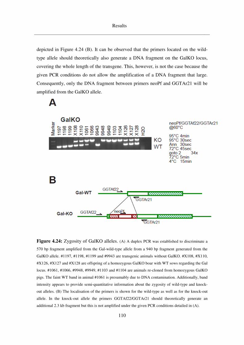
Results
110
depicted in Figure 4.24 (B). It can be observed that the primers located on the wild-
type allele should theoretically also generate a DNA fragment on the GalKO locus,
covering the whole length of the transgene. This, however, is not the case because the
given PCR conditions do not allow the amplification of a DNA fragment that large.
Consequently, only the DNA fragment between primers neoPf and GGTAr21 will be
amplified from the GalKO allele.
Figure 4.24: Zygosity of GalKO alleles. (A) A duplex PCR was established to discriminate a
570 bp fragment amplified from the Gal-wild-type allele from a 940 bp fragment generated from the
GalKO allele. #1197, #1198, #1199 and #9943 are transgenic animals without GalKO. #X108, #X110,
#X126, #X127 and #X128 are offspring of a homozygous GalKO boar with WT sows regarding the Gal
locus. #1061, #1066, #9948, #9949, #1103 and #1104 are animals re-cloned from homozygous GalKO
pigs. The faint WT band in animal #1061 is presumably due to DNA contamination. Additionally, band
intensity appears to provide semi-quantitative information about the zygosity of wild-type and knock-
out alleles. (B) The localisation of the primers is shown for the wild-type as well as for the knock-out
allele. In the knock-out allele the primers GGTAf22/GGTAr21 should theoretically generate an
additional 2.3 kb fragment but this is not amplified under the given PCR conditions detailed in (A).
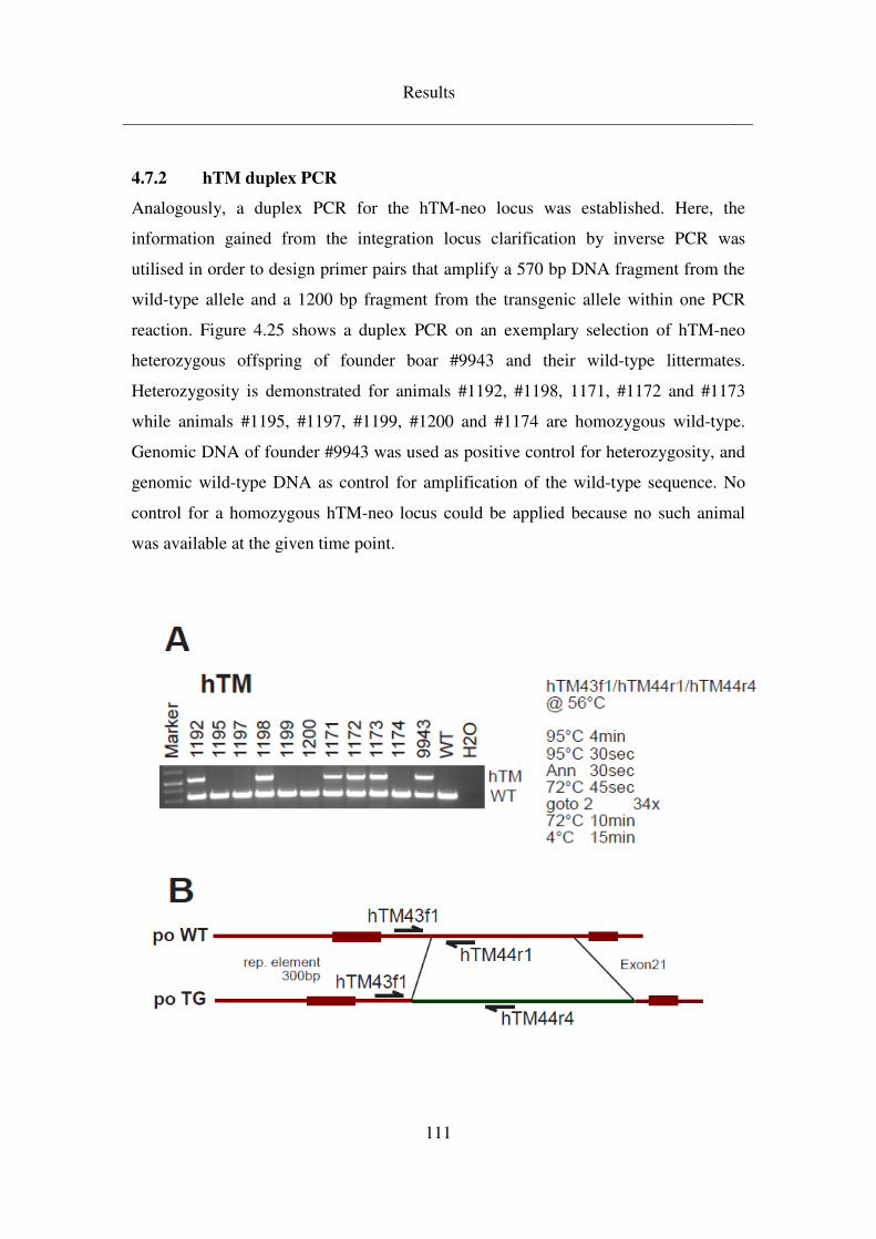
Results
111
4.7.2 hTM duplex PCR
Analogously, a duplex PCR for the hTM-neo locus was established. Here, the
information gained from the integration locus clarification by inverse PCR was
utilised in order to design primer pairs that amplify a 570 bp DNA fragment from the
wild-type allele and a 1200 bp fragment from the transgenic allele within one PCR
reaction. Figure 4.25 shows a duplex PCR on an exemplary selection of hTM-neo
heterozygous offspring of founder boar #9943 and their wild-type littermates.
Heterozygosity is demonstrated for animals #1192, #1198, 1171, #1172 and #1173
while animals #1195, #1197, #1199, #1200 and #1174 are homozygous wild-type.
Genomic DNA of founder #9943 was used as positive control for heterozygosity, and
genomic wild-type DNA as control for amplification of the wild-type sequence. No
control for a homozygous hTM-neo locus could be applied because no such animal
was available at the given time point.
Results
112
Figure 4.25: Zygosity of hTM-neo transgene. (A) A duplex PCR was established to
discriminate a 570 bp fragment amplified from the wild-type allele from a 1200 bp fragment generated
from the hTM-neo transgenic allele and thus identify wild-type as well as hemi- and homozygous
transgenic animals. #1192, #1198, #1171, #1172, #1173 and #9943 are hemizygous transgenic animals.
#1195, #1197, #1199, #1200 and #1174 are wild-type. (B) The localisation of the primers is shown for
the wild-type as well as for the knock-out allele.
113
5 DISCUSSION
The notion that pigs are among the most suitable species for biomedical translational
research and as potential donors in the context of xenotransplantation has been
prevalent for some time and has been discussed extensively by numerous authors
(reviewed in Aigner et al. 2010). This is owed to the fact that the organ systems of
pigs share many anatomical and physiological characteristics with humans (Smith et
al. 1990; Rogers et al. 2008; Spurlock and Gabler 2008). Additionally, their size
corresponds to human dimensions more closely than that of rodents does, which also
makes sampling and handling a less materially limited and demanding task (Roberts et
al. 2009). In their guidelines, national and international regulating authorities such as
the European Medicines Agency require the use of a non-rodent mammal as
experimental animal in clinical development trials (www.ema.europa.eu). Genetically
modified pigs therefore occupy an ever increasing place in biomedical translational
research and xenotransplantation. However, in order to draw meaningful conclusions
from experiments, sizeable numbers of animals are required. Lead times in the
preparation or specific time frames in the conduction of experiments demand
predictable groups of experimental animals of specific size and at specific time points.
This has proven to be a problematic issue in providing transgenic pigs for
experimental purposes.
Novel transgenic lines have been and are being established by a variety of
technologies based on additive gene transfer with unpredictable transgene integration
sites or site directed knock-out or knock-in methods. Somatic cell nuclear transfer
followed by embryo transfer into oestrus synchronised recipient sows has become the
leading technique in generating transgenic pigs (Melo et al. 2007). This method
features beyond 90% transgenesis rates and pooling of transgenic cells derived from
independent integration events leads to more than 90% independent founders, as long
term experience at the MVG has shown (unpublished data). However, it has become
apparent that the process is also very inefficient in terms of total animals numbers
produced. Rates of less than 5% live offspring compared to the total number of
transferred embryos have been reported across the literature (Polejaeva et al. 2000;
Oback 2008; Palmieri et al. 2008) which makes the cloning procedure appropriate for
Discussion
114
establishing novel transgenic pigs but raises the question if this technique is suitable
for reproducing already established lines by nuclear and embryo transfer.
As Tables 4.3, 4.4 and 4.5 demonstrate, re-cloning of pigs after expression analysis of
founder litters and selection of the most suitable founders at the MVG, Badersfeld has
proven to be a tedious issue. For example, six attempts at re-cloning hTM-neo founder
pig #9781 delivered only one single animal which, fortunately, survived the neonatal
period and could be raised until sexual maturity (Table 4.3). Re-cloning INS-LEA
founders #9813 and #9814 has been equally difficult. 14 nuclear and embryo transfers
resulted in only four pregnancies that actually delivered. From these, only two animals
(#1044, #1050) were weaned (Table 4.5). Re-cloning of hTM-bla founder boar #9848
appears to have generated slightly more live offspring. However, taking into account
that 16 nuclear and embryo transfers dates produced only 11 weaned piglets that
could be utilised in experiments or raised for breeding, this calculates to an average of
0.7 piglets derived from one nuclear and embryo transfer . Or, if only nuclear and
embryo transfers from which a pregnancy was established are taken into account, this
number still averages at less than one piglet per NT/ET (Table 4.4). Therefore, this
method had been discarded as a means for routinely re-producing transgenic pigs for
experimental purposes, as outcome is too unpredictable and, consequently, too time,
space and human and material resource intensive.
Breeding transgenic pigs on a demand-based level rectifies these limitations. As has
been reported before, transgenic animals generated by cloning and their offspring
behave similarly in terms of reproductive capacity as their wild-type counterparts
(Martin et al. 2004; Mir et al. 2005; Shibata et al. 2006; Williams et al. 2006).
Additionally, transgenes integrated into the genome appear to be passed on to the next
generation according to Mendelian rules of inheritance (Aigner et al. 1999). Breeding
of transgenic pigs that has been conducted during the preparation of this thesis has
confirmed both of these notions. Litter sizes of transgenic pigs mated to either another
transgenic or to wild-type individuals averaged 10 piglets per litter which is similar to
what has been reported for wild-type litters (McGlone and Pond 2003) and also
reflects the wild-type breeding experience gained at the MVG.
Transgene inheritance appeared to adhere to Mendelian rules. As has been shown in
Discussion
115
Figures 4.16, 4.18 and 4.19, eight matings of GalKO/CD46 boars to either wild-type
or HLA-E transgenic sows generated a total of 84 piglets. Calculated numbers of
offspring transgenic for CD46 and/or HLA-E should have constituted 27.25 CD46,
14.75 HLA-E and 14.75 CD46/HLA-E transgenic piglets. Effectively, gained
transgenic offspring represented 29, 11 and 10, respectively. Even though total
numbers for HLA-E and CD46/HLA-E transgenic offspring are slightly less than
expected, individual single litters have brought more transgenic offspring than
calculated, while others have produced less. Matings involving the hTM-neo boar
#9943 have produced nine transgenic piglets when, mathematically, only eight should
have been generated (Figure 4.21).
Since transgenes are passed on to the next generation adhering to Mendelian rules of
inheritance, they also segregate accordingly. This lowers probabilities of specific
transgene combinations in multiple transgenic animals as long as each transgene is
incorporated as a single vector construct into the genome. However, it also effects
segregation of multiple integration sites of a single transgene. This can become a
problem in offspring generations, as transgene expression capacity of the individuals
cannot be foreseen. Detailed expression analysis would therefore have to be conducted
on each pig, which is in many cases not practicable as animals would have to be
sacrificed for this and could therefore not be utilised in breeding or experiments.
Consequently, segregation of transgenes has to be avoided. This is only possible if
animals are selected for breeding that only possess one single transgene integration
site in the first instance. Segregation of multiple copies of the transgene at one
integration site would only be possible if these transgenes were relocated to multiple
integration sites by recombination first, which would then be able to segregate. This,
however, is a theoretical possibility but extremely unlikely. As Aigner et al. (1999)
have demonstrated, transgene integration sites apparently remain stable in over 20
generations of breeding without reassortment of individual copies. Southern blotting
of the hTM-neo founder and his offspring demonstrated that indeed all transgenic
piglets derived from this boar displayed the same restriction pattern as their father
(Figure 4.22), suggesting cross-generational stability in integration conformation of
the transgenes. Selection of founder animals displaying a single integration site, even
Discussion
116
with more than one transgene copy, is therefore a viable means of preventing
segregation.
If avoidance of transgene segregation is accomplished, expression patterns of founder
animals can be assumed to be passed on to offspring (Bordignon et al. 2003; Brunetti
et al. 2008). Even though epigenetic silencing of promoters or coding sequences has
been observed in lentiviral transgenesis (Kearns et al. 2000, Hofmann et al. 2006), this
effect seems to occur mainly with viral sequences. F1 generation offspring of
GalKO/CD46 and hTM-neo boars could therefore have been expected to display
transgene expression on a level comparable to their fathers’. Indeed, analysis of
immunohistochemically stained ear sections (Figure 4.23) of F1 hTM-neo offspring
reinforced the notion that expression patterns of transgenes are inherited to the next
generation. Thus, expression analysis of founder animals with a single transgene
integration site presumably constitutes a sufficient method of ensuring reliable
expression levels also in later generations.
Furthermore, with the knowledge of transgene vector construction, namely the
promoter part of it, expression of the transgene can be anticipated in cell types or
tissues in which the controlling promoter is active. Therefore, it might appear to be an
option to confine expression analysis of potential founder animals to tissues that can
be harvested without sacrificing the animals in question. For example, CAG-LEA
founders were expressing the immunomodulatory LEA29Y transgene ubiquitously.
Initial expression analysis on ear tissue sections (Figure 4.10) and in blood serum
samples (Figure 4.6) gave a good indication of the expression capacity of each of the
three founders and could be gained without killing the animals. This can be seen as an
important finding, as the extremely low efficiency in re-cloning primary cells from
animals killed for expression analysis hinders generation of breeding stock. However,
specific projects often require the site-specific expression of a particular transgene.
Potential founder animals for INS-LEA transgenic pigs, for example, had to be
sacrificed for analysis because expression of the transgene was expected to be limited
to pancreatic ß-cells which could not feasibly be obtained from living pigs. Moreover,
during the analysis of hTM-bla founders it became apparent that even though initial
expression analysis in the form of immunohistochemically stained ear sections (Figure
Discussion
117
4.2) would lead to the assumption that all three potential founders were equal in their
transgene expression capacity, further examination proved this conjecture wrong. In
depth expression analysis on the basis of immunohistochemical staining of organ
spectra brought to light that expression was indeed on a comparable level in some, but
not in all investigated tissues. For example, animal #9849 expressed hTM well in
heart tissue on a level similar to that of the two litter mates but staining of lung, liver
or kidney tissue was markedly weaker. The same phenomenon could be observed in
the selection of hTM-neo founders. Ear tissue sections of litter mates #9780, #9781
and #9782 all stained equally well in immunohistochemical evaluation. However,
staining of a range of organs showed the differing expression capacity of the litter in,
for example, lung and liver tissue. In both cases, the animal that expressed best in all
examined organs was chosen as founder boar, for hTM-bla pig #9848 and for hTM-
neo pig #9781. This choice, however, would not have been possible, had expression
analysis been confined to a more superficial level.
In-depth expression analysis thus appears to be a prerequisite in the selection of
founder animals that are to be utilised in breeding transgenic pig lines. Choosing
suitable founders with respect to their transgene expression capacity and limiting the
likelihood of transgene segregation in future generations by only employing animals
with a single integration site in breeding ensures viable transgene inheritance. This is
the case for single transgenic animals; however, if multiple transgenic vectors have
been incorporated into the genome, segregation of these transgenes becomes
inevitable. As a result, probabilities for specific transgene combinations in offspring
can decrease to very low numbers, necessitating many litters in order to generate a
certain number of particular pigs.
The low probability of multi-transgene inheritance might be overcome by choosing
only animals for breeding that carry the transgenes on both alleles. In this case, all
offspring would be hemizygous for each transgene. This, however, requires
knowledge of transgene zygosity, which in turn can only be determined if integration
sites are known. Additionally, even though employing homozygous transgenic
animals in breeding or mating animals with identical transgenic properties to each
other increases the numbers of transgenic offspring it also heightens inbreeding levels.
Discussion
118
This is due to the fact that only a very limited amount of breeding stock is available in
the case of transgenic pigs and founder animals are usually confined to only one
genetic background for each transgene. Consequently, homozygosity for a trait can
only be achieved by mating related animals and pigs with identical transgenic
properties will necessarily have been derived from the same founder. But rising
inbreeding coefficients lower productivity of breeding stock and the viability of
offspring (Charlesworth and Charlesworth 1987; Ralls et al. 1988; Lynch 1989; 1991).
This collides with requirements for large numbers of offspring if probabilities for
transgene combinations are low.
Inbreeding should therefore be kept as low as possible within a breeding herd. Thus
matings between animals that are related to each other should be avoided as far as
possible and unrelated wild-type blood should be introduced from the outside on a
regular basis. However, both of these measures again lower probabilities for specific
transgene combinations and make them even impossible, as is the case with traits that
have to be present in homozygosity in order to take effect, such as the GalKO. Hence
it can be concluded that transgene inheritance and inbreeding are two conflicting
issues that cannot be accounted for fully both at the same time. Rather a common
ground has to be identified on which either of these issues presents the least possible
problem.
In order to examine the differing effect a distinct emphasis on either increasing
transgene combination probabilities or on keeping inbreeding levels low can have on
breeding outcome, breeding schedules for establishment of GalKO/CD46/HLA-E and
GalKO/CD46/hTM herds were designed that account for both issues to a different
extent (Figure 4.13). Because GalKO/CD46 was only available in the form of founder
boars generated by re-cloning from cells of one single founder established at
Revivicor, Inc. these transgenes had to be derived from these two boars. On the other
hand, HLA-E was only available as sows derived from breeding the original founders
established in 2006. And, finally, hTM had been established as a new transgene on
two different backgrounds, once by transfecting the hTM-bla vector onto the
Revivicor GalKO/CD46 background and once as hTM-neo on a wild-type cell line,
both again resulting in male animals. As the GalKO needs to be bred to homozygosity
Discussion
119
for an applicable effect, mating of related animals in F1 generation was inevitable
because the GalKO could only be derived from the paternal side. Since the other
transgenes are already functioning in a hemizygous conformation, they could be
obtained from either the paternal or the maternal side. Figure 4.13 (A) and (B)
demonstrate that the choice between half-sibling and full-sibling matings in F1 was
inconsequential for the proportion of suitable transgene combinations in F2. However,
it did have a substantial effect on the development of the inbreeding coefficient in F2
offspring. While half-sibling matings resulted in an inbreeding coefficient of 0.125, it
doubled to 0.25 for full-sibling matings. This can be seen as a considerable difference
if it is kept in mind that several authors have reported average inbreeding coefficients
in commercial pig breeds of below 0.1 (Welsh et al. 2010) and that every ten percent
rise in inbreeding of the sow has been correlated with a 0.2 piglet reduction in litter
size (Dickerson et al. 1954). Father-daughter matings, on the other hand, would have
been able to double probabilities for specific transgene combinations in offspring
(Figure 4.13 (C)). But they, too, would have resulted in inbreeding coefficients of 0.25
in offspring. The decision was thus made to employ half-sibling matings to generate
homozygous GalKO offspring with CD46, HLA-E or both transgenes.
An even greater effect could be observed in deciding on where to derive the hTM
transgene for integration into the breeding herd from. Utilising the GalKO/CD46/hTM
founder boar, suitable animals for breeding would already have been generated within
one generation. However, the genetic background of this founder is identical to the
GalKO/CD46 founders, as all of them are clones of the initial Revivicor cell
population. Thus, mating of F1 generation sows from half-sibling matings to the
GalKO/CD46/hTM founder boar would have constituted father-daughter matings,
resulting in 0.25 inbreeding (Figure 4.14 (A)). On the other hand, employing the
single transgenic hTM founder and mating him to the exactly the same sows will not
generate breeding animals in the following generation as it will be impossible to
establish a homozygous GalKO. But offspring one generation later will not only
display the required transgenes with twice as high a probability than in (A) but will
also feature an inbreeding coefficient of only 0.062 (Figure 4.14 (B)). This constitutes
a value comparable to two generations GalKO/CD46 outbreeding with wild--type

Discussion
120
sows as depicted in Figure 4.15 which can thus be achieved simultaneously with
integrating the hTM transgene into the breeding herd.
Zygosity of transgenes becomes an important issue if transgenes are only effective in
a homozygous conformation or if, on the contrary, homozygosity of a trait implicates
severe disadvantages for the affected individual. For calculation of expectable
proportions of transgenic piglets in offspring, zygosity of the parents is also the
determining factor. In rodents, it is feasible to determine zygosity by out crossing the
individuals in question. This, however, does not appear to be an option in pigs.
Pregnancy lengths of approximately 115 days in pigs would make this a time-
consuming undertaking. Depending on overall body weight, European regulations
dictate minimum floor areas for sows of up to 2.5 m2 per sow (Directive 2010/63/EU)
for experimental animals, thus implying substantial space requirements. Furthermore,
it is doubtful if animal welfare concurs with producing numerous litters of piglets
solely for determining transgene zygosity of the parents. A reliable method for
genomic analysis of the zygosity status of individuals with respect to transgenes must
therefore be identified.
In the establishment of the transgenic breeding herds that are being covered by this
thesis, zygosity was not relevant for the F1 offspring generation. Zygosity of founder
animals was known in each case and since no identical transgenes on the maternal and
the paternal side were present, offspring was necessarily hemi- or heterozygous for the
respective transgene. However, in F2 generation offspring the question of GalKO
zygosity will arise and later on the other transgenes will also become relevant.
Zygosity specific duplex PCRs that are able to amplify two different DNA fragments
from transgenic and wild-type alleles in one PCR reaction and thus discriminate
between homo- and heterozygous transgenic individuals were therefore to be
established.
In the case of the GalKO the location of the relevant DNA sequence within the
genome was already clear, as site directed knock out of a specific gene had been
performed. Thus, primers could be designed that cover the region in question and
from which DNA fragments of two different sizes were amplified from the transgenic
and the wild-type allele. As has been shown in Figure 4.23, this method is able to
Discussion
121
reliably discriminate between hetero- and homozygous GalKO pigs and generates a
570 bp DNA fragment from the wild-type locus and a 940 bp fragment from the
knock out sequence. Because the reverse primer was placed downstream of the known
integration site of the gene disruption vector, it could be utilised for both alleles. Two
forward primers were placed either upstream of the Gal gene or on the knock out
vector sequence. Consequently, DNA fragments from the wild-type allele were
amplified from the reverse and the forward primer that had been placed upstream of
the gene, and DNA fragments from the knock out allele were generated from the
reverse and the second forward primer on the vector sequence. The forward primer
upstream of the Gal gene was also able to bind on the knock out allele; however, since
the vector sequence was larger than 4 kb, the distance between this primer and the
reverse primer downstream of the integration site would have been too long for Taq
polymerase to cover under the used PCR conditions. Thus, no amplicons were
generated from this primer pair.
In order to establish a zygosity specific duplex PCR for the hTM transgene the
integration loci in the founder boars had to be identified first so the comparable wild-
type sequence would be known. Thus, inverse PCR was chosen as a means of
determining integration sites of transgenes in the founders employed in breeding.
From a technological point of view, this method is rather simple and straightforward
(Ochman et al. 1988; 1990). Genomic DNA is fragmented, fragments are circularised
and DNA is amplified from primers binding to the transgene sequence. Flanking
regions of the transgene copy thereby become visible. Using this technique, it has
been possible to successfully identify the hTM-neo integration site in founder #9781
(Figure 4.3) and several transgene-transgene transitions in hTM-bla founders #9948
and #9949 (Figure 4.5), including a number of truncated transgene copies.
Additionally, inverse PCR amplified porcine genomic regions including the porcine
thrombomodulin and the Gal gene. This can be ascribed to the porcine
thrombomodulin promoter and the bGH-polyadenylation site of the resistance
cassettes present in the transgene vectors on which the primer binding sites could be
found.
Identification of the genomic flanking region of transgenes by inverse PCR is based
Discussion
122
on the assumption of complete individual transgene copies. Only then are primer
binding sites, which need to be placed close to the 5’ or the 3’ end of the transgene,
reliably available. Furthermore, increasing numbers of transgene copies at one
integration site lower the probability of ascertaining the inverse PCR amplicons that
contain parts of the genomic flanking region. Thus, failure to identify the hTM-bla
integration locus in founder pigs #9948 and #9949 might be due to (i) the possibility
that only truncated transgene copies are present on either transition site to genomic
DNA or (ii) the fact that such a large number of transgene copies has been inserted at
one site that probabilities of identifying the genomic flanking regions are extremely
low anyway, or (iii) the circularised genomic DNA fragments are too large to be
amplified by the PCR reaction. Several restriction enzymes for genomic fragmentation
and a number of different primer pairs were used in each case but only a minority
delivered amplicons after inverse PCR, which suggests that circularised DNA
fragments might indeed have been too large in many cases for successful
amplification. On the other hand, up to eight different restriction enzymes were tested
in each approach with all of them resulting in calculatory mean fragment sizes of
below 3 kb. Therefore it seems unlikely that so many of the fragments should have
been too large for amplification. In the case of founder animal #9949, only one single
transgene-transgene link could be identified (Figure 4.5 (B)) at all, indicating that
failure to identify genomic flanking regions might have been due to severely truncated
transgene copies on either transition site to the genomic sequence. In founder #9948, a
combination of truncated transgene copies and the sheer number of copies in the
integration site appear to be a probable explanation that the hTM-bla integration locus
could not be clarified. The same can be said for examination of the CD46 integration
locus in Gal/CD46 founder boars, of which results have not been included in this
thesis. Here, too, a number of transgene-transgene transitions could be revealed but no
genomic flanking regions.
In order to rectify problems with too many inserted transgene copies, it might be a
possibility to choose restriction enzymes that result in comparably large fragments
within the transgenic sequence but possess a relatively high restriction frequency
within genomic DNA. Thus fragments resulting from transgene-transgene links might
Discussion
123
become too large for amplification by inverse PCR while fragments from genomic
DNA might be of a suitable size. The majority of amplicons generated would
therefore possibly contain genomic flanking regions. However, this approach would
not account for the presence of truncated transgene copies, limiting the prospects of
success for inverse PCR in clarification of transgene integration sites. Alternative
measures could perhaps be found in the generation of BAC libraries for the founder
animals in question, where probes that recognise parts of the transgenic sequence
would be used to seperate the BACs that contain the transgene from a pool of large
genomic DNA fragments. Sequencing of these particular BACs would then reveal the
surrounding genomic region. Several authors have reported on the construction of
porcine BAC libraries (Liu et al. 2010; Suzuki et al. 2000) however, high throughput
methods for sequencing would have to be employed in order to process the masses of
data this approach would deliver. A third option for clarifying the integration site of a
transgene might be deep sequencing methods which enable the sequencing of whole
genomes or at least large portions of it without requiring extraordinary amounts of
time or money. These methods, however, are still under development and require in
depth knowledge of the genome analysed. Since the presently available sequence of
the porcine genome still contains large gaps, these methods are currently not feasible
but might find their application in the future.
In conclusion, the first systematic approach to establishing breeding herds for
production of donor animals with multiple functional transgenes for
xenotransplantation has been described. The selected breeding schedules represent a
compromise between the conflicting parameters of rising inbreeding coefficients and
efficiency of transgene inheritance. Additionally, novel transgenic pig lines have been
identified that are to be integrated into the breeding herds and duplex PCR screening
methods have been established to facilitate future breeding by discrimination of
transgene zygosity.

124
125
6 SUMMARY
(Re)producing transgenic pigs for xenotransplantation – selection of founder
animals and establishment of breeding herds
Xenotransplantation is discussed as an alternative treatment for end-stage organ
failure. The pig has been widely accepted as a feasible donor species. However, it has
to be genetically modified in order to overcome incompatibilities of the human and
the porcine immune systems. Although profound experience in long-term breeding of
multi-transgenic animals exists for mouse models in biomedical research, the situation
in breeding multi-transgenic pigs is far more complicated. In contrast to small animal
models, several limitations have to be taken into account for the pig: (i) inbreeding has
a detrimental effect on fertility and litter size in pigs while congenic mouse strains are
being bred with small limitations; (ii) generation times of approximately 12 months
and pregnancies of 115 days in pigs necessitate careful breeding management; (iii)
costs and space requirements in pig maintenance far exeed those for mice. In addition,
insight into xenograft rejection mechanisms is still limited and further transgenes or
novel transgene combinations might be required in the future. Thus, effective breeding
schedules have to be designed that accomodate these limitations and allow for
adaptation to changing transgene requirements.
This thesis describes the design and establishment of multi-transgenic
GalKO/CD46/HLA-E and GalKO/CD46/hTM donor herds and the characterisation of
novel hTM and LEA29Y transgenic lines for future incorporation into the breeding
herd. In a first step, two fertile GalKO/CD46 boars and three fertile HLA-E sows were
selected as founders for an initial GalKO/CD46/HLA-E breeding herd into which
further xeno-relevant transgenes were to be incorporated later on. Breeding schedules
that accommodate the conflicting issues of inbreeding, transgene segregation and time
requirements to different extents were designed and the most feasible strategy was
identified. Furthermore, options for incorporation of already fertile hTM transgenic
founder pigs into the core breeding herd were analysed.
Chosen GalKO/CD46 and HLA-E founder animals were mated to each other and half-
sibling matings of the F1 generation offspring were selected as a method for
Summary
126
establishment of a homozygous GalKO with CD46 or HLA-E in F2 generation
offspring. GalKO/CD46 boars were also mated to wild-type sows to facilitate
outbreeding and thus restrict inbreeding levels within the herd. Additionally, the
selected method for integration of the hTM transgene into the breeding herd
necessitated matings of the hTM founder boar to wild-type sows. All F1 generation
offspring was analysed on a genomic level to determine inheritance of transgene
integration patterns. Additionally, inheritance of expression capacity was examined
immunohistochemically. This facilitated selection of breeding animals for generation
of F2 offspring.
In parallel, analyses of potential founder animals for the novel transgenic pig lines
hTM, INS-LEA and CAG-LEA were performed to select the most suitable individuals
for incorporating these transgenes into the breeding herd. In each case, animals were
chosen where Southern blotting indicated the presence of only one single transgene
integration site to avoid segregation of integration loci in offspring. Expression of the
transgene was examined by immunohistochemistry and ELISA in a broad range of
organs to determine the individuals that exhibited the best expression capacity. By
combining results of genomic with expression analysis, founders for the novel lines
were chosen which were subsequently re-cloned to be raised for breeding. While the
generation of a fertile founder had already been accomplished for the hTM transgene,
INS-LEA founders have not reached sexual maturity yet.
Comprehensive genomic analysis included the determination of transgene integration
loci in hTM founder animals. For this, inverse PCR was chosen as a method for
determining unknown genomic flanking regions of transgenes. The transgene
integration site could be identified in the hTM founder that is being employed in
breeding. Additionally, two further potential hTM founders were analysed and
complex transgene integration patterns were clarified. However, the integration locus
itself could not be found in these two cases.
The information gained from hTM integration locus determination in the founder boar
employed in breeding was utilised for establishing a duplex PCR that discriminates
between an hTM transgenic allele and its wild-type counterpart in one single PCR
reaction. This was done in order to facilitate discrimination of hemizygous from
Summary
127
homozygous transgenic pigs, which will be necessary from F2 generation onwards. A
zygosity specific duplex PCR was also established for the genomically assigned
GalKO, discriminating the wild-type from the hetero- or homozygous knockout.
In conclusion, this thesis decribes the systematic reproduction of multi-transgenic
donor pigs for xenotransplantation experiments. While breeding for the well-
characterised transgenes has already commenced, the established schedule also allows
for integration of the additionally analysed transgenes once fertile founder animals
become available.

128
129
7 ZUSAMMENFASSUNG
Transgene Spenderschweine für die Xenotransplantation – Auswahl von
Foundertieren und Etablierung von Zuchtherden
Xenotransplantation gilt angesichts des notorischen Mangels an Spenderorganen als
mögliche Behandlungsmethode für Organversagen im Endstadium. Das Schwein wird
als best-mögliches Spendertier gesehen, allerdings geht man davon aus, dass mehrere
genetische Modifikationen vorgenommen und kombiniert werden müssen, um die
zahlreichen Inkompatibilitäten zwischen humanem und porzinem Immun- und
Gerinnungssystem zu überwinden. Solche multi-transgenen Tiere durch somatischen
Kerntransfer herzustellen bedeutet einen immensen Aufwand. Zucht von multi-
transgenen Tieren wird seit langem für Mausmodelle durchgeführt, aber die
Übertragung dieser Erfahrungswerte auf Schweine unterliegt mehreren
Beschränkungen : (i) während kongene Mausstämme mit wenigen Einschränkungen
gezüchtet werden können, beeinträchtigt schon eine vergleichsweise geringe
Erhöhung des Inzuchtkoeffizienten im Schwein Fertilität und Wurfgrößen; (ii) eine
Generationszeit von 12 Monaten und Trächtigkeitsdauern von 115 Tagen erfordern
eine genaue Zuchtplanung; (iii) Kosten und Raumbedarf in der Schweinehaltung
übersteigen die der Mäusehaltung bei weitem. Zusätzlich zu diesen zucht-spezifischen
Einschränkungen kommt, dass die Abstossungsmechanismen in der
Xenotransplantation bisher nur rudimentär verstanden sind. Man kann davon
ausgehen, dass in Zukunft weitere Transgene und/oder neue Transgenkombinationen
erprobt werden müssen. Zuchtplanungen müssen deshalb diese Limitationen
berücksichtigen und ausserdem an neue Anforderungen angepasst werden können.
Diese Dissertation behandelt Planung und Etablierung von multi-transgenen
Schweineherden mit den Transgenkombinationen GalKO/CD46/HLA-E und
GalKO/CD46/hTM sowie die Charakterisierung neuer hTM- und LEA29Y-transgener
Linien für die zukünftige Implementierung in die Zuchtherden. Aufgebaut wurden die
Bemühungen auf fünf zuchtfähigen Tieren, zwei GalKO/CD46 Ebern und drei HLA-E
Sauen. Mögliche Zuchtpläne wurden auf die gegensätzlichen Aspekte von Inzucht,
Transgensegregation und Zeitaufwand hin untersucht und Anpaarungen nach der
Zusammenfassung
130
günstigsten Variante durchgeführt. Verschiedene Varianten des Einzüchtens von hTM
in die Zuchtherden wurden ebenfalls erwogen.
Die Strategie basierte auf einer männlichen GalKO/CD46 und einer weiblichen
GalKO/HLA-E Linie. Für deren Erstellung wurden GalKO/CD46 und HLA-E
Foundertiere verpaart und Halbgeschwisterverpaarungen aus den resultierende
Würfen geplant. Um den Zuwachs an Inzucht zu reduzieren, wurden Auszuchten von
GalKO/CD46 Ebern mit Wildtypsauen durchgeführt. Alle F1 Tiere wurden
genotypisiert und das Expressionsverhalten wurde immunohistologisch analysiert, um
die Auswahl der Zuchttiere für F2 zu treffen.
Parallel zur Erstellung der ersten Zuchtherde wurden neue Foundertiere mit den
Transgenen hTM, INS-LEA und CAG-LEA untersucht, um die besten Tiere für die
Einbringung dieser Transgene in die Zuchtherden auszuwählen. Dafür wurde die
Transgenintegration durch Southern Blotting analysiert und das Expressionsverhalten
in einer Reihe von Organen immunhistologisch und mit ELISA bestimmt. Nach der
Auswahl der Founderlinien wurden Tiere für die Zucht durch Reklonierung erzeugt.
Während für hTM ein zeugungsfähiger Eber schon vorhanden ist, stehen zwei INS-
LEA Eber kurz vor der Fertilität.
Für die Aufklärung von Transgen-Integrationsorten wurden inverse PCR
durchgeführt. Für den hTM Eber, der in die Zuchtherden integriert werden soll,
konnten die Anzahl der integrierten Kopien und die flankierenden genomischen
Sequenzen bestimmt werden. Für zwei weitere hTM Founder konnten ebenfalls
Transgen-Transgen Verknüpfungen bestimmt werden, allerdings bleibt der
Integrationsort unklar.
Mit der F2 Generation können sowohl hemi- als auch homozygote Varianten von
additiven Transgenen auftreten. Um diese unterscheiden zu können, wurde für die
hTM-Linie mit dem bekannten Integrationsort eine Duplex-PCR etabliert, die das
Wildtyp- vom transgenen Allel unterscheidet. Analog dazu wurde für den GalKO
ebenfalls eine Duplex-PCR entwickelt, die in der Zucht auftretende Wildtyptiere von
hetero- und homozygoten Knockouttieren unterscheidet.
Diese Doktorarbeit beschreibt die systematische Erzeugung von multi-transgenen
Spenderschweinen für die Xenotransplantation. Bereits beschriebene Transgene wurde

Zusammenfassung
131
nach einem Zuchtplan verpaart, der auch die Einbringung neuer Transgene erlaubt.

132
133
8 INDEX OF FIGURES
Figure 3.1 DNA constructs for establishment of novel transgenic pig lines
Figure 3.2 Inverse PCR overview
Figure 3.3 Nested PCR
Figure 3.4 Example of tables for calculation of inbreeding coefficient of
individual ‘x’
Figure 4.1 Genotyping of genomic DNA isolated from hTM founder
animals and littermates
Figure 4.2 Southern blot of genomic DNA derived from hTM transgenic
founder animals
Figure 4.3 Integration site determination of hTM-neo
Figure 4.4 Feasible transgene integration patterns of hTM-neo
Figure 4.5 hTM-bla transgene-transgene links in founder animals
Figure 4.6 Genotyping and Southern blotting of genomic DNA isolated
from INS-LEA transgenic founder animals and littermates
Figure 4.7 INS-LEA expression
Figure 4.8 Pig-to-humanised mouse islet transplantation
Figure 4.9 Genotyping PCR of CAG-LEA founder animals
Figure 4.10 LEA29Y expression in CAG-LEA transgenic founder pigs and
control
Figure 4.11 LEA29Y expression in organ spectrum of CAG-LEA transgenic
founder pigs
Figure 4.12 Binding of transgene expressed from CAG-LEA construct to
antigen presenting cells
Figure 4.13 Breeding schedules for establishment of GalKO/CD46/HLA-E
Index of figures
134
134
breeding herd
Figure 4.14 Breeding schedules for incorporation of hTM into GalKO/CD46
breeding herd
Figure 4.15 Outbreeding schedule for lowering inbreeding coefficients in
transgenic breeding herds
Figure 4.16 Genotyping of F1 generation for establishment of breeding herd
Figure 4.17 Genotyping of F1 generation for outbreeding
Figure 4.18 Genotyping of F1 generation for establishment of breeding herd
Figure 4.19 Genotyping of F1 generation for establishment of breeding herd
Figure 4.20 Inheritance of GalKO
Figure 4.21 Genotyping of hTM single transgenic offspring
Figure 4.22 Inheritance of transgene integration pattern
Figure 4.23 Inheritance of hTM expression capacity
Figure 4.24 Zygosity of GalKO alleles
Figure 4.25 Zygosity of hTM-neo transgene
135
9 INDEX OF TABLES
Table 3.1 Details of genotyping primer pairs used in the identification and
characterisation of transgenic founders and offspring
Table 3.2 Conditions for genomic fragmentation and probe establishment
for Southern blotting
Table 3.3 Restriction enzymes and primers used for nested inverse PCR
Table 3.4 Immunohistochemical procedures
Table 4.1 Planned number of xenotransplantation experiments
Table 4.2 Transgene expression levels of hTM founder animals
determined by immunohistochemical staining of organ sections
Table 4.3 Re-cloning of hTM-neo founders
Table 4.4 Re-cloning of hTM-bla founders
Table 4.5 Re-cloning of INS-LEA founders
Table 4.6 Human IgG concentration in serum and total protein extracts of
organs of CAG-LEA transgenic founder pigs
Table 4.7 Matings
Table 4.8 Mating schedule for establishment of transgenic breeding herds

136
137
10 REFERENCES
Aigner B., Fleischmann M., Muller M. and Brem G. (1999): Stable long-term germ-
line transmission of transgene integration sites in mice Transgenic Res 8(1): 1-
8.
Aigner B., Renner S., Kessler B., Klymiuk N., Kurome M., Wunsch A. and Wolf E.
(2010): Transgenic pigs as models for translational biomedical research J Mol
Med 88(7): 653-664.
Alwine J. C., Kemp D. J. and Stark G. R. (1977): Method for detection of specific
RNAs in agarose gels by transfer to diazobenzyloxymethyl-paper and
hybridization with DNA probes Proc Natl Acad Sci U S A 74(12): 5350-5354.
Alwine J. C., Kemp D. J., Parker B. A., Reiser J., Renart J., Stark G. R. and Wahl G.
M. (1979): Detection of specific RNAs or specific fragments of DNA by
fractionation in gels and transfer to diazobenzyloxymethyl paper Methods
Enzymol 68: 220-242.
Archibald A. L., Bolund L., Churcher C., Fredholm M., Groenen M. A., Harlizius B.,
Lee K. T., Milan D., Rogers J., Rothschild M. F., Uenishi H., Wang J. and
Schook L. B. (2010): Pig genome sequence--analysis and publication strategy
BMC Genomics 11: 438.
Arnold C. N., Xia Y., Lin P., Ross C., Schwander M., Smart N. G., Muller U. and
Beutler B. (2011): Rapid Identification of a Disease Allele in Mouse Through
Whole Genome Sequencing and Bulk Segregation Analysis Genetics 187(3):
633-641.
Bach F. H., Robson S. C., Ferran C., Winkler H., Millan M. T., Stuhlmeier K. M.,
Vanhove B., Blakely M. L., van der Werf W. J., Hofer E. and et al. (1994):
Endothelial cell activation and thromboregulation during xenograft rejection
References
138
Immunol Rev 141: 5-30.
Baggio L. L. and Drucker D. J. (2007): Biology of incretins: GLP-1 and GIP
Gastroenterology 132(6): 2131-2157.
Bataillon T. and Kirkpatrick M. (2000): Inbreeding depression due to mildly
deleterious mutations in finite populations: size does matter Genet Res 75(1):
75-81.
Bereskin B., Shelby C. E., Rowe K. E., Urban, Jr., W. E., Blunn C. T., Chapman A.
B., Garwood V. A., Hazel L. N., Lasley J. F., Magee W. T., Mccarty J. W.
and Whatley, Jr., J. A. (1968): Inbreeding and swine productivity traits Anim
Sci 27: 339-350.
Besenfelder U., Modl J., Muller M. and Brem G. (1997): Endoscopic embryo
collection and embryo transfer into the oviduct and the uterus of pigs
Theriogenology 47(5): 1051-1060.
Betthauser J., Forsberg E., Augenstein M., Childs L., Eilertsen K., Enos J., Forsythe
T., Golueke P., Jurgella G., Koppang R., Lesmeister T., Mallon K., Mell G.,
Misica P., Pace M., Pfister-Genskow M., Strelchenko N., Voelker G., Watt S.,
Thompson S. and Bishop M. (2000): Production of cloned pigs from in vitro
systems Nat Biotechnol 18(10): 1055-1059.
Blackburn H. and Welsh C. (2000): Inbreeding levels in swine The American
Livestock Breeds Conservancy News 27(6): 5-6.
Boakes E. H., Wang J. and Amos W. (2007): An investigation of inbreeding
depression and purging in captive pedigreed populations Heredity 98(3): 172-
182.
References
139
Bonk A. J., Li R., Lai L., Hao Y., Liu Z., Samuel M., Fergason E. A., Whitworth K.
M., Murphy C. N., Antoniou E. and Prather R. S. (2008): Aberrant DNA
methylation in porcine in vitro-, parthenogenetic-, and somatic cell nuclear
transfer-produced blastocysts Mol Reprod Dev 75(2): 250-264.
Bordignon V., Keyston R., Lazaris A., Bilodeau A. S., Pontes J. H., Arnold D.,
Fecteau G., Keefer C. and Smith L. C. (2003): Transgene expression of green
fluorescent protein and germ line transmission in cloned calves derived from
in vitro-transfected somatic cells Biol Reprod 68(6): 2013-2023.
Bradford G. E., Chapman A. B. and Grummer R. H. (1958): Effects of inbreeding,
selection, linecrossing and topcrossing in swine. I. Inbreeding and selection J
Anim Sci 17: 426-440.
Brem G., Brenig B., Goodman H. M., Selden R. C., Graf F., Kruff B., Springman K.,
Hondele J., Meyer J., Winnaker E. L. and Krausslich H. (1985): Production of
transgenic mice, rabbits and pigs by microinjection into pronuclei
Zuchthygiene 20: 251–252.
Brem G. in Kräußlich H. and Brem G., eds. (1997): Tierzucht und allgemeine
Landwirtschaftslehre für Tiermediziner Enke, Stuttgart
Brunetti D., Perota A., Lagutina I., Colleoni S., Duchi R., Calabrese F., Seveso M.,
Cozzi E., Lazzari G., Lucchini F. and Galli C. (2008): Transgene expression of
green fluorescent protein and germ line transmission in cloned pigs derived
from in vitro transfected adult fibroblasts Cloning Stem Cells 10(4): 409-419.
Bryda E. C., Pearson M., Agca Y. and Bauer B. A. (2006): Method for detection and
identification of multiple chromosomal integration sites in transgenic animals
created with lentivirus Biotechniques 41(6): 715-719.
References
140
Bustad L. K. and McClellan R. O. (1966): Swine in biomedical research Science
152(3728): 1526-1530.
Byrne G. W., McCurry K. R., Martin M. J., McClellan S. M., Platt J. L. and Logan J.
S. (1997): Transgenic pigs expressing human CD59 and decay-accelerating
factor produce an intrinsic barrier to complement-mediated damage
Transplantation 63(1): 149-155.
Campbell K. H., McWhir J., Ritchie W. A. and Wilmut I. (1996): Sheep cloned by
nuclear transfer from a cultured cell line Nature 380(6569): 64-66.
Campbell K. H., Alberio R., Choi I., Fisher P., Kelly R. D., Lee J. H. and Maalouf W.
(2005): Cloning: eight years after Dolly Reprod Domest Anim 40(4): 256-268.
Casellas J., Piedrafita J., Caja G. and Varona L. (2009): Analysis of founder-specific
inbreeding depression on birth weight in Ripollesa lambs J Anim Sci 87(1): 72-
79.
Charlesworth B. and Charlesworth D. (1987): Inbreeding depression and its
evolutionary consequences Annual Review of Ecology and Systematics 18:
237-268.
Charlesworth B., Morgan M. T. and Charlesworth D. (1990): Inbreeding depression,
genetic load, and the evolution of outcrossing rates in a multilocus system with
no linkage Evolution 44: 1469-1489.
Charlesworth B. and Charlesworth D. (1999): The genetic basis of inbreeding
depression Genet Res 74(3): 329-340.
Charlesworth D. and Willis J. H. (2009): The genetics of inbreeding depression Nat
Rev Genet 10(11): 783-796.
References
141
Chen K., Baxter T., Muir W. M., Groenen M. A. and Schook L. B. (2007): Genetic
resources, genome mapping and evolutionary genomics of the pig (Sus scrofa)
Int J Biol Sci 3(3): 153-165.
Chen R. H., Naficy S., Logan J. S., Diamond L. E. and Adams D. H. (1999): Hearts
from transgenic pigs constructed with CD59/DAF genomic clones demonstrate
improved survival in primates Xenotransplantation 6(3): 194-200.
Chirgwin J.M., Przybyla A.E., MacDonald R.J. and Rutter W.J. (1979) Isolation of
biologically active ribonucleic acid from sources enriched in ribonuclease
Biochemistry 18(24): 5294-9.
Cho S. K., Kim J. H., Park J. Y., Choi Y. J., Bang J. I., Hwang K. C., Cho E. J., Sohn
S. H., Uhm S. J., Koo D. B., Lee K. K. and Kim T. (2007): Serial cloning of
pigs by somatic cell nuclear transfer: restoration of phenotypic normality
during serial cloning Dev Dyn 236(12): 3369-3382.
Clark A. J., Bissinger P., Bullock D. W., Damak S., Wallace R., Whitelaw C. B. and
Yull F. (1994): Chromosomal position effects and the modulation of transgene
expression Reprod Fertil Dev 6(5): 589-598.
Coghlan J. P., Aldred P., Haralambidis J., Niall H. D., Penschow J. D. and Tregear G.
W.(1985). Hybridization histochemistry Anal Biochem 149(1): 1-28.
Cohen I. T., Meunier K. M. and Hirsh M. P. (1990): The effects of enteral stimulation
on gallbladder bile during total parenteral nutrition in the neonatal piglet J
Pediatr Surg 25(1): 163-167.
Cohen I. T., Nelson S. D., Moxley R. A., Hirsh M. P., Counihan T. C. and Martin R.
F. (1991): Necrotizing enterocolitis in a neonatal piglet model J Pediatr Surg
26(5): 598-601.
References
142
Colledge W. H., Abella B. S., Southern K. W., Ratcliff R., Jiang C., Cheng S. H.,
MacVinish L. J., Anderson J. R., Cuthbert A. W. and Evans M. J. (1995):
Generation and characterization of a delta F508 cystic fibrosis mouse model
Nat Genet 10(4): 445-452.
Costa C., Zhao L., Burton W. V., Bondioli K. R., Williams B. L., Hoagland T. A.,
Ditullio P. A., Ebert K. M. and Fodor W. L. (1999): Expression of the human
alpha1,2-fucosyltransferase in transgenic pigs modifies the cell surface
carbohydrate phenotype and confers resistance to human serum-mediated
cytolysis FASEB J 13(13): 1762-1773.
Coskun S. and Alsmadi O. (2007): Whole genome amplification from a single cell: a
new era for preimplantation genetic diagnosis Prenat Diagn 27(4): 297-302.
Cowan P. J., Aminian A., Barlow H., Brown A. A., Chen C. G., Fisicaro N., Francis
D. M., Goodman D. J., Han W., Kurek M., Nottle M. B., Pearse M. J., Salvaris
E., Shinkel T. A., Stainsby G. V., Stewart A. B. and d'Apice A. J. (2000):
Renal xenografts from triple-transgenic pigs are not hyperacutely rejected but
cause coagulopathy in non-immunosuppressed baboons Transplantation
69(12): 2504-2515.
Cowan P. J. and d'Apice A. J. (2008): The coagulation barrier in xenotransplantation:
incompatibilities and strategies to overcome them Curr Opin Organ
Transplant 13(2): 178-183.
Cozzi E., Langford G. A., Richards A., Elsome K., Lancaster R., Chen P., Yannoutsos
N. and White D. J. (1994): Expression of human decay accelerating factor in
transgenic pigs Transplant Proc 26(3): 1402-1403.
Currat M., Trabuchet G., Rees D., Perrin P., Harding R. M., Clegg J. B., Langaney A.
and Excoffier L. (2002): Molecular analysis of the beta-globin gene cluster in
the Niokholo Mandenka population reveals a recent origin of the beta(S)
References
143
Senegal mutation Am J Hum Genet 70(1): 207-223.
d'Apice A. J. and Cowan P. J. (2009): Xenotransplantation: the next generation of
engineered animals Transpl Immunol 21(2): 111-115.
Dai S. M., Chen H. H., Chang C., Riggs A. D. and Flanagan S. D. (2000): Ligation-
mediated PCR for quantitative in vivo footprinting Nat Biotechnol 18(10):
1108-1111.
Dai Y., Vaught T. D., Boone J., Chen S. H., Phelps C. J., Ball S., Monahan J. A.,
Jobst P. M., McCreath K. J., Lamborn A. E., Cowell-Lucero J. L., Wells K.D.,
Colman A., Polejaeva I. A. and Ayares D. L. (2002): Targeted disruption of
the alpha1,3-galactosyltransferase gene in cloned pigs Nat Biotechnol 20(3):
251-255.
Davenport C. B. (1908): Degeneration, Albinism and Inbreeding Science 28(718):
454-455.
Davis P. B., Drumm M. and Konstan M. W. (1996): Cystic fibrosis Am J Respir Crit
Care Med 154(5): 1229-1256.
Dean F. B., Hosono S., Fang L., Wu X., Faruqi A. F., Bray-Ward P., Sun Z., Zong Q.,
Du Y., Du J., Driscoll M., Song W., Kingsmore S. F., Egholm M. and Lasken
R. S. (2002): Comprehensive human genome amplification using multiple
displacement amplification Proc Natl Acad Sci U S A 99(8): 5261-5266.
Dean W., Santos F., Stojkovic M., Zakhartchenko V., Walter J., Wolf E. and Reik W.
(2001): Conservation of methylation reprogramming in mammalian
development: aberrant reprogramming in cloned embryos Proc Natl Acad Sci
U S A 98(24): 13734-13738.
References
144
Dettmers A., Rempel W. E. and Comstock R. E. (1965): Selection for small size in
swine J Anim Sci 24: 216-220.
Dettmers A. and Rempel W. E. (1968): Minnesota's miniature pigs Lab Anim Care
18(1): 104-109.
Diamond L. E., McCurry K. R., Martin M. J., McClellan S. B., Oldham E. R., Platt J.
L. and Logan J. S. (1996): Characterization of transgenic pigs expressing
functionally active human CD59 on cardiac endothelium Transplantation
61(8): 1241-1249.
Diamond L. E., Quinn C. M., Martin M. J., Lawson J., Platt J. L. and Logan J. S.
(2001): A human CD46 transgenic pig model system for the study of
discordant xenotransplantation Transplantation 71(1): 132-142.
Dickerson G. E. (1954): Evaluation of selection in developing inbred lines of swine
Mo Agr Exp Sta Res Bull 551.
Diehl J. R., Stuart L. D., Goodrowe K. L. and Wildt D. E. (1986): Effects of
altrenogest and exogenous gonadotropins on ovarian function and embryo
recovery in swine leukocyte antigen inbred miniature swine as influenced by
cystic endometrial hyperplasia Biol Reprod 35(5): 1261-1268.
Dietmaier W., Hartmann A., Wallinger S., Heinmoller E., Kerner T., Endl E., Jauch
K. W., Hofstadter F. and Ruschoff J. (1999): Multiple mutation analyses in
single tumor cells with improved whole genome amplification Am J Pathol
154(1): 83-95.
Directive 2010/63/EU on the protection of animals used for scientific purposes
Dixon J. L., Stoops J. D., Parker J. L., Laughlin M. H., Weisman G. A. and Sturek M.
References
145
(1999): Dyslipidemia and vascular dysfunction in diabetic pigs fed an
atherogenic diet Arterioscler Thromb Vasc Biol 19(12): 2981-2992.
Dixon J. L., Shen S., Vuchetich J. P., Wysocka E., Sun G. Y. and Sturek M. (2002):
Increased atherosclerosis in diabetic dyslipidemic swine: protection by
atorvastatin involves decreased VLDL triglycerides but minimal effects on the
lipoprotein profile J Lipid Res 43(10): 1618-1629.
Donald H. P. (1955): Controlled heterozygosity in livestock Proc R Soc Lond B Biol
Sci 144(915): 192-203.
Dorwart M., Thibodeau P. and Thomas P. (2004): Cystic fibrosis: recent structural
insights J Cyst Fibros 3 Suppl 2: 91-94.
Dwyer K. M., Robson S. C., Nandurkar H. H., Campbell D. J., Gock H., Murray-
Segal L. J., Fisicaro N., Mysore T. B., Kaczmarek E., Cowan P. J. and d'Apice
A. J. (2004): Thromboregulatory manifestations in human CD39 transgenic
mice and the implications for thrombotic disease and transplantation J Clin
Invest 113(10): 1440-1446.
Ekser B., Rigotti P., Gridelli B. and Cooper D. K. (2009): Xenotransplantation of
solid organs in the pig-to-primate model Transpl Immunol 21(2): 87-92.
Ekser B., Echeverri G. J., Hassett A. C., Yazer M. H., Long C., Meyer M., Ezzelarab
M., Lin C. C., Hara H., van der Windt D. J., Dons E. M., Phelps C., Ayares D.,
Cooper D. K. and Gridelli B. (2010): Hepatic function after genetically
engineered pig liver transplantation in baboons Transplantation 90(5): 483-
493.
Elston C. and Geddes D. (2007): Inflammation in cystic fibrosis – when and why?
Friend or foe? Semin Respir Crit Care Med 28(3): 286-294.
References
146
Engvall E. and Perlmann P. (1971): Enzyme-linked immunosorbent assay (ELISA).
Quantitative assay of immunoglobulin G Immunochemistry 8(9): 871-874.
Estrada J. L., Collins B., York A., Bischoff S., Sommer J., Tsai S., Petters R. M. and
Piedrahita J. A. (2008): Successful cloning of the Yucatan minipig using
commercial/occidental breeds as oocyte donors and embryo recipients Cloning
Stem Cells 10(2): 287-296.
Fisher T. F., (1993): Miniature swine in biomedical research: applications and
husbandry considerations Lab Anim 22(5): 47-50.
Flibotte S, Edgley ML, Chaudhry I, Taylor J, Neil SE, Rogula A, Zapf R, Hirst M,
Butterfield Y, Jones SJ, Marra MA, Barstead RJ, Moerman DG. (2010):
Whole-genome profiling of mutagenesis in Caenorhabditis elegans Genetics
185(2): 431-441.
Flye M. W., Pennington L., Kirkman R., Weber B., Sindelar W. and Sachs D. H.
(1999): Spontaneous acceptance or rejection of orthotopic liver transplants in
outbred and partially inbred miniature swine Transplantation 68(5): 599-607.
Fodor W. L., Williams B. L., Matis L. A., Madri J. A., Rollins S. A., Knight J. W.,
Velander W. and Squinto S. P. (1994): Expression of a functional human
complement inhibitor in a transgenic pig as a model for the prevention of
xenogeneic hyperacute organ rejection Proc Natl Acad Sci U S A 91(23):
11153-11157.
Forte P., Baumann B. C., Weiss E. H. and Seebach J. D. (2005): HLA-E expression
on porcine cells: protection from human NK cytotoxicity depends on peptide
loading Am J Transplant 5(9): 2085-2093.
Freedman J. E., Sauter R., Battinelli E. M., Ault K., Knowles C., Huang P. L. and
References
147
Loscalzo J. (1999): Deficient platelet-derived nitric oxide and enhanced
hemostasis in mice lacking the NOSIII gene Circ Res 84(12): 1416-1421.
Gal D., Rongione A. J., Slovenkai G. A., DeJesus S. T., Lucas A., Fields C. D. and
Isner J. M. (1990): Atherosclerotic Yucatan microswine: an animal model with
high-grade, fibrocalcific, nonfatty lesions suitable for testing catheter-based
interventions Am Heart J 119 (2 Pt 1): 291-300.
Glauser E. M. (1966): Advantages of piglets as experimental animals in pediatric
research Exp Med Surg 24(2): 181-190.
Glemin S., Ronfort J. and Bataillon T. (2003): Patterns of inbreeding depression and
architecture of the load in subdivided populations Genetics 165(4): 2193-2212.
Glodek P. in Kräußlich H., ed. (1994): Tierzüchtungslehre; 4th ed.; Eugen Ulmer,
Stuttgart 168-194.
Gomez-Raya L., Amoss M.S., Da Y., Beattie C.W., Ash O. and Rauw W.M. (2008):
Role of selection and inbreeding on the incidence of cutaneous malignant
melanoma in Sinclair swine J Anim Breed Genet 126: 242-249.
Goodman D. J., Von Albertini M., Willson A., Millan M. T. and Bach F. H. (1996):
Direct activation of porcine endothelial cells by human natural killer cells
Transplantation 61(5): 763-771.
Graveland G. A., Williams R. S. and DiFiglia M. (1985): Evidence for degenerative
and regenerative changes in neostriatal spiny neurons in Huntington's disease
Science 227(4688): 770-773.
Gregory-Evans K. and Weleber R. G. (1997): An eye for an eye: new models of
genetic ocular disease Nat Biotechnol 15(10): 947-948.
References
148
Grzech M., Dahlhoff M., Herbach N., Habermann F. A., Renner-Muller I., Wanke R.,
Flaswinkel H., Wolf E. and Schneider M. R. (2010): Specific transgene
expression in mouse pancreatic beta-cells under the control of the porcine
insulin promoter Mol Cell Endocrinol 315(1-2): 219-224.
Guilbault C., Saeed Z., Downey G. P. and Radzioch D. (2007): Cystic fibrosis mouse
models Am J Respir Cell Mol Biol 36(1): 1-7.
Hammer R. E., Pursel V. G., Rexroad C. E., Jr., Wall R. J., Bolt D. J., Ebert K. M.,
Palmiter R. D. and Brinster R. L. (1985): Production of transgenic rabbits,
sheep and pigs by microinjection Nature 315(6021): 680-683.
Han J.H., Stratowa C. and Rutter W.J. (1987): Isolation of full-length putative rat
lysophospholipase cDNA using improved methods for mRNA isolation and
cDNA cloning Biochemistry 26(6): 1617-1625.
Hao Y. H., Yong H. Y., Murphy C. N., Wax D., Samuel M., Rieke A., Lai L., Liu Z.,
Durtschi D. C., Welbern V. R., Price E. M., McAllister R. M., Turk J. R.,
Laughlin M. H., Prather R. S. and Rucker E. B. (2006): Production of
endothelial nitric oxide synthase (eNOS) over-expressing piglets Transgenic
Res 15(6): 739-750.
Hara H., Long C., Lin Y. J., Tai H. C., Ezzelarab M., Ayares D. and Cooper D. K.
(2008): In vitro investigation of pig cells for resistance to human antibody-
mediated rejection Transpl Int 21(12): 1163-1174.
Hardy J, Selkoe DJ. (2002): The amyloid hypothesis of Alzheimer's disease: progress
and problems on the road to therapeutics Science 297(5580): 353-356.
Harrison S., Boquest A., Grupen C., Faast R., Guildolin A., Giannakis C., Crocker L.,
McIlfatrick S., Ashman R., Wengle J., Lyons I., Tolstoshev P., Cowan P.,

References
149
Robins A., O'Connell P., D'Apice A. J. and Nottle M. (2004): An efficient
method for producing alpha(1,3)-galactosyltransferase gene knockout pigs
Cloning Stem Cells 6(4): 327-331.
Herbach N., Goeke B., Schneider M., Hermanns W., Wolf E. and Wanke R. (2005):
Overexpression of a dominant negative GIP receptor in transgenic mice results
in disturbed postnatal pancreatic islet and beta-cell development Regul Pept
125(1-3): 103-117.
Herbach N., Rathkolb B., Kemter E., Pichl L., Klaften M., de Angelis M. H., Halban
P. A., Wolf E., Aigner B. and Wanke R. (2007): Dominant-negative effects of
a novel mutated Ins2 allele causes early-onset diabetes and severe beta-cell
loss in Munich Ins2C95S mutant mice Diabetes 56(5): 1268-1276.
Hisashi Y., Yamada K., Kuwaki K., Tseng Y. L., Dor F. J., Houser S. L., Robson S.
C., Schuurman H. J., Cooper D. K., Sachs D. H., Colvin R. B. and Shimizu A.
(2008): Rejection of cardiac xenografts transplanted from alpha1,3-
galactosyltransferase gene-knockout (GalT-KO) pigs to baboons Am J
Transplant 8(12): 2516-2526.
Hofmann A, Kessler B, Ewerling S, Weppert M, Vogg B, Ludwig H, Stojkovic M,
Boelhauve M, Brem G, Wolf E, Pfeifer A. (2003): Efficient transgenesis in
farm animals by lentiviral vectors EMBO Rep 4(11): 1054-1060.
Hofmann A., Kessler B., Ewerling S., Kabermann A., Brem G., Wolf E. and Pfeifer
A. (2006): Epigenetic regulation of lentiviral transgene vectors in a large
animal model Mol Ther 13(1): 59-66.
Horst P. and Gregor G. in Kräußlich H. and Brem G., eds. (1997): Tierzucht und
allgemeine Landwirtschaftslehre für Tiermediziner Enke, Stuttgart
References
150
Huang P. L., Huang Z., Mashimo H., Bloch K. D., Moskowitz M. A., Bevan J. A. and
Fishman M. C. (1995): Hypertension in mice lacking the gene for endothelial
nitric oxide synthase Nature 377(6546): 239-242.
Huang P. L. (2009): eNOS, metabolic syndrome and cardiovascular disease Trends
Endocrinol Metab 20(6): 295-302.
Hubbard D. J., Southwood O. I. and Kennedy B. W. (1990): Rates of inbreeding in
Yorkshire, Landrace, Duroc and Hampshire performance tested pigs in Ontario
Can J Anim Sci 70: 401-407.
Humpherys D., Eggan K., Akutsu H., Hochedlinger K., Rideout W. M., 3rd,
Biniszkiewicz D., Yanagimachi R. and Jaenisch R. (2001): Epigenetic
instability in ES cells and cloned mice Science 293(5527): 95-97.
Huurman V. A., Unger W. W., Koeleman B. P., Oaks M. K., Chandraker A. K.,
Terpstra O. T. and Roep B. O. (2007): Differential inhibition of autoreactive
memory- and alloreactive naive T cell responses by soluble cytotoxic T
lymphocyte antigen 4 (sCTLA4), CTLA4Ig and LEA29Y Clin Exp Immunol
150(3): 487-493.
Issa B. S. and Seeland G. (2001): Einfluss von Zucht und Selektion auf die
Fruchtbarkeit und das Wachstum der Maus Arch Tierz Dummerstorf 44(6):
671-676.
Jiang L., Jobst P., Lai L., Samuel M., Ayares D., Prather R. S. and Tian X. C. (2007):
Expression levels of growth-regulating imprinted genes in cloned piglets
Cloning Stem Cells 9(1): 97-106.
Johansen T., Hansen H. S., Richelsen B. and Malmlof R. (2001): The obese Gottingen
minipig as a model of the metabolic syndrome: dietary effects on obesity,
References
151
insulin sensitivity, and growth hormone profile Comp Med 51(2): 150-155.
Jones R., Baskerville A. and Reid L. (1975): Histochemical identification of
glycoproteins in pig bronchial epithelium: (a) normal and (b) hypertrophied
from enzootic pneumonia J Pathol 116(1): 1-11.
Kearns M., Preis J., McDonald M., Morris C. and Whitelaw E. (2000): Complex
patterns of inheritance of an imprinted murine transgene suggest incomplete
germline erasure Nucleic Acids Res 28(17): 3301-3309.
Keefer C. L. (2004): Production of bioproducts through the use of transgenic animal
models Anim Reprod Sci 82-83: 5-12.
Khalfoun B., Barrat D., Watier H., Machet M. C., Arbeille-Brassart B., Riess J. G.,
Salmon H., Gruel Y., Bardos P. and Lebranchu Y. (2000): Development of an
ex vivo model of pig kidney perfused with human lymphocytes. Analysis of
xenogeneic cellular reactions Surgery 128(3): 447-457.
Khosla S., Dean W., Brown D., Reik W. and Feil R. (2001): Culture of
preimplantation mouse embryos affects fetal development and the expression
of imprinted genes Biol Reprod 64(3): 918-926.
King T. J., Dobrinsky J. R., Zhu J., Finlayson H. A., Bosma W., Harkness L., Ritchie
W. A., Travers A., McCorquodale C., Day B. N., Dinnyes A., De Sousa P. A.
and Wilmut I. (2002): Embryo development and establishment of pregnancy
after embryo transfer in pigs: coping with limitations in the availability of
viable embryos Reproduction 123(4): 507-515.
Kircher M. and Kelso J. (2010): High-throughput DNA sequencing--concepts and
limitations Bioessays 32(6): 524-536.
Klose R., Kemter E., Bedke T., Bittmann I., Kelsser B., Endres R., Pfeffer K.,
References
152
Schwinzer R. and Wolf E. (2005): Expression of biologically active human
TRAIL in transgenic pigs Transplantation 80(2): 222-230.
Klymiuk N., Aigner B., Brem G. and Wolf E. (2010): Genetic modification of pigs as
organ donors for xenotransplantation Mol Reprod Dev 77(3): 209-221.
Kohn F., Sharifi A. R., Taubert H., Malovrh S. and Simianer H. (2008): Breeding for
low body weight in Goettingen minipigs J Anim Breed Genet 125(1): 20-28.
Koike C., Katayama A., Kadomatsu K., Hiraiwa N., Hayashi S., Kobayashi T.,
Hayash S., Yokoyama I. and Takagi H. (1997): Reduction of alpha-Gal
epitopes in transgenic pig by introduction of human alpha 1-2
fucosyltransferase Transplant Proc 29(1-2): 894.
Koketsu Y., Dial G. D. and Marsh W. E. (1994): Reproductive performance and
failure in miniature swine herd analyzed by PigCHAMP® Production System
Proceedings of the 13th
IPVS Congress, Bangkok, Thailand.
Kolber-Simonds D., Lai L., Watt S. R., Denaro M., Arn S., Augenstein M. L.,
Betthauser J., Carter D. B., Greenstein J. L., Hao Y., Im G. S., Liu Z., Mell G.
D., Murphy C. N., Park K. W., Rieke A., Ryan D. J., Sachs D. H., Forsberg E.
J., Prather R. S. and Hawley R. J. (2004): Production of alpha-1,3-
galactosyltransferase null pigs by means of nuclear transfer with fibroblasts
bearing loss of heterozygosity mutations Proc Natl Acad Sci U S A 101(19):
7335-7340.
Koo O. J., Park H. J., Kwon D. K., Kang J. T., Jang G. and Lee B. C. (2009): Effect of
recipient breed on delivery rate of cloned miniature pig Zygote 17(3): 203-207.
Kopp C. W., Siegel J. B., Hancock W. W., Anrather J., Winkler H., Geczy C. L.,
Kaczmarek E., Bach F. H. and Robson S. C. (1997): Effect of porcine
endothelial tissue factor pathway inhibitor on human coagulation factors
References
153
Transplantation 63(5): 749-758.
Kopp C. W., Grey S. T., Siegel J. B., McShea A., Vetr H., Wrighton C. J., Schulte am
Esch J., 2nd, Bach F. H. and Robson S. C. (1998): Expression of human
thrombomodulin cofactor activity in porcine endothelial cells Transplantation
66(2): 244-251.
Kragh P. M., Nielsen A. L., Li J., Du Y., Lin L., Schmidt M., Bogh I. B., Holm I. E.,
Jakobsen J. E., Johansen M. G., Purup S., Bolund L., Vajta G. and Jorgensen
A. L. (2009): Hemizygous minipigs produced by random gene insertion and
handmade cloning express the Alzheimer's disease-causing dominant mutation
APPsw Transgenic Res 18(4): 545-558.
Kurome M., Ueda H., Tomii R., Naruse K. and Nagashima H. (2006): Production of
transgenic-clone pigs by the combination of ICSI-mediated gene transfer with
somatic cell nuclear transfer Transgenic Res 15(2): 229-240.
Kurome M., Ishikawa T., Tomii R., Ueno S., Shimada A., Yazawa H. and Nagashima
H. (2008): Production of transgenic and non-transgenic clones in miniature
pigs by somatic cell nuclear transfer J Reprod Dev 54(3): 156-163.
Kuwaki K., Tseng Y. L., Dor F. J., Shimizu A., Houser S. L., Sanderson T. M.,
Lancos C. J., Prabharasuth D. D., Cheng J., Moran K., Hisashi Y., Mueller N.,
Yamada K., Greenstein J. L., Hawley R. J., Patience C., Awwad M., Fishman
J. A., Robson S. C., Schuurman H. J., Sachs D. H. and Cooper D. K. (2005):
Heart transplantation in baboons using alpha1,3-galactosyltransferase gene-
knockout pigs as donors: initial experience Nat Med 11(1): 29-31.
Lagutina I., Lazzari G., Duchi R., Turini P., Tessaro I., Brunetti D., Colleoni S., Crotti
G. and Galli C. (2007): Comparative aspects of somatic cell nuclear transfer
with conventional and zona-free method in cattle, horse, pig and sheep
References
154
Theriogenology 67(1): 90-98.
Lai L., Kolber-Simonds D., Park K. W., Cheong H. T., Greenstein J. L., Im G. S.,
Samuel M., Bonk A., Rieke A., Day B. N., Murphy C. N., Carter D. B.,
Hawley R. J. and Prather R. S. (2002): Production of alpha-1,3-
galactosyltransferase knockout pigs by nuclear transfer cloning Science
295(5557): 1089-1092.
Langford G. A., Yannoutsos N., Cozzi E., Lancaster R., Elsome K., Chen P., Richards
A. and White D. J. (1994): Production of pigs transgenic for human decay
accelerating factor Transplant Proc 26(3): 1400-1401.
Larsen M. O., Rolin B., Wilken M., Carr R. D. and Gotfredsen C. F. (2003):
Measurements of insulin secretory capacity and glucose tolerance to predict
pancreatic beta-cell mass in vivo in the nicotinamide/streptozotocin Gottingen
minipig, a model of moderate insulin deficiency and diabetes Diabetes 52(1):
118-123.
Larsen M. O. and Rolin B. (2004): Use of the Gottingen minipig as a model of
diabetes, with special focus on type 1 diabetes research ILAR J 45(3): 303-313.
Laval G., Iannuccelli N., Legault C., Milan D., Groenen M. A., Giuffra E., Andersson
L., Nissen P. H., Jorgensen C. B., Beeckmann P., Geldermann H., Foulley J.
L., Chevalet C. and Ollivier L. (2000): Genetic diversity of eleven European
pig breeds Genet Sel Evol 32(2): 187-203.
Lavitrano M., Bacci M. L., Forni M., Lazzereschi D., Di Stefano C., Fioretti D.,
Giancotti P., Marfe G., Pucci L., Renzi L., Wang H., Stoppacciaro A., Stassi
G., Sargiacomo M., Sinibaldi P., Turchi V., Giovannoni R., Della Casa G.,
Seren E. and Rossi G. (2002): Efficient production by sperm-mediated gene
transfer of human decay accelerating factor (hDAF) transgenic pigs for
References
155
xenotransplantation Proc Natl Acad Sci U S A 99(22): 14230-14235.
Lavitrano M., Forni M., Bacci M. L., Di Stefano C., Varzi V., Wang H. and Seren E.
(2003): Sperm mediated gene transfer in pig: Selection of donor boars and
optimization of DNA uptake Mol Reprod Dev 64(3): 284-291.
Lavitrano M., Busnelli M., Cerrito M. G., Giovannoni R., Manzini S. and Vargiolu A.
(2006): Sperm-mediated gene transfer Reprod Fertil Dev 18(1-2): 19-23.
Lee K. F., Salvaris E. J., Roussel J. C., Robson S. C., d'Apice A. J. and Cowan P. J.
(2008): Recombinant pig TFPI efficiently regulates human tissue factor
pathways Xenotransplantation 15(3): 191-197.
Leight G. S., Kirkman R., Rasmusen B. A., Rosenberg S. A., Sachs D. H., Terrill R.
and Williams G. M. (1978): Transplantation in miniature swine. III: effects of
MSLA and A-O blood group matching on skin allograft survival Tissue
Antigens 12(2): 65-74.
LeRoy H. L. (1966): Elemente der Tierzucht; Genetik, Mathematik,
Populationsgenetik BLV Bayerischer Landwirtschaftsverlag, München 150-
156.
Liang Z., Breman A. M., Grimes B. R. and Rosen E. D. (2008): Identifying and
genotyping transgene integration loci Transgenic Res 17(5): 979-983.
Lilienfeld B. G., Crew M. D., Forte P., Baumann B. C. and Seebach J. D. (2007):
Transgenic expression of HLA-E single chain trimer protects porcine
endothelial cells against human natural killer cell-mediated cytotoxicity
Xenotransplantation 14(2): 126-134.
Lin C. C., Ezzelarab M., Shapiro R., Ekser B., Long C., Hara H., Echeverri G., Torres
References
156
C., Watanabe H., Ayares D., Dorling A. and Cooper D. K. (2010): Recipient
tissue factor expression is associated with consumptive coagulopathy in pig-to-
primate kidney xenotransplantation Am J Transplant 10(7): 1556-1568.
Liszewski M. K., Farries T. C., Lublin D. M., Rooney I. A. and Atkinson J. P. (1996):
Control of the complement system Adv Immunol 61: 201-283.
Liu L., Yin J., Li W., Liu K., Peng Y., Tan P. and Ma R. Z. (2010): Construction of a
bacterial artificial chromosome library for the Rongchang pig breed and its use
for the identification of genes involved in intramuscular fat deposition
Biochem Biophys Res Commun 391(2): 1280-1284.
Liu Y. G. and Whittier R. F. (1995): Thermal asymmetric interlaced PCR:
automatable amplification and sequencing of insert end fragments from P1 and
YAC clones for chromosome walking Genomics 25(3): 674-681.
Liu X., Luo M., Zhang L., Ding W., Yan Z. and Engelhardt J. F. (2007): Bioelectric
properties of chloride channels in human, pig, ferret, and mouse airway
epithelia Am J Respir Cell Mol Biol 36(3): 313-323.
Lopez M.F. and Pluskal M.G. (2003): Protein micro- and macroarrays: digitizing the
proteome J Chromatogr B Analyt Technol Biomed Life Sci 787(1): 19-27.
Loveland B. E., Milland J., Kyriakou P., Thorley B. R., Christiansen D., Lanteri M.
B., Regensburg M., Duffield M., French A. J., Williams L., Baker L., Brandon
M. R., Xing P. X., Kahn D. and McKenzie I. F. (2004): Characterization of a
CD46 transgenic pig and protection of transgenic kidneys against hyperacute
rejection in non-immunosuppressed baboons Xenotransplantation 11(2): 171-
183.
Lynch M. (1989): Design and analysis of experiments on random drift and inbreeding
References
157
depression Genetics 120: 791-807.
Lynch M. (1991): The genetic interpretation of inbreeding depression and outbreeding
depression Evolution 45(3): 622-629.
Martin B. C., Warram J. H., Krolewski A. S., Bergman R. N., Soeldner J. S. and Kahn
C. R. (1992): Role of glucose and insulin resistance in development of type 2
diabetes mellitus: results of a 25-year follow-up study Lancet 340(8825): 925-
929.
Martin C., Plat M., Nerriere-Daguin V., Coulon F., Uzbekova S., Venturi E., Conde
F., Hermel J. M., Hantraye P., Tesson L., Anegon I., Melchior B., Peschanski
M., Le Mauff B., Boeffard F., Sergent-Tanguy S., Neveu I., Naveilhan P.,
Soulillou J. P., Terqui M., Brachet P. and Vanhove B. (2005): Transgenic
expression of CTLA4-Ig by fetal pig neurons for xenotransplantation
Transgenic Res 14(4): 373-384.
Martin M., Adams C. and Wiseman B. (2004): Pre-weaning performance and health
of pigs born to cloned (fetal cell derived) swine versus non-cloned swine
Theriogenology 62(1-2): 113-122.
Matsuda T. and Cepko C. L. (2004): Electroporation and RNA interference in the
rodent retina in vivo and in vitro Proc Natl Acad Sci U S A 101(1): 16-22.
Matsunami K., Miyagawa S., Nakai R., Yamada M. and Shirakura R. (2002):
Modulation of the leader peptide sequence of the HLA-E gene up-regulates its
expression and down-regulates natural killer cell-mediated swine endothelial
cell lysis Transplantation 73(10): 1582-1589.
Matsuyama N., Hadano S., Onoe K., Osuga H., Showguchi-Miyata J., Gondo Y. and
Ikeda J. E. (2000): Identification and characterization of the miniature pig
References
158
Huntington's disease gene homolog: evidence for conservation and
polymorphism in the CAG triplet repeat Genomics 69(1): 72-85.
McParland S., Kearney F. and Berry D. P. (2009): Purging of inbreeding depression
within the Irish Holstein-Friesian population Genet Sel Evol 41:16.
McCurry K. R., Kooyman D. L., Alvarado C. G., Cotterell A. H., Martin M. J., Logan
J. S. and Platt J. L. (1995): Human complement regulatory proteins protect
swine-to-primate cardiac xenografts from humoral injury Nat Med 1(5): 423-
427.
McGlone J. and Pond W. (2003): Pig production; Biological principles and
applications Delmar Learning, Clifton Park, NY
Meier J. J., Hucking K., Holst J. J., Deacon C. F., Schmiegel W. H. and Nauck M. A.
(2001): Reduced insulinotropic effect of gastric inhibitory polypeptide in first-
degree relatives of patients with type 2 diabetes Diabetes 50(11): 2497-2504.
Melo E. O., Canavessi A. M., Franco M. M. and Rumpf R. (2007): Animal
transgenesis: state of the art and applications J Appl Genet 48(1): 47-61.
Meuwissen T. H. E. and Woolliams J. A. (1994): Effective sizes of livestock
populations to prevent a decline in fitness Theor Appl Genet 89: 1019-1026.
Meuwissen T. H. E. (2009): Genetic management of small populations, A review Acta
Agriculturae Scandinavica, Section A – Animal Science 59(2): 71-79.
Meyer W. (1996): Comments on the suitability of swine skin as a biological model for
human skin Hautarzt 47(3): 178-182.
Meyerholz D. K., Stoltz D. A., Namati E., Ramachandran S., Pezzulo A. A., Smith A.
References
159
R., Rector M. V., Suter M. J., Kao S., McLennan G., Tearney G. J., Zabner J.,
McCray P. B., Jr. and Welsh M. J. (2010): Loss of cystic fibrosis
transmembrane conductance regulator function produces abnormalities in
tracheal development in neonatal pigs and young children Am J Respir Crit
Care Med 182(10): 1251-1261.
Meyerholz D. K., Stoltz D. A., Pezzulo A. A. and Welsh M. J. (2010a): Pathology of
gastrointestinal organs in a porcine model of cystic fibrosis Am J Pathol
176(3): 1377-1389.
Mezrich J. D., Haller G. W., Arn J. S., Houser S. L., Madsen J. C. and Sachs D. H.
(2003): Histocompatible miniature swine: an inbred large-animal model
Transplantation 75(6): 904-907.
Miller E. R. and Ullrey D. E. (1987): The pig as a model for human nutrition Annu
Rev Nutr 7: 361-382.
Mir B., Zaunbrecher G., Archer G. S., Friend T. H. and Piedrahita J. A. (2005):
Progeny of somatic cell nuclear transfer (SCNT) pig clones are phenotypically
similar to non-cloned pigs Cloning Stem Cells 7(2): 119-125.
Mirenda V., Golshayan D., Read J., Berton I., Warrens A. N., Dorling A. and Lechler
R. I. (2005): Achieving permanent survival of islet xenografts by independent
manipulation of direct and indirect T-cell responses Diabetes 54(4): 1048-
1055.
Miwa Y., Yamamoto K., Onishi A., Iwamoto M., Yazaki S., Haneda M., Iwasaki K.,
Liu D., Ogawa H., Nagasaka T., Uchida K., Nakao A., Kadomatsu K. and
Kobayashi T. (2010): Potential value of human thrombomodulin and DAF
expression for coagulation control in pig-to-human xenotransplantation
Xenotransplantation 17(1): 26-37.
References
160
Miyagawa S., Hirose H., Shirakura R., Naka Y., Nakata S., Kawashima Y., Seya T.,
Matsumoto M., Uenaka A. and Kitamura H. (1988): The mechanism of
discordant xenograft rejection Transplantation 46(6): 825-830.
Miyagawa S., Murakami H., Takahagi Y., Nakai R., Yamada M., Murase A., Koyota
S., Koma M., Matsunami K., Fukuta D., Fujimura T., Shigehisa T., Okabe M.,
Nagashima H., Shirakura R. and Taniguchi N. (2001): Remodeling of the
major pig xenoantigen by N-acetylglucosaminyltransferase III in transgenic
pig J Biol Chem 276(42): 39310-39319.
Miyawaki K., Yamada Y., Yano H., Niwa H., Ban N., Ihara Y., Kubota A., Fujimoto
S., Kajikawa M., Kuroe A., Tsuda K., Hashimoto H., Yamashita T., Jomori T.,
Tashiro F., Miyazaki J. and Seino Y. (1999): Glucose intolerance caused by a
defect in the entero-insular axis: a study in gastric inhibitory polypeptide
receptor knockout mice Proc Natl Acad Sci U S A 96(26): 14843-14847.
Moroi M., Zhang L., Yasuda T., Virmani R., Gold H. K., Fishman M. C. and Huang
P. L. (1998): Interaction of genetic deficiency of endothelial nitric oxide,
gender, and pregnancy in vascular response to injury in mice J Clin Invest
101(6): 1225-1232.
Nickels S. (1997): Von der Domestikation der Schweine zur Entwicklung der
heutigen Rassen in Deutschland GEH (Gesellschaft zur Erhaltung alter und
gefährdeter Haustierrassen) www.g-e-h.de/geh-schweine/11-domest.htm
Nottle M. B., Haskard K. A., Verma P. J., Du Z. T., Grupen C. G., McIlfatrick S. M.,
Ashman R. J., Harrison S. J., Barlow H., Wigley P. L., Lyons I. G., Cowan P.
J., Crawford R. J., Tolstoshev P. L., Pearse M. J., Robins A. J. and d'Apice A.
J. (2001): Effect of DNA concentration on transgenesis rates in mice and pigs
Transgenic Res 10(6): 523-531.
References
161
Oback B. (2008): Climbing Mount Efficiency--small steps, not giant leaps towards
higher cloning success in farm animals Reprod Domest Anim 43 Suppl 2: 407-
416.
Ochman H., Gerber A. S. and Hartl D. L. (1988): Genetic applications of an inverse
polymerase chain reaction Genetics 120(3): 621-623.
Ochman H., Ajioka J. W., Garza D. and Hartl D. L. (1990): Inverse polymerase chain
reaction Biotechnology (N Y) 8(8): 759-760.
O'Connell P. J., Hawthorne W. J., Simond D., Chapman J. R., Chen Y., Patel A. T.,
Walters S. N., Burgess J., Weston L., Stokes R. A., Moran C. and Allen R.
(2005): Genetic and functional evaluation of the level of inbreeding of the
Westran pig: a herd with potential for use in xenotransplantation
Xenotransplantation 12(4): 308-315.
Onishi A., Iwamoto M., Akita T., Mikawa S., Takeda K., Awata T., Hanada H. and
Perry A. C. (2000): Pig cloning by microinjection of fetal fibroblast nuclei
Science 289(5482): 1188-1190.
Ostedgaard L. S., Rogers C. S., Dong Q., Randak C. O., Vermeer D. W., Rokhlina T.,
Karp P. H. and Welsh M. J. (2007): Processing and function of CFTR-
DeltaF508 are species-dependent Proc Natl Acad Sci U S A 104(39): 15370-
15375.
Panepinto L. M. and Phillips R. W. (1981): Genetic selection for small body size in
Yucatan miniature pigs Lab Anim Sci 31(4): 403-404.
Palmieri C., Loi P., Ptak G. and Della Salda L. (2008): Review paper: a review of the
pathology of abnormal placentae of somatic cell nuclear transfer clone
pregnancies in cattle, sheep, and mice Vet Pathol 45(6): 865-880.
References
162
Pennington L. R., Sakamoto K., Popitz-Bergez F. A., Pescovitz M. D., McDonough
M. A., MacVittie T. J., Gress R. E. and Sachs D. H. (1988): Bone marrow
transplantation in miniature swine. I. Development of the model
Transplantation 45(1): 21-26.
Petersen B., Lucas-Hahn A., Oropeza M., Hornen N., Lemme E., Hassel P., Queisser
A. L. and Niemann H. (2008): Development and validation of a highly
efficient protocol of porcine somatic cloning using preovulatory embryo
transfer in peripubertal gilts Cloning Stem Cells 10(3): 355-362.
Petersen B., Ramackers W., Tiede A., Lucas-Hahn A., Herrmann D., Barg-Kues B.,
Schuettler W., Friedrich L., Schwinzer R., Winkler M. and Niemann H.
(2009): Pigs transgenic for human thrombomodulin have elevated production
of activated protein C Xenotransplantation 16(6): 486-495.
Petters R. M., Alexander C. A., Wells K. D., Collins E. B., Sommer J. R., Blanton M.
R., Rojas G., Hao Y., Flowers W. L., Banin E., Cideciyan A. V., Jacobson S.
G. and Wong F. (1997): Genetically engineered large animal model for
studying cone photoreceptor survival and degeneration in retinitis pigmentosa
Nat Biotechnol 15(10): 965-970.
Pfeifer A. (2004): Lentiviral transgenesis Transgenic Res 13(6): 513-522.
Pfeifer A. and Hofmann A. (2009): Lentiviral transgenesis Methods Mol Biol 530:
391-405.
Pfeifer A., Lim T. and Zimmermann K. (2010): Lentivirus transgenesis Methods
Enzymol 477: 3-15.
Phelps C. J., Koike C., Vaught T. D., Boone J., Wells K. D., Chen S. H., Ball S.,
Specht S. M., Polejaeva I. A., Monahan J. A., Jobst P. M., Sharma S. B.,
Lamborn A. E., Garst A. S., Moore M., Demetris A. J., Rudert W. A., Bottino
References
163
R., Bertera S., Trucco M., Starzl T. E., Dai Y. and Ayares D. L. (2003):
Production of alpha 1,3-galactosyltransferase-deficient pigs Science
299(5605): 411-414.
Phelps C. J., Ball S. F., Vaught T. D., Vance A. M., Mendicino M., Monahan J. A.,
Walters A. H., Wells K. D., Dandro A. S., Ramsoondar J. J., Cooper D. K. and
Ayares D. L. (2009): Production and characterization of transgenic pigs
expressing porcine CTLA4-Ig Xenotransplantation 16(6): 477-485.
Phillips R. W., Panepinto L. M., Spangler R. and Westmoreland N. (1982): Yucatan
miniature swine as a model for the study of human diabetes mellitus Diabetes
31(Suppl 1 Pt 2): 30-36.
Polejaeva I. A., Chen S. H., Vaught T. D., Page R. L., Mullins J., Ball S., Dai Y.,
Boone J., Walker S., Ayares D. L., Colman A. and Campbell K. H. (2000):
Cloned pigs produced by nuclear transfer from adult somatic cells Nature
407(6800): 86-90.
Pool J. E., Hellmann I., Jensen J. D. and Nielsen R. (2010): Population genetic
inference from genomic sequence variation Genome Res 20(3): 291-300.
Porter V. (1993): Pigs: A handbook to the breeds of the world Cornell University
Press, Ithaca, NY
Prather R. S., Gibson J., Parry T. W. and Hoffman K. E. (1997): Reproductive
measurements in Sinclair and NIH miniature pigs: a retrospective analysis
Theriogenology 47(2): 433-440.
Prentki M. and Nolan C. J. (2006): Islet beta cell failure in type 2 diabetes J Clin
Invest 116(7): 1802-1812.
Quinton P. M. (2007): Cystic fibrosis: lessons from the sweat gland Physiology
References
164
(Bethesda) 22: 212-225:
Ralls K., Ballou J. D. and Templeton A. (1988): Estimates of lethal equivalents and
the cost of inbreeding in mammals Conservation Biology 2(2):185-193.
Ramos-Vara J.A. (1995): Technical aspects of immunohistochemistry Vet Pathol
42(4): 405-26.
Ramsoondar J. J., Machaty Z., Costa C., Williams B. L., Fodor W. L. and Bondioli K.
R. (2003): Production of alpha 1,3-galactosyltransferase-knockout cloned pigs
expressing human alpha 1,2-fucosylosyltransferase Biol Reprod 69(2): 437-
445.
Rand M. S. (2010): Farm animals as models for biomedical research
www.uac.arizona.edu
Rathje T. A. (2000): Strategies to manage inbreeding accumulation in swine breeding
company nucleus herds: some case studies Proceedings of the Midwestern
Section ASAS
Ren M., Chen Q., Li L., Zhang R. and Guo S. (2005): Successive chromosome
walking by compatible ends ligation inverse PCR Mol Biotechnol 30(2): 95-
102.
Renart J., Reiser J. and Stark G.R. (1979): Transfer of proteins from gels to
diazobenzyloxymethyl-paper and detection with antisera: a method for
studying antibody specificity and antigen structure Proc Natl Acad Sci U S A
76(7): 3116-20.
Renner S., Fehlings C., Herbach N., Hofmann A., von Waldthausen D. C., Kessler B.,
Ulrichs K., Chodnevskaja I., Moskalenko V., Amselgruber W., Goke B.,
References
165
Pfeifer A., Wanke R. and Wolf E. (2010): Glucose intolerance and reduced
proliferation of pancreatic beta-cells in transgenic pigs with impaired glucose-
dependent insulinotropic polypeptide function Diabetes 59(5): 1228-1238.
Rieben R. and Seebach J. D. (2005): Xenograft rejection: IgG1, complement and NK
cells team up to activate and destroy the endothelium Trends Immunol 26(1):
2-5.
Roberts R. M., Smith G. W., Bazer F. W., Cibelli J., Seidel G. E., Jr., Bauman D. E.,
Reynolds L. P. and Ireland J. J. (2009): Research priorities. Farm animal
research in crisis Science 324(5926): 468-469.
Rogers C. S., Abraham W. M., Brogden K. A., Engelhardt J. F., Fisher J. T., McCray
P. B., Jr., McLennan G., Meyerholz D. K., Namati E., Ostedgaard L. S.,
Prather R. S., Sabater J. R., Stoltz D. A., Zabner J. and Welsh M. J. (2008):
The porcine lung as a potential model for cystic fibrosis Am J Physiol Lung
Cell Mol Physiol 295(2): L240-263.
Rogers C. S., Hao Y., Rokhlina T., Samuel M., Stoltz D. A., Li Y., Petroff E.,
Vermeer D. W., Kabel A. C., Yan Z., Spate L., Wax D., Murphy C. N., Rieke
A., Whitworth K., Linville M. L., Korte S. W., Engelhardt J. F., Welsh M. J.
and Prather R. S. (2008a): Production of CFTR-null and CFTR-DeltaF508
heterozygous pigs by adeno-associated virus-mediated gene targeting and
somatic cell nuclear transfer J Clin Invest 118(4): 1571-1577.
Rogers C. S., Stoltz D. A., Meyerholz D. K., Ostedgaard L. S., Rokhlina T., Taft P. J.,
Rogan M. P., Pezzulo A. A., Karp P. H., Itani O. A., Kabel A. C., Wohlford-
Lenane C. L., Davis G. J., Hanfland R. A., Smith T. L., Samuel M., Wax D.,
Murphy C. N., Rieke A., Whitworth K., Uc A., Starner T. D., Brogden K. A.,
Shilyansky J., McCray P. B., Jr., Zabner J., Prather R. S. and Welsh M. J.
(2008b): Disruption of the CFTR gene produces a model of cystic fibrosis in
References
166
newborn pigs Science 321(5897): 1837-1841.
Roussel J. C., Moran C. J., Salvaris E. J., Nandurkar H. H., d'Apice A. J. and Cowan
P. J. (2008): Pig thrombomodulin binds human thrombin but is a poor cofactor
for activation of human protein C and TAFI Am J Transplant 8(6): 1101-1112.
Sachs D. H., Leight G., Cone J., Schwarz S., Stuart L. and Rosenberg S. (1976):
Transplantation in miniature swine. I. Fixation of the major histocompatibility
complex Transplantation 22(6): 559-567.
Sachs D. H. in Swindle M. M., ed. (1992): Swine as models in biomedical research
Iowa State University Press, Ames, Iowa 3-15.
Sachs D. H. and Galli C. (2009): Genetic manipulation in pigs Curr Opin Organ
Transplant 14(2): 148-153.
SanCristobal M., Chevalet C., Haley C. S., Joosten R., Rattink A. P., Harlizius B.,
Groenen M. A., Amigues Y., Boscher M. Y., Russell G., Law A., Davoli R.,
Russo V., Desautes C., Alderson L., Fimland E., Bagga M., Delgado J. V.,
Vega-Pla J. L., Martinez A. M., Ramos M., Glodek P., Meyer J. N., Gandini
G. C., Matassino D., Plastow G. S., Siggens K. W., Laval G., Archibald A. L.,
Milan D., Hammond K. and Cardellino R. (2006): Genetic diversity within and
between European pig breeds using microsatellite markers Anim Genet 37(3):
189-198.
Schena M., Shalon D., Davis R. W. and Brown P. O. (1995): Quantitative monitoring
of gene expression patterns with a complementary DNA microarray Science
270(5235): 467-70.
Schmidt M., Kragh P. M., Li J., Du Y., Lin L., Liu Y., Bogh I. B., Winther K. D.,
Vajta G. and Callesen H. (2010): Pregnancies and piglets from large white sow
References
167
recipients after two transfer methods of cloned and transgenic embryos of
different pig breeds Theriogenology 74(7): 1233-1240.
Schroeder C., Allan J. S., Nguyen B. N., Wu G., Zhang T., Azimzadeh A. M., Madsen
J. C., Schuurman H. J., Sachs D. H. and Pierson R. N., 3rd (2005): Hyperacute
rejection is attenuated in GalT knockout swine lungs perfused ex vivo with
human blood Transplant Proc 37(1): 512-513.
Schuurman H. J., Cheng J. and Lam T. (2003): Pathology of xenograft rejection: a
commentary Xenotransplantation 10(4): 293-299.
Sharma A., Okabe J., Birch P., McClellan S. B., Martin M. J., Platt J. L. and Logan J.
S. (1996): Reduction in the level of Gal(alpha1,3)Gal in transgenic mice and
pigs by the expression of an alpha(1,2)fucosyltransferase Proc Natl Acad Sci U
S A 93(14): 7190-7195.
Sharma A., Naziruddin B., Cui C., Martin M. J., Xu H., Wan H., Lei Y., Harrison C.,
Yin J., Okabe J., Mathews C., Stark A., Adams C. S., Houtz J., Wiseman B. S.,
Byrne G. W. and Logan J. S. (2003): Pig cells that lack the gene for alpha1-3
galactosyltransferase express low levels of the gal antigen Transplantation
75(4): 430-436.
Shibata M., Otake M., Tsuchiya S., Chikyu M., Horiuchi A. and Kawarasaki T.
(2006): Reproductive and growth performance in Jin Hua pigs cloned from
somatic cell nuclei and the meat quality of their offspring J Reprod Dev 52(5):
583-590.
Shimizu A., Hisashi Y., Kuwaki K., Tseng Y. L., Dor F. J., Houser S. L., Robson S.
C., Schuurman H. J., Cooper D. K., Sachs D. H., Yamada K. and Colvin R. B.
(2008): Thrombotic microangiopathy associated with humoral rejection of
cardiac xenografts from alpha1,3-galactosyltransferase gene-knockout pigs in
References
168
baboons Am J Pathol 172(6): 1471-1481.
Shimizu I., Smith N. R., Zhao G., Medof E. and Sykes M. (2006): Decay-accelerating
factor prevents acute humoral rejection induced by low levels of anti-alphaGal
natural antibodies Transplantation 81(1): 95-100.
Sickmann A., Dormeyer W., Wortelkamp S., Woitalla D., Kuhn W. and Meyer H.E.
(2002): Towards a high resolution separation of human cerebrospinal fluid J
Chromatogr B Analyt Technol Biomed Life Sci 771(1-2): 167-96.
Siegel J. B., Grey S. T., Lesnikoski B. A., Kopp C. W., Soares M., Schulte am Esch
J., 2nd, Bach F. H. and Robson S. C. (1997): Xenogeneic endothelial cells
activate human prothrombin Transplantation 64(6): 888-896.
Simianer H. and Kohn F. (2010): Genetic management of the Gottingen Minipig
population J Pharmacol Toxicol Methods 62(3): 221-226.
Simpson D., Crosby R. M. and Skopek T. R. (1988): A method for specific cloning
and sequencing of human hprt cDNA for mutation analysis Biochem Biophys
Res Commun 151(1): 487-92.
Smith A. C., Spinale F. G. and Swindle M. M. (1990): Cardiac function and
morphology of Hanford miniature swine and Yucatan miniature and micro
swine Lab Anim Sci 40(1): 47-50.
Soe O. K., Ohba Y., Imaeda N., Nishii N., Takasu M., Yoshioka G., Kawata H.,
Shigenari A., Uenishi H., Inoko H., Ando A. and Kitagawa H. (2008):
Assignment of the SLA alleles and reproductive potential of selective breeding
Duroc pig lines Xenotransplantation 15(6): 390-397.
Song K., Chen Y., Goke R., Wilmen A., Seidel C., Goke A. and Hilliard B. (2000):
References
169
Tumor necrosis factor-related apoptosis-inducing ligand (TRAIL) is an
inhibitor of autoimmune inflammation and cell cycle progression J Exp Med
191(7): 1095-1104.
Southern E.M. (1975): Detection of specific sequences among DNA fragments
separated by gel electrophoresis J Mol Biol 98(3): 503-17.
Sprangers B., Waer M. and Billiau A. D. (2008): Xenotransplantation: where are we
in 2008? Kidney Int 74(1): 14-21.
Spurlock M. E. and Gabler N. K. (2008): The development of porcine models of
obesity and the metabolic syndrome J Nutr 138(2): 397-402.
Stoltz D. A., Meyerholz D. K., Pezzulo A. A., Ramachandran S., Rogan M. P., Davis
G. J., Hanfland R. A., Wohlford-Lenane C., Dohrn C. L., Bartlett J. A., Nelson
G. A. t., Chang E. H., Taft P. J., Ludwig P. S., Estin M., Hornick E. E.,
Launspach J. L., Samuel M., Rokhlina T., Karp P. H., Ostedgaard L. S., Uc A.,
Starner T. D., Horswill A. R., Brogden K. A., Prather R. S., Richter S. S.,
Shilyansky J., McCray P. B., Jr., Zabner J. and Welsh M. J. (2010): Cystic
fibrosis pigs develop lung disease and exhibit defective bacterial eradication at
birth Sci Transl Med 2(29): 29-31.
Sugarbaker E. V., Thornthwaite J. T., Temple W. T. and Ketcham A. S. (1979): Flow
cytometry: general principles and applications to selected studies in tumor
biology Int Adv Surg Oncol 2: 125-53.
Sullivan T. P., Eaglstein W. H., Davis S. C. and Mertz P. (2001): The pig as a model
for human wound healing Wound Repair Regen 9(2): 66-76.
Suzuki K., Asakawa S., Iida M., Shimanuki S., Fujishima N., Hiraiwa H., Murakami
Y., Shimizu N. and Yasue H. (2000): Construction and evaluation of a porcine
References
170
bacterial artificial chromosome library Anim Genet 31(1): 8-12.
Swiatecka-Urban A., Brown A., Moreau-Marquis S., Renuka J., Coutermarsh B.,
Barnaby R., Karlson K. H., Flotte T. R., Fukuda M., Langford G. M. and
Stanton B. A. (2005): The short apical membrane half-life of rescued
{Delta}F508-cystic fibrosis transmembrane conductance regulator (CFTR)
results from accelerated endocytosis of {Delta}F508-CFTR in polarized
human airway epithelial cells J Biol Chem 280(44): 36762-36772.
Swindle M. M. (2007): Swine in the laborator; Surgery, anaesthesia, imaging and
experimental techniques; 2nd ed.; CRC press, Taylor & Francis Group; Boca
Raton, FL
Swindle M. M. in Conn P. M., ed. (2008): Sourcebook of models for biomedical
research Humana Press Inc., Totowa, NJ 233-239.
Takeuchi A., Irizarry M. C., Duff K., Saido T. C., Hsiao Ashe K., Hasegawa M.,
Mann D. M.,Hyman B. T. and Iwatsubo T. (2000): Age-related amyloid beta
deposition in transgenic mice overexpressing both Alzheimer mutant
presenilin 1 and amyloid beta precursor protein Swedish mutant is not
associated with global neuronal loss Am J Pathol 157(1): 331-339.
Taylor F. B., Jr., Peer G. T., Lockhart M. S., Ferrell G. and Esmon C. T. (2001):
Endothelial cell protein C receptor plays an important role in protein C
activation in vivo Blood 97(6): 1685-1688.
Telenius H., Carter N. P., Bebb C. E., Nordenskjold M., Ponder B. A. and Tunnacliffe
A. (1992): Degenerate oligonucleotide-primed PCR: general amplification of
target DNA by a single degenerate primer Genomics 13(3): 718-725.
Tian X. C., Park J., Bruno R., French R., Jiang L. and Prather R. S. (2009): Altered
References
171
gene expression in cloned piglets Reprod Fertil Dev 21(1): 60-66.
Tseng Y. L., Kuwaki K., Dor F. J., Shimizu A., Houser S., Hisashi Y., Yamada K.,
Robson S. C., Awwad M., Schuurman H. J., Sachs D. H. and Cooper D. K.
(2005): alpha1,3-Galactosyltransferase gene-knockout pig heart transplantation
in baboons with survival approaching 6 months Transplantation 80(10): 1493-
1500.
Tuomi T. (2005): Type 1 and type 2 diabetes: what do they have in common?
Diabetes 54 Suppl 2: S40-45.
Uchida M., Shimatsu Y., Onoe K., Matsuyama N., Niki R., Ikeda J. E. and Imai H.
(2001): Production of transgenic miniature pigs by pronuclear microinjection
Transgenic Res 10(6): 577-582:
Umeyama K., Watanabe M., Saito H., Kurome M., Tohi S., Matsunari H., Miki K.
and Nagashima H. (2009): Dominant-negative mutant hepatocyte nuclear
factor 1 alpha induces diabetes in transgenic-cloned pigs Transgenic Res
18(5): 697-706.
Ursini-Siegel J., Zhang W., Altmeyer A., Hatada E. N., Do R. K., Yagita H. and
Chen-Kiang S. (2002): TRAIL/Apo-2 ligand induces primary plasma cell
apoptosis J Immunol 169(10): 5505-5513.
Visscher P. M., Smith D., Hall S. J. and Williams J. L. (2001): A viable herd of
genetically uniform cattle Nature 409(6818): 303.
von Lengerken G. and Wicke M. in von Lengerken G, Ellendorf F. and von
Lengerken J., eds. (2006): Tierzucht; 6th ed.; Eugen Ulmer, Stuttgart 294-312.
Wang X., Ou J., Huang L., Nishihara M., Li J., Manabe N. and Zhang Y. (2006):
References
172
Genetic characteristics of inbred Wuzhishan miniature pigs, a native Chinese
breed J Reprod Dev 52(5): 639-643.
Watanabe M., Umeyama K., Kawano H. O., Izuno N., Nagashima H. and Miki K.
(2007): The production of a diabetic mouse using constructs encoding porcine
insulin promoter-driven mutant human hepatocyte nuclear factor-1 alpha J
Reprod Dev 53(2): 189-200.
Webster N. L., Forni M., Bacci M. L., Giovannoni R., Razzini R., Fantinati P.,
Zannoni A., Fusetti L., Dalpra L., Bianco M. R., Papa M., Seren E., Sandrin
M. S., Mc Kenzie I. F. and Lavitrano M. (2005): Multi-transgenic pigs
expressing three fluorescent proteins produced with high efficiency by sperm
mediated gene transfer Mol Reprod Dev 72(1): 68-76.
Wehling M. (2008): Translational medicine: science or wishful thinking? J Transl
Med 6:31.
Weiss E. H., Lilienfeld B. G., Muller S., Muller E., Herbach N., Kessler B., Wanke
R., Schwinzer R., Seebach J. D., Wolf E. and Brem G. (2009): HLA-E/human
beta2-microglobulin transgenic pigs: protection against xenogeneic human
anti-pig natural killer cell cytotoxicity Transplantation 87(1): 35-43.
Welsh C. S., Stewart T. S., Schwab C. and Blackburn H. D. (2010): Pedigree analysis
of 5 swine breeds in the United States and the implications for genetic
conservation J Anim Sci 88(5): 1610-1618.
Welsh M. J., Rogers C. S., Stoltz D. A., Meyerholz D. K. and Prather R. S. (2009):
Development of a porcine model of cystic fibrosis Trans Am Clin Climatol
Assoc 120: 149-162.
Wernersson R., Schierup M. H., Jorgensen F. G., Gorodkin J., Panitz F., Staerfeldt H.
References
173
H., Christensen O. F., Mailund T., Hornshoj H., Klein A., Wang J., Liu B., Hu
S., Dong W., Li W., Wong G. K., Yu J., Bendixen C., Fredholm M., Brunak
S., Yang H. and Bolund L. (2005): Pigs in sequence space: a 0.66X coverage
pig genome survey based on shotgun sequencing BMC Genomics 6(1): 70.
Wheeler V. C., Auerbach W., White J. K., Srinidhi J., Auerbach A., Ryan A., Duyao
M. P., Vrbanac V., Weaver M., Gusella J. F., Joyner A. L. and MacDonald M.
E. (1999): Length-dependent gametic CAG repeat instability in the
Huntington's disease knock-in mouse Hum Mol Genet 8(1): 115-122.
Whitelaw C. B., Radcliffe P. A., Ritchie W. A., Carlisle A., Ellard F. M., Pena R. N.,
Rowe J., Clark A. J., King T. J. and Mitrophanous K. A. (2004): Efficient
generation of transgenic pigs using equine infectious anaemia virus (EIAV)
derived vector FEBS Lett 571(1-3): 233-236.
Williams N. E., Walker S. C., Reeves D. E., Sherrer E., Galvin J. M., Polejaeva I.,
Rampacek G., Benyshek L., Christenson R. K., Graves W. M. and Pratt S. L.
(2006): A comparison of reproductive characteristics of boars generated by
somatic cell nuclear transfer to highly related conventionally produced boars
Cloning Stem Cells 8(3): 130-139.
Wissler R. W. and Vesselinovitch D. (1968): Experimental models of human
atherosclerosis Ann N Y Acad Sci 149(2): 907-922.
Wright S. (1922): Coefficients of inbreeding and relationship The American
Naturalist 56: 330-336.
www.chillinghamwildcattle.com
www.ema.europa.eu
References
174
Xi S., Yin W., Wang Z., Kusunoki M., Lian X., Koike T., Fan J. and Zhang Q.
(2004): A minipig model of high-fat/high-sucrose diet-induced diabetes and
atherosclerosis Int J Exp Pathol 85(4): 223-231.
Yamada K., Yazawa K., Shimizu A., Iwanaga T., Hisashi Y., Nuhn M., O'Malley P.,
Nobori S., Vagefi P. A., Patience C., Fishman J., Cooper D. K., Hawley R. J.,
Greenstein J., Schuurman H. J., Awwad M., Sykes M. and Sachs D. H. (2005):
Marked prolongation of porcine renal xenograft survival in baboons through
the use of alpha1,3-galactosyltransferase gene-knockout donors and the
cotransplantation of vascularized thymic tissue Nat Med 11(1): 32-34.
Yamamoto H., Uchigata Y. and Okamoto H. (1981): Streptozotocin and alloxan
induce DNA strand breaks and poly(ADP-ribose) synthetase in pancreatic
islets Nature 294(5838): 284-286.
Yang Y. G. and Sykes M. (2007): Xenotransplantation: current status and a
perspective on the future Nat Rev Immunol 7(7): 519-531.
Young L. E., Fernandes K., McEvoy T. G., Butterwith S. C., Gutierrez C. G., Carolan
C., Broadbent P. J., Robinson J. J., Wilmut I. and Sinclair K. D. (2001):
Epigenetic change in IGF2R is associated with fetal overgrowth after sheep
embryo culture Nat Genet 27(2): 153-154.
Zeiher B. G., Eichwald E., Zabner J., Smith J. J., Puga A. P., McCray P. B., Jr.,
Capecchi M. R., Welsh M. J. and Thomas K. R. (1995): A mouse model for
the delta F508 allele of cystic fibrosis J Clin Invest 96(4): 2051-2064.
Zhang L., Cui X., Schmitt K., Hubert R., Navidi W. and Arnheim N. (1992): Whole
genome amplification from a single cell: implications for genetic analysis Proc
Natl Acad Sci U S A 89(13): 5847-5851.
References
175
Zheng Y. M., Wang N., Li L. and Jin F. (2011): Whole genome amplification in
preimplantation genetic diagnosis J Zhejiang Univ Sci B 12(1): 1-11.
Zhou C. Y., McInnes E., Parsons N., Langford G., Lancaster R., Richards A., Pino-
Chavez G., Dos Santos Cruz G., Copeman L., Carrington C. and Thompson S.
(2002): Production and characterization of a pig line transgenic for human
membrane cofactor protein Xenotransplantation 9 (3): 183-190.
Zielenski J. and Tsui L. C. (1995): Cystic fibrosis: genotypic and phenotypic
variations Annu Rev Genet 29: 777-807.

176

177
11 ACKNOWLEDGEMENTS
I would like to thank Prof. Dr. Eckhard Wolf and all the people at the
Moorversuchsgut for giving me the opportunity to complete this thesis and all their
support and help.