Embed Size (px)
Citation preview

12.1 INTRODUCTION
« Les Cerastes comme außi les viperes en toutes parts, rendent leurs petis en vie,comme aussi faict la Salamandre « (Belon 1554, p. 122).
The above quotation (“Cerastes as vipers in general give birth to livingyoung like the Salamander”) is one of the earliest, if not the earliest, writtenevidence that a urodele (Salamandra) is viviparous. Belon (l.c.) obviouslyassumed knowledge of viviparity in Salamandra. Viviparity of vipers wasdescribed already in the fourth century B.C. by the Greek philosopher Aristotle(Peck 1970).
It is not the aim of the present overview to summarize the history of“viviparity” in salamanders (for further readings see Francis 1934; Grevenand Thiesmeier 1994; Greven 2002). However, the definition of viviparity andthe question whether ovoviviparity (often defined as birth of aquatic larvae),(true) viviparity (birth of metamorphosed juveniles) or, when emphasizing (thehard to recognize) trophic relationships as crucial, lecithotrophy (embryosare exclusively yolk dependent) and matrotrophy (young are supplied withadditional maternal nutrition) occur among salamanders always has been amatter of debate (e.g., Salthe and Mecham 1974; Wake 1982; 1993; Thiesmeierand Haker 1990; Greven and Thiesmeier 1994; Blackburn 1995; Greven 1998).
A generally accepted definition of viviparity does not exist. Use of theterm viviparity in its literal meaning, i.e., the birth of live young, implies thatoviparity, i.e., egg laying, used commonly as the converse of viviparity, is(though perhaps oversubtlely argued) the birth of dead offspring (see the notein Packard 1989). In the following I make, therefore, the state of the bornprogeny the basis of terminology as suggested previously (see Greven 2002)and distinguish among urodeles: oviparous species that deposit eggs fertilized
Larviparity and PueriparityHartmut Greven
C H A P T E R 12
Institut für Zoomorphologie und Zellbiologie der Heinrich-Heine-Universität Düsseldorf,Universitätsstrabe D-40225 Düsseldorf, Germany

��� ����������� ������������������������������
outside the genital tract as in externally fertilizers or when passing throughthe cloaca; larviparous species that release true larvae; and pueriparousspecies that give birth to transformed young (derived from the Latin word puer= child, boy, adolescent).
Data about the reproductive biology of the urodele species that havedeveloped larvi- and pueriparity are widely scattered in the literature. Manyvaluable observations have been largely ignored on account of their regionalcharacter, the language in which they are published, or their age (reviewedpartly by Greven 1998, 2002; Greven and Guex 1994; Guex and Greven 1994).Nevertheless, knowledge of the reproductive biology of larvi- and pueriparousspecies is often sketchy. These species also are often protected by law in thecountries in which they occur, hindering further research. Thus, variousaspects of their reproductive biology are often a matter of speculation.
Reproductive modes affect the whole life history of an animal. However,the following account focuses on structural and physiological specializationsfor internal development in salamanders and does not deal with ecologicalimplications (for further readings see the reviews mentioned and Bruce,chapter 13 of this volume).
12.2 OCCURRENCE OF LARVIPARITY AND PUERIPARITYAMONG URODELES
Apart from a single, but controversial exception of facultative larviparity (seebelow), only the genera Salamandra and Mertensiella, belonging to the “true”salamanders, have achieved obligate larvi- and pueriparity. Young developin the modified posterior-most region of the oviduct, the uterus (see Greven,chapter 5). These reproductive modes enable the animals to lead terrestriallives to a large degree. Only larviparous females must return temporarily intothe water to deposit their offspring.
The genus Salamandra that occurs in Europe, North Africa and the NearEast consists of the fire salamander, S. salamandra complex, and two speciesof Alpine Salamanders, S. atra and S. lanzai. The Fire Salamander complexcurrently includes four species each with its associated subspecies (for detailssee Steinfartz et al. 2000). Structural and physiological data are known foronly a few taxa. Salamandra species and/or subspecies give birth variably tolarvae as well as transformed young. Among others, larviparity is known fromS. salamandra salamandra and S. salamandra terrestris. Most studies have beenmade on these taxa that both will be called simply S. salamandra in portions ofthe following. Also S. infraimmaculata and S. salamandra gallaica are larviparous,whereas S. salamandra fastuosa has a mixed reproductive strategy, i.e., they givebirth variously to larvae and metamorphosed juveniles. Salamandra salamandrabernardezi, however, only bears fully metamorphosed young (for furtherreadings see Greven and Thiesmeier 1994). Both species of the AlpineSalamander (see above) are always pueriparous.
The genus Mertensiella includes the oviparous Mertensiella caucasica andthe Mertensiella luschani complex distributed in southwestern Turkey and

�������������� ��������� ���
the adjacent Aegean islands. As far as known, all taxa in this complex arepueriparous (e.g., Özeti 1979; Guex 1994; for further references, see Greven andThiesmeier 1994). On the basis of mitochondrial DNA sequences, Mertensiellaluschani was recently recommended to be placed in the genus Salamandra(Weisrock et al. 2001).
Larviparity of the usually oviparous paedomorphic olm, Proteus anguinus(Proteidae), was first recorded in 1831 by Michahellis (fide Briegleb 1962) andwas subsequently noticed occasionally in freshly captured specimens or inspecimens kept inadequately (see Briegleb 1962), but these observations werenot included in recent reviews (Parzefall et al. 1999). Proteus anguinus alwaysproduces eggs when properly maintained in the laboratory, and thereforeoviparity is assumed to be the normal reproductive mode (Briegleb 1962). Themultiple and independent records of larviparity, however, may indicate theplasticity of this species to retain eggs for their development in the oviduct ordifferent reproductive strategies among populations.
The evolution of larviparity and pueriparity among salamanders appearslargely to be determined by heterochronous processes. Although not fullydocumented as yet, heterochronisms can be recognized comparing the develop-ment of oviparous, larviparous and pueriparous species. Most obvious are forexample: (1) the different timing of ovulation (stepwise, e.g., in the oviparousTriturus ssp., at once in larvi- and pueriparous salamanders); (2) the timing ofhatching (Salamandra atra hatches at an earlier developmental stage thanSalamandra salamandra and Triturus ssp.); (3) earlier onset of feeding in S. atra;(4) forced hatching of earlier developmental stages by hatching enzymesof more advanced siblings as in S. salamandra fastuosa and S. salamandrabernardezi facilitating adelphophagy; (5) prolonged time of metamorphosisin S. atra; and (6) a general reduction of larval traits during intrauterinedevelopment (discussed in greater detail by Guex and Greven 1994; see alsoDopazo and Alberch 1994; Alcobendas et al. 1996).
Larviparity and pueriparity evolved from oviparity, but selective pressuresfavoring this evolution have not been satisfactorily discussed in the literature.Most authors argue – more or less plausibly at first glance – that thesereproductive strategies evolved as a consequence of selective pressurestending to reduce the aquatic larval period. Various factors that may elicit thisevolution and may act in combination have been considered, e.g., predation ofthe larvae, sweeping away of the larvae by the water current (that favorsdevelopment of larger larvae), deficiency of planktonic food for small larvae,absence of breeding waters in dry habitats, and short seasons for growth dueto cold temperatures as in high mountains (Joly 1968; Häfeli 1971; Fachbach1976a,b; Thiesmeier and Haker 1990; Joly et al. 1994; Dopazo and Alberch 1994).The mixed reproductive strategy (larviparity and pueriparity) of Salamandrasalamandra fastuosa was suggested to be an adaptation to the harsh conditionsof high altitude environments (Joly 1968; Gasser and Joly 1972; Fachbach1969, 1976a,b), but populations of this species are also found in the lowlandsuggesting that such factors may differ even within a given species (Dopazoand Alberch 1994).

��� ����������� ������������������������������
However, selective pressures that favored evolution of larvi- and pueriparity– perhaps the necessity to reduce risks for the female, e.g., during the depositionof eggs, etc. – are very likely not the same that favor recent distribution of suchspecies. Once established, larviparity and pueriparity or at least prolongedegg retention may be favored by the different factors mentioned.
Further, reproductive strategies are costly, but the different costs, such asmechanical, physiological, genetic, immunological and nutritional ones, havenot been estimated for larvi- and pueriparous salamanders as yet.
The question whether larvi-and/or pueriparity evolved once or multiply inSalamandra has not been satisfactorily answered (e.g., Alcobendas et al. 1996;Veith et al. 1998).
12.3 INSEMINATION AND FERTILIZATION
About 90% of extant urodeles practice internal insemination after uptakeof spermatophores preceded by a more or less complex courtship (see Sever,chapter 9; Houck and Arnold, chapter 10 of this volume). Sperm then penetratethe jelly layers when eggs pass the spermathecae, which are situated inthe roof of the cloaca. Thus, fertilization takes place internally, but beyondthe oviduct. Courtship and mating of the known larvi-and pueriparoussalamanders takes place on land (see Houck and Arnold, chapter 10). Inter-estingly, previous authors have recorded rather incidentally gathering ofmales and females of Salamandra atra, in spring during the day or after rain,and a kind of mass mating (Harms, 1946, refers to older literature; Vilteret al. 1959, some details are dubious in this paper). The observation thatnumerous males of S. salamandra are apparently “waiting” for females onrainy nights (Th. Mutz and G. Clemen, personal communication) suggests asimilar phenomenon in this species.
However, when the offspring develops in the oviduct, oviductal fertilizationis a prerequisite. The shift of fertilization into the oviduct was suggested asthe crucial step in the evolution of larvi- and pueriparity in salamanders (vonWahlert 1953).
Early findings report on cleaving eggs in the oviduct in Salamandrasalamandra (e.g., Schwalbe 1896; reviewed by Guex and Greven 1994), andthe presence of different developmental stages along the oviduct, with moreadvanced stages toward the cloaca, confirms fertilization in the upper sub-divisions (Zakrzewski 1976). Joly and Boisseau (1973) detected spermatozoain the posterior and middle subdivision of the glandular pars convoluta ofovulating S. salamandra. Spermatozoa not used for fertilization were resorbedby the gland cells within 48-72 hrs. Zakrzewski (1976) studied the reproductivecycle of S. salamandra in Poland and found small numbers of spermatozoa inthe oviduct of pregnant females in April after hibernation. In May, a highnumber of motile spermatozoa occurs throughout the length of the oviduct infemales starting ovulation, and a small number remain in July when theembryonic development starts. It was speculated that spermatozoa arose from

��������������������� ���
spermatophores transferred in the preceding year, but long sperm storage inthe spermathecae of S. salamandra has been questioned recently (see Sever,chapter 9 of this volume). However, Guex (cited in Guex and Greven 1994)reportedly isolated living sperm from a female S. atra kept for more than twoyears without a male.
Structure and nature of egg-jelly at the time and at the site of fertilization(see Greven, chapter 5; Watanabe and Onitake, chapter 11 of this volume) areunknown in larvi- and pueriparous forms. The jelly layers of eggs fertlized inthe anterior oviduct in Salamandra salamandra (see Joly and Boisseau 1973) maybe incomplete. Lostanlen et al. (1976) illustrated a two-layered jelly (“capsulelarvaire”) surrounding the intrauterine larva of this species and assumedsolubilization of the innermost layer.
The oviduct of Salamandra atra has fewer gland cells. The amount of jelly isnot sufficient to encase all developing eggs (Vilter 1967, 1968; Vilter and Vilter1962), which are also smaller (1.0 to 3.0 mm in diameter; see Fig. 12.5A) thanthose of S. salamandra (that may reach 5 mm in diameter; Wunderer 1910;Häfeli 1971). Therefore, only the “embryonic” egg, usually one per uterus, isprovided with a complete jelly and will be fertilized. Interestingly, the oviductproduces considerably more jelly when artificially stimulated by estradiolbenzoate (Vilter and Vilter 1962; Guex and Greven 1994).
Embryonic eggs are coated by a tough egg-jelly nearly twice or more asthick as the diameter of the egg (Wunderer 1909, 1910; Vilter 1967; Vilterand Vilter 1967) that may be due, however, at least partly to swelling. Itwas suggested that fertilization takes place when the thick egg-jelly of theembryonic egg in the uterus protrudes into the cloacal chamber and brings theuterine mouth close to the spermathecae. The thick jelly layer of the embryonicegg takes up a considerable amount of fluid during early development (seeFig. 12.5C; Häfeli 1971; Guex and Greven 1994). The mode of fertilization seenin S. atra may be considered as a secondary shift of the fertilization site back tothe cloaca or splitting of species occurred earlier.
The remaining 20 to 104 (!) eggs per uterus are either encased incompletelyor lack any jelly. They do not become polyspermic as previously suggestedand attempts to inseminate them have not been successful (Häfeli 1971; Guexand Greven 1994). In the uterus, they disintegrate, forming a creamy mush thatwill be eaten by the young (see Fig. 12.5D; 12.5.5).
12.4 OVIDUCT AND UTERUS
The oviduct and uterus have been described in chapter 5. Here the mostconspicuous differences among the oviducts of larviparous, pureriparous,and oviparous species will be mentioned.
Five main parts are distinguishable in the oviduct of Salamandra salamandra:the pars recta, three glandular subdivisions of the pars convoluta, and the uterus(Fig. 12.1; Greven 1977, 1980a,b, 1981; 1998, 2002; Greven and Rüterbories1984; Greven and Baldus 1984; Greven and Guex 1994). This zonation holds
��� ����������� ������������������������������
also for Mertensiella luschani (Polymeni and Greven 1992) and S. atra (Greven1998; Guex and Greven 1994), and a similar organization is expected for theremaining species and subspecies not examined to date. In S. atra, Vilter (1967)and Niederl (1973) described a fourth glandular subdivision with glandularcells slightly different from those of the previous one. In S. salamandra and M.luschani, tubular glands occur in the oviduct, whereas in S. atra, a simplecolumnar glandular epithelium is predominant. Smaller gland cells occur inboth species (Fig. 12.1; see Greven, chapter 5).
Generally, the larvi- and pueriparous forms examined so far tend to reducethe posterior subdivision of the glandular pars convoluta and to secrete moreacid than neutral mucopolysaccharides (Vilter 1967, 1968; Boisseau 1980;Greven 1980a, 1998, 2002). As the mucopolysaccharides form the egg-jellylayers, these changes should have some consequences for their compositionand strength. Unfortunately details are lacking.
Compared with oviparous species, the pars convoluta in larvi- andpueriparous forms appears less convoluted and is reduced in length in favorof the uterus. The latter is the most obvious modification of the oviduct.
The histology of the uterus in Salamandra salamandra and S. atra has beenknown since the late 19th century (Wiedersheim 1890; Schwalbe 1896; see alsoFrancis 1934). It is an aglandular portion of the pars convoluta exhibitingnumerous smooth muscle cells and a thick connective tissue layer in non-pregnant females (see Greven, chapter 5). Vascularization is rich in bothpregnant and non-pregnant females (Fig. 12.2A,B). Most of the capillaries aresituated immediately under the simple cuboidal or often very thin epithelium(Fig. 12.2B,C) that is largely smooth and lacks ciliated cells (Fig. 12.2D).Epithelial cells are moderately secretory (Fig. 12.2E) and discharge sulphatedglycoproteins, possibly mediated by hormonal and mechanical stimuli(Greven and Robenek 1982), that contribute to the uterine fluid (Lostanlen etal. 1976; Greven 1977). Secretion seems to be asynchronous, i.e., cells behavemore or less individually as indicated by the absence of gap junctions betweenadjacent cells (Greven and Robenek 1980a). Activity of several hydrolases andoxidoreductases has been demonstrated in epithelial cells; occurrence of thelatter indicates the strong oxidative metabolism of the tissue (Greven et al.1986).
Based upon a series of investigations, mainly on pregnant females ofSalamandra salamandra, the uterine epithelium (as most epithelia) is a typicalabsorbing epithelium. Evidence that cells are involved in the transport ofions and solutes toward the vascularized connective tissue (Fig. 12.3) wasdeduced from: (1) their ultrastructure, namely the presence of numerousmitochondria and long, partly distended intercellular spaces (Fig. 12.2E) thatare sealed apically by flexible zonulae occludentes and open basally toward thesubepithelial tissue (Greven 1977, 1980a, 1998; Greven and Robenek 1980a);(2) indirect evidence of an asymmetrical (i.e., basolateral) Na+-K+-ATPaseactivity; (3) precipitation of chloride and cations (among others, sodium) inthe intercellular spaces, known as the main route of ion and solute transport;

��������������������� ���
Fig
. 12
.1D
iagr
am o
f th
e ov
iduc
t of
Sal
aman
dra
sala
man
dra
with
the
diff
eren
t s
ubdi
visi
ons
and
the
rela
tive
freq
uenc
y of
cel
l typ
esth
at f
orm
the
epi
thel
ium
. F
rom
Gre
ven,
H.
1998
. Jo
urna
l of
Exp
erim
enta
l Z
oolo
gy 2
82:
507-
525,
Fig
. 4.

��� ����������� ������������������������������
��������������������� ���
and (4) measurement of a trans-uterine potential of 15 to 25 mV (the insideof the uterus is negative) and a circuit current of 200-300 mA produced byactively transported ions, both of which decrease after inhibition of the Na+-K+-ATPase with ouabain (Greven 1980b). Changes of these parameters shouldbe expected during the reproductive cycle and the seasons, but no furtherinformation is available. Presence of this membrane-bound pump may createa suitable environment for the development of the offspring (see 12.5.3). Suchmechanisms are ubiquitous in vertebrate epithelia and surely exist in the uteriof the species not investigated.
Thinning of the uterine wall during pregnancy in Salamandra salamandrais caused largely by the mechanical stress exerted by the growing offspring(Lostanlen et al. 1976; Greven 1977). Zonulae occludentes described from theuterus of pregnant and non-pregnant S. salamandra obviously can adapt tochanges in this stress (Greven and Robenek 1980a). Structural similaritiesbetween the pregnant and non-pregnant state (Lostanlen et al. 1976; Greven1977; Greven and Robenek 1980b) indicate that the uterine epitheliumof S. salamandra has only basic functions common to almost all epithelialtissue (Greven and Robenek 1980b). After ovariectomy, the epithelium seemsdisordered and coherence of capillaries and the epithelium is weakened(Lostanlen et al. 1976).
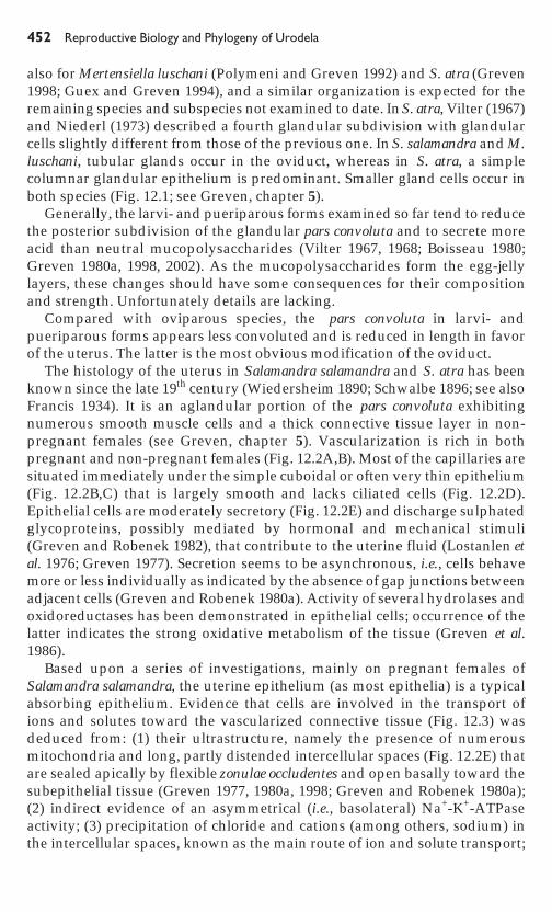
Uterine changes in the course of pregnancy are more conspicuous in thepueriparous Salamandra atra (Vilter and Vilter 1962; Niederl 1981; Guex andGreven 1994), but flattened and cuboidal epithelial cells occur in bothpregnant and non- pregnant females (Fig. 12.4A,B). When larvae have reachedSchwalbe stage I (see 12.5.5), cell and nuclear volume of epithelial cellsincrease continuously, indicating altered metabolic activity of the uterus. Moreprominent cyclic changes, however, occur in a specialized area of the uterus,the zona trophica (see 12.5.5).
12.5 MOTHER-OFFSPRING RELATIONSHIPS
The length of gestation varies from a few months to some years depending onthe species and climatic conditions. Gestation in Salamandra salamandra hasbeen reported or estimated to be five to 14 months; in S. atra and S. lanzai, upto four years; in S. atra aurorae, up to three years; and in Mertensiella luschani,
Fig. 12.2 The uterus of a pregnant female of Salamandra salamandra. A. Densityof blood vessels in the uterine wall as revealed by ink injection (transparency).From Greven, H. 2002. Bonner zoologische Monographien 50: 25-61, Fig. 3. B.Extremely thin (arrow) uterine epithelium with underlying capillaries. Semithin epoxysection stained with toluidine blue-borax. C. Thin epithelium. TEM-micrograph.D. The uterine epithelium is unspecialised, and ciliated cells are lacking. SEM-micrograph. E. Adjacent epithelial cells. Note secretory granules near the apicalplasmalemma, the numerous mitochondria and the partly widened intercellularspace. TEM-micrograph. Abbreviations: ca, capillary; i, intercellular space; lu, uterinelumen; nu, nucleus. Bar for C and E = 1 mm, for D = 10 mm.

�� ����������� ������������������������������
Fig
. 12
.3S
ugge
sted
orig
in a
nd t
rans
port
of
sol
utes
(w
ater
, io
ns,
etc.
) in
the
ute
rus
of p
regn
ant
Sal
aman
dra
sala
man
dra.
Bla
ckar
row
s: a
ctiv
e tr
ansp
ort;
clea
r ar
row
: pa
ssiv
e tr
ansp
ort.
Sod
ium
ent
ers
the
cell
pass
ivel
y, m
oves
dow
n th
e io
n´s
elec
troc
hem
ical
grad
ient
acr
oss
the
lum
enal
cel
l m
embr
ane
and
is p
umpe
d su
bseq
uent
ly f
rom
the
cel
lula
r co
mpa
rtm
ent
into
the
int
erce
llula
rsp
aces
. T
he e
ntry
of
sodi
um c
reat
es a
loc
al i
ncre
ase
in o
smot
ic p
ress
ure
whi
ch d
raw
s w
ater
and
sol
utes
fro
m t
he l
umen
and
the
cells
int
o th
is r
egio
n an
d to
war
d th
e su
bepi
thel
ial
tissu
e. L
arva
e al
so h
ave
to t
ake
up s
olut
es a
nd w
ater
. F
rom
Gre
ven,
H.
1998
.Jo
urna
l of
Exp
erim
enta
l Z
oolo
gy 2
82:
507-
525,
Fig
. 8.

�������������� ��������� ���
up to a little more than one year (Joly 1968; Joly and Picheral 1972; Özeti 1979;Guex 1994; Miaud et al. 2001). Maternal-embryonic interactions during thistime involve respiratory, osmoregulatory, endocrinological, immunological,and trophic relationships.
There is evidence that intrauterine larvae of Salamandra salamandra stopor at least slow down growth (judged by the ossification of the skeleton;Amend and Greven 1996) in winter-time until spring when most parturitionstake place (see also Gasche 1939). Continuous growth of the often numerouslarvae during hibernation could cause some problems regarding the spaceand the limited yolk supply. Tubular bones in intrauterine S. atra showdevelopment throughout the intrauterine period (Fachbach 1988). Lines ofarrested growth, however, occur in the humerus of intrauterine larvae ofS. lanzai, which results in an estimation of the gestation period of up tofour years (Miaud et al. 2001).
In Salamandra salamandra, a central role of the thyroid gland is suggested.The thyroid gland of intrauterine larvae is relatively active just before and atthe beginning of hibernation. Thus, it is speculated that a relative high thyroxinconcentration could keep low the level of the somatotropic hormone prolactin
Fig. 12.4 Uterine epithelium of a pregnant female of Salamandra atra. Note thedifferent aspect of epithelial cells and apical secretory granules. A. Adjacentcuboidal cells. B. Adjacent cells with a more widened intercellular space: comparewith Fig. 12.2E. TEM-micrographs. Abbreviations: i, intercellular space; lu, uterinelumen; nu nucleus. Bar for A = 10 mm and for B = 1 mm.
lu

��� ����������� ������������������������������
and, consequently, could inhibit growth. Thyroid hormones also increaseactivity of the urea cycle, necessary for ureotelism (see 12.5.3) achievedby the intrauterine larvae (Schindelmeiser 1985). Unfortunately, details andcomparable studies on other larvi- or pueriparous species do not exist.
Intrauterine larvae of Salamandra atra and S. salamandra and even larvae ofthe oviparous Triturus alpestris are able to control surface tension by substancesthat appear to be secreted by the skin. Only small amounts of fluid are necessaryto keep the embryo covered with the uterine fluid. Substances from the epidermishave been supposed to serve also as signals for the female to recognizepregnancy or readiness for birth (Guex and Greven 1994).
12.5.1 EndocrinologyThere are only a few investigations regarding the endocrinology of larvi- andpueriparity. As in other urodeles, oocyte maturation coincides with highlyactive glands in the oviduct (Joly and Picheral 1972; Zakrzewski 1976).Concentrations of steroids during the reproductive cycle of female Salamandrasalamandra terrestris and S. infraimmaculata (with an annual cycle) and S.salamandra fastuosa (with a biennial cycle at higher elevations) have beenmeasured in the ovary and the serum of females. Levels increase afterovulation to the end of vitellogenesis, and gonadotropic cells of the pituitaryare active during vitellogenesis (Garnier et al. 1986; Joly et al. 1994; Degani etal. 1997). Administration of estrogen provokes hypertrophy of the secretingoviduct of S. atra (Vilter and Vilter 1962; Guex and Greven 1994).
However, understanding of the endocrine control of reproduction is verylimited. As judged by histology (Salamandra atra; Harms 1946; Vilter and Vilter1960, 1964) as well as histochemistry and ultrastructure (S. salamandra, Jolyand Picheral 1972), corpora lutea are present during the gestation period. Inpregnant females of S. atra, they are present in large numbers and persist forthe first two years of pregnancy, but gradually became reduced towards theterm of parturition. They may control oogenesis and activity of oviductalglands and stage I and stage II features of the uterus. The sharp decline of thenumber of corpora lutea coincides roughly with the onset of oocyte growth andthe formation of the zona trophica at the transition of stage II to stage III ( forstaging see 12.5.5; Häfeli 1971; Niederl 1981; Guex and Greven 1994). Corporalutea of S. salamandra show steroidogenic activity and steroid metabolism,but their exact role is unclear as embryos remain alive and develop afterovariectomy. In ovariectomized pregnant animals, the epithelium seems to bedisordered, and the uterus is less supplied with blood (Joly and Picheral 1972;Lostanlen et al. 1976). However, the ultrastructure of the epithelial cells is notextraordinarily affected by any stage of pregnancy.
Neurohypophysial hormones evoke strong contractions of the oviducts ofSalamandra salamandra and Salamandra atra. Experiments on isolated oviductsof non-pregnant females show strong reactions to vasotocin and extractsof the animal’s own pituitary gland. Reactions to arginine vasopressin andoxytocin were less intense. Pregnant females of S. salamandra injected with

�������������� ��������� ���
high doses of vasotocin intramuscularly gave birth to their larvae (Heller et al.1970). This was difficult to achieve in S. atra females at stage III. Perhaps asignal is needed from the intrauterine offspring about its developmentalstatus, which permits the uterus to react. However, in S. salamandra birth oflarvae extends over several days and in S. atra a few weeks may pass betweenparturition of the two young (Greven and Guex 1994; see 12.5.6.). Changes ofsensitivity dependent on the stage of the reproductive cycle and seasons havenot been investigated. Previous studies by Gasche (1942), who inducedpremature birth in S. salamandra with several hypophysial hormones andexternal stress (light, temperature, etc.), suggest a greater sensitivity toward thespring and even differences of sensitivity among populations.
12.5.2 Respiration and Gas ExchangeInformation on respiration and gas exchange is deduced from anatomical andhistological studies. The rich vascularization of the uterus is conspicuous.Length of the network of blood vessels in the uterus of a pregnant, medium-sized female Salamandra salamandra was calculated to be 38 m (Greven andGuex 1994; Fig. 12.2A). There is no evidence, however, that the length or densityof the network increases during pregnancy. Possibly, blood pressure increasesand causes dilatation of vessels (Kaufman 1913; Greven 1998; Greven andGuex 1994). Thinness of the epithelium (in some areas < 3 mm) and theunderlying capillaries reduce diffusion passage-ways considerably. Larvae ofsalamanders are equipped with long filamentous gills that are povided withnumerous ciliated cells (Greven 1980c; Guex and Greven 1994). Cilia keep theintrauterine fluid in motion. Gills reach their maximal relative length in S. atraat the end of stage II (see 12.5.5). Gills surely absorb oxygen from the highlyvascularized uterus either via the egg-jelly that encases the embryos of manylarviparous species or directly, when the larva is hatched. In addition, thesurface epithelium also may be regarded as a respiratory surface.
To my knowledge, physiological studies on respiration and gas exchangedo not exist, but differences in embryonic and maternal blood oxygen affinitycan be expected.
12.5.3 Osmoregulation and ExcretionIntrauterine development also poses problems with osmoregulation and theremoval of metabolic wastes from the uterine environment.
Ion content (measurements of sodium only) of the uterine fluid of Salamandrasalamandra correspond to that of the blood during pregnancy (approximately80 mmol/l), whereas it is considerably higher (ca. 290 mmol/l) in non-pregnantanimals (Greven 1998; Greven and Guex 1994). This indicates a maternallycontrolled uterine environment, but changes in the course of pregnancy areunknown. Also, larvae are participants of the uterine fluid to some extent, butagain investigations on this subject are lacking. The epidermis and the thingill epithelium of intrauterine larvae of S. salamandra show, as expected,

�� ����������� ������������������������������
chloride in the intercellular spaces and Na+-K+-ATPase activity (Lewinsonet al. 1987a; Greven and Guex 1994). Gills (Greven 1980c) and epidermis(Lewinson et al. 1982, 1984) possess two types of mitochondria-rich cellsthat reach the surface of the epithelium. They were more intensely investigatedonly after birth and show carboanhydrase-activity that increases during larvaldevelopment. High Na+-concentrations inhibit this activity. Mitochondria-rich cells have been suggested to be involved in gas exchange and acid-base regulation (Lewinson et al. 1982, 1984, 1987b). A study of Restaniand Pederzoli (1997) on epidermal mitochondria-rich cells in newborn S.salamandra revealed expression of adhesion molecules and glycoproteins.These molecules perhaps maintain the architecture of skin components. Nofurther studies are available.
Metabolic wastes have to be stored or to be removed via the blood vessels inthe uterine connective tissue. Intrauterine larvae of Salamandra salamandra aresensitive to ammonia, but can tolerate concentrations of urea that exceed thosein the uterus. Content of urea nitrogen in the blood plasma and uterine fluidof pregnant females is higher than in non-pregnant females, suggestingureotelism of larvae. Activity of the hepatic ornithine-urea cycle (measured asactivity of hepatic arginase) is known. When released in water, larvae returnonly partly to ammonotelism (Schindelmeiser and Greven 1981; Schindelmeiseret al. 1983). In S. atra, increasing urea amounts also have been demonstrated inthe uterine fluid during pregnancy (Guex and Greven 1994). The same can bepredicted for the other species, because of the restricted amount of fluid withinthe uterus.
12.5.4 ImmunologyThe intrauterine embryo as well as spermatozoa in the spermatheca andoviduct (Sever 1992) can be regarded as an allograft in their maternal host.Allograft rejection and immune reactions have been shown only toward theintrauterine offspring in Salamandra salamandra. In a series of experimentswith cells in culture, Badet (1984), Badet et al. (1980), and Chateaureynaud etal. (1979) demonstrated that only the serum of pregnant females inhibits acytotoxic reaction of spleen cells, also of pregnant females, against larval cells.One fraction of the serum specifically protects embryonic epithelial cells; asecond one, however, is unspecific. An IgM and an alpha-2-macroglobulin,the latter linked with immunosuppressive properties only during pregnancy,is involved in this process. The protective effect appears to be enhanced withincreasing number of embryos in the uterus, and cytotoxicity and protection islargely specific for the female’s own embryos.
12.5.5 NutritionIn larvi- and pueriparous salamanders, the nutritive support of the youngduring development ranges from the initial yolk stores (lecithotrophy) tooophagy and adelphophagy, both of which are forms of embryonic cannibal-ism, and epitheliophagy. To facilitate nutrient transfer, some adaptations have

�������������� ��������� ���
evolved, probably independently in part. With the exception of Salamandraatra, maternal tissue appears not to be specialized for nutrient transfer butprecocial hatching to ingest food becomes important in some taxa.
Larvae of Salamandra salamandra and some other species and subspecies arearranged like a roll of coins within the uterus and are encased in jelly-layersto full term. Occasionally, unfertilized, but intact eggs can be found betweenthe developing offspring, but these eggs do not serve as food (e.g. Kaufman1913; Gasche 1939). Nutrients other than yolk have to penetrate the eggenvelope. Larvae of S. salamandra take up amino acids that crossed the egg-jelly, however, in negligible amounts (Lostanlen et al. 1976), and amino acidsreach the uterine lumen in S. atra during stage II and III (Guex and Greven1994). Consequently, dry weight decreases during the gestation period(Kaufman 1913; Gasche 1939; Joly 1968; Greven and Guex 1994). Larvae canbe removed from the uterus very early in development and reared in water(Gasche 1939).
In other species and subspecies, nutrients additional to the yolk aresupplied during development and that requires hatching within the uterus.Salamandra salamandra bernardezi from Oviedo (Spain) gives birth to a few fullymetamorphosed, relatively large young (Wolterstorff 1928). Developmentalrates are, however, heterogeneous. Intrauterine larvae feed on degraded,unfertilized eggs (oophagy), that form an embryotrophe, and on less developedsiblings (adelphophagy) (Bas and Gasser 1994; Guex 1994; Dopazo andAlberch 1994; Thiesmeier et al. 1994). Dopazo and Alberch (1994) describedvariation in the pattern of pueriparity in this species: (1) oophagy andadelpho-phagy, facilitated by a great number of unfertilized eggs andheterogenous development, produced large metamorphosed young, as well as(2) a considerable reduction of clutch size and higher fertilization rate; lack ofintrauterine feeding produced smaller transformed juveniles (see alsoAlcobendas et al. 1996).
The behavior of Salamandra salamandra fastuosa is more plastic. The numberof fertilized and unfertilized eggs is variable as well as the size and weightand the developmental stage of larvae. Degrading unfertilized eggs andsmaller siblings are cannibalized, and the same female can bear facultativelynumerous larvae and/or fully metamorphosed young (Joly 1968; Gasser andJoly 1972; Dopazo and Alberch 1994). Oophagy and adelphopagy lead tostrong intrauterine selection.
Mertensiella luschani helverseni normally gives birth to two fully metamor-phosed young. In the uterus they feed on disintegrated eggs. Very likely, azona trophica does not exist (Guex 1994).
With respect to pueriparous salamanders most data are available fromSalamandra atra (among others, Czermak 1843; Wiedersheim 1890; Schwalbe1896; Fachbach 1969; Häfeli 1971; Guex and Chen 1986; review of the olderliterature in Guex and Greven 1994). Young, usually one per uterus, feed onthe yolk when still surrounded by the egg-jelly (lecithotrophic phase; Schwalbestage I; Fig. 12.5C). Once they have left the jelly (Schwalbe stage II), they feed

��� ����������� ������������������������������
on disintegrating eggs (Fig. 12.5D). During transition from stage II to stage III,the position of the larvae within the uterus is fixed; metamorphosis begins(Fig. 12.5E). In stage III, the embrytrophic eggs are consumed and the youngmeasure 4-5 cm in length (Fig. 12.5F). Stage III larvae can be removed from theuterus and reared in water.
Gestation cycles in Salamandra atra last up to 4 years, depending on climaticconditions that are related to the altitude at which individuals are found (seeFig. 12.8; e.g., Wunderer 1909; Vilter and Vilter 1960, 1964; Häfeli 1971; Guexand Greven 1994). Secretion from the uterine epithelium (previously called“uterine milk”) starts with Schwalbe stage II and was assumed to be theessential source for nutrition (Vilter and Vilter 1964; Niederl 1981). There isno evidence for uptake of particulate nutritive substances across the gills assupposed previously (Schwalbe 1896; Häfeli 1971). Uptake of low molecularweight substances across the gills or the surface epithelium, although sus-pected, has never been proven. Of greater importance is a proliferating zone atthe anterior end of the uterus termed the zona trophica by Fachbach (1969) butinitially described in 1890 (Wiedersheim 1890; see Guex and Greven 1994).Cyclic changes were intensely studied by Guex and Chen (1986) and reviewedby Guex and Greven (1994) and Greven (1998). In this zone, mitotic activity isseen (Fig. 12.6 A), and large epithelial cells bulge into the uterine lumen andbecome detached from the underlying connective tissue, probably by apoptoticprocesses and/or necroses (Fig. 12.6B,C). The young ingest cells floating inthe uterine lumen. When positioned with their head to the nutritive zone,embryos may scrape off cells from this area with the help of special toothedareas (“Zahnfelder”) of the upper and lower jaw (Greven 1984; Guex andChen 1986; Fig. 12.5G). Often more than the half area of the zona trophica isfree of epithelial cells independent of the position of the larvae and even bloodvessels will be opened (Fig. 12.6B,C,E). Surprisingly, cells of the trophic zonedo not accumulate glycogen or lipids in considerable amounts (Fig. 12.6C).
The zona trophica forms only in presence of embryos of the Schwalbe stageIII, and its formation cannot be induced by progesterone and/or estrogen.Thus, it develops probably independent from maternal hormones; induction
Fig. 12.5 Pregnancy stages (according to Schwalbe 1896) and position of youngin the uterus of Salamandra atra (A-C, E, F) and Salamandra atra aurorae (D). A.Female near ovulation. B. The embryonic egg enters the uterus. C. Larvae a shorttime before hatching, stage I. Note the swollen jelly envelope. D. Larvae at earlystage II within the creamy mass of embryonic eggs. E. Transition from stage II tostage III. Note twins at the right side and the whitish zone (arrows) of the developingzona trophica. The head of the larva at the left side is directed to the cloaca, thatof one larva at the right side to the zona trophica. F. Stage III larvae. From Guex, G.-D. and Greven, H. 1994. Pp. 161-208. In H. Greven and B. Thiesmeier (eds),Mertensiella Supplement 4, DGHT, Bonn, Germany, Figs. 1a-d, f,h; Figs. 1b and 1cin this article from Häfeli, H.P. 1971. Revue Suisse de Zoologie 78: 235-293, PlateVI, Fig. 2, and Plate VII, Fig. 1. G. Dentition of the lower jaw in a stage III larvae.SEM-micrograph.

�������������� ��������� ���

��� ������ �������������������������������
may be triggered by secretions of the embryo (e.g., by prostaglandins). Theepithelium in this zone continuously regenerates during the period, in whichthe offspring feeds. After birth of the fully transformed young, it is restoredcompletely and looks like the normal uterine epithelium. Alterations of theuterus wall during the reproductive cycle including the zona trophica areillustrated in Fig. 12.7 and the reproductive cycle of Salamandra atra at differentaltitudes in Fig. 12.8 (Guex and Chen 1986; Guex and Greven 1994; Greven1998). Guex (1994) showed that the trophic zone also is present in S. atraaurorae.
The offspring feeding in the uterus stores food in its stomach (Greven andGuex 1994; Guex and Greven 1994) and defecates in the uterus (G.-D. Guex,personal communication).
As yet, epitheliophagy that probably is preceded by oophagy andadelphophagy appears to be realized only in the Salamandra atra group. Thephenomenon appears correlated with a long period of gestation that again isrelated to the duration of winter and the general low temperatures in thehabitat of these species. Metamorphosis starts already at the transition fromstage II to stage III and extends over a considerably long period (see Fig. 12.8).
12.5.6 ParturitionNothing is known on factors signaling female readiness for birth. Contractionsof the uterine muscularis induced by neurohypophysial hormones (12.5.1)may help to expel the young, but in Salamandra atra, a juvenile left the uterusof an anesthetized females (Häfeli 1971). A direct effect of the rich adrenergicinnervation present in the uterus of S. salamandra on the control of thecontraction of the uterine smooth muscle seems not probable because uterinemuscles in general are spontaneously active (Greven et al. 1983).
Pieper (1970) and later Özeti (1979) recorded parturitions of Mertensiellaluschani in the laboratory but did not describe the process in detail. Parturitionsin Salamandra salamandra terrestris and S. salamandra salamandra take place inspring predominantly after hibernaton (e.g., Zakrzewski 1972) and have beenrepeatedly observed. Berweger (1926) observed a female in the shallow waterwith her head above the surface who rested her forelegs on the ground. Afterextending her hind-limbs, she gave birth to free larvae and larvae surroundedby the egg-jelly. This process was accompanied by convulsive contractions of
Fig. 12.6 Zona trophica in pregnant Salamandra atra. A. Uterine epithelium nearthe zona trophica with cuboidal cells. Mitosis indicated by an arrow. B. Zonatrophica at the cranial portion of the uterus. Note the irregularly shaped epithelialcells, some floating in the uterine lumen, and regions devoid of cells (arrowhead).A and B are semithin epoxy sections stained with toluidine blue-borax. C. Largeepithelial cells and area free of them (arrows). TEM-micrograph. D. Intact epitheliumof the zona trophica. E. Zona trophica showing protruding cells (left side) areasdevoid of cells (right side). D and E are SEM-micrographs. Courtesy of Dr. G.-D.Guex. Bar for C = 10 mm, for D = 20 mm and for E = 100 mm.

�������������� ��������� ���

��� ������ �������������������������������
Fig. 12.7 Alterations of the uterus wall during the reproductive cycle in Salamandraatra. The presence of a living embryo is necessary for the development of the zonatrophica. The process of necrosis and of apoptosis and subsequently that ofregeneration is patchy in appearance. From Guex, G.-D. and Greven, H. 1994. Pp.161-208. In H. Greven and B. Thiesmeier (eds), Mertensiella Supplement 4, DGHT,Bonn, Germany, Fig. 8.
the body and discharge of mucus. Birth of altogether 46 larvae and threeabortive eggs lasted three days. The largest number of larvae was born onthe first day. Apart from the mucus discharge and the strong convulsivecontractions, these observations were often confirmed later, i.e., the femaleresting on the fore-limbs only, and parturition of free and encased larvaecovering several days. Characteristic patterns of the female immediately beforeand during birth are lateral spreading, rotation, and bending of the hind-limbs that elevates the body, and raising the tail. In most cases the larvae areborn quickly with either the head or the tail showing first, and without or onlywith remains of the egg-capsule (Fig. 12.9A; e.g., Szabo 1959; Greven 1976).

�������������� ��������� ���
Fig
. 12
.8R
epro
duct
ive
cycl
e of
Sal
aman
dra
atra
. M
atin
g pe
aks
in M
ay a
nd J
une
at 6
50 m
a.s
.l. a
nd i
n Ju
ne a
nd J
uly
at 1
700
ma.
s.l.
Mat
ings
can
be
obse
rved
, ho
wev
er,
until
the
end
of
the
seas
on.
Fer
tiliz
atio
n do
es n
ot n
eed
to c
oinc
ide
with
mat
ing
beca
use
sper
m a
re s
tore
d in
the
spe
rmat
heca
e. T
rans
ition
fro
m s
tage
II
to s
tage
III
coin
cide
s w
ith t
he f
orm
atio
n of
the
zon
a tr
ophi
ca a
ndro
ughl
y w
ith t
he d
eclin
e of
cor
pora
lut
ea a
nd t
he r
esum
ptio
n of
ovi
duct
al a
nd o
varia
n ac
tivity
. F
rom
Gue
x, G
.-D
. an
d G
reve
n, H
.19
94.
Pp.
161
-208
. In
H.
Gre
ven
and
B.
Thi
esm
eier
(ed
s),
Mer
tens
iella
S
uppl
emen
t 4,
DG
HT,
Bon
n, G
erm
any,
Fig
. 9.

�������������� ��������� ���
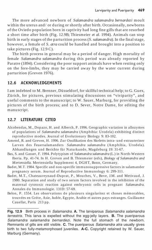
Fig. 12.9 Birth process in Salamandra. A. The larviparous Salamandra salamandraterrestris. This larva is expelled without the egg-jelly layers. B. The pueriparousSalamandra salamandra bernardezi. Note the full stomach of the newborn.Remnants of gills are still visible. C. The pueriparous Salamandra atra usually givesbirth to two fully-metamorphosed juveniles. A-C. Copyright retained by W. Sauer,Marburg (Germany).
The more advanced newborn of Salamandra salamandra bernardezi moultwithin the uterus and/or during or shortly after birth. Occasionally, newbornsof the Oviedo population born in captivity had long fine gills that are resorbeda short time after birth (Fig. 12.9B; Thiesmeier et al. 1994). Animals can stopbirth in early stages of the parturition process (S. salamandra). In the final stage,however, a female of S. atra could be handled and brought into a position totake pictures (Fig. 12.9 C).
The birth process in general may be a period of danger. High mortality offemale Salamandra salamandra during this period was already reported byParatre (1894). Considering the poor support animals have when resting onlyon the fore-limbs, they may be carried away by the water current duringparturition (Greven 1976).
12.6 ACKNOWLEDGEMENTS
I am indebted to M. Brenner, Düsseldorf, for skillful technical help; to G. Guex,Zürich, for pictures, previous stimulating discussions on “viviparity”, anduseful comments to the manuscript; to W. Sauer, Marburg, for providing thepictures of the birth process; and to D. Sever, Notre Dame, for editing themanuscript.
12.7 LITERATURE CITED
Alcobendas, M., Dopazo, H. and Alberch, P. 1996. Geographic variation in allozymesof populations of Salamandra salamandra (Amphibia: Urodela) exhibiting distinctreproductive modes. Journal of Evolutionary Biology 9: 83-102.
Amend, R. and Greven, H. 1996. Zur Ossifikation des Skeletts intra- und extrauterinerLarven des Feuersalamanders Salamandra salamandra (Amphibia, Urodela).Abhandlungen und Berichte für Naturkunde, Magdeburg 19: 31-67.
Bas, S. and Gasser, F. 1994. Polytypism of Salamandra salamandra (L.) in North-WesternIberia. Pp. 41-74. In H. Greven and B. Thiesmeier (eds), Biology of Salamandra andMertensiella. Mertensiella Supplement 4, DGHT, Bonn, Germany.
Badet, M.T. 1984. Specific and non-specific immunosuppressive factors in salamanderpregnancy serum. Journal of Reproductive Immunology 6: 299-311.
Badet, M.T., Chateaureynaud-Duprat, P., Mouches, V., Bove, J.M. and Metivaud, J.1980. Separation and study of two serum factors involved in the inhibition of thematernal cytotoxic reaction against embryonic cells in pregnant Salamandra.Annales du Immunologie. 131D: 57-69.
Belon, P. 1554. Les observations de plusieurs singularitez et choses mémorables,trouvées en Gréce, Asie, Judée, Egypte, Arabie et autres pays estranges. GuillaumeCauellat, Paris 213 pp.

��� ����������� ������������������������������
Berweger, L. 1926. Die Entwicklung der pigmentführenden Zellen in der Haut vonSalamandra. Zeitschrift für mikroskopisch-anatomische Forschung 7: 231-294.
Blackburn, D.G. 1995. Discrepant usage of the term ‘ovoviviparity’ in theherpetological literature. Herpetological Journal 4: 65-72.
Boisseau, C. 1980. Étude ultrastructurale de l´oviducte du Triton Pleurodeles waltlMichah. V. Ultrastructure et cytochimie de l´oviducte postérieur et de l´ »uterus »de la femelle adulte. Annales de Sciences Naturelles, Zoologie et Biologie Animale(Paris) 2 : 67-89.
Briegleb, W. (1962): Zur Biologie und Ökologie des Grottenolms (Proteus anguineusLaur. 1768). Zeitschrift für Morphologie und Ökologie der Tiere 51: 271-335.
Czermak, J.J. 1843. Beiträge zur Anatomie und Physiologie des schwarzenSalamanders. Medicinische Jahrbücher des kaiserlich-königlichen österreichischenStaates 45: 1-13.
Chateaureynaud, P., Badet, M.T. and Voisin, G.A. 1979. Antagonistic maternal immunereactions (rejections and facilitation) to the embryo in the urodele amphibianSalamandra salamandra Lin. Journal of Reproductive Immunology 1: 47-60.
Degani, G., Sharon, R. and Warburg, M. 1997. Ovarian steroid levels in Salamandrasalamandra infraimmaculata during the reproductive cycle. General and ComparativeEndocrinology 106: 356-360.
Dopazo, H. and Alberch, P. 1994. Preliminary results on optional vivipariy andintrauterine siblicide in Salamandra salamandra populations from Northern Spain.Pp. 125-137. In H. Greven and B. Thiesmeier (eds), Biology of Salamandra andMertensiella. Mertensiella Supplement 4, DGHT, Bonn, Germany.
Fachbach, G.1969. Zur Evolution der Embryonal- bzw. Larvalentwicklung beiSalamandra. Zeitschrift für zoologische und systematische Evolutionsforschung7:128-145.
Fachbach, G. 1976a. Biologie, Taxonomie und phylogenetische Beziehungen derverschiedenen Unterarten von Salamandra salamandra im Bereich der IberischenHalbinsel. Zeitschrift für zoologische und systematische Evolutionsforschung 14:59-78.
Fachbach, G. 1976b. Biologie, Taxonomie und phylogenetische Beziehungen derverschiedenen Unterarten von Salamandra salamandra im Bereich der IberischenHalbinsel. Zeitschrift für zoologische und systematische Evolutionsforschung14: 81-103.
Fachbach, G. 1988. Röhrenknochenentwicklung und Altersbestimmung bei Salamandraatra Laur. 1768 (Urodela, Salamandridae). Zoologischer Anzeiger 221:188-200.
Francis, E.T.B. 1934. The Anatomy of the Salamander. Clarendon Press, Oxford, UK.381 pp.
Garnier, D.H. and Joly, J. 1991. Sexual steroids in Salamandra salamandra (L.) duringontogeny. General and Comparative Endocrinology 81: 83-92.
Gasche, P.1939. Beiträge zur Kenntnis der Entwicklungsgeschichte von Salamandrasalamandra L. mit besonderer Berücksichtigung der Winterphase, der Metamor-phose und des Verhaltens der Schilddrüse. Revue Suisse de Zoologie 46: 403-548.
Gasche, P. 1942. Beeinflussung der Larvenablage von Salamandra salamandra L.Verhandlungen der Naturforschenden Gesellschaft Basel 53: 246-264.
Gasser, F. and Joly, J.M.J. 1972. Existénce d´un cycle sexuel biennal chez la femelle deSalamandra salamandra fastuosa Schreiber (Urodéle, Salamandridae) á différentesaltitude dans les Pyrénees centrales: influence des facteurs génetiques et climatiques.Annales des Sciences Naturelles, Zoologie et Biologie Animale (Paris) 14: 427-444.

�������������� ��������� ���
Greven, H. 1976. Notizen zum Geburtsvorgang beim Feuersalamander, Salamandrasalamandra (L.). Salamandra 12: 87-93.
Greven, H. 1977. Comparative ultrastructural investigations of the uterine epitheliumin the viviparous Salamandra atra Laur. and the ovoviviparous Salamandrasalamandra (L.) (Amphibia, Urodela). Cell and Tissue Research 181: 215-237.
Greven, H. 1980a. Licht- und elektronenmikroskopische Untersuchungen zur Strukturund Histochemie des Oviduktepithels von Salamandra salamandra (L.) (Amphibia,Urodela). Zeitschrift für mikroskopisch-anatomische Forschung 94: 387-429.
Greven, H. 1980b. Ultrahistochemical and autoradiographic evidence for epithelialtransport in the uterus of the ovoviviparous salamander, Salamandra salamandra(L.) (Amphibia, Urodela). Cell and Tissue Research 212: 147-162.
Greven, H. 1980c. Ultrastructural investigations of the epidermis and the gill epitheliumin the intrauterine larvae of Salamandra salamandra (L.) (Amphibia, Urodela).Zeitschrift für mikroskopisch-anatomische Forschung 94: 196-208.
Greven, H. 1981. Na-pump sites in the oviduct of Salamandra salamandra (L.) (Amphibia,Urodela). Experientia 327: 771-772.
Greven, H. 1984. Zona trophica and larval dentition in Salamandra atra Laur. (Amphibia,Urodela): Adaptation to intrauterine nutrition. Verhandlungen der DeutschenZoologischen Gesellschaft 77:184.
Greven, H. 1998. Survey of the oviduct of salamandrids with special reference to theviviparous species. Journal of Experimental Zoology. 282: 507-525.
Greven, H. 2002. The urodele oviduct and ist secretions in and after G. von Wahlert´sdoctoral thesis “Eileiter, Laich und Kloake der Salamandriden”. Bonner zoologischeMonographien 50: 25-61.
Greven, H. and Baldus, B. 1984. The distribution of monosaccharides and hexosaminesin the oviduct of Salamandra salamandra (L.) (Amphibia, Urodela). ComparativeBiochemistry and Physiology 79B: 229-232.
Greven, H. and Guex, G.-D. 1994. Structural and physiological aspects of viviparityin Salamandra salamandra. Pp. 139-160. In H. Greven and B. Thiesmeier (eds),Biology of Salamandra and Mertensiella. Mertensiella Supplement 4, DGHT, Bonn,Germany.
Greven, H., Passia, D., Haider, S.G. and Mockenhaupt, U. 1986. Enzymhistochemieam Uterus des Feuersalamanders. Fortschritte der Fertilitätsforschung 13: 71-74.
Greven, H. and Robenek, H. 1980a. Intercellular junctions in the uterine epitheliumof Salamandra salamandra (L.) (Amphibia, Urodela). Cell and Tissue Research 212:163-172.
Greven, H. and Robenek, H. 1980b. On the lumenal plasmalemma and associatedstructures of the uterine epithelium in pregnant and non-pregnant females ofSalamandra salamandra (L.) (Amphibia, Urodela). European Congress of ElectronMicroscopy 1980, The Hague (EUREM) 2: 34-35.
Greven, H. and Robenek, H. 1982. Further freeze-fracture studies on the uterineepithelium of Salamandra salamandra (L.) (Amphibia, Urodela) using the antibioticfilipin. Cell and Tissue Research 226: 441-447.
Greven, H. and Rüterbories, H.J. 1984. Scanning electron microscopy of the oviductof Salamandra salamandra (L.) (Amphibia, Urodela). Zeitschrift für mikroskopisch-anatomische Forschung 98: 49-62.
Greven, H., Schindelmeiser, J. and Straub, H. 1983. The innervation of the uterus inSalamandra salamandra (L.) (Amphibia, Urodela). A morphological and biochemicalstudy. Cell and Tissue Research 232: 421-431 .

��� ����������� ������������������������������
Greven, H. and Thiesmeier, B. (eds). 1994. Biology of Salamandra and Mertensiella.Mertensiella Supplement 4, DGHT, Bonn, Germany. 454 pp.
Guex, G.-D. 1994. Zur Fortpflanzungsbiologie von Salamandra atra aurorae Trevisan,1982, S. salamandra bernadezi Wolterstorff, 1928, S. salamandra fastuosa Schreiber,1892 und Mertensiella luschani helversenii Piper, 1963. Abhandlungen und Berichtefür Naturkunde, Magdeburg 17: 143-150.
Guex, G.-D. and Chen, P.S. 1986. Epitheliophagy: intrauterine cell nourishment in theviviparous alpine salamander, Salamandra atra (Laur.) Experientia 42: 1205-1218.
Guex, G.-D. and Greven, H. 1994. Structural and physiological aspects of viviparity inSalamandra atra. Pp. 161-208. In H. Greven and B. Thiesmeier (eds), Biology ofSalamandra and Mertensiella. Mertensiella Supplement 4, DGHT, Bonn, Germany.
Häfeli, H.-P. 1971. Zur Fortpflanzungsbiologie des Alpensalamanders (Salamandraatra Laur.). Revue Suisse de Zoologie 78: 235-293.
Harms, J. 1946. Die Fortpflanzung von Salamandra atra. Biologisches Zentralblatt 65:254-267.
Heller, H., Ferreri, E.,and Leathers, D.H.G. 1970. The effect of neurohypophysialhormones on the amphibian oviduct in vitro, with some remarks on the histologyof this organ. Journal of Endocrinology 47: 495-509.
Joly, J. 1968. Données ecologiques sur la salamandre tachetée Salamandra salamandraL. Annales de Sciences Naturelles, Zoologie et Biologie Animale (Paris) 10: 301-366.
Joly, J. and Picheral, B. 1972. Ultrastructure, histochimie et physiologie du folliculepréovulatoire et du corps jaune de l´urodèle ovo-vivipare Salamandra salamandra(L.). General and Comparative Endocrinology 18: 235-259.
Joly, J. and Boisseau, C. 1973. Localisation des spermatozoides dans l´oviducte de lasalamandre terrestre Salamandra salamandra (L.) (Amphibien, Urodèle) au momentde la fecondation. Comptes Rendus Hebdomadaires des Séances de l´Académiedes Sciences, Paris, Série D, Sciences Naturelles 277: 2537-2540.
Joly, J., Chesnel, F., Boujard, D. 1994. Biological adaptations and reproductivestrategies in the genus Salamandra. Pp. 255-269. In H. Greven and B. Thiesmeier(eds), Biology of Salamandra and Mertensiella. Mertensiella Supplement 4, DGHT,Bonn, Germany.
Kaufman, L. 1913. Die Degenerationserscheinungen während der intrauterinenEntwicklung bei Salamandra maculata. Archiv für Entwicklungsmechanik derOrganismen 37: 38-84.
Lewinson, D., Rosenberg, M. and Warburg, M.R. 1982. Mitochondria-rich cells insalamander larva epidermis; ultrastructural description and carbonic anhydraseactivity. Biology of Cell 46: 75-84.
Lewinson, D., Rosenberg, M. and Warburg, M.R. 1984. ‘Chloride-cell´-like mitochon-dria-rich cells of salamander larva gill epithelium. Experientia 40: 956-958.
Lewinson, D., Rosenberg, M. and Warburg, M.R. 1987a. Ultrastructural andcytochemical studies of the gill epithelium in the larvae of Salamandra salamandra(Amphibia, Urodela). Zoomorphology 107: 17-25.
Lewinson, D., Rosenberg, M., Goldenberg, S. and Warburg, M.R. 1987b. Carbonicanhydrase cytochemistry in mitochondria-rich cells of salamander larvae gillepithelium as related to age and H+ and Na+ concentrations. Journal of CellPhysiology 130: 125-132.
Lostanlen, D., Boisseau, C. and Joly, J. 1976. Donneés ultrastructurales etphysiologiques sur l´uterus d´un amphibien ovovivipare Salamandra salamandra(L.) (Amphibia, Urodela). Annales de Sciences Naturelles, Zoologie et BiologieAnimale (Paris) 18: 113-144.

�������������� ��������� ���
Miaud, C., Andreone, F., Ribéron, A., DeMichelis, S., Clima, V., Castanet, J.,Francillon-Vieillott, H. and Guyétant, R. 2001. Variations in age, size at maturityand gestation duration among two neighbouring populations of the alpinesalamander (Salamandra lanzai). Journal of Zoology 254 : 251-260.
Niederl, H. 1981. Anatomische und histologische Untersuchungen des weiblichenund männlichen Geschlechtszyklus von Salamandra atra (Laur.). ZoologischeJahrbücher, Abteilung für Anatomie und Ontogenie der Tiere 106: 46-63.
Özeti, N. 1979. Reproductive biology of the salamander Mertensiella luschaniantalyana. Herpetologica 35: 193-197.
Packard, G.C. 1989. How are reproductive systems integrated and how hasviviparity evolved? Pp. 281-293. In D. B. Wake and G. Roth (eds), ComplexOrganismal Functions: Integration and Evolution in Vertebrates. John Wiley and Sons,New York, NY.
Paratre, R. 1894. Notes sur Salamandra maculosa; sa présence aux environs immédiatsde Paris ; remarques sur sa réproduction; époque de sa parturition; développementde la larve. Memoirs de la Société Zoologique de France 10: 132-160.
Parzefall, J., Durand, J.P.and Sket, B. 1999. Proteus anguinus Laurenti, 1768 –Grottenolm. Pp. 57-80. In K. Grossenbacher and B. Thiesmeier (eds), Handbuchder Reptilien und Amphibien Europas. 4/I. Schwanzlurche (Urodela) I. Aula Verlag,Wiesbaden, Germany.
Peck, A.L. (Translator). 1970. Aristotle. Volume X. History of Animals, Books 4-6.Loeb Classical Library Number 438. Harvard University Press, Cambridge,Massachusetts. 424 pp.
Pieper, H. 1970. Neue Beiträge zur Kenntnis der Herpetofauna der südägäischenInseln. Senckenbergiana Biologica, Frankfurt 51: 55-65.
Polymeni, R.M and Greven, H. 1992. Histology and fine structure of the oviduct inMertensiella luschani (Steindachner, 1891). Pp. 361-365. In Z. Korso and I. Kiss (eds),Proceedings of the Sixth Ordinary General meeting of the Societas EuropaeaHerpetologica, Budapest 1991. Hungarian Natural History Museum, Budapest,Hungary.
Restani, C. and Pederzoli, A. 1997. Cytochemical and immunocytochemical investi-gations on epidermal mitochondria-rich cells in Salamandra salamandra salamandra(L.) larvae. Tissue & Cell 29: 619-625.
Salthe, S.N. and Mecham, J.S. 1974. Reproductive and courtship patterns. Pp 309-521.In B. Lofts (ed.), Physiology of the Amphibia. Vol. II. Academic Press, New York, NY.
Schindelmeiser, J. 1985. Structure and activity of the thyroid gland in intra- andextrauterine larvae of the ovoviviparous Salamandra salamandra (L.) (Amphibia,Urodela). Zeitschrift für mikroskopisch-anatomische Forschung 99: 475-497.
Schindelmeiser, J. and Greven, H. 1981. Nitrogen excretion in intra- and extrauterinelarvae of the ovoviviparous salamander, Salamandra salamandra (L.) (Amphibia,Urodela). Comparative Biochemistry and Physiology 75B: 471-473.
Schindelmeiser, J., Schindelmeiser, I. and Greven, H. 1983. Hepatic arginase activityin intra- and extrauterine larvae of the ovoviviparous salamander, Salamandrasalamandra (L.) (Amphibia, Urodela). Comparative Biochemistry and Physiology75B: 471-473.
Schwalbe, G. 1896. Zur Biologie und Entwicklungsgeschichte von Salamandra atra undmaculosa. Zeitschrift für Biologie 34: 340-396.
Sever, D.M. 1992. Spermiophagy by the spermathecal epithelium of the salamanderEurycea cirrigera. Journal of Morphology 212: 281-290.
��� ����������� ������������������������������
Steinfartz, S., Veith, M. and Tautz, D. 2000. Mitochondrial sequence analysis ofSalamandra taxa suggests old split of major lineages and postglacial recolonizationsof Central Europa from distinct source populations of Salamandra salamandra.Molecular Ecology 9: 387-410.
Stieve, H. 1919. Anatomische Untersuchungen über die Fortpflanzung desGrottenolms (Proteus anguineus Laur.). Anatomische Hefte 56: 403-469.
Szabo, I. 1959. Contributions à l´oecologie de la salamandre tachetée (Salamandrasalamandra L.). Vertebrata Hungarica 1: 35-48.
Thiesmeier, B. and Haker, K. 1990. Salamandra salamandra bernadezi Wolterstorff, 1928aus Oviedo, Spanien, nebst Bemerkungen zur Viviparie in der Gattung Salamandra.Salamandra 26: 140-154.
Thiesmeier, B., Sauer, W., and Mutz, Th. 1994. Birth and dates of offspring ofSalamandra salamandra bernardezi from Oviedo. Pp. 347-354 . In H. Greven and B.Thiesmeier (eds), Biology of Salamandra and Mertensiella. Mertensiella Supplement 4,DGHT, Bonn, Germany.
Veith, M., Steinfartz, S., Zardoya, R., Seitz, A. and Meyer, A. 1998. A molecularphylogeny of “ true” salamanders (family Salamandridae) and the evolution ofterrestriality of reproductive modes. Journal of Zoological Systematics andEvolutionary Research 36: 7-16.
Vilter, V. 1967. Histologie de l´oviducte chez Salamandra atra mature, Urodèletotalement vivipare de haute montagne. Comptes Rendus des Séances de laSoiété de Biologie 161: 260-264.
Vilter, V. 1968. Histochimie quantitative des sècrétions mucipares acides del´oviducte mature de Salamandra atra des Alpes vaudoises. Comptes Rendus desSéances de la Société de Biologie 162: 76-80.
Vilter, A. and Vilter, V. 1962. Role de l´oviducte dans l´uniparité utérine de laSalamandre noire des Alpes orientales (Salamandra atra Laur.). Comptes Rendusdes Séances de la Société de Biologie 156: 49-51.
Vilter, V. and Vilter, A. 1960. Sur la gestation de la salamandre noire des Alpes,Salamandra atra Laur. Comptes Rendus des Séances de la Société de Biologie 154 :290-291.
Vilter, V. and Vilter, A. 1964. Sur l´évolution des corps jaunes ovariens chez Salamandraatra Laur. des Alpes vaudoises. Comptes Rendus des Séances de la Société deBiologie 158: 457-461.
Vilter, V., Lugano, A. and Reymond, E. 1959. Comportement printanier de lasalamandre noire d´altitude (Salamandra atra Laur.) dans ses relations biologiques.Comptes Rendus des Séances de la Société de Biologie 153 : 975-978.
Wahlert, G. von. 1953. Eileiter, Laich und Kloake der Salamandriden. ZoologischeJahrbücher, Abteilung für Anatomie und Ontogenie der Tiere 73: 276-310.
Wake, M.H. 1982. Diversity within a framework of constraints. Amphibian repro-ductive modes. Pp. 87-106. In D. Mossakowski and G. Roth (eds), EnvironmentAdaptation and Evolution. Gustav Fischer, Stuttgart, Germany.
Wake, M.H. 1993. Evolution of oviductal gestation in amphibians. Journal ofExperimental Zoology. 266: 394-413.
Weisrock, D.W., Macey, J.R., Ugurtas, J.H., Larson, A. and Papenfuss, T.J. 2001.Molecular phylogenetics and historical biogeography among salamandrids of the“true” salamander clade: rapid banching of numerous highly divergent lineagesin Mertensiella luschani associated with the rise of Anatolia. Molecular Phylogeneticsand Evolution 18: 434-448.

�������������� ��������� ���
Wiedersheim, R. 1980. Beiträge zur Entwicklungsgeschichte von Salamandra atra.Archiv für mikroskopische Anatomie 36: 469-482.
Wolterstorff, W. 1928. Vollmolch-gebärende Feuersalamander aus Oviedo. Blätterfür Aquarien- und Terrarienkunde 39: 132-133.
Wunderer, H. (1909): Beiträge zur Biologie und Entwicklungsgeschichte desAlpensalamanders (Salamandra atra Laur.) Zoologische Jahrbücher, Abteilung fürSystematik, Geographie und Biologie der Tiere 28: 23-80.
Wunderer, H. 1910. Die Entwicklung der äußeren Körperform des Alpensalamanders(Salamandra atra Laur.). Zoologische Jahrbücher, Abteilung für Anatomie undOntogenie der Tiere 29: 368-414.
Zakrzewski, M. 1976. Development of the female reproductive organ and nominalform of the spotted salamander, Salamandra s. salamandra (L.) in the West Beskidregion (Poland) in the annual cycle. Acta Biologica Cracoviensia 19: 23-38.


![Molecular Phylogeny and Evolution. Introduction to evolution and phylogeny Nomenclature of trees Five stages of molecular phylogeny: [1] selecting sequences](https://img.pdfslide.net/doc/110x75/56649e265503460f94b155ae/molecular-phylogeny-and-evolution-introduction-to-evolution-and-phylogeny.jpg)