Embed Size (px)
Citation preview
REPRODUCTIVE BIOLOGY OF INVERTEBRATES
Edited by
K.G. and RITA G. ADIYODI Vals\u\tina Centre of Inverlehrate Reproduction
Calicut University. Kerala 673635, India
VOLUME IV, PART A Fertilization, Development, and Parental Care
OXFORD & IBH PUBLISHING CO. PVT. LTD. NEW DELHI BOMBAY CALCUTTA

14. SIPUNCULA
MARY E. RICE
Smithsonian Marine Station at Link Port, 5612 Old Dixie Highway, Fort Pierce, Florida 34946, U.S.A.
I. INTRODUCTION
Spawning of sipunculans is generally epidemic; eggs and sperm are released into the sea water where fertilization occurs. Prior to spawning, gametes are stored for a short time in the nephridia where they are accumulated from the coelom via the nephrostomes (see Rice, Volume I). With one known exception (Golfingia minuta), sipunculans are dioecious.
The developmental features of sipunculans align the phylum with the Protos- tomia (see Systematic Résumé). Cleavage is spiral, giving rise to a trochophore larva. The stomodaeum and mouth arise from the site of the blastopore, meso- derm is proliferated from a pair of teloblast cells, and the coelom is formed by schizocoely, or a splitting of mesodermal bands.
The first published account of development in sipunculans was that of Krohn in 1851 on Sipunculus nudus. His treatment of early development was incom- plete but his observations of hatching and larval organization were accurate and laid the foundation for later work by Hatschek (1883). The earliest notable contribution to the knowledge of sipunculan development was made in 1875 by Selenka. Working at Villefranche near Nice, he studied the developmental history of Golfingia elongata. By the successful fertilization of coelomic eggs he was able to observe early cleavage as well as later developmental stages. He was impressed by the similarity of sipunculan development to that of polychaetes and suggested a close relationship of the two groups based on common charac- teristics such as cleavage pattern, origin of nerve cord, and types of ciliary bands. In 1883 Hatschek published a treatise on the development of Sipunculus nudus. His material was collected in the plankton at Messina, Sicily. He des- cribed in detail late cleavage, gastrulation, mesoderm formation, larval organo- génesis, and shedding of the egg envelope. As later investigations have shown, the development of S. nudus is much modified from the more typical pattern of development of other sipunculans. In a series of three papers in 1903, 1904, and 1906 Gerould presented a detailed account of development of Golfingia vulga-
264 REPRODUCTIVE BIOLOGY OF INVERTEBRATES
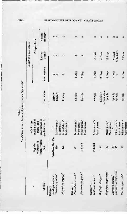
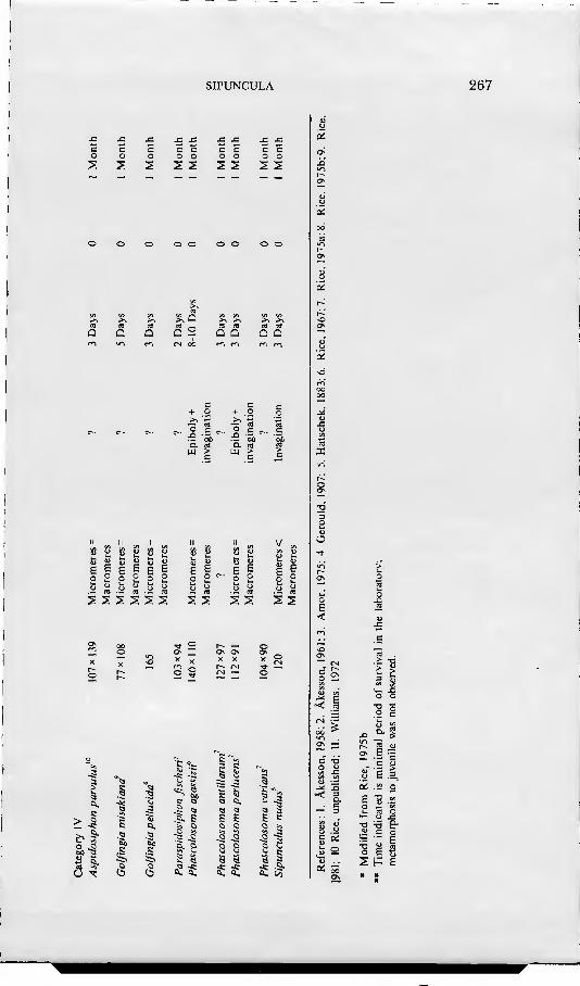
ris and Phascolopsis gouldii, including information on all aspects of the devel- opmental history from gametogenesis and breeding through larval and post-larval development. Using nomenclature established by Conk;lin(1897), he followed cell lineage through the 48-cel] stage, showing that cleavage was of the spiral pattern typical of annelids and molluscs. Gastrulation, mesoderm forma- tion, and organogénesis were described and comparisons made with the find- ings of Selenka (1875) and Hatschek (1883). Apartfrom reports on unidentified oceanic larvae, no studies on sipunculan development were made from the time of Gerould's work until 1958 when Akesson described the development of Phascolion strombi and Golfingia minuta: the latter species was the first known example of direct development in sipunculans. In 1961 Akesson pubhshed an account of development of another species, Golfingia elongata. From addi- tional information provided by a series of recent papers (Rice, 1967, 1975a, b, 1976, 1978; Williams, 1972; Amor, 1975), development is now known for 20 sexually reproducing species of sipunculans. These species, along with selected developmental features, are listed in Table 1.
A review of the known developmental histories of sipunculans reveals four basic patterns of development in the phylum (Table 1). Category I is direct development, in which the embryo develops gradually into the juvenile without passing through a pelagic stage. Category II includes one pelagic stage, the trochopore, which transforms into a vermiform stage and, finally, a juvenile. In the third and fourth categories there are two pelagic stages: the trochophore larva and the pelagosphera larva. The pelagosphera has been defined as a larval stage unique to the Sipuncula which succeeds the trochophore and which is characterized by the loss or reduction of the prototroch and the elaboration of a strongly cihated metatroch as the primary locomotory organ (Rice, 1967). In category III the pelagosphera is lecithotrophic, remaining in the plankton a relatively short time before transforming into the vermiform stage. The pela- gosphera larva of category IV is planktotrophic; it may remain in the plankton up to several months before undergoing a second metamorphosis to the juve- nile. In addition to the references noted in Table 1, there have been numerous studies of planktotrophic pelagospheras of unknown species collected from the plankton of the open ocean (see Rice, 1981, 1985 for reviews).
The developmental features of the four categories designated in Table 1 are closely correlated with yolk content of the egg. Eggs of species in the first three categories have a relatively high concentration of yolk, whereas those of species in the fourth category are low in yolk content. For convenience in presentation and discussion, these four developmental patterns will be referred to frequently in the account given below of embryonic and larval development.
II. FERTILIZATION
At the time of spawning, the eggs of sipunculans are arrested in the first meiotic
SIPUNCULA 265
metaphase (see Rice, Volume I for details of oogénesis and spawning). Matura- tion of the egg is completed following sperm penetration and the subsequent extrusion of the first and second polar bodies. The process of fertilization is then concluded by the formation and union of male and female pronuclei. The initiation of maturation, represented by the breakdown of the germinal vesicle, and the formation of the first meiotic metaphase are prerequisites for the fertilizability of the egg.
Germinal-vesicle dissolution has been reported to occur in fully developed coelomic eggs immediately before nephridial uptake. First noted by Gerouid (1906) in Golfingia viilgaris and Phascolopsis gouldii, breakdown of the germi- nal vesicle of large coelomic oocytes was later reported by Âkesson (1958) in Phascolion strombi about 12 hours before spawning. Similar observations have been reported for Phascolosoma agassizii by Rice (1966).
Little information exists on the regulation of germinal-vesicle breakdown. In vitro studies on maturation of coelomic oocytes of Phascolion strombi have shown that an increase of calcium ions induces dissolution of the germinal vesicle (Pasteéis, 1935). Rice (1966, 1975b), working on coelomic oocytes of Phascolosoma agassizii, found that crude extracts of coelomic sperm, coelomic oocytes, coelomocytes, brain and muscle induced the breakdown of the germi- nal vesicle. Dissolution of the germinal vesicle of coelomic eggs, on transfer to sea water, has been reported in Golfingia pugettensis (Rice, 1967). After five to 10 hours in sea water the germinal vesicle undergoes dissolution and the egg becomes fertilizable. The fertilizability of coelomic eggs in sea water has been reported for Golfingia elongaia (Selenka, 1875) and Golfingia vulgaris (Gerouid, 1906). Attempts to fertilize coelomic eggs in sea water have been unsuccessful in Phascolopsis gouldii (Andrews, 1889), Phascolion strombi (Âkesson, 1958), Golfingia minuta (Gibbs, 1975), G. elongaia and G. rimicola (Gibbs, 1976), and Phascolosoma agassizii (Rice, 1967).
Sperm penetration occurs by formation of a hole in the egg envelope. Sperm entry holes, which may persist for several days in the developing embryo, have been observed in Phascolosoma agassizii, P perlucens, P varians. Goljingia pugettensis, G. pellucida, and Themis te pyro ides (Rice, 1975b). After penetra- tion, the sperm nucleus enlarges and migrates to the centre of the egg to form the male pronucleus. At the same time, maturation of the egg is completed as the first and second polar bodies are extruded, and the egg pronucleus is
formed. On contacting the surface of the egg, the sperm reacts by forming an acro-
somal filament. The acrosomal reaction is most remarkable in Themiste pyroides, in which a thick jelly coat overlies the egg envelope (Rice, 1966). At the time of sperm attachment, the acrosomal filament of the sperm is extended to a length of 50 ¡xm through the jelly coat to the egg envelope. The sperm head lies at the surface of the jelly coat and the sperm tail bends from the head at an angle of 90°. Many sperm may be thus attached to a single egg. The sperm
266 REPRODUCTIVE BIOLOGY OF INVERTEBRATES
c
OH U.
.S D. O O
« c e
o
u <*- C -
• o T3 - DO « " c S ^' 2 .ü 3 " < S •" 7 ë "• • u o c "
u. fa 3
• o .-g y 1- S ^^
••5 c
S S S S
s
il ^ s s e
• .a 5, Q
I'll 8 Si
¡a Ü K Í U
MÛ a ^
.g '5.
"^ -2 >, •- cd o
•a 3'£- "o "o •2 -^ S. S.
O
'ô, u
A
2 S
A A A II A ¡ß Ö E B
g Ö B o ii E
g cd
S S E 2 u
2 Ë
<L>
u M
e I u
O
o cd
5 2 s s
*r\ •«
s s
• in
SS s s 5 5
S
o
• c il fa Ö
3 O
1
^
Í 1 s :: > ^ Ei. à -¡3
^•1 <3
3> S'a i i 1 ^ Öö O cs gfi
SIPUNCULA 267
SS SS SS
5,Q
c C o O >,
o .£ ^• r\ c
a. Q, UJ > LU ^
iß g u ¡g j; u S3 g c Ë Ë 9 a 9
S3 u Ö C
o Ü
ssssss ss ss ss
^ Tf • • ir!
,3 3 c Cl. c
o ^ 00 ^
o X
Cl, a
c
0^ 0-
O Ci,
o o
a 3 a, 1,
3 C
3
3
«1. (/3
O
4J VI
'< 5
c a;
a: 5S
Î3 t/i
n. ;ï «1 111
t _ •= c
Ë -g .Í o w wî L. m O
•5 "ô e-
s Ë s H
268 REPRODUCTIVE BIOLOGY OF INVERTEBRATES
which penetrates leaves behind it a visible track in the jelly. After sperm penetration and before the extrusion of the first polar body, the
cytoplasm of ovoid eggs, such as those of Phascolosoma agassizii, rounds up and separates from the overlying egg envelope at the animal and vegetal poles. ' In the spherical eggs of Golfingia pugettensis and Themiste pyroides, there is a separation of cytoplasm from the egg envelope at the animal pole as the polar bodies are produced.
The events of fertilization following sperm penetration have been described in detail by Gerould (1906) for Golfingia vulgaris and Phascolopsis gouldii. Although he did not observe sperm penetration, Gerould (1906) noted that subsequent to sperm entry the cytoplasmic processes extending through the pores of the egg envelope are withdrawn and that the sperm head, once within the egg, rotates 180°to a position parallel to the egg surface. An aster and centrosome form at the base of the head and, led by the aster, the sperm head moves toward the centre of the egg, leaving a path marking its movement through the cytoplasm. Once in the centre of the egg the sperm nucleus and its astrosphere are enlarged. After sperm penetration the first meiotic metaphase of the egg nucleus proceeds into the telophase and, as division is completed, the first polar body is formed. The division giving rise to the first polar body is equational or longitudinal, and that producing the second polar body is reduc- tional: the reverse of the usual sequence. The number of chromosomes is reduced from 20 to the haploid number of 10 during maturation divisions. After the second polar body is given off, 10 chromatic vesicles are formed which then unite to form the female pronucleus. An associated centrosome lies toward the centre of the egg. The sperm nucleus moves toward the egg nucleus and by the time of contact, the male and female nuclei are approximately equal in size, although the male aster is the more prominent of the two. The first cleavage spindle with 20 chromosomes is soon formed.
III. FMBRYONIC AND LARVAL DEVELOPMENT
A. Cleavage, Blastulation, and Gastrulation
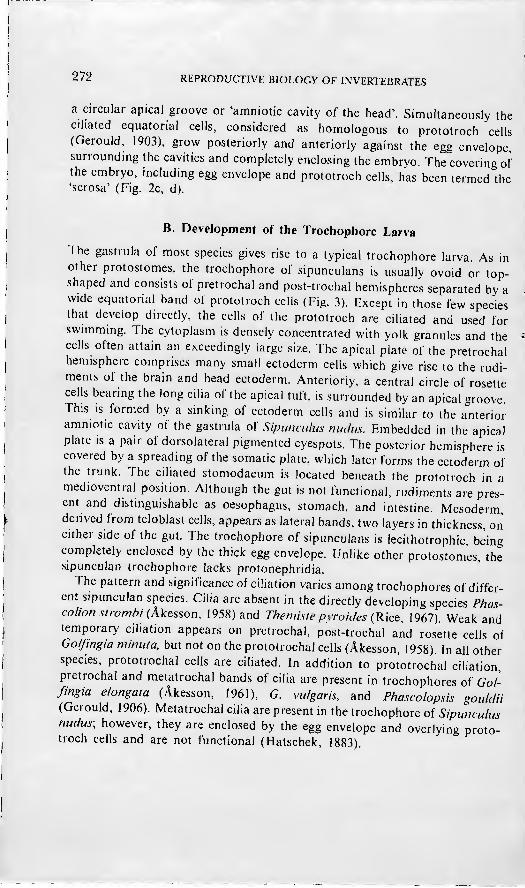
Cleavage in sipunculan eggs is spiral, holoblastic, and unequal. In the only study of cell lineage, Gerould (1906) followed cleavage in the egg of Golfingia vulgaris through the 48-cell stage. The spiral pattern continues to 48 cells, after which it is limited to certain areas of the egg. A variation from typical spiral cleavage is found in the relatively large size of the micromeres at the eight-cell stage. This is reflected in the later prototroch cells. At the 16-cell stage, the micromeres of the A, B, and C quadrants are approximately equal in size and exceed the macromeres with the exception of those in the D quadrant. The 2d or somatoblast is the largest of all cells at this stage and the 2D is next largest. At the 48-cell stage the cross cells of the apical plate (Iq' ) are in the radial
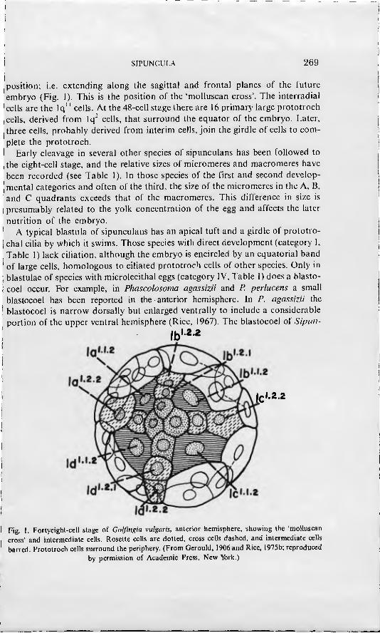
SIPUNCULA 269
position; i.e. extending along tlie sagittal and frontal planes of the future embryo (Fig. 1). This is the position of the 'molluscan cross'. The interradial cells are the Iq" cells. At the 48-cell stage there are 16 primary large prototroch cells, derived from Iq' cells, that surround the equator of the embryo. Later, three cells, probably derived from interim cells, join the girdle of cells to com- plete the prototroch.
Early cleavage in several other species of sipunculans has been followed to the eight-cell stage, and the relative sizes of micromeres and macromeres have been recorded (see Table 1). In those species of the first and second develop- mental categories and often of the third, the size of the micromeres in the A, B, and C quadrants exceeds that of the macromeres. This difference in si/.e is presumably related to the yolk concentration of the egg and affects the later nutrition of the embryo.
A typical blástula of sipunculans has an apical tuft and a girdle of prototro- chal cilia by which it swims. Those species with direct development (category I, Table 1) lack ciliation, although the embryo is encircled by an equatorial band of large cells, homologous to ciliated prototroch cells of other species. Only in blastulae of species with microlecithal eggs (category IV, Table I) does a blasto- coel occur. For example, in Phascolosoma agassizii and P. perlucens a small blastocoel has been reported in the anterior hemisphere. In P. agassizii the blastocoel is narrow dorsally but enlarged ventrally to include a considerable portion of the upper ventral hemisphere (Rice, 1967). The blastocoel of Sipiin-
,bl.2.2
lc'-2.2
Fig. 1. Fortyeight-cell stage of Golfingia vulgaris, anterior hemisphere, showing the 'molluscan cross' and intermediate cells. Rosette cells are doUed, cross cells dashed, and intermediate cells barred. Prototroch cells surround the periphery. (From Gerould, 1906 and Rice, 1975b; reproduced
by permission of Academic Press, New York.)
270 REPRODUCTIVE BIOLOGY OF INVERTEBRATES
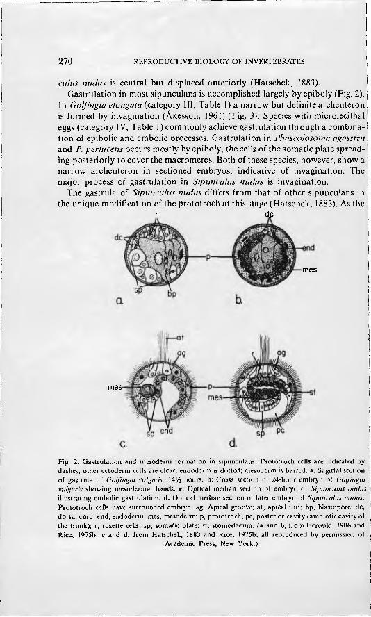
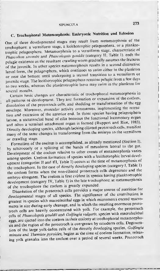
culus mains is central but displaced anteriorly (Hatschek, 1883). ' Gastrulation in most sipunculans is accomplished largely by epiboly (Fig. 2).
In Golfingia elongata (category III, Table 1) a narrow but definite archenteron is formed by invagination (Akesson, 1961) (Fig. 3). Species with microlecithal eggs (category IV, Table 1) commonly achieve gastrulation through a combina- tion of epibolic and embolie processes. Gastrulation in Phascolosoma agassizii, and P. perlucens occurs mostly by epiboly, the cells of the somatic plate spread- ing posteriorly to cover the macromeres. Both of these species, however, show a narrow archenteron in sectioned embryos, indicative of invagination. The major process of gastrulation in Sipunculus nucliis is invagination.
The gastrula of Sipunculus nudus differs from that of other sipunculans in the unique modification of the prototroch at this stage (Hatschek, 1883). As the
r dc
mes
meS'
Fig. 2. Gastrulation and mesüdeim formation in sipunculans. Prototroch cells are indicated by dashes, other ectoderm cells are clear; endoderm is dotted; mesoderm is barred, a: Sagittal section of gastrula of Golfingia viilgaris, 14'/2 hours, b: Cross section of 24-hour embryo of Golfingia vulgwis showing mesodermal bands, e; Optical median section of embryo of Sipunculus nudus illustrating embolie gastrulation. d: Optical median section of later embryo of Sipunculus nudus. Prototroch cells have surrounded embryo, ag, Apical groove; at, apical tuft; bp, blastopore; dc, dorsal cord; end, endoderm; mes, mesoderm; p, prototroch; pc, posterior cavity (amniotic cavity of the trunk); r, rosette cells; sp, somatic plate; st, stomodaeum. (a and b, from Gerould, 1906 and Rice, 1975b; c and d, from Hatschek, 1883 and Rice, 1975b; all reproduced by permission of
Academic Press, New York.)
SIPUNCULA 271
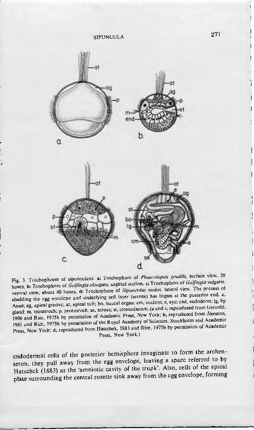
Fig 3 Trochophores of s.punculans. a: Trochophore of Phascolopsis gouldu sudac. v.ew 20 hours b- Trochophore of Golßngia elongaw. sagittal section, c: Trochophore of Golfim,a vulgar. !e•Tal view, about 40 hours, d: Trochophore of Sipuncuk. nudus. lateral v,e^v. The process of Ihling th egg envelope and underlymg cell layer (serosa) has begun at the postenor end a Anus ag ap,cal groove; at, apkal tuft; bo, buccal organ; cm, coelom; e, eye; end, endoderm, Ig. bp dand- m metatroch; p. prototroch; se, serosa; st, stomodaeum. (a and c, reproduced rom Gerould, fZ and RL, 1975b by permission of Academ.c Press, New York; b, reproduced from Akesson
961 and Rice 1975b by permission of the Royal Academy of Sciences, Stockholm and Academ, Press New York; d, reproduced from Hatschek, 1883 and Rice, 1975b by permission of Academic
Press, New York.)
endodermal cells of the posterior hemisphere invaginate to form the archen- teron they pull away from the egg envelope, leaving a space reierred to by Hatschek (1883) as the 'amniotic cavity of the trunk'. Also, cells of the apical plate surrounding the central rosette sink away from the egg envelope, forming
272 REPRODUCTIVK BIOLOGY OF INVERTEBRATES
a circular apical groove or 'amniotic cavity of the head'. Simultaneously the ciliated equatorial cells, considered as homologous to prototroch cells (Gerould, 1903), grow posteriorly and anteriorly against the egg envelope surrounding the cavities and completely enclosing the embryo. The covering of the embryo, including egg envelope and prototroch cells, has been termed the 'serosa' (Fig. 2c, d).
B. Development of the Trochophore Larva
The gastrula of most species gives rise to a typical trochophore larva. As in other protostomes, the trochophore of sipunculans is usually ovoid or top- shaped and consists of pretrochal and post-trochal hemispheres separated by a wide equatorial band of prototroch cells (Fig. 3). Except in those few species that develop directly, the cells of the prototroch are ciliated and used for swimming. The cytoplasm is densely concentrated with yolk granules and the cells often attain an exceedingly large size. The apical plate of the pretrochal hemisphere comprises many small ectoderm cells which give rise to the rudi- ments of the brain and head ectoderm. Anteriorly, a central circle of rosette cells bearing the long cilia of the apical tuft, is surrounded by an apical groove. This is formed by a sinking of ectoderm cells and is similar to the anterior amniotic cavity of the gastrula of Sipimculus nudus. Embedded in the apical plate IS a pair of dorsolateral pigmented eyespots. The posterior hemisphere is covered by a spreading of the somatic plate, which later forms the ectoderm of the trunk. The ciliated stomodaeum is located beneath the prototroch in a medioventra! position. Although the gut is not functional, rudiment.5 are pres- ent and distinguishable as oesophagus, stomach, and intestine. Mesoderm, denved from teloblast cells, appears as lateral bands, two layers in thickness, on either side of the gut. The trochophore of sipunculans is lecithotrophic being completely enclosed by the thick egg envelope. Unlike other protostomes, the sipunculan trochophore lacks protonephridia.
The pattern and significance of ciliation varies among trochophores of differ- ent sipunculan species. Cilia are absent in the directly developing species Phas- co/ion Stromhi (Akesson^ 1958) and Themi.uepyroic/es (Rice, 1967). Weak and temporary ciliation appears on pretrochal, post-trochal and rosette cells of Go/fingia minuta, but not on the prototrochal cells (Akesson, 1958). In all other species, prototrochal cells are ciliated. In addition to prototrochal ciliation pretrochal and metatrochal bands of cilia are present in trochophores of Gol- fingia elongata (ylkesson, 1961), G. vu/garis. and Phascolopsis gouldii (Gerould, 1906). Metatrochal cilia are present in the trochophore of Sipimculus nudus; however, they are enclosed by the egg envelope and overlying proto- troch cells and are not functional (Hatschek, 1883).
SIPUNCULA 273
C. Trochophoral Metamorphosis: Embryonic Nutrition and Eclosión
One of three developmental stages may result from metamorphosis of the trochophore: a vermiform stage, a lecithotrophic pelagosphera, or a plankto- trophic pelagosphera. Metamorphosis to a vermiform stage, characteristic oí PhascoUon slromhi and Phascolopsis goiddii (category II, fable 1), ends the pelagic existence as the resultant crawling worm gradually assumes the leatures of the juvenile. In other species metamorphosis results in a second distinctive larval form the pelagosphera, which continues to swim either in the plankton or near the bottom until undergoing a second transition to a vermiform or juvenile stage. The lecithotrophic pelagosphera remains pelagic Irom a lew days to two weeks, whereas the planktotrophic larva may swim in the plankton lor
several months. . . Certain basic changes arc characteristic of trochophoral metamorphosis in
all patterns of development. They are: formation or expansion oí the coelom, dissolution of the prototroch cells, and shedding or transiormation of the egg envelope Moreover, muscular activity commences, implementing the retrac- tion and extension of the anterior end. In those species having pelagosphera larvae a metatrochal band of cilia becomes the functional locomotory organ and often a terminal attachment organ is formed (Ruppert and Rice, 1983). Directly developing species, although lacking ciliated prototroch cells, manifest many of the same changes in transforming from the embryo to the vermiform
or crawling stage. . Formation of the coelom is accomplished, as already mentioned (Section 1),
by schizocoely or a splitting of the bands of mesoderm lateral to the gut. Development of the coelom relative to other events ot metamorphosis vanes among species. Coelom formation of species with a lecithotrophic larval devel- opment (categories 11 and III, Table 1) occurs at the time of metamorphosis ot the trochophore. In the case of directly developing species (category I, fable 1) the coelom forms when the non-ciliated prototroch cells degenerate and the embryo elongates. The coelom is first evident in species having planktotrophic development (category IV, Table 1) in the late trochophore; at metamorphosis of the trochophore the coelom is greatly expanded.
Dissolution of the prototroch cells provides a major source of nutrition lor developmental stages of all species. The significance of the contribution is greatest in species with macrolecithal eggs in which micromeres exceed macro- meres in si7e during early cleavage, and in which the resulting enormous proto- troch cells are heavily concentrated with yolk. For example, the prototroch cells of Phascolopsis goiddii and Golfingia vulgaris. species with macrolecithal eggs are ejected into the coelom in their entirety at trochophoral metamorpho- sis and the region of the prototroch is overgrown by ectoderm cells. Degenera- tion of the large yolk-laden cells of the directly developing species, Golfingia minuta and Themiste pyroides, begins at the time of coelom formation, releas- ing yolk granules into the coelom over a period of several weeks. Prototroch
274 REPRODUCTIVE BIOLOGY OF INVERTEBRATES
cells of Phascolosoma agassizii. a species with microlecithal eggs, start to break down before metamorphosis of the trochophore, liberating yolk into persistent blastocoelic cavities on the inner side of the prototroch (Rice, 1967). Similarly the uniquely modified prototroch cells of Sipunculus nudus discharge yolk material into an inner embryonic cavity, which Hatschek (1883) termed the 'amniotic cavity' presumably to suggest a nutritive function.
The period of lecithotrophy, or the time of dependence on yolk reserves, ends at trochophoral metamorphosis for species such as Phascolosoma agassizii ana Sipunculus nudus, the developmental patterns of which are characterized by microlecithal eggs and planktotrophic pelagosphera larvae. In species of other developmental patterns, lecithotrophy persists until attainment of the juvenile stage. The lecithotrophic period has been reported as one week for Phascolion crypius (Rice, 1975a), two weeks for Golfingia vulgaris and Phascolopsis goul- dii (GtxouXá, 1906), three weeks for Golfingia elongaia {k^t%son, 1961) four weeks for Phascolion stromhi (Akesson, 1958), and Themis te pyroides (Rice, 1967) and eight weeks for Golfingia minuta (Akesson, 1958).
The egg envelope persists through the trochophore stage of all sipunculans, functioning as the embryonic or larval covering. At metamorphosis of the trochophore, the egg envelope may be either shed or transformed into the cuticle of the succeeding stage. The majority of species have been reported to retam the egg envelope as the larval cuticle. However, Gerould (1906) reported that the egg envelope of the trochophores of Phascolopsis gouldi and Golfingia vulgaris is shed and replaced by a thin underlying cuticle. The egg envelope of Sipunculus nudus is shed, along with the underlying prototroch cells which are diminished in size after release of their component yolk (Hatschek, 1883). A directly developing species, Phascolion cryptus. loses the pretrochal and protot- rochal portions of the envelope, but retains the post-trochal portions as the cuticle of the trunk (Rice, 1975a) (Fig. 4).
D. Development of the Pelagosphera Larva
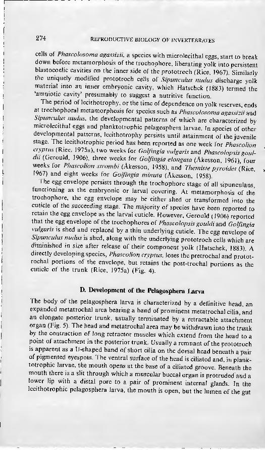
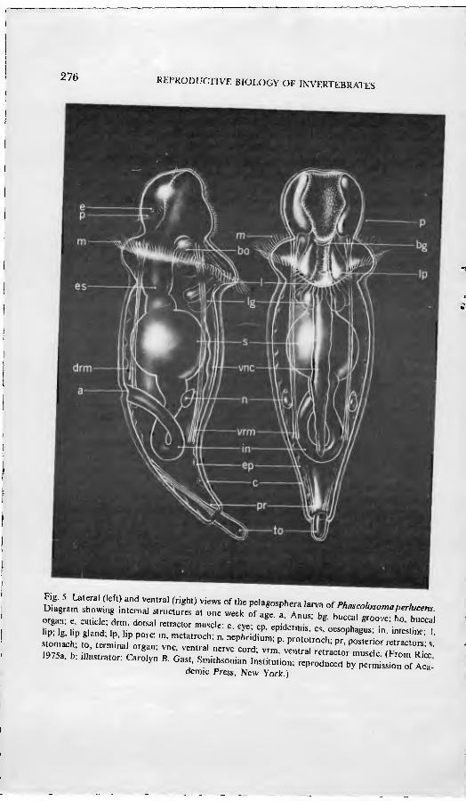
The body of the pelagosphera larva is characterized by a definitive head, an expanded metatrochal area bearing a band of prominent metatrochal cilia, and an elongate posterior trunk, usually terminated by a retractable attachment organ (Fig, 5). The head and metatrochal area may be withdrawn into the trunk by the contraction of long retractor muscles which extend from the head to a point of attachment in the posterior trunk. Usually a remnant of the prototroch is apparent as a U-shaped band of short cilia on the dorsal head beneath a pair of pigmented eyespots. The ventral surface of the head is ciliated and, in plank- totrophic larvae, the mouth opens at the base of a ciliated groove. Beneath the mouth there is a slit through which a muscular buccal organ is protruded and a lower lip with a distal pore to a pair of prominent internal glands. In the lecithotrophic pelagosphera larva, the mouth is open, but the lumen of the gut
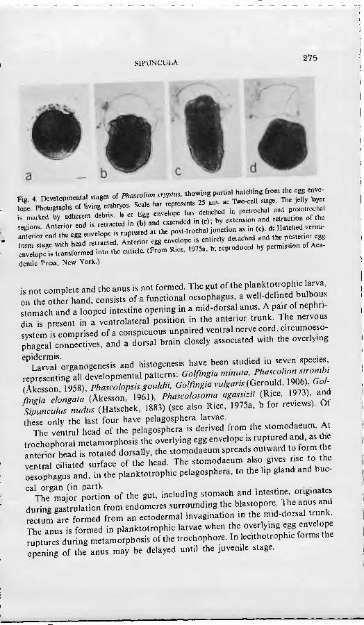
SIPUNCULA 275
•• •,l•l b, .dhe», a=b,• b-^: f.f.J•rd d b(""c...bs». .nd «»«¡o. »r .h.
demie Press, New York.)
is not complete and the anus is not formed. The gut of the planktotrophic larva, onThe otZ hand, consists of a functional oesophagus, a well-detmed bulbous stomach and a looped mtestine opening in a mid-dorsal anus. A pair of nephn- " present in a ventrolateral position m the anterior trunk. The nervous s tl i con^prised of a conspicuous unpaired ventral nerve cord, circumoeso- S'al connectives, and a dorsal brain closely associated with the overlying
''La•'rorganogenesis and histogenesis have been studied in seven species
repre enting'all developmental patterns: Go!ßn,ia -'--•/''-'¡f ^^^l^jf/ (Âkesson, 1958), Phascolopsis gouUJn. Golfingia .ulgans (Ger^là, 1906) Gol- fingia elongata (Âkesson, 1961), Phascolosoma agassm, (R^•, 1973) and ^sZnculusnuäus (Hatschek, 1883) (see also Rice, 1975a, b for reviews). Of these only the last four have pelagosphera larvae.
The ventral head of the pelagosphera is derived from the ^t^-^f J- ^ trochophoral metamorphosis the overlying egg envelope is ruptuied and, as he antenor head is rotated dorsally, the stomodaeum spreads outward to form h ventrlciliated surface of the head. The stomodaeum also gives rise to the oesophagus and, in the planktotrophic pelagosphera, to the lip gland and buc-
"The^m^aÍ; portn of the gut, including stomach and intestine originates during gastrulation from endomeres surrounding the blastopore^ The anus and rectum are formed from an ectodermal invagination in the mid-dorsal trunk^ Th anus is formed in planktotrophic larvae when the overlying egg envelope ruptures during metamorphosis of the trochophore. In lecithotrophic forms the opening of the anus may be delayed until the juvenile stage.
276 RtPRODUCTIVE BIOLOGY OF INVERTEBRATES
Fig. 5. Lateral (left) and ventral (right) views of the pelagosphera larva af Phn.^^i Diagram showing internal structures at one week of age fAn rb" i '''"'"''rr'''- organ; e, cuticle; drm, dorsal re•.c,or muscle; e, eye- ep epL•," es octnh '"" ' ""'
demie Press, New York.)
SIPüNCULA 277
The nephridia of species with lecithotrophic development (categories I, II, and III, Table 1) have a dual origin. The tubular portion arises from ectoderm and the ciliated funnel from coelomic mesoderm. Reports on the development of planktotrophic larvae suggest that the source of the nephridia may be entirely mesodermal. In contrast to larvae of other protostomes, sipunculan larvae lack a protonephridium, as already mentioned (p. 264).
With the exception of Sipiinculus nudus, the retractor muscles have been reported to originate from ectomesoderm. Their origin in 5. nudus is presumed to be from somatic mesoderm.
Usually the ventral nerve cord originates as a single, unpaired proliferation of trunk ectoderm. An exception is that of Phascolosoma agassizii in which the ectodermal proliferation is initially paired, but the two longitudinal cords thus formed later unite to form a single nerve cord. Rudiments of the brain are formed in the trochophore by the inward proliferation of lateral cells of the apical plate. By the pelagosphera stage the brain is well developed.
E. Metamorphosis of the Pelagosphera
Transition of the lecithotrophic pelagosphera to the juvenile stage is usually gradual. It is marked by the loss of metatrochal cilia, elongation of the body, movement of the mouth to a terminal position, formation of terminal tentacles, and the completion of the gut. A vermiform or crawling stage is commonly intermediate between the pelagosphera and juvenile. The changes take place over a period of three to four weeks in Themiste alulacea (Rice, 1975a) and seven weeks in Golfingia pugettensis (Rice, 1967).
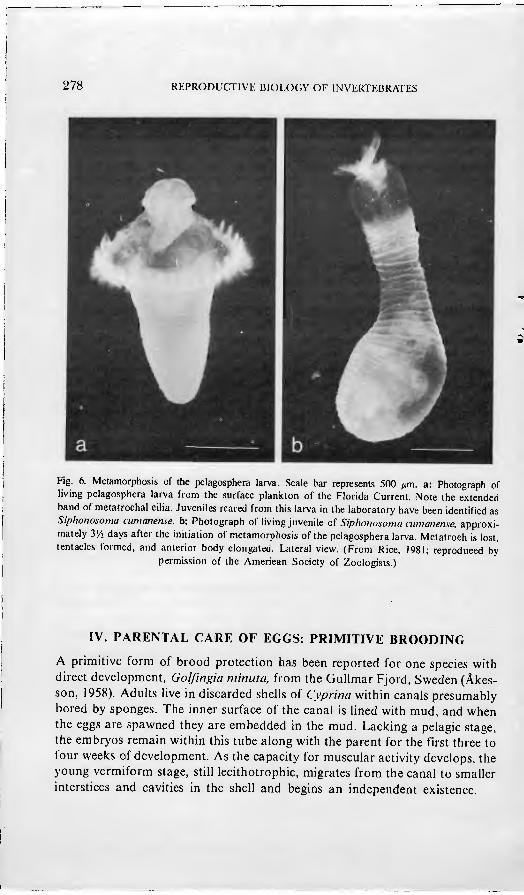
Planktotrophic pelagospheras, after a prolonged period in the plankton, undergo a relatively rapid metamorphosis, lasting two to three days. Metatro- chal cilia are lost, terminal tentacles are formed, the body is elongated, and the habitus of the juvenile is attained (Fig. 6). At the loss of cilia, the pelagic Ufe is ended and a benthic existence begins.
Factors controlling metamorphosis of sipunculans are not well known. Stu- dies of the inducement of metamorphosis in the laboratory have been carried out on a planktotrophic pelagosphera collected from the open ocean and tenta- tively identified as Golfmgia misakiana{Kxct, 1978, 1981, 1986; Rice and Mur- doch, 1978). Experimental evidence suggests that a water-soluble factor of low molecular weight (< 500), produced by adults of G. rnisakiana. will significantly increase the percentage of larval metamorphosis in the presence of substratum. The metamorphosis-inducing factor appears to be species-specific and it is not dependent on the presence of micro-organisms. The significance of the factor under field conditions has not been investigated, nor has the role of the substra- tum been clarified.
278 REPRODUCTIVE BIOLOGY OF INVERTF.BRATES
Fig. 6. Metamorphosis of the pelagosphera larva. Scale bar represents 500 fim. a: Photograph of living pelagosphera larva from the surface plankton of the Florida Current. Note the extended band of metatrochal cilia. Juveniles reared from this larva in the laboratory have been identified as Siphonosoma ciimanense. b: Photograph of living juvenile o{ Siphonosoma cumanense, approxi- mately VA days after the initiation of metamorphosis of the pelagosphera larva. Metatroch is lost, tentacles formed, and anterior body elongated. Lateral view. (From Rice, 1981; reproduced by
permission of the American Society of Zoologists.)
IV. PARENTAL CARE OF EGGS: PRIMITIVE BROODING
A primitive form of brood protection has been reported for one species with direct development, Golfingia minuta, from the Gullmar Fjord, Sweden (Âkes- son, 1958). Adults live in discarded shells of Cyprina within canals presumably bored by sponges. The inner surface of the canal is lined with mud, and when the eggs are spawned they are embedded in the mud. Lacking a pelagic stage, the embryos remain within this tube along with the parent for the first three to four weeks of development. As the capacity for muscular activity develops, the young vermiform stage, still lecithotrophic, migrates from the canal to smaller interstices and cavities in the shell and begins an independent existence.
SIPUNCULA 279
V. CONCLUSIONS
Sipunculans, though comprising a relatively small phylum in terms of taxo- nomic diversity have nevertheless exploited widely different pathways m their developmental histories. Two extremes of ontogenetic patterns are found within the phylum. One is a prolonged lecithotrophic development in which a pelagic stage is lacking; the other is a prolonged planktotrophic development marked by two pelagic stages, including a long-lived oceanic larva. Between these extremes are intermediate modes, manifesting either one or two lecitho- trophic pelagic stages; these swimming stages are followed by a vermiform or crawling stage, transitional between larval and juvenile forms. A chaUenging interpretive analysis, yet to be undertaken, would be an inquiry into the correla- tion between developmental patterns and zoogeographical distribution oí
species. . Other problems, remaining to be explored, concern regulatory mechanisms
governing various developmental processes. Additional studies are necessary for the confirmation of preUminary evidence suggesting that dissolution of the germinal vesicle of the egg and subsequent fertilizability involve hormonal controls Moreover, little is known of the factors inducing larval settlement and metamorphosis. Experiments in the laboratory indicate that a substance asso- ciated with the adult can induce metamorphosis of the larva. The role of this metamorphosis-inducing substance in the field and its ecological s.gniiicance remain to be elucidated by future investigations.
ACKNOWLEDGEMENTS
I wish to thank Julianne Piraino and Hugh Reichardt for technical assistance and aid in the preparation of the figures Carolyn B. Gast is gratefully acknowl- edged for preparation of Figure 5. I appreciate the support and facilities of the Smithsonian Marine Station at Link Port and the Harbor Branch Océanographie In-
stitution in Fort Pierce, Florida.
REFERENCES
Âkesson B (1958) •A study of the nervous system of the Sipunculoideae with some remarks on the
development of Uvo species Phascolion siwmbi Montagu and Golfingia minuia Keferstem,
Undersokningar over Öresimd, 38, 1-249. Akesson, B. (1961). 'The development oí Golfingia elongaw Keferstem (S.puneul.dea) w.th some
remarks on the development of neurosecrelory cells in sipuncuhds', Ark. Zooi. (Ser. 2), 13(23),
Amor A'(1975). 'El desarrollo de Themiste pelricola (Kmox, 1964) (Sipuncula, Golfingi.dae)',
P/,;.« (See. A), 34(89), 359-370. ,,- y , ^ li 140 142 Andrews, E.A. (1889). 'The reproductive organs of Phascolosoma gouldii. Zool. Anz.. 12, 14U-W/. Conklin' EG (1897). 'The embryology of Crepidula', J. Morphol.. 13, 1-226. Gerould, J.H. (1903). 'Studies on the embryology of the Sipunculidae, I. The embryonal envelope
280 REPRODUCTIVE BIOLOGY OF INVERTEBRATES
and its homologue', Mark Anniversary Volume, Henry Holt and Co., New York, pp. 439^52. Gerould, J.H. (1904). 'The development oí Phuscohsoma. Preliminary note', Arch. Zool. exp..
(4)2, xvii-xxix, Notes and Revue, No. 2.
Gerould, B. (1906). 'Studies on the embryology of the Sipunculidae, II. The development of Phascolosoma', Zool. Jb. Abl. Anal. Ontog. Tiere, 23, 77-162.
Gibbs, P.E. (1975). 'Gametogcnesis and spawning in a hermaphroditic population of Go/fingia minina (Sipuncula)', J. mar. biol. Ass. U.K., 55, 69-82.
Gibbs, P.E. 0976). 'Notes on the reproductive cycles of several Golfingia ap^cks (Sipuncula)' ./. mar. biol. Ass. U.K., 56, 909-915.
Halschek, B. (1883). 'Ueber Entwicklung von Sipunculm midus'. Arh. Zool. Ins! Univ Wien Zool Slal. Triesi, 5, 61-140.
Krohn, A. (1851). 'Ueber die Larve des Sipimcukis ?,nclus, nebsl vorausgeschickten Bemerkungen über die Sexualverhältnisse der Sipunculiden'. Arch. Anal. Physiol. und wi.ss Med., 368-379.
Pasteeis, J.J, (1935). 'Recherches sur le déterminisme de l'entrée en maturation de l'oeuf chez divers Invertébrés marins'. Arch. Biol., 46, 229-262.
Rice, M.E. (1966). 'Reproductive biology and development in Sipuncula', Doctoral dissertation, University of Washington, Seattle, 322 pp.
Rice, M.E. (1967). 'A comparative study of the development of Phascolosoma agassizii, Golfingia pugeliensis, and Themisle pyroides with a discussion of developmental patterns in the Sipuncula' Ophelia, 4, 143-171.
Rice, M.E. (1973). 'Morphology, behavior, and histogenesis of the pelagosphera larva of Phascolo- .yowa ago.si/z» (Sipuncula)', Smiihsonian Conlrib. Zool., 132, 1-51.
Rice, M.E. (1975a). 'Observations on the development of six species of Caribbean Sipuncula with a review of development in the phylum', Proc. Iniern. Symp. Biol. Sipuncula and Echiura (Eds. M.E. Rice and M. Todorovic), Naucno Delo Press, Belgrade, pp. 141-160.
Rice, M.E. (1975b). 'Sipuncula', in Reproduction of Marine Inverlebrales, Vol. II (Eds. A.C. Giese and .I.S. Pearse), Academic Press, New York, pp. bl-lll.
Rice, M.E. (1976). 'Larval development and metamorphosis in Sipuncula', Am. Zool., 16,563-571. Rice, M.E. (1978). 'Morphological and behavioral changes at metamorphosis in the SipJncula', in
Selllemeni and Metamorphosis oj Marine Invertebrate Larvae (Eds. F.S. Chia and M.E. Rice), Elsevier/North-Holland Press, New York, pp. 83-102.
Rice, M.E. (I98I). 'Larvae adrift: patterns and problems in life histories of sipunculans' Am Zool 29, (3), 605-619.
Rice, M.E. (1985). 'Sipuncula: developmental evidence for phylogenetic inference', in The Origin and Relaliomhips of Lower Invertebrates (Eàs. S.C. Morris, J.D. George, R. Gibson, and H.M. Platt), Oxford University Press, Oxford, pp. 274-296.
Rice, M.E. (1986). 'Factors influencing larval metamorphosis in Golfingia misakiana(Sivunc^uW Bull. mar. Sei., 39(2), 362-375.
Rice, M.E., and Murdoch, J.D. (1978). 'Influence of adults on metamorphosis of oceanic sipuncu- lan larvae'. Am. Zool, 18(3), 664 (abstract).
Ruppcrt, E.E., and Rice, M.E. (1983). 'Structure, ultrastructure, and function of the terminal organ of a pelagosphera larva (Sipuncula)', Zoomorphology, 102, 143-163.
Selenka, E. (1875). 'Eifurchung und Larvenbildung von Phascolosoma elongatum (Kef)' Z wiss Zool., 25, 442-450.
WiMiams, J. (1972). 'Development of a rock burrowing sipunculid inhabiting stony coral' Am Zool., 12(4), 723 (abstract).