Embed Size (px)
Citation preview

TRANSDUCING SYSTEMS IN THE HORMONAL
REGULATION OF STEROIDOGENESIS
IN RAT LEYDIG CELLS


TRANSDUCING SYSTEMS IN THE HORMONAL REGULATION
OF STEROIDOGENESIS IN RAT LEYDIG CELLS
MECHANISMEN BETROKKEN BIJ DE SIGNAALOVERDRACHT
BIJ DE HORMONALE REGULATIE V AN STEROIDOGENESE
IN LEYDIG CELLEN VAN DE RAT
PROEFSCHRIFT
ter verkrijging van de graad van doctor in de geneeskunde
aan de Erasmus Universiteit Rotterdam op gezag van de rector magnificus
Prof. Dr. A.H.G. Rinnooy Kan en vo1gens het bes1uit van het college van dekanen.
De open bare verdediging za1 p1aatsvinden op woensdag 22 oktober 1986 om 15.45 uur
door
AXEL PETER NICO THEMMEN
geboren te Uden
1986
Offsetdrukkerij Kanters B. V., Alblasserdam

PROMOTIECOMMISSIE
Promotor: Prof. Dr. H.J. van der Molen Overige leden: Prof. Dr. W.C. Hiilsmann
Prof. Dr. S. W.J. Lamberts Prof. Dr. J.J.H.H.M. de Pont
Co-promotor: Dr. F.F.G. Rommerts
Dit proefschrift werd bewerkt in het instituut Biochemie II (Chemische Endocrinologie) van de Faculteit der Geneeskunde, Erasmus Universiteit Rotterdam.

CONTENTS
VOORWOORD
ABBREVIATIONS
CHAPTER GENERAL INTRODUCTION
1~1 Introduction
1~2 Scope of this thesis
CHAPTER 2 TRANSDUCING SYSTEMS
2o1 General introduction
2~2 Transducing systems involving cAMP
2 ~ 2 & 1 introduction
2a2o2 GTP-binding proteins: N5
and Ni
2.2a3 adenylate cyclase and cAMP-dependent
protein kinase
2.3 Transducing systems involving phosphoinositide
metabolism
2. 3.1 introduction
2.3.2 the phosphoinositide cycle
2.3.3 the three messengers
2.4 Role of calcium as a messenger
2. 4. 1 introduction
2.4.2 regulation of the intracellular calcium
concentration
9
11
1 3
14
17
18
1 8
1 9
21
22
22
23
25
27
27
27
2.4.3 functional effects of calcium in the cell 29
2.5 Concluding remarks 30
CHAPTER 3 METHODS
3.1 Introduction
3.2 Isolation and characterization of Leydig cells
from immature rats
3.3 Incubation conditions
3~4 Protein phosphorylation
3~5 Western blotting and photo-affinity
labelling of RII
33
33
34
36
38

CHAPTER 4 THE MECHANISM OF ACTION OF LH
4~1 Introduction 39
4~2 Effects of adenylate cyclase inhibitors 39
4~3 Involvement of calcium ions in the action of LH 40
4.4 Conclusions 44
CHAPTER 5 MECHANISM OF ACTION OF LUTEINIZING HORMONE-
RELEASING HORMONE
5.1 Introduction 45
5.2 Involvement of calcium ions in the action of LHRH 46
5.3 Effects of phorbol ester and phospholipase C 48
5.3~1 pregnenolone production
5.3.2 protein phosphorylation
5.3.3 protein synthesis
5.4 Conclusions
CHAPTER 6 GTP-BINDING PROTEINS IN MATURE RAT LEYDIG CELLS
48
so 53
54
6.1 Introduction 55
6.2 Presence of N5
and Ni in Leydig cells 56
6.3 Effects of cholera toxin and pertussis toxin on
Leydig cell steroid production
6.4 Discussion
6.5 Conclusions
CHAPTER 7 GENERAL DISCUSSION
56
57
59
7.1 Introduction 61
7.2 A model for the regulation of steroid production 61
7.2.1 mechanism of action of LH
7.2.2 mechanism of action of LHRH
7.2.3 stimulation of cholesterol side chain
cleavage
7.3 Concluding remarks
REFERENCES
SUMMARY
61
65
67
68
71
81

SAMENVATTING
CURRICULUM VITAE
APPENDIX PAPER 1
A.P~N. Themmen, JaW. Hoogerbrugge, F.F.G. Rommerts & H.J.
van der Molen (1985) Is cAMP the obligatory second messen
ger in the action of lutropin on Leydig cell steroidogene
sis? Biochemical and Biophysical Research Communications
128, 1164-1172.
APPENDIX PAPER 2
A.P.N. Themmen, J.W. Hoogerbrugge, F.F.G. Rommerts and H.J.
van der Molen (1986) Effects of LH and an LH-releasing her-
mone agonist
regulation of
on different second messenger systems in the
steroidogenesis in isolated rat Leydig cells.
Journal of Endocrinology 108, 431-440.
APPENDIX PAPER 3
A.P.N. Themmen, R. Molenaar, W.J. Visser, J.F. Jongkind,
F.F.G. Rommerts & H.J. van der Molen (1986) Comparison of
the cellular composition and steroidogenic properties of
interstitial cell preparations isolated from immature and
mature rat testis~ Submitted~
APPENDIX PAPER 4
A.P.N Themmen, JGWG Hoogerbrugge, F.FcG. Rommerts and HoJ.
van der Molen (1986) The possible role of protein kinase C
and phospholipids in the regulation of steroid production
in rat Leydig cells. FEBS Letters 203, 116-120.
APPENDIX CHAPTER
A.PoN. Themmen, M. Pigalke, J.W. Hoogerbrugge, W. Rosen
thal, G. Schultz & F~F.G. Rommerts (1986) Stimulatory and
inhibitory guanine nucleotide binding proteins are present
in rat Leydig cells. To be submitted.
85
89
91
103
11 5
133
1 41

Other papers related to this thesis:
F.F.G. Rommerts, R. Molenaar, A.P.N. Themmen & H.J. van der
Molen (1984) Regulation of steroidogenic activities in Leydig
cells by LH and an LHRH agonist. In: Hormonal Control of the
Hypothalamo-pituitary Gonadal Axis, K.W. McKerns & Z. Naor,
eds, Plenum Press, New York, pp. 423-436.
A.P.N. Themmen, J.W. Hoogerbrugge, F.F.G. Rommerts & H.J. van
der Molen (1984) Comparison of LH and LHRH agonist action on
steroidogenesis in rat Leydig cells. Annals of the New York
Academy of Sciences 438, 625-628.
A.P.N. Themmen, J.W. Hoogerbrugge, F.F.G. Rommerts & H.J. van
der Molen (1985) A comparison of the mechanism of action of LH
and an LHRH-agonist on steroidogenesis by immature rat Leydig
cells. In: Recent Progress in Cellular Endocrinology of the
Testis, J.M. Saez, M.G. Forest, A. Dazord & J. Bertrand, eds,
Editions INSERM, Paris, pp. 299-305.
Molen (1985) Hormonal regulation of testicular steroido-
genesis via different transducing systems. In: Proceedings of
the 10th European Symposium on Hormones and Cell Regulation, J.
Nunez, J.E. Dumont & R.J.B. King, eds, Libbey & Co, London, in
press.
A.P.N Themmen, J.W. Hoogerbrugge & F.F.G. Rommerts (1986)
Transducing systems for the stimulation of steroid production
in immature rat Leydig c.ells. In: Proceedings of the IVth
International Testis
Endocrinology of the
Amsterdam, in press.
Workshop on Molecular and Cellular
Testis, M. Stefanini, ed., Elsevier,
F.F.G. Rommerts & A.P.N. Themmen (1986) LHRH, the role of LHRH
(agonists) in the regulation of gonadal function. Acta Endo
crinologica, in press.

9
VOORWOORD
Het tot stand brengen van een proefschrift is zelden of
nooit de verdienste van een persoon$ Daarom wil ik op deze
plaats die mensen bedanken die mij in staat hebben gesteld om
het voor U liggende boekje te schrijven .. Met name:
Focko Rommerts, mijn eo-promotor, voor de positieve en
enthousiaste begeleiding van het onderzoek en bij het schrijven
van het proefschrift, en Henk van der Mol en, mijn promotor, voor
zijn kri tische suggesties bij het onderzoek, en voor zijn niet
geringe inbreng bij het schrijven van di t boekj e .. Ik heb veel van
jullie beiden geleerd.
Jos Hoogerbrugge voor de onmisbare hulp bij de vele experi
menten, waarbij zijn fantasie, nodig bij het oplossen van de fos
foryleringsproblemen, zeker niet onopgemerkt mag blijven~
Herrn prof. dr~ GUnter Schultz, Herrn dr. Walter Rosenthal
und Frau Monika Pigalke (Institut flir Pharmakologie, Freie
Universitat, Berlin) mOchte ich gerne danken fllr die Hilfe mit
den ADP-ribosylarungs Proben.
De leden van de promotiecommissie, prof. dr. HUlsmann, prof.
dr. Lamberts en prof. dr. de Pont voor hun bereidheid het ma
nuscript zo snel te beoordelen.
Hugo de Jonge, voor de vele stimulerende discussies over de
diverse second messenger systemen.
Pim Clotscher, voor. zijn (computer-) technische inspanningen.
De KLM-crew, voor de waardevolle en gezellige samenwerking,
en, niet te vergeten, voor de lol~
Alle medewerkers van Biochemie II voor de prettige tijd op de
afdeling,
Mijn ouders voor de opvoeding die zij mij hebben gegeven~
Els, jij ook natuurlijk, maar niet alleen in dit proefschrift~


11
ABBREVIATIONS AND TRIVIAL NAMES
AC
ACTH
ATP
cAMP
cGMP
cholesterol
cscc cyanoketone
CTP
DAG
db cAMP
DDA
EGF
ER
FSH
GTP
GTPys
hCG
IP
IP2
IP 3 LH
LHRH(-A)
PA
PI
PIP
adenylate cyclase
adrenocorticotropic hormone
adenosine 5'-trisphosphate
adenosine cyclic-3',5'-monophosphate
guanosine cyclic-3' ,5'-monophosphate
5-cholestene-3~-ol
cholesterol side chain cleavage enzyme
2a-cyano-17~-hydroxy-4,4 1 ,17a-trimethyl-
5-androstene-3-one
cytosine 5'-trisphosphate
diacylglycerol
N6-2'-0-dibutyryl adenosine cyclic-
3' ,5'-monophosphate
2' ,5'-dideoxyadenosine
epidermal growth factor
endoplasmic reticulum
follicle stimulating hormone; folli
tropin
guanosine-5'-trisphosphate
guanosine-5'-(3-0-thio)-trisphosphate
human chorionic gonadotropin
inositol-1-phosphate
inositol-1 ,4-bisphosphate
inositol-1 ,4,5-trisphosphate
luteinizing hormone; lutropin
luteinizing hormone releasing hormone
(-agonist); luliberin
inhibitory guanine nucleotide binding
protein
stimulatory guanine nucleotide binding
protein
4~-phorbol-13-monoacetate
phosphatidylinositol
phosphatidylinositol-4-phosphate
phosphatidylinositol-4,5-bisphosphate

12
PL-C
PK-A
PK-C
pregnenolone
R s
SCP 2 SDS-PAGE
SU-10603
phospholipase C
cAMP-dependent protein kinase
ca 2+/phospholipid dependent protein
kinase
4~-phorbol-12-myristate-13-acetate
5-pregnene-3~-ol-20-one
regulatory subunit of PK-AII
receptor coupled to inhibition of
adenylate cyclase
receptor coupled to stimulation of
adenylate cyclase
sterol carrier protein 2
polyacrylamide gel electrophoresis in
the presence of sodium dodecylsulphate
7-chloro-3,4-dihydro-2(3-pyridyl)-1-
(2H)-naphtalenone
testosterone 4-androstene-17~-ol-3-one
TFA 9-(tetrahydro-2-furyl) adenine
TFP trifluoperazine
3~-HSD 3~-hydroxysteroid dehydrogenase
25-hydroxycholesterol 5-cholestene-3~,25-diol

1 3
CHAPTER 1 GENERAL INTRODUCTION
1~1 Introduction
In mammals, the testis can be divided into two specialized
compartments, i.e. the tubular compartment and the interstitial
compartment~ In testicular tubules, the Sertoli cells form the
lining of the inside of the tubular wall and they enclose the
maturing germinal cells with
the male germinal cells are
many cytoplasmic extensions. Thus
in intimate contact with the
Sertoli cells. The development of germinal cells is dependent
on FSH and testosterone, both acting on germinal cells via the
Sertoli cell. In the interstitial compartment many different
cell types are present such as Leydig cells, macrophages,
fibroblasts and endothelial cells. The Leydig cells are the
source of androgens which are important for spermatogenesis and
development of the primary and secondary sex characteristics.
Steroidogenesis in the Leydig cell is mainly under the
control of the pituitary hormone LH. However, it has been shown
that other factors also can affect steroid production by
Leydig cells, e.go factors in interstitial fluid (Sharpe, 1984)
and in medium from cultured Sertoli cells (Verhoeven & Cailleau, 1985) or from cultured testicular macrophages (Yee &
Hutson, 1985). In addition, it has been found that LHRH-A
(Hunter et al, 1982; appendix paper 2), arginine-vasopressin
(Meidan & Hsueh, 1985), ~-adrenergic agonists (Anakwe & Moger,
1984) and the atrial natriuretic factor (Pandey et al, 1985)
can influence the activities of isolated Leydig cells. In
addition
such as
to androgens
prostaglandins
Leydig cells
(Molcho et
can produce other factors
al,
(Valladares & Payne, 1979; Rommerts et al,
1984a,b), estradiol
1982a) and different
peptides derived from pro-opiomelanocortin (Tsong et al, 1982;
Margioris et al, 1983; Valenca & Negro-Vilar, 1986)~
1 4
1.2 Scope of this thesis
Steroid production
regulation by many
by the Leydig cell is
factors (see 1 .1 ) ,
subject to multiple
which suggests that
a complex for transduction of the external signals must be
present in this cell. The factors that can regulate the acti
vities of Leydig cells may utilize different second messenger
systems, suggesting that such systems are present in the Leydig
cell. Synergistic effects between the effect of LHRH and LH on
second messenger
prole~ +;tein phosphorylation synthesis
~ .. ::;;:.~ steroid other
production products
PLASMA MEMBRANE
Figure 1.1 General scheme of transmembrane signalling. The hormone (H) binds to the receptor (R) 1 thereby activating the transducer element (T). This results in an activation of the effector (E), which enhances the production of the intracellular second messenger. The second messenger can cause many effects in the cell e.g. stimulation of a small molecular weight "third messenger", stimulation of protein phosphorylation, or stimulation of protein synthesis. In the Leydig cell these (concomitant?) effects lead to increased productions of androgens and other compounds such as prostaglandins.
15
steroid production have been shown, suggesting that these
hormones utilize independent pathways. However, several trans
ducing systems are eventually coupled to the same target:
regulation of steroid production via regulation of the activity
of the cholesterol side chain cleavage enzyme. Others may be
coupled to other functions of the Leydig cell such as growth
and prostaglandin production.
The studies described in this thesis have mainly been con
cerned with the elucidation of several of the second messenger
systems present in the Leydig cell and the regulation of
steroidogenesis through pathways that involve cAMP and calcium
ions as second messengers. A general, albeit simplified scheme
for transmembrane signalling which result in stimulation of
steroid production is given in figure 1 .1.
To affect Leydig cell function specific stimulators
(dbcAMP, phorbol esters etc.} or inhibitors (P-site agonists,
diltiazem etc.) of the different second messenger systems were
used in addition to physiological stimuli. The effects of these
probes were measured by determining (1) the activity of adenyl
ate cyclase through measurements of the intracellular levels of
cAMP, {2) the activity of protein kinases by studying electro
phoresis patterns of proteins labelled with 32Po4
, (3) stimu
lation of de novo protein synthesis by studying the electro
phoretic patterns of proteins labelled with 35 s-methionine and
(4) the activity of the cholesterol side chain cleavage enzyme
by measuring the production of pregnenolone from endogenous
precursors.
The results of these studies are described in detail in
chapters 3-6 and in the appendix papers 1, 2, 4 and the
appendix chapter. Results of studies on the involvement of cAMP
and calcium as second messengers in LH action are described in
chapter 4. The mechanism of action of LHRH is discussed in
chapter 5 and data on the presence of GTP-binding proteins N s
and Ni in Leydig cells are given in chapter 6. In chapter 7 an
integrated model of the regulation of properties of Leydig cell
function is discussed with special
derived from the results in
summarized.
emphasis on the conclusions
the preceding chapters are


1 7
CHAPTER 2 TRANSDUCING SYSTEMS
2e1 General introduction
Many protein hormones acting on cells, exert their effects
via an interaction with proteins (receptors) on or in the
plasma membrane, which have a high affinity for the hormonee
The receptor activates other proteins in the cell membrane,
eventually resulting in the production of one or more second
messengers inside the cell. This process is called ''trans
duction11 or 11 transmembrane signalling 11 of hormonal signals, and
the different hormone-receptor-transducer-second-messenger sys-
terns are also called "second messenger systems 11•
figure 1 • 1 ) •
(See also
In this chapter background information concerning the
different transducing systems or transmembrane signalling
systems which are discussed in this thesis will be pro-
videde This section is not intended to give a comprehensive
review of all transducing systems, and only those systems which
are important for Leydig cell functions as described in this
thesis will be discussed~ Discussion will include the systems
that use cAMP, IP 3 , calcium or diacylglycerol as second or
third messenger. Other systems that are present in the Leydig
cell such as the tyrosine kinase system (coupled to the
EGF receptor), the guanylate cyclase system (coupled to the
atrial natriuretic factor receptor) will not be discussed.
In the following subsections we will consider the different
second messenger systems, their most important matching
receptors, and their mechanism of coupling to production of the
intracellular messenger& The concluding paragraph presents a
recent idea for a unified theory of transmembrane signalling.

1 8
2.2 Transducing systems involving cAMP
2.2.1 introduction
The role of cAMP as an intracellular
discovered by Sutherland and colleagues who
messenger was
studied the
stimulatory effects of glucagon and epinephrine on phos
phorylase activity, and reported in 1957: 11 The response to the hormones in liver homogenates was
separated in two phases: first, the formation of an acti
ve factor in the particulate fractions in the presence of
hormones and, second, the stimulation by the factor of
liver phosphorylase formation in supernatant fractions of
homogenates in which the hormones themselves had no
effect'1, and ''The factor was heat-stable, dialyzable, and
was purified considerably by chromatography on anion and
cation exchange resins 11 (Rall et al, 1957).
Five years later a partly purified preparation of active
adenylate cyclase was described (Sutherland et al, 1962). In
the ensuing decades considerable effort has been spent on the
investigation of the hormonal regulation of adenylate cyclase
(reviews: Ross & Gilman, 1980; Schramrn & Selinger, 1984). These
studies resulted in a now generally accepted cascade of events
which can be summarized as follows.
There is a multitude of receptors that affect cAMP forma
tion. These receptors (R) can be classified into two sub-types:
Rs receptors, which cause increased
lating adenylate cyclase, and
cAMP formation by stimu-
Ri receptors, which
inhibiting adenylate
cause
cyclase decreased cAMP levels by
(Birnbaumer et al, 1985). Receptors for glucagon, ACTH, LH,
FSH, TSH, adenosine (A 2-receptor) and for ~-cathecholamines
are of the type, whereas
a2-cathecholamine, adenosine
statin receptor are of the R. l
the muscarinic (acetylcholine),
(A1-receptor) and the somato
type. Both types of receptor can
be present in the same cell e.g~ Sertoli cells contain the R s for FSH (Fletcher & Reichert, 1984), glucagon (Eikvar et al,
1985) and ~-adrenergic agonists (Kierzenbaum et al, 1985), and

1 9
the Ri for adenosine (A1
; Monaco et al, 1984), in addition to
tyrosine kinase receptors such as the insulin receptor (Oonk et
al, 1986).
2.2.2 GTP-binding proteins: N5
and Ni
When it was accepted that the interaction between extra-
cellular hormones and membrane receptors could affect the
activity of adenylate cyclase on the inside of the cell
membrane, much effort was invested to elucidate the molecular
mechanism of the coupling of R5
and Ri to this enzyme.
A central role of GTP-binding proteins (N proteins) in hor
monal regulation of adenylate cyclase was first suggested by
Rodbell and colleagues, who demonstrated that GTP was required
for glucagon activation of the enzyme in plasma membranes from
liver (Rodbell et al, 1971b; Harwood et al, 1973). In addition,
they showed that GTP enhanced the rate of dissociation of
radiolabelled glucagon
(Rodbell et al, 1971a).
from its receptor
This mechanism involving
binding sites
the GTP-bind-
ing proteins (N proteins) has been clarified in detail by in
vestigations on the ~-adrenergic receptor system and on ery
throcyte membranes (Gill & r1eren, 1978; Cerione et al, 1985;
reviews: Birnbaumer et al, 1985; Northrup, 1985)~
TheN protein coupled toR. (N.) differs from the N protein l l
coupled to Rs (Ns), although these proteins have a similar
subunit structure~ A model of the structure and function of Ns
and Ni is given in figure 2e1e Ns consists of a GTP-binding
subunit as (42-52 kDa), a #-subunit (35 kDa) and a small ~-sub
unit (ea. 5 kDa)o Ni consists of an ai-subunit (41 kDa), the
same ~-subunit as Ns, and a small y-subunit which may be the
same as inNs. The functional importance of the Y-subunits is
not knowno The N proteins not only bind GTP, but also act as
GTPases. Upon binding of the hormone to the receptor (Rs or Ri'
the mechanism of the N proteins is symmetrical), the a -subunit
of the intact, inactive (a8y) N protein binds GTP, and the
a-GTP and ~y-unit dissociate. The activated as-GTP associates
with the adenylate cyclase thereby activating the enzyme,

20
Rs
~GY Ri PLASMA
MEMBRANE
GTP~GDP
+ 8
cAMP
Figure 2.1 Schematic representation of the involvement of N (consisting of a ~y) and N. (consisting of a.~y) in signaf transduction. F3r explanation see text. AC: ade~ylate cyclase; H
81 Hi: hormones, binding to R8 or Ri respectively.
which results in an increase production of cAMP. Because the
~y-units of N5
and N2
pool of ~y-units may be
may therefore inhibit
are thought to be identical, a single
present in membranes~
cAMP formation in two
Activation of N. l
ways: either via
direct binding of the a-subunit of Ni to adenylate cyclase,
although this could not be confirmed with in vitro studies, or
via enlargement of the pool of ~y-units, which may bind to
a5-subunits leading to 'deactivation of N G Both a -GTP and
s s ai-GTP are inactivated by a GTPase dependent association to the
~y-subunit giving rise to the intact, inactive N proteinG It
has been shown that the affinity of the receptor is affected by
N G Association of N with the ~-adrenergic receptor results in s s
a high affinity state of the receptorG During activation of
adenylate cyclase by as, the receptor changes to a low affinity
state, to be reactivated by the GTPase dependent association of

21
N5
(DeLean et al, 1980; Limbird et al, 1980; Stadel et al,
1980).
Of great interest is the ability of two bacterial toxins,
cholera toxin and pertussis toxin, to catalyze the transfer of
an ADP-ribosyl-group from NAD to the a-subunit of the N pro
teins both in vivo and in vitro. Cholera toxin ADP-ribosyl
ates a5
(Gill & Meren, 1978), whereas pertussis toxin acts on
a. (Katada et al, 1982). The a-subunits can be labelled, l
through incubation with the appropriate toxin in the presence
of 32P-labelled NAD, and can be subsequently detected after
separation on SOS-PAGE (see appendix paper 5). Toxin treatment
of the N proteins has a profound effect on their activity (re
view: Gilman, 1984). ADP-ribosylation of as by cholera toxin
inhibits the GTPase activity, prevents the reassociation of " s to the ~Y-subunit which results in a permanent activation of as
and adenylate cyclase. Pertussis toxin treatment of ai also
inhibits the GTPase activity, but this results in an increased
affinity of ai for ~Y. The low concentration of free ai causes
a stimulation or abolishment of inhibition of adenylate cyclase
in many systems. Pertussis and cholera toxin have proven to be
valuable tools in the study of the involvement of Ns or Ni in
the action of hormones.
2.2.3 adenylate cyclase and cAMP-dependent protein kinase
Little is known about the adenylate cyclase enzyme. The
molecular mass is ea. 150 kDa, and it catalyzes the production
of cAMP from MgATP or MnATP substrates (Schlegel et al, 1979).
The enzyme can be obtained in a 11 resolved state 11, i.e. not
purified,
resolved
but free from Ns or
preparation it has
Ni (Ross et al, 1978). Using this
been found in a reconstituted
system with the p-adrenergic receptor that Ns and Ni are both
necessary to obtain a maximal stimulation by agonists (Cerione
et al, 1985).
The only known role in
adenylate cyclase, cAMP, is
protein kinase (PK-A). The
eukaryotes of the product of
activation of cAMP-dependent
two types of PK-A (PK-AI and
22
PK-AII), have been shown to be similar in size~ They have a
similar subunit composition and mechanism of activation, but
differ in the characteristics of their regulatory subunits
(review: Lohmann
designated RI and
respectively. The
& Walter, 1984)~ The two regulatory subunits,
R11 , have molecular weights of 47 and 54 kDa
catalytic subunit (C; 40 kDa) of both
enzymes is identical. Both kinases are activated by binding of
cAMP to R, resulting in a dissociation of the holoenzyme as
indicated in the following scheme.
R2c 2 (inactive) + 4 cAMP~ R2-(cAMP) 4 + 2 C (active)
The activity is not only regulated by cAMP, but may also be
affected by phosphorylation of the regulatory subunits. PK-A11 undergoes autophosphorylation by the catalytic subunit, and it
has been shown that this phosphorylation results in a slower
reassociation of the R and C subunits (Rangel-Aldao & Rosen,
1977). Both R1
and R11
can be phosphorylated also by other
kinases, but the significance of this phosphorylation in the
regulation of PK-A is not clear (Lohmann & Walter, 1984).
Many different proteins can serve as substrates of PK-A,
but it is beyond the scope of this
proteins. It has been found that
different expressions of cell
chapter
PK-A is
function,
to discuss all these
ihvolved in many
such as metabolic
activity {phosphorylation of liver pyruvate kinase (Engstrom,
1980)), protein synthesis (ribosomal protein S6 (Traugh, 1981 ))
and cell shape (cytoskeleton proteins (Osawa & Hall, 1985)).
Specific phosphoproteins may play a major role depending on the
function of the cell, e.g. phosphorylase in liver.
2.3 Transducing system involving phosphoinositide
metabolism
2 0 3 • 1 introduction
During the last four years evidence has accumulated that in

23
addition to cAMP-dependent signalling systems, another impor
tant system involving phospholipid breakdown is present in all
cells. Hokin & Hokin (1953, 1954) reported that stimulation of
pancreas slices with acetylcholine resulted in an increase in
turnover of phosphatidylinositol (PI). This observation has
been repeated with many different cell types for many different
agonists, and phospholipid breakdown is now generally accepted
as a versatile receptor-activated signalling system, that sti
mulates different activities such as intracellular calcium
levels, protein kinase C and metabolism of arachidonic acid. In
this subsection only the main properties of this
be discussed (for reviews: Berridge, 1984; Takai et
Downes & Michell, 1985; Majerus et al, 1985).
2.3.2 the phosphoinositide cycle
system will
al, 1984;
Many different agonists stimulate PI-breakdown in cells.
Adrenergic agonists (through the a 2-receptor), vasopressin (V1 -
receptor), serotonin (S-HT1-receptor) and acetylcholine {musca
rinic receptor) are all coupled through this system to stimu
lation of intracellular calcium levels (review: Berridge,
ATP ATP
PI \ ~ PIP~PIP2)3 PL-C
1-IP-IP2-1:3
CDP-DAG ·~""""""''\- PA~ DAG
CTP ATP
Figure 2.2 The reactions of the PI-system. All reactions take place in the plasma membrane. For explanation see text. Abbreviations: IP
3: lnOSltol trisphosphate; IP
2: lnOSltol
bisphosphate; IP: lnosltol phosphate; I: lnositol; PI: phosphatidyl inositol; PIP: phosphatidyl inositolmonophosphate; PIP
2: phosphatidyl inositolbisphosphate; PL-C: phospholipase C:
CDP-DAG: cytosine bisphosphate-diacylglycerol; PA: phosphatidic acid.

24
1982). A general outline of the PI-system is given in figures
2.2 and 2.3.
The major step in the PI system is the receptor-mediated
breakdown of phosphatidylinositol bisphosphate (PIP2 ) into
inositol trisphosphate (IP3
) and diacylglycerol (DAG), through
activation of a phospholipase C. IP3
can stimulate the release
of calcium from intracellular stores and DAG can either acti
vate protein kinase C, or DAG can be cleaved to release arachi
donic acid which can serve as a substrate in the formation of
several arachidonic acid metabolites. These three pathways of
the PI-system will be discussed below.
There is increasing evidence that the effect of the hormone-
receptor complex on PL-C is mediated by
With isolated membrane preparations
stimulates hormone-dependent IP3
that
it
a GTP-binding protein.
can be shown that GTP
production (Litosch et al,
1985} 1 and many studies show treatment of cells with per-
phorbol ester
R
PLASMA MEMBRANE
PIP 2 JP 3 DAG arachidonic
• + ~ acid
Ca 2+t PK-CI prosta- leukotriens
1.1.\ gland ins
phospho- thromboxanes proteins
Figure 2.3 General outline of the PI-system, showing the three pathways leading to elevation of intracellular free calcium levels, phosphorylation of proteins and production of arachidonic acid metabolites.

25
tussis toxin inhibits the subsequent hormone-stimulated PIP 2 breakdown (Evans et al, 1985; Uhing et al, 1986). The nature of
this GTP-binding protein is unknown, but awaiting further cha
racterization it has been named Np.
Regeneration of the phosphoinositides occurs as follows.
Cellular IP3
will be metabolized rapidly to IP 2 , IP and finally
to inositol6 DAG is activated using CTP and reacts with inosi
tol to form PI. PI is phosphorylated to form PIP and PIP 2 ,
closing the cycle (figure 2.2). The latter two reactions can be
catalyzed in vitro by two tyrosine kinase oncogene products
(src and res), indicating that growth factors may modulate the
PI system via activation of their receptor-tyrosine kinases.
2.3.3 the three messengers
The first pathway, the production of IP 3 , links the PI sys
tem to the control of intracellular calcium levels. Using
permeabilized pancreatic acinar cells ( Streb e't al, 1 983) and
hepatocytes (Burgess et al, 1984) it was shown that IP 3 can
release calcium from the endoplasmic reticulum. This effect is
IP3
specific with other inositol phosphates being inactive
(Berridge, 1984). The role of increased intracellular levels of
calcium will be discussed below (see subsection 3~4)~
The second pathway, via the production of DAG, is directly
related to protein phosphorylation. DAG can activate calcium/
phospholipid-dependent protein kinase (PK-C), which was first
described by Nishizuka (review: Takai et al, 1984). Both
calcium and DAG are required for activation of PK-C in vitro.
DAG can increase the sensitivity of PK-C to calcium as much as
1,000 foldr and the inactive enzyme can be completely activated
by DAG even at cytosolic calcium concentrations present in
resting cells (Takai et al, 1981). Hence, calcium is an
essential requirement for the enzyme, but not the key regulator
of PK-C activity in vivo.
The discovery that PK-C can be activated directly by
turner-promoting phorbol esters, provided a tool to activate PK-C in intact cells without the possible interference of other

26
second messenger systems (Castagna et al, 1982; Niedel et al,
1983)e In experiments using phorbol esters, or measuring the
DAG-stimulated activity of the isolated enzyme, it was shown
that PK-C is an ubiquitous enzyme, capable of phosphorylation
of proteins associated with many cell functions, e.g. membrane
proteins (Kiss & Luo, 1986), ribosomal protein 86 (Trevillyan
et al, 1984) and cholesterol side chain cleavage enzyme
(Vilgrain et al, 1984). PK-C also phosphorylates Ni thereby
suppressing its activity (Katada et al, 1985), and it has been
shown that phorbol ester treatment of cells can inhibit N s
function (Mukhopadhyay & Schumacher, 1985). These results
suggest that there may be a regulatory link between the cAMP
and the PI-system.
The third pathway of the PI system involves the stimulation
of arachidonic acid metabolism. Phosphoinositides have been
shown to be very rich in 1-stearoyl-2-arachidonyl. After the
breakdown of PIP 2 the DAG can be further metabolized by
1 ,2-DAG-lipase (Lenstra et al, 1984)~ This enzyme sequentially
removes stearic acid from position 1 and arachidonic acid from
position 2 of DAG (Prescott & Majerus, 1983). The released
arachidonic acid can serve as a substrate for the formation of
different bioactive metabolites including prostaglandins, leu
kotrienes and thromboxanes. These metabolites can influence
receptors on the cell of origin or on nearby cells, and in turn
may influence the PI system again (Majerus et al, 1984).
The considerations given above show that the PI-system is a
versatile system that affects many different cell functions
through the three different intermediates: calcium, DAG and
arachidonic acid metabolites. A general model of the PI-system
is given in figure 2.3. It may well depend on the specific
target cell which of these pathways will be most important in
that particular cell. Finally, there are many links between the
cAMP and PI system, e.g. PK-C may act on N proteins, PK-A and
PK-C can phosphorylate identical proteins and prostaglandin can
stimulate adenylate cyclase. These examples show that the two
second messenger systems could be intimately involved with one
another.

27
2~4 Role of calcium as a messenger
2. 4.1 introduction
Like cAMP, calcium is ubiquitously present and may be
involved in many aspects of cell regulation (reviews: Rasmussen
& Barrett, 1984; Huggins & England, 1985). Many hormones
increase intracellular calcium levels in their target cells and
their physiological action is abolished when these increases
are inhibited, or when the actions of calcium ions are pre
vented with specific inhibitors, such as calmodulin inhibitors.
Effects of hormones that depend on the presence of calcium ions
include: the effect of platelet-derived growth factor and epi
dermal growth factor on human fibroblasts (Moolenaar et al,
1984}, thyrotropin releasing hormone acting on anterior
pituitary cells (Ozawa & Kimura, 1982), LH and LHRH-A acting on
Leydig cells (Sullivan & Cooke, 1986) and noradrenaline, vaso
pressin or angiotensin acting on liver cells (Mauger et al,
1984). The calcium ions involved in the increase of the intra
cellular calcium concentration can either originate from out
side the cell, or can be liberated from intracellular stores.
Both sources are under hormonal control, and are important in
the increase of the intracellular free calcium concentration.
In this subsection the regulation of the intracellular calcium
concentration and the effects of calcium in the cell will be
discussed.
2.4.2 regulation of the intracellular calcium concentration
The intracellular concentration of free calcium in the cyto
plasm is extremely low (in the micromolar range) as compared to
the extracellular calcium concentration (in the milimolar
range (Campbell, 1983)), resulting in a steep calcium-gradient
accross the plasma membrane. Influx of calcium through calcium
the extracellu
(ER) and the
channels may occur from three sources: from
lar space, and from the endoplasmic reticulum
mitochondrion, which accumulate calcium against a steep gra-

28
dient~ The mitochondrion is not considered to be involved in
the hormone-stimulated increase in intracellular calcium (see
subsection 2.3e3)o Because high intracellular calcium is cyto
toxic (Rasmussen & Barrett, 1984), the cell has an elaborate
system of pumps that actively decrease the cytoplasmic calcium:
ca2 +-ATP-ases on the plasma membrane, mitochondrion and ER, + 2+ and Na -Ca -exchangers in the plasma membrane. The different
transport systems are outlined in figure 2~4e
It has been found that hormones can regulate cytoplasmic
calcium 1) through activation of PIP2 metabolism, resulting in
IP3 formation that liberates calcium from the ER {see subsec
tion 2.3), 2) through voltage-dependent calcium channels in the
plasma membrane and 3) through release initiated by depola-
CYTOPLASM
ER
PLASMA MEMBRANE
Figure 2~4 Different calcium transport systems in the cell. The mitochondrial and bound calcium pools are not shown. Three different calcium transport systems are shown: l~+calcium channe*; 2~+ATPase-coupled calcium transporter (Ca -ATPase) 1 3) Na -ea -antiporter.

29
rization from internal storeso The latter process is of major
significance in electrically excitable tissues, and will not be
discussedo There is some evidence for the regulation of intra
cellular calcium by voltage-dependent calcium channels in her-
monally regulated cells. Cloned cells from the anterior
pituitary have been shown to contain voltage-dependent calcium
channels, and thyrotropin-releasing hormone stimulates the
entry of calcium by changing some properties in membrane pola
rization in GH3
cells (Oz_awa & Kimura, 1979) o This effect of
thyrotropin-releasing hormone could be blocked by the calcium
channel antagonist verapamil (Ozawa & Kimura, 1982)6 Further
more, it has been shown in isolated rat liver cells, that
noradrenalin, vasopressin and angiotensin increase calcium
influx by opening a common pool of calcium channels (Mauger et
al, 1984). There is increasing evidence that the coupling of
the receptor to the calcium channel may also be mediated by a
GTP-binding protein, although the nature of this N protein is
not known (Gomperts, 1983; Koch et al, 1985; Holz IV et al,
1 986).
2.4.3 functional effects of calcium in the cell
Functional effects of an increase in intracellular calcium
are always mediated by calcium-binding proteins. There are two
classes of these proteins: 1) true calcium receptor proteins
such as calmodulin, parvalbumin and troponin c, which undergo
a conformational change upon calcium binding, and subsequently
interact with enzymes dependent on these proteins for their
activity, and 2) enzymes directly regulated by calcium, such as
ca2 +-activated protease or protein kinase c.
Calmodulin is the most extensively described calcium binding
protein (reviews: Klee et al, 1980; Veigl et al, 1984)e Its
molecular mass is ea. 16 kDa and there are four sites for
binding calcium. Upon binding of two to three calcium ions the
protein changes its conformation, becomes active, and binds to
the calmodulin-target proteins. Many phosphorylation-dephospho
rylation systems such as glycogen phosphorylase are regulated

30
by calmodulin. In addition, calmodulin is very important in
the regulation of the cytoskeleton, because it interacts with
molecules such as caldesmon and spectrin, and can also activate
tubulin kinase. Calmodulin can stimulate cAMP hydrolysis
through activation of a calmodulin dependent phosphodiesterase
(Erneux et al, 1985), and it may serve as one of the links
between the calcium- and the cAMP-systems.
For the second class of proteins the regulatory role of cal
cium is less clear. Many of these enzymes (e.g. ca2+-activated
protease) have been shown to be regulated by calcium in
vitro, but this requires calcium concentrations in the mMolar
range, which are not attained in the intact cell.
In conclusion, available evidence indicates that increases
in intracellular calcium may play a role in many different
aspects of cell regulation ranging from modulations of the cAMP
system, through activation of protein kinases to changes in
cytoskeleton and intermediary metabolismo
2o5 Concluding remarks
GTP-binding proteins appear to play an important role in
the initial action of the hormones on all second messenger
systems discussed
the regulation of
pertussis toxin
and the PI
and Ni are
effects
involved in
of
in this chaptero Ns
adenylate cyclase, and
on the hormone-regulated calcium
GTP and
channels
system have been observedc Transducin, the
protein that couples the effect of light-activated rhodopsin to
cGMP-phosphodiesterase is also an N protein (Fung, 1985)o
Recently an N protein with unknown function has been purified
from brain (N0
) (Sternweis & Robishaw, 1984)o There are
indications that some insulin effects are mediated by an N
protein (Nins) (Houslay, 1985).
These observations suggest that there may be a
GTP-binding proteins which mediate the transduction
family of
of signals
of hormone-receptor binding to various intracellular events.
Based on these observations, Rodbell (1985) has proposed a
theory on hormone action involving programmable messengers as

31
outlined in figure 2.5. A hormone interacting with its receptor
stimulates the release of an activated a-subunit. This a-sub
unit can be covalently modified in different ways by a modifier
M (phosphorylation, methylation, sulphation), each modification
directing the a-subunit to activate another effector E {ade
nylate cyclase, phospholipase C). The modification of the
a-subunits can be considered
messengers". The
as programming, hence the term
effectors emit a signal S (cAMP, "programmable
calcium) that cause the final response R. This theory has yet
to be verified experimentally, although it has been shown with
reconstituted systems, as well as in intact
of hormone to the receptor can result in
cells, that binding
the release of the
a-subunit into the cytoplasm is feasible (Dominguez et al,
1985; Rodbell, 1985; Sternweis, 1986).
------~~~------R
PLASMA MEMBRANE
GTP
Figure 2.5 Proposed model of 11 programmable messengers" .. (Adapted from Rodbell (1985))~ For explanation see text.


33
CHAPTER 3 METHODS
3 o 1 Introduction
This chapter concerns those methods which are of special
interest for the work presented in this thesis, and which are
not described in detail in the appendix paperso All other re
levant materials and methods have been described previously and
can be found in the appendix papers.
3o2 Isolation and characterization of Leydig cells from
immature rats
Interstitial cells obtained from immature (21-24 days old}
rats were used for most of the studies described in this the-
siso Preliminary results had shown that these cells responded
very well to LH and LHRH-A, and could be kept in culture for
72 h without considerable loss of steroid response. Leydig
cells from immature testes were obtained in a high yield (90%)
and with an acceptable (40-50%} purity as follows: after colla
genase dispersion of the decapsulated testes, the cells in the
supernatant were washed and allowed to attach to the plastic
surface of a culture dish in Eagle's minimal essential medium
with Earle's salts and non-essential amino acids (MEM) con
taining 1% (v/v} foetal calf serum (32°C, 5% co2
in air)e Af
ter one hour of culture the floating cells were removed by
washing and the attached cells were kept in MEM with 1% (w/v)
bovine serum albumin (Rommerts et al, 1985)e In this prepa
ration 40-50% of the cells were positive for 3~-hydroxysteroid
dehydrogenase (3~-HSD) activity (for staining method see
Rommerts et al, 1985), which was used to estimate the number of
Leydig cells (see appendix paper 3). To investigate the effect
of hormones on steroid production the cells were incubated with
inhibitors of pregnenolone metabolism (cyanoketone and SU-
10603; see appendix paper 2), and pregnenolone production was
measured in the culture medium by radioimmunoassays Under these

34
conditions pregnenolone production was a good parameter for
measuring cholesterol side chain cleavage (CSCC} activity,
since pregnenolone is the direct product of the cleavage of
cholesterol. Measurement of testosterone may underestimate the
steroidogenic activity because only a small fraction of pregne
nolone is converted to testosterone in Leydig cells of immature
ratS a
Interstitial cell preparations from immature rats also con
tain other cells in addition to Leydig cells, and it could not
be excluded a priori that these non-Leydig cells might contri
bute to steroid production. In this regard we have investigated
the steroidogenic properties of these cells after further frac
tionation of the cell preparation on a 30-60% Percoll density
gradient. After attachment of the cells from different frac
tions to petri dishes, the potential CSCC activity and the
hormonal regulation of the enzyme activity were determined by
measuring the 25-hydroxycholesterol-, the LH-dependent and the
basal pregnenolone production. The presence of 38-HSD activity
was used as a marker for endoplasmic reticular steroidogenic
enzymes~ As shown in figure 2 in appendix paper 3, the ratio
of the different activities (LH/3~-HSD or 25-0H/3~-HSD) in the
fractions was approximately constant. These results indicate
that the LH-stimulated cscc activity is present only in 3e-HsD
positive cells, and that the 3e-HsD-negative cells do not
contribute to the steroidogenic response. Using this Percoll
density gradient centrifugation technique a preparation con
taining 90% Leydig cells could be obtained (see: appendix paper
3). This preparation was used in the experiments on protein
synthesis (chapter 5 and appendix paper 4).
3.3 Incubation conditions
The stimulatory effect of LH on pregnenolone production is
very rapid. Within 2 minutes following the addition of a maxi
mal stimulatory dose of the hormone, the first effects on
steroid production can be detected, and after 20 minutes the
stimulation is maximal (Rommerts et al, 1982b). In contrast,

35
stimulation of steroid production by LHRH-A evolves much slower
(Hunter et al, 1982). This difference in kinetics of steroid
response might reflect a difference in the mechanism of action
of these hormones. Hence, we have determined the rate of preg
nenolone production at different time-points after stimulation
of Leydig cells with LH (100 ng/ml), LHRH-A (40 nM), and a dose
of LH (0.1 ng/ml) which stimulated steroid production to the
same extent as 40 nM LHRH-A (figure 3.1). The results show
that the effect of LHRH-A occurs more slowly than that of LH
(20 minutes for first detection), and that the maximal effect
is reached after 3 h. Stimulation of steroid production with
100 ng/ml LH starts a few minutes after the addition of LH, and
is maximal within 30 minutes. The results also show that the
kinetics of the effect of 0.1 ng/ml LH are very similar to the
kinetics of 40 nM LHRH-A, although after 4 h the rate of preg-
• .. u
~
2 0
• • c c ·;; .2 0
u 0, 0
" c 0 • 0.
• c 0 0 c • c "' • • 0.
2.0
1.5
1.0
0.5
5
LHRH-A
o-~0.......0 2 3 4
0. T ng LH
100 ng LH
/ o 0h 0
.... BASAL .. .. 2 3 4
time ( hr)
Figure 3.1 Kinetics of LH (inset) and LHRH-A action on pregnenolone production by Leydig cells isolated from immature rats. Pregnenolone production per 10 min was calculated from the concentrations of pregnenolone in culture media from cells incubated during different periods. Mean results from triplicate determinations are shown.

36
nenolone production in the presence of LHRH-A declines, whereas
the stimulation with 0.1 ng/ml LH still has not reached its
maximal value$ These data indicate that, depending on the con
centration of LH, LHRH-A and LH have similar effects. This may
suggest an analogous mechanism of action of LH and LHRH-A at
concentrations with a low steroidogenic potency. However, the
experiments concerning the involvement of calcium (chapter 4
and 5), show that the mechanism of LH and LHRH-A is different
at all concentrations of LH.
The results depicted in figure 3.1 show that the effects of
LHRH-A on steroid production are maximal after 3 h. Consequent
ly, for all investigations concerning LHRH-A action 3 h incuba
tions were used.
3.4 Protein phosphorylation
To determine patterns of protein phosphorylation in intact
cells, 10 6 cells, attached to a plastic culture dish (35 mm
diameter), were· incubated with 200 ~Ci/ml 32Po 4 {carrier free)
for 3 h in Krebs-Ringer buffer without phosphate but containing
0.2% {w/v) glucose and 0.1% {w/v) bovine serum albumin. The
proteins were extracted and separated using polyacrylamide
gradient (8-15%) gel electrophoresis in the presence of SOS
{SDS-PAGE) as described by Bakker et al {1981). The labelled
proteins were then visualized by autoradiography. Using this
method a high background radiation which interfered with the
assessment of the phosphorylated proteins was observed. This
background labelling could not be suppressed, although diffe
rent approaches were used: e.g. extraction of RNA and DNA prior
to electrophoresis; decrease of the labelling period; different
extraction methods of proteins using trypsin, 50S-containing
lysis buffer and trichloroacetic acid; variation of the amount
of 32 Po 4 present during the incubation; repeated precipitation
and dissolving of the proteins with 10% trichloroacetic acid
and an 80S-containing buffer, respectively; labelling of the
proteins and
trisphosphate,
washing of the protein pellet in the presence of
to remove labelled polyphosphates which may be

37
present; extraction of phospholipids before SDS-PAGE and last
ly, extensive washing of the gel in the presence of unlabelled
phosphateo These approaches did not improve the autoradiographs
significantly. The same difficulties were encountered in phos
phorylation studies with Leydig cells from mature rats. How
ever, following subfractionation of the cells better results
were obtained. Subfractionation of Leydig cells from immature
rats proved to be difficult because the small diameter of the
cells prevented cell disruption in commercially available
Dounce glass-glass homogenizers (clearance: 0.025-0.030 mm)o
Therefore, to solve the problem of the high background radi
ation in phosphorylation experiments with immature rat Leydig
cells, a glass-glass homogenizer with a smaller clearance, that
will disrupt small cells, should be used~
To study the possible effects of protein kinases on endo
genous substrates in total Leydig cell homogenates or in a
25,000xg supernatant from
method was used (adapted
Leydig tumour cells the following
from de Jonge (1976)). Homogenates
{40-100 ~g protein) were incubated for 2 min at 30°C in 25 mM
2-glycerolphosphate, 2 mM EGTA, 5 mM MgC1 2 , 10 mM Tris-HCl pH
7.4 in the presence of 5 ~Ci (y) 32 P-ATP with 0.01 mM un
labelled ATP, with one of the following additions to activate a
particular kinase: cAMP-dependent protein kinase: 5 ~M cAMP;
cGMP-dependent protein kinase: 1 ~M cGMP; calcium-dependent
protein kinase(s): 2 mM Cacl2
; ca 2+-calmodulin-dependent
protein kinase: 2 mM Cacl2 with 250 ~g/ml calmodulin (partly
purified from rat brain; kind gift from Dr. de Jonge); calcium/
phospholipid-dependent, protein kinase (PK-C): 2.5 mM Cac1 2 ,
0.25 ~g/ml phosphatidylserine (in suspension), 2 ~g/ml PMA. The
volume was adjusted to 50 ~1 with H2o, the reaction was started
by adding (y) 32P-ATP, and terminated by adding SOS-PAGE sample
buffer (Bakker et al, 1981 )~A part of the sample was separated
on SDS-PAGE (8-15%}, and the gel was prepared for autoradio
graphy. After autoradiography results were obtained which did
not have the high background problem such as encountered with
the phosphorylation method used for intact cellso

38
3~5 Western blotting and photo-affinity labelling of R11
For the identification of the regulatory subunit of type II
cAMP-dependent protein kinase (R 11 ) in the 25,000xg supernatant
of a tumour Leydig homogenate two approaches were chosen: a
Western-type blotting experiment using a specific polyclonal
antibody against R11 (kindly provided by Drs~ Walter and
Lohmann, Departments of Physiological Chemistry and Medicine,
University of Wurzburg) and a photoaffinity-labelling method
with ( 3H)8-azido-cAMP.
The Western-blotting experiment was carried out as described
by Ratoosh & Richards (1985), with some modifications~ The
proteins were separated on SOS-PAGE (10% polyacrylamide) and
electrophoretically transferred to a nitrocellulose filter
(Burnette, 1981 )~ The filter was incubated overnight with
phosphate buffered saline (PES), 5% (w/v) BSA. The filter was
subsequently
in PBS, 0 c 1 %
0.3% (w/v)
overnight at
{w/v) Tween,
for 2 h at
protein A
incubated with
(w/v) gelatin,
Triton X-100)
the anti-RII serum (diluted 1 :500
5% (w/v) BSA, 0~05% (w/v) Tween,
for 5 h at room temperature and
8°C~ After 4 washes with wash-buffer (PBS, 0.05%
0.3% (w/v) Triton X-100), the filter was incubated
room temperature
(150,000 cpm/ml
125 with !-labelled Staphylococcal
buffer). After washing, the
in PBS-gelatin-BSA-Tween-Triton
filter was dried (30 min at 60°C)
and placed with Kodak SB-5 X-ray film.
R11 was labelled with {3H)8-azido-cAMP using a photoaffini
ty-labelling method (Richards et al, 1983; Brinkmann et al,
1986). 70 Ml of the 25,000xg supernatant was incubated with
0.34 nmol ( 3H)8-azido-cAMP (10.2 Ci/mmol) in the absence or
presence of 14,000 nmol of unlabelled cAMP. The mixture was
then irradiated with an Osram HBO 100 w/w-2 high pressure mer-
cury lamp. A cm layer of saturated Cuso4
solution was posi-
tioned between the lamp and the sample. The samples were kept
on ice at a distance of 5 cm from the lamp during the irra
diation (18 min). The incubation was terminated by the addition
of 0.1 ml sample-buffer, the proteins were separated on SDS
PAGE (8-15% polyacrylamide gradient) and the gel was prepared
for autoradiography.

39
CHAPTER 4 THE MECHANISM OF ACTION OF LH
4$ 1 Introduction
The pituitary hormone LH is the main regulator of Leydig
cell steroidogenesis. It is generally accepted that LH exerts
its effect through activation of adenylate cyclase resulting in
elevated levels of cAMP, activation of cAMP-dependent protein
kinase and phosphorylation of specific proteins (Cooke et al,
1976; Bakker et al, 1983b). However, the causal and obligatory
interrelationship(s) between these parameters have not been
completely elucidated. Several reports in the literature have
described that the correlation between the effects of LH or hCG
(which is thought to have the same mode of action as LH) on
cAMP levels and steroid production is not always perfect. Dufau
et al (1978) showed that in rat Leydig cells the Eo50
of both
cholera toxin and hCG for cAMP stimulation were approximately
the same, whereas hCG was 60 times more potent than cholera
toxin in stimulation of testosterone production. With isolated
mouse Leydig cells it was found that isoproterenol stimulation
resulted in a small elevation of cAMP levels without a conco
mitant stimulation of testosterone production, whereas a low
dose of LH stimulated steroid production without any effect on
cAMP (Cooke et al, 1982). These results indicate that the in
volvement of cAMP in LH regulation of steroid production is not
completely clear. In this regard, we have studied the effect of
inhibitors of adenylate cyclase on the stimulatory effect of LH
on cAMP levels and steroid production in Leydig cells isolated
from immature rats (appendix paper 1 )o
4.2 Effects of adenylate cyclase inhibitors
Isolated Leydig cells were incubated with two specific
adenylate cyclase
(TFA) (Haslam et
inhibitors: 9-{tetrahydro-2-furyl)adenine
al, 1978; Harris et al, 1979; Simchowitz et
al, 1983) and 2'5'-dideoxyadenosine (DDA) (Fain et al, 1972;

40
Filetti & Rapoport, 1983; Florio & Ross, 1983). Both TFA and
DDA inhibited LH-stimulated cAMP levels (appendix paper 1,
figure 1 & 2)a However, pregnenolone production was not inhi
bited, but rather increased in the presence of TFA as well as
DDA. If cAMP is an obligatory mediator of the action of LH on
steroid production, this increase in steroid production cannot
be readily explained, because a decrease or no effect would
have been anticipated. The observed results are similar to the
synergistic effect of LHRH on LH-dependent steroid production
(cf. chapter 5), although the mechanism of action of LHRH on
cAMP levels on the one hand and of DDA and TFA on the other
hand appear different: LHRH-A probably causes an attenuation of
cAMP levels by activation of a phosphodiesterase {Sullivan &
Cooke, 1984), whereas TFA and DDA act directly on the adenylate
cyclase.
These results show that cAMP may not necessarily be the sole
and obligatory second messenger in the action of LH on steroid
production, albeit cAMP can be important in the mediation of
the signal of LH to the cholesterol side chain cleavage$ For
instance, the phosphodiesterase inhibitor MIX potentiates the
LH-dependent steroid production by immature rat Leydig cells
(appendix paper 2, table 1), and dbcAMP can mimic many effects
of LH (Bakker et al, 1983b). Since there may be more than one
second messenger system involved in relaying the effects of LH
on steroidogenesis, we have investigated the possible involve
ment of an alternative transducing system, i~e. the involve
ment of fluxes of calcium ions through the plasma membrane in
the action of LH.
4.3 Involvement of calcium ions in the action of LH
The involvement of calcium was investigated by determining
the effect on LH-stimulated steroid production of: 1) decreas
ing the extracellular calcium concentration, 2) blocking cal
cium channels with diltiazem, 3) inhibiting the activity of the
calcium-binding protein calmodulin with trifluoperazine, and 4)
artificially enhancing the intracellular free calcium concen-

41
tration using the calcium ionophore A23187$ In all experiments
the steroid production in the presence of 25-hydroxycholesterol
was also measured, to obtain an indication of possible non
specific effects of the different treatments on the cholesterol
side chain cleavage activity (Mason & Robidoux, 1978; Brinkmann
et al, 1984).
In 3 h incubations, A23187 (0.1-10 MM) inhibited both LH
and 25-hydroxycholesterol-dependent pregnenolone production
suggesting non-specific effects on the cholesterol side chain
cleavage enzyme (appendix paper 2, figure 5)~ Treatment of the
cells for 3 h with 0.1-1 ~M A23187 progressively decreased
ATP-levels in the cells, indicating that the ionophore had
deleterious effects on immature rat Leydig cells
paper 2).
(appendix
A decrease of the extracellular calcium concentration re-
sulted in a concomitant decrease of steroid production in the
presence of LH (appendix paper 2, figure 3). The blocking of
calcium fluxes with diltiazem (appendix paper 2, table 2), as
well as inhibiting calmodulin action with trifluoperazine (fi
gure 4.1} also partially inhibited LH actione These results
PREGNENOLONE (PMOLEIHR/106 CELLS)
50 100
ADDITIONS
LH (100 NG/ML)
LH (0.1 NGIML)
LHRHa (40 nMl
2SOH-CHOL ( 30pM)
Figure 4.1 Effect of calmodulin-inhibitor trifluoperazine (TFP) on pregnenolone production by Leydig cells isolated from immature rats. Cells were incubated for 3 h with hormones as indicated, in the absence (open bars) or presence (cross-hatched bars) of 10 gM TFP.

42
show that calcium fluxes through the plasma membrane are in
volved in the effects of LH on steroid productione However,
lowering the extracellular calcium concentrations to very
low levels by the inclusion of the calcium chelator EGTA (Oe1
mM), did not result in a complete block of LH stimulation,
suggesting that the activation of the cholesterol side chain
cleavage enzyme is not completely dependent on extracellular
calciumo This suggestion is substantiated by the observation
that EGTA could only inhibit 70% of the effects of both LHRH-A
and a low concentration of LHo Addition of the different probes
did not influence the 25-hydroxycholesterol-dependent steroid
production, except in the case of trifluoperazine, suggesting
that this compound has non-specific actions. It has been shown
indeed, that trifluoperazine can have rather non-specific ef
fects, since in platelets it inhibits PK-C directly (Sanchez et
al, 1983).
The dependence of steroid production on the presence of ex
tracellular calcium has been shown also in swine granulosa
cells (Veldhuis et al, 1983), rat granulosa cells (Tsang &
Carnegie, 1983), adrenocortical cells (Trzeciak & Mathe, 1981)
and in mature rat Leydig cells (Janszen et al, 1976). Low ex
tracellular calcium concentrations do not cause permanent cell
damage, since the inhibitory effects of low calcium levels on
Leydig cells (Janszen et al, 1976) and Sertoli cells (Oonk,
personal communication) are reversible. The similarity of the
effects of low extracellular calcium concentrations on the
actions of LH and LHRH-A on steroid production suggests that
extracellular calcium may influence the steroidogenic pathway
at or distal to the point where the LH- and LHRH-A-dependent
routes of activation meet.
An increase in the intracellular free calcium concentration
can have several effects including for instance the activation
of calcium-dependent protein kinases or changes in the cyto
skeleton (see subsection 2.4). We have investigated the pre
sence of calcium-activated protein kinases in a homogenate of
Percoll-purified immature rat Leydig cells (figure 4.2). After
incubation in the presence of (~)- 32P-ATP with or without cal-

112r.-
.... , 79~
'!J#IIM'0 .... , ··~ • ... ., ··~
... ,0
- - """ ,_ ... 14
3 4
Figure 4.2 Pattern of in vitro protein phosphorylation~ A hom~~enate of immature rat Leydig cells was incubated with (y) P-ATP as described in section 3~4. Molecular weight markers are indicated on the right~ Lane 1: control; lane 2: 2 mM CaCl ; lane 3: 2 mM CaCl , 250 pg/ml partially purified calmodufin; lane 4: 2 mM CaCl~, 250 pg/ml calmodulin, 50 pM TFP.
43
cium and calmodulin, the proteins were
on SOS-PAGE. The autoradiograph of the
cium-dependent phosphorylations of
extracted and separated
dried gel showed cal-
proteins with relative
molecular masses of 18, 41 and 79 kDa as well as calcium-cal-
modulin-dependent phosphoproteins of 49, 59 and 112 kDa. The
effects of calmodulin on the phosphorylation could be inhibited
specifically by trifluoperazineo Although the physiological
significance of these proteins is not known, these results show
the presence of two calcium-dependent kinase activities with
endogenous substrates in Leydig cells~

44
4e4 Conclusions
The results presented in this chapter show that cAMP is not
the sole and obligatory second messenger in the regulation of
steroid production by LH~ The influx of calcium ions may also
be very important, and the results of the phosphorylation expe
riments with broken cell preparations show that the molecular
mechanisms for the transduction of the calcium effects are
present in the immature rat Leydig cella The presence of ex
tracellular calcium is necessary for a complete stimulation of
steroid productiona However, a small part of the action of LH
and LHRH-A appears to be independent of extracellular calcium.

CHAPTER 5 MECHANISM OF ACTION OF LUTEINIZING HORMONE
RELEASING HORMONE
5.1 Introduction
45
The main physiologic function of the hypothalamic decapep
tide LHRH is probably its effect on the release of LH from the
pituitary, but it has been. shown that LHRH can also act direct
ly on gonadal steroid producing cells (for review: Hsueh & Jones, 1981 )~ Not all steroidogenic cells are sensitive to
LHRH~ Rat Leydig cells give a good response (appendix paper
2), but gonadal cells of murine and human origin show no res
ponse at all, and no LHRH-receptors have been detected in the
tissue from these species (Casper et al, 1982, 1984; Clayton & Huhtanierni, 1982; Hunter et al, 1982}. The physiological rele
vance of the direct action of LHRH on rat testicular cells re
mains unclear, since LHRH is not produced in significant a
mounts within the testis (Hedger et al, 1985). However, the
mechanism of action of LHRH is interesting, since it has been
found to be completely different from the effect of LH. LHRH
has not only a direct effect on basal steroid production in
isolated rat Leydig cells, but also affects the action of LH
(Hunter et al, 1982; Rommerts et al, 1984b; appendix paper 2).
The LHRH controlled pathways may therefore provide an addi
tional system for the regulation of steroidogenesis~ Although
LHRH appears not to be the physiological modulator of Leydig
cells in vivo, it may be that LHRH controlled pathways are
activated under physiological conditions by other locally
produced peptides. Hence, a study of the mechanism of action of
LHRH on Leydig cell steroidogenesis may elucidate yet unknown
regulatory pathways which might play a role in the physiologic
al control of Leydig cell function.
The effects of LHRH on rat Leydig c~lls are mediated by spe
cific high affinity receptors for LHRH on Leydig cell membranes
(Sharpe & Fraser, 1980; Clayton et al, 1980). Studies with iso
lated Leydig cells show a biphasic effect of LHRH and its ago-

46
nists (LHRH-A) on steroid production~ In short term incubations
(less than 3 h) LHRH-A stimulates both basal and LH-dependent
steroid production, but when cells are incubated for 24 h or
longer with LHRH-A, the LH-dependent steroidogenesis is inhi
bited by the releasing hormone (Hunter et al, 1982; Browning et
al, 1983; Rommerts et al, 1984b).
The mechanism of action of LHRH on Leydig cell steroid pro
duction is unclear. Cyclic nucleotides are apparently not in
volved in LHRH action on Leydig cells (Lin, 1984; appendix pa
per 2). In pituitary gonadotrophs, LHRH stimulates PIP2 break
down (Andrews & Conn, 1986) and a rise in inositol phosphates
(Kiesel et al, 1986), and it has been implied that PK-C may be
involved in LHRH action in these cells (Hirota et al, 1985)~
With an interstitial cell preparation
mature rats, Molcho et al {1984a) have
isolated from testes of
shown that LHRH-A sti-
mulates PI-turnover. Because regulation of PI-turnover has been
shown to be involved in calcium-mediated actions (Downes &
Michell, 1985i section 2a3), we have considered the possibility
that the effect of LHRH on PI-turnover could reflect that cal
cium might be involved in the action of LHRH on steroid pro
duction. In this regard we have investigated the involvement of
extracellular calcium and calcium fluxes through the plasma
membrane in the action of LHRH on Leydig cell steroidogenesis
(section 5.2 and appendix paper 2).
5.2 Involvement of calcium ions in the action of LHRH
To study the involvement of calcium ions in the action of
LHRH-A, we have incubated the cells in medium with a low cal
cium concentration (appendix paper 2, figure 3). The low
calcium concentration resulted in an inhibition of LHRH-A
stimulated steroid production which was comparable to the
effect of this medium on LH-dependent pregnenolone production
(cf. section 4.3). However, blocking of calcium channels with
diltiazem (appendix paper 2, figure 4), and inhibition of cal
modulin action with trifluoperazine (figure 4.1) had no effect
on LHRH-A stimulation of steroid production, whereas these

47
treatments resulted in an inhibition of the effect of a sub
maximally stimulatory dose of LHe These results suggest that
the presence of calcium may be essential for the transduction
of any stimulatory signal to the CSCC, but that calcium fluxes
may be involved only in the action of LHe The calmodulin inhi
bitor may have exerted non-specific effects on Leydig cells
{see section 4e3)e From the present results it cannot be exclu
ded that calcium release from intracellular stores is essential
for the action of LHRHe This release may be triggered by IP 3 released from PIP 2 in the plasma membrane after stimulation of
the cells with LHRH-A~ However, until now only changes in turn
over of PI have been studied (Molcho et al, 1984a), and there
is no evidence that IP3
is produced in Leydig cells upon stimu
lation by LHRH. In this respect it may be of interest that the
change of intracellular calcium levels in mature Leydig cells
(measured with the Quin-II method) in response to LH or LHRH-A,
showed a lag time of approximately 2 minutes after stimulation
with LH, whereas no lag time was apparent upon addition of
LHRH-A (Sullivan & Cooke, 1986). This suggests that LH may
trigger the slower entry of calcium through a calcium channel,
whereas LHRH-A, through IP3
formation may trigger calcium
release from intracellular stores, which is generally a much
faster process (Reinhart et al, 1984; Downes & Michell, 1985).
These considerations lead towards the suggestion that PIP 2-
hydrolysis may be involved in LHRH action. Apart from calcium
release from the endoplasmic reticulum, this pathway probably
also employs activation of PK-C through the production of di
acylglycerol (cf. section 2.3 and figure 2.3). We have investi
gated this possibility by comparing the effects of phorbol
ester and phospholipase c, both known activators of PK-C, and
of LHRH-A on steroid production,
protein synthesis (section 5.3 and
protein
appendix
phosphorylation
paper 4).
and

48
5.3 Effects of phorbol ester and phospholipase C
5.3.1 pregnenolone production
LHRH-A has a striking synergistic effect on LH-dependent
pregnenolone production (appendix paper 2, figure 1)~ LH alone
stimulates steroid production 35 fold, but this effect can be
enhanced further (60 fold} by the inclusion of LHRH-A in the
incubation medium, whereas LHRH-A alone stimulates pregnenolone
production only 5 fold (appendix paper 2,
bol-12-myristate-13-acetate (PMA) caused
figure 2). 4~-Phor
effects completely
different from those of LHRH-A* PMA alone stimulated steroid
production 3 fold, but inhibited the effect of intermediate
concentrations of LH, without affecting the steroid production
in maximally stimulated cells {appendix paper 4, figure 2).
When PMA was added to Leydig cells in the presence of different
concentrations of dbcAMP, the stimulatory effects of PMA and
dbcAMP were additive, suggesting that the inhibitory effect of
PMA on LH-stimulated steroid production occurred before the
formation of cAMP, and also that PMA did not have deleterious
effects on the cells (appendix paper 4, figure 2)G These re
sults are consonant with observations in mouse Leydig cells,
where it has been shown that the site of inhibition by PMA of
hCG effects on adenylate cyclase is localized at the regulatory
GTP-binding protein of the adenylate cyclase system (N ) s
(Mukhopadhyay & Schumacher, 1985).
We have also used phospholipase C (PL-C) to study the ef
fects of phospholipid hydrolysis on Leydig cell steroid pro
duction~ PL-C is thought to activate PK-Ce There is no direct
evidence for th·e action Of PL-C on PK-C, but such an action has
been suggested from the similarity of the effects of PMA and
PL-C on protein phosphorylation in intact cells, and from the
ability of PL-C to stimulate the levels of diacylglycerol in
cell membranes (Rozengurt et al, 1983; Hadjian et al, 1984;
Fischer et al, 1985i Jaken, 1985)$ We have used PL-C from
Clostridium perfringens (type XII), because this enzyme has
been shown by Fischer et al (1985) to be effective 1 rather than

49
the enzyme from Bacillus cereus which was not effective in mu
rine epidermal cells (see also Chap et al, 1977; appendix paper
4) •
PL-C stimulated steroid production 3 fold, and acted syner
gistically in stimulating LH-dependent pregnenolone production
2 fold (appendix paper 4, figure 1) • These results showed that
the LHRH effect can be mimicked by PL-C, but not by PMA, sug
gesting that the mechanisms of action of PL-C and PMA are dif
ferent in Leydig cells, although both are thought to activate
the PK-C pathway. The difference between the effects of PL-C
and PMA on pregnenolone production (and protein phosphorylation
and protein synthesis, see below) might arise from the exist
ence of different types of PK-C in the cell (Kiss & Luo, 1986t
de Jonge, personal communication). These results and the fol
lowing results obtained from experiments on protein phosphory
lation and protein synthesis are summarized in Table 5.1. For
the exact figures the reader is referred to appendix paper 4.
Table 5.1 Comparison of protein phosphorylation, production in immature rat
Activity
Steroid production
without added LH in the presence of LH
Protein phosphorylation
17 K 33 K
Protein synthesis
14 K 27/30 K
the effects of different stimuli on protein synthesis and pregnenolone Leydig cells.
LH
25x
+++ +++
+++ +++
Activator
LHRH-A
6x 50x
0 0
+ +
PMA
3x 25x*
+++ ++
++ ++
PL-C
3x 50x
0 0
+ +
*: PMA inhibits pregnenolone production stimulated by submaximally stimulatory concentrations of LH.

50
5~3o2 protein phosphorylation
Bakker et al (1983b) have shown in Leydig tumour cells that
LH-stimulated pregnenolone production is accompanied by the
enhanced phosphorylation of at least six phosphoproteins with
molecular weights ranging from 17 to 76 kDa~ The most pro
nounced effect of LH was on the phosphorylation of a nuclear 17
J.;:Da and the 33 kDa ribosomal protein 56 o hCG-dependent phospho
rylation of proteins in the same molecular weight range has
also been shown in rat Leydig cell extracts (Dufau et al, 1981)
and in intact porcine Leydig cells (Dazord et al, 1984)o In
intact immature rat Leydig cells, we also observed the LH-sti
mulated phosphorylation of the 17 and 33 kDa proteins {appendix
paper 4, figure 3)$ The phosphorylation of these proteins was
also stimulated by PMA to approximately the same extent as the
phosphorylation obtained with LH. PL-C and LHRH-A did not have
any effect on protein phosphorylation (appendix paper 4, fi
gure 3; table 5~1)~ These results show that there is no com
parable effect of PMA and PL-C on protein phosphorylation and
941>-
«11 56
4311>-
1 2 3
Figure 5.1 Fluorograph of proteins labelled wi~h ( 3H)8-azidocAMP~ A homogenate of Leydig tumour cells with ( H)S-azido-cAMP was irradiated in the absence (lane 3) or the presence (lane 2) of an excess u£!abelled cAMP. Lane 1: Marker proteins labelled with ( C)-formaldehyde according to Jentoft & Dearborn (1979).

51
stimulation of steroid production~
The stimulation of phosphorylation of 17 and 33 kDa proteins
by PMA suggests that these proteins are phosphorylated direct
ly by PK-Co Bakker et al (1984} observed a discrepancy between
the stimulation of phosphorylation of the 17 kDa protein by LH
and dbcAMP, and the ability of these compounds to stimulate
steroid production by tumour Leydig cells. The effect of dbcAMP
on the phosphorylation of the 17 kDa protein was almost non
existent when compared to the effect of LH, whereas stimulation
of steroid production by LH was less pronounced than the stimu
lation in the presence of dbcAMP (Bakker et al, 1984). These
observations may be explained by activation of PK-C by LH, but
not by dbcAMP, which results in the stimulation of the phospho
rylation of the 17 kDa proteino
In the case of the ribosomal protein 86 the effect of PMA
could also be a direct phosphorylation of 86 by PK-C. It has
been shown that 86 can be phosphorylated by both PK-A and PK-C
(Burkhard & Traugh 1 1983i Trevillyan et al, 1984}, and this has
led to the suggestion that ribosomal protein 86 can be a site
of multiple regulation of protein synthesis through phosphory
lation at different 1 protein kinase specific sites (Traugh,
1981; Padel & SOling, 1985). There may still be another expla
nation for the effects of PMA on 86 phosphorylation~ PMA also
has a small effect on the phosphorylation of a 57 kDa protein,
which may be the regulatory subunit (RII) of PK-AII (Cooke et
al, 1979a)c Phosphorylation of RII may have a stimulatory
effect on PK-A (see section 2o2o3)o Therefore, we have investi
gated the identity of the 57 kDa protein and whether the phos
phorylation of this 57 kDa protein could be stimulated by PMA
in Leydig tumour cellso These studies might provide evidence
for activation of PK-A by the PK-C pathwayo
The identity of the 57 kDa protein was established in two
independent ways: in a homogenate of Leydig tumour cells cAMP
binding proteins were labelled with the photo-affinity probe
<3H)8-azido-cAMP (figure 5~1}, and a Western-type blotting ex
periment using a specific anti-RII serum was performed (figure
5.2). Both experiments resulted in the specific labelling of a

52
single 57 kDa protein. These results show that the 57 kDa pro
tein is R11
• Protein phosphorylation experiments in intact
Leydig tumour cells also showed increased labelling of the 57
kDa protein in the presence of LH and PMA (figure 5.3). Pre
liminary results from investigations on protein phosphorylation
in Leydig tumour cell homogenates in the presence of (~) 32 P-ATP showed a cAMP-dependent phosphorylation of a 57 kDa protein.
Furthermore, under conditions favourable for activation of PK-C
(subsection 3.4), we could show that phosphorylation of R11 occurred, whereas this phosphorylation was absent under control
conditions (results not shown).
From these results we have concluded that stimulation of
PK-C by PMA in intact cells may lead also to activation of PK-A
via phosphorylation of R11 • In this way the phosphorylation of
S6 might be catalyzed by PK-A, which in turn is activated by
PK-C.
5711o-
.....94 ..... 67
..... 43
..... 20
Figure 5a2 Autoradiograph of a Western blot, incubated with a specific anti RI serum, The autoradiograph of the filter incubated with confrol serum did not show any radioactive bands (not shown). Molecular weight markers are indicated on the right.

~94
57- -67
-43
... 30
17- -20
- 14
2 3 4
57-
Figure 5~3 Protein phosphorylation pattern of intact tumour ~2ydig cells. Cells were incubated for 3 h in the presence of
P04
• Lane 1: control; lane 2: 100 ng/ml LH; lane 3: 40 nM LHRH-A; lane 4: 100 ng/ml PMA~ Molecular weight markers are indicated on the right. Inset: detail of the same gel.
5.3~3 protein synthesis
53
Protein synthesis experiments which may reflect hormonal ef
fects on Leydig cell in a different way, revealed a stimulation
of the synthesis of proteins with molecular weights of 14, 27
and 30 kDa by LH, LHRH-A, PL1A and PL-C (appendix paper, figure
3). Janszen et al (1978} observed in immature rat Leydig cells
that LH stimulated the synthesis of 27 and 29 kDa proteins,
which probably are the same proteins as the 27 and 30 kDa pro
teins described in appendix paper 4. These authors also showed
the LH-stimulated synthesis of 11 and 21 kDa proteins, which
were not apparent in our experimentss However, we investigated
only the proteins in a 10,000xg supernatant of sonicated cells,

54
and the 11 and 21 kDa proteins may have been removed during
the fractionation procedure. The effects of LH on the 14, 27
and 30 kDa proteins are much more pronounced than the effects
of LHRH-A, PL-C and PMA, but we have not correlated the in
tensity of the newly synthesized protein bands with the potency
of the agonist to stimulate steroidogenesis in a quantitative
manner. In this respect, it is not possible to conclude whether
protein synthesis patterns might be a better reflection of the
ability of an agonist to stimulate steroid production than
protein phosphorylation patterns. There is a difference in the
kinetics of the stimulation of synthesis of specific proteins,
which is apparent only after a 5 h incubation period, and the
kinetics of the stimulation of steroid production, which takes
anywhere from 2 min for the initial effect of LH (Rommerts et
al, 1982b) to 3 h for LHRH-A to reach its maximal effect (sec
tion 3.2)~Hence, although the activation of steroid production
is dependent on protein synthesis in Leydig cells (Cooke et al,
1975), a direct correlation between steroidogenesis and the
identity of newly synthesized proteins remains to be establish
ed. Other, as yet unidentified proteins, which might be syn
thesized more rapidly, might play a more important role in the
regulation of steroidogenesis in Leydig cells (cf. Losier &
Younglai, 1981; Pedersen & Brownie, 1983).
5.4 Conclusions
LHRH stimulation of rat Leydig cell steroid production is
independent of LH action and does not involve cyclic nucleo
tides. Both LHRH-A and PL-C did stimulate LH-dependent preg
nenolone production synergistically, and the effects of these
compounds on protein phosphorylation and protein synthesis were
similar. These results suggest that the primary effect of LHRH
is probably on phospholipid hydrolysis, possibly resulting in
the production of IP 3 •

55
CHAPTER 6 GTP-BINDING PROTEINS IN MATURE RAT LEYDIG CELLS
6.1 Introduction
It was discussed in chapter 2 that GTP-binding proteins (N
proteins) are involved in the transduction of the signal from
several hormone-receptor complexes to the intracellular ef
fector systemo One of the actions of LH in Leydig cells is the
stimulation of the production of cAMP, and, similar to other
adenylate cyclase-stimulating hormones, the effect of LH ap
pears to be mediated by the stimulatory N protein (N5
). This
can be inferred from the stimulating effects of cholera
toxin, which specifically
steroidogenesis in intact
activates N5
,
Leydig cells
on cAMP production and
(Cooke et al,
Dufau et al, 1978). Furthermore, the observation that
1 977;
hCG-
stimulation of adenylate cyclase in Leydig cell membrane pre
parations is dependent on the presence of GTP (Dufau et al,
1980; Mukhopadhyay & Schumacher, 1985), is indicative that the
effect of LH (hCG) on adenylate cyclase is mediated by a GTP
binding protein, possibly N . The inhibiting GTP-binding pro-s
tein, Ni, may also be involved in the action of adenylate
cyclase. In reconstitution experiments with Ns, Ni, adenylate
cyclase and ~-adrenergic receptor in liposomes Cerione et al
{1985) showed that the activated receptor may have a dual role,
i.e. 1) activation of Ns, and 2) removal of the inhibition of
adenylate cyclase by Ni. Either one or both of these actions
may result in a significant increase in the hormonal effect on
cAMP production.
In this section both the presence and functional properties
of Ns and Ni in Leydig cells isolated from mature rats have
been investigated, in order to establish whether Leydig cells
may be controlled by these different GTP-binding proteins.

56
6~2 Presence of N5
and Ni in Leydig cells
To study the presence of N5
and Ni in rat Leydig cells, we
have used a membrane preparation obtained from Leydig cells
isolated from mature rats~ This cell preparation was used be
cause sufficient amounts of Leydig cells of high purity could
only be obtained from mature ratso A crude membrane preparation
was incubated with either cholera toxin or pertussis toxin in
the presence of ( 32P)-NADo Cholera toxin stimulates ADP-ribo
sylation of the a5-subunit of N
5 in the presence of Mg2
+ and
GTP, and there are indications that cholera toxin has a pre
ference for the activated form of N5
{Gill, 1977)0 The a-sub
unit of Ni is ADP-ribosylated by pertussis toxino This toxin
has a preference for the form of Ni that predominates in the 2+ absence of Mg , and the ADP-ribosylation can be stimulated by
GDP (Ribeiro-Neto et al, 1985).
Incubation of
in the presence
toxin-dependent
the membrane
of ( 32 P)-NAD
labelling of a
preparation with pertussis toxin
and GDP resulted in a pertussis
39 kDa protein (appendix chap
of GTPYS and MgCl 2 instead of ter, figure 1)$ In the presence
GDP the ADP-ribosylation of this protein was decreased (not
shown)~ Similar experiments with cholera toxin showed labelling
of a 44 kDa protein in the presence of GTP (appendix chapter,
figure 1 )$ Western blotting experiments, using a polyclonal
antibody against the ~-subunit, revealed that the ~-subunit
which is identical in both Ns and Ni was also present (results
not shown)$
6~3 Effects of cholera toxin and pertussis toxin on
Leydig cell steroid production
We have also investigated the functional importance of Ns
and Ni in Leydig cells isolated from 6--8 week old rats~ Isola
ted Leydig cells were preincubated for 2 h in the presence of
either cholera or pertussis toxin (100 ng/ml)$ Preincubation
with the toxins for this period of time is necessary for the
activation or inhibition of N5
or Ni, respectively~ After the

57
preincubation period LH (100 ng/rnl) or LHRH-A (40 nM) were ad-
ded,
was
and the pregnenolone production
determined (appendix chapter,
during the following
figure 2). Cholera
3 h
toxin
stimulated both basal and LHRH-A-dependent pregnenolone pro
duction, but did not affect steroid production in the presence
of LH. Preincubation of Leydig cells with pertussis toxin for 2
h did not have any effect on steroid productione In contrast,
treating the cells for 24 h with pertussis toxin resulted in a
stimulation of basal, LH- and cholera toxin-dependent preg
nenolone production (appendix chapter, figure 2). LHRH-A did
not have stimulatory effects on steroid production after the
cells had been incubated for 24 h (Hunter et al, 1982; Rommerts
et al, 1984b).
6.4 Discussion
The results show that functionally active Ns
present in Leydig cells from mature rats. Cholera
and Ni are
toxin stimu-
lated the ADP-ribosylation of a 44 kDa protein in a crude
Leydig cell membrane preparation. The dependency of the label
ling on cholera toxin, and the observed molecular weight
strongly
Gilman,
suggest that this protein is a s
1984)G Pertussis toxin stimulated
(Kaslow et al, 1980;
the ADP-ribosylation
of a 39 kDa protein, and this labelling was decreased in the
presence of MgC12
and GTP~S, suggesting that this protein is
the a-subunit of Ni (Bokoch et al, 1983; Murayama et al, 1983~
Ribeiro-Neto et al, 1985). In addition, we have observed that
the a-subunit of Ns and
brane preparations~
N. l
is present in the Leydig cell mem-
The stimulation of steroid production in Leydig cells by LH
is accompanied by increased levels of cAMP and activation of
PK-A (Cooke et al, 1976; Podesta et al, 1978). Cholera toxin
stimulates cAMP levels in Leydig cells (Cooke et al, 1977;
Dufau et al, 1978)
the
probably through activation of N5
o This is
observation that cholera toxin treatment substantiated by
of Leydig cells resulted in stimulation of LHRH-A-dependent
pregnenolone production. N5
, activated by cholera toxin, may

58
have stimulated adenylate cyclaseo The increase in cAMP levels
resulted in a stimulation of LHRH-A-dependent steroid product
ion, since the action of LHRH-A on Leydig cells is independent
of cAMP (Sullivan & Cooke, 1984; appendix paper 2).
N. may have a multiple role. In l
it has been shown that N. inhibits l
reconstitution experiments
the basal adenylate cyclase
activity much more than the stimulated enzyme activity
(Cerione et al, 1985). Its presence in the plasma membrane may
be necessary to assure a maximal stimulatory effect of hor
mones on adenylate cyclase. After a 2 h preincubation period
we were not able to show effects of pertussis toxin on steroid
production. However, when pertussis toxin was present for 24 h,
the pregnenolone production in control cells and in the presen
ce of LH or cholera toxin was stimulated. These results show
that Ni is involved in the regulation of steroid production in
rat Leydig cells. It is not clear whether the effect of per
tussis toxin evolves slower compared to the effect of cholera
toxin. It has been observed in other cell systems that the ef
fect of pertussis toxin may take from 60 min to 24 h to become
maximal (Adashi et al, 1984; Holz IV et al, 1986) ~Alternative
ly, the Leydig cells may have acquired a sensitivity to per
tussis toxin during culture. In this respect it is of interest
that Leydig cells in culture become sensitive to adenosine
(Rommerts et al, 1984a) and ~-adrenergic agonists (Anakwe et
al, 1985)e Ni might also mediate the coupling of receptors of
the Ri-type (cf. section 2.2) to inhibition of adenylate cycla
se. It has been shown that 24 h treatment of cultured testicu
lar cells with pertussis toxin abolished the inhibitory effects
of arginine-vasotocin on hCG-dependent steroid production,
suggesting that the receptor for arginine-vasotocin may be
of the R.-type (Adashi et al, 1984). A third role of N. might l l
be the coupling of receptors to hormone-regulated calcium-
channels {Okajima et al, 1985; Koch et al, 1985; Schultz & Rosenthal, 1985; Holz IV et al, 1986), but it is not clear
whether N. functions in this manner in Leydig cells. There is l
no evidence, that N. is involved in the action of LHRH on Leyl
dig cell steroid production, because there are only indications

59
that the effects of LHRH are mediated by an increase in PI
turnover (Molcho et al, 1984a; cf. section 5.3), and this path
way is not activated by N. (Downes & Michell, 1985)~ l
6e5 Conclusions
Two GTP-binding proteins,
rat Leydig cells, and both N
N5 and Ni,
proteins are
are present in mature
involved in the regu-
lation of steroid production. N5 may be required for coupling
of the LH-receptor to adenylate cyclase. N5 and Ni appear
to play no role in the action of LHRH on pregnenolone pro
duction.


61
CHAPTER 7 GENERAL DISCUSSION
7. 1 Introduction
In the preceding chapters the results of our investigations
concerning different second messenger systems operating in tes
tis Leydig cells have been presented and discussed. In this
chapter a model for the mechanism of the regulation of steroid
production consistent with our results will be presented.
Figure 7.1 gives a schematic representation of this model. The
two main pathways triggered by LH and LHRH are described in
sections 7.2.1 and 7.2.2 respectively. The different ways by
which these hormones may stimulate cholesterol side chain clea
vage will be discussed in section 7.2.3. The chapter concludes
with some comments about the specific end-point parameter of
Leydig cell function and the relative importance ascribed to
regulators of the Leydig cell$
7o2 A model for the regulation of steroid production
7o2o1 mechanism of action of LH
The binding of LH to its receptor results in the activation
of two main pathways, viz~ the stimulation of adenylate cyclase
and an increase in intracellular calcium levels. These distinct
effects of LH might be mediated by either two different recep
tor molecules, each coupled to its own N protein, or, alterna
tively, the receptor molecule might interact with two different
N proteins~ Until now there is no evidence based on binding
studies that there are different receptor molecules for LH
(Dufau et al, 1973)o In addition, studies on different receptor
molecules may be complicated by metabolism of the receptor
after internalization in intact cells (Habberfield et al,
1986), and proteolytic modification when membranes are used
(Kellokumpu & Rajaniemi, 1985; Ascoli & Segaloff, 1986). The
stimulation of adenylate cyclase by LH may be mediated by N8
•

62
phorbol
Ca 2+ PK-C cAMP~S'AMP io ~ ~? 1+ ~~CaBP Ca-Pk
~ 1+\ PK-A CaCM-PK
COMPARTMENT 1
protein phosphorylation protein synthesis cholesterol availability direct activation
.o---'------j COMPARTMENT 2
MITOCHONDRION
PLASMA MEMBRANE
DAG
l arachidonic
acid metabolites
l~'"h'='::"::':":''='=P:'":'9="='="=''':'="''===:::j----~---rpregne-nolone
Figure 7ol A model for the regulation of steroid production in the Leydig cell by LH and LHRHo For a complete explanation see text. In summary: LH binds to its receptor resulting in activation of N and N. o
N stimulates the activity of adenylate cyclase, and cAMP stimula~es PK-io N~ may inhibit adenylate cyclase 1 but might also stimulate a calciumcfiannel, resulting in an increase in intracellular calcium levelsa Calcium may activate a calcium- or a calcium-calmodulin-dependent protein kinase. Calcium could also activate phosphodiesterase resulting in a decrease of cAMP levels. LH might also stimulate the activity of PK-C, which in turn could activate PK-A. LHRH binds to its receptor resulting in activation of the PI-system. IP may release calcium ions from intracellular stores. The calcium may activaEe protein kinases, or may stimulate other functions in the cell via calcium-binding proteins. The role of PK-C or arachidonic acid metabolites is not clear. Stimulation of cholesterol side chain cleaving activity may occur via these regulatory systems through stimulation of protein phosphorylation, protein synthesis, cholesterol availability or direct activation of the enzyme. Abbreviations: N : stimulatory guanine nucleotide binding protein; N.: inhibitory guani~e nucleotide binding protein; AC; adenylate cyclase; PIP~: phosphatidyl inositolbisphosphate; IP : inositol trisphosphate; DAG: dfacylglycerol; PDE: phosphodiesterase; C~BP: calcium-binding protein; ER: endoplasmic reticulum CaCM-PK: calcium-calmodulin-dependent protein kinase; Ca-PK: calcium-dependent protein kinase; CSCC: cholesterol side chain cleaving enzyme; PL-C: phospholipase C; PK-C: protein kinase C; PK-A: cAMP-dependent protein kinase~

63
We have shown that the a5-subunit of N
5 is present in Leydig
cells (chapter 6), and it has been shown by others that LH
stimulation of adenylate cyclase in Leydig cell membranes is
dependent on the presence of GTP (Dufau et al 1980; Mukho
padhyay & Schumacher, 1985}~ Furthermore, cholera toxin which
specifically
al, 1977t
activates N5
, stimulates cAMP levels (Cooke et
Dufau et al, 1978) and pregnenolone production in
Leydig cells (chapter 6)~ LH also stimulates intracellular free
calcium levels (Sullivan & Cooke, 1986), and we have shown that
calcium channels may be involved in this effect of LH (chapter
4)o It has been suggested that N. may be involved in the sti-l
mulation of hormone-regulated calcium channels, albeit this
evidence is not direct, but is based on the inhibition of Ni by
pertussis toxin (Okajima et al, 1985; Koch et al, 1985; Schultz
& Rosenthal, 1985; Holz IV et al, 1986}. The a-subunit of Ni
is present in Leydig cells (chapter 6), and may have a similar
function in the regulation of calcium channels as has been
suggested for other cell systems. Another role of Ni could be
the inhibition of adenylate cyclase (Birnbaumer et al, 1985),
thus providing a mechanism for modulation of the adenylate
cyclase system. We have shown a stimulatory effect of pertussis
toxin on steroid production (chapter 6), suggesting that inhi
bition of Ni may lead to an increase in cAMP levels in Leydig
cells~ These results are consonant with the observations of
Adashi et al (1984} who showed that pretreatment of testicular
cells with pertussis toxin resulted in an enhancement of the
effects of hCG on testosterone production. The presence of
Ni may be necessary for establishing a low basal activity of
adenylate cyclase in the cells, assuring a low steroid pro
duction by non-stimulated cells. In analogy with the experi
mental findings of Cerione et al (1985) observed with the
~-adrenergic system, it may be that the LH-receptor complex
evokes activation of Ns and inactivation of Ni, giving rise to
a large effect of the hormone on cAMP levels.
The suggested dual role of the LH receptor (coupling toNs
and Ni) may provide an explanation for the numerous observa
tions of discrepancies between the stimulatory effects of LH on

64
cAMP levels and steroid production (Moyle et al, 1975; Dufau et
al, 1978; Cooke et al, 1977, 1982; appendix paper 1 ). The N5
or
Ni pathway may be preferentially stimulated depending on the
properties of the phospholipids and proteins in the membrane of
the cell. In this way the receptor can be in different 11 Coup
ling states", providing the cell with different modes of regu
lation. In this respect, the results of Anakwe et al (1985) are
of special relevancee They showed that in freshly isolated
Leydig cells ~-adrenergic receptors are present, but these
receptors could not stimulate cAMP levels or steroid produc
tione However, when the cells had been incubated for 3 h or
longer, the coupling between receptors, cAMP and testosterone
production could be demonstrated~ These results show that me
chanisms operate in the cell which can alter the coupling of
receptors to their second messenger systems~ The difference in
effects of hCG and the hormone after partial removal of its
sugar residues on either cAMP levels or steroid production in
Leydig cells, suggests that the carbohydrate moiety of hCG and
perhaps also of LH may be involved in the specificity of the
coupling
(Moyle et
of the receptor
al, 1975).
to the intracellular messenger system
The increase in cAMP levels after stimulation with LH causes
the activation of PK-A, and this kinase can phosphorylate many
proteins in the cell, which in turn may stimulate protein syn
thesis or alter the cytoskeleton~ The consequence of these
events for the stimulation of steroid production is unknown at
present, although it has been shown that continuous protein
synthesis is required for the stimulation of steroid production
(cf. subsection 7~2G3). Another effect of cAMP may be the sti
mulation of the calcium channel as was shown in heart muscle
cells with 8-bromo-cyclic AMP (Cachelin et al, 1983) and in
Leydig cells using dbcAMP (Sullivan & Cooke, 1986)~ However, in
view of observations that LH can have effects without any
elevation of cAMP (see references given above), it is not like
ly that increases in cAMP offer the only mechanism of stimula
tion of calcium influx by LH.
The increase in the intracellular free calcium concentration

65
can result in activation of calmodulin-dependent phosphodies
terases which can inhibit or turn off the cAMP signal (Erneux
et al, 1985). Furthermore, we have shown that both calcium-
dependent and calcium-calmodulin-dependent protein kinases
which may be stimulated by increased levels of calcium and
cause protein phosphorylation even in the absence of cAMP are
present in Leydig cellse On the other hand, calcium-regulated
protein kinases may also act together with cAMP-regulated
protein kinasese In this respect, it has been shown that myosin
light-chain kinase, which is dependent on ca2+-calmodulin, is a
substrate for PK-A reducing its affinity for calmodulin
(Lohmann & Walter, 1984; Manalan & Klee, 1984)8 Similarly,
calcium and cAMP may also have effects on protein synthesis,
since it has been shown that the phosphorylation of ribosomal
protein 86, which
synthesis (Traugh,
may play a role
1 9 81 ) , can be
in the regulation of protein
stimulated by both cAHP- and
calcium-dependent agonists (Padel et al, 1983)e
7o2.2 mechanism of action of LHRH
The second receptor depicted in figure 7~1, the LHRH-recep
tor, may be coupled to the PI-turnover signalling system6 This
suggestion is based mainly on the similarity between the ef
fects of LHRH-A and PL-C on Leydig cells and the absence of
effects of calcium channel-blockers on LHRH-A stimulation of
steroid production in Leydig cells (chapter S)o This may
indicate that LHRH-A stimulates intracellular calcium concen
trations not via influx through the plasma membrane, but by
mobilization of calcium from intracellular stores. The absence
of a lag-time in the rise in intracellular calcium after addi
tion of LHRH-A to Leydig cells (Sullivan & Cooke, 1986) as well
as the stimulation of PI-turnover by LHRH-A in an interstitial
cell preparation (Molcho et al, 1984a) further substantiates
this modelo In analogy with other systems it may be possible
that upon binding of LHRH to the receptor the cleavage of PIP2 by a phospholipase C is triggered through activation of a pu
tative N protein (Downes & Michell, 1985). The products of this

66
metabolic conversion, IP 3 and DAG, might stimulate calcium
release from the endoplasmic reticulum and activate PK-C, res
pectively. We have no indications, however, that LHRH actually
stimulates PK-C through this pathwayG On the other hand phorbol
esters do stimulate protein phosphorylation in
finding which supports the presence of PK-C
Leydig cells, a
(chapter 5; see
also Hirota et al, 198S)G The similarities between the phospho
rylation patterns obtained with LH and PMA (chapter 5), lead
to the suggestion that LH instead of LHRH might stimulate PK-C
activity in addition to intracellular cAMP and calcium. In this
respect, it has been reported that LH can also stimulate the
turnover of various phospholipids in the Leydig cell {Lowitt
et al, 1982), although the effects of LH were small, and these
observations have not been confirmed by other investigators. It
remains to be elucidated whether and how PK-C is activated by
LH in Leydig cellso PK-C may activate PK-A by phosphorylation
of the RII subunit of PK-A (chapter 5), and may inhibit stimu-
lation of adenylate cyclase at the level of N (Mukho-s padhyay & Schumacher, 1985). In this way PK-C may interact
with the cAMP-system at different levelso
It has been observed that LH and LHRH both cause an increase
of intracellular free calcium levels (Sullivan & Cooke, 1986;
chapter 4 and 5). However, LH and LHRH act independently on
steroid production (chapter 5). Hence, if changes in calcium
concentrations are involved in the stimulation of steroid
production the effects of LH and LHRH on intracellular calcium
levels occur via separate intracellular mechanismso A physical
separation may be possible. For example,
calcium influx through the plasma membrane,
LH may stimulate a
whereas LHRH may
stimulate calcium efflux
IP3
• It may well be that
from the endoplasmic reticulum
th ~ t t . f 2 + e ~arge pro e1ns o ea are
via
lo-
cated at or near the source of the calcium ions. In this way a
functional compartimentalization of the effects of calcium re
leased from different sources can be envisaged.
A third messenger involved in the possible actions of the
PI-turnover system could be the arachidonic acid metabolites.
It is not clear, however, whether these metabolites are an
67
endproduct of stimulation by LH and LHRH (Molcho et al, 1984a,
b), or whether they are involved in the regulation of steroido
genesis. Studies with specific inhibitors of lipoxygenase and
cycle-oxygenase have revealed that products of the lipoxy
genase pathway, but not the cycle-oxygenase pathway, may be
involved in the regulation of steroid production by LH and LHRH
in mature rat Leydig cells (Sullivan & Cooke, 1985a). However,
one of the arachidonic metabolites, leukotriene B4 , does not
appear to be involved in the regulation of steroidogenesis by
LH and LHRH (Sullivan & Cooke, 1985b). Moreover, incubation of
Leydig cells with phospholipase A2 did not have an effect on
steroid production (chapter 5).
7.2.3 stimulation of cholesterol side chain cleavage
The mechanism of the regulation of cholesterol side chain
cleavage (CSCC) by the intracellular messengers is not clear.
CSCC is thought to be the key enzyme in steroidogenesis because
the activity of this enzyme determines the rate of production
of androgens in the Leydig cell (Simpson, 1979; Rommerts et al,
1984a). The phosphorylation of proteins catalyzed by different
protein kinases may be important in the regulation of CSCC
(Bakker et al, 1981, 1983a,b}. However, the kinetics of the
phosphorylation of the 17 kDa nuclear protein and the 33 kDa
ribosomal protein S6 did not correlate very well with the ki
netics of the stimulation of steroid production (Bakker et al,
1981), and the role of these and other phosphoproteins (20, 43
and 76 kDa), that may be involved in the cytoskeleton is not
clear (Bakker et al, 1983a,b). We have shown that protein phos-
phorylation patterns after stimulation with LH and phorbol
ester were very similar, whereas there was a 30-fold difference
in the ability of LH and phorbol ester to stimulate pregneno
lone production (chapter 5}. A direct regulation of CSCC
through phosphorylation of the enzyme by a intramitochondrial
protein kinase (PK-C) has been suggested by Vilgrain et al
(1984), although these authors did not show a direct effect
of phosphorylation on the activity of CSCC. As yet, the me-

68
chanism of the effect of stimulation of protein phosphorylation
on the activity of cscc is not clear~
Protein synthesis appears to be obligatory in the stimula
tion of steroid production~ The effects of ACTH and LH on
steroid production are inhibited by inhibitors of protein
synthesis (Schulster et al, 1974; Cooke et al, 1975; Rommerts
et al, 1983). However, in our studies on protein synthesis we
have not been able to show proteins that were synthesized fast
enough to play an important role in the acute regulation of
steroid production {chapter 5)$ In this respect the studies of
Pedersen & Brownie (1983) are of interest$ They have shown the
synthesis of a small peptide in adrenocortical cells that, on
the basis of kinetic studies might be involved in the rapid
effect of ACTH on steroidogenesis~ Moreover, recent studies
with adrenal and luteal cells have revealed a 28 kDa protein
that may play a role in the effect of ACTH or hCG on steroid
production (Pon & Orme-Johnson, 1986). Other proteins less
dependent on de novo synthesis may also be required for the
full activity o_f CSCC. In this respect, it has been suggested
that the 14 kDa sterol carrier protein (SCP 2 ) may be involved
in the regulation of the availability of cholesterol to CSCC~
Although the protein does not appear to act as a carrier pro
tein, it is specifically located in steroid producing cells in
the testis {van Noort et al, 1986), and it has stimulatory
effects on pregnenolone production in isolated Leydig cell
mitochondria (van Noort, unpublished observations). These
observations suggest that SCP 2 may play an important role in
the regulation of steroid production in Leydig cells.
7.3 Concluding remarks
In this thesis we have presented the reults of investiga
tions on the possible transducing systems that could play a
role in regulation of Leydig cell function by LH and LHRH
action. In most cases pregnenolone production has been used as
a parameter for testicular steroid production. This resulted in
the observations that LHRH-A had small effects on basal activi-

69
ties, but stimulated synergistically the LH-stimulated steroid
production. Phorbol esters, however, cause small effects on
steroid production under both conditions~ These results may
suggest that the PK-C system, stimulated with phorbol esters
may not have an important role in the regulation of functional
properties of Leydig cells. However, the stimulation of produc
tion of androgens appears not to be the only effect of hormones
on Leydig cells. Hormones may stimulate growth of Leydig cells,
and Leydig cells can also be the source of products other than
steroids, e.g. different products of the pro-opiomelanocortin
system (Tsong et al, 1982; Margioris et al, 1983), prostaglan
dins (Molcho et al, 1984a,b) and products that affect testi
cular vascular permeability (Setchell & Rommerts, 1986; Veijola
& Rajaniemi, 1986)o These products may have important functions
in the regulation of testis function, such as regulation of the
permeability of blood-vessels or communication between Leydig
cells and Sertoli cells. It might be possible that the PK-C
pathway, as probed with phorbol ester 6 is involved more in the
regulation of one of these activities than in the regulation of
steroid production and that the stimulation of steroid pro
duction with phorbol esters represents an additional effect.
Therefore, the studies described in this thesis should be
interpreted only with respect to the regulation of steroido
genesis, and may not give a complete picture of the importance
of these messenger syste~s for the regulation of all Leydig
cell functions~ To obtain a complete description of the regu-
lation of Leydig cell functions, it is necessary to study
other functional properties in addition to steroid production.
Moreover, the development of specific probes for the different
transducing systems, such as specific inhibitors of the PIP2
-
phospholipase C and PK-C should be attempted.
Our results also show that new studies have to be designed
to investigate the connection between the transmembrane sig
nalling systems and the long term biological effect. For in
stance, it has been shown in long term incubations that hCG
stimulates the synthesis of the different components of the
CSCC complex (Anderson & Mendelson, 1985). Investigations have

70
been initiated to study the role of nucleic acids and protein
synthesis (Cooke et al, 1979b; Rommerts et al, 1983), but at
present no information is available on the mechanism through
which the second messengers can stimulate the transcription of
the CSCC genes. Investigations on this part of the hormonal
signal transduction system may result in a better understanding
of the complex, interdependent, multiple regulatory systems
that are present in cells such as the Leydig cell.

71
REFERENCES
Adashi, EY, CE Resnick, MJ Cronin & EL Hewlett (1984) 11 Antigonadal" activity of the neurohypophysial hormones in cultured testicular cells: abolition by pertussis toxin. Endocrinology 115, 839-841.
Anakwe, 00 & WH Moger (1984) ~2-adrenergic stimulation of androgen production by cultured mouse test1cular interstitial cells. Life Sci 35, 2041-2048.
Anakwe, 00, PR Murphy & WH Moger (1985) Characterization of ~-adrenergic binding sites on rodent Leydig cells. Biol Reprod 33, 815-826~
Anderson, CM & CR Mendelson {1985) Regulation of steroidogenesis in rat Leydig cells in culture: effect of human chorionic gonadotropin and dibutyryl cyclic AMP on the synthesis of cholesterol side chain cleavage cytochrome P-450 and adrenodoxin. Arch Biochem Biophys 238, 378-387.
Andrews, WV & PM Conn (1986) Gonadotropin-releasing hormone stimulates mass changes in phosphoinositides and diacylglycerol accumulation in purified gonadotrope cell cultures. Endocrinology 118, 1148-1158$
Ascoli, M & DL Segaloff (1986) Effects of collagenase on the structure of the lutropin/choriogonadotropin receptor. J Biol Chem 261, 3807-3815.
Bakker, GH, JW Hoogerbrugge, FFG Rommerts & HJ van der Molen (1981) Lutropin-dependent protein phosphorylation and steroidogenesis in rat tumour Leydig cells. Biochem J 198, 339-346.
Bakker, GH (1983a) Phosphoproteins and regulation of steroidogenesis in rat tumour Leydig cells. Thesis.
Bakker, GH, JW Hoogerbrugge, FFG Rommerts & HJ van der Molen {1983b) LH-dependent steroid production and protein phosphorylation in culture of rat tumour Leydig cells. Molec Cell Endocrin 33, 243-253.
Bakker, GH, FFG Rommerts & HJ van der Molen (1984) Actions of luteinizing hormone on protein phosphorylation and steroidogenesis in tumour Leydig cells. In: Adv Cycl Nucl Prot Phosph Res, Vol 17, P Greengard & GA Robison eds, pp 361-374, Raven Press, NY.
Berridge, MJ (1982) A novel cellular signalling system based on the integration of phospholipid and calcium metabolism. In: Calcium and cell function, Vol III, WY Cheung, ed, pp 1-36, Academic Press, NY.
Berridge, MJ (1984) Inositoltriphosphate and diacylglycerol as second messengers. Biochem J 220, 345-360.
Birnbaumer, La J Codina, R Mattera, RA Cerione, JD Hildebrandt, T Sunyer, FJ Rajas, MG Caron, RJ Lefkowitz & R Iyengar (1985) Structural basis of adenylate stimulation and inhibition by distinct guanine nucleotide regulatory proteins. In: Molecular mechanisms of transmembrane signalling, P Cohen & MD Houslay, eds, pp 131-182, Elsevier, Amsterdam.
Bokoch, GM, T Katada, JK Northrup, EL Hewlett & AG Gilrnan (1983) Identification of the predominant substrate for ADP-ribosylation by islet-activating protein. J Biol Chem 258, 2072-2075.
Brinkmann, AO, FG Leemborg, FFG Rommerts & HJ van der Molen (1984) Differences between the regulation of cholesterol side-chain cleavage in Leydig cells from mice and rats. J steroid Biochem 21, 259-264a
Brinkmann, AO, GGJM Kuiper, W de Boer, E Mulder, J Bolt, GJ van Steenbrugge & HJ van der Molen (1986) Cha3acterization of androgen receptors after photoaffinity labelling with ( H)methyltrienolone (R1881). J steroid Biochem 24, 245-249.
Browning, JY, R D1 Agata, A Steinberger, HE Grotjan, Jr, & E Steinberger (1983) Biphasic effect of gonadotropin-releasing hormone and its agonist analog (HOE766) on in vitro testosterone production by purified

72
rat Leydig cells~ Endocrinology 113, 985-991~ Burgess, GM, RF Irvine, MJ Berridge,
2+JS McKinney & JW Putney, Jr (1984)
Actions of inositol phosphates on ea pools in guinea-pig hepatocytes. Biochem J 224, 741-746.
Burkhead, SJ & JA Traugh {1983) Changes in ribosome function by cAMP-dependent and cAMP-independent phosphorylation of ribosomal protein 86. J Biol Chem 258, 14003-14008.
Burnette, WM ( 1981) "Western blotting": electrophoretic transfer of proteins from sodiumdodecyl sulfate-polyacrylamide gels to unmodified nitrocellulose and radiographic detection with antibody and radioiodinated protein A. AnaL Biochem. 112, 195-203.
2+
Cachelin, AB, JE DePeyer, S Kokubun & H Reuter (1983) Ca -channel modulation by 8-bromocyclic AMP in cultured heart cells. Nature 304, 462-464.
Campbell, AK (1983) In: Intracellular Calciumo Its universal role as regulator, Wiley & Sons, NY, p 9.
Casper, RF, GF Erickson, RW Rebar & SSC Yen (1982) The effect of luteinizing hormone-releasing factor and its agonist on cultured human granulosa cellso Fertil Steril 37, 406-409o
Casper, RF, GF Erickson & SSC Yen (1984) Studies on the effect of gonadotropin-releasing hormone and its agonist on human luteal steroidogenesis in vitro. Fertil Steril 42, 39-43.
Castagna, M, Y Takai, K Kaibuchi, K Sano, U Kikkawa & Y Nishizuka (1982) Direct activation of calcium-activated phospholipid-dependent protein kinase by turner-promoting phorbol esters. J Biol Chem 257, 7847-7851o
Cerione, RA, C Staniszewski, MG Caron, RJ Lefkowitz, J Codina & L Birnbaumer (1985) A role of Ni in the hormonal stimulation of adenylate cyclase. Nature 318, 293-295.
Chap, HJ, RFA Zwaa-1 & LLM van Deenen (1977) Action of highly purified phospholipases on blood platelets. Evidence for an asymmetric distribution of phospholipids in the surface membrane. Biochim Biophys Acta 467, 146-164.
Clayton, RN, M Katikineni, V Chan, MML Dufau & KJ Catt (1980) Direct inhibition on testicular function by gonadotropin-releasing hormone: mediation by specific gonadotropin-releasing hormone receptors in interstitial cells. Proc Natl Acad Sci USA 77, 4459-4463.
Clayton, RN & IT Huhtaniemi (1982) Absence of gonadotropin-releasing hormone receptors in human gonadal tissue. Nature 299, 56-59.
Cooke, BA, FHA Janszen, WF Clotscher & HJ van der Molen (1975) Effect of protein-synthesis inhibitors on testosterone production in rat testis interstitial tissue and Leydig cell preparations. Biochem J 150, 413-418.
Cooke, BA, LM Lindh & FHA Janszen (1976) Correlation of protein kinase activity and testosterone production after stimulation of Leydig cells with luteinizing hormone. Biochem J 160, 439-446.
Cooke, BA, LM Lindh & FHA Janszen (1977) Role of cAMP in steroidogenesis in Leydig ce~ls: 'discrepancies' between effects of luteinizing hormone and Cholera toxin. FEES lett 73, 67-71.
Cooke, BA, LM Lindh & HJ van der Molen (1979a) Cyclic AMP-dependent phospphorylation of endogenous proteins in rat testis. J Endocr 83, 32P-33P.
Cooke, BA, FHA Janszen, MJA van Oriel & HJ van der Molen (1979b) Evidence for the involvement of lutropin-independent RNA synthesis in Leydig cell steroidogenesis. Melee Cell Endocr 14, 181-189.
Cooke, BA, M Gelding, CJ Dix & MG Hunter (1982) Cathecholamine stimulation of testosterone production via cyclic AMP in mouse Leydig cells in monolayer culture. Melee Cell Endocrin 27, 221-231.
73
Dazord, A, A Genot, D Langlois-Gallet, C Mombrial, F Haour & JM Saez (1984) hCG-increased phosphorylation of proteins in promary culture of Leydig cells: further characterizationo Biochem Biophys Res Comm 118, 8-13.
DeLean, A, JM Stadel & RJ Lefkowitz (1980) A ternary complex model explains the agonist-specific properties of the adenylate cyclase-coupled ~-adrenergic receptor~ J Biol Chem 255, 7108-7117.
Dominguez, P, F Barros & PS Lazo (1985) The activation of adenylate cyclase from small intestinal epithelium by cholera toxin. Eur J Biochem 146, 533-538.
Downes, PC & RM Michell (1985) Inositolphospholipid breakdown as a receptor-controlled generator of second messengers. mechanisms of transmembrane signalling, P Cohen &
In: Molecular MD Houslay eds,
Elsevier Publ Co, Amsterdam,_ pp 3-56. Dufau, ML, EM Charreau & KJ Catt (1973) Characteristics of a soluble gona
dotropin receptor from the rat testis. J Biol Chem 248, 6973-6982. Dufau, ML, KA Horner, K Hayashi, T Tsuruhura, PM Conn & KJ Catt (1978)
Actions of choleragen and gonadotropin in isolated Leydig cells. Functional compartimentalization of the hormone-activated cyclic AMP response. J Biol Chem 253, 3721-3729.
Dufau, ML, AJ Baukal & KJ Catt (1980) Hormone-induced guanyl nucleotide binding and activation of adenylate cyclase in the Leydig cell. Proc Natl Acad Sci USA 77, 5837-5841.
Dufau, ML, SH Sorrel & KJ Catt (1981) Gonadotropin-induced phosphorylation of endogenous proteins in the Leydig cell. FEES lett 131, 229-234.
Eikvar, L, FO Levy, H Attramadal, NHPM Jutte, A Frosya, SM Tvermyr & V Hansson (1985) Glucagon-stimulated cyclic AMP production and formation of estradiol in Sertoli cell cultures from immature rats. Molec Cell Endocrin 39, 107-113~
Engstrom, L (1980) Regulation of liver pyruvate kinase by phosphorylationdephosphorylation. In: Recently discovered systems of enzyme regulation by reversible phosphorylation, P Cohen, ed, pp 11-31, Elsevier, Amsterdam.
Erneux, C, J VanSande, F Miot, P Cochaux, C Decoster & JE Dumont (1985) A mechanism in the control of intracellular cAMP level: the activation of a calmodulin-sensitive phosphodiesterase by a rise of intracellular free calcium. Molec Cell Endocrin 43, 123-134.
Evans, T, JR Hepler, SB Masters, JH Brown & TK Harden (1985) Guanine nucleotide regulation of agonist binding to muscarinic cholinergic receptors. Relation to efficiacy 2~f agonists for stimulation of phosphoinositide breakdown and Ca mob1l1zat1on. Bioch J 232, 751-757.
Fain, JN, RH Pointer & WF Ward (1972) Effects of adenosine nucleosides on adenylate cyclase, phosphodiesterase, cyclic adenosine monophosphate accumulation, and lipolysis in fat cells. J Biol Chem 247, 6866-6872.
Filetti, S & B Rapoport (1983) Evidence that organic iodine attenuates the adenosine 3',5'-monophosphate response to thyrotropin stimulation in thyroid tissue by an action at or near the adenylate cyclase catalytic unit. Endocrinology 113, 1608-1615.
Fischer, sm, JK Baldwin & LM Adams (1985) Phospholipase C mimics tumor promotor-induced chemiluminescence in murine epidermal cells. Biochem Biophys Res Comm 131, 1103-1108.
Fletcher, PW & LE Reichert Jr (1984) stimulating hormone by Sertoli cells Endocrin 34, 39-49.
Cellular processing of folliclein serum-free culture. Molec Cell
Florio, VA & EM Ross (1983) Regulation of the catalytic component of

74
adenylate cyclasee Potentiative interaction of stimulatory ligands and 2 1 ,5 1 -dideoxyadenosine. Molec Pharmacal 24, 195-202.
Fung, BK (1985) The light-activated cyclic GMP phosphodiesterase system in retinal rods. In: Molecular mechanisms of transmembrane signalling. P Cohen & MD Houslay, eds, pp 183-214, Elsevier, Amsterdam.
Gill, DM (1977) Mechanism of action of cholera toxin. In: Adv Cycl Nucl Res, Vol 8, P Greengard & GA Robison, eds, pp 85-118, Raven Press NY.
Gill, DM & R Meren (1978) ADP-ribosylation of membrane proteins catalyzed by cholera toxin: basis of the activation of adenylate cyclase. Proc Natl Acad Sci USA 75, 3050-3054.
Gilman, AG (1984) G proteins and dual control of adenylate cyclase. Cell 36, 577-579.
Gomperts, .BD (1983~+Involvement of guanine-nucleotide-binding protein in the gat1ng of Ca by receptors. Nature 306, 64-66.
Habberfield, AD, CJ Dix & BA Cooke (1986) Evidence for the rapid internalization and recycling of lutropin receptors in rat testis Leydig cells. Biochem J 233, 369-376.
Hadjian, AJ, M Culty & EM Chambaz (1984) Steroidogenic effect of exogenous phospholipase C on bovine adrenal fasciculata cells. Biochem Biophys Res Comm 123, 33-40.
Harris, DN, MM Asaad, MB Phillips, HJ Goldenberg & MJ Antonaccio (1979) Inhibition of adenylate cyclase in human blood platelets by 9-substituted adenine derivatives. J Cycl Nucl Res 5, 125-134.
Harwood, JP, H LOw & M Rodbell (1973) Stimulatory and inhibitory effects of guanyl nucleotides on fat cell adenylate cyclase. J Biol Chem 248, 6239-6245.
Haslam, RJ, MML Regulation of Cycl Nucl Res, Press NY.
Davidson, T navies, JA Lyndham & MD McClenaghan (1978) blood platelet function by cyclic nucleotides. In: Adv Vol 9, WJ George & LJ Ignaro, eds, pp 533-552, Raven
Hedger, MP, DM Robertson, CA Browne & DM de Kretser (1985) The isolation and measurement of luteinizing hormone-releasing hormone (LHRH) from the rat testis. Molec Cell Endocrin 42, 163-174.
Hirota, K, T Hirota, G Aguilera & KJ Catt (1985) Hormone-induced of calcium-activated, phospholipid-dependent protein kinase in pituitary gonadotrophs. J Biol Chem 260, 3243-3246.
Hokin, MR & LE Hokin (1953) Enzyme secretion and the incorporation of 32P in phospholipids of pancreas slices. J Biol Chem 203, 967-977.
Hokin, MR & LE Hokin (1954) Effects of acetylcholine on phospholipids in the pancreas. J Biol Chem 209, 549-558
Holz IV, GG, SG Rane & K Dunlap (1986) GTP-binding proteins mediate transmitter inhibition of voltage-dependent calcium channels. Nature 319, 670-672.
Houslay, MD (1985) The insulin receptor and signal generation at the plasma membrane. In: Molecular mechanisms of transmembrane signalling, P Cohen & MD Houslay, eds, pp 279-334, Elsevier, Amsterdam.
Hsueh, AJW & PCB Jones (1981) Extrapituitary actions of gonadotropinreleasing hormone. Endocr Rev 2, 437-461.
Huggins, JP & PJ England (1985) The control of calcium pumps and channels and their role in regulating intracellular calcium. In: Molecular mechanisms of transmembrane signalling, P Cohen & MD Houslay, eds, pp 57-87, Elsevier, Amsterdam.
Hunter, MG, MHF Sullivan, CJ Dix, LF Aldred & BA Cooke (1982) Stimulation and inhibition of LHRH analogues of rat Leydig cell function and lack of effect on mouse Leydig cells. Molec Cell Endocrin 27, 31-44.

75
Jaken, S (1985) Increased diacylglycerol content with phospholipase C or hormone treatment: inhibition of phorbol ester binding and induction of phorbol ester-like biological responses. Endocrinology 117, 2301-2306.
Janszen, FHA, BA Cooke, MJA van Oriel & HJ van der Molen (1976) The effect of calcium ions on testosterone production in Leydig cells from rat testis. Biochem J 160, 433-437.
Janszen, FHA, BA Cooke, MJA van Oriel & HJ van der Molen (1978) The effect of lutropin on specific protein synthesis in tumour Leydig cells and in Leydig cell from immature rats. Biochem J 172, 147-153e
Jentoft, N & DG Dearborn (1979) Labeling of proteins by reductive methylation using sodium cyanoborohydride. J Biol Chem 254, 4359-4365.
de Jonge, HR (1976) Cyclic nucleotide-dependent phosphorylation of intestinal epithelium proteins. Nature 262, 590-592.
Kaslow, HR, GL Johnson, VM Brothers & HR Bourne (1980) A regulatory component of adenylate cyclase from human erythrocyte membranes. J Biol Chem 255, 3736-3741.
Katada T, T Amano & M Ui (1982) Modulation by islet-activating protein of adenylate cyclase activity in C6 glioma cells. J Biol Chem 257, 3739-3746.
Katada, T, AG Gilman, Y watanabe, S Bauer & KH Jakobs (1985) Protein kinase C phosphorylates of the guanine-nucleotide-binding regulatory component and apparently suppresses its function in hormonal inhibition of adenylate cyclase. Eur J Biochem 151, 431-437.
Kellokumpu, S & H Rajaniemi (1985) Hormone binding modifies endogenous proteolysis of LH/hCG receptors in rat ovarian plasma membranes. Molec Cell Endocr 42, 157-162.
Kierzenbaum, AL, WA Spruill, MG White, LL Tres & JP Perkins (1985) Rat Sertoli cells acquire a ~-adrenergic response during primary culture. Proc Natl Acad Sci USA 82, 2049-2053.
Kiesel, L, K Bertges, T Rabe & B Runnebaum (1986) Gonadotropin-releasing hormone enhances phosphoinositide hydrolysis in rat pituitary cells. Biochem Biophys Res Comm 134, 861-867.
Kiss, Z & Y Luo (1986) Phorbol ester and equivalent activators of protein kinase C in of membrane proteins in vitro. FEES Lett 198,
1,2-diolein are not fully respect to phosphorylation 203-207.
Klee, CB, TH Crouch & PG Richman (1980) Calmodulin. Ann Rev Biochem 49, 489-515.
Koch, BD, LJ Dorflinger & A Schonbrunn (1985) Pertussis toxin blocks both cyclic AMP-mediated and cyclic AMP-independent actions of somatostatin. Evidence for coupling of Ni to decreases in intracellular calcium. J Biol Chem 260, 13138-13145.
Lenstra, R, G Mauco, M Chap, L Douste-Blazy (1984) Studies on enzymes related to diacylglycerol production in activated platelets. I. Phosphatidylinositol-specific phospholipase C: further characterization using a simple method for determination of activity. Biochim Biophys Acta 792, 199-206.
Limbird, LE, DM Gill & RJ Lefkowitz (1980) ~-adrenergic receptor with the guanine the adenylate cyclase system. Proc Natl
Agonist-promoted coupling of the nucleotide regulatory protein of Acad Sci USA 77, 775-779.
Lin T (1984) Mechanism of action of stimulated Leydig cell steroidogenesis. calcium-dependent and not mediated by 193-200.
gonadotropin-releasing I. The stimulatory
cyclic nucleotides. J
hormoneeffect is Androl 5,
Litosch, I, C Wallis & JN Fain (1985) 5-Hydroxytryptamine stimulates inositolphosphate production in a cell-free system from blowfly salivary

76
glandse Evidence for a role of GTP in coupling receptor activation to phosphoinositide breakdown. J Biol Chem 260, 5464-5471.
Lohmann, SM & U Walter {1984) Regulation of the cellular and subcellular concentrations and distribution of cyclic-nucleotide-dependent protein kinases. In: Adv Cycl Nucl Prot Phosph Res, Vol 18, P Greengard & GA Robison, eds, pp 63-117, Raven Press, NY.
Losier, AJ & EV Younglai (1981) Role of protein synthesis in rabbit follicular testosterone production. J steroid Biochem 14, 285-293.
Lowitt, S, RV Farese, MA Sabir & AW Root (1982) Rat Leydig cell phospholipid content is increased by luteinizing hormone and S-bromo-cyclic AMP~ Endocrinology 111, 1415-1417.
Majerus, PW, EJ Neufeld & DB Wilson (1984) Production of phosphoinositidederived messengers. Cell 37, 701-703.
Majerus, PW, DB Wilson, TM Conolly, TE Brass & EJ Neufeld (1985) Phosphoinositide turnover provides a link in stimulus-response coupling. Trends Biochem Sci 10, 168-171.
Manalan, AS & CB Klee (1984) Calmodulin. In: Adv Cycl Nucl Prot Phosph Res, Vol 18, P Greengard & GA Robison, eds, pp 227-278, Raven Press, NY.
Mauger, JP, J Poggioli, F Guesdon & M2+Claret (1984) Noradrenaline, vasoprz~sin and angiotensin increase ea influx by opening a common pool of ea -channels in isolated rat liver cells. Biochem J 221, 121-127.
Margioris, AN, AS Liotta, H Vandry, CW Bardin & DT Krieger (1983) Characterization of immunoreactive proopiomelanocortin-related peptides in rat testes. Endocrinology 113, 663-671.
Mason, JI & WF Robidoux (1978) Pregnenolone biosynthesis in isolated cells of Snell rat adrenocortical carcinoma 494. Melee Cell Endocrin 12, 299-308.
Meidan, R & AJW Hsueh {1985) Identification and characterization of arginine vasopressin receptors in the rat liver. Endocrinology 116, 416-423.
Melner, MH, WA Lutin & D Puett (1983) Epidermal growth factor and cyclic AMP stimulation of distinct protein kinase activities in Leydig cell turner membranes. Life Sci 30, 1981-1986.
Molcho, J, H Zakut & Z Naor (1984a) Gonadotropin-releasing hormone stimultes phosphatidy1inositol labeling and prostaglandin E production in Leydig cells. Endocrinology 114, 1048-1050.
Molcho, J, Y Eli, H Zakut & Z Naor {1984b) Stimulation of prostaglandin E and testosterone production in rat interstitial cells by a gonadotropinreleasing hormone agonist. Endocrinology 114, 2382-2387.
Moolenaar, WH, LGJ Tertoolen2~ SW de Laat (1984) Growth factors immediately raise cytoplasmic free Ca in human fibroblasts. J Biol Chem 259, 8066-8069.
Monaco, L, MV Toscano & M Conti (1984) Purine modulation of the hormonal response of the rat Sertoli cell in culture. Endocrinology 115, 1616-1624.
Moyle, WR, OP Bahl & L onic gonadotropin 9163-9169.
Marz (1975) Role of the carbohydrate of human choriin the mechanism of hormone action. J Biol Chem 250,
Mukhopadhyay, AK & M Schumacher {1985) Inhibition of hCG-stimulated adenylate cyclase in purified mouse Leydig cells by the phorbol ester PMA. FEES Lett 187, 56-60a
Murayama, T & M Ui (1983) Loss of inhibitory function of the guanine nucleotide regulatory component of adenylate cyclase due to its ADP-ribosylation by islet-activating protein, pertussis toxine in adipocyte membranes. J Biol Chem 258 8 3319-3326.

77
Niedel, JE, LJ Kuhn & GR Vandenbark (1983) Phorbol diester copurifies with protein kinase C~ Proc Natl Acad Sci USA 80, 36-40.
van Noort, M, FFG Rommerts, A van Amerongen, KWA Wirtz & HJ van der Molen (1986} Localization and hormonal regulation of the non-specific lipid transfer protein (sterol carrier protein
2) in the rat testis. J
Endocrin, in press. Northrup, JK (1985) Overview of the guanine nucleotide regulatory protein
systems, Ns and Ni, which control adenylate cyclase activity in plasma membranes. In: Molecular mechanisms of transmembrane signalling, P Cohen & MD Houslay, eds, pp 91-116, Elsevier, Amsterdam.
Okajirna, F, T Takada & M Ui (1985) Coupling of the guanine nucleotide regulatory protein to chemotactic peptide receptors in neutrophil membranes and its uncoupling by isle_t-activating P2~tein, pertussis toxin. A possible role of the toxin substrate in Ca -mobilizing receptor-mediated signal transduction. J Biol Chem 260, 6761-6768.
Oonk, RB, JA Grootegoed & HJ van der Molen (1986} Insulin receptors Sertoli cells, Miniposter B9, IV European workshop on molecular cellular endocrinology of the testis, April 9-12, Capri, Italy.
on and
Osawa, S & PF Hall (1985} Adenosine 3',5'-monophosphate-dependent protein kinase associated with the cytoskeleton of adrenal tumor cells. Endocrinology 117, 2347-2356.
Ozawa, S & N Kimura (1979} Membrane potential changes caused by thyrotropin releasing hormone in the clonal GH cell and their relationship to secretion of pituitary hormones. Pro6 Natl Acad Sci USA 76, 6017-6020.
Ozawa, S & N Kimura (1982) Calcium channel and prolactin release in rat clonal pituitary cells: effects of verapamil. Am J Physiol 243, E68-E73.
Padel, U, J Kruppa, R Jahn & H-D sOling (1983) Phosphopeptide patterns of the ribosomal S6 following stimulation of guinea pig parotid glands by secretagogues involving either cAMP or calcium as second messenger. FEBS lett 159, 112-118.
Padel, U & H-D SOling (1985) Phosphorylation of the ribosomal protein S6 during agonist-induced exocytosis in exocrine glands is catalyzed by calcium-phospholipid-dependent protein kinase (protein kinase C). Eur J Biochem 151, 1-10.
Pandey, KN, WJ Kovacs & T Inagami (1985) The inhibition of progesterone secretion and the regulation of cyclic nucleoties by atrial natriuretic factor in gonadotropin responsive murine Leydig tumor cells. Biochem Biophys Res Comm 133, 800-806.
Pedersen, RC & AC Brownie (1983) Cholesterol side chain cleavage in the rat adrenal cortex: isolation of a cycloheximide sensitive activator peptide. Proc Natl Acad Sci USA SO, 1882-1886.
Podesta, E, ML Dufau, AR Solano & KJ Catt (1978) Hormonal activation of protein kinase in isolated Leydig cells. Electrophoretic analysis of cAMP receptors. J Biol Chem 253, 8994-9001.
Pan, LA & NR Orme-Johnson (1986) Acute stimulation of steroidogenesis in corpus luteum and adrenal cortex by peptide hormones. Rapid induction of a similar protein in both tissues. J Biol Chem 261, 6594-6599.
Prescott, SM & PW Majerus (1983) Characterization of 1,2-diacylglycerol hydrolysis in human platelets. Demonstration of an archidonoyl-monoacylglyceryl intermediate. J Biol Chem 258, 764-769.
Rall, TW, EW Sutherland & J Berthet (1957) The relationship of epinephrine and glucagon to liver phosphorylase. IV. Effect of epinephrine and glucagon on the reactivation of phosphorylase in liver homogenates. J
Biol Che~ 224, 463-475. Rangel-Aldao, R & OM Rosen {1977) Effect of cAMP and ATP on the reassio-

78
ciation of phosphorylated and nonphosphorylated subunits of the cAMPdependent protein kinase from bovine cardiac muscle. J Biol Chern 252, 7140-7145.
Rasmussen, H & PQ Barrett (1984) Calcium messenger system: an integrated view. Physiol Rev 64, 938-984.
Ratoosh, SL & JS Richards (1985) Regulation of the content and phosphorylation of RII by adenosine 3'5'-monophosphate, follicle-stimulating hormone, and estradiol in cultured granulosa cellsa Endocrinology 117, 917-927.
Reinhart, PH, WM Taylor & FL Bygrave (1984) The role of calcium ions in the mechanism of action of a-adrenergic agonists in rat liver. Biochem J 223, 1-13.
Ribeiro-Neto, FAP, R Mattera, JD Hildebrandt, J Codina, JB Field, L Birnbaumer & RD Sekura (1985) ADP-ribosylation of membrane components by pertussis and cholera toxin. In: Meth Enzyme!, Vol 109, Hormone Action, Part I, Peptide Hormones, L Birnbaumer & BW O'Malley, eds, pp 275-288, Academic Press, NY.
Richards, JS, N Sehgal & JS Tash (1983) Changes in content and cAMP-dependent phosphorylation of specific proteins in granulosa cells of preantral and preovulatory follicles and in corpora lutea. J Biol Chem 258, 5227-5232.
Rodbell, MH, MJ Krans, SL Pohl & L Birnbaumer (197la) The glucagon-sensitive adenyl cyclase system in plasma mem£2gnes in rat liver. IV. Effects of guanyl nucleotides on binding of I-glucagon. J Biol Chem 246, 1872-1876~
Rodbell, MH, L Birnbaumer, SL Pohl & HM tive adenyl cyclase system in plasma gatory role of guanyl nucleotides in 1877-1882.
Krans (197lb) The glucagon-sensimembranes of rat liver. V. An obliglucagon action. J Biol Chem 246,
Rodbell, MH (1985) Programmable messengers: a new theory of hormone action. Trends Biochem Sci 10, 461-464.
Rommerts, FFG, FH de Jong, AO Brinkmann & HJ van der Molen (1982a) Development and cellular localization of rat testicular aromatase activity. J Reprod Fert 65, 281-288.
Rornmerts, FFG, MJA van Roemburg, LM Lindh, JAJ Hegge & HJ van der Molen (1982b) The effects of short-term culture and perifusion on LH-dependent steroidogenesis in isolated rat Leydig cells. J Reprod Fert 65, 289-297.
Rornmerts, FFG, GH Bakker & HJ van der Molen (1983) The role of phosphoproteins and newly synthesized proteins in the hormonal regulation of steroidogenesis in Leydig cells. J steroid Bioch 19, 367-373.
Rommerts, FFG, R Molenaar, JW Hoogerbrugge & HJ van der Molen (1984a) Development of adenosine responsiveness after isolation of Leydig cells. Biol Reprod 30, 842-847.
Rommerts, FFG, R Molenaar, APN Themmen & HJ van der Molen tion of steroidogenic activities in Leydig cells by LH nist. In: Hormonal control of the hypothalamo-pituitary McKerns & Z Naor, eds, pp 423-436, Plenum Press, NY.
(1984b) Regulaand an LHRH-agogonadal axis, KW
Rommerts, FFG, R Molenaar & HJ van der Molen (1985) Preparation of isolated Leydig cells. In: Meth Enzymol, Vel 109, Hormone Action, Part I, Peptide Hormones, L Birnbaumer & BW O'Malley, eds, pp 275-288 8 Academic Press, NY,
Ross, EM, AC Howlett, KM Ferguson & AG Gilrnan hormone-sensitive adenylate cyclase activity the enzyme. J Biol Chem 253, 6401-6409.
(1978) Reconstitution with resolved compounds
of of
Ross, EM & AG Gilman (1980) Biochemical properties of hormone-sensitive

79
adenylate cyclase. Ann Rev Biochem 49, 533-564. Rozengurt, E, M Rodriguez-Pena & KA Smith (1983) Phorbol esters, phospho
lipase c, and growth factors rapidly stimilate the phosphorylation of a Mr 80000 protein in intact quiescent 3T3 cells. Proc Natl Acad Sci USA 80, 7244-7248.
Sanchez, A, TJ Hallam & TJ Rink (1983) Trifluoperazine and chlorpromazine block secretion from human platelets evoked at basal cytoplasmic free calcium by activators of C-kinase. FEES Lett 164, 43-46.
Schlegel, W, ES Kempner & M Rodbell (1979) Activation of adenylate cyclase in hepatic membranes involves interactions of the catalytic uni.t with multimeric complexes of regulatory proteins. J Biol Chem 254, 5168-5176.
Schramm, M & Z Selinger (1984) Message transmission: receptor controlled adenylate cyclase systeme Science 225, 1350-1556.
Schu1ster, D, MC Richardson & J Palfreyman (1974) The role of protein synthesis in adrenocorticotrophin action: effects of cycloheximide and puromycin on the steroidogenic response of isolated adrenocortical cells. Molec Cell Endocrin 2, 17-29.
Schultz, G & w Rosenthal (1985) Prinzipien der transmernbranaren Signalumsetzung bei der Wirkung van Horrnonen und Neurotransmittern. Drug Res 35, 1879-1885.
Setchell, BP & FFG Romrnerts (1986) The inportance of the Leydig cells in the vascular response to hCG in the rat testis. Int J Androl 8, 436-440.
Sharpe, RM & HM Fraser (1980) Leydig cell receptors for LHRH and its agonists and their modulation by administration or deprivation of the releasing hormone. Biochem Biophys Res Carum 95, 256-262.
Sharpe, RM (1984) Intratesticular factors controlling tesiticular function. Biol Reprod 30, 29-49.
Simchowitz, L, I Spilberg & JP Atkinson (1983) Evidence that the functional responses of human neutrophils occur independently of transient elevations in cyclic AMP levels. J Cycl Nucl Prot Phosph Res 9, 34-47.
Simpson, ER (1979) Cholesterol side-chain cleavage, cytochrome P450, and the control of steroidogenesis. Melee Cell Endocr 13, 213-227.
Stadel, JM, A DeLean & RJ Lefkowitz (1980) A high affinity agonist ~-adrenergic receptor-complex is an intermediate for catecholamine stimulation of adenylate cyclase in turkey and frog erythrocyte membranes. J Biol Chem. 255, 1436-1441.
Sternweis, PC & JD Robishaw (1984) Isolation of two affinity for guanine nucleotides from membranes of Chem 259, 13806-13813.
proteins with high bovine brain. J Biol
of Go and phospholipid
Gi from bovine vesicles. J Biol
Sternweis, PC (1986) The purified a subunits brain require ~'Y for association with Chem 261, 631-637.
Streb, H, R Irvine, MJ Berridge & I Schulz (1983) Release of ca2+ from
a non-mitochondrial intracellular store in pancreatic acinar cells by inositol-1,4,5-triphosphate. Nature 306, 67-69.
Sullivan, MHF & BA Cooke (1984) LHRH agonist decreases LH- but not forskolin-stimulated cyclic AMP levels in rat Leydig cells in vitro. Melee Cell Endocrin 36, 115-122.
Sullivan, MHF & BA Cooke (1985a) Effects of calmodulin and lipoxygenase inhibitors on LH (lutropin)- and LHRH (luliberin)-agonist-stimulated steroidogenesis in rat Leydig cells. Biochem J 232, 55-59.
Sullivan, MHF & BA Cooke (1985b) Control and production of leukotriene B4
in rat tumour and testicular Leydig cellse ~iochem J 230, 821-824. Sullivan, MHF & BA Cooke (1986) The role of ea in
2fteroidogenesis in Ley
dig cells. Stimulation of intracellular free ea by lutropin (LH), lu-

80
liberin (LHRH) agonist and cyclic AMP. Biochem J 236, 45-Sle Sutherland, EW & TW Rall & T Menon (1962) Adenyl cyclase. I. Distribution,
preparation, and properties. J Biol Chem 237, 1220-1227. Takai, Y, A Kishimoto, Y Kawahara, R Minakuchi, K Sano, U Kikkawa, T Mori,
B Yu, K Kaibuchi & Y Nishizuka (1981) Calcium and phosphatidylinositol turnover as signalling for transmembrane control of protein phosphorylation. In: Adv Cycl Nucl Res, Vol 14, JE Dumont & P Greengard, eds, pp 301-313, Raven Press, NY.
Takai, Y, U Kikkawa, K Kaibuchi & Y Nishizuka (1984) Membrane phospholipid metabolism and signal transduction for protein phosphorylation. In! Adv Cycl Nucl Prot Phosph Res, Vol 18, P Greengard & GA Robison, eds, pp 119-158, Raven Press, NY.
Traugh, JA (1981) Regulation of protein synthesis by phosphorylation. In: Biochemical actions of hormones, Vol 8, G Litwack, ed, pp 167-208, Academic Press, NYe
Trevillyan, JH, RK Kulkarni & CV Byus (1984) Turner-promoting phorbol esters stimulate the phosphorylation of ribosomal protein s
6 in quiescent
Reuber H35 hepatoma cells. J Biol Chem 259, 897-902. Trzeciak, WH & D Mathe (1981} Preparation of adrenocortical cell suspension
hi~~ly responsive to ACTH or dibutyryl-cyclic AMP. Effects of albumin, Ca or stress. FEES Lett 130, 113-118.
Tsang, BK & JA Carnegie (1983) Calcium requirement in the gonadotropic regulation of rat granulosa cell progesterone production. Endocrinology 113, 763-769.
Tsong, SD, D Phillips, N Krieger (1982) ACTH multiple sites in. the 110, 2204-2206.
Halrni, AS Liotta, A Margioris, CW Bardin & DT and ~-endorphin-related peptides are present in
reproductive tract of the male rat. Endocrinology
Uhing, RJ, V Prpic, H Jiang & JH Exton (1986) Hormone-stimulated polyphosphoinositide breakdown in rat liver plasma membranes. Roles for guanine nucleotides and calcium. J Biol Chem 261, 2140-2146.
Valenca, MM & A Negro-Vilar (1986) Proopiomelanocortin-derived peptides in testicular interstitial fluid: characterization and changes in secretion after human chorionic gonadotropin or luteinizing hormone-releasing hormone analog treatment. Endocrinology 118, 32-37.
Valladares, LE & AH Payne (1979) Acute stimulation of aromatization in Leydig cells by human chorionic gonadotropin in vitro. Proc Natl Acad Sci USA 76, 4460-4463.
Veigl, ML, TC Vanaman & WD Sedwick (1984) Calcium and calmodulin in cell growth and transformation. Biochim Biophys Acta 738, 21-48.
Veijola, M & H Rajaniemi (1986) Luteinising hormones activate a factor(s) in testicular interstitial fluid which increases testicular vascular permeability. Melee Cell Endocrin 45, 113-118.
Veldhuis, JD, PA Klase, LM Demers & JG Chafouleas (1983) Mechanisms subserving calcium's modulation of luteinizing hormone action in isolated swine granulosa cells. Endocrinology 114, 441-449e
Verhoeven, G & J Cailleau (1985) A factor in spent media from Sertoli cellenriched cultures that stimulates steroidogenesis in Leydig cells~ Melee Cell Endocrin 40, 57-68.
Vilgrain, I, G Defaye & EM Chambaz (1984) Adrenocortical cytochrome P-450
responsible for cholesterol side chain cleavage (P-450scc) is phosphorylated by the calcium-actvated phospholipid-sensitive protein kinase {protein kinase C). Biochem Biophys Res Comm 125, 554-561.
Yee, JB & JC Hutson (1985) Effects of testicular macrophage-conditioned medium on Leydig cells in culture. Endocrino1ogy 116, 2682-2684.

81
SUMMARY
The work described in this thesis has been concerned mainly
with the elucidation of some of the different second messenger
systems present in testicular Leydig cells, and the regulation
of steroidogenesis through these systems. Leydig cells which
are located in the interstitium of the testis are the source of
androgenic steroids. The androgens are essential for the
development of germinal cells and for the development of male
sex characteristics. The production of steroids (steroido
genesis) in the Leydig cell depends on the activity of several
enzymes. The rate determining step in steroidogenesis is the
conversion of cholesterol into pregnenolone catalyzed by the
cholesterol side chain cleavage enzyme complex (CSCC). The
production of androgens is mainly under the control of the pi
tuitary hormone luteinizing hormone (LH), which regulates the
production of steroids through the activity of CSCC~ Recently
it has been demonstrated that, in addition tq LH, other hor
mones and locally produced factors can also regulate steroid
production in Leydig cells (chapter 1).
Protein hormones exert their effects on cells via an inter
action with receptor proteins in the plasma membraneo The re
ceptor activates other proteins in the cell membrane eventually
resulting in the production of second messengers, which can
activate protein kinases or other enzymes in the cell, and this
may result in stimulation of activities such as protein phos
phorylation, protein synthesis, alterations of the properties
of the cytoskeletono Stimulation of steroidogenesis in Leydig
cells by LH is accompanied by an increase in adenylate cyclase
activity, elevated levels of cAMP and increased phosphoryla
tion of proteins. However, cAMP may not be the only mediator
for the regulation of steroid production, because other effec
tors, such as luteinizing hormone-releasing hormone (LHRH),
which can also stimulate steroid production, appear to utilize
other second messenger systems. In chapter 2 some background
information is given on those regulatory mechanisms that employ
cAMP, phosphoinositide metabolites and calcium ions as second

82
messengers a
In chapter 4 experiments are described that show that
inhibition of adenylate cyclase with adenosine derivatives did
not result in an attenuation of the stimulation of steroido
genesis by LH, but rather potentiated LH effects. It is
concluded that cAMP is not the sole second messenger involved
in the regulation of steroid production by LH~ The involvement
of calcium ions was investigated by determining the effect of
either decreasing the extracellular calcium concentration or
blocking of calcium channels with diltiazem on LH-stimulated
steroid production. It was found that stimulation of steroid
production is partly dependent on the presence of extracellular
calcium, and that calcium fluxes through the plasma membrane
may be necessary for the mediation of the effect of LH~
In chapter 5 experiments on the mechanism of action of
LHRH are described. cAMP is probably not involved in the
actions of LHRH on steroid production, which appear to be in
dependent of those of LH. The presence of extracellular calcium
is necessary for the stimulatory effect of LHRH~ However 1 LHRH
may stimulate increases in intracellular free calcium through
mobilization of calcium ions from intracellular stores, rather
than by an increase in calcium flux through the plasma mem
brane. Experiments on the effects of LHRH, phorbol ester and
phospholipase C on pregnenolone production, protein phospho
rylation and protein synthesis revealed that the effects of
LHRH and phospholipase C on these different activities were
similar, whereas phorbol ester stimulated protein phospho
rylation and protein synthesis approximately to the same extent
as LH. The results indicate that the primary effect of LHRH may
be on phospholipid hydrolysis, possibly resulting in the
production of phosphoinositide metabolites, which may affect
steroid production. It remains to be elucidated whether LH
activates protein kinase c.
In chapter 6 investigations on the presence and functional
properties of the stimulatory (N5
) and the inhibitory (Ni) gua
nine nucleotide binding proteins in Leydig cells are described.
N5
and Ni may be involved in the dual regulation of adenylate

83
cyclase in many cell types. ADP-ribosylation experiments with
cholera toxin or pertussis toxin revealed that both a-subunits
of N5
and Ni are present in Leydig cells~ Preincubation of Ley
dig cells with either cholera toxin or pertussis toxin for 24 h
resulted in a stimulation of both basal and LH-stimulated
steroid production. It is concluded that N5 may mediate the
effect of LH on adenylate cyclase. Ni may also be involved in
the regulation of steroid production, but its exact role
remains to be elucidatede
In chapter 7 a model for the regulation of steroid
production in Leydig cells is presented and discussede LH binds
to its receptor resulting in activation of N which stimulates s
adenylate cyclase activitye The product of adenylate cyclase,
cAMP, stimulates cAMP-dependent protein kinaseG LH may also
stimulate calcium influx through the plasma membrane, possibly
via activation
metabolism, and
LHRH
product
may stimulate phosphoinositide
inositol
calcium ions from intracellular stores.
trisphosphate releases
The role of the other
phosphoinositide products, diacylglycerol and arachidonic acid
metabolites is not cleare An intracellular compartiment-
alization of the effects of calcium is suggested, because both
LH and LHRH increase intracellular calcium concentrations and
the hormones act independently on steroid production. Calcium
may activate protein kinases or may regulate other functions in
the cell via calcium-binding proteinse The combined actions of
the second messengers (cAMP, phosphatidylinositol rnetabolites
and calcium) result in activation of CSCC through stimulation
of either protein phosphorylation and/or protein synthesis,
which may cause an increase in cholesterol availability or
direct activation of the enzyme.

85
SAMENVATTING
Het onderzoek dat in dit proefschrift is beschreven, was
gericht op de opheldering van de verschillende boodschapper
systemen die in Leydig cellen aanwezig zijn, en de regulatie van
de vorming van steroid hormoon productie via deze systemenD
Leydig cellen, die zich in het interstitiele weefsel van de
testikel bevinden, zijn de bran van steroid hormonen (androge
nen) die van essentieel b~lang zijn voor de groei van de man
nelijke geslachtscellen en voor de ontwikkeling van de mannelijke
geslachtskenmerkena De vorming van steroid hormonen door de
Leydig cellen is afhankelijk van de activiteit van verschillende
enzymen, waarbij de snelheid van de productie wordt bepaald door
de snelheid van de omzetting van cholesterol in pregnenolon
door het cholesterol-zijketen splitsend enzym~ De regulatie van
de productie van androgenen door Leydig cellen geschiedt voor
een belangrijk deel door luteiniserend hormoon (LH), dat uit de
hypofyse afkomstig is~ LH reguleert de productie van androgenen
via de activiteit van het cholesterol-zijketen splitsend enzym.
Steroidogenese door Leydig cellen wordt echter niet uitsluitend
gereguleerd door LHi ook andere verbindingen die de Leydig cel
len beinvloeden, hebben een effect op de steroid productie
(hoofdstuk 1).
Veel hormonen oefenen hun werking op cellen uit via binding
aan een specifiek .eiwit (receptor) in de plasmamembraan~
Deze receptor activeert dan andere eiwitten in de membraan,
wat uiteindelijk resulteert in de productie van 11 boodschappers"
met een laag molecuulgewicht. Deze tweede boodschappers kunnen
eiwitkinases of andere enzymen in de eel activeren, hetgeen kan
leiden tot een stimulering van verschillende activiteiten in de
eel zoals eiwitfosforylering, eiwitsynthese of veranderingen in
het cytoskelet6 De stimulering van steroid productie in Leydig
cellen door LH gaat gepaard met een verhoging van de activiteit
van adenylaat cyclase, verhoogde niveaus van cyclisch AMP en
een verhoogde activiteit van eiwitfosforylering. Het cyclisch
AMP systeem is echter waarschijnlijk niet het enige boodschapper
systeem dat betrokken is bU de regulatie van steroid productie.

86
Andere factoren die ook de steroid productie kunnen stimuleren,
zoals LH afgifte bevorderend horrnoon (LHRH), lijken via andere
boodschapper systemen te werken. Hoofdstuk 2 geeft een sarnen
vatting van de belangrijkste informatie over die regulatie
mechanismen waarbij cyclisch AMP, metabolieten van fosfatidyl
inositol en calcium ionen als tweede boodschappers betrokken
zijn ~
In hoofdstuk 4 zijn experimenten beschreven waarui t blijkt
dat remming van adenylaat cyclase door adenosine derivaten niet
resulteerde in remming van de effecten van LH 1 maar veeleer in
een verhoging van de stimulering van steroid productie door LH.
De vraag in hoeverre calcium ionen betrokken zijn bij de regula
tie van steroid productie werd onderzocht door de effecten van
verlaging van de extracellulaire calcium concentratie of het
blokkeren van "kanalen" voor het transport van calcium door de
plasmamembraan op de LH-gestimuleerde steroid productie te
bepalen~ Er werd gevonden dat stimulering van steroid productie
gedeel telijk afhankelijk is van de aanwezigheid van extra
cellulair calcium, en dat calcium fluxen door de plasmamembraan
waarschijnlijk nodig zijn veer het effect van LH op steroid
productie~
In hoofdstuk 5 zijn experimenten over het mechanisme van
werking van LHRH beschreven~ Cyclisch AMP is waarschijnlijk niet
betrokken bij de werking van LHRH, en de effecten van LHRH zijn
onafhankelijk van die van LH o Veer het effect van LHRH is de
aanwezigheid van extracellulair calcium nodig~ De stimulering
van steroid productie door LHRH werd niet geremd wanneer
calcium kanalen werden geblokkeerd, en het lijkt waarschijnlijk
dat ender invloed van LHRH de intracellulaire concentratie van
vrij calcium stijgt doordat calcium wordt vrijgemaakt uit
intracellulaire voorraden. Ui t een vergelijking van de effecten
van LHRH, forbolester (een specifieke activator van eiwit
kinase C) en fosfolipase C op steroid productie, eiwitfosfo
rylering en eiwitsynthese bleek dat de effecten van LHRH en
fosfolipase C vergelijkbaar waren, terwijl forbol ester en LH een
soortgelijk stimulerend effect hadden op eiwi tfosforylering en
eiwitsynthese~ Op grand van deze resultaten kan warden gecon-

87
cludeerd dat LHRH waarschijnlijk in eerste instantie de hydrolyse
van fosfolipiden stimuleert hetgeen mogelijk resulteert in de
productie van metabolieten van fosfatidylinositol, welke dan de
steroid productie beinvloeden~ Of eiwit kinase C door LH wordt
geactiveerd, meet nog verder warden onderzocht.
In hoofdstuk 6 is het onderzoek naar de aanwezigheid en
de functionele eigenschappen van de stimulerende (N5
) en
remmende (Ni) guanine nucleotide bindend eiwitten in Leydig
cellen beschrevenc N5
en Ni zijn waarschijnlijk betrokken bij de
regulatie (stimulatie en remming) van adenylaat cyclase in veel
eel typen. Experimenten over de ADP-ribosylering van eiwitten
met behulp van cholera toxine en pertussis toxine lieten zien
dat de identieke a-subeenheid van Ns en Ni aanwezig is in
Leydig cellen. Een 24 uurs preincubatie met cholera toxine of
pertussis toxine resulteerde in een stimulering van de basale
en de LH-gestimuleerde steroid productie. Op grond van deze
resultaten is het waarschijnlijk dat N betrokken is bij het s
effect van LH op adenylaat cyclase en dat Ni ook een rol
speelt bij de regulatie van steroid productie~
In hoofdstuk 7 wordt een mogelijk model van de regulatie
van steroid productie in Leydig cellen gepresenteerd en bedis
cussieerde Binding van LH aan membraanreceptoren resulteert in
activering van Ns, dat adenylaat cyclase stimuleert. Het
product van adenylaat cyclase, cyclisch AMP, activeert cyclisch
Ar"'P-afhankelijk eiwit kinase~ LH stimuleert ook een influx van
calcium door de plasmamembraan, mogelijk via activering van Ni.
LHRH zou het metabolisme van fosfatidylinositol kunnen stimu
leren, waarbij het product inositoltrifosfaat calcium ionen kan
vrijmaken ui t intracellulaire voorraden. De rol van de andere
producten van het fosfatidylinositol metabolisme, diacyl
glycerol en arachidonzuur metabolieten, is niet duidelijk. Omdat
zowel LH als LHRH de intracellulaire calcium concentratie
verhogen, terwijl de hormonen onafhankelijk van elkaar hun wer
king verrichten, wordt voorgesteld dat de effecten van calcium
via verschillende intracellulaire compartimenten verlopen.
Calcium activeert eiwit kinases en reguleert andere functies in
de eel waarschijnlijk via calciurn-bindende eiwittene De gecombi-

88
neerde acties van de genoemde
fosfatidylinositol metabolieten
boodschappers (cyclisch AMP,
en calcium) resulteert in de activering van CSCC via stimulering van eiwitfosforylering,
en/of eiwitsynthese, waardoor een verhoging van het aanbod van
cholesterol of een directe activering van het enzym plaats
vindt~

89
CURRICULUM VITAE
De schrijver van dit proefschrift werd op 24 mei 1955 geboren
in Volkel (gemeente Uden). Na het behalen van het Atheneum B
diploma aan het Titus Brandsma Lyceum te Oss in 1974, werd in
hetzelfde jaar begonnen met de studie Moleculaire Wetenschappen
aan de Landbouwhogeschool in Wageningen~ Tijdens de praktijktijd
werd onderzoek verricht op het Department of Physiological and
Environmemtal Studies (dr. D. Grierson) aan de Agricultural
School van de University of Nottingham. In juni 1982 werd het
ingenieursdiploma behaald met als vakken Biochemie {prof. dr.
C. Veeger), Plantenfysiologie (pro£. dr. J. Bruinsma) en Kol
loidchemie (pro£. dr~ J. Lyklema). Van 16 augustus 1982 tot 1
februari 1986 was hij werkzaam als wetenschappelijk assistent op
de afdeling Biochemie II (Chemische Endocrinologie) van de Fa
culteit der Geneeskunde van de Erasmus Universiteit Rotterdam
alwaar het in dit proefschrift beschreven onderzoek werd
verricht~


91
APPENDIX PAPER 1


93
Vol. 128, No. 3, 1985
May 16,1985
BIOCHEMICAL AND BIOPHYSICAL RESEARCH COMMUNICATIONS
Pages 1164-1172
IS cAMP THE OBLIGATORY SECOND MESSENGER IN THE ACTION OF LUTROPIN ON LEYDIG CELL STEROIDOGENESIS ?
Axel P. N. Themmen, Jos W. Hoogerbrugge, Focko F. G. Rommerts and Henk J,van der Molen
Department of Biochemistry (Division of Chemical Endocrinology), Medical Faculty, Erasmus University Rotterdam, P.O,Box 1738,
3000 DR Rotterdam, The Netherlands
Received Harch 21, 1985
Tv.'o adenylate cyclase inhibitors: 9-(tetrahydro-2-furyl)adenine and 2'5'-dideoxyadenosine decreased cAMP levels in LH-stimulated immature rat Leydig cells by 20-40%, independent of the concentration of LH. Steroid production was not correlated lo.'ith this decrease in cAMP, but was increased (146%). The phorbol ester 4B-phorbol-l2-myristate-13-acetate stimulated steroidogenesis and the phosphorylation of a 17 kD and a 33 kD protein, which 1-1as also stimulated by LH, whereas the inactive phorbol ester 4~z~horbol-12,13-diacetate did not have any effects. Moreover, the Ca -channel blocker d1ltiazem inhibited LH effects, but had no direct effects on the cholesterol side chain cleavage enzyme. It is concluded that cAMP may not be the only second messenger in LH action, and that other second messenger systems are probably also involved. e 1985 Ac~d""'i~ Press, !ne.
Incubation of testicular Leydig cells with LH, cholera toxin or
forskolin results in increased levels of cAMP (1-5), binding of cAMP to PK-A
(6), activation of PK-A (7,8), protein phosphorylation (9,10) and finally in
an increase in steroid production. Administration of analogues of cAMP such
as dibutyr-yl-cAMP and 8-Bromo-cAMP to Leydig cells also results in a
stimulation of steroid production (1,9). On basis of these results it is
generally accepted that cAMP and specific protein phosphorylation is
important 1n the transmission of the hormonal signal to the cholesterol
side-chain cleavage enzyme 1n the mitochondrion after binding of LH to the
receptor.
Abbreviations:
PMA: 4B-phorbol-l2-myristate-13-acetate; PA: 4B-phorbol-l2,13-diacetate; PKzA: cAMP dependent protein kinase; PK-C: phospholipid dependent, Ca +_activated protein kinase; LH: lutropin (luteinizing hormone); LHRHa: luliberin (lutropin releasing hormone)-analogues; Ptdlns: phosphatidylinositol; EGF: epidermal growth factor; DDA: 2' ,5' dideoxyadenosine; TFA: 9-( tetrahydro-2-furyl )adenine.
0006-29JX/85 $1_50 Copyright 6 1985 by Academic Press. /ne All righrs of rcproducrion m any form reserved.
94
Vol. 128, No. 3, 1985 BIOCHEMICAL AND BIOPHYSICAL RESEARCH COMMUNICATIONS
Several discrepancies have been observed betv:een hormone-induced
dose-response curves for cAMP and for steroid production (2,3,11). This has
been explained frequently by assuming that hormone-induced changes in cAMP
levels occur in specific intracellular pools of cAMP, However, transduction
of hormonal s.ignals 1n cells can occur also independent of cAMP and may
involve PK-C (12), increased Ptdins-turnover (13) or Vla direct activation
of a receptor linked tyrosine kinase (e.g. EGF) (14). Also, in Leydig cells
LHRHa can stimulate steroid production without apparent changes in c.lli'!P
production (15,16), but the exact mechanism of action of LHRHa on these
cells is unknown.
We have investigated whether cAMP has an obligatory role in the
regulation of steroid production of Leydig cells by measuring the effects of
inhibitors of adenylate cyclase on steroid production. The effects of
phorbol ester on steroid production and specific protein phosphorylation
were studied to investigate the possible involvement of PK-C 1n the
regulation of specific protein phosphorylation and steroidogenesis. The
relative importance of LH-induced fluxes of Ca 2+ through the cell
membrane 1n LH action was investigated using the Ca2+ -channel blocker
diltiazem.
Leydig cells were isolated from testes of immature (21-24 day old) \-.'istar rats (substrain R-Amsterda~) essentially as described by Rommerts et al. (17). Approximately O.SxlO cells \,'ere incubated at 32°C under 5% C0
2 and 95% air 1n plastic Coslar Multiv:ell dishes containing
culture medium prepared from modified Eagle's :-ledium Wlth Earle's liquid and non-essential amino acids (Gibco) containing extra glutamine (0.6 mg/ml) and antibiotics (lOO U/ml penicillin, lOO f.!g/ml streptomycin, 0.6 f.!g/ml Fungizone) (HEM) "''ith 1% (v/v) fetal calf serum. After one hour the floating cells were removed by ~,·ashing, and the attached cells ~,·ere incubated in MEM v;ith 0.1% (w/v) BSA for one hour when effects of DDA (PL-Biochemicals Pharmacia) or TFA (SQ22.536, Squibb Institute of Medical Research,USA) Here studied, or three hours in the other experiments. Pregnenolone production was measured in the presence of inhib1tors of pregnenoloue metabolism 20 f.! M SU-10603 (Ciba-Geigy) and 5 J.LH cyanoketone (Stirling-Winthrop) in the culture medium using a radioimmunoassay method as previously described (18), The hormone independent activity of the cholesterol side chain cleaving enzyme Has measured with 25-hydroxycholesterol to evaluate '~·hether additions had non-specific effects (19), For determination of cAMP the cells "''ere lysed with 5% perchloric aci_d. The cell lysate was treated with 0.5 M Iris containing KOH sufficient to neutralize the perchloric acid, stored at -20°C and cAMP "'as measured as described by Schumacher et al. (5) and

95
Vol. 128, No. 3, 1 985 BIOCHEMICAL AND BIOPHYSICAL RESEARCH COMMUNICATIONS
Tovey et al. (20). Protein phosphorylation in intact cells was estimated as described by Bakker et al. (9).
RESULTS AND DISCUSSION
Isolated Leydig cells from immature rats were inctibated with
increasing concentrations of LH in the presencE' of an inhibitor of adenylate
cyclase TFA (21-23) (Fig.l). Steroid production by Leydig cells was
increased 10-30 fold by ng/ml LH w·ithout detectable changes in cAMP
levels, neither could effcts of TFA be demonstrated at ng/ml Ll-1. The
elevated cAMP levels at higher doses of LH '-'ere decreased to 78% ± 7% by
TFA, independent of LH concentrations betv.'een 5 and lOO ng/ml, whereas
pregnenolone production at these concentrations of LH was stimulated to 146%
± 7%, The constant decrease in cAMP levels at LH concentrations bet,;een 5
and lOO ng/ml sho;.'s that the effect of TFA on adenylate cyclase is
independent of the extent of activation of the enzyme. By extrapolation it
l•'<lS inferred th<Jt the same inhibition r:tay occur at lo~>' concentr<Jtions of LH
l•'hich do not cause measur<Jble changes ln cA~lP. Inhibition of adenylate
cyclase v:ithout a decreased, but instead a increased steroid production was
also obtained l>'ith another inhibitor of adenylate cyclase; DDA (24-26)
(fig. 2).
Evidence for the involvement ol other transducing systems '-'as
obtained by uslng phorbolesters ;.•hich specifically activate PK-C (27 ,28),
diltiazem, Ca 2+-channe1 blocker (29). Addition of the
phorbolester P~1A resulted in a small stimulation of steroid production
(Table l). P~A did not have any effect on the maximally stimulating dose of
Ll-I, and the dose response curves for stimulating steroid production by LH ln
the absence or presence of PJviA coincided (results not shown), The inactive
phorbolester PA (27,28) used as an inactive control for PMA, did not have
aay effects steroid production. LH and P~A both stimulated
phosphorylation of a 17 kD and, although less clear, a 33 kD protein to the
same extent, ;.•hereas PA had no effects on protein phosphorylation (fig. 3).

96
Vol. 128, No. 3, 1985 BIOCHEMICAL AND BIOPHYSICAL RESEARCH COMMUNICATIONS
'"'
"" ~
'"" ~
i ""l !
~
~ " ~
" " 0 3_
'"
A
"
' ~
" '" < ~
0
' " '"
.-"' "·' B
ngiml LH ng/ml LH
figure Effect of 9-(tetrahydro-2-furyl)adenine (TFA), an inhibitor of adenylate cyclase, on LH stimulated pregnenolone production and cAMP levels in Leydig cells. A: pregnenolone production stimulated by LH in the absence (o) or presence (Ill) of 100 ,uM TFA. B: intracellular cAHP levels stimulated by LH in the absence (o) or presegce (•) of lOO 1-1M TFA. Basal cAMP levels ;;ere 1.48 + 0.09 (n,.4) pmol/10 cells. LH at concent.ations up to l ng/ml did -not elevate CAMP levels above the basal value, The average percentage inhibition by TFA was 22% ± 7% (SD) and independent of the concentration of LH. The values given are means± SD (n~3). *: P<O.Ol.
' ' ' '"' '"" : ' I 0 I 0' § I
1 I J• 1 )( / 0 ~ '" / '" o'f
/ . ' ,;. " ~
I '" '"'
ng/ml LH
Figure 2 Effect of 2'5'-dideoxyadenosine on LH stimulated pregnenolone production and cAMP levels. Circles: pregnenolone production in the absence (o) or presence (a) of lOO .uM DDA. Triangles: cAMP levels in the absence (L>.) or presence (A) of 100 .uM DDA. Inhibition of dMP levels was 40% ± 3%. Values given are mean percentages ± SD (n,4), with the values at lOO ng/ml in the absence of DDA taken as 100%. ':':P<O.Ol.

97
Vol. 128, No_ 3, 1985 BIOCHEMICAL AND BIOPHYSICAL RESEARCH COMMUNICATIONS
Table 1: EFFEC1' OF PHORBOL ESTERS ON LEYDIG CELL STEROIDOGENESIS
Addition
PA (lOO ng/ml) PMA (lOO ng/ml)
(means ±_ SD; n"'3)
pmol pregnenol one/h/106
cells
l.l + 0.1 1.7+0.5 6,5 :±: l.S
LH (lOO ng/ml)
120.9 + 3.2 !22.5 :±: 8.3 120.4 ± 8.9
Diltiazem, a Ca 2+~channel blocker inhibited the LH stimulated
pregnenolone production from endogenous cholesterol, not the
pregnenolone production in the presence of 25-hydroxycholesterol (table 2),
This indicates that fl uxes may involved the hormonal
control of endogenous steroidogenesis, but not in the cholesterol side-chain
cleavage activity (19),
In this study we have sho~>'n that two :inhibitors of adenylate cyclase
did not inhibit, ':luL actually stimulated the LH response. Also, 1-.'€ have
sho1,·n th8L 8n ac:ti.vator of PK-C also stimulates steroidogenesis and protein
-33000
--17000
Figure 3 Proteir1 phosphorylatior1 pattern of Leydig cells. Lanes from left 3~ right: control, LH, PMA, PA. Intact cells ~·ere incubated "'ith 100 JJCi
PO/well for three hours, ,,•ith the follo1nr1g additions: control, lOO ng/ml LH, lOO ng/ml P~lA, lOO ng/ml PA. Cells "'ere lysed and proteins ~·ere extracted ar1d separated on polyacrylamide gels in the presence of sodiumdodecylsulphale as described hy Bakke-r et al. (9).
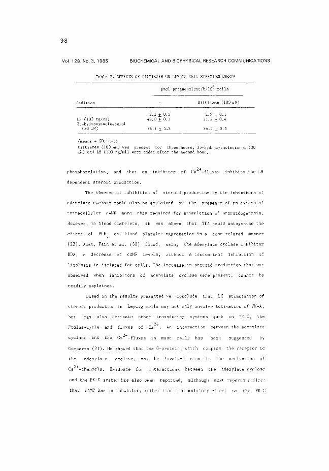
98
Vol. 128. No. 3, 1985 BIOCHEMICAL AND BIOPHYSICAL RESEARCH COMMUNICATIONS
Table 2: EFFECTS OF DILTIAZEM ON LEYDIG CELL STEROIDOGENESIS
Addition
LH (lOO ng/ml) 25-h 'id roxychol esterol
(30 I'M)
(means ±_ SD; n"'3)
pmol pregnenolone/h/106 cells
2.3 ± 0,3 49,0.±_0.1
36.1 ± 5.3
DiltiLlzem (lOO /.1:1)
2.3 + 0.1 31.2 :±: 0.4
36.2 ± 0.5
Diltiazem (lOO J.IM) 'n'as present for three hours, 25-hydroxycholesterol (30 i-'~1) and LH (lOO ng/ml) were added after the second hour.
phosphorylation, and that an inhibitor of Ca 2+-fluxes inhibits the LH
dependent steroid production.
The absence of inhibition of steroid production by the inhibitors of
adenylate cyclase could also be explained by the presence of an excess of
intracellular cAMP more than required for stimulation of steroidogenesis.
However, in blood platelets, it was shown that TFA could antagonise the
effect of PGE1
on blood platelet aggregation in a dose-related manner
(22), Also, Fain et al. (30) found, using the adenylate cyclase inhibitor
DDA, a decrease of cAHP levels, without a concomitant inhibition of
lipolysis in isolated fat cells. The increase 111 steroid production that w·as
observed w·hen inhibitors of adenylate cyclase ~-.·ere present, cannot be
readily explained.
Based on the results presented ~-.·e conclude that LH stir.mlation of
steroid production in Leydig cells may not only involve activation of PK-A,
but may also activate other transducing systems such as PK-C, the
Ptdins-cycle and fluxes of Ca 2+, An interaction bet~;·een the adenylate
cyclase and in mast cells has been suggested by
Gomperts (31). He showed that the G-protein, which couples the receptor Lo
adenylate cyclase, :nay be involved also 111 the activation of
Ca 2+-channels. Evidence for i.nteractions betw·een the adenylate cyclase
and the PK-C system has also been reported, although most reports reflecl
that cAHP has an inhibitory rather than a stimulatory effect on the PK-C

99
Vol. 128, No. 3, 1985 BIOCHEMICAL AND BIOPHYSICAL RESEARCH COMMUNICATIONS
system (13,32-35). The steroidogenic potency of PMA alone is cor.1parable to
a dose of 0,1 ng/ml Ul, and does not reach the high values obtained ,.;ith a
maximal dose of Ul. ~1ukhopadhyay et al.(36) also showed with mouse Leydig
cells a lov stimulation by P~IA, and no effect on the maximally stimulating
dose of LH.
Although PMA was considerably less potent than LH in stimulating
steroid production, che phorbolester and LH had similar effects on
protein phosphorylation. This may indicate that either PK-C is involved ln
LH action on Leydig cells, or that PK-A and PK-C act more or less
independently by phosphorylation of proteins at different sites. Both
interpretations support however that the different transducing systems act
on the same 'target proteins'. Furthermore, the low potency of PMA in
stimulating steroid production in comparison with the efficient stimulation
of protein phosphorylation, suggests that 1n addition to PK-C other
transducing systems such as cAHP and fluxes of Ca 2+ are required for
the stimulation of steroidogenesis by LH.
The possibility that Lll stimulates different messenger systems may
cast new light on old observations, Thusfar the stimulation of steroid
production by low doses of LH \o.'ilhout measurable changes in cAMP has been
explained generally by assuming that elevations of cAMP levels take place in
small intracellular pools but that such changes escape detection because
they are masked by the large cAMP pools ~>·hich are not affected by LH. It is
difficult to envisage such isolated pools, when it cs known that cANP can
pass the cell membrane, and can accumulate in the extracellular space
(5,37), An alternative explanation is that LH activates, en addition to
adenylate cyclase, other messenger systems. This possibility could not be
ruled out 1n earlier experiments, not even when a correlation was
established between binding of cAMP to PK-A, or between the activity of
PK-A, ecd steroid production (2' 6, 7). Involvement of other transducing
systems may also explain Hhy Che EDSO of hCG stimulated testosterone
production by Leydig cells io 60 times lower than Che EDSO of cholera

1 00
Vol. 12B, No. 3, 1985 BIOCHEMICAL AND BIOPHYSICAL RESEARCH COMMUNICATIONS
toxin (hCG: cholera toxin: (2), whereas the
dose response curves for stimulation of cA:-JP production by hCG and cholera
toxin are similar,
In conclusion, the present results show· that cA:tP may not be the
sole messenger for LH action on steroid production 111 Leydig cells. The
criteria as originally proposed by Sutherland and Robison (38) for
evaluating whether cAMP lS the second messenger in the transmission of a
hormonal signal can be improved by including as additional cr1terium the
effects of inhibitors of adenylate cyclase on the physiological response.
ACKNOWLEDGNENT
We thank Dr. Herrmann-Erlee (Dept, of Cell Biology, Leiden) for her
help in obtaining the adenylate cyclase inhibitor TFA.
REFERENCES
l. Marsh, J.M. (1976) Biol. Reprod. 14, 30-53, 2. Dufau, M.L., Horner, K.A., Hayashi, K., Tsuruhura, T., Conn, P.~1., Catt,
K.J. (1978) J. Biol. Chem. 253, 3721-3729. 3. Lin, T, (1983) Life Sciences 33, 2465-2471. 4, Moger, \·LH., Anakwe, 0.0. (1983) Biol. Reprod. 29, 932-937. 5. Schumacher, M., Sch,.;arz, M., Braendle, 'w', (1984) Molec. Cell, Endocrin.
34, 67-80, 6. Dufau, M.L., Tsuruhura, T., Horner, K.A., Podesta, E, , Catt, K .. J.
(1977) Proc. :.latl. Acad. Sci. USA 74, 3419-3423. 7. Cooke, B.A., Lindh, L.M., Janszen, F.H.A, (1976) Biochem. J. 160,
439-446. 8. Podesta, E., Dufau, ~l.L., Solano, A.R., Catt, K.J. (1978) J. Biol.
Chem. 253, 8994-9001. 9. Bakker, G.H., Hoogerbrugge, J.'w',, Rommerts, F.F.G., van der Mo1en, H.J,
(1983) Molec, Cell. Endocrin. 33, 243-253. 10. Rommerts, F.F.G., Bakker, G,H,, van der Molen, H.J. (1983) J. Steroid
Biochem. 19, 367-373, 11. Cooke, B.A., Lindh, L.M., Janszen, F,H.A, (1977) FEES Letters 73,
67-71. 12. Nishizuka, Y. (1984) Trends Biochem. Sci. 9, 163-171. 13. Berridge, M.J. (1984) Biochem. J. 220, 345-360, 14. Heldin, C.-H., Westermark, B. (1984) Cell 37, 9-20, 15. Lin,, T. (1984) J. Androl. 5, 193-200. 16. Sullivan, M.H.F., Cooke, B.A, (1984) Molec, Cell. Endocrin. (1984) 34,
17-22. 17. Rommerts, F.F.G., van Roemburg, M.J.A., Lindh, L.M., Hegge, J.A.J., van
der Mol en, H .J. (1982) J, Re prod, Fert. 65, 289-297. 18. Van der V us se, G .J., Kalkman, M.L. , van der Mol en, H ,J. ( 1975) Biochem,
Biophys, Acta 380, 473-485, 19. Mason, J.I., Robidoux, Vi.F. (1978) Molec, Cell. Endocrin. 12, 299-308.

1 01
Vol. 128, No. 3. 1985 BIOCHEMICAL AND BIOPHYSICAL RESEARCH COMMUNICATIONS
20, Tovey, K,C,, Oldham, K.G., l,..lhelan, J.A.M. (1974) Clin, Chim. Acta 56, 221-234.
21. Haslam, R,J,, Davidson, M.M.L., Davies, T., Lyndham, J,A,, NcClenaghan, M.D. (1978) Adv. Cycl. ){ucl. Res. 9, 533-552 (George, W.J., lgnarro, L.J., eds.) Raven Press, New· York.
22. Harris, D.:-1., Asaad, M.M., Phillips, M.B., Goldenberg, H.J., Antonaccio, M,J, (1979) J. Cycl. Nuc:l. Res. 5, 125-134,
23, Simcho·~·itz, L., Spilberg, I., Atkinson, J.P. (1983) J. Cycl. Nucl. Prot. Phosph. Res. 9, 35-47.
24, Fain, J,N,, Pointer, R.H., Ward, II'.F. (1972) J. Biol.. Chem. 247, 6866-6972.
25. Filetti, S,, Rapoport, B. (1983) Endocrinology 113, 1608-1615. 26, Florio, V.A., Ross, E.M. (1983) Molec. Pharmacal. 24, 195-202. 27. Castagna, M., Takai, Y., Kaibuchi, K., Sano, K., Kikkawa, Ll.,
>iishizuka, Y. (1982) J. Biol. Chem, 257, 7847-7851. 28. Kikkawa, U., Takai, Y., Tanaka, Y., Miyaka, R., 1\ishizuka, Y. (1983) J.
Biol. Chem. 258, 11442-11445. 29. Campbell, A.K. (1983) Intracellular Calcium, J. 1-.iiley and Sons,
Chichester, U.K. 30. Fain, J.N., Li, S.-Y., Horeno, F.J. (1979) J. Cycl. i\ucl. Res. 5,
189-196. 31. Gomperts, B.D. (1983) Nature 306, 64-66. 32. Kennerly, D.A., Secosan, C,J,, Parker, C,\o.'., Sullivan, T.J. (1979) J.
Immunol. 123, 1519-1524. 33, Nishizuka, Y. (1983) Trends fljochem. Sci. 8, 13-16. 34. :\ishizuka, Y. (1984) Nature 308, 693-698. 35, Heyw·orth, C,M,, Whetton, A.D., Kinsella, A.R., Housley, ;1,D, (1984)
FEES Letters 170, 38-42. 36. Mukhopadhyay, A. K., Bohnet, H .G., Leidenberger, F .A. ( 1984) Bioch.
Bioph. Res. Comm. 119, 1062-1067, 37. Pearlmutter, A.F., Rapino, E., Saffram, M. (1973) Endocrinology 92,
679-686. 38, Sutherland, E.H., Robison, G.A, (1966) Pharmacal. Rev. 18, 145-161.

1 03
APPENDIX PAPER 2


1 05
Effects of LH and an LH -releasing hormone agonist on different second messenger systems in the regulation of steroidogenesis in isolated rat Leydig cells
A. P. N. Themmen, J. W. Hoogerbrugge, F. F. G. Rommerts and H. J. van der Molen Department of Biochemistry (Division of Chemical Endocrinology), Medical Faculty, Erasmus University
Rotterdam, 3000 DR Rotterdam, The Netherlands
RECEIVED 2 July !985
ABSTRACT
The stimulation of steroid production in Leydig cells by LH is accompanied by increased cyclic AMP levels, activation of protein kinase A, increased phosphorylation of at least six phosphoproteins and requires protein synthesis. However, an LH-releasing hormone agonist (LHRH-A) can stimulate steroid production without stimulation of cyclic AMP levels. In the present study we have shown that LH action involves calcium ftuxes through the plasma membrane, in addition to activation of protein kinase A. The action of LHRH-A, in contrast, does not require calcium fluxes and is not potentiated by 1-methyl-3-isobutylxanthine, indicating that cyclic AMP is not involved. Extracellular calcium is required for the action of both LH and LHRH-A. An increase in intracellular calcium
INTRODUCTION
It is generally accepted that cyclic AMP (cAMP) plays an important role in the regulation of steroid production, since stimulation of the mitochondrial cholesterol side-chain cleaving enzyme (CSCC) by luteinizing hormone (LH) is accompanied by increased levels of cAMP, activation of protein kinase A (PK-A) and increased phosphorylation of at least six proteins (Cooke, Lindh & Janszen, 1976; Podesta, Dufau, Solano & Catt, 1978; Bakker, Hoogerbrugge, Rommerts & van der Molen, 1983). However, other second messenger systems, not linked to cAMP, have also been implicated in the action of LH on CSCC activity. Blocking calcium channels with cobalt inhibited the effect of LH (Moger, 1983), and the introduction of Ca2 ~ -calmodulin complexes into Leydig
concentration due to the effect of ionophore A23187 did not stimulate steroidogenesis and had deleterious effects on intracellular adenosinetriphosphate levels. LH and 4[3-phorbol-12-myristate-13-acetate (PMA), an activator of protein kinase C, both stimulated phosphorylation of proteins of 17 000 and 33 000 mol. wt, whereas LHRH-A had no effect. However, compared with the effect of LH, PMA had a much smaller effect on steroid production, indicating that even if protein kinase C may be activated by LH its role in the regulation of steroid production may be less important than the role of protein kinase A. Action of LHRH-A does not appear to be mediated by calcium fluxes, protein kinase C activation or active protein phosphorylation. J. Endocr. (1986) 108.431-440
cells via liposomes stimulated CSCC activity (Hall, Osawa & Mrotek, 1981).
In addition to second messenger systems which may be triggered by LH, other LH-independent mechanisms for the regulation of steroid production in Leydig cells exist; e.g. epidermal growth factor (Ascoli, 1981; Melner, Lutin & Puett, 1982), LH-releasing hormone (LHRH) (Hsueh & Jones, 1981; Sharpe, 1982, 1984) and vasopressin (Meidan & Hsueh, 1985), but their mechanism of action in unknown. Of these, the action of LHRH and its analogues has been investigated most. After prolonged administration in vivo in hypophysectomized rats, LHRH agonists reduce testicular LH receptors and testosterone secretion (Bambino, Schreiber & Hsueh, 1980; Cao, Sundaram, Bardin et al. 1982). These direct effects are mediated by specific high affinity receptors for LHRH on Leydig cells
J. Endocr. {1986) 108, 43!-440 © 1986 Journal of Endocrinology Ltd Prinred in Grear Briwin 0022-0795/86/0108-0431 $02.00/0

106
A. 1'. 1\, T!-ll'M~1EN nnd others Second me.l'senger sy.l'lems in Leydig ce/ls -----------------------------
(Sharp.: & Frascr, 1980). Studies with isolated Leydig cells show that the effects ofLHRH agonists on Leydig cell steroidogenesis are biphasic. In short-term incubations ( < 3 h) LH RH agonists stimulate steroid production. but when cells are incubated for 24 h or longer with LHRH agonists. the LH-dependent steroidogenesis is inhibited by the releasing hormone (Hunter, Sullivan. Dix !!! al. 1982; Browning. D'Agata. Steinberger et al. 1983).
The mechanism of LHRH action on Leydig cell steroidogenesis is not dear. Results from different workers indicate that LHRH action is not mediated by cAMP, and it has been postulated that calcium is involved in the regulation of steroid production by LHRH (Lin. 1984; Sullivan & Cooke, 1984a). However. there are many steps between the binding of LH to the receptor and the stimulation of CSCC activity, and the possible locus of the calcium effect is still unknown.
Calcium-dependent. phospholipid-activated protein kinase (PK-C) has been implicated in LHRH action on pituitary gonadotrophs (1-Iirota, Hirota. Aguilera & Catt, 1985). PK-C appears to be present in pig Leydig cells (Kimura, Katoh. Sakurada & Kubo, 1984). and phosphatidylinositol breakdown, which may lead to activation of PK-C. was stimulated by an LHRH agonist in rat Leydig cells (Molcho, Zakut & Naor. 1984). However. there were no indications of a possible effect of LH or an LHRH agonist on calcium Auxes or PK -C in Leydig cells.
We have therefore investigated the relative importance of cAMP. PK-C. calcium fluxes and protein phosphorylation in the LH- and LHRH agonistmediated activation of steroid production in rat Leydig cells.
!VIATERIALS AND METHODS
Isolated cells were incubated in modified Eagle's medium with Earle's salts and non-essential amino acids containing lOO ).lg streptomycin/m!, 0·611g fungizone(ml, 100 i.u. penicillin/m! (MEM) and I% (v/v) fetal calf serum (Gibco, Grand Island. NY, U.S.A.) (MEMiFCS) or 0·1% (w/v) bovine serum albumin (BSA) (MEM/BSA). In some experiments modified Krebs-Ringer buffer with 0·2~~ (w/v) glucose, 0·1% (w/v) BSA (pH 7·4), containing various low concentrations of calcium was used. The inclusion of ethanedioxybis(ethylamine)tetra-acetic acid (EGT A; 0·1 mmolil) in the culture medium assured the virtual absence of any calcium during incubations.
Ovine LH (NIH-LH-Sl8: 1·03 i.u/mg) was a gift from the Endocrinological Study Section of the National Institute of Health. Bethesda, MD, U.S.A,
J. Emlocr. (!986) 108,431-440
[0-Scr{Bu'l', des Gly 10-NH 2]LHRH-ethylamide (HOE 766. an LHRH agonise LHRH-A) and Ac-(D) pC I Phc-( D )pC I Phe-( D )T rp-Ser- Tyr-( D )Phc- Leu-A rgPro-(D)Ala-NH2 (0RG30093, an LHRH antagonist) were kindly provided by Hoechst Pharma (Amsterdam. The Netherlands) and Organon (Oss, The Netherlands) respectively. Di!tiazem. l-methyl~3-isobutylxanthine (MIX). ·collagenase. 4P-phorbol-12-myristatc-13-acelak (P\.1 A). 4P-phorbol~ 13-monoacetate (PA) (Sigma. St Louis. MO, U.S.A.), 5-cholesten-3p,25-diol (25-hydroxycholesterol) (Steraloids, Wilton. NH, U.S.A.) and A23187 (Boehringer. Mannheim. F.R.G.) \Vere used at the concentrations indicated. Carrier-free 32 P04 \vas from Amersham I ntcrnational plc. Bucks. 2-:x-Cyano-17~-hydroxy-4,4', lh-trimethyl-5-androsten-3-one (cyanoketone) and 7 -chloro-3,4-dihydro-2(3-pyrid yl)-l-(2H )-naph thalenone (SU-10603) were kindly donated by Or R. Neher of the Fricdrich Miescher Institute, Basle. Switzerland.
Immature rats from a Wistar substrain (RAmsterdam) were used. Leydig cells were obtained through collagenase treatment of decapsulated testes from 21- to 24-day-old animals as described by Rommerts. Molenaar & van dcr Molen (1985). The cells were allowed to attach to Costar multi well dishes in MEM/FCS. After I h the floating cells were removed by washing, and the cells attached to the dishes were used for the experiments. Incubations were carried out in MEMiBSA at 32 '·C (5% C02 in air) in the presence of the pregnenolone metabolism inhibitors cyanoketone (5)lmOl/l) and SU-10603 (19 J.lmOl/1) (van der Vusse, Kalkman & van der Molen. 1974). At the end of the incubation the medium was collected for determination of pregnenolone by radioimmunoassay (van der Vusse. Kalkman & van der Molen, 1975). The smallest amount that could be measured was 5-10 pmol pregnenolone, the intra-assay variation was in the order of 20~<- and the recovery of added pregnenolone was 107 ± ll'j;,. For the determination of cAMP and adenosinetriphosphate (ATP) the cells were lysed with 5~;, perchloric acid at 4 oc directly after removal of the medium. The lysate (200 J.ll) obtained was mixed with Tris buffer ( 1·1 mol./!, 20 11l). adjusted to pH 7 with KOH, stored at 20''C and assayed for cAMP using a cAMP assay kit (Amersham International plc; smallest amount measurable, 0·5pmol; intra-assay variation 11 ~/;) or for A TP as described by Grootegoed. Jansen & van der Molen (1984). In some experiments pregnenolone production in the presence of 25-hydroxycholesterol (30 ~tmoiil) was measured. This production can be taken as a measure of the CSCC activity and is used to give an indication of nonspecific effects of various additions to the incubations (Mason & Robidoux. 1978; Brinkmann, Leemborg, Rommerts & van der Molen, 1984).
Protein phosphorylation was measured in intact

1 07
Second messenger sysrems in Leydig ceils A. P. N. THEM~EN and others
cells. Cells (10°) were incubated for 3h in 0·5ml Krebs-Ringer buffer without phosphate but containing 0· 2% (w/v) glucose and 0·1% (w/v) BSA, in the presence of 37 MBq 32P04 . The proteins were extracted and separated on polyacrylamide gradient gel electrophoresis in the presence of sodiumdodecylsulphate (8-15% polyacrylamide gradient) as described before (Bakker, Hoogerbrugge, Rommerts & van der Molen, 1981). The gels were dried and autoradiographed using Kodak SB-5 X-ray film. Molecular weight was determined using an electrophoresis calibration kit from Pharmacia Fine Chemicals (Uppsala, Sweden).
The results are given as means ±s.o. of four or more observations obtained using two or more different cell preparations. Each observation is the result of an assay performed in duplicate. The significance of differences
140
120
:? 100
1i c
~ 80 ~
'3 E ~ 0 s 60 0
~ ~ e ~
40
20
0 0 I I 10 100 LH (ng/ml)
FIGURE 1. Effect ofLH-releasing hormone agonist (LHRH-A) on LH-stimulated pregnenolone production by Leydig cells from immature rats. Leydig cells were incubated for 3 h with different concentrations of LH in the absence (O) or presence (.6) ofLHRH-A (40 nmol/1). Values are means ±s.o. (n =4) of two different cell preparations. All values in the presence ofLHRH-A are significantly (P <0·01) different from corresponding productions with LH alone (Studenfs /-test).
between results was calculated using Student's r-test for non-paired data. Differences were considered to be significant when P values were less than 0·0 I.
RESULTS
Steroid production
Leydig cells attached to the plastic surface of culture dishes were incubated for 3 h in the presence of LH (I 00 ng/ml) or in the presence of LH (lOO ng;'ml) and LHRH-A (40nmol/l) (Fig. 1). Pregnenolone production was stimulated 35-fold when cells. were incubated with LH alone compared with controls. The response could be further enhanced (60-fold) when LHRH-A (40 nmol;'l) was added together with LH (Fig. 1). In the presence ofLH, the addition ofLHRHA causF:d an additional steroid production of 60pmol/h per 106 cells; without LH the increase in pregnenolone production was only 8 pmol/h per 106
cells. LHRH-A alone stimulated pregnenolone production in a dose-responsive manner, with a dose for half-maximal stimulation of 0·1 nmol/1 (Fig. 2). Cells cultured for 48 h in the absence of LH or LHRH-A could still be stimulated with LH (control, 8± 1; LH, 145±12pmoljh per 106 cells: n=8 of three different cell preparations), but not with LHRH-A. The effect of LH-stimulated pregnenolone production by cells cultured for 48 h in the presence of both LH and LHRH-A was less than the effect of LH on cells after
10
1i ~
g_
"' 6 0
!
~/ 0 4 8 g
~ ~
0 0·004 O·b4 0·2 d-4 2 4 20 40 LHRH-A (nmo1/1}
FIGURE 2. Effect of LH -releasing hormone agonist (LHRH-A) on pregnenolone production by immature rat Leydig cells. Cells were incubated from 3 h in the presence of various concentrations ofLHRH-A. Values are means ±s.o. (n =4) of two different cell preparations. All productions in the presence of LHRH-A, except the value at 0·004 nmol LHRH-Ajl, were significantly (P <0·01) different from the control (Student's 1-test).
J. Em/on·. (1986) 108.431-440

108
A. P. N. THEM MEN and others Second messenger systems in Leydig cefls
culture with LH alone (180 ± 4 compared with 105 ± 3 pmoljh per 106 cells: n = 8 of three different cell preparations). The effect of LH on cells after culture for 48 h in the presence ofLHRH~A did not differ from the effect of LH on control cel!s. Both the stimulatory (Figs I and 2) and the inhibitory effects of LHRH-A were completely abolished when the LHRH antagonist (400 nmol/1) was present during the incubations or cultures. The LHRH antagonist did not have any effect by itself.
Role of cAMP
The role of cAMP in the mechanism of action of LHRH-A was investigated by measuring the effect of LH and LHRH-A on intracellular cAMP levels, and by comparing the effect of the phosphodiesterase inhibitor MIX on steroid production stimulated by LHRH-A and LH (Table 1). Luteinizing hormone (1 00 ng/ml) enhanced intracellular cAMP levels threefold and a further increase was obtained when, in addition to LH (100 ngjml), MIX (200 J1moljl) was added to the incubation medium. Under these conditions MIX had no effect on steroid production. In cells incubated with a lower concentration of LH (0·2 ngjml) MIX had no effect on cAMP levels, but it stimulated pregnenolone production approximately fourfold. Neither intracellular cAMP levels nor pregnenolone production in the presence of LHRH-A was affected by the addition of MIX.
Role of calcium
Effects of changes in the calcium concentration on steroid production were investigated in different ways: (l) by lowering the extracellular calcium concentration, (2) by blocking the calcium channels with the calcium channel blocking agent diltiazem, and (3) by increasing the calcium flux through the plasma membrane with the calcium ionophore A23187. In all experiments pregnenolone production in the presence
tOO
0 I 10 Ca"-{flmol/1)
100 1000
FIGURE 3. Etfect of extracellular calcium concentration on hormone- and 25-hydroxycholesterol-stimulated steroid production. Cells were incubated for 3 h in the presence of LH (lOO ng/ml, e), LH (0·2 ng,lml. 6), LH-releaSing hormone agonist (LHRH-A; 40 nmol.il, 0) or 25-hydroxycholesterol (30 1-lmol/1, 11!1). Values are mean per-centages ±s.o. (n =4) of two different cell preparations, with values at I mmol Cal+ jl taken as 100%. Pregnenolone production (pmol,ih per 106 cells) at I mmol Cal+/1 was: lH (lOOng/ml) 116·0; LH (0·2ngiml) 20·8; LHRH-A (40 nmolil) 12·3: 25-hydroxycholesterol (30 flmol/1), 120·6. * P<O·OI compared with 25-hydroxycholesterol-stimu1ated production at I mmol Ca2 ~/l. tP<O·Ol compared with LH-stimulated production at 10 ]lmol Ca2
+ /1. All other LHRH-A- and LH-stimulated productions at identical Ca2
T concentrations were not significantly different (Student's Hest).
of 25-hydroxycholesterol (30 J1moljl) was also measured and taken as a measure of CSCC activity (Mason & Robidoux, 1978; Brinkmann et al. 1984).
TABLE I. Etfectst of 1-methyl-3-isobutylxanthine (MIX: 200 JlffiOI/1) on cyclic AMP (cAMP) levels and pregnenolone production in rat Leydig cells in the presence of LH and LH-releasing hormone agonist (LHRH-A). Values are means± S.D. (n=4) of two cell preparations
PregnenOlone (pmo1/h per !06 cells) cAMP (pmol/106 cells)
Additions None LH (I 00 ngjml) LH (0· 2 ngjml) LHRH-A (40 nmolil)
No MIX
1·6±0·3 80·9±3·5 18·6±5·1 13·3±H
MIX
6·0± 1·6* 79-6± 14·5 87·2± 12·6* 18·6±8·5
• P < 0·01 vs corresponding values without MIX (Student':; Hest)
No MIX
7·2±2·2 21·9±2-9 6·0±H 7-3±3-9
MIX
6·3 ± 1·8 36·1±7-4~
7·7± 1·8 H±B
tLeydig cells were incubated in the presence of the additions as indicated. After 3 h the medium was collected for pregnenolone measurement and the cells were lysed for cAMP determination
J. Endocr. (1986) 108,431---440

109
Second messenger systems in Leydig cells A. P. N. THEM MEN and others
After lowering extracellular calcium from I m mol to I Mmol/1 steroidogenic activation in the presence of LHRH-A and different concentrations of LH was inhibited to approximately 30'% of the control values (Fig. 3). Inhibition of the 25-hydroxycholesteroldependent pregnenolone production at calcium concentrations lower than 10 Mmol/1 indicated that at these concentrations mitochondrial CSCC activity was probably directly affected.
Blocking the calcium influx with diltiazem inhibited the LH-stimulated steroid production by 27% both at 0·1 and IOOng/ml, whereas the LHRH-A-stimulated steroid production was not affected. Steroid Production in the presence of 25-hydroxycholesterol was not inhibited (fig. 4).
Treatment of the cells with different concentrations of A23187 inhibited the effect of LH ( 100 ng/ml) and LHRH-A (40nmol/l), but not the effect of LH (0·2 ng/ml) on pregnenolone production (fig. 5). Basal production was not affected. Steroidogenesis in the presence of 25-hydroxycholesterol was also inhibited at low concentrations of A23187, indicating that the ionophore had direct effects on mitochondrial CSCC. The inhibition ofCSCC might have been caused by cell damage, and therefore intracellular A TP levels were measured. After incubation of the cells for 3 h in the presence of different concentrations of A23187 the following ATP levels were measured: control, H±0·1; A23187 (0·1 ~mo1/1). 1·0±0·2 (P<0·01);
50
1i
"' ~
0 25 E
" 5 0
~ ~
" ~
0 m " ~ g ~ -g 0
0 s z ~
~
~ ...::5 o~
~~
~ 0 .. :eo §
0 [ ~
;cc ~" ~~
~
FIGURE 4 Effects of the calcium-channel blocker diltiazem (hatched bars) on pregnenolone production by immature rat Leydig cells in the presence of LH. LH-releasing hormone agonist (LHRH-A) and 25-hydroxycholesterol (25-0H-chol). Cells were incubated for 3 h with the hormone concentrations indicated in the absence or presence of diltiazem (lOO ).lmol/1). Controls are shown by open bars. Values are means± s.o. (n=4) of two cell preparations. *P<O·OJ compared with corresponding incubation without diltiazem (Student's /-test).
A23187 ('"mol/!)
flGL"RE 5. Effects of calcium ionophore A23187 on pregnenolone production by immature rat Leydig cells in the presence ofLH (l00ng/ml: cross-hatched bars). LH (0·2 ng/ml; vertical-hatched bars). LH-releasing hormone agonist (LHRH-A) (40 nmol/1; open bars) and 25-hydroxycholesterol (30 mol/1: stippled bars). Values are mean percentages± s.o. (n=4) of two different cell preparations, with the values in the absence of A23187 taken as lOO%. Pregnenolone productions in the absence of A23187 (pmol/h per 106 cells): LH (lOO ng/ml) 91·2; LH (0·2 ng/ml) 14·7; LHRH-A (40 nmol/1) 9·7; 25-hydroxycholesterol (30 ).lmOl/1) 121·2. * P<O·OI compared with corresponding control (Student's r-test).
J. Emlocr. (1986) 108,431-440

11 0
A. P. N. THl:MMEN and others Sr>cond m('.\'Senger syswns in Leydig cells
A23187 (I ~unol,il). 0·5 ± O·l nmol ATP/106 (P < 0·01) cells (mcans±s.D.) n=3. one cell preparation). These results show that ATP levels were progressively decreased after incubation of the cells with increasing concentrations of A23187.
Role of protein kinase C
To investigate whether LHRH-A acts through the PK -C system, the effect of a specific activator of PK-C, the phorbol ester PMA (Castagna, Takai, Kaibuchi et al. 1982; Kikkawa, Takai, Tanaka et al. 1983) on pregnenolone production was measured. In 3-h incubations PMA (lOOng/ml) alone stimulated steroid production approximately lhreefold (control, 2·0 ±0·1; PMA. 5·9 ±0·2 pmol pregnenolone,ih per 106
cells) (Fig. 6). However, whereas LHRH~A potentiated LH action on pregnenolone production. PMA inhibited pregnenolone production in the presence of low concentrations of LH. The maximal steroid production obtained with high doses of LH was not inhibited by PM A.
The phorbol diester PA ( 100 ng/ml) had no effect on steroid production (results not shown).
Protein phosphorylation
The pattern of protein phosphorylation was investigated to elucidate further the second messenger
JOO•
1l c
~ ~
i3 50
§ g 0
l ~
0
systems for LHRH and LH which may play a role in the regulation of steroid production in the Leydig cell (Fig. 7). Both PMA and LH stimulated the phosphorylation of a protein of I 7 000 mol. wt and of 33 OOOmol. wt, whereas LHRH-A and PA (100 ng/ml) did not have an effect on protein phosphorylation.
DISCUSSlON
The involvement of different second messenger systems in the control of steroid production by LH and LHRH was investigated in Leydig cells from immature rats. The present study confirms that cAMP is not involved in the action of LHRH on Leydig cell steroid production and that calcium may be important, and extends these observations: extracellular calcium, but not calcium fiuxes are essential for LHRH-A action. Moreover, the results indicate that LHRH-A may not activate PK-C.
After incubation of the cells for 2 days with LH and LHRH-A, the subsequent LH-stimulated steroid production was lower than the production in cells preincubated with LH alone. This might be caused by an effect of LHRH-A on cytochrome P450 enzymes as observed for microsomal enzymes by Magoffin & Erickson ( 1982) after incubating ovarian cells with LHRH-A. However. LHRH-A might also have acted
10 LH (ngiml)
FIGURE 6. Effect ofphorbol ester 4~-phorbol-12-myristate-13-acetate (PMA) on LH-stimulated pregnenolone production by immature rat Leydig cells. Cells were incubated for 3 h with different concentrations of LH in the absence (0) or presence (G) of PMA (lOO ng/ml). Values are means ±S.D. (n = 4) of two different cell preparations. All values in the presence of PMA were significantly (P<O·Ol) different from the corresponding value without PMA, except the value at 10 ng LH.iml (Student"s /-test).
J. Endocr. (1986) 108.431-440

111
Sr>cond mr>sscnra sysrem.\' in Leydig cd/.1· A. P. ~-THEM ME~ and others
- 33000
- 17000
FIGURE 7. Protein phosphorylation pattern of immature rat Leydig cells. Cells were incubated alone or with either LH (lOO ng/ml). 4~-phorbol-12-myristate-13-acetate (PMA: lOOng/ml). 4~-phorbol-13-monoacetate (PA: lOOng/ml) or LH-releasing hormone agonist (LHRH-A; 40 nmol/1) in the presence of 32 PO.,.. Proteins were extracted and separated on 8-1 s::~·, polyacrylamide gel electrophoresis in the presence of sodiumdodecylsulphatc. The autoradiograph of the dried gel is shown. Lane 1, control; lane 2. LH: lane 3. LHRH-A: lane 4. PMA and lane 5. PA.
on steps between binding of LH to the receptor and activation of the mitochondrial CSCC. Down-regulation of LH receptors appears to be unlikely because a 60-80~>~ reccptor loss. induced by injection of human chorionic gonadotrophin (hCG), did not cause a decrease of hCG-dependent pregnenolone production in intact rats (Cigorraga, Dufau & Catt, 1978). LHRH-A had long-term inhibitory actions when LH was also present in the preincubation, but enhanced the stimulation of pregnenolone production by LH in short-term incubations. This synergistic effect of LHRH-A on LH-stimulated pregnenolone production suggests that LHRFI-A employs a specific pathway independent of LH. The time-related negative or positive effect of LHRH-A on LH-induced steroid production could be important for the physiological function of LHRH or LHRH-like molecules and suggestions have been made that LHRH-like molecules produced by Sertoli cells could exert paracrine effects on Leydig cells (Sharpe. Fraser. Cooper & Rommerts, 1981: Sharpc. 1982). Hmvever. there is no direct evidence for the possible testicular production of LHRH (Verhoeven & Cailkau, 1985).
Whatever the physiological function of LHRH or
LHRH-likc factors in the testis may be, the mechanism of LHRH action is intriguing because cAMP appears not to play an important role, since LHRH-A does not enhance cAMP levels (Lin, 1984; Sullivan & Cooke, 1984h). However, the absence of increased cAMP levels does not necessarily mean that cAMP is not involved. since low concentrations of LH can cause a large steroidogenic response without a concomitant measurable increase in cAMP levels (Cooke et al. 1976; Podesta et al. 1978). Our experiments w;th MIX show that inhibition of phosphodiesterase can enhance the effect of 0·2 ng LH/ml on pregnenolone production four-to fivefold, and a maximal steroid production is obtained without a measurable rise in cAMP levels. Since MIX inhibits cAMP breakdown, this result shows that cAMP is involved in LH action. MIX did not influence the LHRH-A action on steroid production indicating that cAMP is probably not involved in the action of LHRH-A. Sullivan & Cooke ( (1984b) have observed inibition ofLH-stimulated cAMP levels by LHRH-A, whereas LHRH-A stimulated LHdependent steroid production. We have observed a similar discrepancy between decreased cAMP levels and an increase in steroid production after inhibition of adenylate cyclase (Themmen, Hoogerbrugge, Rommerts & van der Molen, 1985). These observations suggest that, in addition to cAMP, other second messenger systems could be involved in the action of LH. We therefore investigated the possible involvement of calcium and PK-C in LH and LHRH-A action.
A decrease in the extracellular calcium concentration resulted in a concomitant decrease of both LHand LHRH-A-dependent steroid production. The CSCC enzyme itself, or the NADPH generation within the mitochondria, was much less affected by the low extracellular calcium concentration, as judged by the small effects on steroid production in the presence of 25-hydroxycholesterol, which is a good indication of CSCC activity (Mason & Robidoux, 1978; Brinkmann et al. 1984). Similar effects of low extracellular calcium concentrations have been observed in previous work studying Leydig cells (Janszen, Cooke, van Oriel & van der Molen, 1976). adrenocortical cells (Trzeciak & Mathe, \981) and granulosa cells (Veldhuis & Klase, \982). Low extracellular calcium concentrations do not cause damage, since the inhibiting effects of low calcium concentrations on Leydig cells are reversible (Janszen er al. 1976). Moreover, extracellular calcium is not required for insulin action on hexose transport in L6 muscle cells (Klip, Li & Logan, 1984). Calcium can influence the delivery of cholesterol to. or its utilization by. mitochondria in the Leydig cell (Hall et al. 1981}. Hence, calcium ions may regulate intracellular events important for the formation of pregnenolone. Subsequently, the importance of calcium ftuxes
J. Endoa. tln6) 108.431---440

11 2
A. p, N. THEMMEN and others Second messenger systems in Leydig ce!fs
through the plasma membrane for LH and LHRH-A action was investigated.
The inhibitory effects of diltiazem on LH-stimulated steroid production suggest that calcium fl.uxes through the plasma membrane are involved in LH action. Pregnenolone production in the presence of 25-hydroxycholesterol was not inhibited, which may reflect the fact that diltiazem did not affect mitochondrial CSCC. Similar results with ovarian cells have been reported by Veldhuis & Klase (1982). LHRH-A-dependent steroid production was not affected by diltiazem, suggesting that calcium ftuxes through the plasma membrane are less important in LHRH-A action. It was recently reported that another channel blocker, nifedlpine, inhibited LHRH-A-dependent steroid production (Lin, 1984), but in the present study the specificity of the effect of nifedipine was not evaluated. In order to obtain a more conclusive answer on the importance of calcium in LHRH-A action, direct measurement of calcium ions after stimulation of Leydig cells with LHRH-A is necessary. Preliminary experiments with the fluorescent probe Quin-2, however, showed an insufficient loading capacity for the probe of immature rat Leydig cells. Another approach to investigate the effect of artificially increased intracellular calcium concentration with calcium ionophore A23187 was therefore chosen.
Incubation of Leydig cells with A23187 inhibited basal or hormone-stimulated pregnenolone production. The ionophore has been used frequently to investigate the role of calcium ions, and biological effects (such as secretion of proteins) have been shown after application of A23187 (Zawalich, Brown & Rasmussen, 1983; Conn, 1984). Inhibition of the 25-hydroxycholesterol-dependent response indicated that A23187 directly affected the CSCC enzyme. Intracellular A TP levels of the cells were measured after incubation with A23187 to evaluate another mitochondrial function. Levels of A TP were decreased after application of A23187, suggesting that the ionophore has deleterious effects on immature rat Leydig cells. Thus results with the ionophore are inconclusive with respect to the involvement of an increase of intracellular calcium in LHRH-A action, but show that A23187 has deleterious effects in our cell system. It has been shown by Molcho et al. (1984) that LHRH-A can stimulate the turnover of phosphatidylinositol in Leydig cells. We have investigated whether LHRH-A could activate PK-C, since stimulation of phosphatidylinositol breakdown is thought to result in activation of PK-C, in addition to its effect on intracellular calcium release (Berridge, 1984).
PMA stimulates pregnenolone production in immature rat Leydig cells with approximately the same potency as LHRH-A, suggesting that the releasing hormone acts via PK-C. However, in contrast with
J. Endor:r. ( !986) 108. 431-440
LHRH-A, PMA did not potentiate LH-dependent steroid production. Moreover, the stimulatory effect of PMA on protein phosphorylation was very similar to the effect of LH, whereas LHRH-A had either no effects or minor effects, but at least much less than PMA. These results do not support the hypothesis that LHRH-A acts through PK-C.
LH and PMA both stimulate the phosphorylation of the nuclear 17 000 moL wt and the ribosomal 33 000 mol. wt. protein S6 to the same extent (Bakker et al. 1981 ), but LH is tenfold more effective than PMA in stimulation of pregnenolone production. This suggests that phosphorylation of the 17 000 and 33 000 moL wt proteins alone is not sufficient for the acute stimulation of steroidogenesis by LH. The similarity in the patterns of protein phosphorylation stimulated by LH and PMA may suggest a role for PK-C in LH action in addition to cAMP-activated PK-A. However. the inhibition of steroid production by PMA observed in the presence of low doses of LH is puzzling. It may be that PK-C and PK-A, under the influence of different doses of LH, phosphorylate different sites on the same protein, as has been shown for ribosomal protein s6 (Trevillyan, Perisic, Traugh & Byus, 1985), but a connection between phosphorylation sites and function of ribosomal protein s6 has not been demonstrated.
From our present results we conclude that LHRH-A does not mediate its effects on steroid production through PK-A or PK-C, and that calcium fluxes through the plasma membrane do not seem to be important in LHRH action, whereas LH action may involve PK-A and PK-C activation and calcium fluxes. Both hormones require the presence of extracellular calcium. The possible role of PK-C activation in LH action, suggested by the similarity of the patterns of protein phosphorylation, remains to be elucidated.
ACK~OWLEDGE:viENTS
Hoechst Pharma (Amsterdam, The Netherlands) and Organon (Oss The Netherlands) are gratefully acknowledged for their gifts of HOE 766 and ORGJ0093.
REFERENCES
Ascoli, M_ ( 1981 ). Regulation of gonadotropin receptors and gonadotropin responses in a clonal strain of Leydig lumor cells by epidermal growth factor. Joumal of Biological Chemisrry 256. 179~183.
Bakker. G. H., Hoogerbrugge. J. W .. Rommerts, F. F. G. & van der Molen, H. J. ( J 981 ). Lutropin-dependent protein phosphorylation and steroidogenesis in rat tumour Leydig cells Biochemical Journal 198, 339~ 346.

11 3
Second messenger sysrems in Leydir cells A. P. K. THEMMEN and others
Bakker_ G. H .. Hoogcrbrugge. J. W._ Rommerts. F. F. G. & van der Molen, H. J. ( 1983). LH-dependent steroid production and protein phosphorlylation in culture of rat tumour Leydig cells. Molecular alii! Cellular Endocrinoliwr 33, 243-253.
Bambino. T. H._ Schrcibcr. J. R. & Hsueh. A. 1. W. ( 1980). GnRH and its agonist inhibit testicular lutcini1ing hormone rcccptor and steroidogenesis in immature and adult hypophy>ectomizcd rats. Endoainology 107, 908-917.
Bcrridgc. M. J. ( 1984). Inositol triphosphate and diacylglycerol as second messengers. Bioc/r('lllical Journal 220. 345-360
Brinkmann, A. 0., Lecmhorg, F. G._ Rommcrts. F. F. G. & van dcr :vfolen. H. J. ( 1984). Di!Terences between the regulauon of chol~sterol side-chain cleavage in Leydig cells from mice and rats. Journal of Sreroid Biochemisry 21. 259-264.
Browning. J. Y .. D"Agata. R .. Stcinbcrger. A.. Grotjan. H. E .. Jr & Stcinbcrgcr. E. ( 1983). Biphasic c!Tect of gonadotropin-releasing hormone and its agonist analog (HOE766) on in •·irro testosterone production by purified rat Leydig cells. Endocrinology 113. 985-991
Cao, Y.-Q .. Sundaram, K., Bardin, C. W .. Rivicr . .1. & Vale. W ( 1982). Direct inhibition of testicular steroidogenesis and gonadotropin receptor levels by ((imBzi)-His''.Pro''-NEt)GnRH and (o-Trp".Pro9-NEt)GnRH. potent agonists ofGnRH. lntcnwlional Jmmwl o(AndroloRJ" 5. 158-170.
Castagna, M., Takai. Y .. Kaibuchi. K .. Sano. K .. Kikkawa. U. & Ni>hizuka. Y. ( 1982). Direct activation of calcium-activated. phospholipid-dependent protein kinase by tumor-promoting phorbol esters . .foumal <~f Biolor;icul Chemislry 257, 784 7-7851.
Cigorraga. S. B.. Dufau, M. L. & Call. K. 1. (1978). Regulation of l11teinizing hormone receptor,; and steroidogenesis in gonadotropin-d~sensitized Leydig cells. Jmmwl '!f"Biologiwl Chcmisrry 253. 4297-4304.
Conn. P. M. ( 1984). Molecular mechanism of gonadotropinreleasing hormone action. In Biochcmiml A er ions of Hormones. vol. X!. pp. 67-92. Ed. G. Litwack. London: Academic Pr~ss
Cookc. B. A .. Lindh. L. M. & 1anszen. F. H. A. (t976). Correlation of protein kinase activity and testosterone production after stimulation of Leydig cells with luteinizing hormone. Biochemical }oumall60, 439--446.
Grootegoed. J. A .. 1anscn. R. & van der Molcn H. J. (1984). The role of glucose. pyruvate and lactate in ATP production by rat spermatocytcs and spcrmatids. Biochimi("a f/ Biophysica A Cl(t 767. 248-256.
Hall. P. F .. Osawa, S. & Mrotek, J. ( 1981 ). The influence of calmodulin on steroid synthesis in Leydig cells from rat testis Emfocrinology 109. 1677-!682
Hirota, K., Hirota. T .. Aguilera. G. & Catt. K. J. { 1985) Hormone-induced redistribution of calcium-ac;:tivated phospholipid-dependent protein kinas~ in pituitary gonadotrophs. Jownul of Biological Chemisrry 260. 3243--3246.
Hsueh. A. 1. W. & Jones, P. B. C. ( 1981). Extrapituitary actions of gonadotropin-releasing hormone. Endocrine Re•·ie1vs 2, 437-461.
Hunter. M. G., Sullivan. \1. H. F .. Dix. C. L Aldred. L. F. & Cooke. B. A. ( 1982). Stimulation and inhibition of LHRH analogues of cultured rat Leydig cells function and lack of e!Tect on mouse Leydig cells. ;\Jolecular and Cellular Endocrinology 27. 31-44.
Janszen, F. H. A .. Cooke. B. A .. van Oriel. M. 1. A. & van der M olen. H. J. ( 1976). The clfect of calcium ions on testosterone production in Leydig cells from rat testis. Biochemical Journal 160.433-437.
Kikkawa, U., Takai. Y .. Tanaka. Y .. Miyaka. R. & Nishizuka. Y. ( 1983 ). Protein kinase Cas a possible receptor protein of tumor-promoting phorbol esters. Journal of Biological Chemistry 258. 11442-11445.
Kimura. K .. Katoh. N .. Sakurada. K. & Kubo. S. {1984) Phospholipid-sensitive Ca 2 ~-dependent protein kinase sy<;tem in
testi>: Locahsation and endogenous suhstn1tes. Endocrinology 115, 2391 --2399
Klip. A .. LL G. & Logan. W. J. (1984). Rok of calcium ions in insulin action on hexose transport in L6 muscle cells. American Journal of" Physiology 247. E297-E304
Lin. T. ( 1984). Mechanism of action of gonadotropin-releasing hormone-stimulated Leydig cell steroidogenesis. I. The stimulating eiTect is calcium dependent and not mediated by cyclic nucleotidcs. Jounwl ()f Amlro/ogy 5. 193-200.
M(l.gomn. D. A. & Erickson. G. F. ( 1982). Mechanism by which GnRH inhibits androgen synthesis directly in ovarian interstitial cells. M ofaalar and Cellular Endocrino!ogy 27, 191-198.
Muson. J. \. & Robidoux. W. F. ( 1978). Pregnenolone biosynthesis in isolated cells of Snell rat adrenocortical carcinoma 494. ivfolecular rmd Cellular Emlocrino/og_v 12. 299-308.
:vtcidan. R. & Hsuch, A. J. W. ()985).\dentification and characterization of arginine vasopressin receptors in the rat testis. Endoaitwlo?;y 116,416--423.
Mclner. M. H., Lutin, W. A. & Puett, D. (1982). Ep1dermal growth factor and cyclic AMP stimulation of distinct protein kinase activities in Leydig cell tumor membranes. Li{e Sciences 30, 1981-1986
Moger. W. H. ( 1983). Efi"ects of the calcium-channel blockers Cobalt, vcrapamil and 0600 on Leydig cell steroidogenesis. Biolo[;y o(" Reproduction 28. 528"'535
:vtolcho, 1 .. Zakut. H. & Naor, Z. (1984). Gonadotropin-releasing hormone stimulates phosphatidylinositollabeling and prostaglandin E production in Leydig cells. Endocrino/ogy 114. I 048·-1 050.
Podesta. E .. Dufau, M. L., Solano. A. R. &Catt. K. J. (1978). Hormonal activation of protdn kinase in isolated Leydig cells. Electrophoretic analysis of cAMP receptors . .louma! of Biolo~:iml Chemisrrr 253. 8994-9001
Rommerts. F. F. G .. :Ytolenaar. R. & van der Molen. H. J. ( 1985). Prep~ ration of isolated Leydig cells. Methods in En:ymology 109. 275-288.
Sharpe. R. M. (1982). Cellular aspects of the inhibitory actions of LH-RH on the ovary and testis. Journul of Reproducrion und Fenility 64, 517-527.
Sharpc, R. M. ( 1984). lntratcsticubr factors controlling testicular function. Biolo?;y of Reproduction 30, 29-49.
Sharpe. R. \>1. & Fraser. H. :vt. ( 1980). Leydig cell receptors for LJ-IRH(a) and their modulation by administration or deprivation of the releasing hormone. Biochemical and Biophysical Research Communicarions95, 256-262.
Sharpe. R. M .. Fraser, H. M .. Cooper, I. & Rommerts. F. F. G. (198 !). Sertoli-Leydig cell communication via an LHRH-like factor. l'iolurc 290. 785-787.
Sullivan. :vt. H. F. & Cookc. B. A. ( 1984a). The effect of calcium on the potentiation of LH->timulated steroidogenesis and inhibition of LH-stimulated cyclic AMP production by LHRH agonist (I Cl 118630) in rat Leydig cells . .. Uo/eculur ad Cdlu!ur Emlocrinology 34. 17···22.
Sullivan. M. H. F. & Cooke, B. A. (1984h). LHRH agonist decreases LH- but not forskolin-stimulated cyclic AMP levels in rat Leydig cells in1·itro. Molecular and Cdlular Endoerinology 36. 115~122.
Thcmmen. A. P. N .. Hoogcrbrugge. J. W .. Rommerts. F. F. G. & van der Molen. H. 1. (1985). Is cAMP the obligatory second messenger in the action of lutropin on Leydig cell steroidogenesis? Biochemical and Biophysical Research Communications 128. 1164-1172
Trevillyan.J. M., Perisic. 0 .. Traugh.J. A. & Byus. C. V. (1985) Insulin- and phorbol ester-stimulated phosphorylation of ribosomal protein 56 . Journal of Biological Chemisrry 260, 3041-3044.
Trzeciak, W. H. & Mathe, D. (1981). Preparation of adrenocortical cell suspension highly responsive to ACTH or dibutyryl cyclic
J. Endocr. (1986) 108,431-440

11 4
A. P. N. THEM MEN and others Second messenger systems in Leydig ce/fs
AMP. Effects of albumin, calcium or stress. FEES Le/ten; 130. 113-118.
VeldhuisJ D. & Klase. P.A. (1982}. Mechanisms by which calcium ions regulate the steroidogenic actions of luteinizing hormone in isolated ovarian cells in vitro, Endocrino!ogy Ill. l-6.
Verhoeven, G. & Cail!eau, J. (1985). A factor in spent media from Sertoli cell-enriched cultures that stimulates steroidogenesis in Leydig cells. Molecular and Cellular Endocrino/ogy 40, 57--68.
van derVusse. G. 1 .. Kalkman. M. L & van der Molen. H. J. ( 1974). 3~-Hydroxysteroid dehydrogenase in rat testis tissue.
J. Endocr. (1986} 108, 431--440
Inter- and subcellular localisation and inhibition of cyanoketone and nagouse. Biochimim e/ Bioph_vsica Acta 348, 404-414.
van der Vusse, G. J .. Kalkman, M. L. & van der Molen, H. J ( 1975). Endogenous steroid production in cellular and subcellular fractions of rat testis after prolonged treatment with gonadotropins. Biochimica er Biophysica Acta 380,473-485.
Zawalich. W., Brown, C. & Rasmussen, H. {1983). Insulin secretion: combined effects of phorbol ester and A23187. Biochemical and Biophysical Reseanh Communicativns 117. 448-455.

11 5
APPENDIX PAPER 3


117
COMPARISON OF THE CELLULAR COMPOSITION AND STEROIDOGENIC PROPERTIES OF
INTERSTITIAL CELL PREPARATIONS ISOLATED FROM IMMATURE AND MATURE RAT
TESTIS.
A.P.N. Themmen, R. Molenaar, W.J. Visser*, J.F. Jongkind*, F.F.G. Rommerts
& H.J. van der Molen
Department
*Department
of
of
Biochemistry
Cell Biology
(Division of Chemical Endocrinology) and
"and Genetics, Medical Faculty, Erasmus
University Rotterdam, Rotterdam, The Netherlands.
Correspondence to:
A.P.N. Themmen
Department of Biochemistry II
Erasmus University Rotterdam
p.Q, Box 1738
3000 DR Rotterdam
The Netherlands
Running head: .interstitial cells from mature and immature rat testes

118
ABSTRACT
Interstitial cell preparations from both immature (21-24 days old) and
mature (60-90 days old) rats are used in many investigations on the
regulation of steroidogenesis. We have compared the morphological and
steroidogenic properties of interstitial cell preparations isolated from
testes of immature and mature rats. collagenase dispersed cells isolated
from immature rats contained 40-60% Leydig cells, 37% mesenchymal cells and
no macrophages. cell preparations from mature rats, after a Ficoll
purification step, consisted of 80% Leydig cells and 20% macrophages. A 90%
pure Leydig cell preparation could be obtained after further purification
of the preparation isolated from immature rats with a Percoll gradient
centrifugation step. Fractionation of this gradient revealed that the
distribution through the gradient of steroidogenic enzymes and LH-dependent
steroid production was similar. The results indicate that the steroidogenic
activity per Leydig cell from mature rats is fourfold larger than the
activity in immature rat Leydig cells after stimulation with LH, dibutyryl
cyclic AMP or in the presence of 22R-hydroxycholesterol.
INTRODUCTION
Leydig cells from mature rats have been used frequently for studies on
the regulation of steroidogenesis by LH and other factors (Cooke &
Sullivan, 1985; Adashi & Hsueh, 1982; sharpe, 1984). After collagenase
dispersion of testis tissue the isolated cell preparation generally
contains approximately, 10% Leydig cells, although the recovery of intact
Leydig cells from the original testis is less than 10% (Molenaar, Rommerts
& van der Molen, 1983; Rommerts, Molenaar & van der Molen, 1985). The
purity and viability of the isolated cell preparation can be greatly im
proved after density centrifugation and selection through attachment of
intact cells to culture dishes (Cooke, Magee-Brown, Gelding & Dix, 1981;
Molenaar et al~, 1983), Leydig cells from immature (21-24 days old) rats
can be obtained in a rather pure state (40%) and in much higher yield (90%)
after collagenase treatment of the testis, whereas almost no damaged Leydig
cells are present (Molenaar et al., 1983). Isolated immature Leydig cells
retain their LH-dependent steroidogenic properties in culture for 3 days

11 9
(Berns, Brinkmann, Rommerts, Mulder & van der Molen, 1985), in contrast to
isolated Leydig cells from mature rats, which lose more than 90% of their
steroid producing capacity in response to LH under the same incubation
conditions (Hunter, sullivan, Dix, Aldred & Cooke, 1982; Rommerts, van
Roemburg, Lindh, Hegge & van der Molen, 1982), Since Leydig cells from
immature rats have the advantages mentioned above compared with cells
isolated from mature rats, we have used these cells for studies on the
regulation of steroid production (Themmen, Hoogerbrugge, Rommerts & van der
Molen, 1985, 1986).
It is known that during testicular development the Leydig cell under
goes differentiation. Morphological changes such as an increase in the
amount of cells, the size of the cells and the number of LH receptors per
cell have
Tapanainen,
been described (Knorr, Vanha-Perttula
Kuopio, Pelliniemi & Huhtamiemi, 1984;
& Lipsett, 1970;
Clausen, Purvis &
Hansson, 1981). Changes in enzymatic activities: such as esterase
(Molenaar, Rornmerts & van der Molen, 1986), Sa-reductase (van der Molen &
Rornmerts, 1981) and aromatase (Rommerts, de Jong, Brinkmann & van der
Molen, 1982) have also been reported. During this differentiation process
the interstitial tissue may contain precursors for Leydig cells as well as
Leydig cells at different stages of development. For example, the
cholesterol side chain cleavage enzyme (CSCC} activity may be active in
some cells before they can be regulated by LH, or cells may respond to LH
before the appearance of the endoplasmic reticular enzymes, required for
the production of androgens. If this occurs the interstitial cell prepara
tion isolated from i~~ature rats may be heterogeneous in functional proper
ties, which may be a disadvantage of this cell preparation for the investi
gation of the mechanism of regulation of steroid production.
We have characterized with morphological and biochemical techniques
the interstitial cell preparation isolated from immature rats, and the
results were compared with results of preparations isolated from mature
rats.
Particular attention was given to the presence of contaminating macro
phages, because these cells may contribute to the steroid production of
Leydig cells (Milewich, Chen, Lyons, Tucker, Uhr & Macoonald, 1982;
Milewich, Lipscomb, Whisenant & Macnonald, 1983; Lofthus, Marthinsen & Eik
Nes, 1984).

120
MATERIALS AND METHODS
Ovine LH (NIH-LHOS18); 1.03 IU/mg) was a gift from the Endocrinologi
cal study section of the National Institute of Health, Bethesda, MD, USA.
Isolated cells were incubated in modified Eagle's medium with Earle's
salts and non-essential amino acids containing 100 ~g streptomycin/m!, 0.6
~g fungizone/ml and 1% (v/v) fetal calf serum (Gibco, Grand Island, NY,
USA). Trypan blue (Gibco), 25-hydroxycholesterol (5-cholesten-36,25-diol,
Steraloids, Wilton, NH, USA), 22R-hydroxycholesterol (5-cholesten-3S,22[R]
diol), dibutyryl cyclic AMP (dbcAMP) (Sigma, St Louis, MO, USA) were used
at the indicated concentrations.
Mature (60-90 days old) or immature (21-24 days old) rats from a
Wistar substrain (R-Amsterdam) were used. Leydig cells were isolated
through collagenase treatment of decapsulated testes as described
previously (Rommerts et al., 1985}. In the procedure for isolation of
Leydig cells from mature rats a Ficoll purification step was routinely
employed (Rommerts et al., 1985). Subsequently, the cells were allowed to
attach to costar multiwells or Lux dishes. After 1 h the floating cells
were removed by washing, and the cells attached to the dishes were used for
the experiments. In the case of the mature rat Leydig cells, the number of
attached cells was calculated as described by Molenaar et al. (1983). More
than 90% of the interstitial cell preparation isolated from immature rats
attached to the culture well and no correction was applied.Incubations were
carried out at 32 C (5% co2
in air) in the presence of inhibitors of
pregnenolone metabolism (cyanoketone, 2a -cyano-178 -hydroxy-4,4' ,17a -
trimethyl-5-androsten-3-one; 5 ~mol/1) and SU-10603 (7-chloro-3,4-dihydro-
2(3-pyridyll-1-(2H)-naphtalen-one; 19 ~mol/1) (van der vusse, Kalkman & van
der Molen, 1974). At the end of the incubation the medium was collected for
estimation of pregnenolone by radioimmunoassay (van der Vusse, Kalkman &
van der Molen, 1975). The smallest amount that could be detected was 5 to
10 pmol pregnenolone, the intra-assay variation was in the order of 20%,
and the recovery of added pregnenolone was 107%! 11%.
For the Percoll density gradient centrifugation experiments intersti
tial cells from immature rats {3-6 animals) were taken up in 1 ml 0.15 M
NaCl, 0.1% (w/v} BSA containing 1 mg/ml DNase (Sigrna, St Louis, MO, USA).
The cells were layered on a pre-formed continuous 30-60% Percoll gradient
in 0.15 M NaCl, 0.1% (w/v} BSA and centrifuged for 20 minutes at 1500xg.

1 21
The gradient was fractionated by puncturing the bottom of the tube, and 0.5
ml fractions were collected by pumping air in the top of the tube. Small
aliquots were counted and assayed histochemically for 3 S -hydroxysteroid
dehydrogenase (3S-HSD) activity (Rommerts & Molenaar, 1985), Each fraction
was divided into equal parts in two culture dishes for determination of
steroid production.
For labelling of macrophages, animals were injected i.p. with 3% (w/v)
trypan blue in saline (10 ml/kg body weight) 2-3 days before they were
killed. For morphology, te"stes were fixed by perfusion fixation with 2%
(v/v) glutaraldehyde, 3% (v/v) formaldehyde, 3% (w/v) polyvinylpyrrolidon
in Q.1 M phosphate buffer pH 7,3 (Forssmann, Ita, Weine, Aoki, Dym &
Fawcett, 1977). Isolated cells, attached to plastic, were fixed in the same
fixative after a one hour attachment period, Testes and cells were embedded
in Epon and 1 ~m sections were cut and stained with toluidine blue.
RESULTS
A. Morphological characterization of the interstitial cell preparations
from immature and mature rats
In the isolated cell preparations, Leydig cells were identified by
staining for 38-HSD activity in vitro, and macrophages by the presence of
phagocytized trypan blue (injected in vivo), respectively. The results in
Table 1. Proportions of Leydig interstitial cell preparations immature rats.
cells and macrophages isolated from mature
in and
Preparation Leydig cells (%) Macrophages (%)
immature 43 + 7
mature 80 + 7 20 + 6
Leydig cell number was determined by staining for 3S -hydroxysteroid dehydrogenase activity. Macrophages were identified by phagocytosis of trypan blue injected in vivo three days before killing the animals. Values given are means+
SD of 5 different cell preparations of each age.

122
Figure 1 a. Cross-section of a mature rat testis (x720)
~: cluster of Leydig cells Ja-: macrophages
b. Cross-section of an immature rat testis (x720) ~: cluster of Leydig cells -. : peritubular cells
c. Section of an interstitial cell preparation from immature rats (x720)
*: cluster of Leydig cells -.:mesenchymal cells * : undefined cells
Testes (a,b) and cells (c) were fixed, and embedded as described in the Materials and Methods section. 1 ~m sections were cut and stained with toluidine blue.

1 2 3
table show that the Ficoll purified preparation from mature animals
contained approximately 80% Leydig cells and 20% macrophages, whereas
preparations from immature rats, without application of cell purification
techniques contained only 43% Leydig cells and virtually no macrophages.
We have considered the possibility that the preparation from immature
rats contained macrophages not active in phagocytosis, and in this regard
we have characterized the preparations by also determining the structural
morphological properties of the cells. Macrophages which can be
characterized by a pale staining cytoplasm containing large vacuoles, and
an irregular pale staining nucleus with clumped peripheral heterochromatin
(Laws, Wreford & de Kretser, 1985; Kerr & Sharpe, 1985), were frequently
seen in the interstitial tissue from mature rats (fig. 1A), whereas these
cells were very rare in the interstitial tissue from immature rats (fig.
1B). Leydig cells characterized by a round or oval dark staining nucleus,
containing prominent nucleoli and a thin rim of peripheral heterochromatin,
and a dark staining cytoplasm (Laws et al., 1985; Kerr & Sharpe, 1985) were
clearly present in both preparations. Virtually no macrophages were seen in
the cell preparations isolated from immature cells (fig. 1C). Two different
cell types were predominant in this preparation: Leydig cells, and cells
containing a crescent-shaped eccentric nucleus and many interdigitating
processes, which we have called mesenchymal cells (table 2). We have consi
dered the possibility that the latter fraction contained endothelial cells.
Staining for alkaline phosphatase and binding of acetyl-LDL, both characte
ristics of endothelial cells (Voyta, Via, Butterfield & zetter, 1984),
showed less than 2% posit~ve cells, indicating the very low number of
endothelial cells in the preparation (results not shown). The proportion of
Leydig cells based on morp~ological criteria (table 2) was significantly
higher than the figure based on 3S-HSD histochemistry (P<0.01; Student 1 s t
test}.
B. Percoll density gradient purified cells.
To investigate the possible presence of Leydig cells in different
stages of development in the interstitial cell preparation from immature
rats, a 30 - 60% Percoll density gradient centrifugation method was em
ployed. After fractionation of the gradient the steroidogenic properties of
the cells were determined by measuring the LH-stimulated pregnenolone

124
Table 2. Morphological determination of the composition of the interstitial cell preparation from immature rats
cell type Percentage of total
Leydig cells 58 + 2
Mesenchymal cells 37 + 4
Undefined cells 7 + 4
Cells were characterized according to the criteria described in the text. Results are given as means + SD of three different cell preparations. The percentage Leydig cells.
production, and the pregnenolone production in the presence of 25-
hydroxycholesterol (30 ~mol/1) (fig. 2A). The presence of 3S-HSD activity
was determined with histochemistry, performed on a small aliquot of each
fraction (fig. 2B). Fraction 1 (from the bottom of the tube) contained
mostly erythrocytes, whereas the germinal cells concentrated in fractions
14-17. Fractions 10-12 of the gradient contained a large number of small
Sertoli cell aggregates to which Leydig cells may have adhered. This may
account for the aberrant results obtained with these fractions.
The highest LH- as well as 25-hydroxycholesterol-stimulated pregneno
lone productions were found in fraction 7. This fraction also contained the
highest number of 36-HSD-positive cells. The distribution of the LH- and
25-hydroxycholesterol-stimulated pregnenolone and the 36-HSD-positive cells
through the gradient is similar. The LH-stimulated pregnenolone production
when calculated per 106 36-HSD-positive cells is between 30 and 135 pmol/h
(100-430 pmol/h for the 25-hydroxycholesterol-dependent production). The
range in these figures is probably the result of the inaccuracy of the
estimation of the percentage of 38-HSD-positive cells. The pregnenolone
productions expressed per 106 3S-HSD-positive cells appeared not to be
different in the various fractions, indicating that the steroid production
by these cells is not affected by the 36-HSD-negative cells present in the
preparation. A preparation containing 90% 36-HSD-positive cells can be
obtained by pooling fractions 2-8 of the gradient. The total yield with
this method is approximately 25%.

• "' • 0
;
' '"' ~ - -K < 0
l <
, ~
1 -"
[' i ~ s ,E
•"
1 2 3 ~ 5 6 7 s 9 10 11 12 13 14 15 \0 17
(fra<:tion number)
Figure 2 Top: LH- and 25-hydroxycholesterol-dependent pregnenolone productions by interstitial cells from immature rats after separation on a 30-60% Percoll density gradient. Fraction 1 was the most dense fraction. The pregnenolone production is expressed per total cell number. The results from one typical experiment out of three are shown: (.--.: controls; o-o: 100 ng/ml LH; e-e: 30 ]lmol/1 25-hydroxycholesterol), Bottom: Total number of 3S -HSD-posi ti ve cells present in the fractions.
125
c. Steroidogenic activities of Leydig cells from mature and immature
rats.
Pregnenolone productions by interstitial cells isolated from mature
and immature animals were compared under basal conditions, after stimula
tion with LH (100 ng/ml) or dbcAMP (0.5 rnmol/1), or in the presence of 22R
hydroxycholesterol (19 pmol/1) (fig. 3). The steroid production calculated
per 3S-HSD positive cell by cells from mature rats was under all incubation
conditions at least four times higher than the production under correspon
ding conditions by Leydig cells from immature rats.
We have also considered the possibility that the 20% macrophages
present in the interstitial cell preparation from mature rats could have
contributed to the level of pregnenolone production. However, in Leydig

126
cell-free, macrophage-enriched cell preparations (obtained after in vivo
administration of ethane dimethylsulfonate, which is cytotoxic for Leydig
cells; Molenaar et al., 1986), no pregnenolone production could be found
after stimulation of the cells with LH or 22R-hydroxycholesterol (results
not shown).
Pregnenolone
~;,;;lh,no 6 L•yd;g o./1,
D 21-23 days
4000 ~ 60-90 days
1000
500
22R-OHcho!estero!
Figure 3. LH-, dbcAMP- and 22R-hydroxycholesterol-dependent pregnenolone productions by interstitial cell preparations obtained from immature (open bars) and mature (hatched bars) rats. The following concentrations were used: LH: 100 ng/ml; dbcAMP: 0. 5 rnmol/1; 22R-hydroxycholesterol: 19 )J mol/1. The pregnenolone production is expressed per 3S-HSD-positive cell. The Leydig cell preparations from immature and mature rats contained 43 ~ 7 and 80 ~ 7% 3S -HSD-positive cells respectively. Means ~ SD of 5 different cell preparations are given. *Significantly different (P<0.01) from preparation isolated from immature rats (Student's t-test).
DISCUSSION
Leydig cells from immature rats have been used frequently for the
investigation of hormonal regulation of steroid production (Bambino,
schreiber & Hsueh, 1980; Meidan, Lim, McAllister & Hsueh, 1985; Tsai-
Morris, Aquilano & Dufau, 1985; Themmen et al., 1985, 1986). The intersti
tial cell preparation from the immature rat has some advantages: the cells
can be obtained relatively easy with a high yield and viability, and can be

127
cultured for at least 72 h without loss of LH-dependent steroid production
(Molenaar et al., 1983; Rommerts & Molenaar, 1985; Berns et al., 1985).
However, the preparation is not pure, because at this age the Leydig cell
population undergoes developmental changes, and the interstitial cell pre
parations may contain Leydig cells that are in different stages of develop
ment (Knorr et al., 1970; Lording & de Kretser, 1972; Tapanainen et al.,
1984).
In the mature rat, the yield of Leydig cells is low, thus the final
cell preparation may not represent the original Leydig cell population in
the testis. We have also found that the preparation still contains about
20% macrophages (table 1). Several steroidogenic activities have been
reported for macrophages, e.g. 17a-hydroxylase in adrenal macrophages
(Vernon-Roberts, 1969), and 3S-HSD, Ja-HSD, 176-HSD and Sa-reductase were
observed in peritoneal and alveolar macrophages (Milewich et al., 1982,
1983; LOfthus et al., 1984). However, no pregnenolone production could be
found after stimulation of testicular macrophages isolated from mature rat
testes. In cell preparations from immature rats no macrophages could be
detected either by phagocytic properties or by morphological criteria. In
these preparations approximately 37% of the cells present were very similar
to the mesenchymal cells described in testis sections by Kerr & Sharpe
( 1985) and in isolated cell preparations by Laws et al. ( 1985). Therefore
we have referred to this fraction as mesenchymal cells. It is generally
accepted that the adult population of Leydig cells arises from these mesen
chymal cells (Hooker, 1970; Christensen, 1975), and it may be possible that
these cells have some of the characteristic properties of Leydig cells. We
have separated therefore the isolated interstitial cells from immature rats
using density gradient centrifugation. The steroidogenic activity of the
different fractions was estimated. 36-HSD activity was used as a marker for
microsomal enzymes, the cholesterol side chain cleavage enzyme activity was
measured as a mitocho~drial enzyme marker using 25-hydroxycholesterol
(Mason & Robidoux, 1978; Brinkmann, Leemborg, Rommerts & van der Molen,
1984) and the coupling of the mechanism involved in the hormonai regulation
of steroidogenesis was determined by measuring the LH-dependent steroid
production. It was shown that all these different activities are
distributed in a similar fashion through all the fractions of the gradient.
This may indicate that development of the LH-dependent steroidogenic appa
ratus occurs coordinated with similar kinetics. These results also show

1 2 8
that the 3S-HSD-negative cells are probably not steroidogenic, and have no
major effect on the steroid production in Leydig cells. Hence, the inter
stitial cell preparation isolated from immature rat testis is suitable for
studies on the regulation of steroid production. The discrepancy between
the percentage Leydig cells based on morphological criteria and 38 -HSD
activity may indicate that Leydig cells acquire their morphological charac
teristics prior to their steroidogenic activities.
purvis, clausen & Hansson (1978) and van der Molen 1 Roodnat & Rommerts
{1980) have reported that the steroidogenic capacity of the Leydig cell in
the developing testis is constant. We have demonstrated in earlier studies
(Molenaar et al., 1983) that Leydig cell preparations obtained from mature
rats contain damaged cells and that selection of intact cells resulted in a
significant increase in testosterone production per cell. Hence it seems
likely that in the studies mentioned above the steroidogenic capacity of
Leydig cells from mature rats has been underestimated. In the present study
we have shown that the 36-HSD-negative cells do not contribute to steroid
production, and that 36-HSD activity may be used as a marker for steroido
genically active Leydig cells. The results in figure 3 show that Leydig
cells from mature rats have a fourfold larger capacity for production of
pregnenolone compared to cells obtained from immature rats.
The stimulatory effect of dbcAMP was not different from the effect of
LH indicating that the lower number of LH receptors in Leydig cells from
immature rats is not responsible for the difference in steroid production.
Since pregnenolone production in the presence of 22R-hydroxycholesterol
reflects the cholesterol side chain cleavage activity, the capacity of this
enzyme also increases during maturation.
In conclusion, we have shown that interstitial cell preparations
isolated from immature rats contain 40-60% Leydig cells (depending on the
chosen parameter), cells of mesenchymal origin and virtually no
macrophages. All cells that show 36-HSD activity are also LH-responsive and
have CSCC activity, suggesting that the different components of the steroi
dogenic apparatus develop simultaneously in the Leydig cell. The steroido
genic capacity of the Leydig cell appears to increase fourfold during
testicular development.

129
REFERENCES
Adashi, E.Y. & Hsueh, A.J.W, (1982), Direct inhibition of rat testicular androgen biosynthesis by arginine vasotocin: studies on the mechanism of action. Journal of Biological Chemistry 257, 1301-1311.
- Bambino, T.H., Schreiber, J,R. & Hsueh, A.J.W. (1980). Gonadotropinreleasing hormone and its agonist inhibit testicular luteinizing hormone receptor and steroidogenesis in immature and adult hypophysectomized rats. Endocrinology 107, 908-917.
- Berns, Molen, from
E.M.J.J., Brinkmann, A.Q., Rommerts, F.F.G., Mulder, E· & van der H,J, (1985). Changes of oestrogen receptor levels in Leydig cells
mice and rats during culture. Journal of Steroid Biochemistry 22, 293-298.
- Brinkmann, A.O., Leemborg, F.G., (1984). Differences between the cleavage in Leydig cells from Biochemistry 21, 259-264.
Rommerts, F.F.G. & van der regulation of cholesterol mice and rats. Journal
Molen, H.J, side-chain
of Steroid
Christensen, A.K. (1975). Leydig cells. In Handbook of Physiology, section 7, vol. 5, pp. 57-94. Eds n.w. Hamilton & R.o. Greep. Baltimore: Williams & Wilkins. Clausen, Q.P.F., Purvis, K. & Hansson, v. (1981). Age-related changes in 125I-hLH specific binding to rat interstitial cells. Acta Endocrinologica 96, 569-576. Cooke, B.A., Magee-Brown, R., Gelding, M. & Dix, C,J, (1981), The heterogeneity of Leydig cells from mouse and rat testes - evidence for a Leydig cell cycle? International Journal of Andrology 4, 355-366.
- Cooke, B.A. & Sullivan, M.H.F. (1985). The mechanism of LHRH agonist action in gonadal tissues. Molecular and Cellular Endocrinology 41, 115-122.
- Forssmann, W.G., Ito, s., Weine, E., Aoki, A., Dym, M. & Fawcett, n.w. (1977). An improved perfusion fixation method for the testis. Anatomical Records 188, 307-314.
-Hooker, A.o. (1970). The intertubular tissue of the testis. In The Testis, vol. 1, pp. 483-550. Eds A.D. Johnson, W.R. Games & N.L. Vandemark. New York: Academic Press. Hunter, M.G., Sullivan, M.H.F., Dix, C.J,, Aldred, L.F. & Cooke, B.A. (1982). Stimulation and inhibition by LHRH analogues of cultured rat Leydig cell function and lack of effect on mouse Leydig cells. Molecular and Cellular Endocrinology 27, 31-44.
- Kerr, J,B. & Sharpe, R.M. {1985). Follicle-stimulating hormone induction of Leydig cell maturation. Endocrinology 116, 2592-2602.
- Knorr, n.w., vanha-Perttula, T. & Lipsett, M.B. (1970). structure and flli~ction of rat testis through pubescence. Endocrinology 86, 1298-1304.
-Laws, A.Q., Wreford, N.G.M. & de Kretser, D.M. (1985). Morphological and functional characteristis of rat Leydig cells isolated on percoll gradients: is Leydig cell heterogeneity in vitro an artifact? Molecular and Cellular Endocrinology 42, 73-90.
- Lofthus, R., Marthinsen, A.N.L. & Eik-Nes, K.B. (1984). Metabolism of testosterone to 176-hydroxy-5~-androstane-3-one and s~-androstane-3a,17Sdiol in alveolar macrophages from rat lung. Journal of Steroid Biochemistry 20, 1243-1246.
-LOrding, o.w. & de Kretser, o.M. (1972). Comparative ultrastructural and histochemical studies of the interstitial cells of the rat testis during fetal and postnatal development. Journal of Reproduction and Fertility 29, 261-269.
-Mason, J,I. & Robidoux, w.F. {1978). Pregnenolone biosynthesis in iso-

130
lated cells of snell rat adrenocortical carcinoma. Molecular and cellular Endocrinology 12, 299-308.
- Meidan 1 R., Lim, p,, McAllister 1 J.M. & Hsueh, A.J,W. (1985). Hormonal regulation of androgen biosynthesis by primary cultures of testis cells from neonatal rats. Endocrinology 116, 2473-2482. Milewich, L., Chen, G.T., Lyons, c., Tucker, Th.F., Uhr, J,W. & MacDonald, P.C. (1982). Metabolism of androstenedione by guinea-pig peritoneal macrophages: synthesis of testosterone and Sa-reduced metabolites. Journal of Steroid Biochemistry 17, 61-65.
- Milewich, conversion guinea-pig 1611-1615.
L., Lipscomb, M.F., Whisenant, M.G. & Macoonald, p.c. (1983). of androstenedione to testosterone and other androgens in alveolar macrophages. Journal of steroid Biochemistry 19,
van der Molen, H.J,, Roodnat, E.M. & Rommerts, F.F.G. {1980). Effect of age on pregnenolone and testosterone production by rat testis Leydig cells in response to luteinizing hormone. In: Testicular Development, Structure and Function, pp. 221-224· Eds A. Steinberger & E. Steinberger. New York: Raven Press. van der Molen, H.J, & Rommerts, F.F.G. (1981). Testicular steroidogenesis. In: The Testis, pp. 213-238. Eds H. Burger & o. de Kretser. New York: Raven Press.
- Molenaar, R., Rommerts, F.F.G. & van der Molen, H.J. (1983). The steroidogenic activity of isolated Leydig cells from mature rats depends on the isolation procedure. International Journal of Andrology 6, 261-274.
- Molenaar, R., Rommerts, F.F.G. & van der Molen, H.J. (1986). Non-specific esterase: a specific and useful marker enzyme for Leydig cells from mature rats. Journal of Endocrinology 108, 329-334. Purvis, K., Clausen, Q.P.F. & Hansson, v. (1978), Age-related changes in responsiveness of rat Leydig cells to hCG. Journal of Reproduction and Fertility 52, 379-386.
- Rommerts, F.F.G., de Jong, F.H., Brinkmann, A.o. & van der Molen, H.J. (1982a). Development and cellular localization of rat testicular aromatase activity. Journal of Reproduction and Fertility 65, 281-288.
- Rommerts, F.F.G., van Roemburg, M.J.A., Lindh, L.M., Hegge, J.A,J, & van der Molen, H.J, (1982b). The effects of short-term culture and perifusion on LH-dependent steroidogenesis in isolated rat Leydig cells. Journal of Reproduction and Fertility 65, 289-297.
- Rommerts, F.F.G., Molenaar, R. & van der Molen, H,J, (1985). Preparation of isolated Leydig cells. In: Methods of Enzymology, vol. 109, Hormone Action, part I, pp. 275-288. Eds L. Birnbaurner & B.W. O'Malley. New York: Academic Press. Sharpe, R.M. (1985). Intratesticular regulation of testosterone secretion: comparison of the effects and interactions of hCG, an LHRH agonist and testicular interstitial fluid on Leydig cell testosterone secretion in vitro. Molecular and Cellular Endocrinology 41, 247-255.
- Tapanainen, J., Kuopio, T., Pelliniemi, L,J, & Huhtaniemi, I. (1984). Rat testicular endogenous steroids and number of Leydig cells between the fetal period and sexual maturity. Biology of Reproduction 31, 1027-1035.
- Themmen, A.P.N., Hoogerbrugge, J.W., Rommerts, F.F.G. & van der Molen, H.J. {1985). Is cAMP the obligatory second messenger in the action of lutropin on Leydig cell steroidogenesis? Biochemical and Biophysical Research Communications 128, 1164-1172.
- Themmen, A.P.N., Hoogerbrugge, J.W., Rommerts, F.F.G. & van der Molen, H.J. (1986). Effects of LH and on LH-releasing hormone agonist on different second messenger systems in the regulation of steroidogenesis in isolated rat Leydig cells. Journal of Endocrinology 108, 431-440.

1 31
Tsai-Morris, C.-H., Aquilano, D.R. & Oufau, M.L. {1985). Cellular localization of rat testicular aromatase activity during development. Endocrinology 116, 38-46.
- vernon-Roberts, B. (1969). The effects of steroid hormones on macrophage activity. International Reviews on Cytology 25, 131-159. van der vusse, G.J., Kalkman, M.L. & van der Molen, H.J. (1974). 3SHydroxysteroid dehydrogenase in rat testis tissue. Inter- and subcellular localization and inhibition of cyanoketone and nagarse. Biochimica et Biophysica Acta 348, 404-414. van der Vusse, Q,J,, Kalkman, M.L. & van der Molen, H.J, (1975). Endogenous steroid production in cellular and subcellular fractions of rat testis after prolonged treatment with gonadotropins. Biochimica et Biophysica Acta 380, 473-485.
- Voyta, J~c., Via, o.P., Butterfield, c.E. & zetter, B.R. {1984). Identification and isolation of endothelial cells based on their increased uptake of acetylated - low density lipoprotein. Journal of Cell Biology 99, 2034-2040.


1 33
APPENDIX PAPER 4

135
Volume 203, number 2 FEBS 3835 July 1986
The possible role of protein kinase C and phospholipids in the regulation of steroid production in rat Leydig cells
Axel P.N. Themmen, Jos W. Hoogerbrugge, Focko F.G. Rommerts and Henk J. van der Molen
Department of Biochemistry (Division of Chemical Endocrinofogy), Medical Faculty, Erasmus University Rolterdam, PO Box 1738.3000 DR Rotterdam, The Netherlands
Received 17 May 1986
We have studied the possible involvement of the activation of calcium-dependent phospholipid-activated protein kinase (PK-C) in the stimulatory action of LHRH on Leydig cells, using 4.8-phorbol-12-myristate-13-acetate (PMA) and phospholipase C (PL-C). LHRH agonist (LHRH-A) and PL-C had a large synergistic effect on LH-stimulated steroid production, whereas PMA inhibited the effect of LH. However, PMA always caused an increase in steroid production stimulated by various doses of dibutyryl cAMP. LH and PMA stimulated the phosphorylation of 17 and 33 kDa proteins, whereas LHRH-A and PL-C had no effect. Of all effectors used, LH had the most pronounced effect on the synthesis of 14. 27 and 30 kDa proteins. The present results suggest that the mechanisms of action of LHRH-A and PL-C on steroid production in Leydig cells may be similar and different from PMA, and may involve stimulation of a specific type of
PK-C or hydrolysis of a specific pool ofphospholipids.
(Leydig ceff) Phorbol ester Phospholipase C Steroidogenesis LH LHRH
I. INTRODUCTION
It is generally accepted that cAMP plays an important role in the regulation of steroid production in testicular Leydig cells, since stimulation of the mitochondrial side chain cleavage enzyme by LH is accompanied by increased levels of cAMP, activation of protein kinase A and phosphorylation of at least six proteins [i-3]. Recently it was shown that LH action occurs concomitant with changes in the intracellular concentration of Ca2 + [4] and an increase in PI metabolism [5].
The pituitary decapeptide luteinizing hormone releasing hormone and its analogues (LHRH~A) have direct effects on Leydig cell steroid production [6, 7], but the mechanism of action of LHRH
Abbreviations: dbcAMP, f0i-2' -0-dibutyryl adenosine cyclic 3' ,5' -monophosphate; PL-C, phospholipase C; PL-A2, phospholipase A2; PMA, 4,6'-phorbol-12-myristate-13-acetate; PA, 4,6'-phorbol-13~monoacetate; PI, phosphatidylinositol
or LHRH-A on Leydig cell steroidogenesis is not clear. Results from different investigators indicate that the action of LHRH is not mediated by cAMP [8,9]. It has been shown with isolated Leydig cells from mature rats that LHRH-A can cause an increase in the intracellular Ca2+ level [4] and a stimulation of incorporation of labelled phosphate into PI [IO]. These results indicate that the action of both LH and LHRH involves changes in intracellular Ca2+ and PI turnover, although these hormones have different effects on cAMP and steroid production. Increased PI turnover may lead to production of diacylglycerol with subsequent stimulation of calcium-dependent phospholipid-activated protein kinase (PK-C) [11].
To investigate to which degree PK-C activation may play a role in the regulation of steroid production by LHRH, we have compared the steroidogenic effects of PMA and PL-C with those of LHRH-A. In addition, we have measured the effects of these agents on cellular protein phosphorylation and protein synthesis which may
Published by Elsevier Science Publishers B. V. (Biomedical Division) 00145793/86/$3.50 © 1986 Federation of European Biochemical Societies

136
Volume 203, number 2 FEBS LETTERS July 1986
reflect cellular activities after stimulation of specific membrane transducing systems.
2. MATERIALS AND METHODS
2.1. Materials PMA, PA and PL-C (Clostridium perfrilwens,
type XII) were obtained from Sigma (St. Louis, MO). Ovine LH (NIH-LH-$18, 1.03 IU/mg) was a gift from the Endocrinological Study Section of the National Institute of Health, Bethesda, MD. (D-Ser -t-bu6 ,des-Gly10 -NH2)-LHRH-ethylamide (HOE766, LHRH agonist) and Ac-(D)pC 1 Phe-(D)pC 1 Phe-(D)Trp-Ser-Tyr-(D)Phe-LeuArg-Pro-(D)Ala-NHz (ORG30093, an LHRH antagonist) were kindly provided by Hoechst Pharma (Amsterdam, The Netherlands) and Organon (Oss, The Netherlands) respectively.
2.2. Methods Leydig cells were isolated from 21-24-day-old
rats and incubated as described [9,12]. Pregnenolone was measured by radioimmunoassay in the medium after incubation of the cells for 3 h with the indicated additions in the presence of inhibitors of pregnenolone metabolism cyanoketone (5 pM) and SU-10603 (19 pM) [13,14].
Protein phosphorylation was measured in intact
150
~ 100
1 0 . 0 0 0 so 0
! r 0
A
0. 1 10 100
cells. Cells (106) were incubated for 3 h in 0.5 ml
Krebs-Ringer buffer without phosphate but containing 0.20Jo (w/v) glucose, O.lOJo (w/v) BSA, in the presence of 100 p:Ci 32P04 (carrier free). The proteins were extracted and separated using polyacrylamide gradient (8-150Jo) gel electrophoresis in the presence of SDS as described by Bakker et al. [15]. For protein synthesis 106 cells were incubated in medium with 15-30 p:Ci e5S]methionine. Cells were sonicated (2 times for 5 s, high setting) in 1 ml of 10 mM Tris-HCI (pi-I 7.4), 0.!6 M NaCI, 3 mM MgC!,, 5 mM KC!, 1 mM PMSF, and centrifuged (25 min, 10000 x g). Proteins were precipitated from the supernatant with IOOJo trichloroacetic acid, and separated on polyacrylamide gradient (10-20%) gels in the presence of SDS. The gels were dried and autoradiography was performed using Kodak SB-5 X-ray film. M, was determined using an electrophoresis calibration kit from Pharmacia (Uppsala).
3. RESULTS
Isolated Leydig cells from immature rats were incubated with different amounts of LH in the absence or presence of 40 nM LHRH-A (fig. lA).
B
100
~~---.--~-..---0.1 10 100
LH [ng /ml) LH [ng lml]
Fig.l. (A) Effect of LHRH-A on LH-stimulated pregnenolone production. Leydig cells were incubated with different amounts of LH for 3 h in the absence (0) or presence (e) of 40 nM LHRH-A. (B) Effect of PL-C on LH-stimulated pregnenolone production. Leydig cells were incubated with different amounts of LH for 3 h in the absence (0) or presence (o) of I U/ml PL-C. The values given are means ± SO (n "" 4) of two different cell preparations. Steroid productions in the presence of LHRH-A or PL-C are significantly (P < 0.01) different from their respective controls
(Student's !-test).

137
Volume 203, number I FEBS LETTERS July 1986
Pregnenolone production was stimulated up to 35-fold by LH alone, and could be further stimulated (60-fold) by the addition of 40 nM LHRH-A. LHRH-A alone also stimulated pregnenolone production in a dose-dependent manner with an ED5o of 0.1 nM LHRH-A and a 5-fold maximal stimulation (basal: 2.0 ± 0.1; 40 nM LHRH-A: 9.6 ± 0.2 pmol pregnenolone/h per 106 cells). All effects of LHRH-A on steroid production were completely abolished when the LHRH antagonist ORG30093D (400 nM) was included in the incubation medium (not shown).
To investigate the effect of activation of PK-C on steroid production we incubated cells with increasing concentrations of LH in the presence of the phorbol ester PMA (100 ng/ml) (fig.2A). PMA alone stimulated steroid production 3-fold, but inhibited steroidogenesis at intermediate concentrations of LH, without affecting maximally stimulated cells. Results from incubation of cells with PMA in the presence of different concentrations of dbcAMP showed that the small steroidogenic effect of PMA was constant.
PL-C (1 U/ml) was added to the incubations to study the effects of phospholipid hydrolysis on Leydig cell steroid production (fig.1 B). PL-C stimulated basal pregnenolone production 3-fold,
100 A
-" 0
0
~ 0 ;o E
" " 0 0 0 0
" 0 ~ . ~ 0-' ~
ng/ml LH
and was able to stimulate LH-dependent steroid production further. These effects of PL-C were similar to those of LHRH-A.
To characterize further the effects of LH, LHRH-A, PL-C and PMA we have analyzed protein phosphorylation and protein synthesis by Leydig cells. During a 3 h incubation, LH as well as PMA stimulated the phosphorylation of a 17 and a 33 kDa protein, whereas addition of LHRHA or PL-C had no discernable effects on protein phosphorylation. LH, LHRH-A, PL-C and PMA all stimulated the synthesis of 14, 27 and 30 kDa proteins after 5 h incubation, but the effects of LHRH-A, PL-C and PMA were much less pronounced than the effects of LH.
Addition of the phorbol ester PA (lOO ng/ml) or PL-A2 (5 U/ml) did not have any effects on steroid production, protein phosphorylation or protein synthesis.
4. DISCUSSION
The effects of two activators of PK-C on signal transduction pathways ·for regulation of steroid production in Leydig cells have been investigated: PMA which has been shown to activate PK-C [16] and PL-C which increases the concentration of
120 B
o. s so 500
dbcAMP (uM)
Fig.2. (A) Effect of PMA on LH-stimulated pregnenolone production. Leydig cells were incubated with different amounts of LH for 3 h in the absence (0) or presence (e) or 100 ng/ml PMA. (B) Effect of PMA on dbcAMPstimulated pregnenolone production. Leydig cells were incubated with different amounts of dbcAMP in the absence (0) or presence (e) of 100 ng/ml PMA. The values given are means± SD (n = 4) of two different cell preparations. Steroid productions in the presence of LHRH-A or PL-C are significantly (P < 0.0!) different from their respective
controls {Student's !-test).

138
Volume 203, number 1 FEBS LETTERS July 1986
<!= 33000
<!=17000
5 6 7 5
Fig.3. (Left) Protein phosphorylation patterns of Leydig cells. Cells were incubated for 3 h in the presence of nP04 •
Proteins were extracted and separated on SOS-PAGE. The autoradiograph of the dried gel is shown. Lanes: I, control; 2, 100 ng/ml LH; 3, 40 nM LHRH-A; 4, I U/ml PL-C; 5, 5 U/ml PL-A2; 6, !00 ng/ml PMA; 7, lOO ng/ml PA. (Right) Protein synthesis pattern of Leydig cells. Cells were incubated for 5 h in the presence of e5S]methionine. Proteins were extracted and separated on SDS-PAGE. The autoradiograph of the dried gel is shown. Lanes; 1, control;
2, 100 ng/ml LH; 3, 40 nM LHRH-A; 4, 100 ng/ml PMA; 5, lOO ng/ml PA; 6, 1 U/ml PL-C.
diacylglycerol in membranes [17, 18] and elicits effects on protein phosphorylation in 3T3 cells which are supposed to be mediated by PK-C [19).
The synergistic effect of LHRH-A on LHdependent steroid production was not observed with PMA. The phorbol ester stimulated basal steroid production and slightly inhibited the LH effects, but no inhibitory effects on dbcAMP- or maximally LH-stimulated steroidogenesis were found. These results indicate that PMA had no deleterious effects on the cells. Similar results have been obtained for mouse Leydig cells, and the inhibitory effect of PMA appeared to be located at the level of the regulatory GTP-binding protein of adenylate cyclase [20].
PL-C and LHRH-A stimulated basal steroid production as well as LH-dependent steroidogenesis, suggesting a common mechanism of action of PL-C and LHRH-A. LHRH-A and PL-C had also similar effects on protein phosphorylation and protein synthesis: i.e. the rate of protein phosphorylation was not affected, but the synthesis of proteins of 14, 27 and 30 kDa was slightly increased.
These results suggest that LHRH and PL-C may share a common mechanism of action. ln contrast, PMA stimulated protein phosphorylation to approximately the same extent as LH, but the effect of PMA on the newly synthesized proteins was much less pronounced than the effect of LH. These results indicate that patterns of protein synthesis are more sensitive to detecting actions of extracellular stimuli than protein phosphorylation patterns. Enzymatic amplification steps following protein phosphorylation could contribute to this.
PL-C has been used also in other studies on regulation of steroidogenesis. PL-C had a strong synergistic effect on ACTH-stimulated cortisol production in isolated bovine adrenocortical cells [l7], but inhibitory effects on Leydig cells have been reported also [21]. An explanation for these different effects of PL-C may be that these authors used a PL-C from another microbial source (Bacillus cereus). Since it has been shown in murine epidermal cells and in GH4C 1 pituitary cells, that PL-C from B. cereus had very small effects compared to PL-C from C. perjringens, the

139
Volume 203, number 2 FEBS LETTERS July 1986
enzymes may have a different substrate specificity [18,22]. These results indicate that specific phospholipids in the cell membrane may be involved in the control of specific protein kinases. Similar specific phospholipids may be hydrolyzed after activation of the cells by LHRH-A or PL-C resulting in activation of a different type of protein kinase, which is not activated by PMA [23]. Alternatively, PL-C may cause changes in membrane fluidity, which have been shown to influence the number of LH receptors and their coupling to the adenylate cyclase system [24]. Arachidonic acid metabolites, which have been implicated to play a role in the regulation of steroid production [4], are not likely to be involved because phospholipase A2 did not have any effect.
In conclusion, the present results show that the mechanisms of action of PMA and PL-C on Leydig cell steroidogenesis aJre clearly different. The similarities between the effects of LHRH-A and PL-C suggest that one primary effect of LHRH on Leydig cells may be the stimulation of hydrolysis of specific phospholipids. It appears therefore important to elucidate further the exact molecular mechanism of action of PL-C on these cells.
REFERENCES
[I] Bakker, G.H., Hoogerbrugge, J.W., Rommerts, F.F.G. and Van der Molen, H.J. (1983) Mol. Cell. Endocrinol. 33, 243-253.
[2] Cooke, B.A., Lindh, L.M. and Janszen, F.H.A. (1976) Biochem. J. 160, 439-446.
[3] Podesta, E., Dufau, M.L., Solano, A.R. and Catt, K.J. (1978) J. Bioi. Chem. 253, 8994-9001.
[4) Cooke, B.A. and Sullivan, M.H.F. (1985) Med. Bioi. 63, 259-263.
[5] Lowitt, S., Farese, R.F., Sabir, M.A. and Root, A.W. (1982) Endocrinology Ill, 1415-1417.
[6] Hsueh, A.J.W. and Jones, P.C.B. (1982) Endocr. Rev. 2, 437-461.
[7] Cooke, B.A. and Sullivan, M.H.F. (1985) Mol. Cell. Endocrine!. 41, 115-122.
[8] Lin, T. (1984) J. Androl. 5, 193-200. !9) Themmen, A.P.N., Hoogerbrugge, J.W.,
Rommerts, F.F.G. and Van der Molen, H.J. (1986) J. Endocrinol. 108, 431-440.
[10] Molcho, J., Zakut, H. and Naor, Z. (1984) Endocrinology 114, 1048-1050.
[11) Nishizuka, Y. (1984) Nature 308, 693-698. [121 Rommerts, F.F.G., Molenaar, R. and Van der
Molen, H.J. (1985) Methods Enzymol. 109, 275-288.
[13] Van der Vusse, G.J., Kalkman, M.L. and Van der Molen, H.J. (1974} Biochim. Biophys. Acta 348, 404-414.
[14] Van der Vusse, G.J., Kalkman, M.L. and Van der Molen, H.J. (1975) Biochim. Biophys. Acta 380, 473-485.
[15] Bakker, G.H., Hoogerbrugge, J.W., Rommerts, F.F.G. and Van der Molen, H.J. (1981) Biochem. J. 198, 339-346.
[16] Castagna, M., Takai, Y., Kaibuchi, K., Sane, K., Kikkawa, U. and Nishizuka, Y. (1982) J. Bioi. Chem. 257, 7847-7851.
[17] Hadjian, A.J., Culty, M. and Chambaz, E.M. (1984) Biochem. Biophys. Res. Commun. 123, 33-40.
[18] Jaken, S. (1985) Endocrinology ll7, 2301-2306. [19) Rozengurt, E., Rodriguez-Pena, M. and Smith,
K.A. (1983) Proc. Natl. Acad. Sci. USA 80, 7244-7248.
[20] Mukhopadhyay, A.K. and Schumacher, M. (1985) FEBS Lett. 187, 56-60.
[21] Papadopoulos, V., Carreau, S. and Drosdowsky, M.A. (1985) FEBS Lett. 188, 312-316.
[22] Fischer, S.M., Baldwin, J.K. and Adams, L.M. (1985) Biochem. Biophys. Res. Commun. 131, 1103-1108.
[23] Kiss, Z. and Luo, Y. (l986) FEBS Lett. 198, 203-207.
[24] Kolena, J., BlaZiCek, P., Horkovics-Kov<its, S., OndriaS, K. and SebOkov<i, E. (1986) Mol. Cell. Endocrinol. 44, 69-76.


1 41
APPENDIX CHAPTER


143
STIMULATORY AND INHIBITORY GUANINE NUCLEOTIDE BINDING PROTEINS ARE PRESENT
IN RAT LEYDIG CELLS
A.P~N. Themmen, M. Pigalke*, J.W. Hoogerbrugge, w. Rosenthal*, G. Schultz*
& F.F.G. Rommerts
Department of Biochemistry (Division of Chemical Endocrinology) Medical Fa
culty, Erasmus University Rotterdam, The Netherlands and (*) Institute of
Pharmacology, Freie Universitat Berlin, Berlin, Federal Republic of
Germany.
Correspondence to: Ir. A.P.N. Themmen,
Abbreviations: LH: luteinizing hormone;
Dept. of Biochemistry II,
Medical Faculty,
Erasmus University Rotterdam,
P.O. Box 1738,
3000 DR ROTTERDAM,
The Netherlands.
LHRH-(A): luteinizing hormone-
releasing hormone (agonist); GTP~S: guanosine-5 1 -(3-0-thio)-trisphophate:
HEPES: 4-(2-hydroxyethyl)-1-piperazineethanesulfonic acid; BSA: bovine se
rum albumin

1 44
ABSTRACT
Incubation of a crude membrane preparation of rat Leydig cells v;rith
cholera toxin in the presence of GTP and c32P)-NAD resulted in a toxin
dependent c32P)-ADP-ribosylation of a 44 kDa protein, which is probably the
a-subunit of the stimulatory guanine nucleotide binding protein (N8
). The
functional relevance of N8
was indicated by the ability of cholera toxin to
stimulate basal and LHRH-A-dependent pregnenolone production. The presence
of an inhibitory guanine nucleotide binding protein
by incubating Leydig cell membranes in the presence
(N.) was investigated " 32 of GDP and ( P)-NAD.
Pertussis toxin stimulated the labelling of a 39 kDa protein, which is
probably the a-subunit of N1
• Pertussis toxin treatment of the cells for
2 h did not change the LH- and LHRH-A-dependent pregnenolone production in
Leydig cellse However, when cells were incubated for 24 h with pertussis
toxin, the basal, LH- and cholera toxin-dependent pregnenolone production
was stimulated~ We conclude from these results that both N and N. are s l
involved in the regulation of steroidogenesis in rat Leydig cells~ N is s
probably activated by LH and activates adenylate cyclase 1 whereas the role
of Ni in the regulation of Leydig cell steroidogenesis remains to be
elucidatede
INTRODUCTION
Steroid production in rat Leydig cells is mainly under the control of
the pituitary hormone LH~ Stimulation of the mitochondrial cholesterol side
chain cleavage enzyme (CSCC) by LH is accompanied by increased levels of
cAMP, activation of cAMP-dependent protein kinase and increased
phosphorylation of at least six proteins (Cooke et al, 1976; Podesta et al,
1978; Bakker et al, 1983)~ The pituitary decapeptide LHRH can stimulate
also steroid production in Leydig cells. However, the mechanism of action
is not clea+. Both LH and LHRH-agonist (LHRH-A) stimulate intracellular
calcium levels (Sullivan & Cooke, 1986).
Adenylate cyclase activity can be regulated in many different cells by
stimulatory and inhibitory signals (Gilman, 1984; Birnbaumer et al, 1985).
The effects of stimulatory agonists are mediated by a stimulatory guanine
nucleotide binding protein that stimulates adenylate cyclase activity (Ns),
whereas the inhibitory signals are transduced by an analogous, but

145
different inhibitory guanine nucleotide binding protein (N.). It has been l
observed that the activation of adenylate cyclase in Leydig cell membrane
preparations by hCG is dependent on the presence of guanine nucleotides,
suggesting that the effect of LH might be mediated by N8
(Dufau et al,
1980; Mukhopadhyay & Schumacher, 1985)~
An inhibitory protein that modulates adenylate cyclase activity (Ni) has
not yet been demonstrated iri Leydig cells, although the presence of Ni
could have major implications in the regulation of Leydig cell functions.
Ni may mediate effects of inhibitory agonists on Leydig cells, such as
arginine vasotocin (Adashi et al, 1984) or LHRH, which has been shown to
inhibit LH-stimulation of cAMP levels (Sullivan & Cooke, 1984). Further-
more, there are indications that N. is involved in the regulation of cal-l
cium channels in cells (Koch et al, 1985; Okajima et al, 1985; Holz et al,
1986). It has been shown that LH and LHRH-A stimulate intracellular calcium
levels in Leydig eels. (Sullivan & Cooke, 1986), and Ni may be involved in
this action of LH and LHRH.
For these reasons we have investigated whether N8
and Ni are present in
mature rat Leydig cells using the ability of cholera toxin and pertussis
toxin to stimulate the ADP-ribosylation
respectively (Gill & Meren, 1977; Katada
of the a:-subunits of N and s
et al, 1982; Gilman, 1984). In
dition to these efforts to demonstrate Ns and Ni, we also have incubated
intact Leydig cells with the toxins to examine the functional role of N s
and Ni in the regulation of steroid production in the Leydig cell.
MATERIALS AND METHODS
Pertussis toxin (Kakeu Pharmaceutical Co., Tokio, Japan) and cholera
toxin (List Biological Laboratories, Campbell, CA, U.S.A.) were used at the
indicated concentrations. Ovine LH (NIH-LH-818; 1.03 i.u./mg) was a gift
from the Endocrinology Study Section of the National Institutes of Health,
Bethesda, MD, U.S.A. (D-ser(t-bu)6
,des-gly10
-NH2
)LHRH-ethylamide (HOE 766,
an LHRH-agonist) was a kind gift from Hoechst Pharma (Amsterdam, The
Netherlands). c32P)-NAD was prepared as described by Cassel & Pfeuffer
(1978).
Ficoll-purified Leydig cells were isolated from mature (6-9 weeks old)
rats as described previously (Rommerts et al, 1985). Cells were incubated
in Costar 96 multiwell dishes in 200 Ml modified Eagle's medium with

146
Earle's salts and non-essential amino acids containing 100 ~g
streptomycin/m!, 0.6 M9 fungizone/ml, 100 i.u. penicillin/ml and 1% (v/v)
fetal calf serum (Gibco, Grand Island, NY, u.s.A.). After 1 h the floating
cells were removed by washing, and the cells attached to the dishes were
used for the experiments. Incubations were carried out at 32°C (5%
co2
in air) in the presence of the pregnenolone metabolism inhibitors
cyanoketone (5 Mmol/1) and SU-10603 (19 MIDOl/1) (van der Vusse, Kalkman,
& van der Molen, 1974). At the end of the incubation the medium was
collected for determination of pregnenolone by radioimmunoassay (van der
Vusse, Kalkman & van der Molen, 1975). The smallest amount that could be
measured was 5 to 10 pmol pregnenolone, the inter-assay variation was in
the order of 20%, and the recovery of added pregnenolone was 107%±11%.
For the ADP-ribosylation experiments a crude membrane preparation of
mature rat Leydig cells was prepared. Attached cells were harvested using a
Costar cell scraper in 10 mM Tris-HCl pH 7.4, 0.16 M NaCl, 3 mM MgC12
, 5 mM
KCl (TBSS). The cells were washed in TBSS by centrifugation (200xg, 2 min)
and the pellet was resuspended in tenfold diluted TBSS. After 5 minutes the
cells were broken in a Dounce glass-glass homogenizer (clearance 0.025-0.03
mm; ten strokes), and 0.1 volumes of tenfold concentrated TBSS was added to
restore isotonicity. The broken cell suspension was centrifuged for 2 min
at lOOOxg to remove nuclei. The supernatant was centrifuged for 30 min at
100,000xg, and the crude membrane pellet was resuspended and stored at
-80°C. At the start of the experiments the preparation was washed two
times with 10 mM Tris-HCl pH 7.5, 1 mM EDTA, and resuspended to obtain a
protein concentration of 1-5 mg/ml.
The ADP-ribosylation experiments were carried out essentially as
described by Ribeiro-Neto et al (1985) with some modifications. Cholera
toxin and pertussis toxin (0.5 mg/ml) were activated by incubation for 30
min at 37°C in the presence of 20 mM dithiotreitol (DTT), and diluted in
0.1% BSA (carrier protein), 20 mM DTT, 40 mM EDTA, 25 mM Tris-HCl pH 7.5.
For the ADP-ribosylation 10-50 ~g (in 10 ~1) of the membranes were mixed
with 10 pl 100 pg/ml cholera toxin (10 pl 10 ~g/ml pertussis toxin), and 30
pl 10 mM thymidine, 1 mM ATP, 0.1 mN GTPYS, 10 ~M NAD+, 1.5 mM OTT, 20 mM
MgC12
, 300 mM potassium phosphate pH 7.5, 25 mM HEPES/Tris pH 7.5, 1 mM
EDTA, 0.02 mg/ml DNase (in the case of pertussis toxin: 30 ~l 25 mM +
Tris-HCl pH 7.5, 10 mM thymidine, 5 mM ATP, 1 mM GDP, 1 mM EDTA, 1 ~M NAD 1
0.02 mg/ml DNase). The labelling was started with the addition of 2-4x10⁶

147
cpm of <32
P)-NAD, and the tubes were incubated for 1 h at The
incubation was stopped by the addition of 9 volumes of acetone at 0°C. The
protein pellet was washed with 10% (v/v) TCA, chloroform, and dissolved in
sample buffer (Laemmli, 1970). The proteins were separated with polyacryl
amide gel electrophoresis (10%) in the presence of sodiumdodecylsulphate
(Laemmli, 1970). The gel was stained and prepared for autoradiography on
Kodak SB-5 X-ray film. Molecular weight was determined using an electro
phoresis calibration kit from Pharmacia Fine Chemicals (Uppsala, Sweden).
The staining of BSA was taken as a qualitative measure of the recovery of
the protein samples during the washing procedure.
RESULTS
To investigate the possible presence of Ns and Ni in mature rat Leydig
cells, crude Leydig cell membrane preparations were incubated with cholera
toxin or pertussis toxin in the presence of c32P)-NAD (Figure lA,B)~
Cholera toxin stimulated the labelling of a 44 kDa proteine A smaller
protein (28 kDa) was also labelled, but this labelling might reflect the
auto-ADP-ribosylation of the 29 kDa A-subunit of cholera toxin (Gill,
1977). Pertussis toxin stimulated the ADP-ribosylation of a 39 kDa proteine
In the presence of GTP~S and MgC12
instead of GDP the labelling of this
protein was decreased (not shown). Identical amounts of protein were
applied to the gels, as judged by the intensity of the staining of BSA (not
shown).
The effect of the toxins on steroid production was also investigated.
Leydig cells from mature rats were preincubated for 2 h with 100 ng/ml
cholera toxin or pertussis toxine Subsequently, LH (lOO ng/ml) or LHRH
agonist (LHRH-A; 40 nM) was added, and the pregnenolone production in the
presence of inhibitors of pregnenolone metabolism was estimated during the
next 3 h (figure 2). In control incubations LHRH-A and LH both stimulated
pregnenolone production (two- and twelvefold, respectively). Preincubation
with cholera toxin resulted in a large increase in the control and
LHRH-A-dependent steroid production (six- and four-fold respectively), but
did not affect LH-dependent steroid production. Preincubation of Leydig
cells with pertussis toxin for 2 h did not affect LHRH-A or LH-stimulated
pregnenolone production. It may have been possible that the effects of
pertussis toxin evolved more slowly than the effects of cholera toxin.
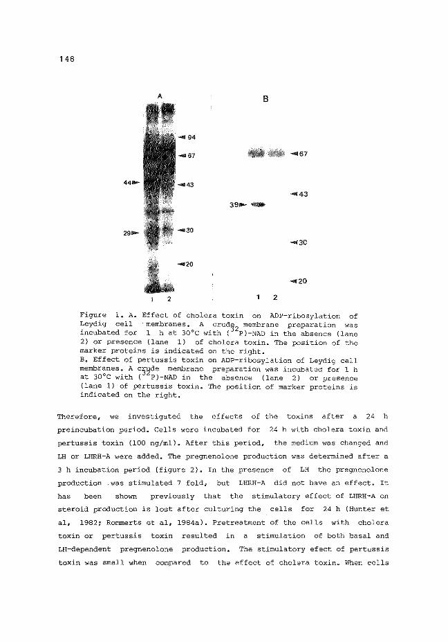
148
A B
.... 94
... 67 .... 67
..... ....3
"'"'43 39 ...... -.-
2910- ... 30
.... 30
... 20
... 20
2 2
Figure l. A. Effect of cholera toxin on ADP-ribosylation of Leydig cell ·membranes. A crud3
2 membrane preparation was
incubated for 1 h at 30°C with ( P)-NAD in the absence (lane 2} or presence (lane 1) of cholera toxin. The position of the marker proteins is indicated on the right. B. Effect of pertussis toxin on ADP-ribosylation of Leydig cell membranes. A c3~de membrane preparation was incubated for 1 h at 30°C with ( P)-NAD in the absence (lane 2) or presence (lane 1) of pertussis toxin. The position of marker proteins is indicated on the right.
Therefore 1 we investigated the effects of the toxins after a 24 h
preincubation period. Cells were incubated for 24 h with cholera toxin and
pertussis toxin (100 ng/ml)e After this period, the medium was changed and
LH or LHRH-A were added. The pregnenolone production was determined after a
3 h incubation period (figure 2). In the presence of LH the pregnenolone
production .was stimulated 7 fold, but LHRH-A did not have an effect. It
has been shown previously that the stimulatory effect of LHRH-A on
steroid production is lost after culturing the cells for 24 h (Hunter et
al, 1982; Rommerts et al, 1984a). Pretreatment of the cells with cholera
toxin or pertussis toxin resulted in a stimulation of both basal and
LH-dependent pregnenolone production~ The stimulatory efect of pertussis
toxin was small when compared to the effect of cholera toxin. When cells

~
Qi u
"' 0
6
5
149
2 h preincubation 24 h preincubation
0 3 E "-
" 2 c 0 0 c " c rn E o "- CT PT
Figure 2. Effect of cholera toxin and pertussis toxin on pregnenolone production in rat Leydig cells. Cells were preincubated for 2 h (left) or 24 h (right) with cholera toxin (100 ng/ml) or pertussis toxin (100 ng/ml), and subsequently incubated for 3 h without hormones (open bars), with 40 nM LHRH-A (cross-hatched bars) or 100 ng/ml LH (striped bars). Values given are means±SD (n=3) of one cell preparation.
CT +
PT
were treated with both toxins, the stimulatory effects of cholera toxin and
pertussis toxin appeared to be additive. The steroid production in cells
that were treated with LHRH-A was not different from control cells.
DISCUSSION
The presence of a stimulatory (Ns) and an inhibitory (Ni) guanine nu
cleotide binding protein in Leydig cells was investigated by using cholera
toxin and pertussis toxin as known modifiers of these proteins~ Our results
indicate that cholera toxin catalyzed the ADP-ribosylation of a 44 kDa
protein in a crude Leydig cell membrane preparation. The cholera toxin
dependency of the labelling~ and the observed molecular weight strongly
suggest that this protein is the a-subunit of Ns (Kaslow et al, 1980; Lai
et al, 1981; Gill, 1984). The functional importance of Ns in Leydig cells
was evaluated by stimulating cholera toxin treated cells with LH or LHRH-A,
and determining pregnenolone, the product of cholesterol side chain clea-

150
vage, which is the rate determining step of steroidogenesis in the Leydig
cell. Pregnenolone production is stimulated in Leydig cells that have been
preincubated with cholera toxin for 2 or 24 h. It has been shown that
cholera toxin stimulates cAMP levels and testosterone production in Leydig
cells (Cooke et al, 1977; Dufau et al, 1978). Moreover, it has been obser-
ved in Leydig cell membrane preparations that the stimulation of adenylate
cyclase by hCG is dependent on the presence of guanine nucleotides (Dufau
et al, 1980; Mukhopadhyay & Schurnacher, 1985)a Hence, it may be suggested
that the effect of LH on steroid production is mediated by LH. Preincu
bation of Leydig cells for 24 h in the presence of cholera toxin resulted
in a potentiation of the effect of LH on steroid production. This may have
been the result of an activation of Ns-proteins which are not coupled to
the LH receptor, in this way stimulating steroid production.
It has been suggested that the presence of both
needed for a maximal effect of hormones on cAMP levels
N and s
(Cerione
N. is l
et al~
1985). The molecular weight of the pertussis toxin substrate (39 kDa), and
the observed decrease in the labelling of the protein when GTP~S and
MgC12
were present in the incubation strongly suggests that
protein is the a-subunit of N. (Bokoch et al, l
1983; Murayama
this
& Ui,
1983; Ribeiro-Neto et al, 1985). However~ preincubation of intact Leydig
cells with pertussis toxin for 2 h did not lead to stimulation of basal,
LH- and LHRH-A-dependent steroid production. When pertussis toxin is
present for 24 h the basal, LH- and cholera toxin-stimulated pregnenolone
production was potentiated. These results are consonant with the
observations of Adashi et al (1984), who showed that that pertussis toxin
stimulated the hCG-dependent testosterone production in testicular cells
after a 24 h preincubation period with the toxin. It may be that the
effects of pertussis toxin evolve more slowly than the effects of cholera
toxin. It has been observed in different cell systems that preincubation
periods ranging from 60 min (Volpi et al, l985i Holz et al, 1986) to 24 h
(Adashi et al, 1984; Veldhuis & Hewlett, 1985) are necessary for pertussis
toxin to exert its effects. Alternatively, the Leydig cells may have
become more sensitive to pertussis toxin during culture. Similar changes in
Leydig cell sensitivity during culture have been shown with adenosine
(Rommerts et al, 1984) or O-adrenergic agonists (Anakwe et al, 1985).
These observations indicate that Ni is involved in the regulation of
steroid production in rat Leydig cells, although its mechanism of action is

1 51
not clearo
Ni may also mediate the effect of receptors for hormones that have
an inhibitory effect on adenylate cyclase (Birnbaumer et al, 1985)~
However, we did not find effects of hormones of this type on steroid
production (unpublished observations). It has been observed that the
inhibitory effects of arginine-vasotocin on beG-stimulated steroid
production could be abolished by pertussis toxinq suggesting that this
peptide may activate Ni-coupled receptors in testicular cells (Adashi et
al, 1984). However, these authors used a testicular cell preparation
containing many other cell types in addition to Leydig cells. Therefore it
is not clear whether the effects of arginine-vasotocin or pertussis toxin
were directly on Leydig cells, or mediated by other cell types~
Furthermore 6 during the 11 day culture period employed by Adashi et al,
(1984) the Leydig cells may have become more sensitive to arginine
vasotocin. It is not clear whether N. is involved in the effects of LHRH on l
steroid production. After Leydig cells have been cultured for 24 h, LHRH
inhibits the LH-dependent steroid production (Hunter et al, 1982; Rommerts
et al, 1984) 1 and it may be necessary to design experiments measuring
this activity of LHRH-A to elucidate the possible involvement in
the action of LHRH on Leydig cell steroidogenesis~
In conclusion, we have shown that the a-subunits of N8
and Ni are pre
sent in mature rat Leydig cells. The cholera toxin substrate Ns may be in-
volved in LH, but not in LHRH regulation of steroid production~
involved in the regulation of steroid production in rat Leydig
the mechanisms involved are not clear.
REFERENCES
N. is also l
cells, but
Adashi, E.Y., Resnick, L.E., Cronin, M.J. & Hewlett E.L. (1984) Endocrinology 115, 839-842~
Anakwe, 0.0., Murphy, P.R. & Moger, W.H. (1985) Biol. Reprod. 33, 815-826. Bakker, G.H., Hoogerbrugge, J.W. & Rommerts, F.F.G. (1983) Molec. Cell.
Endocrin~ 33, 243-253. Birnbaumer, L., Codina, J., Mattera, R., Cerione, R.A., Hildebrandt, J.D.,
Sunyer, T., Rojas, F.J., Caron, M.G., Lefkowitz, R.J. & Iyengar, R. (1985) In: Molecular mechanisms of transmembrane signalling, eds: Cohen, P. & Houslay, M.D. (Elsevier Amsterdam) pp. 131-182.
Bokoch, G.M., Katada, T., Northrup, J.K., Hewlett, E.L. & Gilman, A.G~
(1983) J. Biol. Chem. 258, 2072-2075. Cassel 0 D. & Pfeuffer 1 T. (1978) Proc. Natl. Acad. Sci. USA 75, 2669-2673~ Cerione, R.A., Staniszewski, C., Caron M.G., Lefkowitz, R.J •• Codina, ,J. &

152
Birnbaumer, La (1985) Nature 318, 293-295. Cooke, B.A., Lindh, L~M. & Janszen, F.H.A. (1976) Biochem. J. 160, 439-446. Cooke, B.A., Lindh, L.M. & Janszen, F.H.A. (1977) FEBS lett. 73, 67-71. Dufau, M.L., Horner, K.A., Hayashi, K., Tsuruhura, T., Conn, P.M. & Catt,
K.J. (1978) J. Biol. Chem. 253, 3721-3729. Dufau, M.L., Baukal, A.J. & Catt, K.J. (1980) Proc. Natl. Acad. Sci. USA
77, 5837-5841. Gill, D.M. (1977) In: Advances in cyclic nucleotide research, Vol. 8, eds:
Greengard, P. & Robison, G.A. (Raven Press, New York) pp. 85-118. Gill, D.M. & Meren, R. (1978) Proc. Natl. Acad. Sci. USA 75, 3050-3054. Gilman, A.G. (1984) Cell 36, 577-579. Holz IV, G.G., Rane, S.G. & Dunlap, K. (1986) Nature 319, 670-672. Hunter, M.G., Sullivan, M.H.F., Dix, C.J., Aldred, L.F. & Cooke, B.A.
(1982) Molec. Cell. Endocr. 27, 31-44. Kas1ow, H.R., Johnson, G.L~, Brothers, V.M. & Bourne, H.R. (1980) J. Biol.
Chem 255, 3736-3741. Katada, T., Amano, T. & Ui, M. (1982) J. Biol. Chem. 257, 3739-3746. Koch, B.O., Dorflinger, L.J. & Schonbrunn, A. {1985) J. Biol. Chem. 260,
13138-13145. Laemmli, U.K. {1970) Nature 227, 680-685. Lai, E., Rosen, O.M. & Rubin, c.s. {1981) J. Biol. Chem. 256, 12866-12874. Murayama, T & Ui, M. {1983) J. Biol. Chem. 258, 3319-3326. Mukhopadhyay, A.K. & Schumacher, M. (1985) FEBS Lett. 187, 56-60. Okajima, F., Takada, T. & Ui, M. (1985) J. Biol. Chem. 260, 6761-6768. Podesta 1 E., Dufau, M.L., Solano, A.R. & Catt, K.J. (1978) J. Biol. Chem.
253, 8994-9001. Ribeiro-Neto, F.A.P., Mattera, R. Hildebrandt, J.D.,Codina, J., Field,
J.B., Birnbaumer, L. & Sekura, R.D. (1985) In: Methods of Enzymology, Vol. 109, Hormone Action, Part I, Peptide HormonesF eds: Birnbaumer, L. & O'Malley, B.W. (Academic Press, New York) pp. 275-288.
Rommerts, F.F.G., Molenaar, R., Themmen, A.P.N. & van der Molen, H.J. (1984a) In: Hormonal control of the hypothalamo-pituitary gonadal axis, eds: McKerns, K.W. & Naor, z. (Plenum Press, New York) pp. 423-436.
Rommerts, F.F.G., Molenaar, R., Hoogerbrugge, J.W. & van der Molen, H.J. (1984b) Biol. Reprod. 30, 842-847.
Rommerts, F.F.G., Molenaar, R. & van der Molen, H.J. (1985) In: Methods of Enzymology, Vol. 109, Hormone Action, Part I, Peptide Hormones, eds: Birnbaumer, L. & O'Malley, B.W. (Academic Press, New York) pp~ 423-436.
Sullivan, M~H~F. & Cooke 1 B.A. (1984) Molec. Cell. Endocr. 36, 115-122. Sullivan, M.H.F. & Cooke, B.A. {1986) Biochem. J. 236, 45-51. Veldhuis, J.D. & Hewlett, E.L. (1985) Biochem. Biophys. Res. Comm. 131,
1168-1174. Volpi, Me, Naccache, P.H., Molski, T~F.P., Shefcyk, Je, Huang, Ce-K.,
Marsh, M.L., Munoz 6 J., Becker, E.L~ & Sha 1 afi, R. (1985) Proc. Natl. Acad. Sci. USA 82, 2708-2712.
van der Vuss~, G.J., Kalkman, M.L. & van der Molen, H.J. (1974) Biochim. Biophys. Acta 348, 404-414.
van der Vusse, G~J., Kalkman, M.L. & van der Molen, H.J. (1975) Biochim. Biophys. Acta 380, 473-485.