Embed Size (px)
Citation preview
PATHWAYS OF HOMOLOGOUS RECOMBINATION AND
DNA INTERSTRAND CROSS-LINK REP AIR
Roles of mammalian RAD54 and SNMJ
Routes voor homo loge recombinatie en herstel van kruisverbindingen tussen DNA strengen
De rol van zoogdier-RAD54 en -SNMJ
PROEFSCHRIFT
T er verkrij ging van de graad van doctor
aan de Erasmus Universiteit Rotterdam
op gezag van de Rector Magnificus
Prof dr. ir. J.H. van Bemmel
en volgens besluit van het College voor Promoties
De openbare verdediging zal plaatsvinden op
woensdag 16 januari 2002 om 15.45 uur
door
Maria Louise Geertruda Dronkert
geboren te Borculo
Promotiecommissie
Promotoren: Prof dr. R. Kanaar
Prof dr. J.H.J. Hoeijrnakers
Overige ]eden: Prof dr. J.A. Grootegoed
Dr. E. C. Zwarthotf
Prof. dr. C. Heyting
Acknowledgments The work presented in this thesis was supported by a grant from the Netherlands Organization for Scientific Research (NWO).

CONTENTS
List of abbreviations
Scope ofthe thesis
Chapter 1
DNA double-strand break repair by homologous recombination
1. Introduction 2. Nonhomologous end-joining versus homologous recombination 3. Proteins involved in homologous recombination 4. Pathways of homologous recombination 5. DSB repair assays 6. The choice between homologous recombination pathways 7. Conclusions
Chapter 2
Mouse RAD54 affects DNA double-strand break repair and sister chromatid exchange
Chapter 3
Preliminary characterization of the mouse Rad54B protein
Chapter 4
Repair of DNA interstrand cross-links
6
7
9
11 12 15 22 29 31 34
35
63
75
1. Introduction 77 2. Cross-link formation and properties of DNA interstrand cross-linking agents. 78 3. Clinical relevance of DNA interstrand cross-links 80 4. Detection of DNA interstrand cross-links and repair intermediates 81 5. DNA interstrand cross-link repair in Escherichia coli 82 6. DNA interstrand cross-link repair in Saccharomyces cerevisiae 84 7. Mammalian DNA interstrand cross-link repair 96
Chapter 5
Disruption of mouse SNMJ causes increased sensitivity to the DNA interstrand crosslinking agent mitomycin C
References
Summary
Eenvoudige samenvatting
List of puhlications
Curriculum vitae
Dankwoord
105
133
167
171
174
175
176
Note: For simplicity, both mouse and human genes are indicated by italic capitals (e.g. RAD54), while mouse and human proteins are indicated by one capital followed by lower case (e.g. Rad54). Tbe prefixes 'm' and 'h' are used to indicate mouse and human, respectively.
6
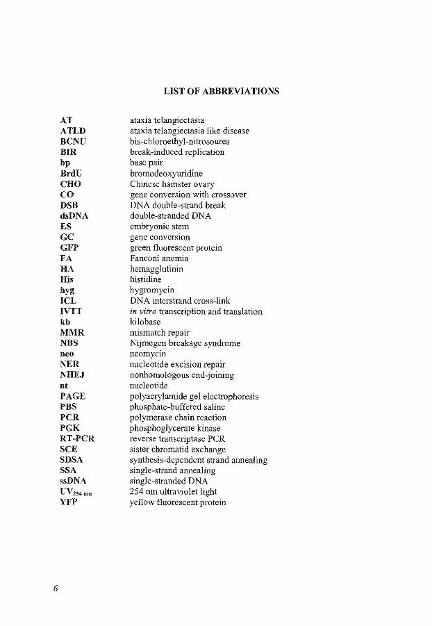
AT ATLD BCNU BIR bp BrdU CHO co DSB dsDNA ES GC GFP FA HA His hyg ICL IVTT kb MMR NBS neo NER NHEJ nt PAGE PBS PCR PGK RT-PCR SCE SDSA SSA ssDNA uv254nm
YFP
LIST OF ABBREVIATIONS
ataxia telangiectasia ataxia telangiectasia like disease bis-chloroethyl-nitrosourea break-induced replication base pair bromodeoxyuridine Chinese hamster ovary gene conversion with crossover DNA double-strand break double-stranded DNA embryonic stem gene conversiOn green fluorescent protein Fan coni anemia hemagglutinin histidine hygromycin DNA interstrand cross-link in vitro transcription and translation kilo base mismatch repair Nijmegen breakage syndrome neomycin nucleotide excision repair nonhomologous end-joining nucleotide polyacrylamide gel electrophoresis phosphate-buffered saline polymerase chain reaction phosphoglycerate kinase reverse transcriptase PCR sister chromatid exchange synthesis-dependent strand annealing single-strand annealing single-stranded DNA 254 rnn ultraviolet light yellow fluorescent protein
SCOPE OF THE THESIS
The aim of this thesis is to investigate mammalian DNA interstrand cross-link (ICL) repair. ICLs are formed by a number of agents used in tumor therapy, like mitomycin C and cisplatin. They constitute one of the most toxic damages to DNA, as they inhibit DNA strand separation. However, little is known about the mechanisms of!CL repair. A number of DNA repair pathways exist, each involved in the repair of specific types of DNA damage that continuously threaten cellular function. An intriguing aspect of ICL repair is the involvement of several of these repair pathways, mainly nucleotide excision repair, homologous recombination, and postreplication/translesion repair. An overview of the involvement of these different repair pathways in ICL repair is given in Chapter 4. This chapter also depicts putative models describing the co-operation of these pathways in repairing ICLs.
Next to genes involved in several repair pathways, other genes have been isolated that are exclusively involved in ICL repair, such as yeast SNMJ. snml mutant yeast cells are sensitive to a number of ICL agents, but they are hardly or not sensitive to other DNAdamaging agents. As described in Chapter 5, we have investigated the human and mouse homologs of Snml. We isolated mouse SNMJ and made embryonic stem cells and mice deficient for SNMJ. Both cells and mice are viable and sensitive to mitomycin C. These results indicate that mammalian Snm 1 is involved in the cellular response to at least some types of!CLs. We also showed that Snml is probably not involved in the homologous recombination pathway of ICL repair, as two parameters for homologous recombination, the formation of mitomycin C-induced Rad51 foci and sister chromatid exchanges, are not affected in SNMJ-deficient mouse cells.
Therefore, as a second approach to gain insight into ICL repair, we analyzed the homologous recombination repair pathway, as discussed in Chapter 1. Homologous recombination is one of the main DNA double-strand break (DSB) repair pathways and is the pathway used to repair DSBs occurring during ICL repair. Homologous recombination uses intact homologous sequences, usually from the sister chromatid or homologous chromosome to repair a DSB. It consists of several subpathways, gene conversion with or without crossover, single-strand annealing, and in yeast, break-induced replication. Key genes involved in homologous recombination are amongst others RADSJ and RAD54. RAD54-deficient mouse embryonic stem cells and mice are sensitive to mitomycin C. As described in Chapter 2, we investigated the involvement of the different subpathways of homologous recombination and the role of RAD54 in mouse embryonic stem cells. As it is currently not possible to create cells with a chromosomal site-specific ICL to enable the analysis of individual repair events, we decided to create site-specific DSBs. We used different recombination-test substrates designed to measure specific subpathways of homologous recombination in wild-type and RAD54-deficient cells. Single-strand annealing is a major pathway to repair these DSBs. Gene conversion with crossover preferentially involves the sister chromatid instead of a homologous sequence on the same chromatid. Mutation of RAD54 results in an increase in single-strand
7

annealing and a decrease in gene conversion with or without crossover using the sister chromatid. RAD54-deficient cells also have a reduced frequency of mitomycin C-induced sister chromatid exchanges, which is the cytological equivalent of gene conversion with crossover. These results suggest that Rad54 promotes error-free gene conversion with or without crossover using the sister chromatid both during DSB and ICL repair at the expense of error-prone single-strand annealing. Finally, we analyzed the cellular localization and interactions of mouse Rad54B, a paralog of mouse Rad54, as described in Chapter 3.
8
CHAPTERl
DNA double-strand break repair by homologous recombination

DNA DOUBLE-STRAND BREAK REPAIR BY HOMOLOGOUS RECOMBINATION
MIES L.G. DRONKERT1, ROLAND KANAAR1
•2
1 Department of Cell Biology and Genetics, Erasmus University Rotterdam, PO Box 1738, 3000 DR Rotterdam, The Netherlands
2 Department of Radiation Oncology, University Hospital Rotterdam/Daniel, The Netherlands
DNA double-strand breaks (DSBs) can be caused by several endogenous and exogenous DNA-damaging agents. Proper repair is needed to protect cells from DSB-promoted mutations and chromosomal aberrations, which may contribute to cell death, uncontrolled cell proliferation or cellular dysfunction. This overview focuses on homologous recombination as a means to repair DSBs. Homologous recombination uses homologous sequences elsewhere in the cell to repair a DSB accurately. Key proteins in homologous recombination are discussed with respect to their biochemistry, cellular biology, and role in different subpathways of homologous recombination. Models for these different subpathways, gene conversion, break-induced replication, and single-strand annealing, are described. The importance of different subpathways to repair a specific DSB can be assessed in specially designed assays. Apparently, the different homologous recombination pathways complement and compete with each other. In addition, the relationship of homologous recombination with the other major DSB repair pathway, nonhomologous end-joining, is also characterized by competition and co-operation. A challenging question for future research will be to elucidate how cells regulate these pathways to minimize the occurrence of chromosomal aberrations.
1 INTRODUCTION
Cell survival depends on the integrity of genetic information contained in DNA. This integrity is continuously at risk due to endogenous and exogenous DNA-damaging agents. In response, cells have developed a number of DNA repair pathways. These repair pathways are specialized in dealing with certain types of damage, but there is a significant overlap and co-operation, creating a network of repair pathways. One of the major threats to DNA integrity is formed by DNA double-strand breaks (DSBs). Unrepaired DSBs can lead to chromosome breaks and loss of chromosomes. Improper repair of DSBs may lead to chromosomal aberrations such as translocations, deletions, inversions, amplifications, loss of heterozygosity, and ring chromosomes, or to mutations [404]. These events will contribute to cell dysfunction, cell death, or tumor formation.
DSBs can occur accidentally during normal cellular metabolism, due to, for example, replication blocks, oxidative damage, and mechanical stress. A DSB can also be formed during the repair of other DNA damages, like DNA interstrand cross-links, or excision repair of two lesions that lie close together on different strands. Some cell types purposely create DSBs to accomplish recombination events. This happens during
Homologous recombination 11
meiosis, V(D)J recombination, immunoglobulin class switching, somatic hypermutation, and, in yeast, during mating-type switching. Exogenous sources of DSBs are ionizing radiation and a number of chemicals. To restore genetic integrity after the formation of a DSB, the cell has to ligate the proper two ends and, if necessary, supply lost information. To accomplish this, most cells contain two main DSB repair pathways: nonhomologous end-joining (NHEJ) and homologous recombination.
This chapter will focus on the repair pathways of homologous recombination. Therefore, we will only shortly describe the NHEJ pathways and discuss the relationships between NHEJ and homologous recombination. Then, we will discuss the proteins involved in homologous recombination and consider in detail the different subpathways of homologous recombination and their requirements. Finally, we will describe the assays that can be used to assess the importance of different pathways in cellular DSB repair and factors that influence the use of these pathways.
2 NONHOMOLOGOUS END-JOINING VERSUS HOMOLOGOUS RECOMBINATION
NHEJ joins the two DSB ends by direct ligation or with the use of a few homologous bases near the ends, referred to as microhomology. NHEJ has no need for extensive homologous sequences and therefore is suitable for DSB repair in haploid cells during G I phase, when no homologous DNA is present. Homologous recombination makes use of homologous sequences, usually from the sister chromatid or the homologous chromosome. Thereby, lost information can be replaced and this repair is principally error-free. Main pathways of NHEJ and homologous recombination are summarized in Figure I.
Key proteins known to be involved in NHEJ in eukaryotes are Ku70, Ku80, Mre II, Rad50, Xrs2/Nbsl, DNA ligase IV, Lifl/Xrcc4, and in vertebrate cells also DNA-PK" [232, 294, 302]. The Ku70-Ku80 heterodimer binds to DNA ends, thereby targeting DNA-PK" to the ends [563]. The DNA-PK complex, consisting of Ku70, Ku80, and DNA-PK", might hold the DSB ends together, prevent end degradation, and phosphorylate other proteins involved in repair [19, 90, 291, 292, 581]. Mrell, Rad50, and Xrs2/Nbs I form a complex that may function in signaling the presence of the break and in processing the DSB [73, 138, 196, 539]. The Xrcc4-ligase IV complex is responsible for ligating the ends [90, 115, 188, 211, 522, 568]. The abovementioned proteins function in the subpathway of NHEJ that assures precise, direct joining of complementary DNA ends (Fig. !AI). This pathway is efficient in both yeast and mammalian cells [54, 134, 355, 432]. When the ends cannot be ligated directly, for example when they are not complementary, the efficiency of repair in yeast declines, but mammalian repair remains efficient, often producing small deletions or insertions using the same set of proteins [134, 270, 355, 432, 535]. Independent of these proteins, joining of the ends using microhomology can be observed (Fig. IA2) [54, !59, 186, 535]. In this pathway, deletion of a few base pairs up to several kb occurs until two to six base pair homology is revealed. These base pairs anneal and the nicks are ligated.
12 Chapter I
g: §l 0
(JQ 0
" ~ g ~ ~: §
w
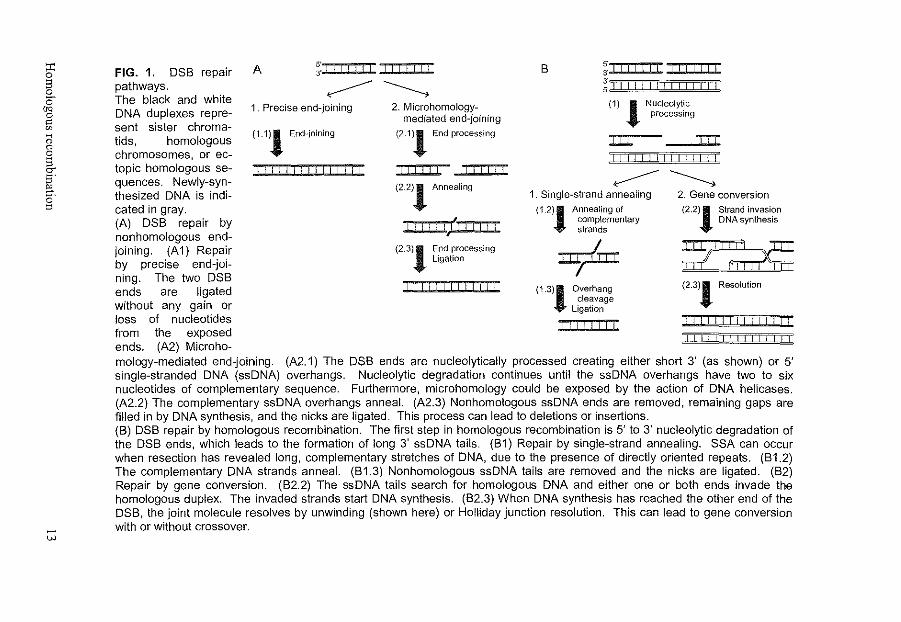
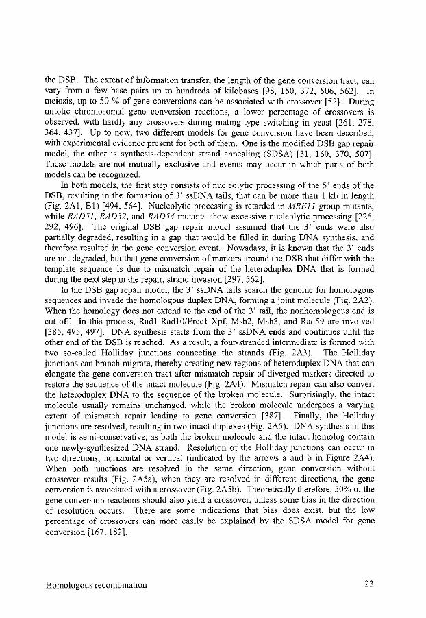
FIG. 1. DSB repair A ;:.:r:o::: ClJI JIDJJI
~~ pathways. The black and white . . .. DNA duplexes repre- 1. Precise end-Jommg
sent sister chromatids, homologous chromosomes, or ectopic homologous sequences. Newly-synthesized DNA is indi-cated in gray. (A) DSB repair by nonhomologous endjoining. (A 1) Repair by precise end-joining. The two DSB ends are ligated without any gain or loss of nucleotides from the exposed ends. (A2) Microho-
(1.1)1 End-joining
n-111 1111111111
2. Microhomologymediated end-joining
(2.1}1 End processing
rTTTTl ITTTn
{2.2)1 Annealing
I TTTTTl TTTTTT
(2.3)1 End processing Ligation
DTl=TTI
B 5
-·~iiii::i~~~~ 3' 3' 5, I I I I I I I I I I I I I I I
(1) 1 Nucleolyt_ic processmg
J:l:l: T1T ITTTf-ll I I I I I I I I
~~ 1. Single-strand annealing
(1.2)1 Annealing of complementary strands
+ (1.3)1 Overhang
cleavage Ligation
rn---rTTTl
2. Gene conversion
(2.2)1 Strand invasion DNA synthesis
:-;ll:nx::
(2.3)1 Resolution
Illlll I I I I I I I I I LJLli I I I I I I I I I I
mology-mediated end-joining. (A2.1) The DSB ends are nucleolytically processed creating either short 3' (as shown) or 5' single-stranded DNA (ssDNA) overhangs. Nucleolytic degradation continues until the ssDNA overhangs have two to six nucleotides of complementary sequence. Furthermore, microhomology could be exposed by the action of DNA helicases. (A2.2) The complementary ssDNA overhangs anneal. (A2.3) Nonhomologous ssDNA ends are removed, remaining gaps are filled in by DNA synthesis, and the nicks are ligated. This process can lead to deletions or insertions. (B) DSB repair by homologous recombination. The first step in homologous recombination is 5' to 3' nucleolytic degradation of the DSB ends, which leads to the formation of long 3' ssDNA tails. (B1) Repair by single-strand annealing. SSA can occur when resection has revealed long, complementary stretches of DNA, due to the presence of directly oriented repeats. (81.2) The complementary DNA strands anneal. (81.3) Nonhomologous ssDNA tails are removed and the nicks are ligated. (B2) Repair by gene conversion. (B2.2) The ssDNA tails search for homologous DNA and either one or both ends invade the homologous duplex. The invaded strands start DNA synthesis. (B2.3) When DNA synthesis has reached the other end of the DSB, the joint molecule resolves by unwinding (shown here) or Holliday junction resolution. This can lead to gene conversion with or without crossover.
Key proteins in homologous recombination are Rad51, Rad52, Rad54, and their homologs/paralogs [253, 386]. Mrel1, Rad50, and Xrs2/Nbs1 are also involved in homologous recombination [253, 386]. The first step of homologous recombination consists of processing of the DSB ends yielding 3' single-stranded DNA (ssDNA) tails [ 494]. The proteins responsible for this processing are not yet known. Exonuclease I and the Mre11-Rad50-Xrs2/Nbsl complex may be involved, as mutations in these proteins slow DSB processing [227, 494, 534]. When the 3' ssDNA tails are homologous, they can anneal onto each other in a process called single-strand annealing (SSA) (Fig. 1B1) [307]. Alternatively, in the major models for homologous recombination, one or both 3' ssDNA tails invade homologous duplex DNA and initiate DNA synthesis, thereby copying information from intact homologous DNA (Fig. 1B2) [160, 507]. After DNA synthesis, the newly-synthesized DNA can unwind from the homologous duplex and anneal with the other DSB end. Alternatively, a four-stranded DNA structure containing Holliday junctions is formed. The Holliday junctions are resolved, resulting in gene conversion with or without crossover. Crossovers of sufficient size can be detected cytologically as sister chromatid exchanges (SCEs).
Both NHEJ and homologous recombination can induce genomic rearrangements. DSB repair by NHEJ can lead to translocations when several DSBs are present and the wrong ends are joined [433]. Furthermore, small insertions or deletions have been found, especially when the DSB ends are not complementary [270, 301,355,432, 535]. Repair by homologous recombination can also induce translocations, or lead to loss of heterozygosity, deletions, inversions, and amplifications [199, 322, 364, 387]. However, chromosomal instability and sensitivity to DSB-inducing agents are much higher in cells deficient for one of the DSB repair pathways, and increase synergistically when both DSB repair pathways are impaired [155, 231, 368, 409, 510, 543].
NHEJ and homologous recombination compete with each other in the repair of DSBs. The importance of either DSB repair pathway differs per species, cell type, and cell cycle phase, as cells apparently try to minimize the risks involved in either pathway [155, 510]. Furthermore, they complement each other in the repair of different specialized types of DSBs that occur in the cell. V(D)J recombination, for example, is achieved by NHEJ, whereas homologous recombination is the main pathway to repair DSBs in DNA interstrand cross-link repair (Chapter 4) [146, 187]. Homologous recombination is important for the pairing of homologous chromosomes during meiosis, which is necessary for proper chromosome segregation during meiosis I [597]. In Saccharomyces cerevisiae, homologous recombination is the main DSB repair pathway and effects of NHEJ can only be detected when homologous recombination is impaired, for example when no homologous DNA is present to repair the break or by genetic ablation of homologous recombination [20, 349, 469]. Most DNA inS. cerevisiae is coding DNA and the progeny of such a single cell organism needs intact genes without mutations. The choice between NHEJ and homologous recombination in yeast mainly depends on the mating-type locus. Cells expressing only one mating-type allele, usually haploid cells, are most proficient in NHEJ. Cells expressing two mating-type alleles, usually diploid cells, are most proficient in homologous recombination [12, 293]. In diploid cells, accurate repair of a DSB by homologous recombination is feasible during the whole cell cycle, in contrast to the
14 Chapter I
situation in haploid cells. The mechanism by which mating-type determines the preference for a certain repair pathway is not known. Part of it may be a difference in the expression of proteins involved in NHEJ versus homologous recombination, or in proteins regnlating these pathways [ 103].
In mammalian cells, NHEJ is an important DSB repair pathway [302]. Mammalian cells might tolerate the inaccuracy of NHEJ better than yeast cells. Much more of the mammalian DNA is noncoding, and small mutations are much less likely to have devastating effects in somatic, differentiated cells than in germ line cells. Furthermore, the presence of many repeats in the mammalian genome increases the risk of homologous recombination using ectopic DNA, which can cause translocations, inversions, and deletions. Homologous recombination nevertheless plays a major role especially early during development, and in the repair of meiotic DSBs, when accurate repair is very important [153, 155, 300, 477]. Mutations in some of the genes involved in homologous recombination in mammalian cells even lead to cellular or embryonic lethality, due to the occurrence of chromosomal abnormalities [527].
NHEJ and homologous recombination are not just separate pathways, they can also interact. Surprisingly, DSB repair events have been found that combine features of homologous recombination and NHEJ [239, 418, 425, 435]. These events apparently start with invasion of one DSB end into homologous DNA. When the homologous DNA lies on a different chromosome, DNA synthesis may continue into nonhomologous sequences. The junction of the newly-synthesized DNA with the other DSB end has to be made by NHEJ. Which proteins from the homologous recombination and NHEJ pathways are involved in these rare coupled events is not yet clear.
3 PROTEINS INVOLVED IN HOMOLOGOUS RECOMBINATION
3.1 RadSl and paralogs
The eukaryotic Rad51 proteins are homologous to Escherichia coli RecA protein, which is the central protein for bacterial homologous recombination. Rad51 is a weak ATPase that can bind both ssDNA and double-stranded DNA ( dsDNA), but it prefers ssDNA ends of a tailed dsDNA molecule (Table I) [34, 333, 338, 588]. On ssDNA, it forms a nucleoprotein filament with one protein molecule per three nucleotides [504]. This nucleoprotein filament can perform D-loop formation and DNA strand exchange reactions in vitro [26, 501]. In yeast, this reaction is inhibited when the ssDNA-binding protein RP A is present before Rad51 is added, due to its higher affinity for ssDNA [ 499, 503]. However, RPA stimulates Rad51-mediated strand exchange when added after Rad51, probably by resolving secondary structures [26, 499, 504]. It may increase the cooperativity ofRad51, when Rad51 is present in a suboptimal concentration [29, 338]. In vivo, Rad51 is thought to be responsible for coating the 3' ssDNA tail and to perform the search for homologous DNA and strand invasion during homologous recombination.
Homologous recombination 15
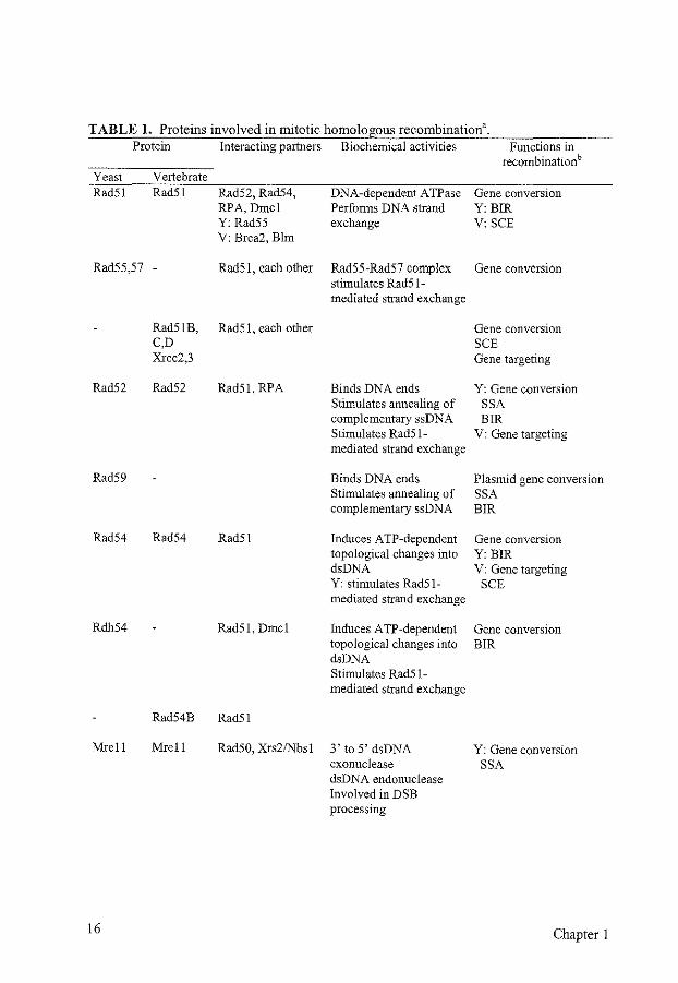
TABLE 1. Proteins involved in mitotic homologous recombinationa. Protein Interacting partners Biochemical activities Functions in
recombinationb Yeast Vertebrate Rad51 Rad51 Rad52, Rad54, DNA-dependent ATPase Gene conversion
RPA, Dmcl Performs DNA strand Y:BIR Y: Rad55 exchange V: SCE V: Brca2, Blm
Rad55,57 - Rad51, each other Rad55-Rad57 complex Gene conversion stimulates Rad51-mediated strand exchange
Rad51B, Rad51, each other Gene conversion C,D SCE Xrcc2,3 Gene targeting
Rad52 Rad52 Rad51, RPA Binds DNA ends Y: Gene conversion Stimulates annealing of SSA complementary ssDNA BIR Stimulates Rad51- V: Gene targeting mediated strand exchange
Rad59 Binds DNA ends Plasmid gene conversion Stimulates annealing of SSA complementary ssDNA BIR
Rad54 Rad54 Rad51 Induces ATP-dependent Gene conversion topological changes into Y: BIR dsDNA V: Gene targeting Y: stimulates Rad51- SCE mediated strand exchange
Rdh54 Rad51, Dmcl Induces ATP-dependent Gene conversion topological changes into BIR dsDNA Stimulates Rad51-mediated strand exchange
Rad54B Rad51
Mrell Mrell Rad50, Xrs21Nbsl 3' to 5' dsDNA Y: Gene conversion exonuclease SSA dsDNA endonuclease Involved in DSB processing
16 Chapter 1
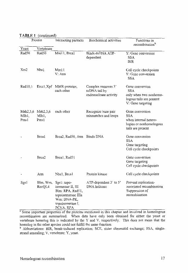
TABLE 1 (continued) Protein
Yeast Rad50
Xrs2
Rad!O,l
Msh2,3,6 Mlhl, Pmsl
Vertebrate Rad50
Nbs!
Erccl,Xpf
Msh2,3,6 Mlhl, Pmsl
Interacting partners
Mrell, Brcal
Mrell V:Atm
MMR proteins, each other
each other
Biochemical activities
Binds dsDNA ATP-dependent
Complex removes 3' ssDNA tail by endonuclease activity
Recognize base pair mismatches and loops
Brcal Brca2, Rad50, Atm Binds DNA
Sgsl
Brca2 Brcal, Rad51
Atm Nbsl, Brcal
Blm, Wrn, Sgsl: topo-RecQL4 isomerase II, III
Elm: RP A, Rad51, topoisomerase Ilia Wm:DNA-PK, topoisomerase I, PCNA,RPA
Protein kinase
ATP -dependent 3' to 5' DNA helicase
Functions in recombinationb
Y: Gene conversion SSA BIR
Cell cycle checkpoints Y: Gene conversion
SSA
Gene conversion SSA
only when two nonhomologous tails are present V: Gene targeting
Gene conversion SSA when internal heterologies or nonhomologous tails are present
Gene conversion SSA Gene targeting Cell cycle checkpoints
Gene conversion Gene targeting Cell cycle checkpoints
Cell cycle checkpoints
Prevent replicationassociated recombination Suppression of recombination
a Some important properties of the proteins mentioned in this chapter and involved in homologous recombination are summarized. When data have only been obtained for either the yeast or vertebrate homolog this is indicated by the Y and V, respectively. This does not mean that the homolog in the other species could not fulfill the same function. b Abbreviations: BIR, break-induced replication; SCE, sister chromatid exchange; SSA, singlestrand annealing; V, vertebrate; Y, yeast.
Homologous recombination 17
Rad51 can be found in damage-induced nuclear foci after exposure to ionizing radiation or treatment with DNA interstrand cross-linking agents [194, 513]. In the cell, these foci are found at sites of DNA damage and are supposed to be sites of recombinational repair of the damage [416, 516]. Rad51 is also involved in the recombinational repair of stalled replication forks, as foci are found in cells which are in the late Sand G2 phases of the cell cycle [299, 515, 516]. This latter function is probably much more important in higher eukaryotes, due to the large size of their genome. This could explain the fact that while yeast cells deficient for Rad51 are viable, though very sensitive to DSB-inducing agents, vertebrate RAD51 is an essential gene [305, 536]. Mutant vertebrate cells accumulate chromosomal breaks before death, probably due to disrupted DSB repair of spontaneous damage [ 481].
In both yeast and mammalian cells, a meiosis-specific homolog of Rad51 is present, called Dmc 1. Both Rad51 and Dmc I are needed for proper homologous recombination during meiosis and they colocalize on the synapsed chromosomes [44, 354, 462]. Dmcl is one of the proteins that ensure a preference for recombination using the homologous chromosome instead of the sister chromatid during meiosis [453].
S. cerevisiae contains two other proteins with homology to Rad51, namely Rad55 and Rad57. These two proteins form a heterodimer, that can interact with Rad51 [203, 241]. They stimulate DNA strand exchange reactions by Rad51, and help to overcome the inhibiting effects of RPA [503]. The ionizing radiation sensitivity and recombination defect of rad55 and rad57 mutants can be suppressed by overexpressing RAD51 and/or RAD52 [203].
Vertebrate cells contain five Rad51 paralogs, Rad51B, Rad51C, Rad51D, Xrcc2, and Xrcc3 [524]. These paralogs interact with each other and with Rad51 and could be involved in stimulating Rad51-mediated DNA strand exchange [60, 142, 310, 451]. Chicken cells deficient for any one of these paralogs are viable, but show chromosomal instability, sensitivity to ionizing radiation and DNA interstrand cross-linking agents, deficient damage-induced Rad51 foci formation, and impaired gene targeting [509, 511]. These defects can be partially rescued by overexpressing RAD51. Mice deficient for RAD51B, RAD51D, and XRCC2 show embryonic lethality, similar to RAD5J-deficient mice [131, 407, 466]. XRCC2 andXRCC3 mutant cells are severely impaired in the repair of DSBs by homologous recombination and in the formation of damage-induced Rad51 nuclear foci [45, 64, 240, 380, 405]. This severe phenotype of mutations in the Rad51 paralogs, with chromosomal instability and chromosome missegregation, leading to cell death, is probably due to an important role for these proteins in homologous recombination [116, 191]. The relatively mild sensitivity ofRad51 paralog mutant cells to ionizing radiation and their severe sensitivity to DNA interstrand cross-linking agents suggests that the Rad5l paralogs are not equally important for the repair of all DSBs [509, 511].
3.2 Rad52 and paralog
Rad52 is the most important protein in homologous recombination in yeast. rad52 mutant cells are almost completely deficient in all pathways of homologous
18 Chapter I
recombination [386]. Nevertheless, vertebrate RAD52-deficient cells are not sensitive to ionizing radiation and have only a mild defect in homologous recombination reactions [428, 579]. In mammalian cells, the function of Rad52 in stimulating Rad51-mediated strand exchange might have been taken over partially by the Rad51 paralogs [353].
Rad52 forms heptameric ring structures that interact with DNA, it binds preferably to ssDNA ends and protects the ends against degradation (Table 1) [360, 391,463,490,541, 542]. The protein catalyzes strand annealing of complementary ssDNA molecules, which is stimulated by adding RPA [360, 421, 463, 498]. In vitro, Rad52 interacts with Rad51 and stimulates Rad51-mediated DNA strand exchange, as it overcomes the inhibition of the strand exchange reaction by RPA [33, 141, 350, 375, 461, 463, 502]. Similar to Rad51, Rad52 forms damage-induced nuclear foci colocalizing with the RPA and Rad51 foci [177, 309,312, 313].
Yeast contains a sequence homolog ofRad52, called Rad59. Rad59 was identified as a protein, needed for Rad51-independent recombination reactions [16, 23]. Similar to Rad52, it also binds ssDNA and stimulates annealing of complementary ssDNA molecules, but this reaction is not stimulated by RPA [401]. Rad52 is dominant over Rad59 as the phenotype of rad59 mutants can be suppressed by overexpression of RAD52, but not the other way around [ 16].
3.3 Rad54 and paralogs
Rad54 is a member of the Swi2/Snf2 family of proteins that contain helicase motifs. Some of these proteins are involved in chromatin remodeling reactions [395]. Rad54 has a strong dsDNA-dependent ATPase activity, but no helicase activity has been identified [ 400, 505]. Rad54 interacts with Rad51 and, in S. cerevisiae, can stimulate Rad51-mediated DNA strand exchange [102, 185,235,332,400,403,479, 546]. In vitro, Rad54 can induce topological changes in DNA, introducing supercoiled regions in a nicked plasmid in an ATP-dependent way [430, 513, 546]. This reaction is stimulated by the presence of a Rad51 nucleoprotein filament [332]. Rad54 could translocate along the DNA, and it could facilitate partial unwinding of the DNA by causing local increases in supercoiled helix density, when rotation around the DNA during translocation is prevented, for example by its binding to the Rad51 nucleoprotein filament [430, 480]. This partial unwinding could facilitate the search for homologous duplex DNA and the strand invasion reaction of the Rad51 nucleoprotein filament by making the DNA more accessible. Consistent with this, Rad54 is found in the nucleus and forms damageinduced foci, that colocalize with Rad51 foci [513]. Rad51 foci formation, however, is not dependent on the presence of Rad54 [509]. In contrast to RAD51, RAD54-deficient chicken and mouse cells are viable, though they are ionizing radiation sensitive and defective in homologous recombination (Chapter 2) [40, 144, 154].
Both in yeast and in mammalian cells, a homolog of Rad54 is found. The yeast homolog, called Rdh4 or Tid!, has similar biochemical properties as Rad54 [264, 402, 465]. It can introduce unconstrained positive and negative supercoils in a plasmid in an ATP-dependent way. Rdh54 interacts with Rad51 and Dmcl and stimulates Rad51-mediated D-loop formation [143, 402]. The functions of Rdh54 and Rad54 in yeast
Homologous recombination 19
partially overlap, with Rad54 being more important for recombination in mitosis, using the sister chromatid as a template, and Rdh54 being more important in meiosis, using the homologous chromosome as a template for homologous recombination [10, 46, 264, 452, 464, 465]. The mammalian homolog of Rad54, called Rad54B, is most homologous to the mammalian Rad54, and is not a clear homolog of Rdh54 [212]. Rad54B is a nuclear protein that shows homomeric interaction and can interact with Rad51 (Chapter 3) [514]. The homology of Rad54B with Rad54 suggests that it could also induce topological changes into the DNA to facilitate Dmcl- and/or Rad51-mediated strand exchange.
3.4 Mrell, RadSO, and Xrs2/Nbsl
Mrell, Rad50, and Xrs2 form a stable complex in yeast cells [245, 378]. In mammalian cells, no obvious sequence homolog of Xrs2 has been detected, with instead Nbs 1 taking its place [73, 138]. The complex formed by Mre 11, Rad50, and Xrs2/Nbs 1 is involved in NHEJ, homologous recombination, and telomere maintenance [55, 65, 295]. During meiosis, the complex is needed for DSB formation and processing [225, 500]. In mitotic recombination, the processing of DSB ends is slowed down in mutant cells, but recombination products can be formed [227, 533]. Nbs! functions both in DSB repair and in DNA damage signaling to activate cell cycle checkpoints [73, 179, 183].
Mre II is a DNA binding protein that can stimulate annealing of complementary ssDNA molecules [125]. In vitro, both Mrell by itself and the whole complex display Mn2+-dependent 3' to 5' dsDNA exonuclease and endonuclease activities [393, 394, 532]. However, nuclease-negative Mre 11 mutants are not significantly impaired in NHEJ, mitotic recombination, or telomere maintenance [356]. Nbsl is needed for translocation of the Mrell-Rad50 complex to the nucleus and phosphorylation of Mrell upon DNA damage [73, 135, 139]. After treatment with ionizing radiation, the complex can be found in foci at the position of DNA damage [328, 371]. Usually, cells show either foci containing Rad51 or foci containing Mrell-Rad50-Nbsl. Mutations in the MREI I gene have been found in patients with ataxia telangiectasia-like disorder (ATLD) and mutations in NBS! in patients with Nijmegen breakage syndrome (NBS) [73, 492]. Cells from patients with these diseases are sensitive to DSB-inducing agents and show radio-resistant DNA synthesis [269]. MRE!l, RAD50, and NBS! null mutations in mice result in inviable cells or embryonic lethality, indicating their importance in cellular metabolism [318, 577, 596].
3.5 Radl!Xpf and Rad!O/Erccl
The yeast Radl-Rad!O heterodimer and its mammalian homolog Xpf-Erccl are primarily known for their role as endonuclease in nucleotide excision repair [169, 473]. In nucleotide excision repair, Radl-Radl 0/Ercc 1-Xpf is responsible for the incision of the damaged DNA strand at the 5' side of the lesion. The complex incises dsDNA near the dsDNA-ssDNA transition when a 3' ssDNA tail is present [18, 126]. In homologous recombination the complex is needed to remove nonhomologous ssDNA tails during SSA and gene conversion reactions, when the tails are longer than 30 nucleotides [1, 18, 223,
20 Chapter 1
385, 448]. Additionally, in mouse cells Erccl-Xpf is essential for gene targeting, even when no nonhomologous ssDNA tails are present. The complex could make incisions at the border of the homologous and nonhomologous DNA in the heteroduplex intermediate, thereby facilitating integration of the targeting DNA (L.J. Niedemhofer, personal communication).
3.6 Mismatch repair proteins
Mismatch repair (MMR) proteins, like Msh2, Msh3, Msh6, Mlhl, and Pmsl, function in the recognition and repair of base pair mismatches and loops that occur due to replication errors [9]. However, they also have several roles in homologous recombination that are not always directly correlated to mismatches. First of all, MMR proteins impair homologous recombination and reduce the frequency of crossovers, when recombination occurs between sequences that are not completely homologous, but show heterologies [121, 122, 149, 457]. In this way, they inhibit ectopic recombination between similar, repeated sequences at different positions in the genome and prevent translocations. Secondly, the MMR proteins are needed for the correction of heteroduplex DNA that is formed during strand invasion and branch migration reactions in homologous recombination [149, 562]. They also limit the length of gene conversion tracts when heterologies are present [93, 94]. Thirdly, Msh2 and Msh3 work together with RadlRadlO/Erccl-Xpf in the removal of nonhomologous ssDNA tails during SSA and gene conversion reactions [385, 446, 497].
3. 7 Other proteins involved in homologous recombination
A number of other proteins are needed for the reactions during homologous recombination. It is not yet quite clear which nuclease( s) are responsible for the resection of the DSB ends, resulting in 3' ssDNA tails, although the Mre l I complex is known to be involved in processing the ends. Later in the reaction, many proteins required for DNA replication, like leading and lagging strand DNA polymerases, are needed for DNA synthesis [21 7]. A complex that could perform branch migration and Holliday junction resolution has been isolated, but its components are not yet known [109]. Apart from these proteins directly involved in enzymatic reactions during homologous recombination, other proteins will be involved in the regulation of homologous recombination reactions.
A number of human diseases are associated with defects in (the regulation of) homologous recombination. A common feature of these diseases is chromosomal instability resulting in an increased risk of cancer. We will discuss some of the proteins involved and indicate how mutations could lead to defects in homologous recombination. Brcal and Brca2 are associated with familiar breast and ovarian cancer [456, 592]. Null mutations are cellular lethal, due to spontaneous chromosomal instability [456]. Both proteins have been shown to be required for homologous recombination after the induction of a DSB [362, 365, 478]. Brcal interacts with the Mreii-Rad50-Nbsl complex, and can be found both in damage-induced Mrel I and Rad5l foci [89, 455, 593]. Brca2 interacts with Rad51 and is needed for the induction of Rad51 foci, with which it
Homologous recombination 21
colocalizes [87, 89,327,571, 587]. The two Brca proteins can also be found in the same foci [88]. Similar to Nbs!, they are not only supposed to be involved in DSB repair but also in cell cycle checkpoints [87, 326, 592]. Upon treatment of cells with ionizing radiation, both Brcal and Nbs! are phosphorylated by Atm, which is involved in the disease ataxia telangiectasia (AT) and which is one of the proteins that play a crucial role in damage-induced cell cycle checkpoints [ 112, 178, 179, 257, 306, 346]. Cells from AT patients are radiosensitive and show radio-resistant DNA synthesis, similar to NBS and ATLD patient cells [347]. All these proteins could have a role in coupling DSB repair to the DNA damage response and cell cycle checkpoints.
A quite different type of defect in homologous recombination is seen in cells from patients with Bloom's syndrome, Werner's syndrome, and Rothmund-Thomson syndrome [168, 256]. These diseases are caused by a mutation in one of the human homologs of E. coli RECQ helicase, BLM, WRN, and RECQL4, respectively [151, 260, 585]. Cells from Bloom's, Werner's, and Rothmund-Thomson syndrome patients show chromosomal instability and the patients have a highly increased risk of tumor formation [168, 170, 180, 256, 308, 547]. BLM mutant cells are characterized by hyperrecombination and an increased number of SCEs and ELM-deficient mice show an increased tumor frequency [180, 317]. WRN mutant cells are characterized by an increase in translocations and deletions [170]. Cells mutant for BLM, WRN, or SCSI, the S. cerevisiae RECQ homolog, are specifically sensitive for perturbations of replication by S phase-specific damaging agents and have abnormal replication intermediates [84]. Interactions have been found between Blm and Rad51, RPA, and topoisomerase lila, and between Wrn and topoisomerase I, PCNA, DNA-PK, and RPA [66, 110, Ill, 290,296,460,487, 572, 573, 584]. Both Blm and Wm form foci that partially colocalize with Rad51 and RPA foci when DSBs have been induced [43, 443]. The RecQ helicases are ATP-dependent 3' to 5' helicases that can unwind both duplex DNA, forked DNA structures and synthetic Holliday junctions [84, 256]. They are supposed to play a role in resolving abnormal replication structures, like those occurring when replication stalls or when replication forks meet each other. Thereby, they could prevent the occurrence of DSBs during replication. Mutations in these genes would then lead to increased homologous recombination due to DSB formation during replication, which would result in increased frequencies of SCEs, translocations, and deletions. Furthermore, the RecQ helicases could have a role during recombination, like E. coli RecQ, in promoting joint molecule formation or in resolving joint molecules by branch migration [200, 201].
4 PATHWAYS OF HOMOLOGOUS RECOMBINATION
4.1 Gene conversion with or without crossover
Gene conversion constitutes the major fom1 of DSB repair by homologous recombination in meiosis and is also very important during mitosis. It consists of the transfer of genetic information from intact homologous sequences to the region containing
22 Chapter I
the DSB. The extent of information transfer, the length of the gene conversion tract, can vary from a few base pairs up to hundreds of kilobases [98, 150, 372, 506, 562]. In meiosis, up to 50 % of gene conversions can be associated -with crossover [52). During mitotic chromosomal gene conversion reactions, a lower percentage of crossovers is observed, with hardly any crossovers during mating-type switching in yeast [261, 278, 364, 437]. Up to now, two different models for gene conversion have been described, with experimental evidence present for both of them. One is the modified DSB gap repair model, the other is synthesis-dependent strand annealing (SDSA) [31, 160, 370, 507]. These models are not mutually exclusive and events may occur in which parts of both models can be recognized.
In both models, the first step consists of nucleolytic processing of the 5' ends of the DSB, resulting in the formation of 3' ssDNA tails, that can be more than 1 kb in length (Fig. 2Al, Bl) [494, 564]. Nucleolytic processing is retarded in MREII group mutants, while RAD51, RAD52, and RAD54 mutants show excessive nucleolytic processing [226, 292, 496]. The original DSB gap repair model assumed that the 3' ends were also partially degraded, resulting in a gap that would be filled in during DNA synthesis, and therefore resulted in the gene conversion event. Nowadays, it is known that the 3' ends are not degraded, but that gene conversion of markers around the DSB that differ with the template sequence is due to mismatch repair of the heteroduplex DNA that is formed during the next step in the repair, strand invasion [297, 562].
In the DSB gap repair model, the 3' ssDNA tails search the genome for homologous sequences and invade the homologous duplex DNA, forming a joint molecule (Fig. 2A2). When the homology does not extend to the end of the 3' tail, the nonhomologous end is cut off. In this process, Radl-RadlO/Erccl-Xpf, Msh2, Msh3, and Rad59 are involved [385, 495, 497]. DNA synthesis starts from the 3' ssDNA ends and continues until the other end of the DSB is reached. As a result, a four-stranded intermediate is formed with two so-called Holliday junctions connecting the strands (Fig. 2A3). The Holliday junctions can branch migrate, thereby creating new regions ofheteroduplex DNA that can elongate the gene conversion tract after mismatch repair of diverged markers directed to restore the sequence of the intact molecule (Fig. 2A4). Mismatch repair can also convert the heteroduplex DNA to the sequence of the broken molecule. Surprisingly, the intact molecule usually remains unchanged, while the broken molecule undergoes a varying extent of mismatch repair leading to gene conversion [387]. Finally, the Holliday junctions are resolved, resulting in two intact duplexes (Fig. 2A5). DNA synthesis in this model is semi-conservative, as both the broken molecule and the intact homolog contain one newly-synthesized DNA strand. Resolution of the Holliday junctions can occur in two directions, horizontal or vertical (indicated by the arrows a and b in Figure 2A4). When both junctions are resolved in the same direction, gene conversion without crossover results (Fig. 2A5a), when they are resolved in different directions, the gene conversion is associated with a crossover (Fig. 2A5b ). Theoretically therefore, 50% of the gene conversion reactions should also yield a crossover, unless some bias in the direction of resolution occurs. There are some indications that bias does exist, but the low percentage of crossovers can more easily be explained by the SDSA model for gene conversion [167, 182].
Homologous recombination 23
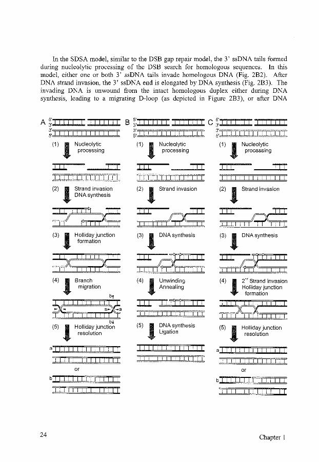
In the SDSA model, similar to the DSB gap repair model, the 3' ssDNA tails formed during nucleolytic processing of the DSB search for homologous sequences. In this model, either one or both 3' ssDNA tails invade homologous DNA (Fig. 2B2). After DNA strand invasion, the 3' ssDNA end is elongated by DNA synthesis (Fig. 2B3). The invading DNA is unwound from the intact homologous duplex either during DNA synthesis, leading to a migrating D-loop (as depicted in Figure 2B3), or after DNA
A 3. I I I I I
s·~~~~~~i~~
24
3' 5• I I I I I I I I I I I I I I
(1) l ILL
Nucleolytic processing
ILL I I I I I I I I I I I I I I
(2) l Strand invasion DNA synthesis
(3) l Holliday junction formation
(4) l Branch migration
j)f: : I 1
1
1 : : : :af~a bl
(5) l Holliday junction resolution
a I I I I I I I I I I I I I I
I I I I I I I I I I I I I I
or
b I I I I I I I I I I I I
I I I I I I I I I I I I I
s·=~s·=~ B 3. I C 3.
3' 3' 5. 5. I I I I I I I I I I I I I I I
(1) l ILL
Nucleolytic processing
ILL I I I I I I I I I I I I I I
(1) l ILL
Nucleolytic processing
ILL
(2) l Strand invasion (2) l Strand invasion
~1111~ ~1111~ (3) l DNA synthesis
~,/¢;X: : : : : : (4) l Unwinding
Annealing
JJl II:! jIll
(5) l DNA synthesis Ligation
I I I I I I I I I I
(3) l DNA synthesis
(4) 12"6 Strand invasion
Holliday junction formation
~:::::: (5) l Holliday junction
resolution
a I II I I I I I I I I I I I I
or
Chapter 1
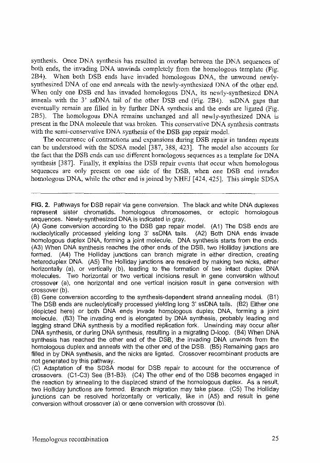
synthesis. Once DNA synthesis has resulted in overlap between the DNA sequences of both ends, the invading DNA unwinds completely from the homologous template (Fig. 284). When both DS8 ends have invaded homologous DNA, the unwound newlysynthesized DNA of one end anneals with the newly-synthesized DNA of the other end. When only one DS8 end has invaded homologous DNA, its newly-synthesized DNA anneals with the 3' ssDNA tail of the other DS8 end (Fig. 284). ssDNA gaps that eventually remain are filled in by further DNA synthesis and the ends are ligated (Fig. 285). The homologous DNA remains unchanged and all newly-synthesized DNA is present in the DNA molecule that was broken. This conservative DNA synthesis contrasts with the semi-conservative DNA synthesis of the DS8 gap repair model.
The occurrence of contractions and expansions during DSB repair in tandem repeats can be understood with the SDSA model [387, 388, 423]. The model also accounts for the fact that the DS8 ends can use different homologous sequences as a template for DNA synthesis [387]. Finally, it explains the DS8 repair events that occur when homologous sequences are only present on one side of the DSB, when one DSB end invades homologous DNA, while the other end is joined by NHEJ [424, 425]. This simple SDSA
FIG. 2. Pathways for DSB repair via gene conversion. The black and white DNA duplexes represent sister chromatids, homologous chromosomes, or ectopic homologous sequences. Newly-synthesized DNA is indicated in gray. (A) Gene conversion according to the DSB gap repair model. (A 1) The DSB ends are nucleoly1ically processed yielding long 3' ssDNA tails. (A2) Both DNA ends invade homologous duplex DNA, forming a joint molecule. DNA synthesis starts from the ends. (A3) When DNA synthesis reaches the other ends of the DSB, two Holliday junctions are formed. (A4) The Holliday junctions can branch migrate in either direction, creating heteroduplex DNA. (A5) The Holliday junctions are resolved by making two nicks, either horizontally (a), or vertically (b), leading to the formation of two intact duplex DNA molecules. Two horizontal or two vertical incisions result in gene conversion without crossover (a), one horizontal and one vertical incision result in gene conversion with crossover (b). (B) Gene conversion according to the synthesis-dependent strand annealing model. (B1) The DSB ends are nucleoly1ically processed yielding long 3' ssDNA tails. (B2) Either one (depicted here) or both DNA ends invade homologous duplex DNA, forming a joint molecule. (83) The invading end is elongated by DNA synthesis, probably leading and lagging strand DNA synthesis by a modified replication fork. Unwinding may occur after DNA synthesis, or during DNA synthesis, resulting in a migrating D-loop. (84) When DNA synthesis has reached the other end of the DSB, the invading DNA unwinds from the homologous duplex and anneals with the other end of the DSB. (85) Remaining gaps are filled in by DNA synthesis, and the nicks are ligated. Crossover recombinant products are not generated by this pathway. (C) Adaptation of the SDSA model for DSB repair to account for the occurrence of crossovers. (C1-C3) See (81-83). (C4) The other end of the DSB becomes engaged in the reaction by annealing to the displaced strand of the homologous duplex. As a result, two Holliday junctions are formed. Branch migration may take place. (C5) The Holliday junctions can be resolved horizontally or vertically, like in (A5) and result in gene conversion without crossover (a) or qene conversion with crossover (b).
Homologous recombination 25
model, however, does not account for the occurrence of crossovers. Crossovers can be explained when elements of the SDSA model are combined with elements from the DSB gap repair model (Fig. 2C) [160]. One way of obtaining crossovers is the following. One DSB end primarily invades the homologous duplex DNA and starts DNA synthesis, for example via a migrating D-loop (Fig. 2C2, 3). Then the other DSB end anneals to the displaced strand of the homologous DNA and two Holliday junctions are formed (Fig. 2C4). Resolution of the Holliday junctions can lead to crossover events (Fig. 2C5).
DSB repair via gene conversion in yeast strongly requires the presence of Rad52 (Table 1) [226, 324, 496]. During mitotic chromosomal gene conversion, Rad51, Rad54, Rad55, and Rad57 also play an important role, whereas for gene conversion reactions in plasmids, these proteins are much less important [226, 322, 471, 496]. The difference is probably due to the chromatin structure which complicates strand invasion in chromosomal sequences. DNA unwinding is much easier in plasmid DNA, and the activity of Rad52 in stimulating annealing might be sufficient to result in strand invasion of the 3' ssDNA tail. Rad59 is also important for Rad51-independent plasmid DSB repair, especially when the homology length is limited and nonhomologous tails are present [16, 23, 495]. rad59 mutants are, however, hardly impaired in chromosomal DSB repair by heteroallelic gene conversion [471].
In vertebrate cells, as far as the different mutants have been tested, DSB repair by gene conversion is reduced in BRCAJ, BRCA2, XRCC2, XRCC3, and RAD54 mutant cells and in cells expressing dominant negative alleles of RAD51 (Table 1) (Chapter 2) [40, 64, 144, 240, 280, 362, 365, 405]. Gene targeting is affected in BRCAJ, BRCA2, RAD52, RAD54, and RAD51 paralog mutant cells, while ELM mutant cells show increased gene targeting frequencies [40, 154, 317, 362, 365, 478, 509, 511, 555].
4.2 Break-induced replication
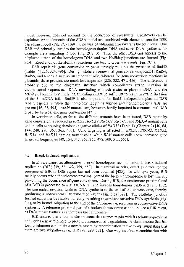
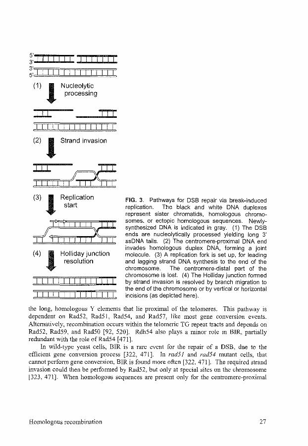
In S. cerevisiae, an alternative form of homologous recombination is break-induced replication (BIR) [39, 53, 322, 359, 550]. In mammalian cells, direct evidence for the presence of BIR in DSB repair has not been obtained [425]. In wild-type yeast, BIR mainly occurs when the telomere-proximal part of the broken chromosome is lost, thereby preventing the occurrence of gene conversion. During BIR, the centromere-proximal end of a DSB is processed to a 3' ssDNA tail and invades homologous dsDNA (Fig. 3.1, 2). The one-ended invasion leads to DNA synthesis to the end of the chromosome, thereby producing a nonreciprocal translocation event (Fig. 3.3) [322]. The Holliday junction formed can either be resolved directly, resulting in semi-conservative DNA synthesis (Fig. 3.4), or by branch migration to the end of the chromosome, resulting in conservative DNA synthesis. A telomere-proximal part of a broken chromosome cannot induce a BIR event, as DNA repair synthesis cannot pass the centromere.
BIR ensures that a broken chromosome that cannot rejoin with its telomere-proximal end, gains a new telomere to prevent chromosomal degradation. A chromosome that has lost its telomere can obtain a new telomere by recombination in two ways, suggesting that there are two subpathways ofBIR [92, 289, 521]. One way involves recombination with
26 Chapter 1
s·~~~~~~~~i~~~ 3' I 3' s· I I I I
(1) J Nucleolytic processing
Jll Jll I I I I I I I I I I I I I I
(2) J Strand invasion
Jll Ill~ I I I I
(3) J Replication start
,~::::::~ (4) J Holliday junction
resolution
FIG. 3. Pathways for DSB repair via break-induced replication. The black and white DNA duplexes represent sister chromatids, homologous chromosomes, or ectopic homologous sequences. Newlysynthesized DNA is indicated in gray. (1) The DSB ends are nucleolytically processed yielding long 3' ssDNA tails. (2) The centromere-proximal DNA end invades homologous duplex DNA, forming a joint molecule. (3) A replication fork is set up, for leading and lagging strand DNA synthesis to the end of the chromosome. The centromere-distal part of the chromosome is lost. (4) The Holliday junction formed by strand invasion is resolved by branch migration to the end of the chromosome or by vertical or horizontal incisions (as depicted here).
the long, homologous Y elements that lie proximal of the telomeres. This pathway is dependent on Rad52, Rad51, Rad54, and Rad57, like most gene conversion events. Alternatively, recombination occurs within the telomeric TG repeat tracts and depends on Rad52, Rad59, and Rad50 [92, 520]. Rdh54 also plays a minor role in BIR, partially redundant with the role ofRad54 [471].
In wild-type yeast cells, BIR is a rare event for the repair of a DSB, due to the efficient gene conversion process [322, 471]. In rad51 and rad54 mutant cells, that cannot perform gene conversion, BIR is found more often [322, 471]. The required strand invasion could then be performed by Rad52, but only at special sites on the cbromosome [323, 471]. When homologous sequences are present only for the centromere-proximal
Homologous recombination 27
part of the DSB, and normal gene conversion therefore is impossible, the DSB is repaired by BIR in about 70% of wild-type cells, which is completely Rad52 dependent [53].
4.3 Single-strand annealing
One of the simplest ways of homologous recombination is SSA [307]. SSA can only occur between two stretches of directly oriented homologous DNA sequences and causes deletion of one of the repeats and the sequence in between the repeats. The minimal length of homology to allow for SSA in a plasmid containing a DSB is around 30 bp in yeast, but SSA is much more efficient when at least 200 bp are homologous [163, 423, 494, 495]. A biologically relevant role of SSA could be resolving recombination intermediates in the repair of DNA interstrand cross-links (Chapter 4) [146]. DSB repair by recombination between repeated sequences, like the Alu repeats in mammalian cells, could also occur by SSA. However, similar to gene conversion, SSA is inhibited when the two repeated DNA sequences are not completely homologous, which reduces the inappropriate use of SSA [ 497]. In recombination assays, directly repeated DNA sequences are used frequently to test homologous recombination pathways, conditions, and genes involved, which makes SSA an important pathway in these assays.
The first step of DSB repair in SSA is nucleolytic processing of the DSB ends yielding 3' ssDNA tails (Fig. IBI) [164]. Once resection has uncovered complementary sequences on both DSB ends, these sequences anneal (Fig. IB1.2). Rad52 and Rad59 could be involved in this annealing step [360, 401, 495]. Annealing does not necessarily occur with the other end of the DSB. Haber and Leung induced two DSBs in different chromosomes in yeast, that could be repaired by two intrachromosomal or two interchromosomal SSA events [199]. Both events were equally frequent, suggesting that the DSB ends search the whole genome for homology. Nonhomologous sequences present at the end of the ssDNA are removed and the nicks are ligated (Fig. IB 1.3). In yeast, the removal of the nonhomologous tails could be performed by Polymerase 8, when the tails are shorter than 30 bp [385]. Longer tails are usually removed by Radl-Rad!O [163, 385, 411]. SSA between homologous sequences shorter than I kb is also dependent on Msh2 and Msh3 when long nonhomologous tails are present [ 446, 497]. The MMR proteins probably stabilize the annealed sequence and thereby enable Radl-Rad!O to cleave the tails. An increasing distance between the repeated DNA sequences slows the rate of SSA, probably due to rate-limiting resection of the DSB ends needed to expose the complementary DNA [164, 411]. Nevertheless, SSA can occur efficiently between repeated DNA sequences that are 15 kb apart [437].
Alternative events can give rise to the same genotypic outcome, consisting of the deletion of intervening sequences between directly oriented repeats. One of the alternatives is intrachromatid gene conversion with cross-over (Fig. 5A2). This event yields a circular DNA containing the deleted sequence as a second product. As this second product is either not found or found in a low frequency, intrachromatid gene conversion with cross-over is not very frequent compared to SSA [164, 172, 280, 420]. Other alternative events are unequal sister chromatid gene conversion with crossover (Fig.
28 Chapter l

5A3), or unequal sister chromatid gene conversion with deletion of the intervening sequences [2, 11, 411].
SSA in yeast depends on Rad52, but much less than gene conversion (Table 1) [263, 377, 383, 495]. For SSA events, RAD52 is synergistic with RADJ, indicating the presence ofRad52-independent, Radl-dependent SSA events [450, 526]. Rad59 seems to function in the Radl-dependent SSA pathway and is possibly involved in annealing the ssDNA and stabilizing the intermediate for tail removal, independent of the MMR proteins [228, 495]. Mutations in the MREJJ group genes cause mainly a delay but hardly a reduction in product formation during SSA, probably due to slower processing of the DSB ends [226, 494]. Rad51, Rad54, Rad55, and Rad57 are not needed for SSA [226]. On the contrary, RAD51- and RAD54-deficient cells show an increase in SSA events, probably due to the impairment of gene conversion in these cells (Chapter 2) [144, 226, 280, 465].
5 DSB REP AIR ASSAYS
DSB repair assays are often based on the presence of two homologous, repeated sequences on plasmids or in chromosomes. The two repeats can be placed in direct or inverse order on the same molecule allowing measurement of intrachromatid and sister chromatid recombination (Fig. 4a, b, c). Alternatively, when present on homologous chromosomes, they measure heteroallelic recombination (Fig. 4d). Ectopic recombination can be assessed when they are placed on nonhomologous chromosomes (Fig. 4e ). A plasmid containing a DSB that recombines with chromosomal homologous DNA yields a gene targeting event.
FIG. 4. Types of homologous recombination, defined by the position of the homologous sequence used to repair the DSB. The thick arrows represent repeated sequences. A white block in the arrow represents a restriction site where a DSB can be induced. Centromeres are indicated by circles. The chromosome shown at the top is homologous to the chromosome in the middle. Only for the chromosome in the middle both sister chromatids are indicated. The chromosome at the bottom is nonhomologous. (a) lntrachromatid recombination, (b) equal sister chromatid recombination, (c) unequal sister chromatid recombination, (d) heteroallelic recombination, (e) ectopic recombination. a, b, and c together constitute intrachromosomal recombination.
Homologous recombination 29
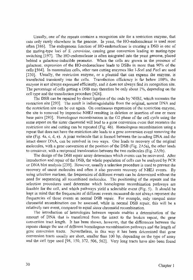
Usually, one of the repeats contains a recognition site for a restriction enzyme, that cuts only rarely elsewhere in the genome. In yeast, the HO-endonuclease is used most often [386]. The endogenous function of HO-endonuclease is creating a DSB in one of the mating-type loci of S. cerevisiae, causing gene conversion leading to mating-type switching [ 197]. The HO-endonuclease is often integrated into the yeast genome, placed behind a galactose-inducible promoter. When the cells are grown in the presence of galactose, expression of the HO-endonuclease leads to DSBs in more than 90% of the cells [564]. In mammalian cells, other rare cutting enzymes like !-See! and Pacl are used [230]. Usually, the restriction enzyme, or a plasmid that can express the enzyme, is transfected transiently into the cells. Transfection efficiency is far below 100%, the enzyme is not always expressed efficiently, and it does not always find its recognition site. The percentage of cells getting a DSB may therefore be only about 1%, depending on the cell type and the transfection procedure [426].
The DSB can be repaired by direct ligation of the ends by NHEJ, which recreates the restriction site [293]. The result is indistinguishable from the original, noncut DNA and the restriction site can be cut again. On continuous expression of the restriction enzyme, the site is removed by imprecise NHEJ resulting in deletion or insertion of one or more base pairs [293]. Homologous recombination in the G2 phase of the cell cycle using the same repeat on the sister chromatid will lead to a gene conversion event that recreates the restriction site and cutting can be repeated (Fig. 4b). Homologous recombination using a repeat that does not have the restriction site leads to a gene conversion event removing the site (Fig. 4a, c, d, e). A joint molecule that is formed between the invading DNA and the intact donor DNA, can be resolved in two ways. One leads to recovery of the original molecules, with a gene conversion at the position of the DSB (Fig. 2A5a), the other leads to crossover, with a reciprocal translocation between the two molecules (Fig. 2A5b).
The design of the DSB repair assay determines which events can be recovered. After introduction and repair of the DSB, the whole population of cells can be analyzed by PCR or DNA blot analysis [239]. However, usually a selection procedure is used to prevent the recovery of uncut molecules and often it also prevents recovery of NHEJ events. By using selection markers, the frequencies of different events can be determined without the need for sequencing all recombined molecules. The positioning of the repeats and the selection procedures used determine which homologous recombination pathways are feasible for the cell, and which pathways yield a selectable event (Fig. 5). It should be kept in mind that the frequencies of different events measured do not always represent the frequencies of those events in normal DSB repair. For example, only unequal sister chromatid recombination can be assessed, while in normal DSB repair, this will be a relatively rare event, compared to equal sister chromatid recombination.
The introduction of heterologies between repeats enables a determination of the amount of DNA that is transferred from the intact to the broken repeat, the gene conversion tract length. It has been shown, however, that the differences between the repeats change the use of different homologous recombination pathways and the length of gene conversion tracts. Nevertheless, in this way it has been determined that gene conversion tracts usually are short, smaller than I 00 bp, depending on the type of assay and the cell type used [98, !50, 372, 506, 562]. Very long tracts have also been found
30 Chapter I
[364]. These long tract gene conversion events cannot readily be distinguisted from crossover events, when the restriction site used to distinguish gene conversion from crossover on a DNA blot is positioned relatively close to the DSB [239]. Although the DSB repair assays using reporter genes have been highly informative, all the artificial aspects of these assays should be borne in mind when interpreting their results.
A totally different kind of assay to measure the efficiency of homologous recombination in DSB repair is the observation of SCEs. SCEs occur mainly due to gene conversion events that are accompanied by crossovers [ 482]. A low level of SCEs can already be found without any treatment of the cells, as a result of repair of spontaneously occurring damage. The frequency of SCEs increases by DNA-damaging treatments, like UV, DSB-inducing agents, and DNA interstrand cross-linking agents. Cells deficient in genes involved in gene conversion, show a lower frequency of SCEs (Chapter 2) [144, 509, 511]. The advantage of the measurement of SCEs is that no artificial sequences possibly influencing the frequency of different DSB repair processes have to be introduced into the cells. The disadvantage is that only gene conversion events associated with crossovers large enough to be cytologically detected can be measured and no knowledge concerning other DSB repair pathways is obtained.
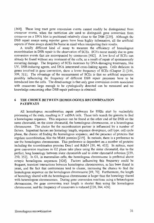
6 THE CHOICE BETWEEN HOMOLOGOUS RECOMBINATION PATHWAYS
All homologous recombination repair pathways for DSBs start by nucleolytic processing of the ends, resulting in 3' ssDNA tails. These tails search the genome to find a homologous sequence. This sequence can be found at the other end of the DSB on the same chromatid, on the sister chromatid, the homologous chromosome, or a heterologous chromosome. The choice for the recombination partner is influenced by a number of factors. Important factors are homology length, sequence divergence, cell type, cell cycle phase, the chance of finding the homologous sequence, and the presence of proteins that regulate recombination, like the MMR proteins [219]. In meiosis, there is a preference to use the homologous chromosome. This preference is dependent on a number of proteins including the recombination proteins Dmcl and Rdh54 [10, 46, 453]. In mitosis, most gene conversion reactions in G2 phase take place using the sister chromatid, due to the perfect, long homology between sister chromatids and to sister chromatid cohesion [239, 250, 352]. In Gl, in mammalian cells, the homologous chromosome is preferred above ectopic homologous sequences [426]. Factors influencing this frequency could be frequent, transient interactions between homologous chromosomes, as has been found in yeast, and the fact that centromeres tend to cluster, increasing the chance to find the homologous sequence on the homologous chromosome [69, 70]. Furthermore, the length of homology shared with the homologous chromosome is larger than the homology shared with heterologous chromosomes. During gene conversion reactions using a heterologous chromosome, the gene conversion tract length is shorter than using the homologous chromosome, and the frequency of crossovers is reduced [220, 364, 426].
Homologous recombination 31
A..,,_ 1 .. 52 "5'51
0 - + 2-111-
51
3[: • 4-111-
8411- 1 -10- 'j
52 "5'"3'51
+ 0 2-1!1- -
{''~--<&- •
4 ... ~·
c ,,... II 1-o-"5'51 52 51
2+ +
3["""' -· 4-
o .. If,, 1 -· 51 52 "5'51 2-1!1-
3[: f
32 Chapter 1
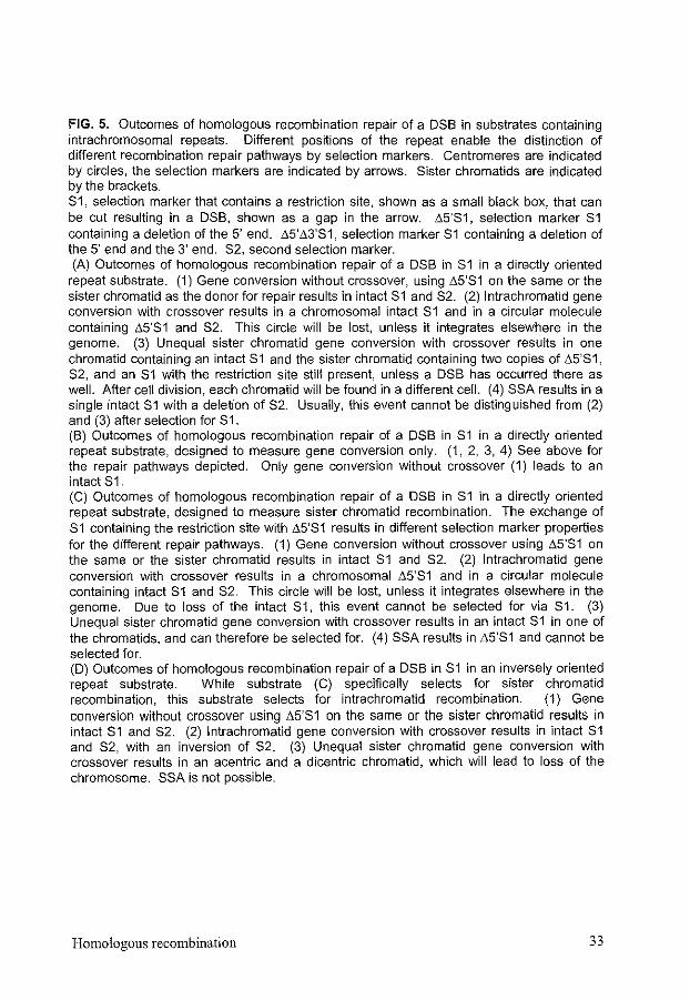
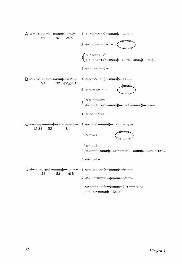
FIG. 5. Outcomes of homologous recombination repair of a DSB in substrates containing intrachromosomal repeats. Different positions of the repeat enable the distinction of different recombination repair pathways by selection markers. Centromeres are indicated by circles, the selection markers are indicated by arrows. Sister chromatids are indicated by the brackets. S1, selection marker that contains a restriction site, shown as a small black box, that can be cut resulting in a DSB, shown as a gap in the arrow. ~5'81, selection marker S1 containing a deletion of the 5' end. ~5'~3'81, selection marker S1 containing a deletion of the 5' end and the 3' end. S2, second selection marker. (A) Outcomes of homologous recombination repair of a DSB in S1 in a directly oriented
repeat substrate. (1) Gene conversion without crossover, using ~5'81 on the same or the sister chromatid as the donor for repair results in intact S1 and S2. (2) lntrachromatid gene conversion with crossover results in a chromosomal intact 81 and in a circular molecule containing ~5'81 and S2. This circle will be lost, unless it integrates elsewhere in the genome. (3) Unequal sister chromatid gene conversion with crossover results in one chromatid containing an intact S1 and the sister chromatid containing two copies of A5'S1, S2, and an S1 with the restriction site still present, unless a DSB has occurred there as well. After cell division, each chromatid will be found in a different cell. (4) SSA results in a single intact S1 with a deletion of S2. Usually, this event cannot be distinguished from (2) and (3) after selection for S 1. (B) Outcomes of homologous recombination repair of a DSB in S1 in a directly oriented repeat substrate, designed to measure gene conversion only. (1, 2, 3, 4) See above for the repair pathways depicted. Only gene conversion without crossover (1) leads to an intact S1. (C) Outcomes of homologous recombination repair of a DSB in S1 in a directly oriented repeat substrate, designed to measure sister chromatid recombination. The exchange of S 1 containing the restriction site with Ll5'S 1 results in different selection marker properties for the different repair pathways. (1) Gene conversion without crossover using ~5'81 on the same or the sister chromatid results in intact S 1 and S2. (2) lntrachromatid gene conversion with crossover results in a chromosomal Ll5'S1 and in a circular molecule containing intact S1 and S2. This circle will be lost, unless it integrates elsewhere in the genome. Due to loss of the intact S 1, this event cannot be selected for via S 1. (3) Unequal sister chromatid gene conversion with crossover results in an intact S1 in one of the chromatids, and can therefore be selected for. (4) SSA results in ~5'81 and cannot be selected for. (D) Outcomes of homologous recombination repair of a DSB in S1 in an inversely oriented repeat substrate. While substrate (C) specifically selects for sister chromatid recombination, this substrate selects for intrachromatid recombination. (1) Gene conversion without crossover using ~5'81 on the same or the sister chromatid results in intact S1 and S2. (2) lntrachromatid gene conversion with crossover results in intact S1 and S2, with an inversion of S2. (3) Unequal sister chromatid gene conversion with crossover results in an acentric and a dicentric chromatid, which will lead to loss of the chromosome. SSA is not possible.
Homologous recombination 33
Apart from this competition between different repair templates, there is also competition between the different homologous recombination pathways, When a DSB can be repaired by either gene conversion or SSA, as with a directly oriented repeat, the ratio of the events varies widely for different repair assays (Chapter 2) [144, 164, 390, 508, 574], Factors influencing this ratio are the cell type, the homology length, the distance between the repeats, and the position of the DSB. In cells mutant for one or more of the recombination repair genes, the ratio will change, Mutations in for example RAD51 and RAD54 will reduce gene conversion frequencies and increase SSA, while RAD52 mutants show a larger decrease in gene conversion than in SSA (Chapter 2) [144, 226, 280, 377, 383, 465], A similar competition occurs between gene conversion and BIR [322, 471, 550], A mutation in one of the genes involved in homologous recombination may not only lead to a shift in recombination pathways, it may also lead to increased lethality, even when the broken chromosome or plasmid is not essential for cell survival [265, 322], This may be due to the formation of recombination intermediates that cannot be resolved anymore and lead to cell death,
7 CONCLUSIONS
Cells contain an intricate network of DSB repair pathways to protect the integrity of the DNA, The proteins involved in DSB repair work together in a number of different complexes, often performing functions in different repair pathways, Some of the protein interactions are very stable, others are much more dynamic (J. Essers, personal communication). Apart from repair proteins, replication and cell cycle checkpoint proteins are also involved in the cellular reaction to DSBs. Repair is localized to the sites of the DSBs, but the DSB ends can search the whole genome to find homologous DNA The in vitro biochemistry of gene conversion and strand annealing reactions is currently being elucidated, However, how the cellular repair proteins are able to find first the DSBs, and then homologous sequences in the large excess of DNA present is still a mystery, Similarly, future research should provide further clues as to how cells regulate DSB repair and decide which pathway is most suitable to prevent chromosomal instability,
34 Chapter I
CHAPTER2
Mouse RAD54 affects DNA double-strand break repair and sister chromatid exchange
Modified from Mol. Cell. Biol. 20(9): 3147-3156, 2000

MOUSE RAD54 AFFECTS DNA DOUBLE-STRAND BREAK REPAIR AND SISTER CHROMATID EXCHANGE
MIES L.G. DRONKERT1, H. BERNA BEVERL001
, ROGER D. JOHNSON2, JAN H.J.
HOEIJMAKERS 1, MARIA JASIN2
, AND ROLAND KANAAR1•3
1 Department of Cell Biology and Genetics, Erasmus University Rotterdam, PO Box 1738, 3000 DR Rotterdam, The Netherlands
2 Cell Biology and Genetics Program, Sloan-Kettering Institute and Cornell University Graduate School of Medical Sciences, New York, New York 10021, USA
3 Department of Radiation Oncology, University Hospital Rotterdam/Daniel, The Netherlands
Cells can achieve error-free repair of DNA double-strand breaks (DSBs) by homologous recombination through gene conversion with or without crossover. In contrast, an alternative homology-dependent DSB repair pathway, single-strand annealing (SSA), results in deletions. In this study, we analyzed the effect of mRAD54, a gene involved in homologous recombination, on the repair of a site~specific I~Scei-induced DSB located in a repeated DNA sequence in the genome of mouse embryonic stem cells. We used six isogenic cell lines differing solely in the orientation of the repeats. The combination of the three recombination-test substrates used discriminated among SSA, intrachromatid gene conversion, and sister chromatid gene conversion. DSB repair was most efficient for the substrate that allowed recovery of SSA events. Gene conversion with crossover, which was indistinguishable from long tract gene conversion, preferentially involved the sister chromatid rather than the repeat on the same chromatid. Comparing DSB repair in mRAD54 wild-type and knockout cells revealed direct evidence for a role of mRad54 in DSB repair. The substrate measuring SSA showed an increased efficiency of DSB repair in the absence of rnRad54. The substrate measuring sister chromatid gene conversion showed a decrease in gene conversion with and without crossover. Consistent with this observation, DNA damage-induced sister chromatid exchange was reduced in mRAD54-deficient cells. Our results suggest that mRad54 promotes gene conversion with predominant use of the sister chromatid as the repair template at the expense of errorprone SSA In promoting error-free DSB repair, mRad54 will contribute to the maintenance of genomic stability.
INTRODUCTION
DNA double-strand breaks (DSBs), induced for example by endogenously produced radicals or ionizing radiation, form a major threat to the integrity of chromosomes and viability of cells. Unrepaired or incorrectly repaired DSBs may lead to translocations or loss of chromosomes, which could result in cell death or uncontrolled cell proliferation. Eukaryotes have developed several mechanisms to repair DSBs, including nonhomologous DNA end-joining (NHEJ) and homologous recombination. In
Mouse RAD54 and chromosomal rearrangements 37
Saccharomyces cerevisiae, DSBs are efficiently repaired through homologous recombination by the RAD52 group genes, while a contribution ofNHEJ to DSB repair is only observed in the absence of homologous recombination [20, 173, 270, 355, 386, 398, 469]. In mammalian cells, NHEJ plays a major role in DSB repair [233]. More recently, it has become clear that in addition to NHEJ, homologous recombination can play an important role in DSB repair in mammalian cells as well [28, 224, 253, 300, 399].
Several pathways of homology-dependent DSB repair have been described for S. cerevisiae (Chapter I) (386]. One of these pathways, single-strand annealing (SSA), specifically occurs when a DSB is made between directly repeated DNA sequences. The DSB is processed by removal of part ofthe 5' strand on each side of the break, exposing long 3' overhangs [263]. The single-stranded DNA (ssDNA) overhangs anneal to a long complementary stretch of DNA and nonhomologous ssDNA ends are removed. As a result, one of the repeats and the intervening sequence are deleted. In vertebrates, a similar pathway has been described [75].
An alternative homology-dependent DSB repair pathway, mediated by the RAD52 group genes, is gene conversion [386, 489, 507]. DSB repair through this pathway also requires the DNA around the DSB to be degraded to produce 3' ssDNA overhangs. One or both of these ends invade a homologous DNA sequence, which can be found either on the homologous chromosome or, in the S and G2 phases of the cell cycle, on the sister chromatid. Several models for this invasion have been described, including DSB gap repair and synthesis-dependent strand annealing (See Chapter I) [160, 370, 387]. In a model for DSB gap repair, both ends invade the homologous duplex and the gap is filled by semi-conservative DNA synthesis. The resulting Holliday junctions are resolved either with or without crossover [507]. For reasons of brevity, we will use the term 'crossover' for events involving gene conversion with crossover, and 'gene conversion' for gene conversion without crossover. In the simplest model for synthesis-dependent strand annealing, only one end invades the homologous sequence. After DNA synthesis primed from the invading end, the newly-synthesized strand reanneals with the other end of the DSB. Then the second strand is synthesized, resulting in conservative DNA synthesis and a strong bias towards noncrossover events (See Figure 2 in Chapter 1) (160]. However, if a long tract of DNA is synthesized, the result of gene conversion without crossover may appear similar to a crossover event.
RAD52 is important for almost all gene conversion and crossover pathways in S. cerevisiae (386]. Other genes involved include RAD51, RAD54, RDH54/TIDI, RAD55, and RAD57 (Chapter 1) [162, 264, 386]. RAD51, RAD54, RAD55, and RAD57 are required for gene conversion. RDH54, a homolog of RAD54, is mainly required for gene conversion using the homologous chromosome, while RAD54 is involved in gene conversion with both the sister chromatid and the homologous chromosome (10, 264, 465]. Mutations in MREII, RAD50, and XRS2 cause a slower repair of DSBs by SSA, gene conversion and crossover, most likely because the 5' to 3' degradation of the DNA is slowed down in these mutants [226, 292, 494]. In mammalian cells, similar gene conversion and crossover pathways have been found, but very little is known about the genetic requirements of the different pathways. Most of the above-mentioned genes have a homolog in mammals (Table I in Chapter I) [253, 399]. Nevertheless, the importance
38 Chapter 2
of each gene can differ in mammalian and S. cerevisiae cells. For example, the mouse RAD52 (mRAD52) gene can be mutated without a major effect on recombination, while it is the most important gene inS. cerevisiae [428].
One of the other RAD52 group genes, RAD54, is clearly important in mammalian cells. The Rad54 protein belongs to the Swi2/Snf2 protein family whose members modulate protein-DNA interactions in an ATP-dependent manner [148, 259, 395]. The S. cerevisiae and human Rad54 proteins are double-stranded DNA-dependent ATPases that interact with Rad51, a key player in the search for homologous template DNA [102, 185, 235, 400, 505, 513]. Compared to wild-type cells, RAD54-deficient mouse embryonic stern (ES) cells are two- to four-fold more sensitive to ionizing radiation, the alkylating agent methyl rnethanesulfonate, and the DNA interstrand cross-linking agent mitomycin C [154]. In addition, homologous recombination in mRAD54-deficient cells is 5- to 10-fold reduced, as measured by targeted integration of exogenous DNA [154]. This reduction in homologous recombination can explain the sensitivity of cells lacking mRad54 to DSBinducing agents, although a direct involvement of mRad54 in DSB repair has not yet been demonstrated.
Much information concerning the mechanisms of DSB repair in S. cerevisiae has been obtained using a site-specific DSB induced by the rare-cutting HO-endonuclease. Recently, rare-cutting enzymes suitable for site-specific DSB induction in mammalian cells have become available [230]. One of these is the S. cerevisiae mitochondrial enzyme !-See!, which recognizes and cuts a nonpalindrornic 18-bp site leaving 4-bp 3' overhangs [106]. Transfection of an 1-Scel-expressing plasmid into mouse or hamster cells induces a DSB at I-Scel sites present on a plasmid or integrated in a chromosome, but is not toxic to these cells [99, 436]. Analysis of the repair products of the site-specific DSB allows quantitation of the relative conttibution of NHEJ and different homologydependent pathways of DSB repair in mammalian cells [140, 240, 300, 301, 436, 447, 508].
In this study, we have investigated the relative contribution of different homologydependent pathways to the repair of an 1-Scel-induced chromosomal DSB in mouse ES cells. The repair of the DSB induced in different recombination-test substrates that discriminate among gene conversion, crossover using the homologous sequence on the sister chromatid or the same chromatid, and SSA was monitored through genetic selection. Importantly, the different recombination-test substrates were integrated at the same site in the genome, which excludes differences due to local effects of DNA sequences or chromatin and allows a direct comparison between the results obtained with different substrates. In addition, we analyzed the effect of mRAD54, by comparing the distribution of repair events from the different recombination-test substrates in mRAD54+!and mRAD541- mouse ES cells that were otherwise isogenic. The effects of mRAD54 on DSB repair were compared with its effect on the number of both spontaneous and DNA damage-induced sister chromatid exchanges (SCEs) observed in mRAD54-proficient anddeficient cells.
Mouse RAD54 and chromosomal rearrangements 39
MATERIALS AND METHODS
Construction of mRAD54 targeting vectors. Targeting vectors were constructed to integrate three different recombination-test substrates into the mRAD54 genomic locus. The substrates were cloned into the unique Sful site of exon 4, thereby disrupting mRAD54. The first targeting vector was made by inserting the DRneo construct [301], linearized with Xhol, into the Sfol site of a 9-kb EcoRI fragment from mRAD54 encompassing exons 4, 5, and 6 (Fig. !A) [!54]. The 5' ssDNA overhangs of both restriction sites were removed by incubation of the digested DNA with the Klenow fragment of E. coli DNA polymerase I. The resulting mRAD54 allele is referred to as mRAD54°R"" (Fig. !A). The second and third targeting vectors were made by inserting the !Rneo and SCneo recombination-test substrates in a similar manner [240]. The resulting mRAD54 alleles are referred to as mRAD541
Rneo and mRAD54SCneo, respectively (Fig. lA). The presence of an intact !-See! restriction site was tested by digestion of the targeting vectors with the purified enzyme.
ES cell culture and electroporation. Heterozygous mRAD54 ES cells of the genotype mRAD54+!JOlpur were electroporated with the different targeting vectors and cultured on gelatinized dishes as described previously [154, 595]. The cells were split 24 h after electroporation, and hygromycin B was added to a final concentration of 200 fig/ml. After 7 to I 0 days, colonies were isolated and expanded. Genomic DNA from individual clones was digested with Stu! and analyzed by DNA blotting using a flanking probe (Fig. !B). The blot was rehybridized with a 700-bp 3' neomycin (neo) fragment to confirm a single integration of the targeting vector.
Immunoblot analysis. The expression of mRad54 protein was checked for all cell lines used in this study by immunoblot analysis of whole cell extracts. Blots were hybridized with a polyclonal a-hRad54 antibody [254].
Cell survival after gamma ray treatment. To determine whether integration of the recombination-test substrates into the mRAD54 locus resulted in mRAD54 deficiency, the sensitivity of mRAD54+JDRneo and mRAD54-IDRneo ES cells towards increasing doses of ionizing radiation was determined by measuring their colony-forming ability. ES cells were trypsinized and counted. Various cell dilutions were aliquoted onto gelatinized 60-mm dishes, and after 12 to 24 h, cells were irradiated by a 137Cs source. They were grown for 7 to 10 days, fixed, stained, and counted. All measurements were performed in triplicate.
I-Scel transfections. ES cells containing the recombination-test substrates were cultured in medium containing hygromycin at a concentration of 200 )J.g/ml. Transfection of 3.2xl0
6 cells was done by electroporation with 6 fig of either pPGK3xnlsl-Scel or
pCBA3xnls-I-Scel, which transiently express 1-Scel from the phosphoglycerate kinase I (PGK) and the chicken ~-actin promoter, respectively [150, 426]. The pCBA3xnls-I-Scel
40 Chapter 2
construct was slightly more efficient in the introduction of a DSB than pPGK3xnlsi-Scel, as was apparent from the number of 0418-resistant colonies obtained. However, no differences in the ratio of 0418-hygromycin-resistant colonies to 0418-resistant colonies were found (data not shown). To determine the transfection efficiency, 6 !Lg of pPGKCAS-eGFP, containing the green fluorescent protein (GFP) gene under the control of a PGK promoter, was cotransfected in a number of experiments. In parallel, cells were electroporated without DNA, with pBluescript II KS, or pPGKCAS-eGFP alone. After electroporation, 103 cells were plated without selection to determine the cloning efficiency. The remaining cells were grown for one day without selection before they were split and cultured in medium containing 0418 (200 f.Lg/ml) or 0418 (200 f.Lg/ml)hygromycin (200 f.Lg/ml). When pPGKCAS-eGFP had been cotransfected with the 1-Scei-expressing plasmid, a portion of the cells was subjected to fluorescence-activated cell sorting analysis one day after transfection to detennine the percentage of cells positive for GFP expression. After 8 to 11 days, cells were fixed, stained, and counted. The number of clones from the cells transfected with the 1-Scel-expressing plasmid was corrected for the number of clones from the mock-transfected cells. To enable comparison between the number of clones from different cell lines and experiments, the absolute number of clones was divided by the cloning efficiency and transfection efficiency. Recombination frequencies can then be calculated by dividing the corrected number of clones by the number of cells transfected. The data on the number of 0418- and 0418-hygromycinresistant clones is based on three to seven independent experiments, using two or three independent cell lines for each genotype. In several of the experiments, colonies were isolated and expanded. Genomic DNA from individual clones was analyzed for recombination events by digestion with either Neal or EcoRI and DNA blotting using the 700-bp 3' neo fragment as a probe. Colonies from all recombination substrates that had aberrations in the hybridization pattern which were difficult to interpret, were not included in the analysis. Inclusion of these aberrant clones did not alter the conclusions. To determine the influence of a low dose of gamma rays from a 137Cs source on the distribution of recombination events, ES cells were irradiated with 1 or 2 Gy just before or 4 h after electroporation ofpCBA3xnls-1-Scel. After one day without selection, the cells were split and grown in medium containing 0418 or 0418-hygromycin and processed as described above.
Sister chromatid exchanges. SCEs in ES cell lines of the genotype mRAD54+1~, mRAD54"1
-, mRAD541-, and a derivative of the same mRAD541
- line expressing the hRAD54 eDNA were analyzed [154, 505]. The mRAD54 knockout allele in these lines was mRAD54307
neo. The cell lines were coded to prevent bias in the analysis. The code was revealed only after the SCE analysis of all cell lines was completed. Cells were seeded in 60-mm dishes and after 24 h, the medium was removed and replaced with medium containing 10 ~M bromodeoxyuridine (BrdU) with or without 0.2 ~g mitomycin C/ml. After 1 to 2 h, cells were washed with phosphate-buffered saline (PBS) and fresh medium supplemented with 10 f.LM BrdU was added. Cells were cultured for two rounds of DNA replication (23 to 26 h), collected by mitotic shake off 2 h after the addition of
Mouse RAD54 and chromosomal rearrangements 41
colcemid to a final concentration ofO.l ~g/ml, and treated with trypsin/EDT A for 5 min. Cells were treated with cold hypotonic solution (0.075 M KCI) immediately followed by centrifugation. Subsequently, cells were fixed with three changes of methanol/acetic acid (3:1) and metaphase preparations were made. To visualize the SCEs, slides were stained in diluted acridine orange for 5 min, mounted in 0.07 M Na2HP04/KH2P04 (pH 6.5) with a coverslip, and exposed to UV light of 265 nm for I 0-15 min. SCEs were observed through a fluorescence microscope (Leitz Orthoplan) using an IOOx objective. At least 40 metaphases per cell line were analyzed for both the number of chromosomes and SCEs. Metaphase spreads were also analyzed for the presence of gross chromosomal abnormalities.
RESULTS
The recombination-test substrates. The three different recombination-test substrates that were used to measure recombination frequencies in mouse ES cells are schematically depicted in Fignre lA. They contain a hygromycin-selectable marker gene, flanked by two inactive neomycin-selectable marker genes, S2neo and 3' neo. For brevity, we will
FIG. 1. Generation of mRAD54•t- and mRA054-t- ES cells containing recombination-test substrates. (A) Structure of the genomic mRAD54 locus and targeting vectors containing the recombination-test substrates. The two upper lines represent the wild-type (mRAD54+) and the puromycin-targeted knockout (mRAD54307
P"') alleles, respectively. The 18 exons that encode mRad54 are indicated by boxes. The dashed line above exons 7 and 8 indicates the position of the probe used to distinguish the different mRA054 alleles after digestion of the genomic DNA with Stul. The arrow shows the position of the puromycin (pur)-selectable marker gene. The locations of selected restriction sites are shown; E, EcoRI; N, Ncol; Sf, Sful; St, Stu!. The third line shows a generic representation of the targeting vectors. The three lower lines show the three different recombination-test substrates inserted into the mRAD54 locus in more detail. The black arrow indicates the hygromycin-selectable marker gene (hyg). The gray arrow on the left represents the 700-bp 3' neomycin-selectable marker gene (3' neo). The gray arrow on the right represents the full-length S2neo gene, which contains a 4-bp deletion at the 18-bp 1-Scel site insertion (indicated in black). Recombination between the S2neo and 3' neo genes can lead to restoration of the original Neal site, resulting in an intact neo gene. The targeting constructs differ solely in the orientation of the 3' neo and S2neo genes. (B) DNA blot of ES cells containing wild-type(+) and knockout(-) mRAD54 alleles in addition to alleles with recombination-test substrates. mRAD54+1307pur ES cells were electroporated with the mRAD54DRneo, mRAD541Rneo, and mRAD54SCneo targeting vectors. After selection, genomic DNA was isolated from individual clones and digested with Stu!. The DNA blot was hybridized with the probe indicated in panel A. Phage fi. DNA digested with Psti was used as a size marker. The lengths of marker fragments are indicated in kilo bases on the right and the positions of the different mRAD54 alleles are shown on the left.
42 Chapter 2
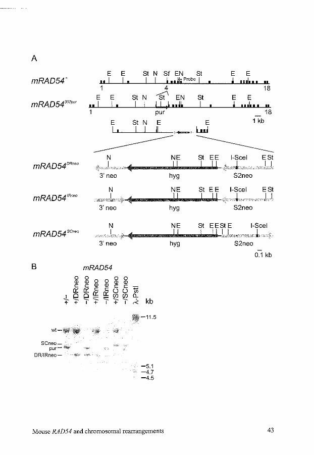
A
E E St N Sf EN St E E mRAD54+ nl I I I I • 11 ~i Probe 1 I j ... 1 ... II
1 4 18
E E St N --St' EN St E E mRAD54307
P"' ul I I I I I I ~~ .,jl, I j ... ! ... II pur 18
E St N E E 1 kb
I I I I II -+---;" luJ
------------ -------------N NE St EE 1-Scel ESt ). ..J.V.V• .II
3'neo hyg S2neo
N NE St EE 1-Scel ESt .J < -i-/// ·•··v.kvv·. 1 •.. 1
3'neo hyg S2neo
N NE St EESt E 1-Scel mRA054scoeo J I I J ..
3'neo hyg S2neo
0.1 kb
B mRAD54 0 0
0 0 0 0
(]) (]) (]) (]) c c (]) (]) c c :;::; 0:: 0:: c c 0 0 oQ; 0::
(/)
-!_ 0 (/)(/) (L --- "" -- ~ kb + + I + I + I
-11.5
wt-!illf
SCneo-pur-
DR/IRneo- --5.1 -4.7 -4.5
Mouse RAD54 and chromosomal rearrangements 43
hereafter refer to these selectable marker genes as the hyg and neo genes, respectively. One of the crippled neo genes, 3' neo, consists of the 3' 700 bp of the neo gene. The other, S2neo, is a full-length neo gene, which contains a 4-bp deletion and the 18-bp insertion of the !-See! site at the position of the Neal site at bp 576 of the neogene [301]. Expression of the I -See I enzyme can create a DSB in the S2neo gene. By recombination between the S2neo and 3' neo gene, the original Neal site of the Slneo gene, which is present in the 3' neo gene, can be restored, creating an intact neo gene. The three recombination-test substrates differ solely in the relative orientation of the two crippled neogenes (Fig. !A). One substrate, DRneo, contains both crippled neogenes in a directly repeated orientation. Transcription of the S2neo gene occurs towards the 51 end of the 3' neo gene. The second substrate, IRneo, contains both genes in an inversely repeated orientation, because the 3' neogene has been inverted relative to its orientation on DRneo. The third, SCneo, contains the genes in a directly repeated orientation, but in contrast to DRneo, transcription of the S2neo gene occurs away from the 31 end of the 3' neogene [240].
Homologous integration of the recombination-test substrates in the mRAD54 locus. We targeted the recombination-test substrates to the mRAD54 gene of mouse ES cells to obtain single integration of the substrates at a defined and transcriptionally active position in the genome. Consequently, the targeted cell lines are isogenic and differ only in the presence of an mRAD54+ or an mRAD54- allele and the orientation of the crippled neo genes of the recombination-test substrates. To achieve this, the recombination-test substrates were subcloned into ex on 4 of the mRAD54 gene to create targeting vectors that would result in disruption of the gene [154]. The resulting mRAD54 alleles are referred to as mRAD54DRneo, mRAD5lRneo, and mRAD54SCneo, respectively. The targeting vectors were transfected into mRAD54+!- ES cells of the genotype mRAD54+1307
'"' [154]. After selection with hygromycin, targeted clones were identified by DNA blotting with a unique probe outside the targeting construct (Fig. 1). To confirm single integration, the blots were rehybridized with a 700-bp 3' neo probe (data not shown). Targeting efficiencies to the mRAD54+ allele were around 13% and to the mRAD54307
P"' allele around 5%. The disruption of mRAD54 by the recombination-test substrates was confirmed by the hypersensitivity of mRAD54307pur!DRneo ES cells to gamma irradiation (data not shown). The survival curve of the mRAD54 ... !DRneo cell line after gamma irradiation was similar to that of wild-type cells, as expected, because heterozygote mRAD54 cells show no obvious phenotype [154]. Immunoblot analysis using a-hRad54 showed that mRad54 protein was present in all mRAD54+ cell lines containing the recombination-test substrates, but could not be detected in any of the mRAD54 knockout cell lines (data not shown).
The DRneo substrate: DSB repair events. For clarity, we first describe possible pathways of DSB repair on the DRneo recombination-test substrate and their resulting molecular outcomes. Subsequently, we will present the results obtained with this recombination-test substrate in wild-type ES cells, in mRAD54-deficient ES cells, and the effect of ionizing radiation on 1-Seel-induced DSB repair.
44 Chapter 2
Transfection of an 1-Scel-expressing plasmid in cells containing the DRneo substrate can result in a DSB in the S2neo gene (Fig. 2). The DSB can be repaired by NHEJ with or without a deletion or insertion [300]. NHEJ will not result in the restoration of an intact neo gene, and therefore NHEJ events will not be recovered on selection with G418. An alternative repair patbway is SSA (Fig. 2). During SSA within the DRneo substrate, complementary strands of the S2neo gene and the 3' neo gene will anneal, resulting in an intact neo gene and deletion of the intervening hyg gene. A third pathway to repair the DSB is homologous recombination by gene conversion (Fig. 2). Gene conversion by recombination with the S2neo gene on the sister chromatid will result in restoration of the nonfunctional S2neo gene, and therefore, these events will not be recovered. To obtain an intact neo gene by gene conversion or crossover, the S2neo gene containing the DSB needs to pair with either the 3' neo gene on the same chromatid or, in the S and G2 phases of the cell cycle, with the 3' neo gene on the sister chromatid. These modes of homologous pairing are referred to as intrachromatid and sister chromatid pairing in Figure 2. If the intermediate is resolved without a crossover, the resulting clone will contain intact neo and hyg genes and a 3' neo gene, and the cell will be resistant to 0418 and hygromycin. On the other hand, if a single crossover takes place, the resulting clone will contain an intact neogene without a hyg gene and will only be resistant to 0418. At the DNA level, the outcome of crossover is therefore identical to the outcome of SSA (Table 1). The reciprocal product for intrachromatid crossover, a circular DNA molecule containing a hyg and a 3' neo gene, will usually be lost. The reciprocal product for sister chromatid crossover, that contains a duplication of the hyg and 3' neo genes, will segregate from the intact neo gene on cell division and be lost on selection with G418 (See Figure SA in Chapter 1).
In summary, the different ways of processing the 1-Scel-induced DSB in the DRneo recombination-test substrate result in differences in the 0418 and hygromycin resistance of the cells. NHEJ results in resistance to hygromycin only, which is identical to the original resistance of the cells before induction of a DSB and therefore, these events cannot be detected. SSA and crossover result in resistance to 0418 only, while gene conversion results in resistance to G418 and hygromycin (Table 1).
The DRneo substrate: relative efficiency of DSB repair events. The relative contribution of the different homology-dependent DSB repair pathways was investigated by transfection of the 1-Scel-expressing plasmids pPGK3xnlsl-Scel or pCBA3xnls-1-Scel into mRAD54+!DRneo ES cells. All transfections were carried out at least three times with two independently obtained mRAD54+!DR"'" cell lines. As a control, a mock transfection was performed with either no DNA, pBluescript II KS or pPGKCAS-eGFP. Before transfection, the cells were grown on hygromycin selection to reduce the background due to spontaneous recombination events. After transfection, the cells were grovm for one day without selection. Subsequently, they were divided over multiple dishes and cultured in either 0418-containing medium or 0418-hygromycin-containing medium. After 8 to 11 days, the cells were fixed and the number of colonies on each dish was counted.
The frequency of spontaneously arising G418-resistant colonies varied between 1 o-5
and 10-6 No significant differences in the induction of G418-resistant colonies were
Mouse RAD54 and chromosomal rearrangements 45
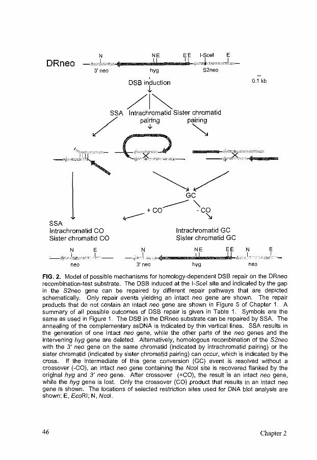
DRneo N
,JFG'' 3' neo
NE E E I,Scel E ............................. ........__.,;,. ~"' ' ,) hyg S2neo
' DSB induction
"' /I~ SSA lntrachromatid Sister chromatid
/ patng p~
k ~ _,,. v:tr:c' -\: :~.":;:~ --+n :~·y·
~/
SSA lntrachromatid CO Sister chromatid CO
N . ,.), ..
neo
E ,I..
GC
------ " +CO -CO ~ '\,
N 1 .
3' neo
lntrachromatid GC Sister chromatid GC
NE EE
hyg
0.1 kb
neo
E J
FIG. 2. Model of possible mechanisms for homology-dependent DSB repair on the DRneo recombination-test substrate. The DSB induced at the 1-Scel site and indicated by the gap in the S2neo gene can be repaired by different repair pathways that are depicted schematically. Only repair events yielding an intact neo gene are shown. The repair products that do not contain an intact neo gene are shown in Figure 5 of Chapter 1. A summary of all possible outcomes of DSB repair is given in Table 1. Symbols are the same as used in Figure 1. The DSB in the DRneo substrate can be repaired by SSA. The annealing of the complementary ssDNA is indicated by thin vertical lines. SSA results in the generation of one intact neo gene, while the other parts of the neo genes and the intervening hyg gene are deleted. Alternatively, homologous recombination of the S2neo with the 3' neo gene on the same chromatid (indicated by intrachromatid pairing) or the sister chroma!'1d (indicated by sister chromatid pairing) can occur, wh'1ch is indicated by the cross. If the intermediate of this gene conversion (GC) event is resolved without a crossover (-CO), an intact neo gene containing the Ncol site is recovered flanked by the original hyg and 3' neo gene. After crossover (+CO), the result is an intact neo gene, while the hyg gene is lost. Only the crossover (CO) product that results in an intact neo gene is shown. The locations of selected restriction sites used for DNA blot analysis are shown; E, EcoRI; N, Neal.
46 Chapter 2
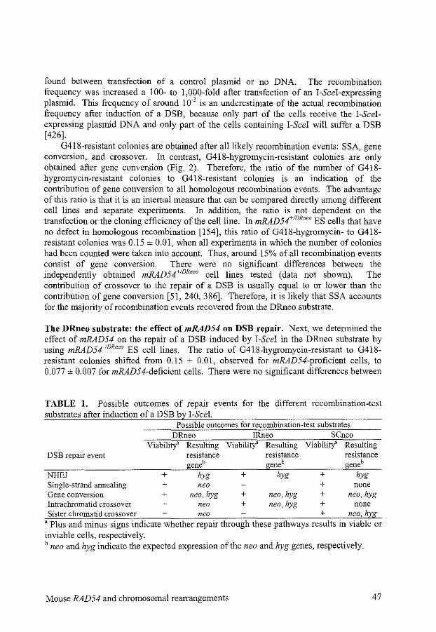
found between transfection of a control plasmid or no DNA. The recombination frequency was increased a 100- to 1,000-fold after transfection of an 1-Scel-expressing plasmid. This frequency of around 1 o·' is an underestimate of the actual recombination frequency after induction of a DSB, because only part of the cells receive the 1-Scelexpressing plasmid DNA and only part of the cells containing !-See! will suffer a DSB [426].
G418-resistant colonies are obtained after all likely recombination events: SSA, gene conversion, and crossover. In contrast, G418-hygromycin-resistant colonies are only obtained after gene conversion (Fig. 2). Therefore, the ratio of the number of G418-hygrornycin-resistant colonies to G418-resistant colonies is an indication of the contribution of gene conversion to all homologous recombination events. The advantage of this ratio is that it is an internal measure that can be compared directly among different cell lines and separate experiments. In addition, the ratio is not dependent on the transfection or the cloning efficiency of the cell line. In mRAD54+!DR"'" ES cells that have no defect in homologous recombination [154], this ratio of G418-hygromycin- to G418-resistant colonies was 0.15 ± 0.01, when all experiments in which the number of colonies had been counted were taken into account. Thus, around 15% of all recombination events consist of gene conversion. There were no significant differences between the independently obtained mRAD54+!DR"" cell lines tested (data not shown). The contribution of crossover to the repair of a DSB is usually equal to or lower than the contribution of gene conversion [51, 240, 386]. Therefore, it is likely that SSA accounts for the majority of recombination events recovered from the DRneo substrate.
The DRneo substrate: the effect of mRAD54 on DSB repair. Next, we determined the effect of mRAD54 on the repair of a DSB induced by 1-Scel in the DRneo substrate by using mRAD541DR"" ES cell lines. The ratio of G418-hygromycin-resistant to G418-resistant colonies shifted from 0.15 ± 0.01, observed for mRAD54-proficient cells, to 0.077 ± 0.007 for mRAD54-deficient cells. There were no significant differences between
TABLE 1. Possible outcomes of repair events for the different recombination-test substrates after induction of a DSB by !-See!.
Possible outcomes for recombination-test substrates DRneo IRneo SCneo
Viabilitf Resulting Viabilitf Resulting Viabilitl Resulting DSB repair event resistance resistance resistance
geneb geneb geneb NHEJ + hyg + hyg + hyg Single-strand annealing + neo + none Gene conversion + neo, hyg + neo, hyg + neo, hyg Intrachromatid crossover + neo + neo, hyg + none Sister chromatid crossover + neo + neo, hyg
a Plus and minus signs indicate whether repair through these pathways results in viable or inviable cells, respectively. b neo and hyg indicate the expected expression of the neo and hyg genes, respectively.
Mouse RAD54 and chromosomal rearrangements 47
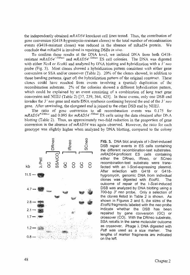
the independently obtained mRAD54 knockout cell lines tested. Thus, the contribution of gene conversion (0418-hygromycin-resistant clones) to the total number of recombination events (0418-resistant clones) was reduced in the absence of mRad54 protein. We conclude that mRad54 is involved in repairing DSBs in vivo.
To confirm these results at the DNA level, we isolated DNA from both 0418-resistant mRAD54+!DRneo and mRAD541DRnea ES cell colonies. The DNA was digested with either Nco I or EcoRI and analyzed by DNA blotting and hybridization with a 3' neo probe (Fig. 3). Most clones showed a hybridization pattern consistent with either gene conversion or SSA and/or crossover (Table 2). 20% of the clones showed, in addition to those banding patterns, (part of) the hybridization pattern of the original construct. These clones could have resulted from events involving a (partial) duplication of the recombination substrate. 2% of the colonies showed a different hybridization pattern, which could be explained by an event consisting of a combination of long tract gene conversion and NHEJ (Table 2) [37, 239, 364, 425]. In these events, only one DSB end invades the 3' neogene and starts DNA synthesis continuing beyond the end of the 3' neo gene. After unwinding, the elongated end is joined to the other DSB end by NHEJ.
The ratio of gene conversion to all recombination events was 0.175 for mRAD54+!DRneo and 0.095 for mRAD541
DRneo ES cells using the data obtained after DNA blotting (Table 2). Thus, an approximately two-fold reduction in the proportion of gene conversion in the absence of mRAD54 was again observed. However, the ratio for each genotype was slightly higher when analyzed by DNA blotting, compared to the colony
0 Q) c
0:: 0
1jj a_ 0
kb ~ <.9
11.5-·
47-· "*'*
2.8---2.1-·
1.7--
1.2-
0.8-
48
0 OJ c
0::
o' 0 :;;: (f) () 0 (f) <.9 ()
-- ••
0 Q) c 0 (f)
() <.9
•
0 0
FIG. 3. DNA blot analysis of 1-Scel-induced DSB repair events in ES cells containing the different recombination-test substrates. mRA054-proficient ES cells conlaining either the DRneo, IRneo, or SCneo recombination-test substrate were transfected with an 1-Scel-expressing plasmid. After selection with G418 or G418-hygromycin, genomic DNA from individual clones was digested with EcoRI. The outcome of repair of the 1-Scel-induced DSB was analyzed by DNA blotting using a 700-bp 3' neo probe. Only a selection of the clones listed in Table 2 is shown. As shown in Figures 2 and 5, the sizes of the EcoRI fragments labeled with the neo probe indicate whether the DSB has been repaired by gene conversion (GC) or crossover (CO). With the DRneo substrate, SSA results in the same molecular outcome as crossover. Phage A DNA digested with Pstl was used as a size marker. The lengths of marker fragments are indicated on the left.
Chapter 2
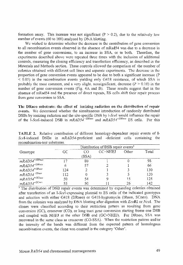
formation assay. This increase was not significant (P > 0.2), due to the relatively low number of events ( 60 to 1 00) analyzed by DNA blotting.
We wished to determine whether the decrease in the contribution of gene conversion to all recombination events observed in the absence of rnRad54 was due to a decrease in the number of gene conversions, to an increase in SSA, or to both. Therefore, the experiments described above were repeated three times with the inclusion of additional controls, measuring the cloning efficiency and transfection efficiency, as described in the Materials and Methods section. These controls allowed the comparison of the number of colonies obtained with different cell lines and separate experiments. The decrease in the proportion of gene conversion events appeared to be due to both a significant increase (P < 0.05) in the recombination events yielding only G418 resistance, of which SSA is probably the most common, and a very slight, nonsignificant, decrease (P > 0.1 0) in the number of gene conversion events (Fig. 4A and B). These results suggest that in the absence of mRad54 and the presence of direct repeats, ES cells shift their repair process from gene conversion to SSA.
Tile DRneo substrate: the effect of ionizing radiation on the distribution of repair events. We determined whether the simultaneous introduction of randomly distributed DSBs by ionizing radiation and the site-specific DSB by I-Scei would influence the repair of the I-Scei-induced DSB in mRAD54+-IDRneo and mRAD5riDRneo ES cells. For this
TABLE 2, Relative contribution of different homology-dependent repair events of IScei-induced DSBs in mRAD54-proficient and -deficient cells containing the recombination-test substrates.
Distribution ofDSB repair eventsa Genotype GC CO GC+NHEJ Other Total
(SSA) mRAD54+ "" 17 80 1 0 98 mRAD54-IDR"w 6 57 2 1 66 mRAD54+!IR"'" 124 2 1 3 130 mRAD54-11R"w 112 0 3 5 120 mRAD54+!SC"" 50 54 9 15 128 mRAD5415c"" 55 50 11 26 142
a The distribution of DSB repair events was determined by expanding colonies obtained after transfection of an l-Scel-expressing plasmid to ES cells of the indicated genotypes and selection with either G418 (DRneo) or G418-hygromycin (!Rneo, SCneo). DNA from the colonies was analyzed by DNA blotting after digestion with EcoRI or Neal. The clones were classified according to their restriction pattern as resulting from gene conversion (GC), crossover (CO), or long tract gene conversion starting frorne one DSB end coupled with NHEJ at the other DSB end (GC+NHEJ). For DRneo, SSA was recovered in the same class as crossover (CO-S SA). VVhen the restriction pattern and/or the intensity of the bands was different from the expected pattern of homologous recombination events, the clone was counted in the category 'Other'.
Mouse RAD54 and chromosomal rearrangements 49
A SSA B GC GC GC GC co
~
~ m
80 c m -§ c _Q u m
~ 60 ~ ·v; = 0 ·v; a. g_ 40 ~
0 ~
m ~ c 20 m a c a
0 0 0 0 0 0 0 0 • • • • • • • 0 0 0 0 0 0 0
"' "' "' " " 0 0 0 0 0
-'. -:- "' "' -:- i -:- + -'. -:-+
Cell line Cell line
FIG. 4. Homologous recombination frequencies for the recombination-test substrates. 1.6x1 06 mRAD54-proficient and -deficient ES cells containing the different recombinationtest substrates (DRneo, IRneo, or SCneo) in the identical genomic location were transfected with pCBA3xnls-I-Scel and grown in G418- and/or hygromycin-containing media. After 8 to 11 days, colonies were fixed, stained, and counted. The number of colonies obtained was normalized for the cloning efficiency and the transfection efficiency (see Materials and Methods). Shown is the normalized number of G418- or G418-hygromycin-resistant colonies ± standard error of the mean for three independent experiments with two cell lines from all six genotypes. (A) Homologous recombination frequency of mRAD54+/DRoeo (+/-)and mRAD54'0R"" (-/-) ES cells. Colonies containing an intact neo gene were obtained after repair of the 1-Scelinduced DSB by SSA, gene conversion (GC), or crossover (CO). (B) Frequencies of gene conversion and crossover events for ES cells containing the recombination-test substrates. For all three recombination-test substrates, neo- and hygcontaining colonies were obtained after repair of the 1-Scel-induced DSB by intrachromatid gene conversion and sister chromatid gene conversion. The IRneo- and SCneo-containing cell lines each have one additional possibility to yield G418-hygromycin-resistant colonies. In the IRneo-containing lines, these clones can be formed by intrachromatid crossover (CO intra). In the SCneo-containing lines, they can be formed by crossover after pairing with the sister chromatid (CO sister).
purpose, mRAD54+!DRneo and mRAD541DRneo cells were irradiated with 2 and 1 Gy of
gamma rays, respectively, either just before or 4 h after transfection ofpCBA3xnls-!-SceL These radiation doses induce 30-100 DSBs per cell and give about 50% survival of the cells [!54, 413]. As a control, unirradiated cells were transfected with pCBA3xnls-I-Scel or pB!uescript II KS. The irradiated cells showed no significant difference in the ratio of G418-hygromycin-resistant colonies to G418-resistant colonies, compared to unirradiated cells transfected with pCBA3xnls-1-Scel (data not shown). Thus, a relatively high level of randomly-introduced DSBs does not saturate one of the DSB repair pathways that lead to G418- and G418-hygromycin-resistant colonies.
50 Chapter 2
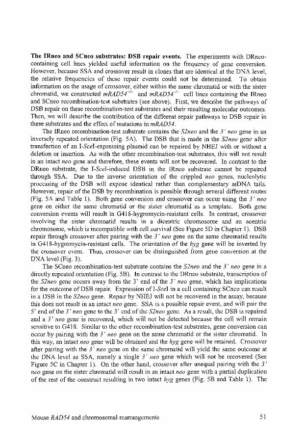
The IRneo and SCneo substrates: DSB repair events. The experiments with DRneocontaining cell lines yielded useful information on the frequency of gene conversion. However, because SSA and crossover result in clones that are identical at the DNA level, the relative frequencies of these repair events could not be determined. To obtain information on the usage of crossover, either within the same chromatid or with the sister chromatid, we constructed mRAD54+!- and mRAD54_1
_ cell lines containing the IRneo and SCneo recombination-test substrates (see above). First, we describe the pathways of DSB repair on these recombination-test substrates and their resulting molecular outcomes. Then, we will describe the contribution of the different repair pathways to DSB repair in these substrates and the effect of mutations in mRAD54.
The IRneo recombination-test substrate contains the S2neo and the 3' neo gene in an inversely repeated orientation (Fig. SA). The DSB that is made in the S2neo gene after transfection of an 1-Scel-expressing plasmid can be repaired by NHEJ with or without a deletion or insertion. As with the other recombination-test substrates, this will not result in an intact neo gene and therefore, these events will not be recovered. In contrast to the DRneo substrate, the 1-Scel-induced DSB in the !Rneo substrate cannot be repaired through SSA. Due to the inverse orientation of the crippled neo genes, nucleolytic processing of the DSB will expose identical rather than complementary ssDNA tails. However, repair of the DSB by recombination is possible through several different routes (Fig. SA and Table 1). Both gene conversion and crossover can occur using the 3' neo gene on either the same chromatid or the sister chromatid as a template. Both gene conversion events will result in G418-hygromycin-resistant cells. In contrast, crossover involving the sister chromatid results in a dicentric chromosome and an acentric chromosome, which is incompatible with cell survival (See Figure SD in Chapter 1). DSB repair through crossover after pairing with the 3' neo gene on the same chromatid results in G418-hygromycin-resistant cells. The orientation of the hyg gene will be inverted by the crossover event. Thus, crossover can be distinguished from gene conversion at the DNA level (Fig. 3).
The SCneo recombination-test substrate contains the S2neo and the 3' neo gene in a directly repeated orientation (Fig. 5B). In contrast to the DRneo substrate, transcription of the S2neo gene occurs away from the 3' end of the 3' neo gene, which has implications for the outcome ofDSB repair. Expression ofi-Scel in a cell containing SCneo can result in a DSB in the S2neo gene. Repair by NHEJ will not be recovered in the assay, because this does not result in an intact neo gene. SSA is a possible repair event, and will pair the 5' end of the 3' neogene to the 3' end of the S2neo gene. As a result, the DSB is repaired and a 3' neo gene is recovered, which will not be detected because the cell will remain sensitive to G418. Similar to the other recombination-test substrates, gene conversion can occur by pairing with the 3' neo gene on the same chromatid or the sister chromatid. In this way, an intact neo gene will be obtained and the hyg gene will be retained. Crossover after pairing with the 3' neo gene on the same chromatid will yield the same outcome at the DNA level as SSA, namely a single 3' neo gene which will not be recovered (See Figure SC in Chapter 1 ). On the other hand, crossover after unequal pairing with the 3' neo gene on the sister chromatid will result in an intact neo gene with a partial duplication of the rest of the construct resulting in two intact hyg genes (Fig. 5B and Table 1). The
Mouse RAD54 and chromosomal rearrangements 51
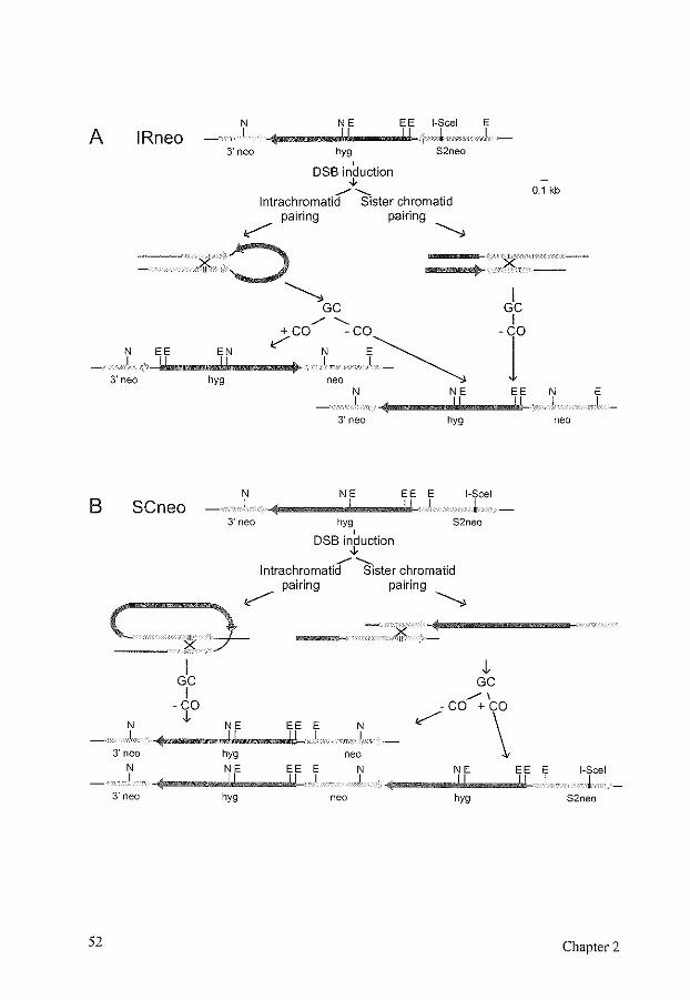
A
B
52
IRneo N I
NE EE 1-Scel E l . I
3' neo hyg S2neo
DSB induction ,. ./----
lntrachromatid Sister chromatid pairing pairing
~GC
·n·· X
I GC
./"- I
~ _E.j,Ei--,_.i,Ej,N,__~~m+~C!>O ~ -C~~ - ClO
3'neo hyg neo .. - ~
0.1 kb
N I
NE EE N E I
SCneo
( *· I
GC I
-CO
N .J,
.I 3' neo
N I
3' neo
N I
3' neo
3' neo
NE EE E I
hyg
1-Scel
l hyg S2neo
' DSB induction ,. ./----lntrachromatid Sister chromatid
pairing pairing
~ ~
.);f:
,j, GC
./I -CO +co
NE EE E N ~ \ I I hyg neo
NE EE E N NE I I
hyg neo hyg
I
neo
EE E 1-Scel I I
S2neo
Chapter 2
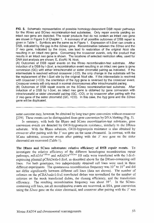
FIG. 5. Schematic representation of possible homology-dependent DSB repair pathways for the IRneo and SCneo recombination-test substrates. Only repair events yielding an intact neo gene are depicted. The repair products that do not contain an intact neo gene are shown in Figure 5 of Chapter 1. A summary of all possible outcomes of DSB repair is given in Table 1. Symbols are the same as in Figure 1. Expression of 1-Scel can create a DSB, indicated by the gap in the S2neo gene. Recombination between the S2neo and the 3' neo gene, indicated by the cross, can lead to restoration of the original Neal site resulting in an intact neo gene. Concerning the crossover events, only the product that results in an intact neo gene is shown. The locations of selected restriction sites, used for DNA blot analysis are shown; E, EcoRI; N, Neal. (A) Outcomes of DSB repair events on the IRneo recombination-test substrate. After induction of a DSB by 1-Scel, a recombination event resulting in an intact neo gene is gene conversion (GC) with either intrachromatid or sister chromatid pairing as indicated. If the intermediate is resolved without crossover (-CO), the only change in the substrate will be the replacement of the 1-Scel site by the original Neal site. If the intermediate is resolved with crossover (+CO), the orientation of the hyg gene is reversed by the crossover event. Crossover events will only result in normal chromosomes after intrachromatid pairing. (B) Outcomes of DSB repair events on the SCneo recombination-test substrate. After induction of a DSB by 1-Scel, an intact neo gene is obtained by gene conversion with intrachromatid or sister chromatid pairing (GC, -CO), or by crossover after pairing with the 3' neogene of the sister chromatid (GC, +CO). In this case, the hyg gene and the S2neo gene will be duplicated.
same outcome may, however, be obtained by long tract gene conversion without crossover [239]. These events can be distinguished from gene conversion by DNA blotting (Fig. 3).
In summary, with both the IRneo and SCneo recombination-test substrates, gene conversion events are detected by G418-hygromycin resistance, similarly to the DRneo substrate. With the !Rneo substrate, G4!8-hygromycin resistance is also obtained by crossover after pairing with the 3' neo gene on the same chromatid. In contrast, with the SCneo substrate, crossover events after pairing with the 3' neo gene on the sister chromatid are recovered (Table 1 ).
The IRneo and SCneo substrates: relative efficiency of DSB repair events. To investigate the relative efficiency of the different homologous recombination repair pathways, mRAD54+!IRneo and mRAD54+!SCnr?o ES cells were transfected v.rith the I-Scelexpressing plasmid pCBA3xnls-I-Scel, as described above for the DRneo-containing cell Jines. For both genotypes, two independently obtained cell Jines were used in three different experiments. The spontaneous recombination frequency was 1 o-5 to 1 o-6 and did not differ significantly between different cell lines (data not shown). The number of colonies on the pCBA3xnls-I-Scel transfected dishes was normalized for the number of colonies on the mock transfected dishes, the cloning efficiency, and the transfection efficiency. The resulting recombination frequency was about 10·2 In the SCneocontaining cell lines, not all recombination events are recovered, as SSA, gene conversion using the S2neo gene on the sister chromatid, and crossover after pairing with the 3' neo
Mouse RAD54 and chromosomal rearrangements 53
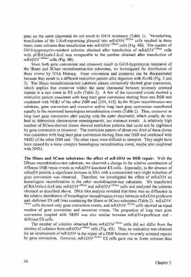
gene on the same chromatid do not result in G418 resistance (Table 1). Nevertheless, transfection of the I-Scel-expressing plasmid into mRAD54+!SCneo cells resulted in three times more colonies than transfection into mRAD54+IIRneo cells (Fig. 4B). The number of G418-hygromycin-resistant colonies obtained after transfection of mRAD54+!IRneo cells with pCBA3xnls-I -Scel was comparable to the number obtained after transfection of mRAD54+!DR'" cells (Fig. 4B).
Since both gene conversion and crossover result in G418-hygromycin resistance of the !Rneo and SCneo recombination-test substrates, we investigated the distribution of these events by DNA blotting. Gene conversion and crossover can be discriminated because they result in a different restriction pattern after digestion with EcoRl (Fig. 5 and 3). The !Rneo recombination-test substrate almost exclusively showed gene conversion, which implies that crossover within the same chromatid between inversely oriented repeats is a rare event in ES cells (Table 2). A few of the recovered events showed a restriction pattern consistent with long tract gene conversion starting from one DSB end combined with NHEJ of the other DSB end [239, 418]. In the SCneo recombination-test substrate, gene conversion and crossover and/or long tract gene conversion contributed equally to the recovered homologous recombination events (Table 2). Thus, crossover or long tract gene conversion after pairing with the sister chromatid, which usually do not lead to deleterious chromosome rearrangements, are common events. A relatively high number of SCneo-derived clones showed restriction patterns that could not be explained by gene conversion or crossover. The restriction pattern of about one third of these clones was consistent with long tract gene conversion starting from one DSB end combined with NHEJ ofthe other DSB end. The other cases were difficult to interpret. They might have been caused by a more complex homologous recombination event, maybe also combined with NHEJ.
The IRneo and SCneo substrates: the effect of mRAD54 on DSB repair. With the DRneo recombination-test substrate, we observed a change in the relative contribution of different DSB repair events in mRAD54 knockout ES cells. Especially, in the absence of mRad54 protein, a significant increase in SSA with a concomitant very slight reduction of gene conversion was observed. Therefore, we investigated the effect of mRAD54 on homologous recombination in the other recombination-test substrates. We transfected pCBA3xnls-I-Scel into mRAD54-11
R"" and mRAD5r5c"'" cells and analyzed the colonies obtained as described above. DNA blot analysis revealed that there was no difference in the relative distribution of homologous recombination events between mRAD54-proficient and -deficient ES cell lines containing the !Rneo or SCneo substrates (Table 2). mRAD54 IIRneo cells showed only gene conversion events, and mRAD541
scneo cells showed an equal number of gene conversion and crossover events. The proportion of long tract gene conversion coupled with NHEJ was also similar between mRAD54-proficient and -deficient ES cells.
The number of colonies obtained from mRAD5411R"" cells did not differ from the
number of colonies from mRAD54'11R"" cells (Fig. 4B). Thus, no indication was obtained for an involvement of mRAD54 in the repair of a DSB between inversely oriented repeats by gene conversion. However, mRADS~SCneo ES cells gave rise to fewer colonies than
54 Chapter 2
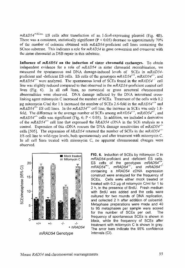
mRAD54+!sc," ES cells after transfection of an I-Scei-expressing plasmid (Fig. 4B). There was a consistent, statistically significant (P < 0.05) decrease to approximately 70% of the number of colonies obtained with mRAD54-proficient cell liues containing the SCneo substrate. This indicates a role for mRAD54 in gene conversion and crossover with the sister chromatid in DSB repair in this substrate.
Influence of mRAD54 on the induction of sister chromatid exchanges. To obtain independent evidence for a role of mRAD54 in sister chromatid recombination, we measured the spontaneous and DNA damage-induced levels of SCEs in mRAD54-proficient and -deficient ES cells. ES cells of the genotypes mRAD54'1', mRAD5~1-, and mRAD54_1
_ were analyzed. The spontaneous level of SCEs found in the mRAD541- cell
line was slightly reduced compared to that observed in the mRAD54-proficient control cell lines (Fig. 6). In all cell lines, no numerical or gross structural chromosomal abnormalities were observed. DNA damage inflicted by the DNA interstrand crosslinking agent mitomycin C increased the number ofSCEs. Treatment of the cells with 0.2 ~g mitomycin C/ml for 1 h increased the number of SCEs 2.6-fold in the mRAD5~1' and mRAD54'1- ES cell lines. In the mRAD54'- cell line, the increase in SCEs was only 1.8-fold. The difference in the average number of SCEs among mRAD54'1', mRAD54+1
-, and mRAD54_1
_ cells was significant (Fig. 6; P < 0.05). In addition, we included a derivative of the mRAD54_1
_ cell line that expressed the hRAD54 eDNA in the SCE analysis as a control. Expression of this eDNA rescues the DNA damage sensitivities of mRAD541
-
cells [505]. The expression of hRAD54 returned the number of SCEs in the mRAD541-
ES cell line to wild-type levels, both spontaneously and after treatment with mitomycin C. In all cell lines treated with mitomycin C, no apparent chromosomal changes were observed.
+I+ +I- -1- -/-+ hRAD54
mRAD54 Genotype
FIG. 6. Induction of SCEs by mitomycin C in mRA054-proficient and -deficient ES cells. ES cells of the genotypes mRAD54'1', mRAD54'1-, mRAD54-I-, and mRA054'containing a hRAD54 eDNA expression construct were analyzed for the frequency of SCEs. Cells were either mock treated or treated with 0.2 !19 of mitomycin C/ml for 1 to 2 h, in the presence of BrdU. Fresh medium with BrdU was added and the cells were cultured for two rounds of DNA replication, and collected 2 h after addition of colcemid. Metaphase preparations were made and 40 to 95 metaphases per sample were scored for the number of SCEs per cell. The frequency of spontaneous SCEs is shown in black, while the frequency of SCEs after treatment with mitomycin C is shown in gray. The error bars indicate the 95% confidence intervals (CI).
Mouse RAD54 and chromosomal rearrangements 55
DISCUSSION
In this study, we have analyzed the repair of a single site-specific DSB located in a repeated DNA sequence in the genome of mouse ES cells. Three recombination-test substrates have been used, all integrated in the same genomic location, but different in the orientation of the repeated sequences, allowing optimal comparison. DSB repair involving homologous recombination was found to be most efficient, when the DSB can be healed through SSA. The second most efficient homology-dependent repair pathway is gene conversion from the sister chromatid. We have determined the effect of the mouse Rad54 protein on homology-dependent DSB repair using these substrates in mRAD54-proficient and -deficient cells. Our data provides the first evidence for involvement of mRad54 in repairing DSBs. The DSB repair phenotype of mRAD541
- cells differs depending on the type of recombination-test substrate. mRAD541
- cells display a hyperrecombination phenotype for DSB repair between directly oriented DNA sequences and no phenotype when the DSB is located between inversely oriented repeats. For a recombination-test substrate that measures repair by gene conversion and crossover using the sister chromatid, a hypo-recombination phenotype is observed. Consistent with this observation, we find that lack of mRAD54 results in less efficient induction of sister chromatid exchanges in response to DNA damage.
A site-specific DSB in mouse ES cells induces homologous recombination. In this study, we have analyzed homologous recombination in mouse ES cells. These cells have two major advantages over other mammalian cell types. First, they are nontransformed and therefore, all DNA damage and cell cycle checkpoints are likely intact [ 440]. Second, efficient protocols for gene targeting in these cells have been developed. This allows the generation of series of isogenic cell lines, which differ solely in defined genetic loci.
The analysis of homologous recombination after induction of a site-specific DSB between repeated DNA sequences requires a low level of spontaneous recombination between the repeats. We find that the spontaneous frequency of recombination in mouse ES cells is in the order of 10-5 to 10·6 for all three recombination-test substrates used in this study. Similar frequencies of spontaneous recombination between directly oriented repeats have been observed in Chinese hamster ovary (CHO) cells [240, 301, 376]. Induction ofi-Scei expression in the ES cells containing the recombination-test substrates increases the frequency of homologous recombination at least 100- to 1000-fold. Similar increases in DSB-induced recombination have been observed in a number of different cell lines including ES cells [150, 240, 301, 364, 426, 447, 508]. In this study, we find that, after correction for cloning and transfection efficiencies, 3.6 to 4.8% of the DRneocontaining cells undergo homologous recombination upon DSB induction (Fig. 4). This percentage of cells undergoing homologous recombination is still an underestimate, because not all transfected cells will receive a DSB [426]. Furthermore, homologous recombination using the S2neo gene on the sister chromatid will not be detected. The contribution of homology-dependent processes towards DSB repair can be as high as the one by DNA end-joining, as was recently demonstrated using the DRneo and SCneo substrates randomly integrated in the genome of CHO cells [240, 300].
56 Chapter 2
The major homology-dependent DSB repair pathway for the Dllneo substrate is SSA. Using the DRneo substrate, a distinction can be made between DSB repair through SSA and crossover on one hand and gene conversion on the other hand (Fig. 2). Our results show that approximately 15% of all homology-dependent DSB repair events within the DRneo substrate in mouse ES cells occur through recombination between the directly oriented repeats via gene conversion. These results appear not to be specific for mouse ES cells, because 25% of the DSB repair events in CHO cells containing the integrated DRneo substrate occur through gene conversion [300]. The separate contributions of SSA and crossover to DSB repair cannot be determined with the DRneo substrate. However, a comparison to the results with the SCneo substrate, which also contains directly oriented repeats, suggests that gene conversion and crossover occur at similar frequencies (Table 2). This implies that SSA accounts for 70% of aU homologydependent DSB repair events within the DRneo substrate in mouse ES cells.
Consistent with our results, recombination between directly repeated sequences through gene conversion, when compared to SSA, accounts for the minority of detected events in a number of other assay systems, including DSB-induced events on plasmids and in chromosomes in S. cerevisiae and vertebrate cells [164, 234, 300, 377, 437]. However, there are a number of exceptions, both in S. cerevisiae and mammalian cells, in which gene conversion accounts for the majority of spontaneous and DSB-induced events [51, 184, 376, 420, 508]. For example, after induction of a DSB with 1-Scel between directly oriented repeats containing restriction fragment length polymorphisms, spaced by 100 bp, more than 90% of the DSB repair events in CHO cells are gene conversions [508]. This shift in repair pathways could be due to the heterology in the repeats. Other variables that might contribute to observed differences among assay systems include the length and sequence context of the repeats, the distance between the repeats, the position of the DSB and heterology at the ends [164, 184, 377, 385]. Both gene conversion and SSA use 3' ssDNA tails as intermediates. Gene conversion requires search for homology followed by joint molecule formation actively mediated by Rad51-coated ssDNA. On the other hand, SSA involves the more passive process of annealing of complementary single strands, although it does also, at least partially, depend on Rad52. Depending on the presence of nonhomologous ends and the length and sequence context of the repeat, these two processes might be affected differentially. Finally, the distribution of repair events may also be dependent on the stage of the cell cycle, as gene conversion using the sister chromatid is only possible inS and 02.
DSB repair associated with DNA crossovers occurs mostly from the sister chromatid. DSB repair products of both the !Rneo and SCneo recombination-test substrates differ depending on whether they have been generated through gene conversion or crossover (Fig. 5). While the !Rneo substrate detects intrachromatid crossovers, the SCneo substrate detects unequal crossovers between sister chromatids. It should be noted, however, that if a long tract of DNA is synthesized during gene conversion, the result appears similar to a crossover. With the IRneo substrate, DSB repair through crossover occurs only in 1.5% of the analyzed repair events, while gene conversion accounts for over 95% of the events
Mouse RAD54 and chromosomal rearrangements 57
(Table 2). This also indicates that gene conversion tracts are generally shorter than 2.7 kb, because otherwise, the outcome would have been scored as a crossover. In contrast to the lack of crossovers with IRneo, we find that gene conversion and crossover contribute equally to DSB repair using SCneo. Thus, it appears that crossovers preferably arise when the sister chromatid, instead of a homologous sequence on the same chromatid, is used as the repair template. A similar preference for crossover events using the sister chromatid has been observed in mouse L cells during spontaneous recombination between repeated sequences [51]. A somewhat lower contribution of crossovers using SCneo has been found in CHO cells after induction of a DSB [239]. In addition, evidence has been obtained for a restriction of gene conversion to the S and G2 phases of the cell cycle after induction of a DSB in the DRneo substrate in CHO cells, again indicating a preference for pairing with the sister chromatid to perform gene conversion [300]. In contrast, in S. cerevisiae a preference for intrachromatid interactions has been found, as indicated by a low percentage of crossover events in an SCneo-like substrate [262]. A high percentage of crossovers after intrachromatid interactions has been found in the repair of an induced DSB on a plasmid or spontaneous DSBs on chromosomes using inversely oriented repeats inS. cerevisiae [3, 419,437, 497].
Using the SCneo substrate in CHO cells, the nature of the crossover events after induction of a DSB has been investigated [239]. All crossover events in these cells appeared to be due to long tract gene conversion without crossover, as the reciprocal product that should be formed during crossover could not be found in unselected colonies, while the second product of long tract gene conversion, an unchanged SCneo substrate, was found (See Figure 5 in Chapter 1 ). Due to our selection procedures, we have not been able to discriminate between actual crossover and long tract gene conversion. Long tract gene conversion requires a gene conversion tract length of 3.1 kb in the SCneo substrate, which contrasts with the shorter gene conversion tract lengths we have found for the !Rneo substrate in ES cells. Half of the long tract gene conversion events found in CHO cells are actually the result of gene conversion starting from one DSB end and NHEJ of the other DSB end [239]. In our study, these combined recombination and NHEJ events are detected as a separate class on DNA blots, and constitute about 7% of all analyzed repair events, while crossovers constitute 40%. If these combined events also accounted for half of the long tract gene conversion events in ES cells, 33% of all events would still be due to actual crossovers after pairing with the sister chromatid.
The preference for sister chromatid interactions during homologous recombination in mammalian cells could have arisen because sister chromatid recombination is, in general, less prone to the generation of chromosomal rearrangements than intrachromatid recombination. A significant fraction of mammalian genomes consists of repetitive DNA sequences. Crossovers between these sequences will result in deleterious chromosomal rearrangements, except when the same sequence on the sister chromatid is used. Evidence thus far suggests that genome rearrangements are indeed suppressed during recombination between sequence repeats on nonhomologous chromosomes [ 425, 426]. Furthermore, the presence of mismatches between the repeats also prevents recombination, due to the mismatch repair system [ 150, 518]. S. cerevisiae contains hardly any repetitive sequences and will undergo less selection against allowing intrachromatid recombination. The
58 Chapter 2
preference for the sister chromatid in mammalian cells might occasionally result in unequal crossover events between sister chromatids, but if those events occur in a limited region, their potential deleterious effects could be minimized.
Comparison of the frequency of DSB repair events on the different substrates. We find that the frequency of gene conversion is comparable among the different recombination-test substrates. The estimated frequency of gene conversion between intrachromosomal repeats after induction of a DSB is 5.9 x 10·3 for DRneo, 6.6 x 10·3 for SCneo and 5.1 x 10·3 for IRneo (Table 2 and Figure 4). The frequency of crossover events for DRneo cannot be determined due to the occurrence of SSA events. With IRneo, we hardly find any crossover events compared to SCneo. It seems reasonable to assume that with all three recombination-test substrates, a similar fraction of the cells receives a DSB and that a similar fraction of these DSBs is channeled into a homology-dependent repair pathway. Therefore, it is striking that with the IRneo substrate, the number of events involving gene conversion is not higher than with the DRneo or SCneo substrate. Repair by SSA will result in correct DSB repair in cells containing DRneo and SCneo, but cells containing IRneo that attempt SSA will fail to repair the DSB. Apparently, these cells will not undergo gene conversion or crossover, but instead, the homologous repair event will be aborted and resolved by another pathway not resulting in an intact neo gene, or it will not be resolved at all and result in cell death. Cells that repair their DSB by gene conversion using the sister chromatid, which is the predominant route for recombination, can resolve the intermediate without a crossover in all three recombination-test substrates. However, if a cell attempts to resolve the intermediate by crossover in the IRneo substrate, this will result in chromosomal rearrangements and cell death. It seems that either finding the homologous repair template on the sister chromatid is more difficult with the IRneo substrate or cells containing the inversely oriented repeat fail to prevent crossovers with the sister chromatid and die. Othen:vise, more gene conversion events should have been recovered with the IRneo substrate. In S. cerevisiae. induction of a DSB by HOendonuclease in directly or inversely oriented repeats on a plasmid, results in a higher loss of the plasmid containing the inversely oriented repeats [437]. In mouse L cells containing thymidine kinase repeats in a direct or inverted orientation integrated in the chromosome, the frequency of spontaneous recombination is three-fold lower for the repeats in inverted orientation [50]. These results also indicate that recombinational repair between inversely oriented repeats is less efficient.
mRad54 influences the repair of DSBs in the DRneo recombination-test substrate. A role for the mRad54 protein in the repair of DSBs has been postulated based on the ionizing radiation sensitivity and homologous recombination deficiency of mRAD541
- ES cells [154]. The results of our study provide direct evidence that mRad54 is involved in DSB repair in vivo. The difference in DSB repair between mRAD54-proficient and -deficient cells is most clearly seen when the DSB is induced between directly oriented repeated sequences, as is the case with the DRneo and SCneo recombination-test substrates (Fig. 4A and B). The absence ofmRad54 causes a very slight reduction in gene conversion during DSB repair of the DRneo substrate. This reduction is accompanied by
Mouse RAD54 and chromosomal rearrangements 59
a statistically significant increase in the number of crossovers and SSA, the latter of which is the most frequent. In S. cerevisiae, a similar increase in homologous recombination is seen in rad54 mutants, both with directly oriented repeats on plasmids and in chromosomes [226, 303, 465]. The frequency of SSA (or crossover) is 2- to 27-fold higher in rad54 cells than in wild-type cells [303, 465]. With an HO-endonucleaseinduced DSB between chromosomal, directly oriented repeats, a decreased survival of rad54 mutant cells has been found compared to wild-type cells. Although this indicates a deficiency in DSB repair, the number of SSA events increased [226]. In addition to the increase in SSA events, a decrease in both spontaneous and DSB-induced gene conversions has been observed [226, 303, 465]. These results suggest that there might be a competition between SSA and gene conversion (see below).
mRad54 influences recombination between sister chromatids. In cells containing the SCneo recombination-test substrate, the effect of mRAD54 on gene conversion is more pronounced than in cells containing the DRneo substrate. Repair of the DSB through SSA is possible in SCneo, although those events are not detected, because they do not yield an intact neo gene. A statistically significant 27% decrease in the frequency of gene conversion and crossover and/or long tract gene conversion is observed in the absence of mRAD54 (Fig. 4B). Since all crossover events take place after pairing with the sister chromatid, mRAD54 is clearly involved in sister chromatid recombination. This is also the case for S. cerevisiae RAD54 [10]. The contribution of gene conversion and crossover remains about equal in mRAD541
- cells compared to mRAD54+!- cells, which indicates that mRAD54 is involved in both gene conversion and crossover recombination events (Table 2). In contrast to these results, S. cerevisiae RAD54 appears to be mainly involved in gene conversion, although this has been investigated only with inversely oriented repeats [ 419].
Crossovers resulting in restoration of the neo gene in the SCneo recombination-test substrate are the consequence of interactions with the sister chromatid and result in SCEs at the chromosomal level. Therefore, our results with the SCneo substrate predict a reduction in the level of SCEs in the absence of mRAD54. Indeed, we find a slight~ lower level of spontaneous SCEs in mRAD541
- ES cells compared to that in mRAD54+ +
ES cells (Fig. 6). Because SCEs are induced by DNA-damaging agents, we have also tested whether mRAD541
- ES cells respond differently to mitomycin C treatment in the SCE assay than mRAD54+!+ or mRAD54+1
- cells. Treatment of mRAD541- cells with
mitomycin C yields a 1.5-fold lower induction of SCEs, compared to mRAD54+t+ cells (Fig. 6). This effect of RAD54 on spontaneous and DNA damage-induced SCEs corresponds to results obtained with chicken-derived cells, in which a reduction in the frequency of SCEs in RAD54- and RAD51-deficient chicken B-lymphocytes is observed [482]. From these results, we conclude that genes required for homologous recombination are also involved in promoting SCEs. The decrease in SCEs induced by DNA damage corresponds to the similar decrease in the number of crossover events during DSB repair on the SCneo recombination-test substrate.
The observation that mRAD54 influences DNA damage-induced SCEs adds significantly to our results with the recombination-test substrates. The results of the SCE
60 Chapter 2
experiments show that mRAD54 is involved in homology-dependent DNA repair of DNA damages that are present in naturally occurring genomic sequences. The SCE experiments overcome two restrictions of the experiments with recombination-test substrates. First, the restriction enzyme-induced DSB that initiates repair in the experiments involving the recombination-test substrates might be recognized differently from other types of DNA damages, including DSBs introduced by ionizing radiation or DNA interstrand crosslinking agents. Second, in the experiments involving the recombination-test substrates, the introduction of repeated DNA sequences is necessary in order to select for successful DSB repair events. However, the presence of these repeated sequences -will influence the distribution of observed repair events. SSA relies especially on the presence of repeated sequences and will be used less frequently in a more physiological situation,
The absence of mRad54 has no influence on recombination within the IRneo substrate. We find no change in the frequency of gene conversion after induction of a DSB in the !Rneo substrate in mRAD54-deficient cells compared to that in mRAD54-proficient cells (Fig. 4B), With the !Rneo recombination-test substrate, the induced DSB can be repaired by gene conversion involving either the same chromatid or the sister chromatid, or by crossover after pairing with the 3' neo gene on the same chromatid (Fig. SA). The latter events are rare in mRAD54--r-IIRneo cells and have not been detected in mRAD5411
R"" cells (Table 2), Similar to our results with chromosomal substrates in mouse ES cells, disruption of RAD54 in S. cerevisiae has no effect on the repair of an induced DSB in inversely oriented repeats located on a plasmid [226], In contrast, the rate of spontaneous gene conversion between chromosomal, inversely oriented repeats is decreased 25-fold in a rad54 S, cerevisiae strain [419], The frequency of gene conversion is severely reduced, while events involving crossovers are relatively less affected. The difference between the results obtained with rad54 S cerevisiae and mRAD54_1
_ ES cells might be due to a difference between spontaneous and DSB-induced recombination events. Furthermore, because S. cerevisiae cells display a different distribution of events, with a predominance of crossovers during intrachromatid recombination, a direct comparison between S cerevisiae rad54 and mRAD541
- ES cells is difficult, The lack of an effect of mRAD54 on DSB repair between inversely oriented repeats in mouse ES cells also contrasts with the effects of mRAD54 on DSB repair between directly oriented repeats (Fig, 4), As we will discuss below, this could be due to the possibility to repair a DSB in directly oriented repeats by SSA, which is not possible in inversely oriented repeats,
Does mRad54 promote gene conversion at the expense of SSA? ssDNA tails are formed as a common intermediate in SSA and gene conversion with or without crossover. Because of this common intermediate, it is likely that a certain degree of competition exists between these two pathways [164]. rnRad54 could have a role in promoting gene conversion, either directly or indirectly by blocking DSBs from being processed through the SSA pathway, This would explain the increase in the number of 0418-resistant colonies in mRAD54-deficient cells with DRneo, which results from an increase in SSA. It would also explain the decrease in 0418-hygromycin-resistant colonies with SCneo,
Mouse RAD54 and chromosomal rearrangements 61
because an increase in SSA, which is not recovered, would cause a decrease in the recovered gene conversion events. With IRneo, SSA is not possible, and therefore, lack of rnRad54 would not have any effect on DSB repair in this substrate. Manunalian chromosomes contain a significant amount of repetitive sequences that could be used to repair a DSB by SSA, thereby resulting in deletions. Inhibition of SSA by rnRad54 is therefore even more relevant in mammalian cells than in S. cerevisiae, where similar effects of Rad54 on SSA have been found. Direct stimulation of gene conversion pathways by rnRad54, possibly by its interaction with rnRad51, would decrease the contribution of SSA to DSB repair. It has been shown that the purified human and S. cerevisiae Rad54 proteins have ATP-dependent DNA supercoiling activity [403, 513]. This activity would be ideally suited for the the stimulation of mRad51-mediated homologous DNA pairing and strand exchange (See Chapter I) [ 400].
ACKNOWLEDGMENTS
We thank M. de Bruijn for technical support and J. Essers for mRAD54+!- cells. This work was supported by grants from the Netherlands Organization for Scientific Research (NWO), the Dutch Cancer Society (KWF), and the Human Frontier Science Program Organization. R.K. was a fellow of the Royal Netherlands Academy of Arts and Sciences.
62 Chapter 2
CHAPTER3
Preliminary characterization ofthe mouse Rad54B protein

PRELIMINARY CHARACTERIZATION OF THE MOUSE RAD54B PROTEIN
MIES L.G. DRONKERT1, HANNY ODIJK1
, JEAN-MARIE BUERSTEDDE2, OLGA
BEZZUBOVA2, ROLAND KANAARt.3
1 Department of Cell Biology and Genetics, Erasmus University Rotterdam, PO Box 1738, 3000 DR Rotterdam, The Netherlands
2 Heinrich-Pette-Institute, 20251 Hamburg, Germany
3 Department of Radiation Oncology, University Hospital Rotterdam, The Netherlands
Homologous recombination is one of the important pathways to repair DNA double-strand breaks. Central proteins involved are Rad51 and Rad54, which are conserved from yeast to human. Rad51 can perform DNA strand exchange, stimulated by Rad54 via a direct interaction. Upon ionizing radiation, colocalizing nuclear foci are formed by Rad51 and Rad54. In mammals, a second sequence homolog of Rad54 is present, called Rad54B. Mouse embryonic stem cells deficient for both mouse RAD54 (mRAD54) and mRAD54B, are more sensitive to ionizing radiation than mRAD54-deficient cells, suggesting a role for mRad54B in DSB repair, similar to mRad54. In this study, we show that mRad54B resides in the nucleus. In contrast to mRad54, we could not observe foci formation of mRad54B coupled to Yellow Fluorescent Protein (YFP) upon treatment with ionizing radiation. We performed immunoprecipitations from Chinese hamster ovary cells transfected with YFP-mRAD54B-Flag and from reticulocyte lysates containing in vitro translated hRAD51 and mRAD54B(-Flag) using a-hRad51, C<-mRad54B, and a-Flag antibodies. We observed an association between Rad51 and Rad54B both without treatment and after ionizing radiation, although at a low efficiency. Furthermore, we demonstrated homomeric association ofmRad54B, mediated by the carboxy-terminal 140 amino acids. These results and the conservation betvveen mRad54 and mRad54B suggest a role for mRad54B in homologous recombination either together with mRad54 or in a different subpathway.
INTRODUCTION
Homologous recombination is one of the important pathways for the repair of DNA double-strand breaks (DSBs) that is conserved from Saccharomyces cerevisiae to human (See Chapter 1). This pathway is essentially error-tree by using homologous DNA as a template for DSB repair. Two of the central proteins in homologous recombination both in yeast and mammalian cells are Rad51 and Rad54 [253, 386]. In vertebrate cells, deletion of RAD51 is lethal, most likely due to defective repair ofDSBs that occur during DNA replication [114, 165, 198, 268, 305, 325, 434, 481, 536]. Deletion of vertebrate RAD54 makes cells sensitive to ionizing radiation, reduces the frequency of homologous recombination, and alters DSB repair, but is not lethal (Chapter 2) [40, 144, 154]. In vitro, Rad51 can form a nucleoprotein filament on a single-stranded DNA (ssDNA)
Mouse Rad54B protein characterization 65
molecule and perform strand exchange using a homologous double-stranded DNA (dsDNA) molecule [27, 338, 472, 501, 504]. Human Rad54 (hRad54) is a dsDNAdependent ATPase that can induce supercoiling into DNA [430, 505, 513]. In vitro, it can bind to hRad51 on nucleoprotein filaments [185]. In vivo, binding of mouse Rad54 (mRad54) to mRad51 is observed only after induction of DSBs [513]. Yeast Rad54 (ScRad54) stimulates DNA strand exchange by ScRad51, possibly through its supercoiling ability [332, 400,403, 546].
The close cooperation between Rad51 and Rad54 in DSB repair identified biochemically, is also found at the cell biological level. The mammalian Rad51 and Rad54 proteins cannot be detected by immunofluorescence in untreated cells [513]. However, the majority of hRad51 fused to the green fluorescence protein (GFP) is localized in the cytosol in transfected Chinese hamster ovary (CHO) cells. In contrast, GFP-hRad54 is found in the nucleus (J. Essers, personal communication). After the induction of DSBs by ionizing radiation, mammalian Rad51 and Rad54 molecules form nuclear foci, that colocalize [513]. Rad51 foci can also be observed in S phase cells, probably due to DSB repair during replication [299, 515, 516].
Both in S. cerevisiae and in mammalian cells, a second homolog of RAD54 has been identified. The yeast homolog, called ScRDH54 or ScTIDJ, fimctions in different subpathways of homologous recombination compared to ScRAD54, although the function of ScRad54 and ScRdh54 is partially redundant [264, 402, 464, 465, 471]. The mammalian RAD54 homolog is called RAD54B [212]. 50% of the amino acids of mRad54B and mRad54 are similar. mRad54B is more homologous to ScRad54 than to ScRdh54. Mice deficient for mRAD54B are viable and show no gross abnormalities, similar to mRAD54-deficient mice (J.-M. Buerstedde, personal communication in [153]). The ionizing radiation sensitivity of mouse embryonic stem cells that are deficient for mRAD54B is between the sensitivity of wild-type and mRAD54-deficient cells (A. Wesoly, personal communication). Cells deficient for both mRAD54 and mRAD54B are viable but more sensitive to ionizing radiation than either mRAD54-deficient or mRAD54B-deficient cells (J. Essers and A. Wesoly, personal communication). Chicken DT40 cells, deficient for RAD54B, have a minor defect in homologous recombination [358].
In this study, we have investigated the cellular localization of mRad54B both in untreated cells and in cells treated with ionizing radiation. Furthermore, we have demonstrated that mRad54B and hRad51 interact by immunoprecipitation from CHO cell extracts and from reticulocyte lysates containing in vitro translated proteins. By the same methods, we have observed homomeric (di- or polymeric) interaction of mRad54B and determined the domain responsible for this interaction.
MATERIALS AND METHODS
eDNA constructs. The full length mRad54B eDNA was cloned using four overlapping PCR fragments. The following tags were added by PCR: an amino-terminal histidine tag (His6: single letter amino acid code: MGHHHHHHGGI) and a carboxy-terminal Flag tag
66 Chapter 3
(DYKDDDDK). All PCR fragments were sequenced The His-mRAD54B-Flag eDNA was subcloned into pEYFPCl (Yellow Fluorescent Protein, Clontech), yielding YFPmRAD54B-Flag. The constructs were subcloned into the appropiate vectors for transfection and in vitro transcription and translation (IVTT).
Generation of a-mRad54B antibodies. 390 bp encoding the carboxy terminus of mRad54B were subcloned into pRP261, derived from pGex2T. The fusion protein, mRad54B-Cterm, derived from this plasmid, contained amino acid 754 to 884 of mRad54B fused to a glutathione S-transferase tag. Similarly, a 386-bp Nhei-Pstl fragment was subcloned in pMalC2 and yielded a fusion protein containing amino acid 100 to 229 fused to maltose binding protein. The fusion proteins were produced in E. coli strain BL21-codon+. The proteins were purified from the insoluble fraction by preparative SDS-polyacrylamide gel electrophoresis (PAGE) and used to immunize two rabbits. The antibody directed against the carboxy terminus is called o:-mRad54B-Cterm, the antibody against amino acid I 00 to 229 a-mRad54B-Nterm. The antibodies did not cross-react with mRad54 on immunoblots (data not shown).
Transfection of mRAD54B into CH09 cells. YFP-mRAD54B-Flag was transfected using lipofectin into CH09 cells, grown in FlO-DMEM containing 10% fetal calf serum. To obtain stable transfectants, cells were split after one day and put on G418 selection (1 mg/ml). Single colonies containing YFP-mRad54B-Flag protein were expanded. The cellular localization of the YFP signal was determined using an Olympus IX70 microscope. The expression of full-length fusion protein was investigated with o:-YFP, o:mRad54B-Cterm and a-Flag antibodies on immunoblots of cell extracts from transfected CH09 cells, using untransfected cells as a control.
Immunofluorescence. Cells were grown on glass slides for two days and treated with 12 Gy of gamma irradiation or left untreated. Two to six hours after treatment, the slides were fixed in 2% paraformaldehyde and washed twice with 0.1% Triton-X 100 in phosphate-buffered saline. They were incubated with o:-hRad51, a-Flag (M2, Sigma), or a-mRad54B for 90 min, washed again with 0.1% Triton-X 100 and incubated for 90 min with the secondary antibodies (Alexa, Molecular probes). Nuclei were counterstained with 4' ,6-diamidino-2-phenylindole.
IVTT and immunoprecipitation. In vitro transcription and translation of pcDNAmRAD54B, pcDNA-His-mRAD54B-FLAG, pRSET-His-mRAD54B-Cterm, pcDNAhRAD51 and pZeo-His-hRad54-HA in the presence of 35S methionine produced the corresponding mRad54B, hRad51 and hRad54 proteins (Promega TnT T7 coupled reticulocyte lysate system). The antibodies used for immunoprecipitation were amRad54B-Cterm, a-Flag, o:-hRad51, and a-HA (3Fl0, Roche). Interactions of the in vitro translated proteins were investigated by mixing 5 ~1 of reticulocyte lysates containing the proteins with 5 ~1 NETT buffer (100 mM NaCI, 5 mM EDTA, 50 mM Tris-HCI (pH 8.0), 0.5% Triton-X 100) and incubating for 1 hat 30'C. 5 ~I antibody was
Mouse Rad54B protein characterization 67
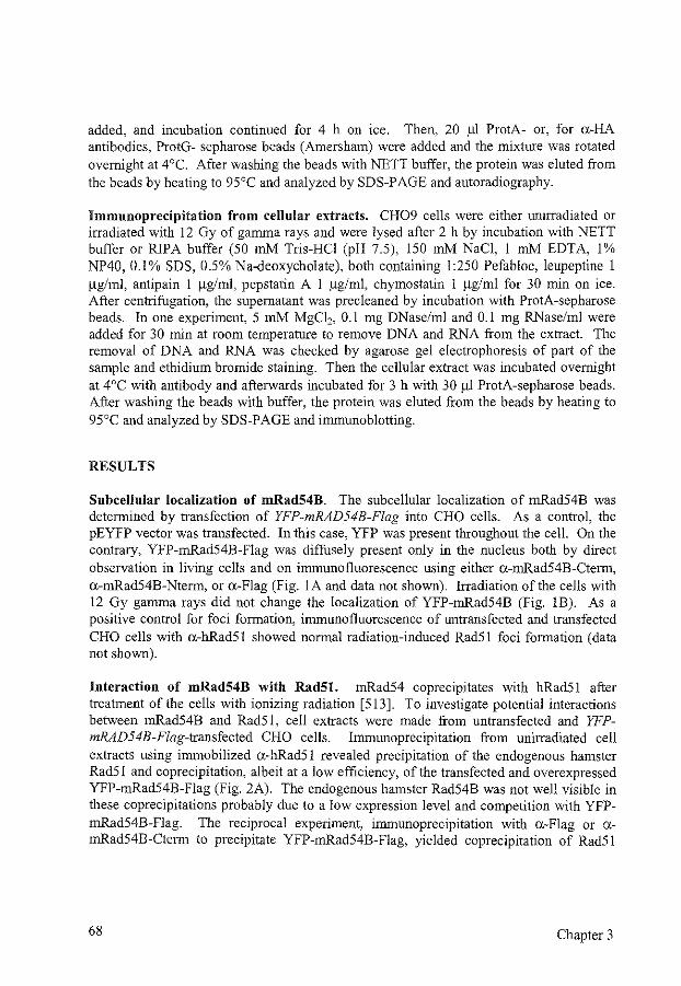
added, and incubation continued for 4 h on ice, Then, 20 fll ProtA- or, for a-HA antibodies, ProtG- sepharose beads (Amersham) were added and the mixture was rotated overnight at 4°C, After washing the beads with NETT buffer, the protein was eluted from the beads by heating to 95°C and analyzed by SDS-PAGE and autoradiography,
Immunoprecipitation from cellular extracts. CH09 cells were either unirradiated or irradiated with 12 Gy of gamma rays and were lysed after 2 h by incubation with NETT buffer or RJPA buffer (50 mM Tris-HCl (pH 7,5), 150 mM NaCl, 1 mM EDTA, 1% NP40, 0,)% SDS, 0,5% Na-deoxycholate), both containing 1:250 Pefabloc, leupeptine 1 !lgiml, antipain 1 !lgiml, pepstatin A 1 !lgiml, chymostatin 1 !lgiml for 30 min on ice. After centrifugation, the supernatant was precleaned by incubation with ProtA-sepharose beads. In one experiment, 5 mM MgC12, 0.1 mg DNase/ml and 0.1 mg RNase/ml were added for 30 min at room temperature to remove DNA and RNA from the extract. The removal of DNA and RNA was checked by agarose gel electrophoresis of part of the sample and ethidium bromide staining. Then the cellular extract was incubated overnight at 4°C with antibody and afterwards incubated for 3 h with 30 111 ProtA-sepharose beads. After washing the beads with buffer, the protein was eluted from the beads by heating to 95'C and analyzed by SDS-PAGE and immunoblotting.
RESULTS
Subcellular localization of mRad54B. The subcellular localization of mRad54B was determined by transfection of YFP-mRAD54B-Flag into CHO cells. As a control, the pEYFP vector was transfected. In this case, YFP was present throughout the cell. On the contrary, YFP-mRad54B-Flag was diffusely present only in the nucleus both by direct observation in living cells and on immunofluorescence using either o:-rnRad54B-Cterm, a-mRad54B-Nterm, or a-Flag (Fig. !A and data not shown). Irradiation of the cells with 12 Gy gamma rays did not change the localization of YFP-mRad54B (Fig. !B). As a positive control for foci formation, immunofluorescence of untransfected and transfected CHO cells with a-hRad51 showed normal radiation-induced Rad51 foci formation (data not shown).
Interaction of mRad54B with Rad51. mRad54 coprecipitates with hRad51 after treatment of the cells with ionizing radiation [513]. To investigate potential interactions between mRad54B and Rad51, cell extracts were made from untransfected and YFPmRAD54B-Flag-transfected CHO cells. Immunoprecipitation from unirradiated cell extracts using immobilized a-hRad51 revealed precipitation of the endogenous hamster Rad51 and coprecipitation, albeit at a low efficiency, of the transfected and overexpressed YFP-mRad54B-Flag (Fig. 2A). The endogenous hamster Rad54B was not well visible in these coprecipitations probably due to a low expression level and competition with YFPmRad54B-Flag. The reciprocal experiment, immunoprecipitation with a-Flag or amRad54B-Cterm to precipitate YFP-mRad54B-Flag, yielded coprecipitation of Rad51
68 Chapter 3
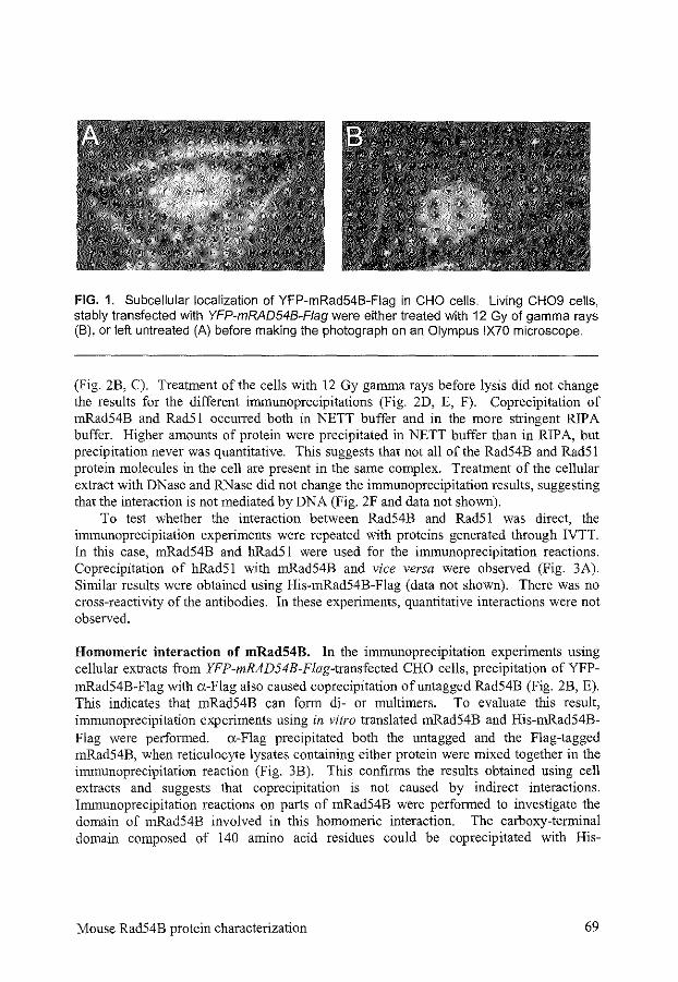
FIG. 1. Subcellular localization of YFP-mRad54B-Fiag in CHO cells. Living CH09 cells, stably transfected with YFP-mRAD54B-Fiag were either treated with 12 Gy of gamma rays (B), or left untreated (A) before making the photograph on an Olympus IX70 microscope.
(Fig. 2B, C). Treatment of the cells with 12 Gy gannna rays before lysis did not change the results for the different innnunoprecipitations (Fig. 2D, E, F). Coprecipitation of mRad54B and Rad51 occurred both in NETT buffer and in the more stringent RIP A buffer. Higher amounts of protein were precipitated in NETT buffer than in RIPA, but precipitation never was quantitative. This suggests that not all of the Rad54B and Rad51 protein molecules in the cell are present in the same complex. Treatment of the cellular extract with DNase and RNase did not change the immunoprecipitation results, suggesting that the interaction is not mediated by DNA (Fig. 2F and data not shown).
To test whether the interaction between Rad54B and Rad51 was direct, the innnunoprecipitation experiments were repeated with proteins generated through IVTT. In this case, mRad54B and hRad51 were used for the innnunoprecipitation reactions. Coprecipitation of hRad51 with mRad54B and vice versa were observed (Fig. 3A). Similar results were obtained using His-mRad54B-Flag (data not shown). There was no cross-reactivity of the antibodies. In these experiments, quantitative interactions were not observed.
Homomeric interaction of mRad54B. In the immunoprecipitation experiments using cellular extracts from YFP-mRAD54B-Flag-transfected CHO cells, precipitation of YFPmRad54B-Flag with a-Flag also caused coprecipitation ofuntagged Rad54B (Fig. 2B, E). This indicates that mRad54B can form di- or multimers. To evaluate this result, innnunoprecipitation experiments using in vitro translated mRad54B and His-mRad54BFlag were performed. a-Flag precipitated both the untagged and the Flag-tagged mRad54B, when reticulocyte lysates containing either protein were mixed together in the innnunoprecipitation reaction (Fig. 3B). This confirms the results obtained using cell extracts and suggests that coprecipitation is not caused by indirect interactions. Irnmunoprecipitation reactions on parts of mRad54B were performed to investigate the domain of mRad54B involved in this homomeric interaction. The carboxy-terminal domain composed of 140 amino acid residues could be coprecipitated with His-
Mouse Rad54B protein characterization 69
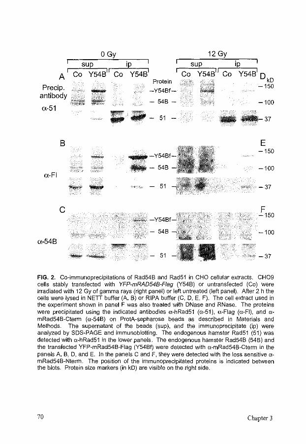
I A
Precip. antibody
a-51
8
a-FI
c
a-54B
OGy 12 Gy
ip sup ip sup Co Y54B
11 Co Y54B
1 Co Y54B
11 Co Y54B
1 D
kD -150
-
-
Protein
-Y54Bf-
54B-
II¢ ;;; - 51 -
·"'1!111!111111
--548
- 51
-Y54Bf-'·: _x:;
548 ~~~~,
-100
---37
E -150
-100
-37
F -150
-100
-37
FIG. 2. Co-immunoprecipitations of Rad54B and Rad51 in CHO cellular extracts. CH09 cells stably transfected with YFP-mRAD548-Fiag (Y54B) or untransfected (Co) were irradiated with 12 Gy of gamma rays (right panel) or left untreated (left panel). After 2 h the cells were lysed in NETT buffer (A, B) or RIP A buffer (C. D. E, F). The cell extract used in the experiment shown in panel F was also treated with DNase and RNase. The proteins were precipitated using the indicated antibodies o:-hRad51 (o:-51 ), a-Flag (o:-FI), and o:mRad54B-Cterm (o:-548) on ProtA-sepharose beads as described in Materials and Methods. The supernatant of the beads (sup), and the immunoprecipitate (ip) were analyzed by SDS-PAGE and immunoblolting. The endogenous hamster Rad51 (51) was detected with o:-hRad51 in the lower panels. The endogenous hamster Rad54B (54B) and the transfected YFP-mRad54B-Fiag (Y54Bf) were detected with o:-mRad54B-Cterm in the panels A, B, D, and E. In the panels C and F, they were detected with the less sensitive o:mRad54B-Nterm. The position of the immunoprecipitated proteins is indicated between the blots. Protein size markers (in kD) are visible on the right side.
70 Chapter 3
mRad54B-Flag using a-Flag (Fig. 3C), suggesting that the interaction occurs via the carboxy terminus of the protein.
DISCUSSION
In this study, we determined that mRad54B is a nuclear protein, similar to mRad54. mRad54B foci formation after ionizing radiation of the cells was not observed in contrast to mRad54 foci formation [513]. mRad54B interacts with hRad51, and shows homomeric interaction. This homomeric interaction is mediated by the carboxy terminus of the protein. The interactions are independent of ionizing radiation treatment.
Nuclear localization and foci formation. Using a YFP-mRad54B-Flag fusion protein, overexpressed in CHO cells, we showed that the protein is uniformly distributed in the nucleus. The localization did not change on ionizing radiation (Fig. I). With the use of immunofluorescence, we could not observe the endogenous Rad54B protein, maybe due to a low expression level. On irnrnunoblots from CHO cell extracts, we could see the endogenous Rad54B using a-mRad54B-Ctenm. Using a different a-Rad54B antibody, the endogenous protein has also been observed on an immunoblot using a mouse embryonic stern cell extract, but in that cell line expression was very low as well [514]. The expression level did not change upon treatment with ionizing radiation. Focally concentrated mRad54B in the nucleus on immunofluorescence has been reported for several tumor cell lines, with an increase in cells containing foci from 71% to 89% after ionizing radiation. The foci colocalized with both Rad51 and Rad54 foci after ionizing radiation [514]. This focus formation is different from that observed for mRad54 and mRad51, where no foci are seen without irradiation of the cells [513]. We may have missed this focal pattern because the tags interfered with focus formation, due to the overexpression of our mRad54B fusion protein, or to the different cell type used. We also used a different immunofluorescence protocol and different antibodies. Radiationinduced foci likely reflect protein relocalization events upon DNA damage. The Rad51 foci co localize with sites of DNA damage, where repair proteins accumulate [516]. The high percentage of foci found in unirradiated cells by Tanaka et al. is difficult to reconcile with this hypothesis about the origin of foci formation and the relevance of the foci they observed is therefore unclear [514]. Whether or not Rad54B can be found in radiationinduced foci at the sites of DNA damage remains to be determined.
Interaction of mRad54B with bRadSl. Our finding of co-irnmunoprecipitation of Rad54B and the endogenous Rad51 from CHO cell extracts confirms previous findings on this interaction [514]. Contrary to the situation for Rad54, the interaction in vivo is not dependent on the previous induction of DNA damage (Fig. 2A, B, C) [513]. The lack of interaction between Rad51 and Rad54 in unirradiated cells could be due to the nuclear localization of Rad54, while Rad51 is predominantly located in the cytoplasm. In vitro, Rad54 and Rad51 readily interact (data not shown) [185]. Possibly, the Rad54B protein interacts with the small amount of Rad51 present in the nucleus. The interaction between
Mouse Rad54 B protein characterization 71
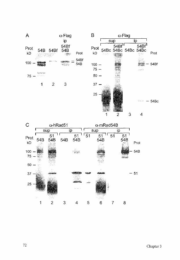
A
Prot 548 548f kD
100--
75 -
1 2
a-Flag ip
548f 548
Prot
_ 54Bf 88"'' - 54B
3
c a-hRad51 sup
51 Prot 548 548 kD
100-75 -
50-
ip 51
548 548
B a-Flag sup ip
1 548f 11 548f1
Prot 548c 548c 548c 548c kD Prot
100-75-
- 54Bf
50-
25
1 2
a-mRad548 sup
I 51 51 II 51 548
ip
3
51 548
:;, Prot
~~-54B '~
- 54Bc
4
37 --- - 51
25 -1 2 3 4 5 6 7 8
72 Chapter 3
FIG. 3. Co-immunoprecipitations of reticulocyte lysates containing in vitro translated mRad54B and hRad51. By IVTT radioactive mRad54B (54B). His-mRad54B-Fiag (54Bf). mRad54B-Cterm (54Bc), and hRad51 (51) were produced. The IVTT reactions yielded secondary protein bands due to alternative translation start sites. The proteins were precipitated with the antibodies (indicated at the top) a-Flag, a-hRad51, and a-mRad54B(Cterm) in NETT buffer on ProtA-sepharose beads as described in Materials and Methods. The proteins, the supernatant of the beads (sup), and the immunoprecipitate (ip) were analyzed by SDS-PAGE and autoradiography. Protein size markers (in kD) are indicated on the left side. The immunoprecipitated proteins (Prot) are indicated on the right side. In panels B and C, to prevent overflow, the lanes were loaded in a different order, but on the same gel and then electronically rearranged. (A) mRad54B shows homomeric interaction. mRad54B and His-mRad54B-Fiag were immunoprecipitated with a-Flag. Lane 1, IVTT of mRAD54B; lane 2, IVTT of HismRAD54B-FLAG; lane 3, co-immunoprecipitate of mRad54B and His-mRad54B-Fiag by a.Flag. (B) The homomeric interaction of mRad54B is mediated by its carboxy terminus. mRad54B-Cterm alone (lanes 1, 3) or mixed with His-mRad54B-Fiag (lanes 2, 4) was precipitated with a-Flag. Lanes 1, 2, supernatant; lanes 3, 4, immunoprecipitate. (C) mRad54B and hRad51 interact as detected by precipitation with a-hRad51 and amRad54B. mRad54B alone (lanes 1, 3) or mixed with hRad51 (lanes 2, 4) was precipitated with a-hRad51. hRad51 alone (lanes 5, 7) or mixed with mRad54B (lanes 6, 8) was precipitated with a-mRad54B. Lanes 1, 2, 5, 6, supernatant; lanes 3, 4, 7, 8, immunoprecipitate.
Rad54B and Rad51 was not quantitative (Fig. 2). Tanaka et a!. reported that the interaction was mediated by the amino terminus of hRad54B [514]. The interaction between hRad54 and hRad51 is also mediated by the amino terminus of hRad54 [185]. As hRad54B and hRad51 did not show any interaction in a yeast two-hybrid screen, the interaction was presumed to be indirect in contrast to the interaction between Rad54 and Rad51 [514]. The lack of interaction could, however, also be due to the fusion proteins used in this two-hybrid screen. The interaction is probably not mediated by DNA, because DNase and RNase treatment of the cell extracts did not inhibit the coimmunoprecipitation of Rad54B and Rad51 (Fig. 2F). We also observed this interaction using reticulocyte lysates containing in vitro translated proteins, suggesting a direct interaction (Fig. 3A). Nevertheless, the interaction might be mediated by other proteins present in the reticulocyte lysate. The co-immunoprecipitation of in vitro translated RAD54B and RAD51 was weaker than that of RAD54 and RAD51 (data not shown). This is another difference between the interaction of Rad51 with Rad54 and Rad54B and suggests that Rad54 and Rad54B perform at least slightly different functions.
Homomeric interaction of mRad54B. Apart from the interaction between mRad54B and hRad51, we also observed homomeric interaction ofmRad54B. This interaction was mediated by the carboxy terminus of mRad54B. Scanning force microscope observations on hRad54 suggest that hRad54 also has homomeric interaction and forms complexes on
Mouse Rad54B protein characterization 73
DNA [430]. The carboxy-terminal homomeric interaction ofmRad54B probably does not interfere with the interaction with Rad51, which takes place via the amino terminus of mRad54B.
Role of mRad54B. mRad54B has conserved the ATPase and helicase motifs characteristic for proteins belonging to the Swi2/Snf2 family of proteins [395]. These proteins are involved in chromatin remodeling. The homology between mRad54 and mRad54B suggests that the functions of both proteins are similar. The nuclear localization and the interactions observed for mRad54B are compatible with a role for the protein in homologous recombination. This is also suggested by the increased sensitivity of mRAD54B-mRAD54-deficient mouse cells to ionizing radiation compared to mRAD54-deficient cells. Mutations in RAD54 and RAD54B have been observed in a low percentage oftumors [212, 331].
In yeast, the two Rad54 homologs function in different subpathways of recombination, but are also partially redundant. Both proteins can stimulate DNA strand exchange by ScRad51 [400, 402]. ScRad54 is more involved in mitotic recombination and interacts with ScRad51 (I 02, 235]. ScRdh54 on the other hand is more important for meiotic recombination and interacts with ScRad51 and Dmcl, the yeast Rad51 homolog only active during meiosis [462, 464]. Consistent with this, Scrad54 mutants are deficient in recombination using the sister chromatid or the homologous chromosome, while Scrdh54 mutants are only deficient in recombination using the homologous chromosome, which is more important during meiosis (10, 264, 465]. The defects in all these recombination reactions are more severe in Scrad54-rdh54 double mutants.
On the basis of its sequence, mRad54B is expected to have similar ATPase and DNA supercoiling activities as mRad54. mRad54B might also stimulate Rad51-mediated DNA strand exchange. Similar to the situation in S. cerevisiae, the mammalian Rad54 homo logs may have partially redundant roles. Such a redundancy is relatively common in mammalian DNA repair pathways. The nucleotide excision repair gene RAD23, the translesion repair gene RAD6, the recombination gene RAD51, and the DNA interstrand cross-link repair gene SNMJ all have more than one homolog/paralog in mammalian cells (Chapter 5) [145, 266, 330, 524]. mRad54B and mRad54 may be important for different subpathways of recombination. Alternatively, they could also work together in the same pathways, by interaction with each other or by common interactions with other homologous recombination proteins, like Rad5l. For further clues on the function of mRad54B, biochemical analysis of the protein, results on recombination assays, and on the phenotype of mRAD54-mRAD54B double knockout cells will be necessary.
ACKNOWLEDGMENTS
We thank Cecile Beerens for a-hRad5l. This work was supported by the Netherlands Organization for Scientific Research (NWO).
74 Chapter 3
CHAPTER4
Repair of DNA interstrand cross-links
Modified from Mutat. Res. 486: 217-247, 2001

REPAIR OF DNA INTERSTRAND CROSS-LINKS
MIES L.G. DRONKERT1, ROLAND KANAARL2
1 Department of Cell Biology and Genetics, Erasmus University Rotterdam, PO Box 1738, 3000 DR Rotterdam, The Netherlands
2 Department of Radiation Oncology, University Hospital Rotterdam/Daniel, The Netherlands
DNA interstrand cross-links (ICLs) are very toxic to dividing cells, because they induce mutations, chromosomal rearrangements, and cell death. Inducers of ICLs are important drugs in cancer treatment. We discuss the main properties of several classes ofiCL agents and the types of damage they induce. The current insights in ICL repair in bacteria, yeast, and mammalian cells are reviewed. An intriguing aspect of ICLs is that a number of multi-step DNA repair pathways including nucleotide excision repair, homologous recombination, and postrephcation/translesion repair all impinge on their repair. Furthermore, the breast cancer-associated proteins Brcal and Brca2, the Fanconi anemiaassociated FA proteins, and cell cycle checkpoint proteins are involved in regulating the cellular response to ICLs. We depict several models that describe possible pathways for the repair or replicational bypass of ICLs.
1 INTRODUCTION
DNA interstrand cross-links (ICLs) are among the most toxic DNA damages. A single ICL can kill repair-deficient bacteria and yeast, and about 40 ICLs can kill repairdeficient mammalian cells [287, 320]. Moreover, ICLs can induce mutations and rearrangements of DNA, possibly resulting in uncontrolled cell proliferation and tumor formation. Many different agents are capable of inducing ICLs into the genome. A wellknown class of ICL-inducers are chemotherapeutic agents like mitomycin C, cisplatin, nitrogen mustard, nitrosourea, and their derivatives. However, cells are also exposed to environmental ICL agents, like furocoumarins that are present in many plants and cosmetics [454]. Moreover, endogenous agents that are formed during lipid peroxidation, such as malondialdehyde, can lead to ICLs. However, the clinical relevance of these endogenous agents is unclear, because they are unstable and hardly form ICLs under physiological conditions [24, 86]. Organisms have developed strategies to deal with DNA damage in order to survive. A number of specialized repair pathways have evolved that each process specific kinds of DNA damage. An intriguing aspect of ICL repair is that several of these pathways have to work together in order to remove or bypass an ICL. While the major ICL repair pathway in bacteria is well-characterized, both genetically and biochemically, ICL repair in eukaryotes is less well understood.
First, we will discuss a number of important properties of ICL agents and the DNA damage with which they confront the cells. Next, the clinical relevance of knowledge concerning ICL repair for the treatment of cancer will be surveyed, followed by a review
DNA interstrand cross-link repair 77
of the current insights in the mechanisms ofiCL repair in bacteria, yeast, and mammalian cells.
2 CROSS-LINK FORMATION AND PROPERTIES OF DNA INTERSTRAND CROSS-LINKING AGENTS
ICL agents have different toxicities depending on a number of factors, including cellular uptake aud metabolic activation. First, most ICL agents have to be trausported into the cell and then into the nucleus. Second, a number of ICL agents, such as the nitrosoureas, are metabolically activated in the cell, yielding an agent with a much higher activity [566]. Metabolism also yields other damaging agents. Mitomycin C, for example, undergoes a cycle of oxydation and reduction, thereby activating the agent and generating reactive oxygen species that cau cause additional damage to the cell [68, 414, 529, 530]. Nitrogen mustard, cisplatin, and 8-methoxypsoralen also give rise to reactive oxygen species [15, 101]. On the other hand, metabolism also inactivates ICL agents, often via the cytochrome P450 reductase system [566]. Furthermore, intracellular thiols, such as glutathione and metallothionein, can detoxifY ICL agents [100].
ICL agents form a number of adducts with DNA. The adducts are not always produced randomly throughout the genome, because chromatin structure can influence adduct formation [222, 284, 552]. Mostly, ICLs only represent a small fraction of the adducts formed. Nevertheless, they are thought to be the main determinaut of the toxicity of ICL agents, due to their inhibition of DNA strand separation and therefore of DNA replication, transcription, and segregation. To adequately respond to the ICLs, cells have to recognize them. They could recognize the ICL itself by its distortion ofthe DNA helix. However, the three-dimensional structures of the ICLs are variable, which could influence the efficiency of!CL recognition and repair. Cells could also recognize the transcription or replication block induced by the ICLs. The damage response will lead to cell cycle arrest and, if repair fails, to cell death. The characteristics of a few classes of!CL agents are discussed below and summarized in Table 1.
Psora/ens. Psoralens can be activated by UV A light to induce thymine monoadducts and ICLs between thymines at d(TpA) sequences in DNA. They are used in the treatment of several skin diseases like psoriasis, and belong to the group of furocoumarins, present in plants aud cosmetics [454]. The ratio of!CL to monoadduct can be influenced by the wavelength and dose ofUV A. In this way, up to 40% of the adducts can be converted to ICLs [63]. Compared to monoadducts, ICLs are more cytotoxic, mutagenic, and recombinogenic [15, 79, 189]. Psoralen-induced ICLs on an oligonucleotide induce relatively minor changes in the architecture of the DNA helix. There is no bend in the helix axis and only a 25' local unwinding of the helix [485, 486]. The cross-link is asymmetric, because it consists of a furan ring with one thymine and a pyrone ring with the other [63]. Repair of this ICL also shows asymmetry. Usually, there is a preference for incision of the furan side [21, 544].
78 Chapter 4
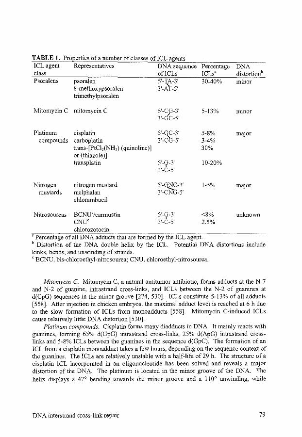
TABLE 1. Properties of a number of classes of!CL agents ICL agent class Psoralens
Mitomycin C
Platinum compounds
Nitrogen mustards
Representatives DNA sequence ofiCLs
psoralen 8-methoxypsoralen trimethylpsoralen
mitomycin C
cisplatin carboplatin trans-[PtC12(NH3) (quinoline)] or (thiazole)] transplatin
nitrogen mustard melphalan chlorambucil
5'-TA-3' ' 3'-AT-5'
5'-CG-3' 3'-GC-5'
5'-GC-3' 3'-c'o-s·
5'-G-3' 3'-C-s·
5'-GNC-3' 3'-C'NG-5'
Percentage ICLs' 30-40%
5-13%
5-8% 3-4% 30%
10-20%
1-5%
DNA distortionb minor
minor
major
major
Nitrosoureas BCNU'/carmustin 5'-G-3' <8% unknown CNU' ' 3'-C-5' 2.5% chlorozotocin
'Percentage of all DNA adducts that are formed by the ICL agent. b Distortion of the DNA double helix by the ICL. Potential DNA distortions include kinks, bends, and unwinding of strands. 'BCNU, bis-chloroethyl-nitrosourea; CNU, chloroethyl-nitrosourea.
Mitomycin C. Mitomycin C, a natural antitumor antibiotic, forms adducts at the N-7 and N-2 of guanine, intrastrand cross-links, and ICLs between the N-2 of guanines at d(CpG) sequences in the minor groove [274, 530]. ICLs constitute 5-13% of all adducts [558]. After injection in chicken embryos, the maximal adduct level is reached at 6 h due to the slow formation of ICLs from monoadducts [558]. Mitomycin C-induced ICLs cause relatively little DNA distortion [530].
Platinum compounds. Cisplatin forms many diadducts in DNA. It mainly reacts with guanines, forming 65% d(GpG) intrastrand cross-links, 25% d(ApG) intrastrand crosslinks and 5-8% ICLs between the guanines in the sequence d(GpC). The formation of an ICL from a cisplatin monoadduct takes a few hours, depending on the sequence context of the guanines. The ICLs are relatively unstable with a half-life of 29 h. The structure of a cisplatin ICL incorporated in an oligonucleotide has been solved and reveals a major distortion of the DNA. The platinum is located in the minor groove of the DNA. The helix displays a 47° bending towards the minor groove and a IJ0° unwinding, while
DNA interstrand cross-link repair 79
cytosines are extruded from the helix [321]. Carboplatin adducts are similar to cisplatin adducts, with 3-4% ICLs [47]. Transplatin forms different intrastrand cross-links and ICLs. The ICLs are formed between a complementary cytosine and guanine. Although the percentage of!CLs is higher, transplatin has a much lower cytotoxicity than cisplatin [57]. This difference could be due to the different structrue of the ICL allowing faster repair, slower conversion ofmonoadducts to ICLs, or lack of the toxic d(GpG) intrastrand adducts [321, 389]. Up to 30% of the adducts induced can be ICLs when cells are treated with trans-[PtC!,(NH3)(quinoline)] and trans-[PtC12(NH3)(thiazole)], which are derivatives of transplatin. These ICLs form between guanines in the sequence d(GpC), similar to cisplatin-induced ICLs [58].
Nitrogen mustards. ICLs constitute up to 5% of the DNA damage caused by nitrogen mustard and its derivatives. These ICLs cause a major DNA distortion [ 429]. Next to the N-7 guanine ICLs, formed in the sequence d(GpNpC), nitrogen mustards cause guanine monoadducts, DNA-protein cross-links and they also react with RNA. Melphalan and chlorambucil also cause N-3 adenine adducts [63, 410]. Nitrogen mustard ICLs are quite uustable with a half-life of 2 h, which decreases their toxicity compared to other, more stable ICLs [207].
Nitrosoureas. Nitrosoureas, for example bis-chloroethyl-nitrosourea (BCNU or carmustin), form guanine and cytosine monoadducts, guanine intrastrand cross-links, and guanine-cytosine ICLs after metabolic activation. ICLs constitute only a low percentage of total adducts. Nevertheless, the cytotoxicity of nitrosoureas is clearly linked to the number of ICLs formed [566]. Formation of the ICL from 0-6 guanine adducts takes several hours. Furthermore, DNA strand breaks and DNA-protein cross-links are formed by BCNU and proteins are also attacked by its carbamoylating activity.
3 CLINICAL RELEVANCE OF DNA INTERSTRAND CROSS-LINKS
As mentioned above, many ICL-inducing agents are used in the treatment of cancer. Knowledge about the formation and repair of these ICLs enables us to understand the clinical effectiveness and side-effects of these agents and to improve cytostatic therapy. Knowledge about ICL repair is also relevant for Fanconi anemia patients, whose cells are specifically sensitive to ICL agents. Fanconi anemia will be discussed later.
Cytostatic therapy is based on selective killing of tumor cells, while normal cells are spared as much as possible. For example, the activation of mitomycin C is stimulated in the hypoxic environment in many solid tumors, while the well-oxygenated cells in the rest of the body show less mitomycin C activation [ 530]. Part of the selectivity of all ICL agents for tumor cells is achieved by interfering with DNA transcription and replication. These processes are more active in rapidly dividing cells and in general, tumor cells divide more rapidly than nontumorigenic cells. ICLs inhibit DNA metabolism, they cause mutations and chromosomal damage, and can induce apoptosis. Unfortunately, tumors can be intrinsically resistant to certain agents, or become so after treatment [161]. For example, intrinsic resistance to cisplatin occurs in hereditary nonpolyposis colorectal carcinomas which are mismatch repair deficient. The reason is that mismatch repair-
80 Chapter 4
deficient cells can tolerate cisplatin adducts better than repair-proficient cells. This is not true for all platinating agents, so some of them can still be used in treating these tumors [ 161, 540]. Mismatch repair deficiency is often induced in ovarian tumors, also yielding cisplatin-resistant cells [ 67].
Resistance to ICL agents can be caused by many mechanisms, which often operate together, increasing resistance even more. In many cases, there is a clear correlation between the level of ICLs formed or the repair activity on ICLs and the sensitivity of tumor cells [244, 488, 566, 591]. In some cases, though, resistance is not correlated to the ICL level, but to other adducts formed [540]. In many tumors, overexpression of multidrug transport proteins causes resistance to chemotherapeutic agents by increased export of the agents from the cell [285]. Tumor cells often have a different metabolism than nontumorigenic cells, reducing the formation of active metabolites, or increasing deactivation of the drug. A higher level of glutathione or metallothionein can increase the detoxification of!CL agents [6, 397, 470]. Increased base excision repair can remove, for example, the 0-6 guanine monoadducts formed by BCNU, thereby preventing ICL formation, which takes a few hours [566]. Conversely, inactivation of the responsible alkyltransferase can make tumors more sensitive to certain ICL agents [156, 204, 412]. Finally, repair of the ICLs can be increased in resistant tumors [6, 244, 379, 488, 582, 591]. Some of these mechanisms cause cross-resistance to ICL agents of several classes, while others are agent-specific [59, 161, 204, 566]. Knowledge of the mechanisms by which a tumor has become resistant to treatment may guide the development of additional treatments that could still be effective.
4 DETECTION OF DNA INTERSTRAND CROSS-LINKS AND REPAIR INTERMEDIATES
It is important to be aware of the methods used to assess the level of ICLs and their repair intermediates to correctly interpret the data that are obtained. It has to be kept in mind that ICLs form only a small fraction of the induced DNA damage, except when one ICL has been produced at a specific position on a plasmid. The damage response, repair activity, and survival of the cell are also determined by the other types of damage. The main difference between an ICL and any other DNA damage is that ICLs covalently bind the two strands of the DNA double helix together. Thereby, separation of the two strands by denaturation is made impossible. This property yields different sedimentation rates of native versus cross-linked DNA in alkaline sedimentation methods and causes different mobility in agarose and polyacrylamide gels. After incision in at least one DNA strand near the ICL, the DNA strands can be separated again by denaturation. This step of repair can thus be followed in time by measuring the fraction of DNA that can be denatured. A drawback of these methods is their relatively low sensitivity and therefore they require a high level of ICLs, which is lethal for almost all cells and may saturate repair pathways. Furthermore, the nature of the incision, on one or both sides of the ICL, in one or both DNA strands, cannot be determined. The recovery of the ability to denature cross-linked DNA is often used to compare the repair proficiency in sensitive and resistant cell lines,
DNA interstrand cross-link repair 81
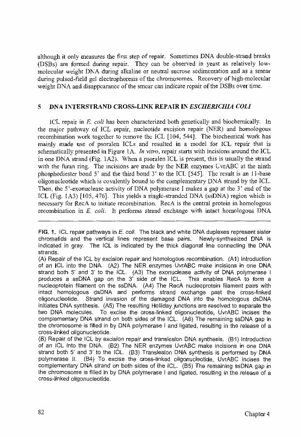
although it only measures the first step of repair. Sometimes DNA double-strand breaks (DSBs) are formed during repair. They can be observed in yeast as relatively lowmolecular weight DNA during alkaline or neutral sucrose sedimentation and as a smear during pu!sed-f1eld gel electrophoresis of the chromosomes. Recovery of high-molecular weight DNA and disappearance of the smear can indicate repair of the DSBs over time.
5 DNA INTERSTRAND CROSS-LINK REPAIR IN ESCHERICHIA COLI
!CL repair in E. coli has been characterized both genetically and biochemically. In the major pathway of ICL repair, nucleotide excision repair (NER) and homologous recombination work together to remove the ICL [104, 544]. The biochemical work has mainly made use of psoralen ICLs and resulted in a model for ICL repair that is schematically presented in Figure lA In vitro, repair starts with incisions around the ICL in one DNA strand (Fig. IA2). When a psoralen ICL is present, this is usually the strand with the furan ring. The incisions are made by the NER enzymes UvrABC at the ninth phosphodiester bond 5' and the third bond 3' to the ICL [545]. The result is an !!-base oligonucleotide which is covalently bound to the complementary DNA strand by the ICL Then, the 5' -exonuclease activity of DNA polymerase I makes a gap at the 3' end of the ICL (Fig. IA3) [105, 476]. This yields a single-stranded DNA (ssDNA) region which is necessary for RecA to initiate recombination. RecA is the central protein in homologous recombination in E. coli. It performs strand exchange with intact homologous DNA
FIG. 1. ICL repair pathways in E. coli. The black and white DNA duplexes represent sister chromatids and the vertical lines represent base pairs. Newly-synthesized DNA is indicated in gray. The ICL is indicated by the thick diagonal line connecting the DNA strands. (A) Repair of the ICL by excision repair and homologous recombination. (A1) Introduction of an ICL into the DNA (A2) The NER enzymes UvrABC make incisions in one DNA strand both 5' and 3' to the ICL. (A3) The exonuclease activity of DNA polymerase I produces a ssDNA gap on the 3' side of the ICL. This enables RecA to form a nucleoprotein filament on the ssDNA (A4) The RecA nucleoprotein filament pairs with intact homologous dsDNA and performs strand exchange past the cross-linked oligonucleotide. Strand invasion of the damaged DNA into the homologous dsDNA initiates DNA synthesis. (A5) The resulting Holliday junctions are resolved to separate the two DNA molecules. To excise the cross-linked oligonucleotide, UvrABC incises the complementary DNA strand on both sides of the ICL. (A6) The remaining ssDNA gap in the chromosome is filled in by DNA polymerase I and ligated, resulting in the release of a cross-linked oligonucleotide. (B) Repair of the ICL by excision repair and translesion DNA synthesis. (81) Introduction of an ICL into the DNA (82) The NER enzymes UvrABC make incisions in one DNA strand both 5' and 3' to the ICL. (83) Translesion DNA synthesis is performed by DNA polymerase II. (84) To excise the cross-linked oligonucleotide, UvrABC incises the complementary DNA strand on both sides of the ICL. (85) The remaining ssDNA gap in the chromosome is filled in by DNA polymerase I and ligated, resulting in the release of a cross-linked oligonucleotide.
82 Chapter 4
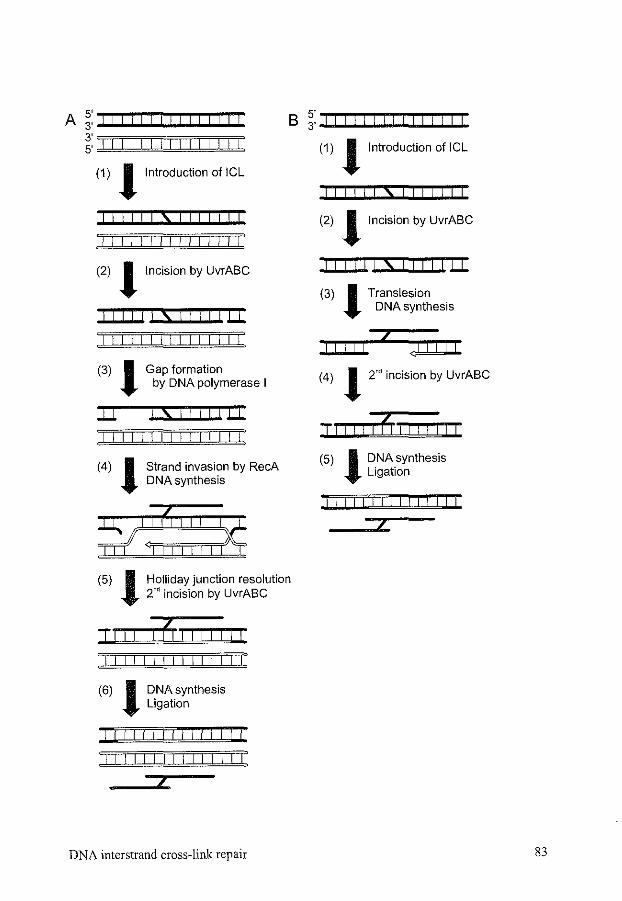
A 5' I I I I I I I I I I I I I 3' B ~: I I I! I I I I I I I I I I I 3' 5' I I I I I I I I I I I I I I I
(1) !Introduction of ICL
iiii!!Siiiii!
I I I I I I II I II I I I I
(2) !Incision by UvrABC
!liii!S!iillll
I I I I I I I I I I II I I I
(3) 1 Gap formation by DNA polymerase I
ll s Ill I I I I I I I I I I I
(4) 1 Strand invasion by RecA DNA synthesis
(5) 1 Holliday junction resolution 2'' incision by UvrABC
II I I j LJ I I I I
I I I I I I I I I I I
(6) 1 DNA synthesis Ligation
I I
I ! I I I I ! ! I I I i I I 1
/11/ll/11111111
DNA interstrand cross-link repair
(1) !Introduction of ICL
I II I I I S I I I I I I I
(2) !Incision by UvrABC
ilii!!Siiillil
(3) J Translesion DNA synthesis
j I I I II
(4) J 2" incision by UvrABC
j Ji I I I I I] I I I I I I
(5) J DNA synthesis Ligation
I 1 ! II I I I I I I I I I
83
resulting in heteroduplex DNA encompassing the region containing the cross-linked oligonucleotide (Fig. 1A4) [476]. As a result, a three-stranded region occurs with predominantly base pairing of the full-length strands. This enables UvrABC to incise the other DNA strand, containing the pyrone ring (Fig. lAS). Thereby, UvrABC enables the release of the ICL-containing DNA fragment [96, 476]. The resulting gap can be filled in by DNA polymerase I and ligated (Fig. 1A6).
In a RecA-deficient strain, repair of psoralen ICLs decreases to 2% of wild-type repair, while repair of a nitrogen mustard ICL decreases only to 30% [36, 406]. This suggests that the stmcture of the ICL is important for the repair efficiency of different pathways. Repair in RecA-deficient strains is dependent on NER and DNA polymerase II (Fig. lB) [35]. Repair is once again inititated by UvrABC incising one strand of the cross-linked DNA (Fig. 1B2). But because recombination is impaired, DNA polymerase II synthesizes DNA across the region spanning the cross-linked oligonucleotide (Fig. 1B3). Then, UvrABC removes the ICL by incisions in the complementary DNA strand (Fig. IB4). Thus, besides the major RecA-dependent recombination repair, a minor DNA polymerase ll-dependent translesion repair pathway is capable of repairing ICLs.
In vivo, more proteins are involved in the repair process. Bacteria deficient in one of a number of recombination proteins, such as RecB, C, D, F, G, 0, R, are sensitive to ICL agents [216, 589]. RecFOR promotes the binding of RecA to ssDNA and facilitates homologous pairing by RecA, especially in the absence of a DSB [538, 559]. RecBCD is necessary when a DSB is formed during ICL repair. DSBs can be formed by the direct action of nucleases. Alternatively, when a nick has been made next to an ICL, the encounter of a replication fork will yield a DSB. Finally, a replication fork that is stalled by an ICL can also result in a DSB without a pre-existing nick. RecBCD resects DSBs producing a 3' ssDNA overhang and it loads RecA onto the ssDNA [8]. RecG or RuvABC are responsible for the resolution of the recombination intermediates [113, 268]. Finally, some ICL repair is seen in uvr mutants, which are unable to follow either of the pathways mentioned above. After replication is stalled due to the adduct, recombinational repair or bypass may be induced without the need for UvrABC in the process, possibly using other nucleases [553].
6 DNA INTERSTRAND CROSS-LINK REPAIR IN SACCHAROMYCES CEREVISIAE
ICL repair inS. cerevisiae has not been characterized in as much detail as repair in E. coli. A significant amount of information is available about the genes involved in ICL repair, but not specifically about their involvement in ICL repair (Table 2). Most ofthese genes belong to one of the DNA repair pathways that process other types of lesions as well: NER, homologous recombination, and postreplication/translesion repair [63]. Survival after treatment with ICL agents is also affected by mutations in base excision repair and mismatch repair. Base excision repair can prevent the formation of ICLs by repair of precursor lesions [336]. Mismatch repair sensitizes cells to ICL agents as it interferes with translesion synthesis past monoadducts and intrastrand cross-links [147].
84 Chapter 4
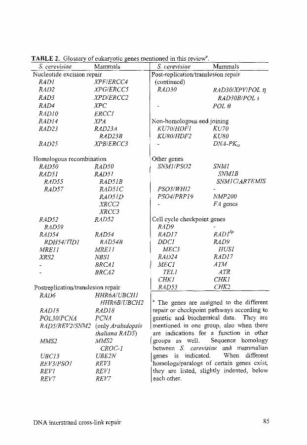
TABLE 2. Glossary of eukaryotic genes mentioned in this review'. S. cerevisiae Mammals S. cerevisiae Mammals
Nucleotide excision repair Post-replication/translesion repair RADI XPFIERCC4 (continued) RAD2 XPGIERCC5 RAD30 RAD30/XPV/POL 1]
RAD30B/POL t
POLe
RAD3 XPDIERCC2 RAD4 XPC RADIO RADI4 RAD23
RAD25
ERCCI XPA RAD23A
RAD23B XPB/ERCC3
Homologous recombination RAD50 RAD50 RAD5I RAD5I
RAD55 RAD5IB RAD57 RAD5IC
RAD52 RAD59
RAD54 RDH54/TIDI
MREII XRS2
RAD51D XRCC2 XRCC3
RAD52
RAD54 RAD54B
MREJI NBSI BRCAI BRCA2
PostreplicationJtranslesion repair RAD6 HHR6AIUBCHI
RADI8 POL30/PCNA RAD5/REV2/SNM2
MMS2
UBCJ3 REV3/PSOI REVI REV7
HHR6B/UBCH2 RADI8 PCNA (only Arabidopsis thaliana RAD5) MMS2
CROC-I UBE2N REV3 REVI REV7
DNA interstrand cross-link repair
Non-homologous end joining KU70/HDFI KU70 KU80/HDF2 KU80
Other genes SNMI/PS02
DNA-PKc,
SNMI SNMIB SNMIC/ARTEMIS
PS03/WHI2 PS04/PRPI9 NMP200
FA genes
Cell cycle checkpoint genes RAD9 RAD17 DDCI
MEC3 RAD24 MECI
TELl CHKI RAD53
RADISp
RAD9 HUSI
RADI7 ATM
ATR CHKI CHK2
a The genes are assigned to the different repair or checkpoint pathways according to genetic and biochemical data. They are mentioned in one group, also when there are indications for a function in other groups as well. Sequence homology between S. cerevisiae and mammalian genes is indicated. When different homologs/paralogs of certain genes exist, they are listed, slightly indented, below each other.
85
Next to the genes that were already known for a role in DNA repair, some genes involved in ICL repair have been identified by searching for mutations that sensitize yeast cells to ICL agents, namely SNMJ, SNM2, PSOJ, PS02, PS03, and PS04 [208, 210, 439]. SNM2 and PSOI later appeared to be allelic to RAD5/REV2 and REV3, respectively, and therefore belong to the postreplication/translesion repair group of genes [82, 468]. The other genes, PS03, PS04, and SNMI which is allelic to PS02, do not clearly belong to one of the abovementioned DNA repair pathways and will be dealt with separately below [82].
6.1 Cell cycle effects of DNA interstrand cross-links
DNA damage induces cell cycle arrest to allow DNA repair and prevent genetic instability. This arrest is achieved by activating cell cycle checkpoint pathways (for reviews see [166, 316, 561, 594]). The intricate network of proteins involved in DNA damage-induced cell cycle checkpoints recognizes the presence of damage, arrests the cell cycle after signal transduction, affects transcription of repair genes and post-translational modifications of repair proteins, and influences DNA repair processes. It is not yet known how the cell achieves recognition of all different kinds of DNA lesions. The so-called sensor proteins in S. cerevisiae, including Rad9, Ddcl, Mec3, Radl7, and Rad24 may recognize lesions directly, or after processing to a common intermediate such as ssDNA. Alternatively, they may sense changes in chromatin structure or interact with DNA repair proteins. Different damages may be recognized by different sensor proteins, thereby affecting the type of checkpoint response. During S phase, the replication complex is also involved in signaling damage. The damage response signal is transduced by Mecl, Rad53, and Chkl, which activate proteins further downstream that affect cell cycle progression and DNA repair.
Although much is known about the response of cells to 254-nm UV light (UV254 mn) or ionizing radiation, little is known about cell cycle arrest after treatment with ICL agents. Different ICL agents trigger different cell cycle responses. 8-methoxypsoralen plus UV A in a high dose, inducing three ICLs per replicon, causes G2 delay after treatment in S or G2 phase. Cells treated in G l show G l arrest [345]. In contrast, after treatment with cisplatin, no G l arrest is found. This might be due to differences in the detection of cisplatin and psoralen DNA lesions. At a relatively low dose of cisplatin, 40% survival and less than one ICL per replicon, wild-type yeast cells show only a G2 delay after treatment in Gl [192, 193]. Repair-deficient cells treated with a low dose of cisplatin and wild-type cells treated with a high dose show a permanent G2 arrest [193]. This G2 arrest is not directly dependent on the number of adducts induced, but may depend on repair or replication intermediates. Apparently, replication can tolerate or bypass a low number of cisplatin ICLs. Nevertheless, at a high dose, cisplatin also induces S phase delay [193]. This S phase delay and the G2 arrest depend on MECJ, one of the cell cycle checkpoint genes [192, 193].
86 Chapter4
6.2 Processes during DNA interstrand cross-link repair
6.2.1 Recognition and incisions The first step in the repair of a DNA lesion is recognition. Up to now, no specific
proteins involved in ICL recognition have been found. Recognition could occur when the replication or transcription machinery encounter an ICL and become blocked. Alternatively, or in addition, the NER DNA damage recognition proteins or other ICL structure-specific DNA binding proteins could recognize the ICLs and initiate repair.
Shortly after ICLs are formed in yeast as measured by the inhibition of DNA denaturation, this inhibition is overcome [229, 320, 344, 348]. This indicates either that incisions surrounding the ICL are made in one strand, as in E. coli, or that incisions are made in both strands resulting in DSBs. Formation of DSBs during ICL repair is much more prominent in S. cerevisiae than in E. coli, indicating an important difference in the processing ofiCLs in bacteria versus fungi [119, 120, 229, 320].
The first incision near the ICL is supposed to be made by NER enzymes, since in radl, rad2, and rad3 cells, cross-linked DNA cannot be denatured after incubation of the cells to allow repair, in contrast to the situation in mutants defective in homologous recombination or postreplicationltranslesion repair [344, 348]. It is not known whether the NER nucleases are also responsible for incisions made later in the process of ICL repair. NER is known for the repair ofUV 254 nm-induced lesions and bulky adducts, which damage only one strand. A structure-specific endonuclease consisting of a heterodimer of the Radl and RadiO proteins makes an incision on the 5' side of the lesion. A second endonuclease involved in NER, Rad2, makes the incision on the 3' side of the lesion. Together, they facilitate the removal of an oligonucleotide containing the lesion. NER consists of two subpathways, global genome repair and transcription-coupled repair. The latter subpathway provides fast repair of transcriptionally active genes [169]. ICL repair in transcriptionally active genes is also often faster than the repair in transcriptionally inactive chromatin regions [21, 344, 345]. This could be due to the presence of more accessible chromatin in transcriptionally active regions or to a potential transcriptioncoupling activity associated with the NER enzymes acting early in ICL repair. The latter hypothesis is supported by the finding that mutations induced by psoralen-induced ICLs are most often found at the position of the thymine in the nontranscribed strand [21].
The NER nucleases are supposed to make incisions in only one of the strands of double-stranded DNA ( dsDNA). DSB formation could be caused either by two closely separated incisions on opposite strands or by replication of nicked DNA This would explain the observation that in exponentially growing cells, usually a higher level of DSB formation is found than in stationary cells [119, 337]. However, some DSB formation has been found in cells deficient for the NER proteins Rad1, Rad2, and Rad4, suggesting there are other ways of making incisions [33 7].
6.2.2 Recombination The DSBs formed after incision of the ICL have to be repaired by one of the DSB
repair pathways. The major pathway inS. cerevisiae is homologous recombination using either the sister chromatid or the homologous chromosome. Some of the key genes
DNA interstrand cross-link repair 87
involved in homologous recombination are RAD51, RAD52, and RAD54 (Table 2) (See also Chapter I) [253]. Yeast cells mutant for one of these genes are sensitive to ICL agents, and they show a decrease in the recovery of high-molecular weight DNA after DSB formation during the repair of ICLs [14, 229, 337]. Homologous recombination is less important for ICL repair in haploid yeast treated in stationary phase, as only few DSBs are formed and there is no homologous DNA available for recombination [337, 467]. It should be noted, however, that treatment of yeast cells in stationary phase is followed by proliferation of the cells when survival is assessed. On cell proliferation, homologous recombination is feasible again. Therefore, survival is also reduced in homologous recombination-deficient cells, treated in stationary phase. The second DSB repair pathway is nonhomologous end-joining (NHEJ) in which for example the yKU70 andyKU80 are involved. Mutants in NHEJ are not sensitive to ICL agents and ku70rad52 double mutants are as sensitive as rad52 mutants, suggesting that NHEJ cannot compensate for a defect in homologous recombination [337]. Nevertheless, while rad52 mutants perform some DSB repair during nitrogen mustard ICL repair, in a ku70rad52 double mutant no DSB repair is fouud [337]. NHEJ may therefore still have a minor role in processing ICL repair intermediates.
In some cases, homologous recombination was found to be epistatic to NER with regard to ICL repair, suggesting that they participate in one pathway as described above [209]. In most cases, however, synergism is observed, with double mutants being more sensitive than either single mutant, which suggests that they are involved, at least partially, in alternative routes of repair [85, 319]. This means that other pathways supplement NER and recombination, as it is difficult to imagine complete repair of ICLs by just the one or the other pathway.
6.2.3 Postreplication/translesion repair Postreplication/translesion repair provides at least one additional route for repair of
ICLs. Postreplication/translesion repair mutants usually are synergistic with both NER and recombination repair mutants [85]. The mechanism of postreplication/translesion repair is less well known than that of NER or homologous recombination. Mostly, this pathway does not repair a lesion, but helps the cell to tolerate or bypass the lesion. The major genes in postreplication/translesion repair are RAD6, encoding a ubiquitinconjugating enzyme that is also involved in other cellular pathways, and RAD18 [288]. These two genes take part in most routes of postreplication/translesion repair. Postreplication/translesion repair consists of several subpathways, which are either errorfree or error-prone [288, 575]. One error-free pathway involves Pol30/PCNA and DNA Polymerase 8, the other Rad5/Rev2/Surn2 [5, 242, 468, 575]. The mechanisms of the error-free pathways are still unknown. Rad5 and Rad!S are both ssDNA-dependent ATPases, bind DNA, and can form a complex with each other and the ubiquitinating proteins Ubcl3-Mms2 and Rad6, respectively [17, 243, 537]. The error-prone pathway consists of the trans!esion DNA polymerase<; (zeta) complex, formed by Rev3/Psol and Rev7, but the translesion synthesis of this complex also depends on Rev! [82, 288, 357, 373, 374]. RAD5 plays a role in the error-prone pathway as well [304]. RAD30, which
88 Chapter 4
encodes DNA polymerase 11 (eta), is also part of the postreplication/translesion repair pathway [334].
rad6 and rad18 mutants are sensitive to ICL agents both after treatment during exponential growth and during stationary phase followed by exponential growth [209, 337]. The induction of DSBs after ICL formation is normal, but they are slowed down in the restitution of high-molecular weight DNA [80, 119]. In contrast to NER and homologous recombination mutants, which show an increase in ICL-induced mutagenesis, rad6 and radl8 mutants show a decrease in mutagenesis in response to JCL agents [22, 80]. Mutagenesis induced by ICLs formed during stationary phase is probably mainly dependent on translesion DNA synthesis by DNA polymerase c;, that can efficiently bypass UV254 om and bulky adducts but at the expense of a high mutation rate [30, 288, 304, 374, 431]. A DNA polymerase c; mutant, rev3, is not sensitive to ICL agents when the cells grow exponentially [337]. rev3 mutants are also less sensitive to DNA-damaging agents than rad6 or rad18 mutants [288]. This implies that the error-free subpathways of the postreplicationltranslesion repair pathway also have an important function in ICL repmr.
6.3 Additional genes involved in DNA interstrand cross-link repair
SNMI. Some genes that are involved in ICL repair do not quite fit into one of the major repair pathways. One of them is SNMI/PS02 [427]. snml mutants are sensitive to JCL agents, but in contrast to the abovementioned genes, they are only slightly or not sensitive to UV254 nm' ionizing radiation, and monofunctional alkylating agents [63, 208, 439]. The relative sensitivity of snml cells compared to wild-type cells is much higher for nitrogen mustard than for psoralens, suggesting some ICL agent specificity [81]. Expression of SNMI is induced by JCL agents and UV254 om in exponentially growing cells [570]. In stationary cells, the expression level is two times lower and is not induced by DNA damage [570]. There are no indications for a role of Snml in cell cycle checkpoints, as the cell cycle arrest after cisplatin treatment of snml cells is similar to the arrest of wild-type cells [193].
SNMI encodes a nuclear protein, that is a member of the NER epistasis group with respect to monoadducts and ICLs and shows the same preference for repair of transcriptionally active genes [207, 209, 344, 467]. However, with respect to treatment of cells with ICL agents, the SNMI gene also appears to be epistatic to REV3, a postreplication/translesion repair gene that is synergistic to the NER genes [209]. snml cells show a reduced mutagenesis after ICL agent treatment, which also suggests that Snml is involved in an error-prone repair pathway [78, 80, 81]. Nevertheless, snml shows synergism with the other postreplication/translesion repair mutants rad5 and rad6 [ 467]. Similar to postreplication/translesion repair mutants but in contrast to NER mutants, snml cells are able to incise lCLs and produce DSBs, but they are not able to reconstitute high-molecular weight DNA [193, 320, 567]. Similar to the recombination mutants, they do not show the increased resistance in 02 or in diploid yeast cells that is observed in wild-type cells [78, 208]. Damage-induced recombination is also decreased in snml cells after treatment with 8-methoxypsoralen plus UV A, but not after nitrogen
DNA interstrand cross-link repair 89
mustard treatment [ 441]. The repair of a psora! en ICL on exogenous plasmid DNA is not impaired in snml mutants in contrast to NER and recombination mutants [319]. This suggests that Smnl could be involved in some chromatin modulation activity. Snml could function in a mutagenic, recornbinogenic repair pathway that is specific for ICL repair and does not belong to the recombination or postreplication/translesion repair pathways. It might also link incision of the ICL by NER to recombination or trans lesion synthesis. Alternatively, it could have a role in the regulation of!CL repair.
PS03. Another gene involved in ICL repair is PS03. pso3 mutant yeast cells are sensitive to both psoralen monoadducts and ICLs, but also to H20 2• They are, however, not sensitive to UV254 nm, ionizing radiation, or alkylating agents [62, 207]. With regard to ICL repair, PS03 is epistatic to RAD3, SNMI, and REV3, but synergistic with RAD6 [32, 207]. Compared to wild-type cells, pso3 cells show reduced mutagenesis and gene conversion frequencies, but an increased number of crossovers after ICL agent treatment, both in haploid and diploid cells [62, 78, 123, 342]. These features are similar to those of snml cells. PS03 may be allelic to WHI2, a gene involved in the regulation of cell growth and cell division [207, 415, 449]. Therefore, it might form one ofthe connections between repair and cell growth regulation.
PS04. PS04 is another gene identified by sensitivity of mutant cells to treatment with psoralens [210]. It is allelic to PRP19, an essential gene that is involved in prernRNA splicing [97, 190]. The pleiotropic DNA repair defective phenotype ofpso4 cells could be explained by a requirement for Pso4 function in different repair pathways and in splicing. Alternatively, its involvement in DNA damage repair could be explained by inhibited splicing ofpre-rnRNAs of DNA repair genes inpso4 mutant cells.
6.4 Conclusions on Saccharomyces cerevisiae DNA interstrand cross-link repair
Surveying the data available, there is no single pathway responsible for all ICL repair. Different ways of dealing with ICLs are used, dependent on position and type of the ICL, the cell cycle phase, ploidy, chromatin structure, and availability of ICL repair proteins. In Figures I, 2, and 3, we have depicted models showing different hypothetical pathways by which ICLs could be repaired or bypassed in yeast in accordance with the genetic and biochemical data described above. The first two pathways described are similar to the E. coli ICL repair pathways (Fig. lA, B). Repair starts with incisions next to the ICL in one DNA strand. These incisions could be made by the NER enzymes and they may occur preferentially in transcriptionally active genes. When a sister chromatid or homologous chromosome is available, homologous recombination could continue the repair reaction, similar to the situation in the major E. coli ICL repair pathway, without the need for a DSB (Fig. !A). Resolution ofthe homologous recombination intermediate leads to a gene conversion event with or without a crossover. Only the latter outcome is depicted. NER enzymes could finally remove the ICL by incisions around it in the complementary DNA strand. This repair route is principally error-free.
In the absence of homologous DNA, recombination repair is not possible. A more error-prone postreplication/translesion repair route might then take over. After incision
90 Chapter 4
next to the ICL in one DNA strand by the NER enzymes or some other nuclease, DNA polymerase S could bypass the base on the other strand which is bound to the cross-linked oligonucleotide (Fig. 1B3). Subsequently, a second round of NER activity could remove the cross-linked oligonucleotide leading to restoration of intact DNA (Fig. 1B4, 5). Base pair substitutions are expected to occur frequently dming translesion synthesis. When NER is eliminated, ICL repair becomes more error-prone, not only yielding base pair substitutions but also insertions [22]. Other nuclease(s) might incise the DNA near the ICL, or breaks could occur upon a replication block. They could be processed either by a recombinational pathway or a translesion synthesis pathway.
In yeast, DSBs are often found after the induction of ICLs. These DSBs, either formed by NER or other nucleases, could be the intermediate for initiating recombination (Fig. 2). DSBs might occur on both sides of the ICL, thereby immediately removing the ICL (Fig. 2Al). Homologous recombination could then continue the repair reaction similar to the repair of DSBs induced by other damaging agents, like restriction enzymes or ionizing radiation (See Chapter 1). After nucleolytic processing, strand invasion of one or both ends takes place, leading to DNA synthesis to bridge the gap (Fig. 2A2, 3). Formation and resolution of Holliday junctions or reaunealing of the elongated DSB ends result in two intact DNA molecules (Fig. 2A4, 5). In Figure 2, we have only depicted the outcome after gene conversion without crossover, but crossovers could equally well occur. When DSBs are formed, as described here, on both sides of the ICL, the DSBs could also be repaired by NHEJ. This would result in deletion of the sequences around the ICL. However, yeast cells mutant for NHEJ are not sensitive to ICL agents, which leads to the conclusion that this pathway cannot play a major role in ICL repair [337].
Alternatively, ICL repair could proceed via a single DSB (Fig. 2B, C). Only the DSB end that does not contain the ICL can be exonucleolytically processed and invade homologous DNA on the sister chromatid or homologous chromosome (Fig 2B2, B3, C2, C3). The continuity of the chromosome with the ICL could be restored by break-induced replication initiated from the invaded end (Fig. 2B4) (See also Figure 3 in Chapter 1) [386]. Break-induced replication cannot pass the centromere, so for this pathway the DSB has to be made on the centromere-proximal side of the ICL. The result is replacement of the centromere-distal part of the chromosome, including the ICL, by homologous DNA (Fig. 2B5). The chromosome fragment containing the ICL will be lost. As an alternative to break-induced replication, the invading end could, after being extended by DNA synthesis, unwind again from the homologous DNA molecule. This model requires the introduction of an additional nick in one DNA strand on the other side of the ICL (Fig. 2C3). Resection or unwinding of the 5' end of the nick would result in a ssDNA region. The unwound newly-synthesized DNA strand could anneal with this ssDNA (Fig. 2C4). Cleaving off the unpaired tails or flaps including the ICL, and DNA synthesis and ligation ofthe remaining gap would result in intact DNA molecules (Fig. 2C5). The Radl-RadlO heterodimer is known to remove unpaired tails during homologous recombination, and could therefore also be responsible for the removal of these unpaired tails during ICL repair [ 18].
An alternative to removing ICLs is to bypass them during DNA replication. Two pathways that could lead to ICL bypass after replication stalling and formation of a DSB
DNA interstrand cross-link repair 91
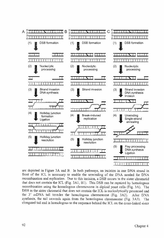
A ;·:.: 1~1 ~I ~I il~l~\~~1~1 ~I il ~~~liB ~:JI~~I~I ~I il ~I~S~I~I ~I ~I il~li C ;: I I I I I 1 S 1 I I I I I I ~::1 I I I I I I I I I I I I I ~:J I I I I I I I I I I I I I I ;: I I I I I I I I I I I I I I I
(1) 1 DSB formation (1) 1 DSB formation (1) 1 DSB formation
!iiii!Si!ili! S
I I I I I I I I I I I I I I I I I I I I I I I I I I I
(2) J Nucleolytic processing
:u IT
I I I I I I I I I I I I I I I
(3) J Strand invasion DNA synthesis
*?I I 1'
(4) J Holliday junction formation
Ligation
(5) J Holliday junction resolution
I I I I I I I I I I I I I I I I I I I I I I I I I I I I
I I
(2) J Nucleolytic process1ng
I I \ IT
I I I I I I I I I I I I I I I
(3) J Strand invasion
(4) J Break-induced replication
,~;:::',::,1:~ (5) J Holliday junction
resolution
I I I I I I I I I I I I I I I I I I I I I I I I I I I
(2) J Nucleolytic processmg
I I I IS IT
I I I I I I I I I I I I I I I
(3) J Strand invasion DNA synthesis Incision
ili!ILS:IJ
11:11!11~ (4) J Unwinding
Single-strand annealing
:t;l I I I I I I I I I I I I I I I I
(5) J Flap processing DNA synthesis Ligation
I I I I
are depicted in Figure 3A and B. In both pathways, an incision in one DNA strand in front of the ICL is necessary to enable the unwinding of the DNA needed for DNA recombination and replication. Due to this incision, a DSB occurs in the sister chromatid that does not contain the ICL (Fig. 3Al, Bl). This DSB can be repaired by homologous recombination using the homologous chromosome in diploid yeast cells (Fig. 3A). The DSB in the sister chromatid that does not contain the ICL is nucleolytically processed and the 3' ssDNA tail invades the homologous chromosome (Fig. 3A2). After DNA synthesis, the tail unwinds again from the homologous chromosome (Fig. 3A3). The elongated tail end is homologous to the sequence behind the ICL on the cross-linked sister
92 Chapter 4
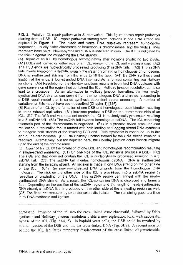
FIG. 2. Putative ICL repair pathways in S. cerevisiae. This figure shows repair pathways starting from a DSB. ICL repair pathways starting from incisions in one DNA strand are depicted in Figure 1. The black and white DNA duplexes represent homologous sequences, usually sister chromatids or homologous chromosomes, and the vertical lines represent base pairs. Newly-synthesized DNA is indicated in gray. The ICL is indicated by the thick diagonal line connecting the DNA strands. (A) Repair of an ICL by homologous recombination after incisions producing two DSBs. (A1) DSBs are formed on either side of an ICL, removing the ICL and yielding a gap. (A2) The DSB ends are nucleolytically processed producing 3' ssDNA tails. (A3) The ssDNA tails invade homologous dsDNA, usually the sister chromatid or homologous chromosome. DNA is synthesized starting from the ends to Jill the gap. (A4) By DNA synthesis and ligation of the ends, a four-stranded DNA intermediate is formed containing two Holliday junctions. (A5) Resolution of the Holliday junctions results in two intact DNA duplexes with gene conversion of the region that contained the lCL. Holliday junction resolution can also lead to a crossover. As an alternative to Holliday junction formation, the two newlysynthesized DNA strands can unwind from the homologous DNA and anneal according to a DSB repair model that is called synthesis-dependent strand annealing. A number of variations on this model have been described (Chapter 1) [386]. (B) Repair of an ICL by the formation of one DSB and homologous recombination resulting in break-induced replication. (B1) Incisions produce a DSB on the centromeric side of an ICL. (B2) The DSB end that does not contain the ICL is nucleolytically processed resulting in a 3' ssDNA tail. (B3) The ssDNA tail invades homologous dsDNA. The ICL-containing telomeric part of the chromosome is degraded. (B4) In a process called break-induced replication, a replication fork is formed with both leading and lagging strand DNA synthesis to elongate both strands of the invading DSB end. DNA synthesis is continued up to the end of the chromosome. (B5) The Holliday junction formed by the DNA strand invasion is resolved. Alternatively, but not depicted here, the Holliday junction could branch migrate up to the end of the chromosome. (C) Repair of an ICL by the formation of one DSB and homologous recombination resulting in single-strand annealing. (C1) On one side of the ICL, incisions produce a DSB. (C2) The DSB end that does not contain the ICL is nucleolytically processed resulting in a 3' ssDNA tail. (C3) The ssDNA tail invades homologous dsDNA. DNA is synthesized starting from the invading strand. An incision is made in one DNA strand on the other side of the ICL. (C4) The newly-synthesized DNA unwinds from the homologous DNA molecule. The nick on the other side of the ICL is processed into a ssDNA region by resection or unwinding of the DNA. This ssDNA region can anneal with the newlysynthesized DNA strand. As a result, the ICL-containing DNA is displaced and forms a flap. Depending on the position of the ssDNA region and the length of newly-synthesized DNA strand, a ssDNA flap is produced on the other side of the annealing region as well. (C5) The flaps are removed by an endonucleolytic incision. The remaining gaps are filled in by DNA synthesis and ligation.
chromatid. Invasion of the tail into the cross-linked sister chromatid, followed by DNA synthesis and Holliday junction resolution yields a new replication fork, with successful bypass of the !CL (Fig. 3A4, 5). In haploid yeast cells, the DSB could be repaired by strand invasion of the DSB end into the cross-linked DNA (Fig. 3B2). A second incision behind the ICL facilitates temporary displacement of the cross-linked oligonucleotide.
DNA interstrand cross-link repair 93
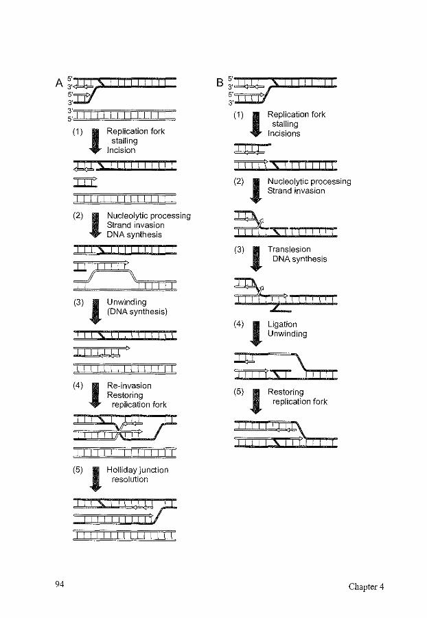
A~:~s!lllllitl 5' 3'
;:·"'ln=l "'I "'I ""l"'ln=l T'l 'i'l "'TI=cln=l T'l '1'1 'T
(1) l Replication fork stalling
Incision
rus ! ! ! ! I
I I I I I I I I I I I I I I I
(2) l Nucleolytic processing Strand invasion DNA synthesis
I I IS
*?II
(3) l
I I I !! I I I
I If \ "'r"'lrr=l orl "'I 'PI
Unwinding (DNA synthesis)
I I IS\\ I I I I\\!
I,IQ >
I I I I I I I I I I I I
(4) l Re-invasion Restoring
replication fork
I I I I I I I I I I I I I I I
(5) l Hollidayjunction resolution
:::,',::::&? I I I I I I I I I I I I
B ~:~ S II II I I I 5'TIT'T/ 3 ..............
(1) l Replication fork stalling
Incisions
1\!!j'XJ!!!!\\\
(2) l Nucleolytic processing Strand invasion
iTi:rrsr I I I I I I I (3) l Translesion
DNA synthesis
(4) l Ligation Unwinding
(5) l Restoring replication fork
FIG. 3. Putative pathways to bypass an ICL during replication in S. cerevisiae. The black and white DNA duplexes represent homologous chromosomes and the vertical lines represent base pairs. Newly-synthesized DNA is indicated in gray. The ICL is indicated by the thick diagonal line connecting the DNA strands. (A) Bypass of an ICL at a stalled replication fork by recombinational mechanisms. This pathway is only feasible in diploid cells. (A 1) The presence of an ICL causes arrest of a replication fork. An incision in the leading strand DNA template nex1 to the ICL produces a nick nex1 to the ICL in the lagging strand and a DSB on the DNA containing the leading strand. Repair is similar when the first incision is made in the lagging strand template. (A2) The DSB is nucleolytically processed, forming a 3' ssDNA tail, that invades the homologous chromosome. DNA is synthesized from the DSB end to bridge the region of the ICL. (A3) The newly-synthesized DNA unwinds from the homologous chromosome. The DNA could be made double-stranded again by DNA synthesis. (A4) The elongated 3' ssDNA end can now invade the cross-linked DNA molecule behind the ICL. Both leading and lagging DNA synthesis are started. (A5) Resolution of the Holliday junction formed by the second invasion step results in the recovery of a normal replication fork with successful bypass of the ICL. (B) Bypass of an ICL at a stalled replication fork by recombination and translesion synthesis. This pathway is feasible in both haploid and diploid cells. (B1) The presence of an ICL causes arrest of a replication fork. Incisions in the lagging strand template next to the ICL produce nicks on both sides of the ICL in the leading strand and a DSB on the DNA containing the lagging strand. Repair is similar when the first incisions are made in the leading strand template. (B2) The DSB is nucleolytically processed, forming a 3' ssDNA tail, that invades the sister chromatid in front of the ICL. (B3) The cross-linked oligonucleotide is displaced and DNA is synthesized from the invading end past the ICL by translesion DNA synthesis. (B4) The newly-synthesized DNA ligates to the parental lagging strand. The DNA unwinds, and the cross-linked oligonucleotide can come back into the helix. (B5) The cross-linked oligonucleotide is ligated to the rest of the leading strand and the replication fork is restored.
The invading end can perform translesion synthesis past the ICL and upon unwinding restore the replication fork (Fig. 3B3, 4, 5). Up to now, there is no direct evidence for these pathways, and several other solutions for bypass or repair ofiCLs during replication are feasible.
Proteins exclusively involved in ICL repair, like Snml and Pso3, may play a role in one or several of these repair routes by assisting other repair proteins in overcoming specific ICL-related difficulties. NER-mediated incision ofiCLs could be hindered by the covalent bond between the DNA strands, that impairs unwinding of the DNA. Recombination could be more difficult as well, if there is only a DSB on one side of the ICL and the ICL persists, while translesion synthesis requires displacement of an oligonucleotide containing the ICL. Adaptations in repair to deal with difficulties like these, could also require additional unknown proteins.
DNA interstrand cross-link repair 95
7 MAMMALIAN DNA INTERSTRAND CROSS-LINK REP AIR
7.1 Finding genes involved in mammalian DNA interstrand cross-link repair
Knowledge about ICL repair in mammalian cells is mainly derived from two sources. The first of these is knowledge of yeast ICL repair. DNA repair pathways have been conserved in eukaryotes from S. cerevisiae to human (Table 2). Similar to the situation in S. cerevisiae, genes required for NER, homologous recombination, and postreplication/ translesion repair are involved in mammalian ICL repair. A number of genes involved in mammalian ICL repair, including RAD6, RAD18, RAD54, SNMJ, REV3, and REV7 have been identified by their sequence homology to yeast genes [181, 254, 266, 367, 369, 517, 576, 578]. However, differences exist in the importance of certain pathways for the repair of certain damages. To repair DSBs, yeast cells mainly use homologous recombination, while NHEJ plays an important role in many mammalian cell types [169, 253]. Furthennore, mammalian DNA repair is often more complex than DNA repair in yeast, which is reflected in the increase in the number of proteins involved. Many yeast DNA repair proteins, such as RAD6, RAD23, RAD30, RAD51, and SNMJ, have more than one homolog/paralog in mammals (Chapter 5) [145, 266, 329, 330, 335, 369, 524].
A second way in which mammalian genes involved in ICL repair have been identified is the analysis of mutant cell lines for their sensitivity to ICL agents. Most ICL-sensitive mammalian cell lines are derived from hamster cell lines. For a number of the highly ICL-sensitive cell lines, the gene responsible for the DNA repair defect has been identified (Table 3). For others, the genetic defect is still unknown [107, 247]. Many cell lines show a moderate sensitivity to ICL agents, probably not due to a repair defect of ICLs, but to a defect in the repair of other adducts induced by ICL agents. A striking contrast exists between the involvement of NER in yeast and in mammalian cells. Yeast NER mutants all show a similar, rather severe, sensitivity to ICL agents [169]. Most mammalian NER mutant hamster cell lines show only a moderate sensitivity to ICL agents, while cell lines with a mutation in the mammalian homologs of RADJ 0 and RAD 1, ERCCJ and ERCC4/XPF, respectively, are very sensitive to ICL agents [107]. An explanation for this difference between yeast and mammals has not yet been found (see below). Apart from the ICL-sensitive hamster cell lines, the Fanconi anemia patient cell lines are also sensitive to ICL agents. Fanconi anemia will be discussed separately below. Finally, Drosophila melanogaster mus308 mutant cell lines are sensitive to ICLs among other types of damage [4, 56]. The gene involved appeared to be a DNA polymerase, that also contains helicase motifs [202, 381]. The human homolog was recently cloned and named DNA polymerase 6 (theta) [459].
In addition to the molecular biological and genetic approaches mentioned above, a number of biochemical attempts have been made to unravel the mechanisms of mammalian ICL repair. A few groups have undertaken the formidable challenge to set up a system to measure aspects of ICL repair in vitro using cell extracts or purified proteins. To date, the results obtained are difficult to interpret in the context of the available genetic
96 Chapter4
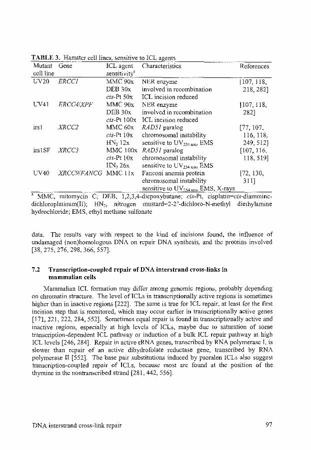
TABLE 3. Hamster cell lines, sensitive to ICL agents Mutant Gene ICL agent Characteristics cellline sensitivit/ UV20 ERCCJ MMC 90x
DEB 30x
UV41 ERCC41XPF cis-Pt SOx MMC90x DEB 30x
NER enzyme involved in recombination ICL incision reduced NER enzyme involved in recombination
irs 1 XRCC2 cis-Pt IOOx ICL incision reduced MMC 60x RAD51 paralog
irs! SF XRCC3
UV40 XRCC91FANCG
cis-Pt lOx HN2 12x MMC !OOx cis-Pt I Ox HN2 26x MMC llx
chromosomal instability sensitive to UV 254 nm• EMS RAD51 paralog chromosomal instability sensitive to UV 254 nm• EMS Fanconi anemia protein chromosomal instability sensitive to UVzs4nm• EMS, X-rays
References
[107, 118, 218, 282]
[107, 118, 282]
[77, 107, 116,118, 249, 512]
[107, 116, 118,519]
[72, 130, 311]
' MMC, mitomycin C; DEB, 1,2,3,4-diepoxybutane; cis-Pt, cisplatin=cis-diamminedichloroplatinum(II); HN2, nitrogen mustard=2-2 '-dichloro-N-methyl diethylamine hydrochloride; EMS, ethyl methane sulfonate
data. The results vary with respect to the kind of incisions found, the influence of undamaged (non)homologous DNA on repair DNA synthesis, and the proteins involved [38, 275, 276, 298, 366, 557].
7.2 Transcription-coupled repair of DNA interstrand cross-links in mammalian ce1ls
Mammalian ICL formation may differ among genomic regions, probably depending on chromatin structure. The level ofiCLs in transcriptionally active regions is sometimes higher than in inactive regions [222]. The same is true for ICL repair, at least for the first incision step that is monitored, which may occur earlier in transcriptionally active genes [171, 221, 222, 284, 552]. Sometimes equal repair is found in transcriptionally active and inactive regions, especially at high levels of ICLs, maybe due to saturation of some transcription-dependent ICL pathway or induction of a bulk ICL repair pathway at high ICL levels [246, 284]. Repair in active rRNA genes, transcribed by RNA polymerase I, is slower than repair of an active dihydrofolate reductase gene, transcribed by RNA polymerase II [552]. The base pair substitutions induced by psoralen ICLs also suggest transcription-coupled repair of ICLs, because most are found at the position of the thymine in the nontranscribed strand [281, 442, 556].
DNA interstrand cross-link repair 97
7.3 Cell cycle effects of DNA interstrand cross-links
The organization of DNA damage-induced cell cycle checkpoints in mammalian cells is comparable to the situation in yeast cells [594]. A number of proteins, like Radl, Rad9, Husl, and Radl7, are present to detect DNA damage or repair intermediates. One of the signal transduction proteins is Atm, a homolog of the yeast checkpoint protein Mec 1, and responsible for the human genome instability and cancer predisposition syndrome ataxia telangiectasia [286]. Together with other transducer proteins, like Atr, Chkl, and Chk2, it phosphorylates a number of effector proteins like p53, Nbs!, and Brcal, that induce cell cycle arrest, apoptosis and DNA repair [594]. A number of proteins, including Nbs!, Brcal, and Brca2, are supposed to play a role both in cell cycle checkpoint responses and in DNA repair [87, 136, 326, 592]. NBSJ is the gene involved in Nijmegen breakage syndrome. The cellular phenotypes of NBSJ and ATM mutations are similar, including chromosomal instability, radiosensitivity, defective cel1 cycle checkpoints, and reduced p53 responses [136]. Nbs! forms a complex with Mrell and Rad50 that is involved both in homologous recombination and NHEJ. Nbs 1 targets Mre 11 and Rad50 to repair sites for DSBs [73]. Brcal and Brca2 are involved in familial breast and ovarian carcinomas [133, 592]. Apart from its role in damage-induced cell cycle checkpoints, Brcal is involved in transcription and DSB repair. After ionizing radiation, Brcal can be fotmd in association with Mrell-Rad50-Nbsl, Rad51, and Brca2 [88, 89, 455, 593].
The role of cell cycle checkpoints in the cellular response to ICL agents has hardly been investigated. Similar to the situation in yeast, the induction of cell cycle checkpoints depends on the ICL agent used. After treatment with mitomycin C, Chinese hamster ovary (CHO) cells continue to replicate [283]. On the other hand, treatment with cisplatin induces G2 arrest, followed by either apoptosis or continuation of cell cycling [283, 484]. Mutations in ATM, BRCAJ, BRCA2, and NBSJ cause sensitivity to ICL agents, but it is not known, whether this is due to their role in cell cycle checkpoints or in DNA repair [ 41, 176, 363, 586].
7.4 Processes during mammalian DNA interstrand cross-link repair
7.4.1 Recognition and incisions The proteins responsible for recognition of ICLs in mammalian cells are not known.
There could be specific DNA damage recognition proteins, or recognition could occur when transcription or replication are blocked. While NER enzymes probably make many of the incisions required during ICL repair in yeast, their role in mammalian ICL repair is unclear. Mammalian cells may use other enzymes to make the incisions, because most NER mutant cell lines are only moderately sensitive to ICL agents, and therefore are thought to be defective only in the repair of monoadducts and intrastrand cross-links. In some assays, an ICL repair deficiency is found in NER mutants, suggesting they could play a minor role in ICL repair [556, 557, 590]. The only NER genes that have a major influence on mammalian ICL repair are ERCCJ and ERCC4/XPF, the homo logs of S. cerevisiae RADIO and RADJ [169].
98 Chapter 4
Erccl and Ercc4/Xpf form a complex that functions as an endonuclease that nicks dsDNA near the transition between dsDNA and ssDNA, when the ssDNA forms a 3' ssDNA tail [18, 126, 474]. InNER, Erccl-Xpf incises the DNA at the 5' side of the lesion, while Xpg incises at the 3' side, thereby removing an oligonucleotide containing the lesion [169]. Furthermore, Erccl-Xpf and RadlO-Radl are involved in removing nonhomologous ssDNA tails during homologous recombination [1, 18, 223, 448]. Mammalian ERCCJ knockout mice show a much more severe phenotype than other NER mutant mice. They are not only UV254 run-sensitive, but have a severe growth failure, many mice die before birth, and their maximal age is around 80 days. The mice are infertile, show nuclear abnormalities in liver cells, renal dysfunction, and no subcutaneous fat [340, 560]. Cells from ERCCJ knockout mice show early senescence and genomic instability [343, 560]. They are also very sensitive to mitomycin C and other ICL agents. The role of the Erccl-Xpf complex in ICL repair may differ from its function inNER, as some ERCCJ and ERCC4 mutant CHO cells that have been characterized, are only mildly sensitive to mitomycin C, but still are sensitive to UV 254 nm [71].
Incision of ICLs is reduced in ERCCJ mutant cells, but normal in XPG mutants [118, 282]. In vitro, Erccl-Xpfcan cut a Y-shaped oligonucleotide with an ICL in the dsDNA near the transition between dsDNA and ssDNA. The incisions occur both at the 5' and 3' side of the ICL [279]. Such a Y shape could be formed in vivo by a stalled replication fork, a transcription complex, or a helicase. If the in vivo incision activity ofErccl-Xpfis the same as the in vitro activity, Erccl-Xpfincisions next to the ICL in one strand will create a similar situation to that after the first incisions in E. coli (Fig. 1A2). Homologous recombination could then continue the repair reaction. Erccl-Xpf could also perform incisions later during ICL repair. One of its additional roles could be the removal of nonhomologous DNA tails during homologous recombination in ICL repair (see Figure 2C). The loss ofErccl-Xpffunction in this late step of!CL repair may therefore be the cause of the extreme sensitivity of ERCCJ mutant cells, compared to cells mutant in other NER genes. Maybe, the repair intermediate formed by strand invasion after the occurrence of a DSB next to an ICL is preferentially resolved by break-induced replication in yeast, but by single-strand annealing in mammalian cells (Fig. 2B, C). This could be an explanation why loss of the yeast hornologs of Ercc1-Xpf does not lead to a higher lethality than loss of other NER proteins.
7.4.2 Recombination The extent ofDSB induction during the repair of!CLs in mammalian cells is not well
known, because their formation and disappearance are much more difficult to monitor than in yeast cells. Some data suggest that DSBs are indeed formed [118, 549]. While ionizing radiation-induced DSBs in many mammalian cell types are often repaired via NHEJ, mutations in NHEJ do not cause sensitivity to ICL agents [42]. Whether or not DSBs play an important role, homologous recombination genes are important for ICL repair. After treatment with ionizing radiation or ICL agents, proteins involved in homologous recombination, like Rad51 and Rad54, form nuclear foci, which could be centers for the repair of DNA damage [194, 513, 516]. Vertebrate cells mutant for mRAD54 and the RAD51 paralogs are known for their sensitivity to ICL agents [107, 154,
DNA interstrand cross-link repair 99
176, 509, 511]. Sensitivity to ICL agents is also found in BRCA I mutant tumor cells, and in Brcal- and Brca2-deficient mouse cells [41, 176, 363, 586]. Brcal and Brca2 associate with each other and Rad51 and they are required for the formation of damage-induced Rad51 nuclear foci [41, 455, 458, 587, 592]. Furthermore, Brcal- and Brca2-deficient cells are deficient in homologous recombination [362, 365, 478]. The proteins could have a regulatory role in coupling the DNA damage response, cell cycle checkpoints, and DNA repair.
Another indication for the importance of recombination during ICL repair is the recombinogenicity of ICLs. Short tract gene conversions sometimes accompanied by crossover have been observed after induction ofiCLs [158, 445]. Treatment of cells with ICL agents induces sister chromatid exchanges (SCEs), another measure for recombination, while SCEs are hardly induced by monofunctional agents [49]. The toxicity of several types of ICL agents is correlated with the number of SCEs that are induced by the ICLs formed [49, 566]. SCEs induced by mitomycin C are found only after the first replication, suggesting that these ICLs cannot be bypassed efficiently during replication, causing cell death when repair has failed [569]. Psoralen ICLs are also hardly found in replicated DNA, in contrast to psoralen monoadducts [551]. The induction of SCEs by ICL agents is reduced in cells mutant for RAD54, RAD51, or its paralogs (Chapter 2) [144, 482,509, 511].
Mutations in the vertebrate RAD51 paralogs yield a higher sensitivity to ICL agents than mutations in the other genes from the homologous recombination pathway, suggesting different subpathways of recombination that are not equally important for ICL repair [154, 509, 511]. Therefore, the RAD51 paralogs will be discussed here in more detail. Both yeast and human Rad51 play a central role in homologous recombination and can perform DNA strand exchange in vitro [27, 501]. In yeast, two RAD51 paralogs are known, RAD55 and RAD57, that stimulate the Rad51-mediated DNA strand exchange reaction [255, 315, 503]. In mammalian cells, three mitotically active paralogs of RAD51 have been identified by their amino acid sequence, RAD51B, RAD51C, and RAD5JD [7, 76, 142, 258, 408, 422]. Two other paralogs were identified as the genes responsible for the ICL sensitivity of the mutant CHO cell lines irs! and irslSF, and they are called XRCC2 and XRCC3, respectively [77, 310, 512, 519]. The Rad51 paralogs interact with each other and with Rad51 [60, 142, 310, 451, 524]. RAD51 deficiency leads to cellular lethality, while chicken cells deficient in any one of the RAD51 paralogs are viable. Nevertheless, both chicken and mammalian cells deficient for a RAD51 paralog show chromosomal instability [116, 191,509,511, 519]. Mice deficient for RAD51, RAD51B, RAD5ID, and XRCC2 show embryonic lethality, signifying the importance of homologous recombination for genomic integrity and cell proliferation [131, 305, 407, 466, 536]. The unhooking of ICLs is not affected in XRCC2 and XRCC3 mutant cells, as expected for homologous recombination mutants [118]. Repair ofDSBs by homologous recombination is affected in mutant XRCC2 and XRCC3 cells, but NHEJ is not affected [64, 240, 405]. ICL-induced Rad51 nuclear foci formation is impaired in XRCC3-deficient cells [45].
The Rad51 paralogs could play a supporting role in Rad51-mediated DNA strand exchange. The RecA-mediated DNA strand exchange reaction in E. coli is stimulated by
100 Chapter 4
RecBCD when starting from a DSB intermediate and by RecFOR in the absence of a DSB [8, 268, 538]. Homologous recombination in mammalian cells probably needs some adaptations during ICL repair compared to DSB repair, because there may be repair pathways in which only one end of a DSB is available for strand invasion (Fig. 2B, C and Fig. 3) and others in which strand invasion is required in the absence of a DSB (see Figure lA). Cells with a mutation in the Rad51 paralogs are much more sensitive to ICL agents, than to DSB-inducing agents [107, 248, 519, 525]. This may be explained by a specific role of these paralogs, like the role of RecBCD and RecFOR, in adapting the strand invasion reaction by Rad51 to the requirements ofiCL repair.
7.4.3 Postreplicationltranslesion repair Little is known about the influences of the mammalian postreplicationltranslesion
repair group of enzymes on the cellular response to ICL agents. Human Rad6/UbcHI can rescue yeast rad6 sensitivity to 8-methoxypsoralen plus UVA [251]. RADJ8-deficient vertebrate cells are very sensitive to cisplatin and have increased levels of SCEs, suggesting a switch from postreplication/translesion repair to homologous recombination (Y.M. Yamashita, S. Takeda, personal communication). Expression of a dominantnegative mutant of human RAD18 makes cells sensitive to mitomycin C [517]. Human RAD30/XPV mutant cells show a decreased mutagenesis and replication bypass of ICLs compared to wild-type cells [351, 417]. The mutagenicity of ICLs compared to monoadducts differs per species and cell type, suggesting different preferences for certain ICL repair pathways [15, 61, 281, 583].
7.5 Mammalian Snml homologs
The mammalian homolog of the yeast Smnl/Pso2 protein is also involved in the response to ICL agents. SNMI-deficient mouse embryonic stem cells are sensitive to mitomycin C, but not to 8-methoxypsoralen plus UV A, melphalan, or cisplatin (Chapter 5) [145]. SNMJ knockout mice show a two-fold higher sensitivity to mitomycin C, which is similar to the sensitivity of the homologous recombination-deficient RAD54 knockout mice (Chapter 5) [145, 155]. SCE induction by mitomycin Cis similar in SNMJ-deficient and wild-type cells, suggesting that Snml plays no role in the homologous recombination pathway of ICL repair (Chapter 5). In mammalian cells, at least two other proteins that display similarity to Snml are present (Chapter 5) [145]. They could have redundant functions in the cellular response to different ICLs. However, Snm!B and Snm!C could also be involved in other cellular processes. The human SNMJ CIARTEMIS gene has been isolated as one of the genes responsible for severe combined immunodeficiency, which is probably due to a defect in NHEJ [361].
7.6 Fan coni anemia
Fanconi anemia is a rare, autosomal recessive disorder, characterized and diagnosed by the sensitivity of patient cells to ICL agents. Clinical symptoms of Fanconi anemia include progressive bone marrow failnre, leading to pancytopenia, developmental
DNA interstrand cross-link repair 101
abnormalities, such as aplasia of the thumb and radius, growth retardation, hyperpigmentation of the skin, kidney and urinary tract malformations, and microcephaly. Patients are predisposed to the development of cancer, particularly acute myeloid leukemia and squamous cell carcinoma [13, 238]. Increased spontaneous and ICLinduced chromosomal breakage is observed, which places Fanconi anemia among the chromosomal instability disorders, like ataxia telangiectasia, Bloom's syndrome, Nijmegen breakage syndrome and hereditary nonpolyposis colorectal carcinoma. Further characteristics of Fanconi anemia cells are sensitivity to oxidative DNA damage, cell cycle abnormalities, changes in certain cytokines and abnormal apoptosis [68, 83, 101, 238, 444]. There is no clear defect in the formation and repair of ICLs in Fanconi anemia cell lines, although they do show prolonged G2 arrest and highly increased cell death on ICL agent treatment [68, 101, 444].
Thus far, eight Fanconi anemia complementation groups have been defined and six genes have been mapped and cloned, FANCA, FANCC, FANCD2, FANCE, FANCF, and FANCG [108, 127, 128, 130,205,236,237,314,493, 528]. A correlation has been found between the complementation group, the type of mutation, and the clinical severity [!57]. Both FANCA and FANCC knockout mice have been generated [91, 95, 565]. The mice and cell lines isolated from them are very sensitive to ICL agents and also show ICLinduced chromosomal instability and cell cycle abnormalities. Otherwise, their phenotype is very mild under normal breeding conditions, except for reduced fertility [74, 91, 95, 382, 565].
Although Fanconi anemia is a very intriguing human disease that will be of great value in understanding human ICL repair mechanisms, the function of the Fanconi anemia (FA) proteins is still unclear. The FA genes encode novel proteins, whose sequence provides no immediate clues for a biochemical function. Up to now, FANCD2 is the only FA gene that has homologs in lower eukaryotes, namely Caenorhabditis elegans, D. melanogaster, and Arabidopsis thaliana, but there is no homolog in S. cerevisiae [528]. The role of the other, probably upstream ofF ANCD2 functioning FA proteins may be fulfilled by sequence-unrelated proteins in lower eukaryotes. Because there are no homologous proteins in yeast, it is much more difficult to unravel the role of the FA proteins, than the role of most of the other proteins involved in !CL repair that have been discussed above.
A substantial amount of work has been done to determine the cellular localization and interactions of the FA proteins. FANCA, FANCC, and FANCG are found both in the cytoplasm and in the nucleus, while F ANCD2, F ANCE, and FANCF are predominantlynuclear [129, 174-176, 213, 271]. Interactions have been observed among FANCA, FANCC, FANCE, FANCF, and FANCG, but only the interaction between FANCA and FANCG has been shown to be direct [129, 174, 175, 271, 273, 277, 341, 554]. These interactions suggest the presence of a large complex formed by the FA proteins in the nucleus. All the interactions found are diminished or absent in cells belonging to other Fanconi anemia complementation groups, except for group Dl and D2 cells [129, 174, 175,554, 580].
An interaction model has been proposed in which the different FA proteins sequentially form a complex and translocate to the nucleus [129]. This complex may be
102 Chapter 4
required for DNA damage-dependent activation of FANCD2 to a monoubiquitinated isoform, that forms nuclear foci after treatment with UV 254 nm• ionizing radiation, and ICL agents [176]. The ubiquitination of FANCD2 also depends on Brcal, and ubiquitinated FANCD2 can be coprecipitated with BrcaL After ionizing radiation, FANCD2 and Brcal colocalize in nuclear foci. Interactions with other cellular proteins have also been investigated. FANCC interacts with NADPH cytochrome P450 reductase, with glutathione S-transferase, with a transcriptional repressor protein, and with a nonerythroid a spectrin, which may function as a scaffold for interacting proteins [117, 214, 272, 339]. These interactions suggest that the FA proteins may have other functions apart from their role in the response to ICL agents [384].
The FA proteins may work together in a stable bolo-complex of all FA proteins, or in dynamically interacting complexes containing a subset ofF A proteins [23 8]. They might have some direct role in ICL repair, but could as well have a more indirect role, influencing ICL formation, damage recognition, or the cellular response to ICLs. The interaction with Brcal may provide a link between the FA proteins and the cellular damage response, cell cycle regulation, or homologous recombination. Whatever their role, the FA proteins are essential for chromosomal stability, especially when cells are exposed to ICL agents.
7.7 Conclusions on mammalian DNA interstrand cross-link repair
Generally, ICL repair in mammalian cells is comparable to ICL repair in bacteria and in yeast The pathways described in Figures I, 2, and 3 are also feasible for ICL repair in mammals. However, as with most other cellular processes, mammalian ICL repair is more complex than yeast ICL repair. Several mammalian homo logs of S. cerevisiae ICL repair genes have a number of paralogs. This expansion could be a sign of the specialization of proteins in the repair of ICLs, or they could be active in different cell types or cell cycle stages. Furthermore, proteins without a homolog in S. cerevisiae, such as the Brca and FA proteins play a role in mammalian ICL repair.
A remarkable difference between mammals and yeast is that only Ercc 1 and Xpf play a major role in ICL repair in mammalian cells in contrast to the involvement of most other NER proteins in S. cerevisiae. Although the other NER proteins are important for making incisions in yeast cells, they may have only a limited or even no role in making incisions next to ICLs in mammalian cells, while the incisions are still dependent on Erccl-Xpf. Alternatively, the difference between mammals and yeast may be due to an additional role for Ercc!-Xpf later during ICL repair. For example, Erccl-Xpf could possibly be responsible for the removal of nonhomologous ssDNA tails during recombinational repair of an ICL (Fig. 2C). Break-induced replication is a frequently used pathway during homologous recombination in yeast, but in mammalian cells, there is no direct evidence for the existence of break-induced replication [386]. The reason may be the length and complexity of mammalian chromosomes, compared to yeast chromosomes. Instead of using the pathway that depends on break-induced replication (Fig. 2B), mammalian cells could be much more dependent on single-strand annealing to resolve recombination
DNA interstrand cross-link repair 103
intermediates (Fig. 2C). It is likely that Erccl-Xpf plays an essential role in this recombinational subpathway.
Furthermore, the importance of the induction of DSBs during ICL repair in mammalian cell lines is unknown. Nevertheless, homologous recombination is very important in mammalian ICL repair. The Rad51 paralogs could play a special role in adapting homologous recombination to ICL repair, especially when no DSBs are induced, or when only one end of a DSB is available for homologous recombination (Fig. lA, 2B, 2C, 3A, 3B). While in yeast, Rad52 is even more important than Rad51 for a number of homologous recombination subpathways, the role of Rad52 in mammalian homologous recombination is minor [386, 428]. Mammalian cells could more strongly depend on the Rad51 paralogs to stimulate Rad51-mediated DNA strand exchange than on Rad52 [353]. The idea of a redundant role for the Rad51 paralogs and Rad52 in stimulating Rad51-mediated DNA strand exchange is supported by the fact that while chicken DT 40 cells deficient for either RAD52 or XRCC3 are viable, cells deficient for both genes are not viable (unpublished observations in [483]).
The Snml, FA, and Brca proteins may function in the regulation of the cellular damage response to ICLs. The Brca proteins are involved in damage-induced cell cycle checkpoints via Atm and in homologous recombination, and Brcal has been associated with transcription. The pleiotropic phenotype of Fanconi anemia and the link between FANCD2 and Brcal are also consistent with a regulatory role for the FA proteins. The transport of some of the FA proteins from the cytoplasm to the nucleus, and their activation by phosphorylation and ubiquitination suggest a role in a signal transduction pathway. The specific roles for the large number of proteins involved directly or indirectly in the repair of!CLs will have to be elucidated by further research.
ACKNOWLEDGMENTS
Research on ICL repair in our laboratory is supported by a grant from the Netherlands Organization for Scientific Research (NWO).
104 Chapter 4
CHAPTERS
Disruption of mouse SNMJ causes increased sensitivity to the DNA interstrand cross-linking agent mitomycin C
Modified from Mol. Cell. Biol. 20(13): 4553-4561, 2000

DISRUPTION OF MOUSE SNMI CAUSES INCREASED SENSITIVITY TO THE DNA INTERSTRAND CROSS-LINKING AGENT MITOMYCIN C
MIES L.G. DRONKERT1, JAN DE WIT1
, MIRANDA BOEVE2, M. LUISA VASCONCELOSu,
HARRY VAN STEEG2, H. BERNA BEVERLOO\ T.L. RAOUL TAN1
, JAN H.J. HOEIJMAKERS 1 AND ROLAND KANAARt.4
1 Department of Cell Biology and Genetics, Erasmus University Rotterdam, PO Box 1738, 3000 DR
Rotterdam, The Netherlands
2 Laboratory of Health Effects Research, National Institute of Public Health and the Environment,
PO Box 1, 3720 BA Bilthoven, The Netherlands
3 Present address: Department of Biological Chemistry, Howard Hughes Medical Institute, University of California, 5-748 MacDonald Bldg, Los Angeles, CA 90095-1662, USA
4 Department of Radiation Oncology, University Hospital Rotterdam/Daniel, The Netherlands
DNA interstrand cross-links (ICLs) represent lethal DNA damages, because they block transcription, replication, and segregation of DNA Because of their genotoxicity, agents inducing ICLs are often used in antitumor therapy. The repair of ICLs is complex and involves proteins belonging to nucleotide excision, recombination, and postreplication/translesion DNA repair pathways, in Escherichia coli, Saccharomyces cerevisiae, and mammals. The molecular mechanisms of ICL repair in mammalian cells are still unknown. We cloned and analyzed mammalian homologs of the S. cerevisiae gene, SNMJ(PS02), which is specifically involved in ICL repair. Human Snml, a nuclear protein, was ubiquitously expressed at a very low level. We generated mouse SNMr1
-
embryonic stem cells, and showed that these cells were sensitive to mitomycin C. In contrast to S. cerevisiae snml mutants, they were not significantly sensitive to other ICL agents, probably due to redundancy in mammalian ICL repair and the existence of other SNMJ paralogs. The sensitivity to mitomycin C was complemented by transfection of the hSNMJ eDNA and by targeting of a genomic-eDNA murine SNMJ fusion construct to the disrupted locus. We also generated mice deficient for murine SNMJ. They were viable and fertile and showed no major abnormalities. However, they were sensitive to mitomycin C. Snml is probably not involved in recombinational repair, because the response ofRad5 1 towards ICL agents and the induction of sister chromatid exchanges by DNA damaging agents are not affected by the absence of Snml. The ICL sensitivity of the mammalian SNMJ mutant suggests that SNMJ function and, by implication, ICL repair are at least partially conserved between S. cerevisiae and mammals.
INTRODUCTION
DNA interstrand cross-links (ICLs) prevent strand separation, thereby physically blocking transcription, replication, and segregation of DNA. In bacterial and yeast cells,
Mouse SNMJ and DNA interstrand cross-link repair 107
the presence of one unrepaired ICL can be lethal [287, 320], Humans are exposed to environmental ICL agents, such as furocoumarins, from plants and cosmetics [454], Due to their extreme genotoxicity, agents that induce cross-links in DNA are widely used in antitumor therapy. Examples include cisplatin, mitomycin C, and derivatives of nitrogen mustard such as melphalan and cyclophosphamide (See Chapter 4 ). Most of these agents cause a number of different lesions in DNA, including monoadducts, DNA-protein crosslinks, DNA intrastrand cross-links, and ICLs. The latter are the main cause of cell death, although they constitute only a few percent of the total number of adducts [63, 287]. Resistance of tumors to cross-linking agents can be caused by a variety of mechanisms, including increased repair of ICLs [6, 25, 59, 152, 244, 488, 582, 591], Therefore, an understanding of how ICLs are repaired is important.
To elucidate the mechanism of ICL repair in yeast, genetic screens have been performed to find genes specifically involved in ICL repair. A number of snm and pso mutants have been isolated after screening for strains with increased sensitivity towards nitrogen mustard and 8-methoxypsoralen plus UV A light, respectively [208, 438]. Some of the mutants isolated are sensitive to several classes of DNA-damaging agents, while others are almost exclusively sensitive to ICL agents. The gene mutated in one of these strains is SNMJ, which is allelic with PS02 and encodes a nuclear 76-kDa protein [82, 207, 427]. snml mutants are sensitive to a number of agents that cause ICLs, but they are only mildly sensitive to monofunctional a1kylating agents and 254-nm UV light (UV254 ,m) and are not hypersensitive to gamma rays [63, 208, 439]. In exponentially growing cells, the expression of SNMJ can be induced by ICL agents or UV254 "m [570]. Overexpression of SNMJ results in an increased resistance to nitrogen mustard and cisplatin [206].
Many genes involved in DNA damage repair are conserved between yeast and higher eukaryotes. One way to elucidate the mechanisms of ICL repair in mammalian cells is by studying mammalian homologs of S. cerevisiae genes specifically involved in ICL repair. A human homolog of SNMJ has been isolated and comparison of its predicted protein product (hSnml) to S. cerevisiae Snml (ScSnml) has shown that 39% of the ScSnml amino acids have a similar amino acid in hSnml [369]. The hSNMJ gene is located on chromosome 10q25, a region often found to be rearranged in tumors [48, 267, 396, 475, 491,531].
Here, we report on the characterization of the mammalian SNMJ homologs. We generated mouse embryonic stem (ES) cells in which both alleles of mouse SNMJ (mSNMJ) are disrupted. These cells were sensitive to mitomycin C, but not to 8-methoxypsoralen plus UV A light, cisplatin, melphalan, UV254 "m' methyl methanesulfonate or gamma rays. The sensitivity to mitomycin C was restored to wild-type sensitivity by transfecting either a plasmid expressing hSnm1 or by targeting a genomiceDNA mSNMJ fusion construct to the disrupted locus. We also generated mice deficient for mSNMJ. They were viable and fertile and showed no major abnormalities. However, the mice were sensitive to mitomycin C. Our results show that mammalian SNM I is a functional homolog of yeast SNMJ, even though only 22% of the ScSnm1 amino acids are identical to hSnml amino acids. This indicates that at least parts of the DNA repair mechanisms specific for the repair of ICLs are conserved between S. cerevisiae and mammals.
108 Chapter 5
MATERIALS AND METHODS
hSNMI eDNA constructs. The eDNA of hSNMI (kindly provided by N. Nomura) was used to make several constructs containing tags at the 5'- and 3' -termini of hSNMI (Table 1). For this purpose, the 5' terminus of hSNMI was modified to generate an Xhol restriction enzyme site just in front of the expected initiation codon, thereby deleting the 900-bp 5' untranslated region that contains 15 additional ATG sequences. The constructs were generated in pBluescript II KS (Stratagene) and subsequently subcloned into appropriate expression vectors (Table 1). The sequence of all DNA fragments produced by PCR was determined by sequence analysis. The following tags were added to hSnml: an amino-terminal histidine tag (His 10: single letter amino acid code: MGHHHHHHHHHHGGSR), a carboxy-terminal hemagglutinin tag (HA: PGGYPYDVPDY AS) and a carboxy-terminal histidine-hemagglutinin tag (His6HA: PHHHHHHGGSAYPYDVPDYAS). The first 570 bp of hSNMI, the first 1515 bp of hSNMI and the last 1470 bp and the total hSNMI eDNA with a carboxy-tenninal His6HA tag were subcloned into pEGFPC vectors (Clontech) to obtain hSnml protein with an amino-terminal Green Fluorescent protein (GFP) tag (Table 1).
Subcellular localisation of hSNMJ. The constructs GFP-hSNMI, GFP-570hSNMJ, GFP-1515hSNMJ, and GFP-ChSNMI were transfected with lipofectin into CH09 cells, grown in F!O-DMEM containing 10% fetal calf serum. To obtain stable transfectants, cells were split after one day and put on G4!8 selection (! mg/ml). Single colonies expressing GFP-hSnml proteins were expanded. The GFP-hSNMJ construct was also microinjected into multinucleated fibroblasts according to previously described procedures [215]. The cellular localization of the GFP was determined using an Olympus IX70 microscope.
Generation of a-hSnml antibodies. A 354-bp Psti-Hindlll fragment from the hSNMJ eDNA was subcloned into pTrcHisC (Table I). The fusion protein derived from this plasmid contained amino acid 644 to 763 ofhSnml fused to a His6 tag and was produced in E. coli strain DH5a. The protein was present in the insoluble fraction and was dissolved in 6 M urea. It was purified on a Ni-nitrilotriacetic acid column, eluted with 200 mM imidazole and used to immunize two rabbits. The detection limit of the polyclonal antibodies was determined by immunoblotting using a range of 1 to 1,000 ng of antigen and was shown to be below I ng. The antibodies were affinity purified using fusion protein immobilized on a nitrocellulose filter.
Expression of recombinant hSnml. The eDNA encoding His10-hSnml-HA was subcloned into pFastBacl (BAC-TO-BAC Baculovirus Expression System, GibcoBRL) (Table l ). The resulting plasmid was transformed into DH! OBac E. coli cells to allow site-specific transposition into bacmid bMONI4272. High molecular weight recombinant bacmid DNA was isolated and transfected into Sf2! cells to produce virus stocks that were amplified as described by the manufacturer. Protein extracts of these cells were made as described [505].
Mouse SNMI and DNA interstrand cross-link repair 109
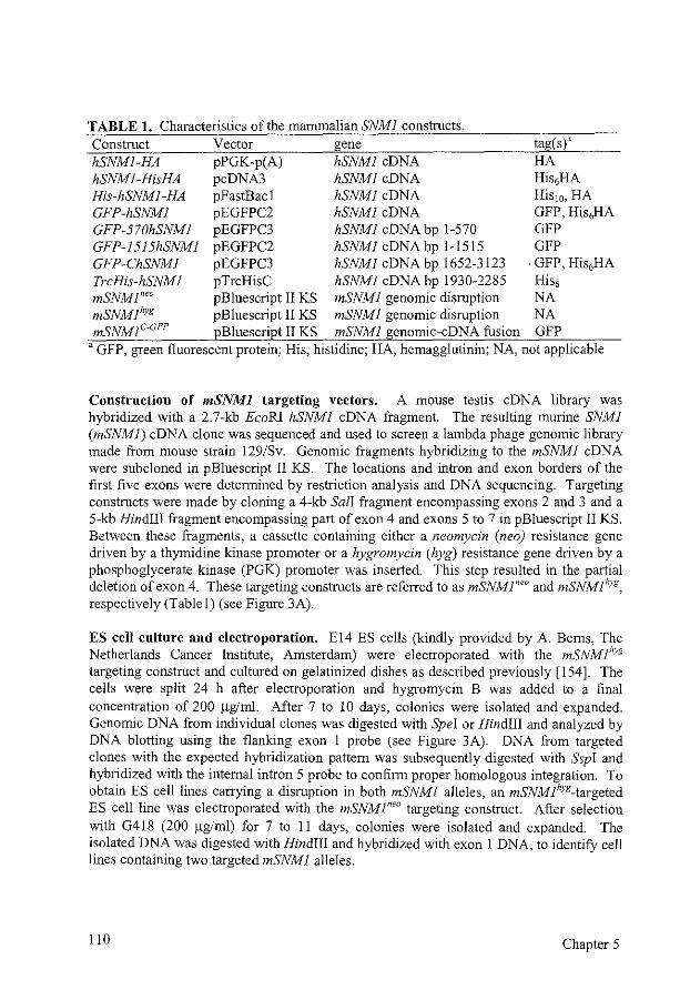
TABLE I. Characteristics of the mammalian SNMJ constructs. Construct Vector gene tag(s)a hSNMI-HA pPGK-p(A) hSNMJ eDNA HA hSNMI-HisHA pcDNA3 hSNMJ eDNA His6HA His-hSNMJ-HA pFastBacl hSNMJ eDNA His10, HA GFP-hSNMI pEGFPC2 hSNMJ eDNA GFP, His6HA GFP-570hSNMJ pEGFPC3 hSNMJ eDNA bp 1-570 GFP GFP-1515hSNMJ pEGFPC2 hSNMJ eDNA bp 1-1515 GFP GFP-ChSNMI pEGFPC3 hSNMJ eDNA bp 1652-3123 · GFP, His6HA TrcHis-hSNMI pTrcHisC hSNMJ eDNA bp 1930-2285 His6
mSNMI'" pBluescript II KS mSNMI genomic disruption NA mSNMihyg pBluescript II KS mSNMI genomic disruption NA mSNMJC-GFP pBluescript II KS mSNMl genomic-eDNA fusion GFP
'GFP, green fluorescent protein; His, histidine; HA, hemagglutinin; NA, not applicable
Construction of mSNMJ targeting vectors. A mouse testis eDNA library was hybridized with a 2.7-kb EcoRI hSNMI eDNA fragment. The resulting murine SNMJ (mSNMJ) eDNA clone was sequenced and used to screen a lambda phage genomic library made from mouse strain 129/Sv. Genomic fragments hybridizing to the mSNMJ eDNA were subcloned in pBluescript II KS. The locations and intron and exon borders of the first five exons were detennined by restriction analysis and DNA sequencing. Targeting constructs were made by cloning a 4-kb Sal! fragment encompassing exons 2 and 3 and a 5-kb Hindiii fragment encompassing part of exon 4 and exons 5 to 7 in pBluescript II KS. Between these fragments, a cassette containing either a neomycin (neo) resistance gene driven by a thymidine kinase promoter or a hygromycin (hyg) resistance gene driven by a phosphoglycerate kinase (PGK) promoter was inserted. This step resulted in the partial deletion of ex on 4. These targeting constructs are referred to as mSNMreo and mSNMlhyg' respectively (Table!) (see Figure 3A).
ES cell culture and electroporation. E 14 ES cells (kindly provided by A. Berns, The Netherlands Cancer Institute, Amsterdam) were electroporated with the mSNMJ"Yg targeting construct and cultured on gelatinized dishes as described previously [154]. The cells were split 24 h after electroporation and hygromycin B was added to a final concentration of 200 ~glml. After 7 to 10 days, colonies were isolated and expanded. Genomic DNA from individual clones was digested with Spel or Hindiii and analyzed by DNA blotting using the flanking exon 1 probe (see Figure 3A). DNA from targeted clones with the expected hybridization pattern was subsequently digested with Ssp! and hybridized with the internal intron 5 probe to confirm proper homologous integration. To obtain ES cell lines carrying a disruption in both mSNMI alleles, an mSNMlhyg_targeted ES cell line was electroporated with the mSNMreo targeting construct. After selection with G4!8 (200 ~g/ml) for 7 to II days, colonies were isolated and expanded. The isolated DNA was digested with Hindiii and hybridized with exon I DNA, to identifY cell lines containing two targeted mSNMJ alleles.
110 Chapter 5
Rescue of mSNMl-1- cells by hSNMJ eDNA constructs. The hSNMI-HA eDNA was subcloned into pPGK-p(A) to express the gene under the control of the PGK promoter with a carboxy-terminal HA tag (Table 1 ). This eDNA expression construct was coelectroporated with a puromycin-expressing plasmid into mSNMreo!hyg cells. Clones were selected with puromycin (1 ~g/ml) for 10 days. Integration of the eDNA construct was confirmed by DNA blotting.
Rescue of mSNMl-1- cells by mSNMI genomic constructs. An mSNMJ-GFP targeting fusion construct was made under the control of the mSNMJ promoter (Fig. 3B). To obtain expression of mSNMJ with 3'-terminal GFP, the 3' terminus of mSNMJ eDNA was modified by PCR to create an EcoRV site in front of the stop codon. In this site, the GFP eDNA was cloned, and the borders were sequenced. Then, the 5.6-kb genomic mSNMJ Hindiil fragment encompassing exons I to 4 was cloned into the eDNA, thereby fusing exon 4 from the genomic fragment with eDNA exons 4 to 9. Behind the endogenous polyadenylation signal, a neomycin cassette and the 5-kb mSNMJ Hindiil fragment were cloned (see Figure 3B). The construct, named mSNMJC-GFP, was transfected into mSNMlhyg_targeted ES cells, and clones were selected with G4!8 (200 ~g/ml) and expanded. The isolated DNA was digested with Ssp! and hybridized with the intron 5 probe to screen for cell lines containing the homologously integrated target DNA.
RT-PCR reactions. mRNA was isolated from 106 to 107 ES cells as described (Pharmacia Quick Prep Micro mRNA purification). eDNA was made using Pharmacia 'Ready to go you prime first strand' beads and either a specific primer or a random hexamer primer. Then, PCR was performed on I to 10 ~1 eDNA preparation. The PCR reaction to detect mRNA from mSNMr1
- cells was performed with 5'GAGACGGACTCGTGGCAGGAC and 5'-AGTCGCACATGTCTGTGGTTATGA as a forward and reverse primer, respectively, and gave a product of 740 bp. To detect mRNA from mSNMJC-GFP' the forward and reverse primers were 51
-
GAGACGGACTCGTGGCAGGAC and 5'-TTGTGGCCGTTTACGTCGCCG, respectively. These primers yielded a PCR product of 1.2 kb.
Cell survival assays. The sensitivity of ES cells to increasing doses of DNA-damaging agents was determined by measuring their colony-forming ability as described before [!54]. The cloning efficiency of untreated cells varied between 10 and 30%. Cells were incubated in drug-containing media for I h. To detect sensitivity to 8-methoxypsoralen plus UV A light, cells were incubated for 30 min in medium with 8-methoxypsoralen in the dark. Then, the cells were irradiated with 12 kJ of 320- to 380-mn UV A light at 2 mW/cm2 in phosphate-buffered saline (PBS) containing 8-methoxypsoralen. Ionizing radiation sensitivity was determined by comparing the colony-forming ability of targeted ES cells after irradiation 'With a 137Cs source. For U\1254 run irradiation, the medium was replaced with PBS prior to exposure. Cells were grown for 7 to 10 days, fixed, stained, and counted. All measurements were performed in triplicate.
Mouse SNMJ and DNA interstrand cross-link repair Ill
Immunofluorescence. ES cells were grown on gelatinized glass slides and treated with 12 Gy gamma rays, 2.4 ~g mitomycin C/ml, or left untreated. Two hours after treatment, the slides were fixed with methanol/acetone and processed as described [328]. Slides were incubated for 1.5 hat 20'C with a-HA, a-hSnml or a-hRad51 antibody, followed by a 1.5 h incubation with Alexa 594-conjugated goat a-rat or goat a-rabbit secondary antibody obtained from Molecular Probes. Nuclei were counterstained with 4' ,6-diarnidino-2-phenylindole. Cells with two or more nuclear foci were considered positive. The data were obtained in tbree independent experiments and based on the analysis of at least 100 nuclei in every experiment.
Sister chromatid exchanges. Sister cbromatid exchanges (SCEs) in ES cell lines of the genotype mSNMJ+1+, mSNMJ+1
-, mSNMr1·, and mSNMJC·GFPt- were analyzed. The cell
lines were coded to prevent bias in the analysis. The code was revealed only after the SCE analysis of all cell lines was completed. Cells were seeded in 60-mm dishes and after 24 h, the medium was removed and replaced with medium containing I 0 ~M bromodeoxyuridine (BrdU) with or without 0.2 ~g mitomycin C/ml. After I to 2 h, cells were washed with PBS and fresh medium supplemented with I 0 ~M BrdU was added. Cells were cultured for two rounds of DNA replication (23 to 26 h), collected by mitotic shake off 2 h after the addition of colcemid to a final concentration of 0.1 ~g/ml, and treated with trypsin/EDT A for 5 min. Cells were treated with cold hypotonic solution (0.075 M KCI) immediately followed by centrifugation. Subsequently, cells were fixed with tbree changes of methanol/acetic acid (3: I) and metaphase preparations were made. To visualize the SCEs, slides were stained in diluted acridine orange for 5 min, mounted in 0.07 M Na2HP04/KH2P04 (pH 6.5) with a coverslip, and exposed to UV light of 265 mn for 10-15 min. SCEs were observed tbrough a fluorescence microscope (Leitz Orthoplan) using an IOOx objective. At least 47 metaphases per cell line were analyzed for both the number of chromosomes and SCEs. Metaphase spreads were also analyzed for the presence of gross chromosomal abnormalities.
Generation of mSNMJ mutant mice. Cells of mSNMJ"yg_targeted clones were karyotyped, and ES cells from four independent clones with 40 chromosomes were used for injection into C57BL/6 blastocysts [595]. Male chimeric mice were mated with C57BL/6 females and found to transmit the disrupted mSNMJ allele to their offspring. mSNMr1- mice were obtained by intercrossing Fl heterozygous mice. Genotyping was performed by DNA blot analysis with a flanking probe, PCR analysis, or both.
In vivo mitomycin C survival. For in vivo mitomycin C survival, 6- to 8-weeks old mSNMJ+I- and mSNMJ-1- animals were intraperitoneally injected with 7.5, 10, or 15 mglkg bodyweight mitomycin C. After injection, mice were kept in sterile isolators and observed for 14 days. After this period, surviving animals were euthanized.
112 Chapter 5
RESULTS
Comparison of Snml from different species. A computer database search using either ScSnm1 or hSnm1 as a query sequence revealed the existence of homologs of Snm1 in many different species, including Schizosaccharomyces pombe, Aspergillus niger, Arabidopsis thaliana, Caenorhabditis elegans, and Drosophila melanogaster (Fig. 1 and appendix). We isolated and sequenced the mouse homolog (mSnml) as described below. In addition, we identified two additional human and mouse Snml paralogs that we refer to as Snm1B and Snm1C, respectively. hSNMJC has been isolated recently as one of the genes responsible for severe combined immunodeficiency, which is probably due to a defect in the end-joining of DNA double-strand breaks (DSBs) and was called ARTEMIS [361]. Comparison of these different mammalian paralogs showed that hSnm1 and mSnm1 displayed the highest degree of similarity to each other and to ScSnml. hSnmlB and mSnmlB showed a high degree of similarity to each other but less similarity to ScSnml. The same is true for hSnm1C and mSnm1C. The carboxy-terminal region of mammalian Snml appeared to be similar to the amino-terminal region of Snm1B and SnmlC. In A. thaliana, three Snml paralogs were also found, but these were not clear homologs of either hSnm1, hSnmlB, or hSnmlC. The length of the protein varied between different homologs/paralogs and species. Some homologs/paralogs showed similarity in the amino-terminal region of Snml, including the putative Zn finger domain present in ScSrnnl. Others did not contain this Zn finger domain and showed no significant similarity in the amino-terminal region. Deletion of the Zn finger in S. cerevisiae did not render cells more sensitive to nitrogen mustard [207]. The middle part of the protein was least conserved among all species, while conservation was highest in the carboxy-terminal region. Eight regions stood out in particular, and we will refer to these as motifs (Fig. 1). However, no clues regarding the function of these motifs could be obtained through database searches.
Subcellular localization of hSnml. The subcellular localization of hSnm1 was determined by microinjecting human fibroblasts and by transfecting CH09 cells with a GFP-hSNMJ fusion construct (Table 1 and Fig. 2). As a control, the pEGFPC2 vector was transfected. In this case, the GFP protein was diffusely present throughout the cell. In contrast, the location of GFP-hSnml was clearly restricted to the nucleus after both microinjection and transfection, as revealed by inspection of the cells under a fluorescence microscope after one to several days (Fig. 2B). GFP-hSnml was excluded from the nucleolus. In cells expressing large amounts of protein, there was also weak staining of the cytoplasm. We conclude that hSnml is a nuclear protein. A few days after transfection or microinjection, many of the cells expressing high amounts of GFP-hSnm1 underwent morphological changes that were consistent with apoptosis. This finding indicates that Snml could be toxic when overexpressed in cells. This notion would also explain why extensive attempts to generate stable protein-expressing clones after G418 selection failed.
We made GFP-hSNMJ fusion constructs expressing parts ofhSnm1 to determine the position of the nuclear localization signal (Fig. 2A). Constructs expressing the amino-
Mouse SNMJ and DNA interstrand cross-link repair 113
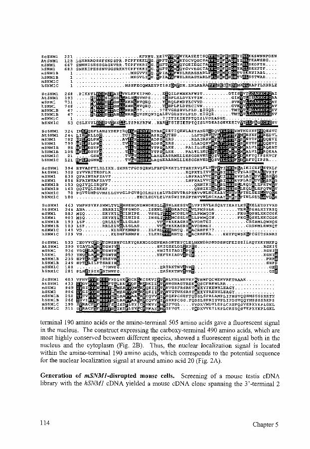
ScSNMJ. AtSNMJ. mSNMJ. hSNMl mSNMl:S hSNMlB mSNMJ.C hSNMJ.C
ScSNMl AtSNMl mSNMJ. hSNMl mSNMJ.B hSNMJ.B mSNMlC hSNMlC
ScSNMJ. AtSNMJ. mSNMl hSNMl mSNMlB hSNMlB mSNMlC hSNMlC
ScSNMl AtSNMl mSNMl hSNMJ. mSNMlB hSNMlB mSNMlC hSNMlC
ScSNMl AtSNMJ. mSNMl hSNMl mSNMlB hSNMl:S mSNMlC hSNMlC
ScSNMl AtSNMl mSNMl hSNMl mSNMlB hSNMJ.B mSNMJ.C hSNMlC
ScSNMl AtSNMl mSNMJ. hSNMJ. mSNMlB hSNMlB mSNMlC hSNMlC
221 128 667
'" 1 1 1 1
268 193 731 7<8
"
...................... KFNNG.HEI LGKRRROSSFSNDSPR.PCPFYKKLIG IPFT GNMNISESSGAGEVRR.TCPFYKRI•G: GFT
~~~~~~~~~~~~~~~~~~~~~~~~i:~· ~~T . . . . . . . . . . . . . . . . . . . MNGVLI·H. PI
•G NYKASETISQII~·SI' IKSWNNPDEN 1 RYGCVQGCSA T"F: •: I TKAWSHG .. . 1 QYGEIEGCTA ·F:S•• SKDFTR ... . oA QYGVVEGCTA T-F:So.Y SKHFTF ... . o WSLRRAGSARLF T· ·C••TVSKHWIRAL ... . 1 WSLRRAGTARLF S: :so:Tv{fi!SSTWAR ... .
J• E GN KKK RVQEQ Y RQLPMDTECVVD ,. E GN KNK HVQ:EQ Y HPLPLDTECIVN ·L P C HRR ?? . ??EVGESBVLPLD.EIGQE.
PIKK~-i ~~s~~=~~F~~~~~~ . ~~g~~~~~~~~:i~
47 •L P 8 HRH• QVSKQ:WIQALEVGESHVLPLD EIGQE 1 ... ? ?ITIEIETPTQISLVDEASGE ................. .
53 CSiKVYL~PV~KE~iTSPKYRFW: .KKRUISIEIETPTQISLVDEASGEKEEIV~T~~~G~SV
324 244 782 799
95 105
21 121
'" 302 839 856 153 163
78 180
463
IIEFLANS LIH LLDI ... .
I QLPN ... .
LPN ... . F EGYF ... .
EGYF ... .
~WQ~~= : ::· -ETVADFTLRLIKH.GKNKTFGDSQRNLFBFQRKKTLTTHRYRVLFLITITIIK~IKICEFITKLF SYVVRITKDFLR .......................... KQPKTLIV S SI CVYLAirn~·KA VKIF QFAINTAFEAVT .......................... LNPRALVVC T CI KVFLAI·DV SK G RFAINTAFEAVT .......................... LNPHALVVC T SI KVFLAI·DV• SK G QQIVQLIRQFP .............................. QBNIKI L SL 'SJ LEQL·LEFRTW QQIVQLIRKHP .............................. QHNIKI L SL S• EQL'LEFQTW PGVTDMPGVMSLLGVVGLPGVEQCLRGILELVRSWVTRSPBHVVWLNCKAAY Y YatTNLSEE~VQ~ ..................... EECLSGVLELVRSWITRSPYHVVWLNCKAAY Y Y~TNLSEE~VQ~H
346 ANA. 863 MSQ. 900 MSQ. 193 LSP. 203 LSP. 148 vo. 229 VN.
IEAYLKIELETDYVKD ..... VE EHLKIYREQ ..... FKG QSBLKKCGGK ..... FKG QSBLKKCGGK
.CHSAMLQWNQS
.CHSNMLRWNQT
... EEYFQWS~CGITSRNRI
533 399 936 953 239 249 180 281
603 03 969 986 258 268 199 311
IPHSV<!Q>>M' S S S RKTN .FQDSLSPRISVPLIPDSVQQYMSSSSRKPS
.... TVDKVMDVLKPLCRSPQSVEPKYKPLGKL
.... T~KVVEILKPLCRSSQSTEPKYKPLGKL
tenninal 190 amino acids or the amino-terminal 505 amino acids gave a fluorescent signal in the nucleus. The construct expressing the carboxy-terminal 490 amino acids, which are most highly conserved between different species, showed a fluorescent signal both in the nucleus and the cytoplasm (Fig. 2B). Thus, the nuclear localization signal is located within the amino-tenninal 190 amino acids, which corresponds to the potential sequence for the nuclear localization signal at around amino acid 20 (Fig. 2A).
Generation of mSNMJ-disrupted mouse cells. Screening of a mouse testis eDNA library with the hSNMJ eDNA yielded a mouse eDNA clone spanning the 3'-terminal 2
114 Chapter 5
FIG. 1. Comparison of the deduced amino acid sequences of SNM1 homologs/paralogs. The amino acid sequences of the carboxy termini of S. cerevisiae (Sc), A. thaliana (At), mouse (m) and human (h) Snm1 are aligned and shown in the single-letter code. From the second and third human and mouse Snm1 paralogs, called Snm1 B, and Snm1C/Artemis, the amino termini are shown. A more extensive version of this Figure, including Snm1 homologs from additional species, can be found in the appendix. Identical and similar amino acids are shown in black and gray boxes, respectively. Dots denote gaps; question marks denote unknown sequences. Amino acid positions are shown on the left. The eight motifs with high conservation are underlined. GenBank accession numbers are X64004 (ScSnm1), X98130 (AtSnm1), 042045 (hSnm1), AK024060 (hSnm1B), AJ296101 (hSnm1C), AF241240 (mSnm1), Al530740 and mCP20194 (Celera database [548]) (mSnm1B), mCP15877 and mCP36740 (Celera database [548]) (mSnm1C).
kb of mSNMJ. This clone was sequenced and used as a probe to obtain genomic mouse DNA. Two clones spanning the first seven exons of the mSNMJ genomic locus were obtained. The locus was characterized by restriction site mapping, PCR, and sequencing of the first two exons (Fig. 3A). A targeting construct was made by subcloning into pBluescript II KS a 4-kb Sal! fragment encompassing exons 2 and 3 and a 5-kb Hindiii fragment encompassing part of exon 4 and exons 5 to 7 with a selectable marker gene in between. In this way, part of intron 3 and 25 bp of ex on 4 were deleted. This procedure resulted in e1imination of the carboxy-terminal 287 amino acids of mSnm 1, which contain most conserved motifs (Fig. I). Moreover, aberrant RNA splicing that skips the targeted exon would result in a frameshift mutation. Disruption constructs were made containing the neomycin or the hygromycin selectable marker gene. They are referred to as mSNMlneo and mSNMlhyg' respectively.
The mSNMlhyg construct was electroporated into ES cells. After selection, targeted clones were identified by DNA blotting using a unique probe outside the targeting construct (Fig. 3A, C). Four independent cell lines containing one disrupted mSNMJ allele were obtained. The frequency of homologous versus random integration of the targeting construct was 2.5%. The second wild-type allele was disrupted by targeting with the mSNMI"" construct. Three double-knockout cell lines in which the remaining wild-type allele was replaced by mSNMI"'" were obtained (Fig. 3C).
RT -PCRs with RNAs isolated from mSNMI +!- and mSNMr1- cells were performed
using primers for exons 2 and 7. RNA from mSNMI+I- cells showed a clear signal from the wild-type mSNMJ allele, and both cell lines showed three weak signals from the knockout allele (data not shown and Fig. 3D). These three bands were cloned and sequenced. One product would result in a frameshift and a premature stop codon. The other two products, one containing an insert from the neomycin cassette and the other splicing around exon 4, used an alternative splice site in exon 5 which restored the reading frame. Both would result in disruption of conserved motifs 2 and 3 of mSnml (Fig. 1). No significant difference in growth rate between mSNMJ+I+ ES cells and all mSNMI+Iand mSNM r 1
- cell lines was detected. We conclude that, similar to the situation in S. cerevisiae, disruption of the mSNMJ in mouse cells results in viable cells [427].
Mouse SNMJ and DNA interstrand cross-link repair 115
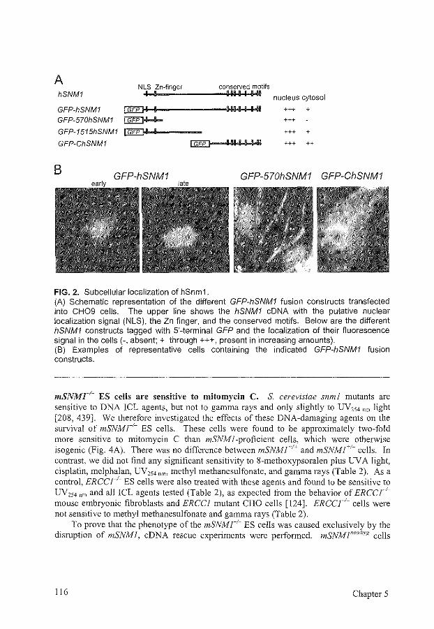
A hSNM1
NLS Zn-finger I I
conserved motifs 11111111 nucleus cytosol
GFP-hSNM1 []EEJI-!1-i-1-------111-11+1 +1-1-1-111-4111 +++ +
GFP-570hSNM1 ~ +++
GFP-1515hSNM1 IJ:iEE]-1-1-11----- +++ +
GFP-ChSNM1 +++ ++
B GFP-hSNM1 GFP-570hSNM1 GFP-ChSNM1
FIG. 2. Subcellular localization of hSnm1. (A) Schematic representation of the different GFP-h$NM1 fusion constructs transfected into CH09 cells. The upper line shows the hSNM1 eDNA with the putative nuclear localization signal (NLS), the Zn finger, and the conserved motifs. Below are the different hSNM1 constructs tagged with 5'-terminal GFP and the localization of their fluorescence signal in the cells(-, absent;+ through+++, present in increasing amounts). (B) Examples of representative cells containing the indicated GFP-hSNM1 fusion constructs.
mSNMl-1- ES cells are sensitive to mitomycin C. S. cerevisiae snml mutants are sensitive to DNA ICL agents, but not to gamma rays and only slightly to UV254 nm light [208, 439]. We therefore investigated the effects of these DNA-damaging agents on the survival of mSNMr1
- ES cells. These cells were found to be approximately two-fold more sensitive to mitomycin C than mSNM ]-proficient cells, which were otherwise isogenic (Fig. 4A). There was no difference between mSNlvfl+l+ and mSNMI+I- cells. In contrast, we did not find any significant sensitivity to 8-methoxypsoralen plus UV A light, cisplatin, melphalan, UV254 nm• methyl methanesulfonate, and gamma rays (Table 2). As a control, ERCCr1
- ES cells were also treated with these agents and found to be sensitive to UV,,4 nm and all ICL agents tested (Table 2), as expected from the behavior of ERCCJ·Imouse embryonic fibroblasts and ERCCJ mutant CHO cells [124]. ERCCr1
- cells were not sensitive to methyl methanesulfonate and gamma rays (Table 2).
To prove that the phenotype of the mSNMr1- ES cells was caused exclusively by the
disruption of mSNMJ, eDNA rescue experiments were performed. mSNMreolhyg cells
116 Chapter 5
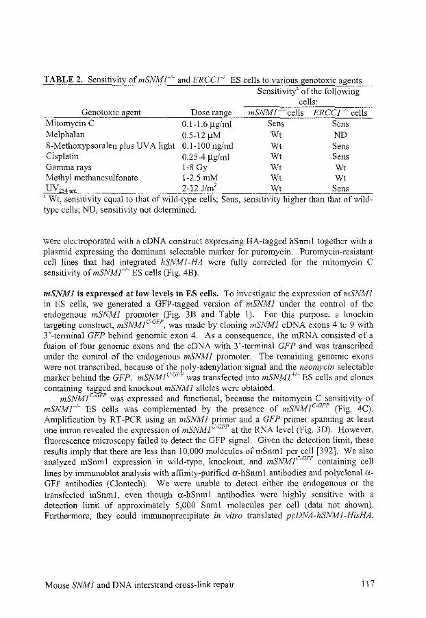
TABLE 2. Sensitivity of mSNMr1- and ERCCI_1
_ ES cells to various genotoxic agents Sensitivity' of the following
cells: Genotoxic agent Dose range mSNMI cells ERCCJ ;. cells
Mitomycin C 0.1-1.6 flg/ml Sens Sens Melphalan 0.5-12 f.LM Wt ND 8-Methoxypsoralen plus UV A light 0.1-100 ng/ml Wt Sens Cisplatin 0.25-4 flg/ml Wt Sens Gamma rays 1-8 Gy Wt Wt Methyl methanesulfonate 1-2.5 mM Wt Wt UV254 ,m 2-12J/m2 Wt Sens
"Wt, sensitivity equal to that of wild-type cells; Sens, sensitivity higher than that of wildtype cells; ND, sensitivity not determined.
were electroporated with a eDNA construct expressing HA-tagged hSnml together with a plasmid expressing the dominant selectable marker for puromycin. Puromycin-resistant cell lines that had integrated hSNMJ-HA were fully corrected for the mitomycin C sensitivity ofmSNMr1
- ES cells (Fig. 4B).
mSNMI is expressed at low levels in ES cells. To investigate the expression of mSNMI in ES cells, we generated a GFP-tagged version of mSNMI under the control of the endogenous mSNMI promoter (Fig. 3B and Table 1). For this purpose, a knockin targeting construct, mSNMJC-GFP' was made by cloning mSNMJ eDNA exons 4 to 9 with 3 '-terminal GFP behind genomic exon 4. As a consequence, the mRNA consisted of a fusion of four genomic exons and the eDNA with 3 '-terminal GFP and was transcribed under the control of the endogenous mSNMl promoter. The remaining genomic exons were not transcribed, because of the poly-adenylation signal and the neomycin selectable marker behind the GFP. mSNMIC-GFP was transfected into mSNMJ+I- ES cells and clones containing tagged and knockout mSNMI alleles were obtained.
mSNMJC-GFP was expressed and functional, because the mitomycin C sensitivity of mSNMr1- ES cells was complemented by the presence of mSNMIC-GFP (Fig. 4C). Amplification by RT -PCR using an mSNMI primer and a GFP primer spanning at least one intron revealed the expression of mSNMIC-GFP at the RNA level (Fig. 3D). However, fluorescence microscopy failed to detect the GFP signal. Given the detection limit, these results imply that there are less than 10,000 molecules of mSnml per cell [392]. We also analyzed mSnml expression in wild-type, knockout, and mSNMJC-GFP containing cell lines by immunoblot analysis with affinity-purified o:-hSnm I antibodies and polyclonal o:GFP antibodies (Clontech). We were unable to detect either the endogenous or the transfected mSnml, even though o:-hSnml antibodies were highly sensitive with a detection limit of approximately 5,000 Snml molecules per cell (data not shown). Furthermore, they could immunoprecipitate in vitro translated pcDNA -hSNM 1-HisHA.
Mouse SNMI and DNA interstrand cross-link repair 117
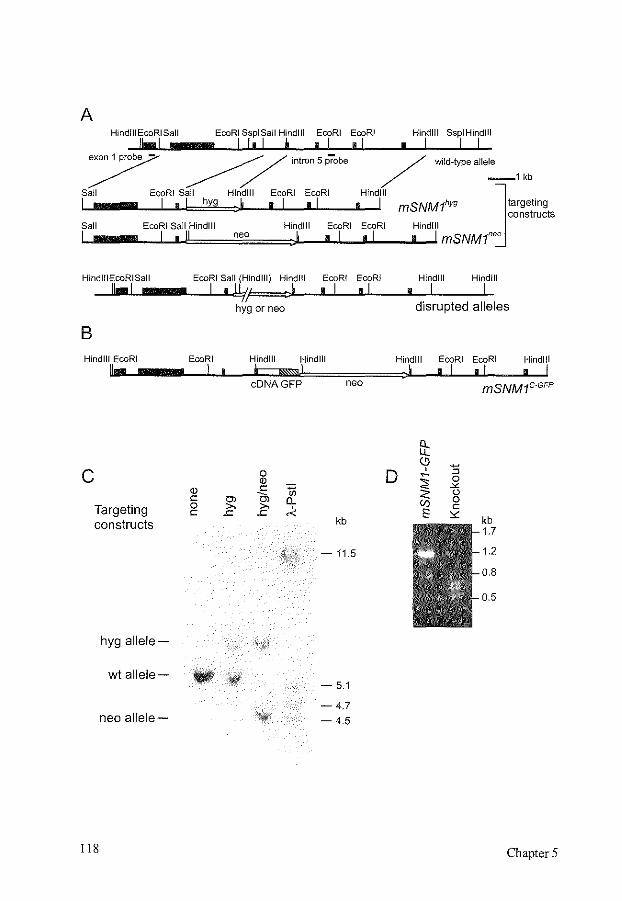
A HindiiiEcoRISall EcoRI SspiSall Hindi II EcoRI EcoRI
11 ... 1 I !, I I • I , I • Hindlll SspiHindlll
I I I 7 ~/ lntcon 5pcobe / wlld-typealle-le-1kb
Sail EcoRI Sail Hindlll EcoRI EcoRI Hindlll J L..IIIIIIIIIIIIL_ll ...1.• .J,I~hQ)Y~9=,J.I_...J.o_IL..JI•JI __ _ao_ll mSNM1hyg targeting
constructs Sail EcoRI Sail Hindlll Hindi! I EcoRI EcoRI Hindlll
I I • II neo :I • I • I • I mSNMt''
HindiiiEcoRISall !Ia!
B Hindlll EcoRI
u ...
c Targeting constructs
hyg allele-
wt allele-
neo allele-
118
EcoRI Sail (Hindlll) Hindl!l
I ,~, :1 EcoRI EcoRI , I , I
hyg or neo
EcoRI Hindlll Hindlll I ~ t\~W!)
cDNAGFP neo
0
" " c U3 0, c OJ o._
0 >- >-
'"" c .c .c
kb
- 11.5
- -5.1
-4.7 -4.5
Hindlll Hind!ll
• I I d·;srupted alleles
Hindi! I : ~
a: (')
D ' ~ ~ <: U)
E:
EcoRI EcoRI Hindlll • I , I , I
mSNM1C-GFP
::; 0 -"' () 0 c ~
kb 1.7
1.2
0.8
0.5
Chapter 5
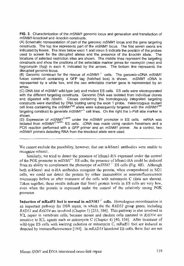
FIG. 3. Characterization of the mSNM1 genomic locus and generation and transfection of mSNMJ-knockout and -knockin constructs. (A) Schematic representation of part of the genomic mSNM1 locus and the gene targeting constructs. The top line represents part of the mSNM1 locus. The first seven exons are indicated by boxes. The lines below exon 1 and intron 5 indicate the position of the probes used to screen for the disrupted alleles and the presence of the knockin allele. The locations of selected restriction sites are shown. The middle lines represent the targeting constructs and show the positions of the selectable marker genes for neomycin (neo) and hygromycin (hyg) in exon 4, indicated by the arrows. The bottom line represents the disrupted genomic locus. (B) Genomic construct for the rescue of mSNM1-'- cells. The genomic-eDNA mSNM1 fusion construct containing a GFP tag (hatched box) is shown. mSNM1 eDNA is represented by a white box, and the neo selectable marker gene is represented by an arrow. (C) DNA blot of mSNM1 wild-type (wt) and mutant ES cells. ES cells were electroporated with the different targeting constructs. Genomic DNA was isolated from individual clones and digested with Hindlll. Clones containing the homologously integrated targeting constructs were identified by DNA blotting using the exon 1 probe. Heterozygous mutant cell lines containing the mSNM1hyg allele were subsequently targeted with the mSNM1"" targeting construct to generate mSNM1-t- cell lines. On the right the lc-Pstl size marker is shown. (D) Expression of mSNM1c-GFP under the mSNM1 promoter in ES cells. mRNA was isolated from mSNM1c-GFPI~ ES cells. eDNA was made using random hexamers and a PCR reaction performed with a GFP primer and an mSNM1 primer. As a control, two mSNM1 primers detecting RNA from the knockout allele were used.
We cannot exclude the possibility, however, that our a-hSrnnl antibodies were unable to recognize mSnml.
Similarly, we tried to detect the presence of hSrnnl-HA expressed under the control of the PGK promoter in mSNMr1
- ES cells; the presence ofhSrnni-HA could be deduced from its ability to complement the phenotype of mSNMr1
- ES cells (Fig. 4B). Although both a-hSrnnl and a-HA antibodies recognize the protein, when overproduced in Sf21 cells, we could not detect the protein by either immunoblot or immunofluorescence microscopy before or after treatment of the cells with mitomycin C (data not shown). Taken together, these results indicate that Snml protein levels in ES cells are very low, even when the protein is expressed under the control of the relatively strong PGK promoter.
Induction of mRad51 foci is normal in mSNMJ-1- cells. Homologous recombination is an important pathway for DSB repair, in which the the RAD52 group genes, including RAD51 and RAD54 are involved (Chapter I) [253, 386]. This pathway is also involved in ICL repair in vertebrate cells, because mouse and chicken cells mutated in RAD54 are sensitive to ICL agents such as mitomycin C (Chapter 4) [40, !54]. After treatment of wild-type ES cells with ionizing radiation or mitomycin C, mRad51 foci are induced as detected by immunofluorescence [194]. In mRAD54 knockout ES cells, these foci are not
Mouse SNMJ and DNA interstrand cross-link repair 119
c
10
'V mSNM1 +I- 5
0 mSNM1+!- 144
8 mSNM1-!- 96
II mSNMr!- 195 5~~==~-r--r-.-~
0 0.2 0.4 0.6 0.8 1.0 1.2
[mitomycin C) (~g/ml)
B 100
10
5
• ~!
=~~ 1,! !:::.mSNM1+/+ • 8 mSNM1-I- 96
II mSNM1-I- 195
0 mSNMr!- + hSNM1-HA 3
0 0.2 0.4 0.6 0.8 1.0 1.2
[mitomycin C) (~g/ml)
100~~~~~~~~------------------~
'iJ mSNM1+/- 5
0 mSNM1-!- 96
0 mSNMr!- + mSNM1-GFP 204
t3mSNM1+ +mSNM1-GFP249
0 0.2 0.4 0.6 0.8 1.0 1.2 1.4 1.6
[mitomycin C) (~g/ml)
FIG. 4. Effect of mitomycin C on wild-type and mutant mSNM1 ES cells. ES cells were treated with mitomycin C for 1 h, washed with PBS and then grown for 7 to 1 0 days in fresh medium. Subsequently, they were fixed, stained, and counted. Cloning efficiencies of untreated cells varied between 10% and 30%. All measurements were performed in triplicate. Error bars represent standard errors of the mean. Numbers following symbol definitions (figure insets) indicate the cell line isolation numbers. (A) Clonogenic survival of mSNM1•t- and mSNMr'· ES cells after treatment with increasing concentrations of mitomycin C. (B) Rescue of the mitomycin C sensitivity of mSNM1-t- cells by hSNM1-HA. An mSNMJ+ cell line containing randomly integrated hSNM1-HA eDNA expression constructs was obtained as described in Materials and Methods. The survival of this cell line was compared to the survival of mSNM1•t• and mSNMr'· ES cells. (C) Rescue of the mitomycin C sensitivity of mSNM1-t- cells by mSNM1°·GFP Two mSNM1c-GFPt- cell lines were obtained as described in Materials and Methods. The survival of these cells was compared to the survival of mSNM1•t- and mSNMr'· ES cells.
120 Chapter 5
seen using methanol/acetone fixation, suggesting that mRad54 functions at an earlier or the same step as mRad51 [513]. In contrast, mSNMJ knockout ES cells showed wild-type induction of mRad51 foci both after gamma ray and mitomycin C treatment (data not shown).
Induction of sister chromatid exchanges is normal in mSNMr1- cells. Homologous
recombination events can result in SCEs. Treatment with mitomycin C increases the number of SCEs detected in cells, due to recombinational repair of the DNA damage induced. In RAD51- and RAD54-deficient chicken B lymphocytes and in RAD54-deficient mouse ES cells, a reduced frequency of SCEs is observed after treatment with mitomycin C (Chapter 2) [144, 482]. Normal mRad51 foci formation in mSNMI knockout cells does not exclude a role for mSnml later in the homologous recombination repair ofiCLs. Therefore, we measured the spontaneous and mitomycin C-induced levels of SCEs in mSNMJ-proficient and -deficient ES cells. ES cells of the genotype mSNMJ+I+, mSNM1+1
-, mSNMr1-, and mSNMlc.GFPI- were analyzed. Mitomycin C
treatment induced a dose-dependent increase in SCEs. No differences in the level of SCEs were observed between the different cell lines (data not shown). In all cell lines treated with mitomycin C, no apparent chromosomal changes were observed. These results suggest that Snml is not involved in repair ofiCLs by homologous recombination. Alternatively, there could be redundancy between Snml and another protein for its function in homologous recombination during ICL repair.
Generation of mSNMI--1- mice. The experiments described above show that disruption of mSNM I is compatible with normal ES cell proliferation. Subsequently, we investigated whether mSnrn 1 is required for normal mouse development. Cells from four targeted ES clones, carrying the mSNMlhyg allele, were injected into C57BL/6 blastocysts and 11 chimeric mice were obtained. The disrupted mSNMJ allele was transmitted to the mouse germline. F 1 heterozygous offspring were intercrossed, and F2 offspring were genotyped by DNA blotting, PCR analysis, or both (data not shown). Among 78 genotyped animals, 20 mSNMJ+I+, 30 mSNMJ+1
-, and 28 mSNMr1- animals were identified. This outcome is
compatible with normal Mendelian segregation of the disrupted mSNMI allele. Thus, disruption of mSNMJ does not result in embryonic or neonatal lethality. No statistically significant difference in weight was observed among mSNM1+1+, mSNM1+1
-, and mSNMr /-littermates (data not shown). Importantly, the mSNMr1
- mice exhibited no macroscopic abnormalities up to at least 12 months of age. Both mSNMr1
- males and females were fertile.
mSNMI--1- mice are sensitive to mitomycin C. We tested whether the sensitivity of mSNMr1
- ES cells to the ICL agent mitomycin C was also found in mSNMJ-1- mice. mSNMl+l- and mSNMr1
- mice were treated with various doses of mitomycin C, ranging from 7.5 to 15 mg/kg of body weight. ll)iection of 10 and 15 mg/kg in female mice resulted in enhanced sensitivity in mSNMr- mice (Fig. 5). In addition, with 15 mg/kg, a shorter latency period in mSNMr1
- mice was observed. In male mice, 7.5 mg/kg was lethal for 30% of the knockout mice, while all heterozygous mice survived. At 10 mg/kg,
Mouse SNMJ and DNA interstrand cross-link repair 121
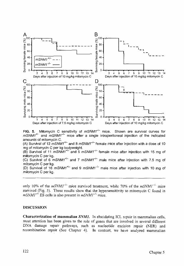
A B ~100..-v~~,_-_-_~~ ~100.,.-i,~--,
~80 t ~80 -,
~60---- Ll ___ ~60 ~ 40 ,-------,-/---, ~ 40
01 mSNM1 + ~ - - · rn
-, __ _
'-, .. _1 ____ _
-~ 20 mSNM1 ~!- -- -~ 20 1
~ 0 t~~::;=;:::::::.-~~~-~~., -~ 0 .!..,,..,...~~~.-~~~-+~-~~., 3 4 5 6 7 8 9 10 11 12 13 14 (fJ 3 4 5 6 7 8 9 10 11 12 13 14
Days after injection of 10 mg/kg mitomycin C Days after injection of 15 mg/kg mitomycin C
c 100.,.-i,~------r-------
*' -80 ~
.E eo ID
" E 40 ~ c ·:;: 20 ·~
~ o.J..,,..,...~~-r-~-,-~-T~.-~~., 3 4 5 6 7 8 9 10 11 12 13 14
Days after injection of 7.5 mg/kg mitomycin C
---, _____ _
3 4 5 6 7 8 9 10 11 12 13 14 Days after injection of 10 mg/kg mitomycin C
FIG. 5. Mitomycin C sensitivity of mSNMr'- mice. Shown are survival curves for mSNM1+J- and mSNMr1
- mice after a single intraperitoneal injection of the indicated amounts of mitomycin C. (A) Survival of 12 mSNM1•t- and 8 mSNM1--t- female mice after injection with a dose of 10 mg of mitomycin C per kg bodyweight (B) Survival of 11 mSNM1•t- and 5 mSNMr1
- female mice after injection with 15 mg of mitomycin C per kg. (C) Survival of 6 mSNM1H- and 7 mSNMr1
- male mice after injection with 7.5 mg of mitomycin C per kg. (D) Survival of 16 mSNM1•t- and 9 mSNMr1
- male mice after injection with 10 mg of mitomycin C per kg.
only 10% of the mSNMr1- mice survived treatment, while 70% of the mSNMJ+!- mice survived (Fig. 5). These results show that the hypersensitivity to mitomycin C found in mSNMr1- ES cells is also present in mSNMr1- mice.
DISCUSSION
Characterization of mammalian SNMJ. In elucidating ICL repair in mammalian cells, most attention has been given to the role of genes that are involved in several different DNA damage repair pathways, such as nucleotide excision repair (NER) and recombination repair (See Chapter 4). In contrast, we have analyzed mammalian
122 Chapter 5
homo logs of the S. cerevisiae SNMJ gene, which specifically provides resistance to ICL agents, Like yeast snml knockout cells, mSNMr1
- ES cells and mice are viable, showing that SNMJ is not essential for viability under normal culture and breeding conditions [195], The mice do not show any major abnormalities and are fertile. mSNMr1- ES cells and mice are hypersensitive to mitomycin C, but the cells are not sensitive to UV254 nm•
methyl methanesulfonate or gamma rays, in agreement with the results obtained with S. cerevisiae snml cells [208, 439], These results argue for a specific role of mammalian SNMJ in the cellular response to ICL agents, similar to yeast SNMJ.
hSnml resides in the nucleus (Fig. 2), similar to its yeast homolog, a location expected for a DNA repair protein [207], It has a putative nuclear localization signal at the amino terminus, and there may be another signal in the carboxy terminus of the protein. The gene is expressed in all tissues tested [369], The promoter region contains a consensus sequence for AP 1 and NFtcB, that can induce the expression of genes after being induced themselves by stress factors including DNA damage [132], hSNMJ contains an exceptionally long, 911-bp 5'-terminal untranslated region, containing several ATGs in all reading frames, which might cause inefficient translation. We could not detect any hSnml-HA or mSnml-GFP after transfection into mSNMJ+ cells, although the detection limit of our antibodies was below 1 ng, and the fluorescence microscopy detection limit of GFP is about 10,000 molecules per cell [392]. This suggests that the level of expression of Snml is very low, in agreement with the codon usage and the lack of a TATA box. Nevertheless, we could complement the mitomycin C sensitivity of mSNMr1
- cells with hSnml-HA and mSnm!-GFP. Thus, the protein levels must have been sufficient, and the HA and GFP tags at the carboxy terminus of Snml must not have interfered with its function. ScSNMJ is also expressed at a very low level [427],
Cross-link repair pathways, The formation and repair of ICLs in cells are complex processes, with major differences between ICL agents. The sensitivity of a cell to a certain ICL agent is dependent on all steps in the processing of ICLs, ranging from uptake and metabolization to damage recognition and repair (Chapter 4) [63]. A number of major DNA damage repair pathways are involved in the repair of ICLs. In E. coli, the major pathway involves NER and recombination repair (Fig. I in Chapter 4). First, UvrABC incises the DNA in one strand on both sides ofthe ICL. After a single-stranded DNA gap has been made by the exonuclease activity of DNA polymerase I, RecA performs strand exchange past the cross-link. Once recombination is complete, UvrABC makes incisions in the other strand around the cross-link, resulting in removal of the cross-linked oligonucleotide. Repair is completed by repair synthesis and ligation. Other, RecA independent, pathways depend on translesion synthesis by DNA polymerase II after incision by UvrABC [35]. This RecA-independent ICL repair is more efficient in the repair of nitrogen mustard ICLs, than psoralen ICLs [36]. In the absence of UvrABC, some error-prone ICL repair still takes place [544].
In S. cerevisiae, mutants m the NER, recombination repair, and postreplication!translesion repair pathways are sensitive to ICL agents [209]. Moreover, a number of ICL-sensitive mutants have been found that do not quite fit into one of these pathways, such as pso2 to pso4 [207]. The major repair pathway of ICLs inS cerevisiae
Mouse SNM 1 and DNA interstrand cross-link repair 123
is supposed to start with incision by the NER system, resulting in a DSB, in contrast to the incisions in E. coli, which do not result in a DSB [119, 229, 320, 348, 544]. The DSB can be repaired by the recombination repair pathway (Fig. 2 in Chapter 4) [14, 229]. Repair may also occur via the postreplication/translesion repair pathway, involving RAD6 and RAD18, which is more error-prone [467]. Yeast postreplication/translesion repair mutants are not defective in the repair of an ICL on a plasmid, in contrast to recombination repair mutants, suggesting that chromatin structure may have an important influence [319].
In mammals, a large number of mutant cell lines are known to be sensitive to ICL agents (Table 3 in Chapter 4) [107]. Many of the known mutated genes in those eel! lines belong to the NER pathway, the recombination repair pathway, and/or the error-prone postreplication!translesion repair pathway [107, 154, 251]. A number of genes cannot be attributed to one of the known repair pathways [68, 107]. The molecular mechanisms of mammalian ICL repair are still unknown, but they are probably similar to S. cerevisiae and E. coli ICL repair (See Chapter 4). For some ICL agents, there seems to be a transcription-dependent repair pathway [171, 221, 222, 284]. Incision of the ICLs is supposed to be performed by some of the NER proteins, as mutations in ERCCJ result in a decrease in the incision of ICLs and reduced repair-associated replication [282]. In contrast to the situation in S. cerevisiae, where no significant differences among NER mutants are found, most mammalian NER-deficient cells are only moderately sensitive to ICL agents [107]. However, ERCCJ and XPF mutants are among the cell lines that are most sensitive to mitomycin C. Therefore, in addition to their role inNER, ERCCJ and XPF are likely to be involved in the incision of ICLs or subsequent recombination, independent of the other NER genes. The sensitivity of the other NER mutants might be caused solely by a defect in the repair of monoadducts. Hamster cell lines with a mitomycin C sensitivity similar to that of ERCCJ mutants are irs! and irslSF [107]. The genes mutated in these cell lines are XRCC2 and XRCC3, which are paralogs of the recombination repair gene RAD51 and are important for chromosomal stability and DSB repair [77, 116,240,310,405,519, 524]. The mechanism by which these genes work is still unknown, but they could have an important role in recombination repair ofiCLs.
Another group of genes involved in the response to ICL agents, consists of the Fanconi anemia (FA) genes. Fanconi anemia is a rare, autosomal recessive disorder, characterized clinically by developmental abnormalities, pancytopenia of blood cells and a predisposition to the development of leukemia and other tumors [13, 137]. Fanconi anemia patient cells are sensitive to ICL agents and to oxidative DNA damage and they show cell cycle abnormalities [68, 101]. To date, eight complementation groups have been defined, six genes have been cloned, FANCA, FANCC, FANCD2, FANCE, FANCF, and FANCG [108, 127, 128, 130, 205, 236, 237, 314, 493, 528]. Interactions have been observed between FANCA, FANCC, FANCE, FANCF, and FANCG [108, 129, 130, 174, 175, 237, 271, 314, 341, 493, 554]. FANCC knockout mice are very sensitive to mitomycin C, although otherwise, their phenotype is very mild [74, 91, 565]. The FA proteins do not show any homology to other known proteins and Fanconi anemia patient cells do not show a consistent picture of a deficiency in ICL repair [68, 101]. For this reason and because of their pleiotropic phenotype, they may not be involved in ICL repair directly, but rather have some regulatory role in response to DNA damage.
124 Chapter 5
Role of Snml in ICL repair. The results for S. cerevisiae SNMJ show that Snml is not required for all ICL repair. S. cerevisiae snml cells show synergism with mutants from both the postreplicationltranslesion and the recombination repair pathways with regard to sensitivity to ICL agents. suggesting that Srnnl functions in an alternative pathway [209. 467]. Moreover, snml cells are capable of repairing an ICL on a plasmid, suggesting that the activity of Srnnl may be related to the modulation of chromosomal structure [319]. The sensitivity of snml mutants to ali!CL agents tested suggests that Snml is involved in an ICL agent-independent step and therefore probably after the formation of ICLs [63, 208, 439]. This notion is consistent with the fact that snml mutants are capable of creating DSBs after treatment with 8-methoxypsoralen plus UV A light, iri contrast to NER mutants, which are not able to incise the DNA near the ICL [320, 344]. snml mutants are, however, epistatic to NER mutants, suggesting that they play a role in this repair pathway [209, 467]. Snml may be involved in restoring the continuity ofthe DNA, because snml cells are not able to repair the DSBs formed [320]. Alternatively, Srnnl could also play a more indirect role, in the regulation of!CL repair.
mSNMJ knockout ES cells are specifically sensitive to mitomycin C, but not to cisplatin, melphalan, or 8-methoxypsoralen plus UV A light, in contrast to S. cerevisiae snml cells. Therefore, mammalian SNMJ is probably not essential for a general ICL repair pathway. Mammalian cells could have different proteins for the recognition and repair of different ICLs, while yeast cells could depend on a single protein. Alternatively, some postreplication/translesion repair pathways in mammals could be more efficient than those in yeast, taking over most ICL repair in mSNMJ knockout ES cells, but being insufficient to repair mitomycin C-induced ICLs. We cannot exclude the possibility of the presence in our knockout ES cells of some mutated mSnml that could fulfill some of the functions of the protein and thereby mitigate the phenotype. However, this notion is not a likely explanation of the mild phenotype, because two highly conserved motifs were deleted. Some of the conserved motifs of Snml constitute a metallo-lyase domain, which is found in a wide variety of proteins with different functions, like bacterial ~-lactamase II, cyclic phosphodiesterases, glyoxylases, and proteins involved in the uptake of DNA (I.S. Mian, personal communication). Despite this clue about a possible function for Snml, we can only speculate about its role. mSnml could be involved in the activation of mitomycin C, decreasing the number of ICLs or other damage caused by tbis agent. Alternatively, it could be responsible for the recognition of mitomycin C-induced ICLs and other structurally related ICLs. These explanations would be inconsistent with the yeast snml phenotype, although a direct comparison is not possible because of the existence of multiple mammalian SNMI paralogs and because mitomycin C has not been tested inS. cerevisiae. It is also possible that Snml is involved in a regulatory pathway, influencing the response of the cell to the damage caused by mitomycin C. The most attractive assumption for the function of rnSnml is a direct role in the repair of ICLs, as suggested by the results for its homolog in S. cerevisiae.
Induction of mRad51 foci and sister chromatid exchanges is normal in mSNMJ-1-cells. One of the pathways for ICL repair is constituted by the homologous recombination repair pathway, in which Rad54, Rad51, and its paralogs play an important role [154, 253,
Mouse SNMJ and DNA interstrand cross-link repair 125
386, 523]. Upon treatment with mitomycin C, mRad51 forms nuclear foci [194]. Immunofluorescence on mSNMl knockout ES cells after mitomycin C treatment showed normal foci formation by Rad51 (data not shown). This is in contrast to RAD51 paralog mutant chicken cells, XRCC3 mutant CHO cells, and mRAD54 mutant ES cells that do not show Rad51 foci after treatment with cisplatin and mitomycin C, respectively [45, 509, 511, 513]. Using a different fixation technique than was used for mRAD54 mutant ES cells, Rad51 foci formation was increased in chicken RAD54 mutant cells, suggesting arrest of homologous recombinational repair of!CLs after Rad51 has accumulated [509]. SCE induction by mitomycin C is reduced in cells deficient for RAD51, its paralogs, or RAD54 (Chapter 2) [144, 482]. mSNMr1
- cells, however, show normal induction of SCEs. These results on Rad51 foci formation and SCE induction suggest that Snml plays a role independent of the recombination repair pathway. This explanation is supported by the results inS. cerevisiae, which place Snml and Rad52 in different pathways [209, 467].
Redundancy of mammalian Snml function. The relatively mild phenotype of mSNMr ; .. ES cells and mice is reminiscent of the phenotype found for mRAD54_1
_ cells and mice, which are also about two-fold more sensitive to mitomycin C than wild-type cells and mice [154, 155]. This mild phenotype can probably be attributed in part to the existence of parallel pathways for the repair of ICLs. In addition, known paralogs for both mSNMl and mRAD54 could take over part of the function (Fig. 1) [212]. In fact, redundancy is a phenomenon that is quite common in mammalian DNA damage repair pathways. The NER gene RAD23, the postreplicationltranslesion repair gene RAD6, and the recombination repair gene RAD51 have several paralogs [252, 266, 330, 524]. These duplications provide cells with the possibility of functional differentiation in the repair of different types of damage, in different phases of the cell cycle, or in different cell types. Moreover, the cell can fall back on the alternative repair pathway in case of dysfunction of one of the proteins involved, a strategy which may prevent inappropriate repair and cell transformation. It will be important to analyze the other SNMJ paralogs and to look at the effects of ICL agents on double knockout cells. Furthermore, study of cells with mutations in SNMJ and other genes functioning in ICL repair, such as RAD54, will yield information on the relative importance of the different ICL repair pathways and the level of redundancy between different pathways. Nevertheless, the sensitivity of mSNMl knockout cells to mitomycin C shows that there is no complete redundancy in mammalian cells. Both the sequence conservation and the functional conservation with yeast Snml underline the significance of SNMJ in ICL repair.
ACKNOWLEDGMENTS
We thank W. Vermeulen for the microinjection experiment. This work was supported by grants from the Netherlands Organization for Scientific Research (NWO) and the Dutch Cancer Society (KWF). R.K. was a fellow of the Royal Netherlands Academy of Arts and Sciences.
126 Chapter 5
APPENDIX
FIGURE. Comparison of the deduced amino acid sequences of SNMI homologs/ paralogs. The amino acid sequences of Saccharomyces cerevisiae (Sc ), Schizosaccharomyces pombe (Sp). Arabidopsis thaliana (At), Drosophila melanogaster (Dm), Caenorhabditis elegans (Ce), mouse (m), and human (h) Snml are aligned and shown in the single-letter code. The second and third human and mouse Snm 1 paralogs are called SnmlB and SnmlC, and the A. thaliana homologs/paralogs AtSnmlB, C, and D. Identical and similar amino acids are shown in black and gray boxes, respectively. Dots denote gaps, question marks denote unknown sequences. Amino acid positions are shown on the left. The putative nuclear localization signal lies around amino acid 20. The Znfinger domain at the N-terminus and the eight motifs with high conservation are underlined. Gene bank accession numbers are X64004 (ScSnml), Z69728 (SpSnrnl), X98130 (AtSnml), AC004665 (AtSnrnlB), AC004557 (AtSnmlC), AC013288 (AtSnmlD), AE003602 (DmSnml), Z81080 (CeSnml), D42045 (hSnrnl), AK024060 (hSnmlB), AJ296101 (hSnmlC), AF241240 (mSnml), Al530740 and mCP20194 (Celera database, www.celera.com, [548]) (mSnmlB), mCP15877 and mCP36740 (Celera database) (mSnm 1 C).
Mouse SNMJ and DNA interstrand cross-link repair 127
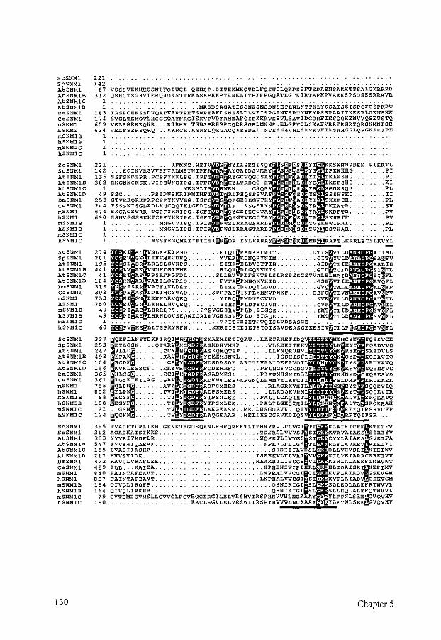
ScSNMl 221 SpSNMl 142 AtSNMl 67 AtSNMlB 312 AtSNMlC 1 AtSNMlD 1 DmSNMl 183 CeSNMl 174 mSNMJ. 609 hSNMJ. '" mSNMlB 1 hSNMlB 1 mSNMJ.C 1 hSNMJ.C 1
ScSNMl 221 SpSNMl 1<2 AtSNMl 135 AtSNMlB 382 AtSNMlC 1 AtSNMlD " DmSNMl 253 CeSNMl 244 mSNMl 674 hSNMl '" mSNMlB 1 hSNMlB 1 mSNMlC 1 hSNMlC 1
ScSNMl 274 SpSNMl 2 01 AtSNMl 195 AtSNMlB '" AtSNMlC <1 AtSNMlD 10' OmSNMl 313 CeSNMl 303 mSNMJ. 733 hSNMl 750 mSNMlB " hSNMlB " mSNMlC 1 hSNMlC 60
ScSNMl 327 SpSNMl 253 AtSNMl 2<7 AtSNMlB '" AtSNMlC 10< AtSNMlD 156 DmSNMl 365 CeSNMl 361 mSNMJ. n5 hSNMl 802 mSNMlB 98 hSNMlB 108 mSNMlC 21 hSNMlC 12<
ScSNMl 395 SpSNMl 312 AtSNMl 303 AtSNMlB 5<7 AtSNMlC 165 AtSNMlD 217 OmSNMl '" CeSNMl 429 mSNMJ. '" hSNMl 857 mSNMlB 15< hSNMJ.B 154 mSNMlC 79 hSNMlC 180
130
VSSSVKKMKQSNLFQIWGL.QENSP.DTTKRMKQTDLFQSWGLQRPSPFTSPASNSAKKTTSALGKRRRD QSBCTSGHVTERQRDKSTTRKASEPKKPTANKLITEFFPGQATEGTKIRTAPKPVAEKSPSDSSSRRAVR
.. MASDSAGATISGNFSNSDNSETLNLNTTKLYSSAISSISPQFPSPKPT IASPCEKSSDVQATKFRTPETSMPEAKLSNSSLDLVEISPGPNKSPTNNFYRSSPAAITKESPLGKNSKK SVGLTEMQVLHGGGQAYNRGISKVPVDFRNEAFQIFKKRVESVLEAVTDCONFIEFQQKENVVQSETSTQ VGLSGEKRQHR ... RKRHK.TSNSPREGPCQRRSGHLMNNP.ELGPVSLSKAFVRRTRGRTQRGNMNISE VELSSERSQRQ ... KKRCR.KSNSLQEGACQKRSDBLINTESEAVNLSKVKVFTKSAHGGLQRGNKKIPE
.KFNNG.HEI NYKASETISQY ... KQINVRSVVPFYKLMPYNIPF YGAIDGVJ::AY SSFSNDSPR.PCPFYKKLPG.TPFT RYGCVQGCSAY RNGNNGKSK.VIPHWNCIPG.TPF KYLTRDCC.HW
.... MESGLIS •RWRN .... GSQAY sse .. .PSIPNSKRIPNTNFI L RLP.IIQSSSVA GTVRKQRKPKPCPPYKVVEG.TSFC •G QFGEIEGVTHY TSSSNTSGDADLKHCQQIKIGEDIS •Y LK .. KSGSRYN SSGAGEVRR.TCPFYKRIPG.TGFT QYGEIEGCTAY SSNVGGSRKKTCPFYKKIPG.TGFT QYGVVEGCTAY
... MNGVVIPQ.TPI WSLRRAGSARLF
. . . MNGVLIPH.TPIA •FWSLRRAGTARLF
~PVDKE~LLTSPKYRFW:: .... KKRr
QEFLANSYDKPI ETLQSN. QTR~.~~~~S RLLD~ .... EPAN .. RGDF .
VKLESSGF. KLSS~. EGSKIEEIAG.
~~:~ :::: EGYF .... . EGYF ... .
.. GSN .. !QGN .
s' F"S• ·YI KKSWNNPDEN .PIKKTL S• "YG TPKWKHG. . .... PI
T F • · YI TKAWSHG. . .... PI T. F.L• YQ TKSFSHG. . .... KI
" I :S • TR SGGWSQG. ..... PL
'' F·s• -ys SSSWSKG. ..... II T F • ·YI TKKFCH. ..... PL T. S• "YR DKKWTR. . .... sv T F-S• :YA SKDFTR. ..... f?V
·p S• "YA SKHFTF_ ... PV T c•:TVSKHWIRAL. ..... PL s S•"TVWSSTWAR . ..... PL
TVADFTLRL I KH :G~~RT> G~>SilR;"~~RFOi~~OKOOL~:g~~;~:.~~:;~~; i ACADKAISIKKS. YVVRITKDFLR. FVVEAIQAEAF. LVADIIASHP. YVVSVIDK .. RAVDLVRAFLEK. ILL ... KAIEA. FAINTAFEAVT. FAINTAFEAVT.
lRQFP. IRKBP.
Chapter 5
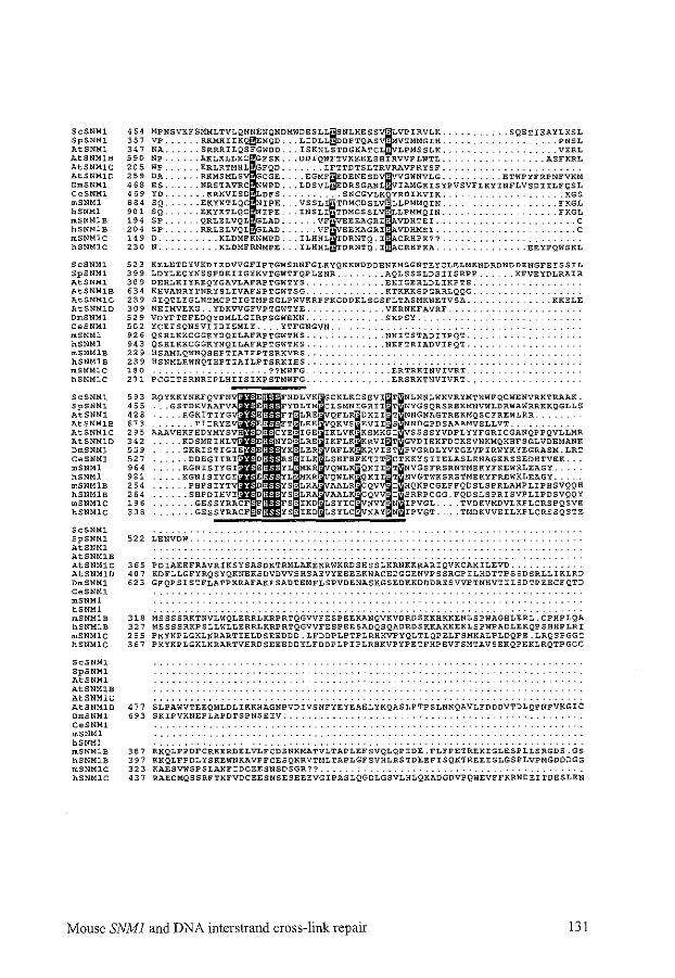
ScSNMl SpSNMl. AtSNMl AtSNMlB AtSNMlC AtSNMlD DmSNMl CeSNMl mSNMl hSNMl. mSNMlB hSNMl.B mSNMl.C hSNMlC
ScSNMl. SpSNMl AtSNMl. AtSNMlB AtSNMl.C AtSNMl.D DmSNMl. CeSNMl mSNMl hSNMl mSNMl.B hSNMlB m.SNMlC hSNMlc
ScSNMl .SpSNMl AtSNMl AtSNMl.B AtSNMlC AtSNMl.D DmSNMl CeSNMl mSNMl hSNMl mSNMlB hSNMlB mSNMlC hSNMlC
ScSNMl SpSNMl AtSNMl AtSNMlB AtSNMlC AtSNMlD OmSNMl CeSNMl mSNMl hSNMl mSNMlB hSNMlB mSNMlC hSNMlC
ScSNMl SpSNMl AtSNMl AtSNMlB AtSNMlC AtSNMlD OmSNMl CeSNMl mSNMl. hSNMl mSNMlB hSNMlB mSNMl.C hSNMlC
464 MPNSVKFSMMLTVLQNNENQNDMWDESLL~SNLHESS~LVPIRVLK SQETIEAYLKSL 357 VP RKMHIIKQ~ENQD LIDLL~DDPTQASV: VTMMGIH PNSL 347 NA SRRRILQSFGWDD ISKNLSTDGKATCL: LPMSSLK VERL 590 NP AKLKLLECIFSK DDIQWFTVKEEESHIHVVPLWTL ASFKRL 205 WP ERLRTMHL GFQD IFTTDTSLTRVRAVPRYSF 259 DA RKMSMLSV GCGE EGMF~EDENESDVIDvVGWNVLG ETWPYFRPNFVKM 468 ES NRSTAVRC WPD LDSVL~EDRSGANLWVIAMGKISYPVSVFLKYINFLVSDIILFQSL 469 YO ERKVISD LDFS SNCGVLKQYRDIKVIK KGS 884 SQ EKYKTLQC IPE VSSLI~DMCDSLI.LLPMMQIN FKGL 901 SQ EKYKTLQC IPE INSLI TDMCSSL LLPMMQIN FKGL 194 SP QRLELVQL GLAD VF EEEAGRI VOHTEI C 204 Sp RRLELVQL GLAD VF EEKAGRI VDHMEI C 149 D ......... KLDMFKNMPO ... ILHHL TDRNTQ.I CRHPK?? .. 230 N .......... KLOMFRNMPE ... ILHHL TDRNTQ. I CRHPKA. . . .. EEYFQWSKL
523 KELETDYVKDIEDVVGFIPTGWSHNFGLKYQKKNDDDENEMSGNTEYCLELMKNORDNDDENGFEISSIL 399 LDYLEQYNSSFDKIIGYKVTGWTFQPLENR ....... AQLSSSLDSIISRPP ...... KFVEYDLRAIR 389 DEHLKIYREQYGAVLAFRPTGWTYS. . ........ EKIGEHLDLIKPTS. 634 KHVANRYTNRYSLIVAFSPTGWTSG. . ...... KTKKKSPGRRLQQG. 239 SIQTLEGLNTMCPTIGIMPSGLPWVKRPFKGDDKLSGSFLTASMKNETVSA. . .KKELE 309 NEIMVEKG .. YDKVVGFVPTGWTYE. . ...... VKRNKFAVRF .. 529 VOYFTEFEDQYDMLLGIRPSGWEKN. .SKPSY. 502 YCEISQNSVIIDISMLY .... YTFGNGVN .. 926 QSHLKKCGGKYDQILAFRPTGWTHS. .NNITSTADIIPQT. 943 QSHLKKCGGKYNQILAFRPTGWTHS.. . .NKFTRIADVIPQT. 229 HSAMLQWNQSHPTIAIFPTSRKVRS .. 239 HSNMLRWNQTHPTIAILPTSRKIRS .. ~80 ... ??MWFG .. 271 PCGITSRNRIPLHIISIKPSTMWFG ..
593 455 428
'" 295 342 559 527 964 981 254 264 196 308
522 LENVDW.
. ..... ERTRKTNVIVRT. . .. ERSRKTNVIVRT.
365 PDIAEEFRAVRIKSYSASDKTRMLAKEKRWKRDSHSSLKRNKKRARIQVKCAKILEVD .. 407 KDFLLGFYRQSYQKNEKSDVDVVSHSAEVYEEEEKNACEDGGENVPSSRGPILHDTTPSSDSRLLIKLRD 623 GFQPSISTFLATPKRAFAKFSADTEMFLSPVDENASKGSEDKKDDDRESVVPTNHVTIISDTPEECFQTD
318 MSSSSRKTNVLWQLERRLKRPRTQGVVFESPEEKANQVKVDROSKKRKKENLSPWAGHLERL.CPHPLQA 327 MSSSSRKPSLLWLLERRLKRPRTQGVVFESPEESADQSQADRDSKKAKKEKLSPWPADLEKQPSHHPLRI 255 PKYKPLGKLKRARTIHLDSEEDDD.LFDDPLPTPLRHKVPYQLTLQPELFSMKALPLDQPE.LRQSPGGC 367 PKYKPLGKLKRARTVHRDSEEEDOYLFDDPLPIPLRRKVPYPETFHPEVFSMTAVSEKQPEKLRQTPGCC
477 S~PAWVTEEQMLDLIKK~AGNPVDIVSNFYEYEAEiYKQASiPTPSLNNQAVLFDDDVTDLQPNPVRGIC 693 SKIPVKNEPLAPOTSPNSEIV.
387 RKQLFPOFCRKEROELVLFCOSNKMATVLTAPLEFSVQLQPIDE.FLFPETREKIGLESPLLSRGDS.GS 397 KKQLFPDLYSKEWNKAVPFCESQKRVTMLTAPLGFSVHLRSTDEEFISQKTREEIGLGSPLVPMGDODGG 323 KAESVWSPSLANFIDCEESNSDSGR?? .. 437 RAECMQSSRFTNFVDCEESNSESEEEVGIPASLQGOLGSVLHLQKADGDVPQWEVFFKRNDEITDESLEN
Mouse SNMJ and DNA interstrand cross-link repair 131
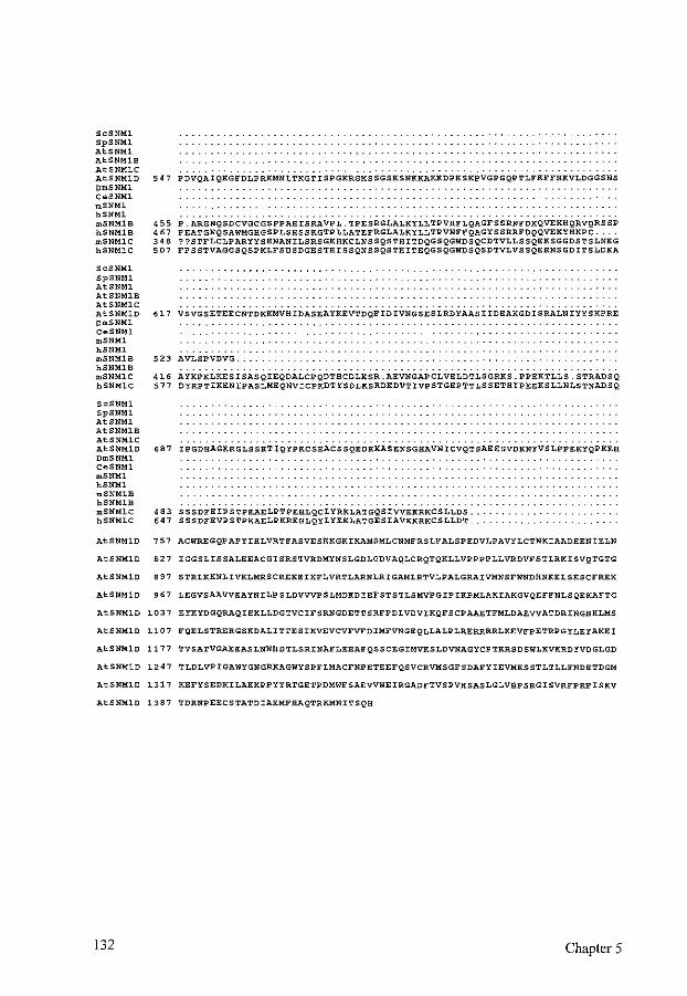
ScSNM1 SpSNM1 AtSNM1 AtSNM1B AtSNM1C AtSNM1D 547 PDVQAIQKGFDLPRKMNLTKGTISPGKRGKSSGSKSNKKAKKDPKSKPVGPGQPTLFKFFNKVLDGGSNS OmSNM1 CeSNM1 mSNM1 hSNM1 mSNM1B hSNM1B mSNM1C hSNM1C
ScSNM1 SpSNM1 AtSNM1 AtSNM1B AtSNM1C AtSNM1D OmSNM1 CeSNM1 mSNM1 hSNM1
455 P.ARGNQSDCVGCGSPPAHISRAVPL.TPESRGLALKYLLTPVHFLQAGFSSRNFDKQVEKHQRVQRSSP 467 PEATGNQSAWMGHGSPLSHSSKGTPLLATEFRGLALKYLLTPVNFFQAGYSSRRFDQQVEKYHKPC. 348 ??STFLCLPARYYSHNANILSRSGKHKCLNSSQSTHITDQGSQGWDSQCDTVLLSSQEKSGGDSTSLNKG 507 FPSSTVAGGSQSPKLFSDSDGESTHISSQNSSQSTHITEQGSQGWDSQSDTVLVSSQERNSGDITSLDKA
617 VSVGSETEECNTDKKMVHIDASEAYKEVTDQFIDIVNGSESLRDYAASIIDEAKGDISRALNIYYSKPRE
mSNM1B 523 AVLSPVDVG. hSNM1B mSNM1C 416 AYKPKLKESISASQIEQDALCPQDTHCDLKSR.AEVNGAPCLVELDTLSGRKS.PPEKTLLS.STRADSQ hSNM1C 577 DYRPTIKENIPASLMEQNVICPKDTYSDLKSRDKDVTIVPSTGEPTTLSSETHIPEEKSLLNLSTNADSQ
ScSNMl SpSNMl AtSNM1 AtSNM1B AtSNM1C AtSNM1D 687 IPGDHAGERGLSSKTIQYPKCSEACSSQEDKKASENSGHAVNICVQTSAEESVDKNYVSLPPEKYQPKEH OmSNM1 CeSNM1 mSNM1 hSNM1 mSNM1B hSNM1B mSNM1C 483 SSSDFEIPSTPEAELPTPEHLQCLYRKLATGQSIVVEKRKCSLLDS. hSNM1C 647 SSSDFEVPSTPEAELPKREHLQYLYEKLATGESIAVKKRKCSLLDT.
AtSNM1D 757 ACWREGQPAPYIHLVRTFASVESEKGKIKAMSMLCNMFRSLFALSPEDVLPAVYLCTNKIAADHEN!ELN
AtSNM1D 827 IGGSLISSALEEACGISRSTVRDMYNSLGDLGDVAQLCRQTQKLLVPPPPLLVRDVFSTLRKISVQTGTG
AtSNM1D 897 STRLKKNLIVKLMRSCREKEIKFLVRTLARNLRIGAMLRTVLPALGRAIVMNSFWNDHNKELSESCFREK
AtSNM1D 967 LEGVSAAVVEAYNILPSLDVVVPSLMDKDIEFSTSTLSMVPGIPIKPMLAKIAKGVQEFFNLSQEKAFTC
AtSNMlD 1037 EYKYDGQRAQIHKLLDGTVCIFSRNGDETTSRFPDLVDVIKQFSCPAAETFMLDAEVVATDRINGNKLMS
AtSNM1D 1107 FQELSTRERGSKDALITTESIKVEVCVFVFDIMFVNGEQLLALPLRERRRRLKEVFPETRPGYLEYAKEI
AtSNM1D 1177 TVSATVGAEEASLNNHDTLSRINAFLEEAFQSSCEGIMVKSLDVNAGYCPTKRSOSWLKVKRDYVDGLGD
AtSNM1D 1247 TLDLVPIGAWYGNGRKAGWYSPFLMACFNPETEEFQSVCRVMSGFSDAFYIEVMKSSTLTLLFNDETDGM
AtSNM1D 1317 KEFYSEDKILAKKPPYYRTGETPDMWFSAEVVWEIRGADFTVSPVHSASLGLVHPSRGISVRFPRFISKV
AtSNM1D 1387 TDRNPEECSTATDIAEMFHAQTRKMNITSQH
132 Chapter 5
REFERENCES
1. Adair, G. M., R. L. Rolig, D. Moore-Faver, M. Zabelshansky, J. H. Wilson, and R. S. Nairn. Role ofERCCl in removal oflong non-homologous tails during targeted homologous recombination. EMBO J. 19:5552-5561 (2000).
2. Aguilera, A., S. Chavez, and F. Malagon. Mitotic recombination in yeast: elements controlling its incidence. Yeast 16:731-754 {2000).
3. Aguilera, A., and H. L. Klein. Yeast intrachromosomal recombination: long gene conversion tracts are preferentially associated with reciprocal exchange and require the RADJ and RAD3 gene products. Genetics 123:683-694 (1989).
4. Aguirrezabalaga, I., L. M. Sierra, and M. A. Comendador. The hypermutability conferred by the mus308 mutation of Drosophila is not specific for cross-linking agents. Mutat. Res. 336:243-250 (1995).
5. Ahne, F., M. Baur, and F. Eckardt-Schupp. The REV2 gene of Saccharomyces cerevisiae: cloning and DNA sequence. Curr. Genet. 22:277-282 (1992).
6. Alaoui-Jamali, M., B. B. Loubaba, S. Robyn, H. Tapiero, and G. Batist. Effect of DNArepair-enzyme modulators on cytotoxicity of L-phenylalanine mustard and cisdiamminedichloroplatinum (II) in mammary carcinoma cells resistant to alkylating drugs. Cancer Chemother. Pharmacal. 34:153-158 (1994).
7. Albala, J. S., M.P. Thelen, C. Prange, W. Fan, M. Christensen, L. H. Thompson, and G. G. Lennon. Identification of a novel human RAD51 homolog, RAD5JB. Genornics 46:476-479 (1997).
8. Anderson, D. G., and S.C. Kowalczykowski. The translocating RecBCD enzyme stimulates recombination by directing RecA protein onto ssDNA in a chi-regulated manner. Cell 90:77-86 (1997).
9. Aquilina, G., and M. Bignami. Mismatch repair in correction of replication errors and processing of DNA damage. J. Cell. Physiol. 187:145-154 (2001).
10. Arbel, A., D. Zenvirth, and G. Simchen. Sister chromatid-based DNA repair is mediated by RAD54, not by DMCI or TIDI. EMBO J. 18:2648-2658 (1999).
11. Arnaudeau, C., C. Lundin, and T. Helleday. DNA double-strand breaks associated with replication forks are predominantly repaired by homologous recombination involving an exchange mechanism in mammalian cells. J. Mol. Biol. 307:1235-1245 (2001).
12. Astrom, S. U., S.M. Okamura, and J. Rine. Yeast cell-type regulation of DNA repair. Nature 397:310 (1999).
13. Auerbach, A. D., M. Buchwald, and H. Joenje. Fanconi anemia, p. 317-332. In B. Vogelstein, and K. W. Kinzler (eds), The Genetic Basis of Human Cancer. McGraw-Hill, New York (1998).
14. Averbeck, D., M. Dardalhon, N. Magana-Schwencke, L. B. Meira, V. Meniel, S. Boiteux, and E. Sage. New aspects of the repair and genotoxicity ofpsoralen photoinduced lesions in DNA. J. Photochem. Photobiol. B. 14:47-63 (1992).
15. Averbeck, D., D. Papadopoulo, and E. Moustacchi. Repair of 4,5',8-trimethylpsoralen plus light-induced DNA damage in normal and Fanconi's anemia cell lines. Cancer Res. 48:2015-2020 (1988).
16. Bai, Y., and L. S. Symington. A Rad52 homolog is required for RAD5J-independent mitotic recombination in Saccharomyces cerevisiae. Genes Dev. 10:2025-2037 (1996).
17. Bailly, V., S. Lauder, S. Prakash, and L. Prakash. Yeast DNA repair proteins Rad6 and Rad18 form a heterodimer that has ubiquitin conjugating, DNA binding, and ATP hydrolytic
133
activities. J. Bioi. Chern. 272:23360-23365 (1997). 18. Bardwell, A. J., L. Bardwell, A. E. Tomkinson, and E. C. Friedberg. Specific cleavage of
model recombination and repair intermediates by the yeast Radl-RadlO DNA endonuclease. Science 265:2082-2085 (1994).
19. Barlev, N. A., V. Poltoratsky, T. Owen-Hughes, C. Ying, L. Liu, J. L. Workman, and S. L. Berger. Repression ofGCN5 histone acetyltransferase activity via bromodomain-mediated binding and phosphorylation by the Ku-DNA-dependent protein kinase complex. MoL Cell. Biol.18:1349-1358 (1998).
20. Barnes, G., and D. Rio. DNA double-strand-break sensitivity, DNA replication, and cell cycle arrest phenotypes of Ku-deficient Saccharomyces cerevisiae. Proc. Natl. Acad. Sci. USA 94:867-872 (1997).
21. Barre, F. X., U. Asseline, and A. Harel-Bellan. Asymmetric recognition of psoralen interstrand crosslinks by the nucleotide excision repair and the error-prone repair pathways. J. Mol. Bioi. 286:1379-1387 (1999).
22. Barre, F. X., C. Giovannangeli, C. Helene, and A. Harel-Bellan. Covalent crosslinks introduced via a triple helix-forming oligonucleotide coupled to psoralen are inefficiently repaired. Nucleic Acids Res. 27:743-749 (1999).
23. Bartsch, S., L. E. Kang, and L. S. Symington. RAD51 is required for the repair of plasmid double-stranded DNA gaps from either plasmid or chromosomal templates. Mol. Cell. Biol. 20:1194-1205 (2000).
24. Basu, A. K., L. J. Marnett, and L. J. Romano. Dissociation of malondialdehyde mutagenicity in Salmonella typhimurium from its ability to induce interstrand DNA crosslinks. Mutat. Res. 129:39-46 (1984).
25. Batist, G., S. Torres-Garcia, J. M. Demuys, D. Greene, S. Lehnert, M. Rochon, and L. Panasci. Enhanced DNA cross-link removal: the apparent mechanism of resistance in a clinically relevant melphalan-resistant human breast cancer cell line. Mol. Pharmacal. 36:224-230 (1989).
26. Baumann, P., F. E. Benson, and S. C. West. Human Rad51 protein promotes ATPdependent homologous pairing and strand transfer reactions in vitro. Cell 87:757-766 (1996).
27. Baumann, P., and S. C. West. The human Rad51 protein: polarity of strand transfer and stimulation by hRP-A. EMBO J. 16:5198-5206 (1997).
28. Baumann, P., and S. C. West. Role of the human RAD51 protein in homologous recombination and double-stranded-break repair. Trends Biochem. Sci. 23:247-251 (1998).
29. Baumann, P., and S.C. West. Heteroduplex formation by human Rad51 protein: effects of DNA end-structure, hRPA and hRad52. J. Mol. Biol. 291:363-374 (1999).
30. Baynton, K., A. Bresson-Roy, and R. P. Fuchs. Analysis of damage tolerance pathways in Saccharomyces cerevisiae: a requirement for Rev3 DNA polymerase in translesion synthesis. Mol. Cell. Biol. 18:960-966 (1998).
31. Belmaaza, A., and P. Chartrand. One-sided invasion events in homologous recombination at double-strand breaks. Mutat. Res. 314:199-208 (1994).
32. Benfato, M. S., M. Brendel, and J. A. Henriques. The DNA repair gene PS03 of Saccharomyces cerevisiae belongs to the RAD3 epistasis group. Curr. Genet. 21:85-90 (1992).
33. Benson, F. E., P. Baumann, and S. C. West. Synergistic actions of Rad51 and Rad52 in recombination and DNA repair. Nature 391:401-404 (1998).
34. Benson, F. E., A. Stasiak, and S. C. West. Purification and characterization of the human Rad51 protein, an analogue of E. coli RecA. EMBO J. 13:5764-5771 (1994).
35. Berardini, M., P. L. Foster, and E. L. Loechler. DNA polymerase II (polB) is involved in a new DNA repair pathway for DNA interstrand cross-links in Escherichia coli. J. Bacterial. 181:2878-2882 (1999).
134
36. Berardini, M., W. Mackay, and E. L. Loechler. Evidence for a recombination-independent pathway for the repair of DNA interstrand cross-links based on a site-specific study with nitrogen mustard. Biochemistry 36:3506-3513 (1997).
37. Berinstein, N., N. Pennell, C. A. Ottaway, and M. J. Shulman. Gene replacement with onesided homologous recombination. Mol. Cell. Bioi. 12:360-367 (1992).
38. Bessho, T., D. Mu, and A. Sancar. Initiation of DNA interstrand cross-link repair in humans: the nucleotide excision repair system makes dual incisions 5' to the cross-linked base and removes a 22- to 28-nucleotide-long damage-free strand. MoL CelL Biol. 17:6822-6830 (1997).
39. Bethke, B. D., and J. Galin. Long-tract mitotic gene conversion in yeast evidence for a triparental contribution during spontaneous recombination. Genetics 137:439-453 (1994).
40. Bezzubova, 0., A. Silbergleit, Y. Yamaguchi-Iwai, S. Takeda, and J.-M. Buerstedde. Reduced X-ray resistance and homologous recombination frequencies in a RAD54-i- mutant of the chicken DT40 cell line. Cell89:185-l93 (1997).
41. Bhattacharyya, A., U.S. Ear, B. H. Koller, R. R. Weichselbaum, and D. K. Bishop. The breast cancer susceptibility gene BRCAI is required for subnuclear assembly of Rad51 and survival following treatment with the DNA cross-linking agent cisplatin. J. BioL Chem. 275:23899-23903 (2000).
42. Biedermann, K. A., J. R. Sun, A. J. Giaccia, L. M. Tosto, and J. M. Brown. scid mutation in mice confers hypersensitivity to ionizing radiation and a deficiency in DNA double-strand break repair. Proc. Nat!. Acad. Sci. USA 88:1394-1397 (1991).
43. Bischof, 0., S. H. Kim, J. Irving, S. Beresten, N. A. Ellis, and J. Campisi. Regulation and localization of the Bloom syndrome protein in response to DNA damage. J Cell Bioi. 153:367-380 (2001).
44. Bishop, D. K. RecA homologs Dmcl and Rad51 interact to form multiple nuclear complexes prior to meiotic chromosome synapsis. Cell 79:1081-1092 (1994).
45. Bishop, D. K., U. Ear, A. Bhattacharyya, C. Calderone, M. Beckett, R. R. Weichselbaum, and A. Shinohara. Xrcc3 is required for assembly of Rad51 complexes in vivo. J. Biol. Chern. 273:21482-21488 (1998).
46. Bishop, D. K., Y. Nikolski, J. Oshiro, J. Chon, M. Shinohara, and X. Chen. High copy number suppression of the meiotic arrest caused by a dmcl mutation: REC114 imposes an early recombination block and RAD54 promotes a DMCl-independent DSB repair pathway. Genes Cells 4:425-444 (1999).
47. Blommaert, F. A., H. C. van Dijk-Knijnenburg, F. J. Dijt, L. den Engelse, R. A. Baan, F. Berends, and A. M. Fichtinger-Schepman. Formation of DNA adducts by the anticancer drug carboplatin: different nucleotide sequence preferences in vitro and in cells. Biochemistry 34:8474-8480 (1995).
48. Bockmuhl, U., S. Petersen, S. Schmidt, G. Wolf, V. Jahnke, M. Dietel, and I. Petersen. Patterns of chromosomal alterations in metastasizing and nonmetastasizing primary head and neck carcinomas. Cancer Res. 57:5213-5216 (1997).
49. Bodell, W. J. Molecular dosimetry for sister-chromatid exchange induction and cytotoxicity by monofunctional and bifunctional alkylating agents. Mutat. Res. 233:203-210 (1990).
50. Bollag, R. J., and R. M. Liskay. Conservative intrachromosomal recombination bet\veen inverted repeats in mouse cells: association betw"een reciprocal exchange and gene conversion. Genetics 119:161-169 (1988).
51. Bollag, R. J., and R. M. Liskay. Direct-repeat analysis of chromatid interactions during intrachromosomal recombination in mouse cells. MoL Cell. Bioi. 11:4839-4845 (1991).
52. Barts, R. H., and J. E. Haber. Length and distribution of meiotic gene conversion tracts and crossovers in Saccharomyces cerevisiae. Genetics 123:69-80 (1989).
135
53. Bosco, G., and J. E. Haber. Chromosome break-induced DNA replication leads to nonreciprocal translocations and telomere capture. Genetics 150:1037-1047 (1998).
54. Boulton, S. J., and S. P. Jackson. Saccharomyces cerevisiae Ku?O potentiates illegitimate DNA double-strand break repair and serves as a barrier to error-prone DNA repair pathways. EMBO J. 15:5093-5103 (1996).
55. Boulton, S. J., and S. P. Jackson. Components of the Ku-dependent non-homologous endjoining pathway are involved in telomeric length maintenance and telomeric silencing. EMBO J. 17:1819-1828 (1998).
56. Boyd, J. B., M. D. Golino, K. E. Shaw, C. J. Osgood, and M. M. Green. Thirdchromosome mutagen-sensitive mutants of Drosophila melanogaster. Genetics 97:607-623 (1981).
57. Brabec, V., and M. Leng. DNA interstrand cross-links of trans-diamminedichloroplatinum(II) are preferentially formed between guanine and complementary cytosine residues. Proc. Nat!. Acad. Sci. USA 90:5345-5349 (1993).
58. Brabec, V., K. Neplechova, J. Kasparkova, and N. Farrell. Steric control of DNA interstrand cross-link sites of trans platinum complexes: specificity can be dictated by planar nonleaving groups. J. Bioi. lnorg. Chern. 5:364-368 (2000).
59. Bramson, J., A. McQuillan, R. Aubin, M. Alaoui-Jamali, G. Batist, G. Christodoulopoulos, and L. C. Panasci. Nitrogen mustard drug resistant B-cell chronic lymphocytic leukemia as an in vivo model for crosslinking agent resistance. Mutat. Res. 336:269-278 (1995).
60. Braybrooke, J. P., K. G. Spink, J. Thacker, and I. D. Hickson. The RAD51 family member, RAD51L3, is a DNA-stimulated ATPase that forms a complex with XRCC2. J. Bioi. Chern. 275:29100-29106 (2000).
61. Bredberg, A., and N. Nachmansson. Psoralen adducts in a shuttle vector plasmid propagated in primate cells: high mutagenicity of DNA cross-links. Carcinogenesis 8:1923-1927 (1987).
62. Brendel, M., M. Grey, A. F. Maris, J. Hietkamp, Z. Fesus, C. T. Pich, A. L. Dafre, M. Schmidt, F. Eckardt-Schupp, and J. A. Henriques. Low glutathione pools in the original pso3 mutant of Saccharomyces cerevisiae are responsible for its pleiotropic sensitivity phenotype. Curr. Genet. 33:4-9 (1998).
63. Brendel, M., and A. Ruhland. Relationships between functionality and genetic toxicology of selected DNA-damaging agents. Mutat. Res. 133:51-85 (1984).
64. Brenneman, M. A., A. E. Weiss, J. A. Nickoloff, and D. J. Chen. XRCC3 is required for efficient repair of chromosome breaks by homologous recombination. Mutat. Res. 459:89-97 (2000).
65. Bressan, D. A., B. K. Baxter, and J. H. Petrini. The Mrell-Rad50-Xrs2 protein complex facilitates homologous recombination-based double-strand break repair in Saccharomyces cerevisiae. Mol. Cell. Bioi. 19:7681-7687 (1999).
66. Brosh, R. M., Jr., J. L. Li, M. K. Kenny, J. K. Karow, M.P. Cooper, R P. Kureekattil, I. D. IDckson, and V. A. Bohr. Replication protein A physically interacts with the Bloom's syndrome protein and stimulates its helicase activity. J. Bioi. Chern. 275:23500-23508 (2000).
67. Brown, R., G. L. Hirst, W. M. Gallagher, A. J. Mcllwrath, G. P. Margison, A. G. van der Zee, and D. A. Anthoney. hMLHl expression and cellular responses of ovarian tumour cells to treatment with cytotoxic anticancer agents. Oncogene 15:45-52 (1997).
68. Buchwald, M., and E. Moustacchi. Is Fanconi anemia caused by a defect in the processing of DNA damage? Mutat. Res. 408:75-90 (1998).
69. Burgess, S. M., and N. Kleckner. Collisions betvveen yeast chromosomal loci in vivo are governed by three layers of organization. Genes Dev. 13:1871-1883 (1999).
70. Burgess, S. M., N. Kleckner, and B. M. Weiner. Somatic pairing of homo logs in budding
136
yeast: existence and modulation. Genes Dev. 13:1627-1641 (1999). 71. Busch, D. B., H. van Vuuren, J. de Wit, A. Collins, M. Z. Zdzienicka, D. L. Mitchell, K.
W. Brookman, M. Stefanini, R. Riboni, L. H. Thompson, R. B. Albert, A. J. van Gool, and J. Hoeijmakers. Phenotypic heterogeneity in nucleotide excision repair mutants of rodent complementation groups 1 and 4. Mutat. Res. 383:91-106 (1997).
72. Busch, D. B., M. Z. Zdzienicka, A. T. Natarajan, N. J. Jones, W. J. Overkamp, A. Collins, D. L. Mitchell, M. Stefanini, E. Botta, R. B. Albert, N. Lin, D. A. White, A. J. van Gool, and L. H. Thompson. A CHO mutant, UV40, that is sensitive to diverse mutagens and represents a new complementation group of mitomycin C sensitivity. Mutat. Res. 363:209-221 (1996).
73. Carney, J. P., R. S. Maser, H. Olivares, E. M. Davis, M. Le Beau, J. R. Yates, 3rd, L. Hays, W. F. Morgan, and J. H. J. Petrini. The hMre11/hRad50 protein complex and Nijmegen breakage syndrome: linkage of double-strand break repair to the cellular DNA damage response. Cell93:477-486 (1998).
74. Carreau, M., 0. I. Gan, L. Lin, M. Doedens, C. McKerlie, J. E. Dick, and M. Buchwald. Bone marrow failure in the Fanconi anemia group C mouse model after DNA damage. Blood 91:2737-2744 (1998).
75. Carroll, D. Homologous genetic recombination in Xenopus: mechanism and implications for gene manipulation. Prog. Nucleic Acid Res. Mol. Bioi. 54:101-125 (1996).
76. Cartwright, R., A.M. Dunn, P. J. Simpson, C. E. Tambini, and J. Thacker. Isolation of novel human and mouse genes of the recA/RAD51 recombination-repair gene family. Nucleic Acids Res. 26:1653-1659 (1998).
77. Cartwright, R., C. E. Tambini, P. J. Simpson, and J. Thacker. The XRCC2 DNA repair gene from human and mouse encodes a novel member of the recA!RAD51 family. Nucleic Acids Res. 26:3084-3089 (1998).
78. Cassier, C., R. Chanet, J. A. Henriques, and E. Moustacchi. The effects of three PSO genes on induced mutagenesis : a novel class of mutationally defective yeast. Genetics 96:841-857 (1980).
79. Cassier, C., R. Chanet, and E. Moustacchi. Mutagenic and recombinogenic effects of DNA cross-links induced in yeast by 8-methoxypsoralen photoaddition. Photochem. Photobiol. 39:799-803 (1984).
80. Cassier, C., R. Chanet, and E. Moustacchi. Repair of 8-methoxypsoralen photoinduced cross-links and mutagenesis: role of the different repair pathways in yeast. Photochem. PhotobioL 41:289-294 (1985).
81. Cassier, C., and E. Moustacchi. Mutagenesis induced by mono- and bi-functional alkylating agents in yeast mutants sensitive to photo-addition offurocoumarins (pso). Mutat. Res. 84:37-47 (1981).
82. Cassier-Chauvat, C., and E. Moustacchi. Allelism betweenpsol-1 and rev3-l mutants and between pso2-J and snml mutants in Saccharomyces cerevisiae. Curr. Genet. 13:37-40 (1988).
83. Centurion, S. A., H. R. Kuo, and W. C. Lambert. Damage-resistant DNA synthesis in fanconi anemia cells treated with a DNA cross-linking agent. Exp. Cell Res. 260:216-221 (2000).
84. Chakraverty, R. K., and I. D. Hickson. Defending genome integrity during DNA replication: a proposed role for RecQ family helicases. Bioessays 21:286-294 (1999).
85. Chanet, R., C. Cassier, and E. Moustacchi. Genetic control of the bypass ofmono-adducts and of the repair of cross-links photoinduced by 8-methoxypsoralen in yeast. Mutat. Res. 145:145-155 (1985).
86. Chaudhary, A. K., M. Nokubo, G. R. Reddy, S. N. Yeola, J.D. Morrow, I. A. Blair, and
137
L. J. Marnett. Detection of endogenous malondialdehyde-deoxyguanosine adducts in human liver. Science 265:1580-1582 (1994).
87. Chen, C. F., P. L. Chen, Q. Zhong, Z. D. Sharp, and W. H. Lee. Expression of BRC repeats in breast cancer cells dismpts the BRCA2-Rad51 complex and leads to radiation hypersensitivity and loss of G(2)/M checkpoint controL J. Bioi. Chern. 274:32931-32935 (1999).
88. Chen, J., D.P. Silver, D. Walpita, S. B. Cantor, A. F. Gazdar, G. Tomlinson, F. J. Couch, B. L. Weber, T. Ashley, D. M. Livingston, and R. Scully. Stable interaction between the products of the BRCAJ and BRCA2 tumor suppressor genes in mitotic and meiotic cells. Mol. Cel12:317-328 (1998).
89. Chen, J. J., D. Silver, S. Cantor, D. M. Livingston, and R. Scully. BRCAl, BRCA2, and Rad51 operate in a common DNA damage response pathway. Cancer Res. 59:1752s-1756s (1999).
90. Chen, L., K. Trujillo, P. Sung, and A. E. Tomkinson. Interactions of the DNA ligase IVXRCC4 complex with DNA ends and the DNA-dependent protein kinase. J. Biol. Chern. 275:26196-26205 (2000).
91. Chen, M., D. J. Tomkins, W. Auerbach, C. McKerlie, H. Youssoufian, L. Lin, 0. Gan, M. Carreau, A. Auerbach, T. Groves, C. J. Guidos, M. H. Freedman, J. Cross, D. H. Percy, J. E. Dick, A. L. Joyner, and M. Buchwald. Inactivation of Fac in mice produces inducible chromosomal instability and reduced fertility reminiscent of Fanconi anaemia. Nat. Genet. 12:448-451 (1996).
92. Chen, Q., A. Ijpma, and C. W. Greider. Two survivor pathways that allow growth in the absence of telomerase are generated by distinct telomere recombination events. MoL Cell. Bioi. 21:1819-1827 (2001).
93. Chen, W., and S. Jinks-Robertson. Mismatch repair proteins regulate heteroduplex formation during mitotic recombination in yeast. MoL Cell. Biol. 18:6525-6537 (1998).
94. Chen, W., and S. Jinks-Robertson. The role of the mismatch repair machinery in regulating mitotic and meiotic recombination between diverged sequences in yeast. Genetics 151:1299-1313 (1999).
95. Cheng, N.C., H. J. van De Vrugt, M.A. van Der Valk, A. B. Oostra, P. Krimpenfort, Y. de Vries, H. Joenje, A. Berns, and F. Arwert. Mice with a targeted disruption of the Fanconi anerniahomologfanca. Hum. Mol. Genet. 9:1805-1811 (2000).
96. Cheng, S., A. Sancar, and J. E. Hearst. RecA-dependent incision of psoralen-crosslinked DNA by (A)BC excinuclease. Nucleic Acids Res. 19:657-663 (1991).
97. Cheng, S. C., W. Y. Tarn, T. Y. Tsao, and J. Abelson. PRP19: a novel spliceosomal component. Mol. Cell. Bioi. 13: 1876-1882 (1993 ).
98. Cho, J. W., G. J. Khalsa, and J. A. Nickoloff. Gene-conversion tract directionality is influenced by the chromosome environment. Curr. Genet. 34:269-279 (1998).
99. Choulika, A., A. Perrin, B. Dujon, and J. F. Nicolas. Induction of homologous recombination in mammalian chromosomes by using the 1-Scel system of Saccharomyces cerevisiae. MoL Cell. Bioi. 15:1968-1973 (1995).
100. Chu, G. Cellular responses to cisplatin. The roles of DNA-binding proteins and DNA repair. J. Bioi. Chern. 269:787-790 (1994).
101. Clarke, A. A., J. C. Marsh, E. C. Gordon-Smith, and T. R. Rutherford. Molecular genetics and Fanconi anaemia: new insights into old problems. Br. J. Haematol. 103:287-296 (1998).
102. Clever, B., H. Interthal, J. Schmuckli-Maurer, J. King, M. Sigrist, and W.-D. Heyer.
138
Recombinational repair in yeast functional interactions between Rad51 and Rad54 proteins. EMBO J. 16:2535-2544 (1997).
103. Clikeman, J. A., G. J. Khalsa, S. L. Barton, and J. A. Nickoloff. Homologous recombinational repair of double~ strand breaks in yeast is enhanced by MAT heterozygosity throughyKU~dependent and -independent mechanisms. Genetics 157:579-589 (2001).
104. Cole, R S. Repair of DNA containing interstrand crosslinks in Escherichia coli: sequential excision and recombination. Proc. Natl. Acad. Sci. USA 70:1064-1068 (1973).
105. Cole, R S., D. Levitan, and R. R. Sinden. Removal ofpsoralen interstrand cross-links from DNA of Escherichia coli: mechanism and genetic control. J. Mol. Bioi. 103:39-59 (1976).
106. Colleaux, L., L. D' Auriol, F. Galibert, and B. Dujon. Recognition and cleavage site of the intron-encoded omega transposase. Proc. Natl. Acad. Sci. USA 85:6022-6026 (1988).
107. Collins, A. R. Mutant rodent cell lines sensitive to ultraviolet light, ionizing radiation and cross~linking agents: a comprehensive survey of genetic and biochemical characteristics. Mutat. Res. 293:99-118 (1993).
108. Consortium, F. A. B. C. Positional cloning of the Fanconi anaemia group A gene. The Fanconi anaemia/breast cancer consortium. Nat. Genet. 14:324-328 (1996).
109. Constantinou, A., A. A. Davies, and S. C. West. Branch migration and Holliday junction resolution catalyzed by activities from mammalian cells. Cell104:259-268 (2001).
110. Constantinou, A., M. Tarsounas, J. K. Karow, R. M. Brosh, V. A. Bohr, I. D. Hickson, and S. C. West. Werner's syndrome protein (WRN) migrates Holliday junctions and colocalizes with RPA upon replication arrest. EMBO Rep. 1:80-84 (2000).
111. Cooper, M.P., A. Machwe, D. K. Orren, R. M. Brosh, D. Ramsden, and V. A. Bohr. Ku complex interacts with and stimulates the Werner protein. Genes Dev. 14:907-912 (2000).
112. Cortez, D., Y. Wang, J. Qin, and S. J. Elledge. Requirement of ATM-dependent phosphorylation of brca1 in the DNA damage response to double-strand breaks. Science 286:1162116-6 (1999).
113. Cox, M. M. A broadening view ofrecombinational DNA repair in bacteria. Genes Cells 3:65-78 (1998).
114. Cox, M. M., M. F. Goodman, K. N. Kreuzer, D. J. Sherratt, S. J. Sandler, and K. J. Marians. The importance of repairing stalled replication forks. Nature 404:37-41 (2000).
115. Critchlow, S. E., R. P. Bowater, and S. P. Jackson. Mammalian DNA double-strand break repair protein XRCC4 interacts with DNA ligase IV. Curr. BioL 7:588-598 (1997).
116. Cui, X., M. Brenneman, J. Meyne, M. Oshimura, E. H. Goodwin, and D. J. Chen. The XRCC2 and XRCC3 repair genes are required for chromosome stability in mammalian cells. Mutat. Res. 434:75-88 (1999).
117. Cumming, R. C., J. Lightfoot, K. Beard, H. Youssoufian, P. J. O'Brien, and M. Buchwald. Fanconi anemia group C protein prevents apoptosis in hematopoietic cells through redox regulation ofGSTPl. Nat. Med. 7:814-820 (2001).
118. Da Silva, I. U., P. J. McHugh, P. H. Clingen, and J. A. Hartley. Defining the roles of nucleotide excision repair and recombination in the repair of DNA interstrand cross-links in mammalian cells. Mol. Cell. BioL 20:7980-7990 (2000).
119. Dardalhon, M., and D. Averbeck Pulsed-field gel electrophoresis analysis of the repair of psoralen plus UV A induced DNA photoadducts in Saccharomyces cerevisiae. Mutat. Res. 336:49-60 (1995).
120. Dardalhon, M., B. de Massy, A. Nicolas, and D. Averbeck. Mitotic recombination and localized DNA double-strand breaks are induced after 8-methoxypsoralen and UV A irradiation in Saccharomyces cerevisiae. Curr Genet 34:30-42 (1998).
121. Datta, A., A. Adjiri, L. New, G. F. Crouse, and S. Jinks Robertson. Mitotic crossovers between diverged sequences are regulated by mismatch repair proteins in Saccaromyces cerevisiae. MoL CelL BioL 16:1085-1093 (1996).
122. Datta, A., M. Hendrix, M. Lipsitch, and S. Jinks-Robertson. Dual roles for DNA sequence
139
identity and the mismatch repair system in the regulation of mitotic crossing-over in yeast. Proc. Nat!. Acad. Sci. USA 94:9757-9762 (1997).
123. de Andrade, H. H., E. Moustacchi, and J. A. Henriques. The PS03 gene is involved in error-prone intragenic recombinational DNA repair in Saccharomyces cerevisiae. Mol. Gen. Genet. 219:75-80 (1989).
124. de Boer, J., and J. H. Hoeijmakers. Cancer from the outside, aging from the inside: mouse models to study the consequences of defective nucleotide excision repair. Biochimie 81:127-137 (1999).
125. de Jager, M., M. L. G. Dronkert, M. Modesti, C. E. Beerens, R. Kanaar, and D. C. van Gent DNA-binding and strand-annealing activities of human Mre 11: implications for its roles in DNA double-strand break repair pathways. Nucleic Acids Res. 29:1317-1325 (200 1 ).
126. de Laat, W. L., E. Appeldoorn, N. G. J. Jaspers, and J. H. J. Hoeijmakers. DNA structural elements required for ERCCl-XPF endonuclease activity. J. Bioi. Chern. 273:7835-7842 (1998).
127. de \Vinter, J.P., F. Leveille, C. G. van Berkel, M.A. Rooimans, L. van Der Weel, J. Steltenpool, I. Demuth, N. V. Morgan, N. Alon, L. Bosnoyan-Collins, J. Lightfoot, P. A. Leegwater, Q. Waisfisz, K. Komatsu, F. Arwert, J. C. Pronk, C. G. Mathew, M. Digweed, M. Buchwald, and H. Joenje. Isolation of a eDNA representing the Fanconi anemia complementation groupE gene. Am. J. Hum. Genet. 67:1306-1308 (2000).
128. de Winter, J. P., M. A. Rooimans, L. van Der Weel, C. G. van Berkel, N. Alon, L. Bosnoyan-Collins, J. de Groot, Y. Zhi, Q. Waisfisz, J. C. Pronk, F. Arwert, C. G. Mathew, R. J. Scheper, M. E. Hoatlin, M. Buchwald, and H. Joenje. The Fanconi anaemia gene FANCF encodes a novel protein with homology to ROM. Nat. Genet. 24:15-16 (2000).
129. de Winter, J.P., L. van Der Weel, J. de Groot, s. Stone, Q. Waisfisz, F. Arwert, R. J. Scheper, F. A. Kruyt, M. E. Hoatlin, and H. Joenje. The Fanconi anemia protein FANCF forms a nuclear complex with FANCA, FANCC and FANCG. Hum. Mol. Genet. 9:2665-2674 (2000).
130. de Winter, J.P., Q. Waistisz, M.A. Rooimans, C. G. van Berkel, L. Bosnoyan-Collins, N. Alon, M. Carreau, 0. Bender, I. Demuth, D. Schindler, J. C. Pronk, F. Arwert, H. Hoehn, M. Digweed, M. Buchwald, and H. Joenje. The Fanconi anaemia group G gene FANCG is identical withXRCC9. Nat. Genet. 20:281-283 (1998).
131. Deans, B., C. S. Griffin, M. Maconochie, and J. Thacker. Xrcc2 is required for genetic stability, embryonic neurogenesis and viability in mice. EMBO J. 19:6675-6685 (2000).
132. Demuth, 1., and M. Digweed. Genomic organization of a potential human DNA-crosslink repair gene, KJAA0086. Mutat. Res. 409:11-16 (1998).
133. Deng, C. X., and S. G. Brodie. Roles of BRCA1 and its interacting proteins. Bioessays 22:728-73 7 (2000).
134. Derbyshire, M. K., L. H. Epstein, C. S. Young, P. L. Munz, and R. Fishel. Nonhomologous recombination in human cells. Mol. CelL Bioi. 14:156-169 (1994).
135. Desai-Mehta, A., K. M. Cerosaletti, and P. Concannon. Distinct functional domains of nibrin mediate Mre 11 binding, focus formation, and nuclear localization. Mol. Cell. Bioi. 21:2184-2191 (2001).
136. Digweed, M., A. Reis, and K. Sperling. Nijmegen breakage syndrome: consequences of defective DNA double strand break repair. Bioessays 21:649-656 (1999).
137. Digweed, M., and K. Sperling. Molecular analysis of Fanconi anaemia. BioEssays 18:579-585 (1996).
138. Dolganov, G. M., R. S. Maser, A. Novikov, L. Tosto, S. Chong, D. A. Bressan, and J. H.
140
J. Petrini. Human Rad50 is physically associated with hMrel1: identification of a conserved multiprotein complex implicated in recombinational DNA repair. Mol. Cell. Biol. 16:4832-
4841 (1996). 139. Dong, Z., Q. Zhong, and P. L. Chen. The Nijmegen breakage syndrome protein is essential
for Mrell phosphorylation upon DNA damage. J. Bioi. Chern. 274:19513-19516 (1999). 140. Donoho, G., M. Jasin, and P. Berg. Analysis of gene targeting and intrachromosomal
homologous recombination stimulated by genomic double-strand breaks in mouse embryonic stem cells. MoL CelL Bioi. 18:4070-4078 (1998).
141. Donovan, J. W., G. T. Milne, and D. T. Weaver. Homotypic and heterotypic protein associations control Rad51 function in double-strand break repair. Genes Dev. 8:2552-2562 (1994).
142. Dosanjh, M. K., D. W. Collins, W. Fan, G. G. Lennon, J. S. Albala, Z. Shen, and D. Schild. Isolation and characterization of RAD51 C, a new human member of the RAD51 family of related genes. Nucleic Acids Res. 26:1179-1184 (1998).
143. Dresser, M. E., D. J. Ewing, M. N. Conrad, A.M. Dominguez, R. Barstead, H. Jiang, and T. Kodadek. DMCJ functions in a Saccharomyces cerevisiae meiotic pathway that is largely independent of the RAD51 pathway. Genetics 147:533-544 (1997).
144. Dronkert, M. L. G., H. B. Beverloo, R. D. Johnson, J. H. J. Hoeijmakers, M. Jasin, and R. Kanaar. Mouse RAD54 affects DNA double-strand break repair and sister chromatid exchange. MoL Cell. Bioi. 20:3147-3156 (2000).
145. Dronkert, M. L. G., J. de Wit, M. Boeve, M. L. Vasconcelos, H. van Steeg, T. L. Tan, J. H. J. Hoeijmakers, and R. Kanaar. Disruption of mouse SNMJ causes increased sensitivity to the DNA interstrand cross-linking agent mitomycin C. Mol. CelL Bioi. 20:4553-4561 (2000).
146. Dronkert, M. L. G., and R. Kanaar. Repair of DNA interstrand cross-links. Mut. Res. 486:217-247 (2001).
147. Durant, S. T., M. M. Morris, M. Illand, H. J. McKay, C. McCormick, G. L. Hirst, R. H. Borts, and R. Brown. Dependence on RAD52 and RADJ for anticancer drug resistance mediated by inactivation of mismatch repair genes. Curr. Biol. 9:51-54 (1999).
148. Eisen, J. A., K. S. Sweder, and P. C. Hanawalt. Evolution of the SNF2 family of proteins: subfamilies with distinct sequences and functions. Nucleic Acids Res. 23:2715-2723 (1995).
149. Elliott, B., and M. Jasin. Repair of double-strand breaks by homologous recombination in mismatch repair-defective mammalian cells. MoL Cell. Biol. 21:2671-2682 (2001).
150. Elliott, B., C. Richardson, J. Winderbaum, J. A. Nickoloff, and M. Jasin. Gene conversion tracts from double-strand break repair in mammalian cells. Mol. Cell. Biol. 18:93-101 (1998).
151. Ellis, N. A., J. Groden, T. Z. Ye, J. Straughen, D. J. Lennon, S. Ciocci, M. Proytcheva, and J. German. The Bloom's syndrome gene product is homologous to RecQ helicases. Cell 83:655-666 (1995).
152. Erickson, L. C., R. Osieka, and K. W. Kohn. Differential repair of 1-(2-chloroethyl)-3-(4-methylcyclohexyl)-1- nitrosourea-induced DNA damage in two human colon tumor cell lines. Cancer Res. 38:802-808 (1978).
153. Essers, J. Role of human and mouse Rad54 in DNA recombination and repair. Ph.D. thesis. Erasmus University Rotterdam (1999).
154. Essers, J., R W. Hendriks, S.M. A. Swagemakers, C. Troelstra, J. de Wit, D. Bootsma, J. H. J. Hoeijmakers, and R. Kanaar. Disruption of mouse RAD54 reduces ionizing radiation resistance and homologous recombination. Cell 89:195-204 (1997).
155. Essers, J., H. van Steeg, J. de Wit, S.M. Swagemakers, M. Vermeij, J. H. Hoeijmakers, and R. Kanaar. Homologous and non-homologous recombination differentially affect DNA damage repair in mice. EMBO J. 19:1703-1710 (2000).
156. Esteller, M., J. Garcia-Foncillas, E. Andion, S. N. Goodman, 0. F. Hidalgo, V. Vanaclocha, S. B. Baylin, and J. G. Herman. Inactivation of the DNA-Repair Gene MGMT
141
and the Clinical Response of Gliomas to Alkylating Agents. N. Engl. J. Med. 343:1350-1354 (2000).
157. Faivre, L., P. Guardiola, C. Lewis, I. Dokal, W. Ebell, A. Zatterale, C. Altay, J. Poole, D. Stones, M. L. Kwee, M. van Weel-Sipman, C. Havenga, N. Morgan, J. de Winter, M. Digweed, A. Savoia, J. Pronk, T. de Ravel, S. Jansen, H. Joenje, E. Gluckman, and C. G. Mathew. Association of complementation group and mutation type with clinical outcome in Fanconi anemia. Blood 96:4064-4070 (2000).
158. Faruqi, A. F., M. M. Seidman, D. J. Segal, D. Carroll, and P.M. Glazer. Recombination induced by triple-helix-targeted DNA damage in mammalian cells. MoL CelL Bioi. 16:6820-6828 (1996).
159. Feldmann, E., V. Schmiemann, W. Goedecke, S. Reichenberger, and P. Pfeiffer. DNA double-strand break repair in cell-free extracts from Ku80-deficient cells: implications for Ku serving as an alignment factor in non-homologous DNA end joining. Nucleic Acids Res. 28:2585-2596 (2000).
160. Ferguson, D. 0., and W. K. Holloman. Recombinational repair of gaps in DNA is asymmetric in Ustilago maydis and can be explained by a migrating D-loop model. Proc. N atl. Acad. Sci. USA 93:5419-5424 (1996).
161. Fink, D., s. Aebi, and S. B. Howell The role of DNA mismatch repair in drug resistance. Clin. Cancer Res. 4:1-6 (1998).
162. Firmenich, A. A., M. Elias-Arnanz, and P. Berg. A novel allele of Saccharomyces cerevisiae RFAJ that is deficient in recombination and repair and suppressible by RAD52. Mol. Cell. Bioi. 15:1620-1631 (!995).
163. Fishman-Lobell, J., and J. E. Haber. Removal of nonhomologous DNA ends in doublestrand break recombination: the role of the yeast ultraviolet repair gene RADJ. Science 258:480-484 (!992).
164. Fishman-Lobell, J., N. Rudin, and J. E. Haber. Two alternative pathways of double-strand break repair that are kinetically separable and independently modulated. Mol. Cell. Bioi. 12:1292-1303 (!992).
165. Flores-Rozas, H., and R. D. Kolodner. Links between replication, recombination and genome instability in eukaryotes. Trends Biochem. Sci. 25:196-200 (2000).
166. Foiani, M., A. Pellicioli, M. Lopes, C. Lucca, M. Ferrari, G. Liberi, M. Muzi Falconi, and P. Plevani. DNA damage checkpoints and DNA replication controls in Saccharomyces cerevisiae. Mutat. Res. 451:187-196 (2000).
167. Foss, H. M., K. J. Hillers, and F. W. Stahl The conversion gradient at H!S4 of Saccharomyces cerevisiae. II. A role for mismatch repair directed by biased resolution of the recombinational intermediate. Genetics 153:573-583 (1999).
168. Frei, C., and S.M. Gasser. RecQ-like helicases: the DNA replication checkpoint connection. J. Cell. Sci.113:264l-2646 (2000).
169. Friedberg, E. C., G. C. Walker, and W. Siede. DNA repair and mutagenesis. ASM Press, Washington DC (1995).
170. Fukuchi, K., G. M. Martin, and R. J. Monnat, Jr. Mutator phenotype of Werner syndrome is characterized by extensive deletions. Proc. Natl. Acad. Sci. USA 86:5893-5837 (1989).
171. Futscher, B. W., R. 0. Pieper, W. S. Dalton, and L. C. Erickson. Gene-specific DNA interstrand cross-links produced by nitrogen mustard in the human tumor cell line Colo320HSR. Cell Growth Differ. 3:217-223 (!992).
172. Galli, A., and R. H. Schiestl. On the mechanism of UV and gamma-ray-induced intrachromosomal recombination in yeast cells synchronized in different stages of the cell cycle. Mol. Gen. Genet. 248:301-310 (!995).
173. Game, J. C. DNA double-strand breaks and the RAD50-RAD57 genes in Saccharomyces.
142

Semin. Cancer Bioi. 4:73-83 (1993). 174. Garda-Higuera, 1., Y. Kuang, J. Denham, and A. D. D'Andrea. The Fanconi anemia
proteins F ANCA and F ANCG stabilize each other and promote the nuclear accumulation of the Fanconi anemia complex. Blood 96:3224-3230 (2000).
175. Garda-Higuera, I., Y. Kuang, D. Naf, J. Wasik, and A. D. D'Andrea. Fanconi anemia proteins FANCA, FANCC, and FANCG/XRCC9 interact in a functional nuclear complex. Mol. Cell. Bioi. 19:4866-4873 (1999).
176. Garda-Higuera, 1., T. Taniguchi, S. Ganesan, M. S. Meyn, C. Timmers, J. Hejna, M. Grompe, and A. D. D'Andrea. Interaction of the Fanconi anemia proteins and BRCAI in a common pathway. Mol. Cell 7:249-262 (2001).
177. Gasior, S. L., A. K. Wong, Y. Kora, A. Shinohara, and D. K. Bishop. Rad52 associates with RP A and functions with rad55 and rad57 to assemble meiotic recombination complexes. Genes Dev. 12:2208-2221 (1998).
178. Gatei, M., S. P. Scott, I. Filippovitch, N. Soronika, M. F. Lavin, B. Weber, and K. K. Khanna. Role for ATM in DNA damage-induced phosphorylation of BRCAl. Cancer Res. 60:3299-3304 (2000).
179. Gatei, M., D. Young, K. M. Cerosaletti, A. Desai-Mehta, K. Spring, S. Kozlov, M. F. Lavin, R. A. Gatti, P. Concannon, and K. Khanna. ATM-dependent phosphorylation of nibrin in response to radiation exposure. Nat. Genet. 25:115-119 (2000).
180. German, J., L. P. Crippa, and D. Bloom. Bloom's syndrome. III. Analysis of the chromosome aberration characteristic of this disorder. Chromosoma 48:361-366 (1974).
181. Gibbs, P. E., W. G. McGregor, V. M. Maher, P. Nisson, and C. W. Lawrence. A human homolog of the Saccharomyces cerevisiae REV3 gene, which encodes the catalytic subunit of DNA polymerase zeta. Proc. Nat!. Acad. Sci. USA 95:6876-6880 (1998).
182. Gilbertson, L. A., and F. W. Stahl. A test of the double-strand break repair model for meiotic recombination in Saccharomyces cerevisiae. Genetics 144:27-41 (1996).
183. Girard, P.M., N. Foray, M. Stumm, A. Waugh, E. Riballo, R. S. Maser, W. P. Phillips, J. Petrini, C. F. Arlett, and P. A. Jeggo. Radiosensitivity in Nijmegen Breakage Syndrome cells is attributable to a repair defect and not cell cycle checkpoint defects. Cancer Res. 60:4881-4888 (2000).
184. Godwin, A. R., and R. M. Liskay. The effects of insertions on mammalian intrachromosomal recombination. Genetics 136:607-617 (1994).
185. Golub, E. I., 0. V. Kovalenko, R. C. Gupta, D. C. Ward, and C. M. Radding. Interaction of human recombination proteins Rad51 and Rad54. Nucleic Acids Res. 25:4106-4110 (1997).
186. Gottlieb, B., S. Reichenberger, E. Feldmann, and P. Pfeiffer. Rejoining of DNA doublestrand breaks in vitro by single-strand annealing. Eur. J. Biochem. 258:387-395 (1998).
187. Grawunder, U., and E. Harfst. How to make ends meet in V(D)J recombination. Curr. Opin. Immunol. 13:186-194 (2001).
188. Grawunder, U., M. Wilm, X. Wu, P. Kulesza, T. E. Wilson, M. Mann, and M. R. Lieber. Activity of DNA ligase IV stimulated by complex formation with XRCC4 protein in mammalian cells. Nature 388:492-495 (1997).
189. Greenberg, R. B., M. Alberti, J. E. Hearst, M. A. Chua, and W. A. Saffran. Recombinational and mutagenic repair of psoralen interstrand cross-links in Saccharomyces cerevisiae. I. Bioi. Chern. 276:31551-31560 (2001 ).
190. Grey, M., A. Dusterhoft, J. A. Henriques, and M. Brendel. Allelism of PS04 and PRP 19 links pre-mRNA processing with recombination and error-prone DNA repair in Saccharomyces cerevisiae. Nucleic Acids Res. 24:4009-4014 (1996).
191. Griffin, C. S., P. J. Simpson, C. R. Wilson, and J. Thacker. Mammalian recombination-
143
repair genes XRCC2 and XRCC3 promote correct chromosome segregation. Nat. Cell Bioi. 2:757-761 (2000).
192. Grossmann, K. F., J. C. Brown, andRE. Moses. Cisplatin DNA cross-links do not inhibit S-phase and cause only a G2/M arrest in Saccharomyces cerevisiae. Mutat. Res. 434:29-39 (1999).
193. Grossmann, K. F., A.M. Ward, andRE. Moses. Saccharomyces cerevisiae lacking snml, rev3 or rad51 have a normal S-phase but arrest permanently in G2 after cisplatin treatment. Mutat. Res. 461:1-13 (2000).
194. Haaf, T., E. I. Golub, G. Reddy, C. M. Radding, and D. C. Ward. Nuclear foci of mammalian Rad51 recombination protein in somatic cells after DNA damage and its localization in synaptonemal complexes. Proc. Natl. Acad. Sci. USA 92:2298-2302 (1995).
195. Haase, E., D. Riehl, M. Mack, and M. Brendel. Molecular cloning of SNMI, a yeast gene responsible for a specific step in the repair of cross-linked DNA Mol. Gen. Genet. 218:64-71 (1989).
196. Haber, J. E. The many interfaces ofMrell. Cell95:583-586 (1998). 197. Haber, J. E. Mating-type gene switching in Saccharomyces cerevisiae. Annu. Rev. Genet.
32:561-599 (1998). 198. Haber, J. E. DNA recombination: the replication connection. Trends Biochem. Sci. 24:271-
275 (1999). 199. Haber, J. E., and W. Y. Leung. Lack of chromosome territoriality in yeast: promiscuous
rejoining of broken chromosome ends. Proc. Nat!. Acad. Sci. USA 93:13949-13954 (1996). 200. Harmon, F. G., R. J. DiGate, and S.C. Kowalczykowski. RecQ helicase and topoisomerase
III comprise a novel DNA strand passage function: a conserved mechanism for control of DNA recombination. Mol. Cell3:611-620 (1999).
201. Harmon, F. G., and S. C. Kowalczykowski. RecQ helicase, in concert with RecA and SSB proteins, initiates and disrupts DNA recombination. Genes Dev. 12:1134-1144 (1998).
202. Harris, P. V., 0. M. Mazina, E. A. Leonhardt, R B. Case, J. B. Boyd, and K. C. Burtis. Molecular cloning of Drosophila mus308, a gene involved in DNA cross-link repair with homology to prokaryotic DNA polymerase I genes. Mol. Cell. Biol. 16:5764-5771 (1996).
203. Hays, S. L., A. A. Firmenich, and P. Berg. Complex formation in yeast double-strand break repair: participation ofRad51, Rad52, Rad55, and Rad57 proteins. Proc. Natl. Acad. Sci. USA 92:6925-6929 (1995).
204. Heim, M. M., W. Eberhardt, S. Seeber, and M. R. Muller. Differential modulation of chemosensitivity to alkylating agents and platinum compounds by DNA repair modulators in human lung cancer cell lines. J. Cancer Res. Clin. Oneal. 126:198-204 (2000).
205. Hejna, J. A., C. D. Timmers, C. Reifsteck, D. A. Bruun, L. W. Lucas, P. M. Jakobs, S. Toth-Fejel, N. Unsworth, S. L. Clemens, D. K. Garcia, S. L. Naylor, M. J. Thayer, S. B. Olson, M. Grompe, and R. E. Moses. Localization of the Fanconi anemia complementation group D gene to a 200-kb region on chromosome 3p25.3. Am. J. Hum. Genet. 66:1540-1551 (2000).
206. Henriques, J. A., and M. Brendel. The role of PSO and SNM genes in DNA repair of the yeast Saccharomyces cerevisiae. Curr. Genet. 18:387-393 (1990).
207. Henriques, J. A., J. Brozmanova, and M. Brendel. Role of PSO genes in the repair of photoinduced interstrand cross-links and photooxidative damage in the DNA of the yeast Saccharomyces cerevisiae. J. Photochem. Photobiol. B. 39:185-196 (1997).
208. Henriques, J. A., and E. Moustacchi. Isolation and characterization of pso mutants sensitive to photo- addition of psoralen derivatives in Saccharomyces cerevisiae. Genetics 95:273-288 (1980).
209. Henriques, J. A., and E. Moustacchi. Interactions between mutations for sensitivity to
144
psoralen photoaddition (pso) and to radiation (rad) in Saccharomyces cerevisiae. J. Bacterial. 148:248-256 (1981).
210. Henriques, J. A., E. J. Vicente, K V. Leandro da Silva, and A. C. Schenberg. PS04: a novel gene involved in error-prone repair in Saccharomyces cerevisiae. Mutat. Res. 218:111-124 (1989).
211. Herrmann, G., T. Lindahl, and P. Schar. Saccharomyces cerevisiae L!Fl: a function involved in DNA double-strand break repair related to mammalian XRCC4. EMBO J. 17:4188-4198 (1998).
212. Hiramoto, T., T. Nakanishi, T. Sumiyoshi, T. Fukuda, S. Matsuura, H. Tauchi, K. Komatsu, Y. Shibasaki, H. Inui, M. Watatani, M. Yasutomi, K. Sumii, G. Kajiyama, N. Kamada, K. Miyagawa, and K. Kamiya. Mutations of a novel human RAD54 homologue, RAD54B, in primary cancer. Oncogene 18:3422-3426 (1999).
213. Hoatlin, M. E., T. A. Christianson, W. W. Keeble, A. T. Hammond, Y. Zhi, M. C. Heinrich, P. A. Tower, and G. C. Bagby, Jr. The Fanconi anemia group C gene product is located in both the nucleus and cytoplasm of human cells. Blood 91:1418-1425 (1998).
214. Hoatlin, M. E., Y. Zhi, H. Ball, K. Silvey, A. Melnick., S. Stone, S. Arai, N. Hawe, G. Owen, A. Zelent, and J. D. Licht. A novel BTB/POZ transcriptional repressor protein interacts with the Fanconi anemia group C protein and PLZF. Blood 94:3737-3747 (1999).
215. Hoeijmakers, J. H. J. Use ofmicroneedle injection to study DNA repair in mammalian cells. In F1iedberg, E.C. and Hanawalt, P.C. (eds.), A laboratory manual of research procedures. Marcel Dekker, New York. 3:133-150 (1988).
216. Holland, J., I. B. Holland, and S. I. Ahmad. DNA damage by 8-methoxypsoralen plus near ultraviolet light (PUV A) and its repair in Escherichia coli: genetic analysis. Mutat. Res. 254:289-298 (1991).
217. Holmes, A.M., and J. E. Haber. Double-strand break repair in yeast requires both leading and lagging strand DNA polymerases. Cell 96:415-424 (1999).
218. Hoy, C. A., L. H. Thompson, E. P. Salazar, and S. A. Stewart. Different genetic alterations underlie dual hypersensitivity of CHO mutant UV-1 to DNA methylating and cross-linking agents. Somal. Cell. Mol. Genet. 11:523-532 (1985).
219. Inbar, 0., and M. Kupiec. Homology search and choice of homologous partner during mitotic recombination. Mol. Cell. Bioi. 19:4134-4142 (1999).
220. Inbar, 0., B. Liefshitz, G. Bitan, and M. Kupiec. The relationship bet\veen homology length and crossing over during the repair of a broken chromosome. J. Biol. Chern. 275:30833-30838 (2000).
221. Islas, A. L., F. J. Baker, and P. C. Hanawalt. Transcription-coupled repair of psoralen cross-links but not monoadducts in Chinese hamster ovary cells. Biochemistry 33:10794-10799 (1994).
222. Islas, A. L., J. M. Vos, and P. C. Hanawalt. Differential introduction and repair ofpsoralen photoadducts to DNA in specific human genes. Cancer Res. 51 :2867-2873 (1991 ).
223. Ivanov, E. L., and J. E. Haber. RADJ and R4DIO, but not other excision repair genes, are required for double-strand break-induced recombination in Saccharomyces cerevisiae. MoL Cell. Bioi. 15:2245-2251 (1995).
224. Ivanov, E. L, and J. E. Haber. DNA repair: RAD alert. Curr. Bioi. 7:R492-R495 (1997). 225. Ivanov, E. L., V. G. Korolev, and F. Fabre. XRS2, a DNA repair gene of Saccharomyces
cerevisiae, is needed for meiotic recombination. Genetics 132:651-664 (1992). 226. Ivanov, E. L., N. Sugawara, J. Fishman-Lobell, and J. E. Haber. Genetic requirements for
the single-strand annealing pathway of double-strand break repair in Saccharomyces cerevisiae. Genetics 142:693-704 (1996).
227. Ivanov, E. L., N. Sugawara, C. I. White, F. Fabre, and J. E. Haber. Mutations inXRS2 and
145
RAD50 delay but do not prevent mating-type switching in Saccharomyces cerevisiae. Mol. Cell. Biol. 14:3414-3425 (1994).
228. Jablonovich, Z., B. Liefshitz, R. Steinlauf, and M. Kupiec. Characterization of the role played by the RAD59 gene of Saccharomyces cerevisiae in ectopic recombination. Curr. Genet. 36:13-20 (1999).
229. Jachymczyk, W. J., R. C. von Borstel, M. R. Mowat, and P. J. Hastings. Repair of interstrand cross-links in DNA of Saccharomyces cerevisiae requires two systems for DNA repair: the RAD3 system and the RAD51 system. Mol. Gen. Genet. 182:196-205 (1981).
230. Jasin, M. Genetic manipulation of genomes with rare-cutting endonucleases. Trends Genet. 12:224-228 (1996).
231. Jasin, M. Chromosome breaks and genomic instability. Cancer. Invest. 18:78-86 (2000). 232. Jeggo, P. A. DNA breakage and repair. Adv. Genet. 38:185-218 (1998). 233. Jeggo, P. A., G. E. Taccioli, and S. P. Jackson. Menage a trois: double strand break repair,
V(D)J recombination and DNA-PK. BioEssays 17:949-957 (1995). 234. Jeong-Yu, S. J., and D. Carroll. Test of the double-strand-break repair model of
recombination in Xenopus laevis oocytes. Mol. Cell. Bioi. 12:112-119 (1992). 235. Jiang, H., Y. Xie, P. Houston, K. Stemke-Hale, U. H. Mortensen, R. Rothstein, and T.
Kodadek. Direct association between the yeast Rad51 and Rad54 recombination proteins. J. Biol. Chern. 271:33181-33186 (1996).
236. Joenje, H., M. Levitus, Q. Waisfisz, A. D'Andrea, I. Garcia-Higuera, T. Pearson, C. G. van Berkel, M. A. Rooimans, N. Morgan, C. G. Mathew, and F. Arwert. Complementation analysis in Fanconi anemia: assignment of the reference FA-H patient to group A. Am. J. Hum. Genet. 67:759-762 (2000).
237. Joenje, H., A. B. Oostra, M. Wijker, F. M. di Summa, C. G. van Berkel, I\tl. A. Rooimans, W. Ebell, M. van Weel, J. C. Pronk, M. Buchwald, and F. Arwert. Evidence for at least eight Fanconi anemia genes. Am. J. Hum. Genet. 61:940-944 (1997).
238. Joenje, H., and K. J. Patel The emerging genetic and molecular basis of Fanconi anaemia. Nat. Rev. Genet. 2:446-457 (2001).
239. Johnson, R. D., and M. Jasin. Sister chromatid gene conversion is a prominent double-strand break repair pathway in mammalian cells. EMBO J. 19:3398-3407 (2000).
240. Johnson, R. D., N. Lin, and M. Jasin. Mammalian XRCC2 promotes the repair of DNA double-strand breaks by homologous recombination. Nature 401:397-399 (1999).
241. Johnson, R. D., and L. S. Symington. Functional differences and interactions among the putative RecA homologs Rad51, Rad55, and Rad57. Mol. Cell. Biol. 15:4843-4850 (1995).
242. Johnson, R. E., S. T. Henderson, T. D. Petes, S. Prakash, M. Bankmann, and L. Prakash. Saccharomyces cerevisiae RAD5-encoded DNA repair protein contains DNA helicase and zinc-binding sequence motifs and affects the stability of simple repetitive sequences in the genome. Mol. Cell. Biol.12:3807-3818 (1992).
243. Johnson, R. E., S. Prakash, and L. Prakash. Yeast DNA repair protein RAD5 that promotes instability of simple repetitive sequences is a DNA-dependent ATPase. J. Biol. Chern. 269:28259-28262 (1994).
244. Johnson, S. W., R. P. Perez, A. K. Godwin, A. T. Yeung, L. M. Handel, R. F. Ozols, and T. C. Hamilton. Role of platinum-DNA adduct formation and removal in cisplatin resistance in human ovarian cancer cell lines. Biochem. Pharmacal. 47:689-697 (1994).
245. Johzuka, K., and H. Ogawa. Interaction of Mrell and Rad50: two proteins required for DNA repair and meiosis-specific double-strand break formation in Saccharomyces cerevisiae. Genetics 139:1521-1532 (1995).
246. Jones, J. C., W. P. Zhen, E. Reed, R. J. Parker, A. Sancar, and V. A. Bohr. Gene-specific formation and repair of cisplatin intrastrand adducts and interstrand cross-links in Chinese
146
hamster ovary cells. J. Bioi. Chern. 266:7101-7107 (1991). 247. Jones, N.J. Genetic analysis of mitomycin C-hypersensitive Chinese hamster cell mutants.
Mutagenesis 9:477-482 (1994). 248. Jones, N. J., R. Cox, and J. Thacker. Isolation and cross-sensitivity of X-ray-sensitive
mutants ofV79-4 hamster cells. Mutat Res. 183:279-286 (1987). 249. Jones, N. J., S. Ellard, R. Waters, and E. M. Parry. Cellular and chromosomal
hypersensitivity to DNA crosslinking agents and tapa isomerase inhibitors in the radiosensitive Chinese hamster irs mutants: phenotypic similarities to ataxia telangiectasia and Fanconi's anaemia cells. Carcinogenesis 14:2487-2494 (1993).
250. Kadyk, L. C., and L. R Hartwell. Sister chromatids are preferred over homologs as substrates for recombinational repair in Saccharomyces cerevisiae. Genetics 132:387-402 (1992).
251. Kaiser, P., H. A. Mansour, T. Greeten, B. Auer, M. Schweiger, and R. Schneider. The human ubiquitin-conjugating enzyme UbcHl is involved in the repair of UV -damaged, alkylated and cross-linked DNA FEBS Lett. 350:1-4 (1994).
252. Kaiser, P., W. Seufert, L. Hofferer, B. Kofler, C. Sachsenmaier, H. Herzog, S. Jentsch, M. Schweiger, and R. Schneider. A human ubiquitin-conjugating enzyme homologous to yeast UBC8. J. Bioi. Chem. 269:8797-8802 (1994).
253. Kanaar, R., J. H. J. Hoeijmakers, and D. C. van Gent. Molecular mechanisms of DNA double-strand break repair. Trends CelL Bioi. 8:483-489 (1998).
254. Kanaar: R., C. Troelstra, S. M. A. Swagemakers, J. Essers, B. Smit, J.-H. Franssen, A. Pastink, 0. Bezzubova, J.-M. Buerstedde, B. Clever, W. D. Heyer, and J. H. J. Hoeijmakers. Human and mouse homo logs of the Saccharomyces cerevisiae RAD54 DNA repair gene: evidence for functional conservation. Curr. Biol. 6:828-838 (1996).
255. Kans, J. A., and R. K. Mortimer. Nucleotide sequence of the RAD57 gene of Saccharomyces cerevisiae. Gene 105:139-140 (1991).
256. Karow, J. K., L. Wu, and I. D. Hickson. RecQ family helicases: roles in cancer and aging. Curr. Opin. Genet. Dev. 10:32-38 (2000).
257. Kastan, M. B., and D. S. Lim. The many substrates and functions of ATM. Nat. Rev. MoL Cell. Bioi. 1:179-186 (2000).
258. Kawabata, M., and K. Saeki. Sequence analysis and expression of a novel mouse homolog of Escherichia coli recA gene. Biochim. Biophys. Acta 1398:353-358 (1998).
259. Kingston, R. E., C. A. Bunker, and A. N. Imbalzano. Repression and activation by multiprotein complexes that alter chromatin structure. Genes Dev. 10:905-920 (1996).
260. Kitao, S., A. Shimamoto, M. Goto, R. W. Miller, W. A. Smithson, N. M. Lindor, and Y. Furuichi. Mutations in RECQL4 cause a subset of cases of Rothmund-Thomson syndrome. Nat. Genet. 22:82-84 (1999).
261. Klar, A. J., and J. N. Strathern. Resolution of recombination intennediates generated during yeast mating type switching. Nature 310:744-748 (1984).
262. Klein, H. L. Different types of recombination events are controlled by the RADJ and RAD52 genes of Saccharomyces cerevisiae. Genetics 120:367-377 (1988).
263. Klein, H. L. Genetic control of intrachromosomal recombination. BioEssays 17:147-159 (1995).
264. Klein, H. L. RDH54, a RAD54 homologue in Saccharomyces cerevisiae, is required for mitotic diploid-specific recombination and repair and for meiosis. Genetics 147:1533-1543 (1997).
265. Klein, H. L. Mutations in recombinational repair and in checkpoint control genes suppress the lethal combination of srs2Delta with other DNA repair genes in Saccharomyces cerevisiae. Genetics 157:557-565 (2001).
147
266. Koken, M. H., P. Reynolds, I. Jaspers-Dekker, L. Prakash, S. Prakash, D. Bootsma, and J. H. Hoeijmakers. Structural and functional conservation of two human homo logs of the yeast DNA repair gene RAD6. Proc. Nat!. Acad. Sci. USA 88:8865-8869 (1991).
267. Komiya, A., H. Suzuki, T. Ueda, R. Yatani, M. Emi, H. Ito, and J. Shimazaki. Allelic losses at loci on chromosome 10 are associated with metastasis and progression of human prostate cancer. Genes Chromosomes Cancer 17:245-253 (1996).
268. Kowalczykowski, S. C. Initiation of genetic recombination and recombination-dependent replication. Trends Biochem. Sci. 25:156-165 (2000).
269. Kraakman-van der Zwet, M., W. J. Overkamp, A. A. Friedl, B. Klein, G. W. Verhaegh, N. G. Jaspers, A. T. Midro, F. Eckardt-Schupp, P. H. Lohman, and M. z. Zdzienicka. Immortalization and characterization ofNijmegen Breakage syndrome fibroblasts. Mutat. Res. 434:17-27 (1999).
270. Kramer, K. M., J. A. Brock, K. Bloom, J. K Moore, and J. E. Haber. Two different types of double-strand breaks in Saccharomyces cerevisiae are repaired by similar RAD52-independent, nonhomologous recombination events. Mol. CelL Bioi. 14:1293-1301 (1994).
271. Kruyt, F. A., F. Abou-Zahr, H. Mok, and H. Youssoufian. Resistance to mitomycin C requires direct interaction between the Fanconi anemia proteins FANCA and FANCG in the nucleus through an arginine-rich domain. J. Bioi. Chern. 274:34212-34218 (1999).
272. Kruyt, F. A., T. Hoshino, J. M. Liu, P. Joseph, A. K. Jaiswal, and H. Youssoufian. Abnormal microsomal detoxification implicated in Fanconi anemia group C by interaction of the FAC protein with NADPH cytochrome P450 reductase. Blood 92:3050-3056 (1998).
273. Kuang, Y., I. Garcia-Higuera, A. Moran, M. Mondoux, M. Digweed, and A. D. D'Andrea. Carboxy terminal region of the Fanconi anemia protein, FANCG/XRCC9, is required for functional activity. Blood 96:1625-1632 (2000).
274. Kumar, G. S., R. Lipman, J. Cummings, and M. Tomasz. Mitomycin C-DNA adducts generated by DT -diaphorase. Revised mechanism of the enzymatic reductive activation of mitomycin C. Biochemistry 36:14128-14136 (1997).
275. Kumaresan, K. R., B. Hang, and M. W. Lambert. Human endonucleolytic incision of DNA 3' and 5' to a site-directed psoralen monoadduct and interstrand cross-link J. Bioi. Chern. 270:30709-30716 (1995).
276. Kumaresan, K. R., and M. W. Lambert. Fanconi anemia, complementation group A, cells are defective in ability to produce incisions at sites of psoralen interstrand cross-links. Carcinogenesis 21:741-751 (2000).
277. Kupfer, G. M., D. Naf, A. Soliman, M. Pulsipher, and A. D. D'Andrea. The Fanconi anaemia proteins, FAA and FAC, interact to form a nuclear complex. Nat. Genet. 17:487-490 (1997).
278. Kupiec, M., and T. D. Petes. Allelic and ectopic recombination between Ty elements in yeast. Genetics 119:549-559 (1988).
279. Kuraoka, I., W. R. Kobertz, R. R. Ariza, M. Biggerstaff, J. M. Essigmann, and R. D. Wood. Repair of an interstrand DNA cross-link initiated by ERCC1-XPF repair/recombination nuclease. J. Biol. Chern. 275:26632-26636 (2000).
280. Lambert, S., and B. S. Lopez. Characterization of mammalian RAD51 double strand break repair using non-lethal dominant-negative forms. EMBO J. 19:3090-3099 (2000).
281. Laquerbe, A., C. Guillouf, E. Moustacchi, and D. Papadopoulo. The mutagenic processing of psoralen photo lesions leaves a highly specific signature at an endogenous human locus. J. Mol. Biol. 254:38-49 (1995).
282. Larminat, F., and V. A. Bohr. Role of the human ERCC-1 gene in gene-specific repair of cisplatin- induced DNA damage. Nucleic Acids Res. 22:3005-3010 (1994).
283. Larminat, F., G. Cambois, M. Z. Zdzienicka, and M. Defais. Lack of correlation between
148
repair of DNA interstrand cross-links and hypersensitivity of hamster cells towards mitomycin C and cisplatin. FEBS Lett. 437:97-100 (1998).
284. Larminat, F., W. Zhen, and V. A. Bohr. Gene-specific DNA repair of interstrand cross-links induced by chemotherapeutic agents can be preferentiaL J. Biol. Chern. 268:2649-2654 (1993).
285. Larsen, A. K., A. E. Escargueil, and A. Skladanowski. Resistance mechanisms associated with altered intracellular distribution of anticancer agents. Pharmacol. Ther. 85:217-229 (2000).
286. Lavin, M. F., and K. K. Khanna. ATM: the protein encoded by the gene mutated in the radiosensitive syndrome ataxia-telangiectasia. Int. J. Radiat. Bioi. 75:1201-1214 (1999).
287. Lawley, P. D., and D. H. Phillips. DNA adducts from chemotherapeutic agents. Mutat. Res. 355:13-40 (1996).
288. Lawrence, C. W., and D. C. Hinkle. DNA polymerase zeta and the control of DNA damage induced mutagenesis in eukaryotes. Cancer Surv. 28:21-31 ( 1996).
289. Le, S., J. K. Moore, J. E. Haber, and C. W. Greider. RAD50 and RAD51 define two pathways that collaborate to maintain telomeres in the absence of telomerase. Genetics 152:143-152 (1999).
290. Lebel, M., E. A. Spillare, C. C. Harris, and P. Leder. The Werner syndrome gene product co-purifies with the DNA replication complex and interacts with PCNA and topoisomerase I. J. Biol. Chern. 274:37795-37799 (1999).
291. Leber, R., T. W. Wise, R. Mizuta, and K. Meek The XRCC4 gene product is a target for and interacts with the DNA-dependent protein kinase. J. Bioi. Chem 273:1794-1801 (1998).
292. Lee, S. E., J. K. Moore, A. Holmes, K. Umezu, R. D. Kolodner, and J. E. Haber. Saccharomyces Ku70, Mrell/Rad50 and RPA proteins regulate adaptation to G2/M arrest after DNA damage. Cell94:399-409 (1998).
293. Lee, S. E., F. Paques, J. Sylvan, and J. E. Haber. Role of yeast SIR genes and mating type in directing DNA double-strand breaks to homologous and non-homologous repair paths. Curr. Biol. 9:767-770 (1999).
294. Lewis, L. K., and M. A. Resnick Tying up loose ends: nonhomologous end-joining in Saccharomyces cerevisiae. Mutat. Res. 451:71-89 (2000).
295. Lewis, L. K., J. W. Westmoreland, and M. A. Resnick Repair of endonuclease-induced double-strand breaks in Saccharomyces cerevisiae: essential role for genes associated with nonhomologous end-joining. Genetics 152:1513-1529 (1999).
296. Li, B., and L. Comai. Functional interaction between Ku and the Werner syndrome protein in DNA end processing. J. Biol. Chern. 275:28349-28352 (2000).
297. Li, J., and M.D. Baker. Use of a small palindrome genetic marker to investigate mechanisms of double-strand-break repair in mammalian cells. Genetics 154:1281-1289 (2000).
298. Li, L., C. A. Peterson, X. Lu, P. Wei, and R. J. Legerski. Interstrand cross-links induce DNA synthesis in damaged and undamaged plasmids in mammalian cell extracts. MoL CelL Bioi. 19:5619-5630 (1999).
299. Li, M. J., and N. Maizels. Nuclear Rad51 foci induced by DNA damage are distinct from Rad51 foci associated with B cell activation and recombination. Exp. Cell Res. 237:93-100 (1997).
300. Liang, F., M. Han, P. J. Romanienko, and M. Jasin. Homology-directed repair is a major double-strand break repair pathway in mammalian cells. Proc. Natl. Acad. Sci. USA 95:5172-5177 (1998).
301. Liang, F., P. J. Romanienko, D. T. Weaver, P. A. Jeggo, and M. Jasin. Chromosomal double-strand break repair in Ku80-deficient cells. Proc. Natl. Acad. Sci. USA 93:8929-8933 (1996).
149
302. Lieber, M. R. The biochemistry and biological significance of nonhomologous DNA end joining: an essential repair process in multicellular eukaryotes. Genes Cells 4:77-85 (1999).
303. Liefshitz, B., A. Parket, R. Maya, and M. Kupiec. The role of DNA repair genes in recombination between repeated sequences in yeast. Genetics 140:1199-1211 (1995).
304. Liefshitz, B., R. Steinlauf, A. Friedl, F. Eckardt-Schupp, and M. Kupiec. Genetic interactions between mutants of the 'error-prone' repair group of Saccharomyces cerevisiae and their effect on recombination and mutagenesis. Mutat. Res. 407: 135-145 (1998).
305. Lim, D. S., and P. Hasty. A mutation in mouse rad51 results in an early embryonic lethal that is suppressed by a mutation in p53. MoL Cell. Bioi. 16:7133-7143 (1996).
306. Lim, D. S., S. T. Kim, B. Xu, R. S. Maser, J. Lin, J. H. Petrini, and M. B. Kastan. ATM phosphorylates p95/nbs1 in an S-phase checkpoint pathway. Nature 404:613-617 (2000).
307. Lin, F. L., K. Sperle, and N. Sternberg. Model for homologous recombination during transfer of DNA into mouse L cells: role for DNA ends in the recombination process. Mol. CelL Bioi. 4:1020-1034 (1984).
308. Lindor, N. M., E. M. Devries, V. V. Michels, C. R. Schad, S.M. Jalal, K. M. Donovan, W. A. Smithson, L. K. Kvols, S. N. Thibodeau, and G. W. Dewald. Rotlunund-Thomson syndrome in siblings: evidence for acquired in vivo mosaicism. Clin. Genet. 49:124-129 (1996).
309. Lisby, M., R. Rothstein, and U. H. Mortensen. Rad52 forms DNA repair and recombination centers during S phase. Proc. Nat!. Acad. Sci. USA 98:8276-8282 (2001).
310. Lin, N., J. E. Lamerdin, R. S. Tebbs, D. Schild, J.D. Tucker, M. R. Shen, K. W. Brookman, M. J. Siciliano, C. A. Walter, W. Fan, L. S. Narayana, Z. Q. Zhou, A. W. Adamson, K. J. Sorensen, D. J. Chen, N. J. Jones, and L. H. Thompson. XRCC2 and XRCC3, new human Rad51-family members, promote chromosome stability and protect against DNA cross-links and other damages. MoL Celli :783-793 (1998).
311. Liu, N., J. E. Lamerdin, J. D. Tucker, Z. Q. Zhou, C. A. Walter, J. S. Albala, D. B. Busch, and L. H. Thompson. The human XRCC9 gene corrects chromosomal instability and mutagen sensitivities in CHO UV40 cells. Proc. Nat!. Acad. Sci. USA 94:9232-9237 (1997).
312. Liu, Y., M. Li, E. Y. Lee, and N. Maizels. Localization and dynamic relocalization of mammalian Rad52 during the cell cycle and in response to DNA damage. Curr. Bioi. 9:975-978 (1999).
313. Liu, Y., and N. Maizels. Coordinated response of mammalian Rad51 and Rad52 to DNA damage. EMBO Rep. 1:85-90 (2000).
314. Lo Ten Foe, J. R., M.A. Rooimans, L. Bosnoyan-Collins, N. Alon, M. Wijker, L. Parker, J. Lightfoot, M. Carreau, D. F. Callen, A. Savoia, N. C. Cheng, C. G. van Berkel, M. H. Strunk, J. J. Gille, G. Pals, F. A. Kruyt, J. C. Pronk, F. Arwert, M. Buchwald, and H. Joenje. Expression cloning of a eDNA for the major Fanconi anaemia gene, FAA. Nat. Genet. 14:320-323 (1996).
315. Lovett, S. T. Sequence of the RAD55 gene of Saccharomyces cerevisiae: similarity of RAD55 to prokaryotic RecA and other RecA-like proteins. Gene 142:103-106 (1994).
316. Lowndes, N. F., and J. R. Murguia. Sensing and responding to DNA damage. Curr. Opin. Genet. Dev.10:17-25 (2000).
317. Luo, G., I. M. Santoro, L. D. McDaniel, I. Nishijima, M. Mills, H. Youssoufian, H. Vogel, R. A. Schultz, and A. Bradley. Cancer predisposition caused by elevated mitotic recombination in Bloom mice. Nat. Genet. 26:424-429 (2000).
318. Luo, G., M.S. Yao, C. F. Bender, M. Mills, A. R. Bladl, A. Bradley, and J. H. Petrini.
!50
Disruption of mRad50 causes embryonic stem cell lethality, abnormal embryonic development, and sensitivity to ionizing radiation. Proc. Natl. Acad. Sci. USA 96:7376-7381 (1999).
319. Magana-Schwencke, N., and D. Averbeck. Repair of exogenous (plasmid) DNA damaged by photoaddition of 8- methoxypsoralen in the yeast Saccharomyces cerevisiae. Mutat. Res. 251:123-131 (1991).
320. Magana-Schwencke, N., J. A. Henriques., R Chanet, and E. Moustacchi. The fate of 8-methoxypsoralen photoinduced crosslinks in nuclear and mitochondrial yeast DNA: comparison of wild-type and repair-deficient strains. Proc. Natl. Acad. Sci. USA 79:1722-1726 (1982).
321. Malinge, J. M., M. J. Giraud-Panis, and M. Leng. Interstrand cross-links of cisplatin induce striking distortions in DNA. J. Inorg. Biochem. 77:23-29 (1999).
322. Malkova, A, E. L. Ivanov, and J. E. Haber. Double-strand break repair in the absence of RAD51 in yeast: a possible role for break-induced DNA replication. Proc. Natl. Acad. Sci. USA 93:7131-7136 (1996).
323. Malkova, A., L. Signon, C. B. Schaefer, M. L. Naylor, J. F. Theis, C. S. Newlon, and J. E. Haber. RAD51-independent break-induced replication to repair a broken chromosome depends on a distant enhancer site. Genes Dev. 15:1055-1060 (2001).
324. Malone, R. E., and R E. Esposito. The RAD52 gene is required for homothallic interconversion of mating types and spontaneous mitotic recombination in yeast. Proc. NatL Acad. Sci. USA 77:503-507 (1980).
325. Marians, K. J. Replication and recombination intersect. Curr. Opin. Genet. Dev. 10:151-156 (2000).
326. Marmorstein, L. Y., A. V. Kinev, G. K. Chan, D. A. Bochar, H. Beniya, J. A. Epstein, T. J. Yen, and R. Shiekhattar. A human BRCA2 complex containing a structural DNA binding component influences cell cycle progression. Cell104:247-257 (2001).
327. Marmorstein, L. Y., T. Ouchi, and S. A. Aaronson. The BRCA2 gene product functionally interacts with p53 and RAD5L Proc. NatL Acad_ Sci. USA 95:13869-13874 (1998).
328. Maser, R. S., K. J. Monsen, B. E. Nelms, and J. H. J. Petrini. hMre11 and hRad50 nuclear foci are induced during the normal cellular response to DNA double-strand breaks. MoL Cell. BioL 17:6087-6096 (1997).
329. Masutani, C., R. Kusumoto, A. Yamada, N. Dohmae, M. Yokoi, M. Yuasa, M. Araki, S. Iwai, K. Takio, and F. Hanaoka. The XPV (xeroderma pigmentosum variant) gene encodes human DNA polymerase eta. Nature 399:700-704 (1999).
330. Masutani, C., K. Sugasawa, J. Yanagisawa, T. Sonoyama, M. Ui, T. Enomoto, K. Takio, K. Tanaka, P. J. van der Spek, D. Bootsma, and et al. Purification and cloning of a nucleotide excision repair complex involving the xeroderma pigmentosum group C protein and a human homologue of yeast RAD23. EMBO J. 13:1831-1843 (1994).
331. Matsuda, M., K. Miyagawa, M. Takahashi, T. Fukuda, T. Kataoka, T. Asahara, H. Inui, M. Watatani, M. Yasutomi, N. Kamada, K. Dohi, and K. Kamiya. Mutations in the RAD54 recombination gene in primary cancers. Oncogene 18:3427-3430 (1999).
332. Mazin, A. V., C. J. Bornarth, J. A. Solinger, W. D. Heyer, and S. C. Kowalczykowski. Rad54 protein is targeted to pairing loci by the Rad51 nucleoprotein filament. MoL Cell 6:583-592 (2000).
333. Mazin, A. V., E. Zaitseva, P. Sung, and S. C. Kowalczykowski. Tailed duplex DNA is the preferred substrate for Rad51 protein-mediated homologous pairing. EMBO J. 19:1148-1156 (2000).
334. McDonald, J. P., A. S. Levine, and R. Woodgate. The Saccharomyces cerevisiae RAD30 gene, a homologue of Escherichia coli dinE and umuC, is DNA damage inducible and functions in a novel error-free postreplication repair mechanism. Genetics 147:1557-1568 (1997).
335. McDonald, J. P., V. Rapic-Otrin, J. A. Epstein, B. C. Broughton, X. Wang, A. R.
151
Lehmann, D. J. Wolgemuth, and R. Woodgate. Novel human and mouse homologs of Saccharomyces cerevisiae DNA polymerase eta. Genomics 60:20-30 (1999).
336. McHugh, P. J., R. D. Gill, R. Waters, and J. A. Hartley. Excision repair of nitrogen mustard-DNA adducts in Saccharomyces cerevisiae. Nucleic Acids Res. 27;3259-3266 (1999).
337. McHugh, P. J., W. R. Sones, and J. A. Hartley. Repair of intermediate structures produced at DNA interstrand cross-links in Saccharomyces cerevisiae. MoL CelL BioL 20:3425-3433 (2000).
338. Mcllwraith, M. J., E. VanDyck, J. Y. Masson, A. Z. Stasiak, A. Stasiak, and S.C. West. Reconstitution of the strand invasion step of double-strand break repair using human rad51, rad52 and RPA proteins. J. MoL Bioi. 304:151-164 (2000).
339. McMahon, L. W., C. E. Walsh, and M. W. Lambert. Human alpha spectrin II and the Fanconi anemia proteins FANCA and FANCC interact to form a nuclear complex. J Bioi. Chern. 274:32904-32908 (1999).
340. McWhir, J., J. Selfridge, D. J. Harrison, S. Squires, and D. W. Melton. Mice with DNA repair gene (ERCC-1) deficiency have elevated levels of p53, liver nuclear abnormalities and die before weaning. Nat. Genet. 5:217-224 (1993).
341. Medhurst, A. L., P. A. Huber, Q. Waisfisz, J.P. de Winter, and C. G. Mathew. Direct interactions of the five lmown Fanconi anaemia proteins suggest a common functional pathway. Hum. Mol. Genet. 10:423-429 (2001).
342. Meira, L. B., N. Magana-Schwencke, D. Averbeck, and J. A. Henriques. Involvement of the PS03 gene of Saccharomyces cerevisiae in intrachromosomal mitotic recombination and gene amplification. Mol. Gen. Genet. 245:750-759 (1994).
343. Melton, D., A. M. Ketchen, F. Nuii.ez, S. Bonatti-Abbondandolo, A. Abbondandolo, S. Squires, and R. Johnson. Cells from ERCCJ-deficient mice show increased genome instability and a reduced frequency of S-phase-dependent illegitimate chromosome exchange but a normal frequency of homologous recombination. J. Cell Sci. 111:395-404 (1998).
344. Meniel, V., N. Magana-Schwencke, and D. Averbeck Preferential repair in Saccharomyces cerevisiae rad mutants after induction of interstrand cross-links by 8-methoxypsoralen plus UVA. Mutagenesis 10:543-548 (1995).
345. Meniel, V., N. Magana-Schwencke, D. Averbeck, and R. Waters. Preferential incision of interstrand crosslinks induced by 8-methoxypsoralen plus UV A in yeast during the cell cycle. Mutat. Res. 384:23-32 (1997).
346. Meyn, M. S. Ataxia-telangiectasia, cancer and the pathobiology of the ATM gene. Clin. Genet. 55:289-304. (1999).
347. Michelson, R. J., and T. Weinert. Closing the gaps among a web of DNA repair disorders. Bioessays 22:966-969 (2000).
348. Miller, R. D., L. Prakash, and S. Prakash. Genetic control of excision of Saccharomyces cerevisiae interstrand DNA cross-links induced by psoralen plus near-UV light. MoL Cell. Bioi. 2:939-948 (1982).
349. Milne, G. T., S. Jin, K. B. Shannon, and D. T. Weaver. Mutations in t\.vo Ku homologs define a DNA end-joining repair pathway in Saccharomyces cerevisiae. MoL Cell. Bioi. 16:4189-4198 (1996).
350. Milne, G. T., and D. T. Weaver. Dominant negative alleles of RAD52 reveal a DNA repair/recombination complex including Rad51 and Rad52. Genes Dev. 7:1755-1765 (1993).
351. Misra, R. R., and J. M. Vos. Defective replication ofpsoralen adducts detected at the genespecific level in xeroderma pigmentosum variant cells. Mol. Cell. Bioi. 13:1002-1012 (I 993).
352. Miyazaki, W. Y., and T. L. Orr-Weaver. Sister-chromatid cohesion in mitosis and meiosis. Annu. Rev. Genet. 28:167-187 (1994).
!52
353. Modesti, M., and R. Kanaar. Homologous recombination: from model organisms to human disease. Genome Bioi. 2:1014.1-1014.5 (2001).
354. Moens, P. B., R. Freire, M. Tarsounas, B. Spyropoulos, and S. P. Jackson. Expression and nuclear localization of BLM, a chromosome stability protein mutated in Bloom1s syndrome, suggest a role in recombination during meiotic prophase. J. Cell Sci. 113:663-672 (2000).
355. Moore, J. K., and J. E. Haber. Cell cycle and genetic requirements of two pathways of nonhomologous end-joining repair of double-strand breaks in Saccharomyces cerevisiae. MoL Cell. Biol.I6:2164-2173 (1996).
356. Moreau, S., J. R. Ferguson, and L. S. Symington. The nuclease activity of Mrell is required for meiosis but not for mating type switching, end joining, or telomere maintenance. Mol. Cell. Bioi. 19:556-566 (1999).
357. Morrison, A., R. B. Christensen, J. Alley, A. K. Beck., E. G. Bernstine, J. F. Lemontt, and C. W. Lawrence. REV3, a Saccharomyces cerevisiae gene whose function is required for induced mutagenesis, is predicted to encode a nonessential DNA polymerase. J. BacteriaL 171:5659-5667 (1989).
358. Morrison, C., and S. Takeda. Genetic analysis of homologous DNA recombination in vertebrate somatic cells. Int. J. Biochem. Cell. Bioi. 32:817-831 (2000).
359. Morrow, D. M., C. Connelly, and P. Hieter. 11Break copy" duplication: a model for chromosome fragment formation in Saccharomyces cerevisiae. Genetics 147:371-382 (1997).
360. Mortensen, U. H., C. Bendixen, I. Sunjevaric, and R. Rothstein. DNA strand annealing is promoted by the yeast Rad52 protein. Proc. Nat!. Acad. Sci. USA 93:10729-10734 (1996).
361. Moshous, D., I. Callebaut, R. de Chasseval, B. Corneo, M. Cavazzana-Calvo, F. Le Deist, I. Tezcan, 0. Sanal, Y. Bertrand, N. Philippe, A. Fischer, and J.P. de Villartay. Artemis, a novel DNA double-strand break repairN(D)J recombination protein, is mutated in human severe combined immune deficiency. Cell105:177-186 (2001).
362. Moynahan, M. E., J. W. Chiu, B. H. Koller, and M. Jasin. Brcal controls homologydirected DNA repair. Mol. Cell4:511-518 (1999).
363. Moynahan, M. E., T. Y. Cui, and M. Jasin. Homology-directed dna repair, mitomycin-c resistance, and chromosome stability is restored with correction of a brcal mutation. Cancer Res. 61:4842-4850 (2001).
364. Moynahan, M. E., and M. Jasin. Loss of heterozygosity induced by a chromosomal doublestrand break. Proc. Nat!. Acad. Sci. USA 94:8988-8993 (1997).
365. Moynahan, M. E., A. J. Pierce, and M. Jasin. BRCA2 is required for homology-directed repair of chromosomal breaks. Mol. Cell 7:263-272 (2001).
366. Mu, D., T. Bessho, L. V. Nechev, D. J. Chen, T. M. Harris, J. E. Hearst, and A. Sancar. DNA interstrand cross-links induce futile repair synthesis in mammalian cell extracts. MoL Cell. Bioi. 20:2446-2454 (2000).
367. Murakumo, Y., T. Roth, H. Ishii, D. Rasio, S. Numata, C. M. Croce, and R. Fishel. A human REV7 homolog that interacts with the polymerase zeta catalytic subunit hREV3 and the spindle assembly checkpoint protein hMAD2. J. Bioi. Chern. 275:4391-4397 (2000).
368. Myung, K., C. Chen, and R. D. Kolodner. Multiple pathways cooperate in the suppression of genome instability in Saccharomyces cerevisiae. Nature 411:1073-1076 (2001 ).
369. Nagase, T., N. Miyajima, A. Tanaka, T. Sazuka, N. Seki, S. Sato, S. Tabata, K. Ishikawa, Y. Kawarabayasi, H. Kotani, and et al. Prediction of the coding sequences of unidentified human genes. III. The coding sequences of 40 new genes (KIAA0081-KIAA0120) deduced by analysis of eDNA clones from human cell line KG-1 (supplement). DNA Res. 2:51-59 (1995).
370. Nassif, N., J. Penney, S. Pal, W. R. Engels, and G. B. Gloor. Efficient copying of nonhomologous sequences from ectopic sites via P- element-induced gap repair. Mol. CelL Bioi. 14:1613-1625 (1994).
153
371. Nelms, B. E., R. S. Maser, J. F. MacKay, M. G. Lagally, and J. H. J. Petrini. In situ visualization of DNA double-strand break repair in human fibroblasts. Science 280:590-592 (1998).
372. Nelson, H. H., D. B. Sweetser, and J. A. Nickoloff. Effects of terminal nonhomology and homeology on double-strand-break-induced gene conversion tract directionality. Mol. Cell. Biol.16:2951-2957 (1996).
373. Nelson, J. R., C. W. Lawrence, and D. C. Hinkle. Deoxycytidyl transferase activity of yeast REV! protein. Nature 382:729-731 (1996).
374. Nelson, J. R., C. W. Lawrence, and D. C. Hinkle. Thymine-thymine dimer bypass by yeast DNA polymerase zeta. Science 272:1646-1649 (1996).
375. New, J. H., T. Sugiyama, E. Zaitseva, and S.C. Kowalczykowski. Rad52 protein stimulates DNA strand exchange by Rad51 and replication protein A. Nature 391:407-410 (1998).
376. Nickoloff, J. A. Transcription enhances intrachromosomal homologous recombination in mammalian cells. Mol. Cell. Biol. 12:5311-5318 (1992).
377. Nickoloff, J. A., J. D. Singer, M. F. Hoekstra, and F. Heffron. Double-strand breaks stimulate alternative mechanisms of recombination repair. J. MoL Bioi. 207:527-541 (1989).
378. Ogawa, H., K. Johzuka, T. Nakagawa, S. H. Leem, and A. H. Hagihara. Functions of the yeast meiotic recombination genes, MREJJ and MRE2. Adv. Biophys. 31:67-76 (1995).
379. O'Neill, C. F., B. Koberle, J. R. Masters, and L. R. Kelland. Gene-specific repair of Pt!DNA lesions and induction of apoptosis by the oral platinum drug JM216 in three human ovarian carcinoma cell lines sensitive and resistant to cisplatin. Br. J. Cancer 81:1294-1303 (1999).
380. O'Regan, P., C. Wilson, S. Townsend, and J. Thacker. XRCC2 is a nuclear RAD51-like protein, required for damage-dependent RAD51 focus formation without the need for ATP binding. J. Biol. Chern. 11:11 (2001).
381. Oshige, M., N. Aoyagi, P. V. Harris, K. C. Burtis, and K. Sakaguchi. A new DNA polymerase species from Drosophila melanogaster: a probable mus308 gene product. Mutat. Res. 433:183-192 (1999).
382. Otsuki, T., J. Wang, I. Demuth, M. Digweed, and J. M. Liu. Assessment of mitomycin C sensitivity in Fanconi anemia complementation group C gene (Fac) knock-out mouse cells. Int. J. Hematol. 67:243-248 (1998).
383. Ozenberger, B. A., and G. S. Roeder. A unique pathway of double-strand break repair operates in tandemly repeated genes. Mol. Cell. Biol. 11:1222-1231 (1991 ).
384. Pang, Q., T. A. Christianson, W. Keeble, J. Diaz, G. R. Faulkner, C. Reifsteck, S. Olson, and G. C. Bagby. The Fanconi anemia complementation group C gene product: structural evidence ofmultifunctionality. Blood 98:1392-1401 (2001).
385. Paques, F., and J. E. Haber. Two pathways for removal of nonhomologous DNA ends during double-strand break repair in Saccharomyces cerevisiae. Mol. Cell. Bioi. 17:6765-6771 (1997).
386. Paques, F., and J. E. Haber. Multiple pathways of recombination induced by double-strand breaks in Saccharomyces cerevisiae. Micro bioi. Mol. Bioi. Rev. 63:349-404 (1999).
387. Paques, F., W. Y. Leung, and J. E. Haber. Expansions and contractions in a tandem repeat induced by double-strand break repair. Mol. Cell. Bioi. 18:2045-2054 (1998).
388. Paques, F., G. F. Richard, and J. E. Haber. Expansions and contractions in 36-bp minisatellites by gene conversion in yeast. Genetics 158:155-166 (200 1).
389. Paquet, F., M. Boudvillain, G. Lancelot, and M. Leng. NMR solution structure of a DNA dodecamer containing a transplatin interstrand GN7-CN3 cross-link Nucleic Acids Res. 27:4261-4268 (1999).
390. Parket, A., 0. lobar, and M. Kupiec. Recombination of Ty elements in yeast can be induced
154
by a double-strand break. Genetics 140:67-77 (1995). 391. Parsons, C. A., P. Baumann, E. Van Dyck, and S. C. West. Precise binding of single
stranded DNA termini by human RAD52 protein. EMBO J. 19:4175-4181 (2000). 392. Patterson, G. H., S. M. Knobel, W. D. Sharif, S. R. Kain, and D. W. Piston. Use of the
green fluorescent protein and its mutants in quantitative fluorescence microscopy. Biophys. J. 73:2782-2790 (1997).
393. Pauli, T. T., and M. Gellert. The 3' to 5' exonuclease activity ofMrell facilitates repair of DNA double-strand breaks. MoL Cell1:969-979 (1998).
394. Paull, T. T., and M. Gellert. Nbsl potentiates ATP-driven DNA unwinding and endonuclease cleavage by the Mre11/Rad50 complex. Genes Dev. 13:1276-1288 (1999).
395. Pazin, M. J., and J. T. Kadonaga. SWI2/SNF2 and related proteins: ATP-driven motors that disrupt protein- DNA interactions? Cell 88:737-740 (1997).
396. Peiffer-Schneider, S., F. C. Noonan, D. G. Mutch, S. B. Simpkins, T. Herzog, J. Rader, A. Elbendary, D. J. Gersell, K. Call, and P. J. Goodfellow. Mapping an endometrial cancer tumor suppressor gene at 1 Oq25 and development of a bacterial clone contig for the consensus deletion intervaL Genomics 52:9-16 (1998).
397. Perego, P., S. Romanelli, N. Carenini, I. Magnani, R. Leone, A. Bonetti, A. Paolicchi, and F. Zunino. Ovarian cancer cisplatin-resistant cell lines: multiple changes including collateral sensitivity to Taxol. Ann. Oncol. 9:423-430 (1998).
398. Petes, T. D., R. E. Malone, and L. S. Symington. Recombination in yeast. The Molecular and Cellular Biology of the Yeast Saccharomyces (Broach, J.R., Pringle, J.R and Jones, E.W., eds.) pp.407-521, Cold Spring Harbor Laboratory Press, Cold Spring Harbor. (1991 ).
399. Petrini, J. H. J., D. A. Bressan, and M. S. Yao. The RAD52 epistasis group in mammalian double strand break repair. Semin. lmmunol. 9:181-188 (1997).
400. Petukhova, G., S. Stratton, and P. Sung. Catalysis of homologous DNA pairing by yeast Rad51 and Rad54 proteins. Nature 393:91-94 (1998).
401. Petukhova, G., S. A. Stratton, and P. Sung. Single strand DNA binding and annealing activities in the yeast recombination factor Rad59. J. Bioi. Chern. 274:33839-33842 (1999).
402. Petukhova, G., P. Sung, and H. Klein. Promotion ofRad51-dependent D-loop formation by yeast recombination factor Rdb54/Tidl. Genes Dev. 14:2206-2215 (2000).
403. Petukhova, G., S. Van Komen, S. Vergano, H. Klein, and P. Sung. Yeast rad54 promotes Rad51-dependent homologous DNA pairing via ATP hydrolysis-driven change in DNA double helix conformation. J. Bioi. Chern. 274:29453-29462 (1999).
404. Pfeiffer, P., W. Goedecke, and G. Obe. Mechanisms of DNA double-strand break repair and their potential to induce chromosomal aberrations. Mutagenesis 15:289-302 (2000).
405. Pierce, A. J., R. D. Johnson, L. H. Thompson, and M. Jasin. XRCC3 promotes homologydirected repair of DNA damage in mammalian cells. Genes Dev. 13:2633-2638 (1999).
406. Piette, J., H. B. Gamper, A. van de Vorst, and J. E. Hearst. Mutagenesis induced by site specifically placed 4'-hydroxyrnethyl-4,5',8-trimethylpsoralen adducts. Nucleic Acids Res. 16:9961-9977 (1988).
407. Pittman, D. L., and J. C. Schimenti. Midgestation lethality in mice deficient for the RecArelated gene, Rad5Id/Rad5113. Genesis 26:167-173 (2000).
408. Pittman, D. L., L. R. Weinberg, and J. C. Schimenti. Identification, characterization, and genetic mapping of Rad51 d, a new mouse and human R4D51/RecA-related gene. Genomics 49:103-111 (1998).
409. Pluth, J. M., L. M. Fried, and C. U. Kirchgessner. Severe combined immunodeficient cells expressing mutant hR4D54 exhibit a marked DNA double-strand break repair and error-prone chromosome repair defect. Cancer Res. 61:2649-2655 (2001 ).
410. Povirk, L. F., and D. E. Shuker. DNA damage and mutagenesis induced by nitrogen
155
mustards. Mutat. Res. 318:205-226 (1994). 41 L Prado, F., and A. Aguilera. Role of reciprocal exchange, one-ended invasion crossover and
single-strand annealing on inverted and direct repeat recombination in yeast: different requirements for the RADJ, RADIO, and RAD52 genes. Genetics 139:109-123 (1995).
412. Preuss, I., R. Thust, and B. Kaina. Protective effect of 06-methylguanine-DNA methyltransferase (MGMT) on the cytotoxic and recombinogenic activity of different antineoplastic drugs. Int. J. Cancer 65:506-512 (1996).
413. Prise, K. M., G. Ahnstrom, M. Belli, J. Carlsson, D. Frankenberg, J. Kiefer, M. Lobrich, B. D. Michael, J. Nygren, G. Simone, and B. Stenerlow. A review of dsb induction data for varying quality radiations. Int. J. Radial. Bioi. 74:173-184 (1998).
414. Pritsos, C. A., and A. C. Sartorelli. Generation of reactive oxygen radicals through bioactivation of mitomycin antibiotics. Cancer Res. 46:3528-3532 (1986).
415. Radcliffe, P. A., K. M. Binley, J. Trevethick, M. Hall, and P. E. Sudbery. Filamentous growth of the budding yeast Saccharomyces cerevisiae induced by overexpression of the WHI2 gene. Microbiology 143:1867-1876 (1997).
416. Raderschall, E., E. I. Golub, and T. Haaf. Nuclear foci of mammalian recombination proteins are located at single-stranded DNA regions formed after DNA damage. Proc. Natl. Acad. Sci. USA 96:1921-1926 (1999).
417. Raha, M., G. Wang, M. M. Seidman, and P. M. Glazer. Mutagenesis by third-stranddirected psoralen adducts in repair-deficient human cells: high frequency and altered spectrum in a xeroderma pigmentosum variant. Proc. Natl. Acad. Sci. USA 93:2941-2946 (1996).
418. Rattray, A. J., C. B. McGill, B. K. Shafer, and J. N. Strathern. Fidelity of Mitotic DoubleStrand-Break Repair in Saccharomyces cerevisiae. A role for sae2/coml. Genetics 158:109-122 (2001).
419. Rattray, A. J., and L. S. Symington. Multiple pathways for homologous recombination in Saccharomyces cerevisiae. Genetics 139:45-56 (1995).
420. Ray, A., I. Siddiqi, A. L. Kolodkin, and F. W. Stahl Intra-chromosomal gene conversion induced by a DNA double-strand break in Saccharomyces cerevisiae. J. Mol. Bioi. 201:247-260 (1988).
421. Reddy, G., E. I. Golub, and C. M. Radding. Human Rad52 protein promotes single-strand DNA annealing followed by branch migration. Mutat. Res. 377:53-59 (1997).
422. Rice, M. C., S. T. Smith, F. Bullrich, P. Havre, and E. B. Kmiec. Isolation of human and mouse genes based on homology to REC2, a recombinational repair gene from the fungus Ustilago maydis. Proc. Nat!. Acad. Sci. USA 94:7417-22 (1997).
423. Richard, G. F., B. Dujon, and J. E. Haber. Double-strand break repair can lead to high frequencies of deletions within short CAG/CTG trinucleotide repeats. Mol. Gen. Genet. 261:871-882 (1999).
424. Richard, M., N. Gusew, A. Belmaaza, and P. Chartrand. Homologous junctions formed between a vector and human genomic repetitive LINE-I elements as a result of one-sided invasion. Somat. Cell. Mol. Genet. 23:75-81 (1997).
425. Richardson, C., and M. Jasin. Coupled homologous and nonhomologous repair of a doublestrand break preserves genomic integrity in mammalian cells. Mol. Cell. Bioi. 20:9068-9075 (2000).
426. Richardson, C., M. E. Moynahan, and M. Jasin. Double-strand break repair by interchromosomal recombination: suppression of chromosomal translocations. Genes Dev. 12:3831-3842 (1998).
427. Richter, D., E. Niegemann, and M. Brendel. Molecular structure of the DNA cross-link repair gene SNMJ (PS02) of the yeast Saccharomyces cerevisiae. Mol. Gen. Genet. 231:194-200 (1992).
!56
428. Rijkers, T., J. Van Den Ouweland, B. Morolli, A. G. Rolink, W. M. Baarends, P. P. H. Van Sloun, P. H. M. Lohman, and A. Pastink. Targeted inactivation of mouse RAD52 reduces homologous recombination but not resistance to ionizing radiation. Mol. CelL Bioi. 18:6423-6429 (1998).
429. Rink, S. M., and P. B. Hopkins. A mechlorethamine-induced DNA interstrand cross-link bends duplex DNA. Biochemistry 34:1439-1445 (1995).
430. Ristic, D., C. Wyman, C. Paulusma, and R Kanaar. The architecture of the human Rad54-DNA complex provides evidence for protein translocation along DNA Proc. Natl. Acad. Sci. USA 98:8454-8460 (2001).
431. Roche., H., R D. Gietz, and B. A. Kunz. Specificities of the Saccharomyces cerevisiae rad6, radl8, and rad52 mutators exhibit different degrees of dependence on the REV3 gene product, a putative nonessential DNA polymerase. Genetics 140:443-456 (1995).
432. Roth, D. B., and J. H. Wilson. Nonhomologous recombination in mammalian cells: role for short sequence homologies in the joining reaction. MoL Cell. Biol. 6:4295-4304 (1986).
433. Rothkamm, K., M. Kuhne, P. A. Jeggo, and M. Lobrich. Radiation-induced genomic rearrangements formed by nonhomologous end-joining of DNA double-strand breaks. Cancer Res. 61:3886-3893 (2001).
434. Rothstein, R., B. Michel, and S. Gangloff. Replication fork pausing and recombination or "gimme a break". Genes Dev. 14:1-10 (2000).
435. Rouet, P., F. Smih, and M. Jasin. Expression of a site-specific endonuclease stimulates homologous recombination in mammalian cells. Proc. Natl. Acad. Sci. USA 91:6064-6068 (1994).
436. Rouet, P., F. Smih, and M. Jasin. Introduction of double-strand breaks into the genome of mouse cells by expression of a rare-cutting endonuclease. Mol. Cell. Bioi. 14:8096-8106 ( 1994).
437. Rudin, N., E. Sugarman, and J. E. Haber. Genetic and physical analysis of double-strand break repair and recombination in Saccharomyces cerevisiae. Genetics 122:519-534 (1989).
438. Ruhland, A., E. Haase, W. Siede, and M. Brendel Isolation of yeast mutants sensitive to the bifunctional alkylating agent nitrogen mustard. MoL Gen. Genet. 181:346-351 (1981).
439. Ruhland, A., M. Kircher, F. Wilborn, and M. Brendel. A yeast mutant specifically sensitive to bifunctional alkylation. Mutat. Res. 91:457-462 (1981).
440. Sabapathy, K., M. Klemm, R. Jaenisch, and E. F. Wagner. Regulation of ES cell differentiation by functional and conformational modulation of p53. EMBO J. 16:6217-6229 (1997).
441. Saeki, T., C. Cassier, and E. Moustacchi. Induction in Saccharomyces cerevisiae of mitotic recombination by mono and bifunctional agents: Comparison of the pso2-l and rad52 repair deficient mutants to the wild-type. Mol. Gen. Genet. 190:255-264 (1983).
442. Sage, E., E. A. Drobetsky, and E. Moustacchi. 8-Methoxypsoralen induced mutations are highly targeted at crosslinkable sites of photoaddition on the non-transcribed strand of a mammalian chromosomal gene. EMBO J. 12:397-402 (1993).
443. Sakamoto, S., K. Nishikawa, S. J. Heo, M. Goto, Y. Furuichi, and A. Shimamoto. Werner helicase relocates into nuclear foci in response to DNA damaging agents and co-localizes with RPA and Rad51. Genes Cells 6:421-430 (2001).
444. Sala-Trepat, M., D. Rouillard, M. Escarceller, A. Laquerbe, E. Moustacchi, and D. Papadopoulo. Arrest of S-phase progression is impaired in Fanconi anemia cells. Exp. Cell Res. 260:208-215 (2000).
445. Sandor, Z., and A. Bredberg. Triple helix directed psoralen adducts induce a low frequency of recombination in an SV40 shuttle vector. Biochim. Biophys. Acta 1263:235-240 (1995).
446. Saparbaev, M., L. Prakash, and S. Prakash. Requirement of mismatch repair genes MSH2
157
and MSH3 in the RADI-RADJO pathway of mitotic recombination in Saccharomyces cerevisiae. Genetics 142:727-736 (1996).
447. Sargent, R. G., M. A. Brenneman, and J. H. Wilson. Repair of site-specific double-strand breaks in a mammalian chromosome by homologous and illegitimate recombination. Mol. Cell. Bioi. 17:267-277 (1997).
448. Sargent, R. G., J. L. Meservy, B. D. Perkins, A. E. Kilburn, Z. Intody, G. M. Adair, R. S. Nairn, and J. H. Wilson. Role of the nucleotide excision repair gene ERCCJ in formation of recombination-dependent rearrangements in mammalian cells. Nucleic Acids Res. 28:3771-3778 (2000).
449. Saul, D. J., and P. E. Sudbery. Molecular cloning of WH12, a gene involved in the regulation of cell proliferation in Saccharomyces cerevisiae. J. Gen. MicrobiaL 131:1797-1806 (1985).
450. Schiestl, R. H., and S. Prakash. RADI, an excision repair gene of Saccharomyces cerevisiae, is also involved in recombination. MoL Cell. Biol. 8:3619-3626 (1988).
451. Schild, D., Y. Lio, D. W. Collins, T. Tsomondo, and D. J. Chen. Evidence for simultaneous protein interactions between human Rad51 para1ogs. J. Biol. Chern. 275:16443-16449 (2000).
452. Schmuckli-Maurer, J., and W. D. Heyer. Meiotic recombination in RAD54 mutants of Saccharomyces cerevisiae. Chromosoma 109:86-93 (2000).
453. Schwacha, A., and N. Kleckner. Interhomolog bias during meiotic recombination: meiotic functions promote a highly differentiated interhomolog-only pathway. Cell 90:1123-1135 (1997).
454. Scott, B. R., M. A. Pathak, and G. R. Mohn. Molecular and genetic basis of furocoumarin reactions. Mutat. Res. 39:29-74 (1976).
455. Scully, R., J. Chen, A. Plug, Y. Xiao, D. Weaver, J. Feunteun, T. Ashley, and D. M. Livingston. Association ofBRCA1 with Rad51 in mitotic and meiotic cells. Cell88:265-275 (1997).
456. Scully, R., N. Puget, and K. Vlasakova. DNA polymerase stalling, sister chromatid recombination and the BRCA genes. Oncogene 19:6176-6183 (2000).
457. Selva, E. M., L. New, G. F. Crouse, and R. S. Lahue. Mismatch correction acts as a barrier to homeologous recombination in Saccharomyces cerevisiae. Genetics 139:1175-1188 (1995).
458. Sharan, S. K., M. Morimatsu, U. Albrecht, D. S. Lim, E. Regel, C. Dinh, A. Sands, G. Eichele, P. Hasty, and A. Bradley. Embryonic lethality and radiation hypersensitivity mediated by Rad51 in mice lacking Brca2. Nature 386:804-810 (1997).
459. Sharief, F. S., P. J. Vojta, P. A. Ropp, and W. C. Copeland. Cloning and chromosomal mapping of the human DNA polymerase theta (POLQ), the eighth human DNA polymerase. Genomics 59:90-96 (1999).
460. Shen, J. C., M.D. Gray, J. Oshima, and L.A. Loeb. Characterization of Werner syndrome protein DNA helicase activity: directionality, substrate dependence and stimulation by replication protein A. Nucleic Acids Res. 26:2879-2885 (1998).
461. Shen, Z., K. G. Cloud, D. J. Chen, and M.S. Park. Specific interactions between the human RAD51 and RAD52 proteins. J. Bioi. Chern. 271:148-152 (1996).
462. Shinohara, A., S. Gasior, T. Ogawa, N. Kleckner, and D. K. Bishop. Saccharomyces cerevisiae recA homologues RAD51 and DMCI have both distinct and overlapping roles in meiotic recombination. Genes Cells 2:615-629 (1997).
463. Shinohara, A., M. Shinohara, T. Ohta, S. Matsuda, and T. Ogawa. Rad52 forms ring structures and co-operates with RPA in single-strand DNA annealing. Genes Cells 3:145-156 (1998).
464. Shinohara, M., S. L. Gasior, D. K. Bishop, and A. Shinohara. Tidl!Rdh54 promotes colocalization ofrad51 and dmcl during meiotic recombination. Proc. Natl. Acad. Sci. USA 97:10814-10819 (2000).
158
465. Shinohara, M., E. Shita-Yamaguchi, J.-M. Buerstedde, H. Shinagawa, H. Ogawa, and A. Shinohara. Characterization of the roles of the Saccharomyces cerevisiae RAD54 gene and a homologue of RAD54, RDH54/TIDJ, in mitosis and meiosis. Genetics 147:1545-1556 (1997).
466. Shu, Z, S. Smith, L. Wang, M. C. Rice, and E. B. Kmiec. Disruption ofmuREC2/RAD51Ll in mice results in early embryonic lethality which can be partially rescued in a p53(-/-) background. Mol. Cell. Biol.19:8686-8693 (1999).
467. Siede, W., and M. Brendel. Interactions among genes controlling sensitivity to radiation (RAD) and to alkylation by nitrogen mustard (SNM) in yeast. Curr. Genet. 5:33-38 (1982).
468. Siede, W., and M. Brendel. Mutant gene snm2-lts, conferring thermoconditional mutagen sensitivity in Saccharomyces cerevisiae is allelic with Rad5. Curr. Genet 5:93-95 (1982).
469. Siede, W., A. A. Friedl, I. Dianova, F. Eckardt-Schupp, and E. C. Friedberg. The Saccharomyces cerevisiae Ku autoantigen homologue affects radiosensitivity only in the absence ofhomologous recombination. Genetics 142:91-102 (1996).
470. Siegsmund, M. J., C. Marx, 0. Seemann, B. Schommer, A. Steidler, L. Toktomambetova, K. U. Kohrmann, J. Rassweiler, and P. Aiken. Cisplatin-resistant bladder carcinoma cells: enhanced expression of metallothioneins. Urol. Res. 27:157-163 (1999).
471. Signon, L., A. Malkova, M. L. Naylor, H. Klein, and J. E. Haber. Genetic requirements for RAD51- and RAD54-independent break-induced replication repair of a chromosomal doublestrand break. Mol. Cell. Bioi. 21:2048-2056 (2001).
472. Sigurdsson, S., K. Trujillo, B. Song, S. Stratton, and P. Sung. Basis for avid homologous dna strand exchange by human rad51 and rpa J. Bioi. Chem 276:8798-8806 (200 I).
473. Sijbers, A.M., W. L. de Laat, R. R. Ariza, M. Biggerstaff, Y. F. Wei, J. G. Moggs, K. C. Carter, B. K. Shell, E. Evans, M. C. de Jong, S. Rademakers, J. de Rooij, N. G. Jaspers, J. H. Hoeijmakers, and R. D. Wood. Xeroderma pigmentosum group F caused by a defect in a structure- specific DNA repair endonuclease. Cell 86:811-822 (1996).
474. Sijbers, A.M., W. L. de Laat, R. R. Ariza, M. Biggerstaff, Y. F. Wei, J. G. Moggs, K. C. Carter, B. K. Shell, E. Evans, M. C. de Jong, S. Rademakers, J. de Rooij, N. G. Jaspers, J. H. Hoeijmakers, and R. D. Wood. Xeroderma pigmentosum group F caused by a defect in a structure-specific DNA repair endonuclease. Cell 86:811-822 (1996).
475. Simon, R., H. Burger, C. Brinkschmidt, W. Boeker, L. Hertle, and H. J. Terpe. Chromosomal aberrations associated with invasion in papillary superficial bladder cancer. J. Pathol. 185:345-351 (1998).
476. Sladek, F. M., M. M. Muon, W. D. Rupp, and P. Howard-Flanders. In vitro repair of psoralen-DNA cross-links by RecA, UvrABC, and the 5'-exonuclease of DNA polymerase I. J. Bioi. Chern. 264:6755-6765 (1989).
477. Smith, K. N., and A. Nicolas. Recombination at work for meiosis. Curr. Opin. Genet. Dev. 8:200-211 (1998).
478. Snouwaert, J. N., L. C. Gowen, A.M. Latour, A. R. Mohn, A. Xiao, L. DiBiase, and B. H. Koller. BRCAJ deficient embryonic stem cells display a decreased homologous recombination frequency and an increased frequency of non-homologous recombination that is corrected by expression of a brcal trans gene. Oncogene 18:7900-7907 (1999).
479. Solinger, J. A., and W. D. Heyer. Rad54 protein stimulates the postsynaptic phase ofRad51 protein-mediated DNA strand exchange. Proc. Nat!. Acad. Sci. USA 98:8447-8453 (2001).
480. Solinger, J. A., G. Lutz, T. Sugiyama, S. C. Kowalczykowski, and W. D. Heyer. Rad54 protein stimulates heteroduplex DNA formation in the synaptic phase of DNA strand exchange via specific interactions with the presynaptic Rad51 nucleoprotein filament. J. MoL Bioi. 307:1207-1221 (2001).
481. Sonoda, E., M. S. Sasaki, J.-M. Buerstedde, 0. Bezzubova, A. Shinohara, H. Ogawa, M.
!59
Takata, Y. Yamaguchi-Iwai, and S. Takeda. Rad51-deficient vertebrate cells accumulate chromosomal breaks prior to cell death. EMBO 1.17:598-608 (1998).
482. Sonoda, E., M. S. Sasaki, C. Morrison, Y. Yamaguchi-Iwai, M. Takata, and S. Takeda. Sister chromatid exchanges are mediated by homologous recombination in vertebrate cells. Mol. Cell. Biol. 19:5!66-5!69 (1999).
483. Sonoda, E., M. Takata, Y. M. Yamashita, C. Morrison, and S. Takeda. Homologous DNA recombination in vertebrate cells. Proc. Natl. Acad. Sci. USA 98:8388-8394 (2001).
484. Sorenson, C. M., M. A. Barry, and A. Eastman. Analysis of events associated with cell cycle arrest at G2 phase and cell death induced by cisplatin. J. Natl. Cancer Inst. 82:749-755 (1990).
485. Spielmann, H. P., T. J. Dwyer, J. E. Hearst, and D. E. Wemmer. Solution structures of psoralen monoadducted and cross-linked DNA oligomers by NMR spectroscopy and restrained molecular dynamics. Biochemistry 34:12937-12953 (1995).
486. Spielmann, H. P., T. J. Dwyer, S. S. Sastry, J. E. Hearst, and D. E. Wemmer. DNA structural reorganization upon conversion of a psoralen furan-side monoadduct to an interstrand cross-link: implications for DNA repair. Proc. Natl. Acad. Sci. USA 92:2345-2349 (1995).
487. Spillare, E. A., A. I. Robles, X. W. Wang, J. C. Shen, C. E. Yu, G. D. Schellenberg, and C. C. Harris. p53-mediated apoptosis is attenuated in Werner syndrome cells. Genes Dev. 13:1355-1360 (1999).
488. Srivenugopal, K. S. Formation and disappearance of DNA interstrand cross-links in human colon tumor cell lines with different levels of resistance to chlorozotocin. Biochem. Pharmacal. 43:1159-1163 (1992).
489. Stahl, F. Meiotic recombination in yeast: coronation of the double-strand-break repair modeL Cell87:965-968 (1996).
490. Stasiak, A. Z., E. Larquet, A. Stasiak, S. Muller, A. Engel, E. VanDyck, S. C. West, and E. H. Egelman. The human Rad52 protein exists as a heptameric ring. Curr. Biol. 10:337-340 (2000).
491. Steck, P. A., H. Lin, L. A. Langford, S. A. Jasser, D. Koul, W. K. Yung, and M. A. Pershouse. Functional and molecular analyses of lOq deletions in human gliomas. Genes Chromosomes Cancer 24:135-143 (1999).
492. Stewart, G. S., R. S. Maser, T. Stankovic, D. A. Bressan, M. I. Kaplan, N. G. Jaspers, A. Raams, P. J. Byrd, J. H. Petrini, and A. M. Taylor. The DNA double-strand break repair gene hMREJJ is mutated in individuals with an ataxia-telangiectasia-like disorder. Cell 99:577-587 (1999).
493. Strathdee, C. A., H. Gavish, W. R. Shannon, and M. Buchwald. Cloning of cDNAs for Fanconi's anaemia by functional complementation. Nature 356:763-767 (1992).
494. Sugawara, N., and J. E. Haber. Characterization of double-strand break-induced recombination: homology requirements and single-stranded DNA formation. Mol. Cell. Bioi. 12:563-575 (1992).
495. Sugawara, N., G. Ira, and J. E. Haber. DNA length dependence of the single-strand annealing pathway and the role of Saccharomyces cerevisiae RAD59 in double-strand break repair. Mol. Cell. Biol. 20:5300-5309 (2000).
496. Sugawara, N., E. L. Ivanov, J. Fishman-Lobell, B. L. Ray, X. Wu, and J. E. Haber. DNA structure-dependent requirements for yeast RAD genes in gene conversion. Nature 373:84-86 (1995).
497. Sugawara, N., F. Paques, M. Colaiacovo, and J. E. Haber. Role of Saccharomyces cerevisiae Msh2 and Msh3 repair proteins in double-strand break-induced recombination. Proc. Nat!. Acad. Sci. USA 94:9214-9219 (1997).
160
498. Sugiyama, T., J. H. New, and S.C. Kowalczykowski. DNA annealing by RAD52 protein is stimulated by specific interaction with the complex of replication protein A and singlestranded DNA. Proc. Nat!. Acad. Sci. USA 95:6049-6054 (1998).
499. Sugiyama, T., E. M. Zaitseva, and S.C. Kowalczykowski. A single-stranded DNA-binding protein is needed for efficient presynaptic complex formation by the Saccharomyces cerevisiae Rad51 protein. J. BioL Chern. 272:7940-7945 (1997).
500. Sun, H., D. Treco, and J. W. Szostak. Extensive 3'-overhanging, single-stranded DNA associated \Vith the meiosis-specific double-strand breaks at the ARG4 recombination initiation site. Cell64:1155-1161 (1991).
501. Sung, P. Catalysis of ATP-dependent homologous DNA pairing and strand exchange by yeast RAD51 protein. Science 265:1241-1243 (1994).
502. Sung, P. Function of yeast Rad52 protein as a mediator between replication protein A and the Rad51 recombinase. J. Biol. Chern. 272:28194-28197 (1997).
503. Sung, P. Yeast Rad55 and Rad57 proteins form a heterodimer that functions with replication protein A to promote DNA strand exchange by Rad51 recombinase. Genes Dev. 11:1111-1121 (1997).
504. Sung, P., and D. L. Robberson. DNA strand exchange mediated by a RAD51-ssDNA nucleoprotein filament with polarity opposite to that ofRecA Cell 82:453-461 (1995).
505. Swagemakers, S.M. A., J. Essers, J. de Wit, J. H. J. Hoeijmakers, and R. Kanaar. The human Rad54 recombinational DNA repair protein is a double-stranded DNA-dependent ATPase. J. Biol. Chern. 273:28292-28297 (1998).
506. Sweetser, D. B., H. Hough, J. F. Whelden, M. Arbuckle, and J. A. Nickoloff. Fineresolution mapping of spontaneous and double-strand break-induced gene conversion tracts in Saccharomyces cerevisiae reveals reversible mitotic conversion polarity. Mol. Cell. Biol. 14:3863-3875 (1994).
507. Szostak, J. W., T. L. Orr-Weaver, R. J. Rothstein, and F. W. Stahl. The double-strandbreak repair model for recombination. Cell33:25-35 (1983).
508. Taghian, D. G., and J. A. Nickoloff. Chromosomal double-strand breaks induce gene conversion at high frequency in mammalian cells. MoL CelL Biol. 17:6386-6393 (1997).
509. Takata, M., M. S. Sasaki, E. Sonoda, T. Fukushima, C. Morrison, J. S. Albala, S.M. Swagemakers, R. Kanaar, L. H. Thompson, and S. Takeda. The Rad51 paralog Rad51B promotes homologous recombinational repair. Mol. Cell. Bioi. 20:6476-6482 (2000).
510. Takata, M., M. S. Sasaki, E. Sonoda, C. Morrison, M. Hashimoto, H. Utsumi, Y. Yamaguchi-Iwai, A. Shinohara, and S. Takeda. Homologous recombination and nonhomologous end-joining pathways of DNA double-strand break repair have overlapping roles in the maintenance of chromosomal integrity in vertebrate cells. EMBO J. 17:5497-5508 (1998).
511. Takata, M., M. S. Sasaki, S. Tachiiri, T. Fukushima, E. Sonoda, D. Schild, L. H. Thompson, and S. Takeda. Chromosome instability and defective recombinational repair in knockout mutants of the five rad51 paralogs. MoL Cell. Biol. 21:2858-2866 (200!).
512. Tambini, C. E., A. M. George, J. M. Rommens, L. C. Tsui, S. W. Scherer, and J. Thacker. The XRCC2 DNA repair gene: identification of a positional candidate. Genomics 41:84-92 (1997).
513. Tan, T. L., J. Essers, E. Citterio, S. M. Swagemakers, J. de Wit, F. E. Benson, J. H. Hoeijmakers, and R. Kanaar. Mouse Rad54 affects DNA conformation and DNA-damageinduced Rad51 foci formation. Curr. Biol. 9:325-328 (1999).
514. Tanaka, K., T. Hiramoto, T. Fukuda, and K. Miyagawa. A novel human rad54 homologue, Rad54B, associates with Rad51. J. BioL Chern. 275:26316-26321 (2000).
515. Tashiro, S., N. Kotomura, A. Shinohara, K. Tanaka, K. Ueda, and N. Kamada. S phase
161
specific formation of the human Rad51 protein nuclear foci in lymphocytes. Oncogene 12:2165-2170 (1996).
516. Tashiro, S., J. Walter, A. Shinohara, N. Kamada, and T. Cremer. Rad51 accumulation at sites of DNA damage and in postreplicative chromatin. J. Cell Bioi. 150:283-291 (2000).
517. Tateishi, S., Y. Sakuraba, S. Masuyama, H. Inoue, and M. Yamaizumi. Dysfunction of human Rad18 results in defective postrephcation repair and hypersensitivity to multiple mutagens. Proc. Natl. Acad. Sci. USA 97:7927-7932 (2000).
518. te Riele, H., E. R. Maandag, and A. Berns. Highly efficient gene targeting in embryonic stem cells through homologous recombination 'With isogenic DNA constructs. Proc. Natl. Acad. Sci. USA 89:5128-5132 (1992).
519. Tebbs, R. S., Y. Zhao, J.D. Tucker, J. B. Scheerer, M. J. Siciliano, M. Hwang, N. Liu, R. J. Legerski, and L. H. Thompson. Correction of chromosomal instability and sensitivity to diverse mutagens by a cloned eDNA of the XRCC3 DNA repair gene. Proc. Natl. Acad. Sci. USA 92:6354-6358 (1995).
520. Teng, C. S., J. Chang, B. McCowan, and A. V. Zakian. Telomerase-independent lengthening of yeast telomeres occurs by an abrupt Rad50p-dependent, Rif-inhibited recombinational process. Mol. Cell6:947-952 (2000).
521. Teng, S. C., and V. A. Zakian. Telomere-telomere recombination is an efficient bypass pathway for telomere maintenance in Saccharomyces cerevisiae. Mol. Cell. Bioi. 19:8083-8093 (1999).
522. Teo, S. H., and S. P. Jackson. Liflp targets the DNA ligase Lig4p to sites of DNA doublestrand breaks. Curr. Bioi. 10:165-168 (2000).
523. Thacker, J. The role of homologous recombination processes in the repair of severe forms of DNA damage in mammalian cells. Biochimie 81:77-85 (1999).
524. Thacker, J. A surfeit of RAD51-like genes? Trends Genet. 15:166-168 (1999). 525. Thacker, J., C. E. Tambini, P. J. Simpson, L. C. Tsui, and S. W. Scherer. Localization to
chromosome 7q36.1 of the human XRCC2 gene, detennining sensitivity to DNA-damaging agents. Hum. Mol. Genet. 4:113-120 (1995).
526. Thomas, B. J., and R. Rothstein. The genetic control of direct-repeat recombination in Saccharomyces: the effect of rad52 and radl on mitotic recombination at GALlO, a transcriptionally regulated gene. Genetics 123:725-738 (1989).
527. Thompson, L. H., and D. Schild. Homologous recombinational repair of DNA ensures mammalian chromosome stability. Mutat. Res. 477:131-153 (2001).
528. Timmers, C., T. Taniguchi, J. Hejna, C. Reifsteck, L. Lucas, D. Bruun, M. Thayer, B. Cox, S. Olson, A. D. D'Andrea, R. Moses, and M. Grompe. Positional cloning of a novel Fanconi anemia gene, FANCD2. Mol. Cell 7:241-248 (2001).
529. Tomasz, M. H20 2 generation during the redox cycle of mitomycin C and DNA-bound mitomycin C. Chern. Bioi. Interact. 13:89-97 (1976).
530. Tomasz, M. Mitomycin C: small, fast and deadly (but very selective). Chern. Bioi. 2:575-579 (1995).
531. Tong, C. Y., H. K. Ng, J. C. Pang, A. B. Hui, H. C. Ko, and J. C. Lee. Molecular genetic analysis of non-astrocytic gliomas. Histopathology 34:331-341 (1999).
532. Trujillo, K. M., S. S. Yuan, E. Y. Lee, and P. Sung. Nuclease activities in a complex of human recombination and DNA repair factors Rad50, Mrell, and p95. J. Bioi. Chern. 273:21447-21450 (1998).
533. Tsubouchi, H., and H. Ogawa. A novel mrell mutation impairs processing of double-strand breaks of DNA during both mitosis and meiosis. Mol. Cell. Bioi. 18:260-268 (1998).
534. Tsubouchi, H., and H. Ogawa. Exol roles for repair of DNA double-strand breaks and meiotic crossing over in Saccharomyces cerevisiae. Mol. Bioi. Cell. 11:2221-2233 (2000).
162
535. Tsukamoto, Y., J. Kato, and H. Ikeda. Budding yeast Rad50, Mrell, Xrs2, and Hdfl, but not Rad52, are involved in the formation of deletions on a dicentric plasmid. Mol. Gen. Genet. 255:543-547 (1997).
536. Tsuzuki, T., Y. Fujii, K. Sakumi, Y. Tominaga, K. Nakao, M. Sekiguchi, A. Matsushiro, Y. Yoshimura, and MoritaT. Targeted disruption of the Rad51 gene leads to lethality in embryonic mice. Proc. Nat!. Acad. Sci. USA 93:6236-6240 (1996).
537. Inrich, H. D., and S. Jentsch. Two RING finger proteins mediate cooperation between ubiquitin-conjugating enzymes in DNA repair. EMBO J. 19:3388-3397 (2000).
538. Umezu, K., and R. D. Kolodner. Protein interactions in genetic recombination in Escherichia coli. Interactions involving RecO and RecR overcome the inhibition of RecA by singlestranded DNA-binding protein. J. Biol. Chern. 269:30005-30013 (1994).
539. Usui, T., T. Ohta, H. Oshiumi, J. Tomizawa, H. Ogawa, and T. Ogawa. Complex formation and functional versatility ofMre11 of budding yeast in recombination. Cell95:705-716 (1998).
540. Vaisman, A., M. Varchenko, A. Umar, T. A. Kunkel, J. I. Risinger, J. C. Barrett, T. C. Hamilton, and S. G. Chaney. The role of hMLHJ, hMSH3, and hMSH6 defects in cisplatin and oxaliplatin resistance: correlation with replicative bypass of platinum-DNA adducts. Cancer Res. 58:3579-3585 (1998).
541. Van Dyck, E., N. M. Hajibagheri, A Stasiak, and S. C. West Visualisation of human rad52 protein and its complexes with hRad51 and DNA J. MoL Biol. 284:1027-1038 (1998).
542. VanDyck, E., A. Z. Stasiak, A. Stasiak, and S.C. West. Binding of double-strand breaks in DNA by human Rad52 protein. Nature 398:728-731 (1999).
543. van Gent, D. C., J. H. Hoeijmakers, and R. Kanaar. Chromosomal stability and the DNA double-stranded break connection. Nat. Rev. Genet. 2:196-206 (2001).
544. Van Hooten, B. Nucleotide excision repair in Escherichia coli. MicrobiaL Rev. 54:18-51 (1990).
545. Van Hooten, B., H. Gamper, S. R. Holbrook, J. E. Hearst, and A. Sancar. Action mechanism of ABC excision nuclease on a DNA substrate containing a psoralen crosslink at a defined position. Proc. Nat!. Acad. Sci. USA 83:8077-8081 (1986).
546. Van Komen, S., G. Petukhova, S. Sigurdsson, S. Stratton, and P. Sung. Superhelicitydriven homologous DNA pairing by yeast recombination factors Rad51 and Rad54. Mol. Cell 6:563-572 (2000).
547. Vennos, E. M., and W. D. James. Rothmund-Thomson syndrome. Dermatol. Clin. 13:143-150 (1995).
548. Venter, J. C., et al. The sequence of the human genome. Science 291:1304-1351 (2001). 549. Vock, E. H., W. K. Lutz, P. Hormes, H. D. Hoffmann, and S. Vamvakas. Discrimination
between genotoxicity and cytotoxicity in the induction of DNA double-strand breaks in cells treated with etoposide, melphalan, cisplatin, potassium cyanide, Triton X-100, and garmnairradiation. Mutat. Res. 413:83-94 (1998).
550. Voelkei-Meiman, K., and G. S. Roeder. Gene conversion tracts stimulated by HOT1-promoted transcription are long and continuous. Genetics 126:851-867 (1990).
551. Vos, J. M., and P. C. Hanawalt. Processing of psoralen adducts in an active human gene: repair and replication of DNA containing monoadducts and interstrand cross-links. Cell 50:789-799 (1987).
552. Vos, J. M., and E. L. Wauthier. Differential introduction of DNA damage and repair in mammalian genes transcribed by RNA polyrnerases I and II. Mol. Cell. Bioi. 11:2245-2252 (1991 ).
553. Vos, J.-M. H. Cis and trans mechanisms of DNA repair. Curr. Opin. Cell. Bioi. 4:385-395 (1992).
163
554. Waisfisz, Q., J.P. de Winter, F. A. Kruyt, J. de Groot, L. van der Weel, L. M. Dijkmans, Y. Zhi, F. Arwert, R. J. Scheper, H. Youssoufian, M. E. Hoatlin, and H. Joenje. A physical complex of the Fanconi anemia proteins FANCG/XRCC9 and FANCA. Proc. Natl. Acad. Sci. USA 96:10320-10325 (1999).
555. Wang, W., M. Seki, Y. Narita, E. Sonoda, S. Takeda, K. Yamada, T. Masuko, T. Katada, and T. Enomoto. Possible association of BLM in decreasing DNA double strand breaks during DNA replication. EMBO I. 19:3428-3435 (2000).
556. Wang, X., C. A. Peterson, H. Zheng, R S. Nairn, R. J. Legerski, and L. Li Involvement of nucleotide excision repair in a recombination-independent and error-prone pathway of DNA interstrand cross-link repair. MoL Cell. Bioi. 21:713-720 (200 1 ).
557. Warren, A. J., M.A. Ihnat, S. E. Ogdon, E. E. Rowell, and J. W. Hamilton. Binding of nuclear proteins associated with mammalian DNA repair to the mitomycin C-DNA interstrand crosslink. Environ. MoL Mutagen. 31:70-81 (1998).
558. Warren, A. J., A. E. Maccubbin, and J. W. Hamilton. Detection of mitomycin C-DNA adducts in vivo by 32P-postlabeling: time course for formation and removal of adducts and biochemical modulation. Cancer Res. 58:453-461 (1998).
559. Webb, B. L., M. M. Cox, and R. B. Inman. Recombinational DNA repair: the ReeF and RecR proteins limit the extension of RecA filaments beyond single-strand DNA gaps. Cell 91:347-56. (1997).
560. Weeda, G., I. Donker, J. de Wit, H. Morreau, R. Janssens, C. J. Vissers, A. Nigg, H. van Steeg, D. Bootsma, and J. H. J. Hoeijmakers. Disruption of mouse ERCCJ results in a novel repair syndrome with growth failure, nuclear abnormalities and senescence. Curr. Biol. 7:427-439 (1997).
561. Weinert, T. DNA damage and checkpoint pathways: molecular anatomy and interactions with repair Cell94:555-558 (1998).
562. Weng, Y. S., and J. A. Nickoloff. Evidence for independent mismatch repair processing on opposite sides of a double-strand break in Saccharomyces cerevisiae. Genetics 148:59-70 (1998).
563. West, R. B., M. Yaneva, and M. R. Lieber. Productive and nonproductive complexes ofKu and DNA-dependent protein kinase at DNA termini. Mol. Cell. Bioi. 18:5908-5920 (1998).
564. White, C. I., and J. E. Haber. Intermediates of recombination during mating type switching in Saccharomyces cerevisiae. EMBO J. 9:663-673 (1990).
565. Whitney, M.A., G. Royle, M. J. Low, M.A. Kelly, M. K. Axthelm, C. Reifsteck, S. Olson, R. E. Braun, M. C. Heinrich, R. K. Rathbun, G. C. Bagby, and M. Grompe. Germ cell defects and hematopoietic hypersensitivity to gamma- interferon in mice with a targeted disruption of the Fanconi anemia C gene. Blood 88:49-58 (1996).
566. Wieneke, J. K., and J. Wiemels. Genotoxicity of 1,3-bis(2-chloroethyl)-l-nitrosourea (BCNU). Mutat. Res. 339:91-119 (1995).
567. Wilborn, F., and M. Brendel. Formation and stability of interstrand cross-links induced by cis- and trans-dianuninedichloroplatinum (II) in the DNA of Saccharomyces cerevisiae strains differing in repair capacity. Curr. Genet. 16:331-338 (1989).
568. Wilson, T. E., U. Grawunder, and M. R. Lieber. Yeast DNA ligase IV mediates nonhomologous DNA end joining. Nature 388:495-498 (1997).
569. Wolff, S., and V. Afzal. Segregation of DNA polynucleotide strands into sister chromatids and the use of endoreduplicated cells to track sister chromatid exchanges induced by crosslinks, alkyl at ions, or x-ray damage. Proc. Nat!. Acad. Sci. USA 93:5765-5769 (1996).
570. Wolter, R., W. Siede, and M. Brendel. Regulation of SNMJ, an inducible Saccharomyces cerevisiae gene required for repair of DNA cross-links. Mol. Gen. Genet. 250: 162-168 (1996).
571. Wong, A. K., R. Pero, P. A. Ormonde, S. V. Tavtigian, and P. L. Bartel. RAD51 interacts
164
with the evolutionarily conserved BRC motifs in the human breast cancer susceptibility gene brca2. J. Biol. Chern. 272:31941-31944 (1997).
572. Wu, L., S. L. Davies, N. C. Levitt, and I. D. Hickson. Potential role for the BLM helicase in recombinational repair via a conserved interaction with RAD5L J. BioL Chern. 276:19375-19381 (2001).
573. Wu, L., S. L. Davies, P. S. North, H. Gou1aouic, J. F. Riou, H. Turley, K. C. Garter, and L D. Hickson. The Bloom's syndrome gene product interacts with topoisomerase III. J. Bioi. Chern. 275:9636-9644 (2000).
574. Wu, X., C. Wu, and J. E. Haber. Rules of donor preference in saccharomyces mating-type gene switching revealed by a competition assay involving two types of recombination. Genetics 147:399-407 (1997).
575. Xiao, W., B. L. Chow, S. Broomfield, and M. Hanna. The Saccharomyces cerevisiae RAD6 group is composed of an error-prone and two error-free postreplication repair pathways. Genetics 155:1633-1641 (2000).
576. Xiao, W., T. Lechler, B. L. Chow, T. Fontanie, M. Agustus, K. C. Carter, andY. F. Wei. Identification, chromosomal mapping and tissue-specific expression of hREV3 encoding a putative human DNA polymerase zeta. Carcinogenesis 19:945-949 (1998).
577. Xiao, Y., and D. T. Weaver. Conditional gene targeted deletion by Cre recombinase demonstrates the requirement for the double-strand break repair Mre11 protein in murine embryonic stem cells. Nucleic Acids Res. 25:2985-2991 (1997).
578. Xin, H., W. Lin, W. Sumanasekera, Y. Zhang, X. Wu, and Z. Wang. The human RADI8 gene product interacts with HHR6A and HHR6B. Nucleic Acids Res. 28:2847-2854 (2000).
579. Yamaguchi-Iwai, Y., E. Sonoda, J. M. Buerstedde, 0. Bezzubova, C. Morrison, M. Takata, A. Shinohara, and S. Takeda. Homologous recombination, but not DNA repair, is reduced in vertebrate cells deficient in RAD52. MoL CelL BioL 18:6430-6435 (1998).
580. Yamashita, T., G. M. Kupfer, D. Naf, A. Soliman, H. Joenje, S. Asano, and A. D. D'Andrea. The Fanconi anemia pathway requires FAA phosphorylation and F AA!F AC nuclear accumulation. Proc. Nat!. Acad. Sci. USA 95:13085-13090 ( 1998).
581. Yaneva, M., T. Kowalewski, and M. R. Lieber. Interaction of DNA-dependent protein kinase with DNA and with Ku: biochemical and atomic-force microscopy studies. EMBO J. 16:5098-5112 (1997).
582. Yang, L. Y., L. Li, M. J. Keating, and W. Plunkett. Arabinosyl-2-fluoroadenine augments cisplatin cytotoxicity and inhibits cisplatin-DNA cross-link repair. MoL Phannacol. 47:1072-1079 (1995).
583. Yang, S.C., J. G. Lin, C. C. Chiou, L. Y. Chen, and J. L. Yang. Mutation specificity of 8-methoxypsoralen plus two doses of UV A irradiation in the hprt gene in diploid human fibroblasts. Carcinogenesis 15:201-207 (1994).
584. Yannone, S. M., S. Roy, D. W. Chan, M. B. Murphy, S. Huang, J. Campisi, and D. J. Chen. Werner syndrome protein is regulated and phosphorylated by DNA-dependent protein kinase. J. Biol. Chern. 27:27 (2001).
585. Yu, C. E., J. Oshima, Y. H. Fu, E. M. Wijsman, F. Hisama, R. Alisch, S. Matthews, J. Nakura, T. Miki, S. Ouais, G. M. Martin, J. Mulligan, and G. D. Schellenberg. Positional cloning of the Werner's syndrome gene. Science 272:258-262 (1996).
586. Yu, V. P., M. Koehler, C. Steinlein, M. Schmid, L. A. Hanakahi, A. J. van Gool, S. C. West, and A. R. Venkitaraman. Gross chromosomal rearrangements and genetic exchange between nonhomologous chromosomes following BRCA2 inactivation. Genes Dev. 14:1400-1406 (2000).
587. Yuan, S. S., S. Y. Lee, G. Chen, M. Song, G. E. Tomlinson, and E. Y. Lee. BRCA2 is required for ionizing radiation-induced assembly of Rad51 complex in vivo. Cancer Res.
165
59:3547-3551 (1999). 588. Zaitseva, E. M., E. N. Zaitsev, and S. C. Kowalczykowski. The DNA binding properties of
Saccharomyces cerevisiae Rad51 protein. J. Biol. Chern. 274:2907-2915 (1999). 589. Zdraveski, Z. Z., J. A. Mello, M.G. Marinos, and J. M. Essigmann. Multiple pathways of
recombination define cellular responses to cisplatin. Chern. Biol. 7:39-50 (2000). 590. Zhen, W., M. K. Evans, C. M. Haggerty, and V. A. Bohr. Deficient gene specific repair of
cisplatin-induced lesions in Xeroderma pigrnentosum and Fanconi's anemia cell lines. Carcinogenesis 14:919-924 (1993).
591. Zhen, W., C. J. Link, Jr., P.M. O'Connor, E. Reed, R. Parker, S. B. Howell, and V. A. Bohr. Increased gene-specific repair of cisplatin interstrand cross-links in cisplatin-resistant human ovarian cancer cell lines. MoL CelL Bioi. 12:3689-3698 (1992).
592. Zheng, L., S. Li, T. G. Boyer, and W. H. Lee. Lessons learned from BRCAl and BRCA2. Oncogene 19:6159-6175 (2000).
593. Zhong, Q., C. F. Chen, S. Li, Y. Chen, C. C. Wang, J. Xiao, P. L. Chen, Z. D. Sharp, and W. H. Lee. Association of BRCAl with the hRad50-hMrell-p95 complex and the DNA damage response. Science 285:747-750 (1999).
594. Zhou, B. B., and S. J. Elledge. The DNA damage response: putting checkpoints in perspective. Nature 408:433-439 (2000).
595. Zhou, X. Y., H. Morreau, R. Rattier, D. Davis, E. Booten, N. Gillemans, D. Wenger, F. G. Grosveld, P. Doherty, K. Suzuki, and et al. Mouse model for the lysosomal disorder galactosialidosis and correction of the phenotype with overexpressing erythroid precursor cells. Genes Dev. 9:2623-2634 (1995).
596. Zhu, J., S. Petersen, L. Tessarollo, and A. Nussenzweig. Targeted disruption of the Nijmegen breakage syndrome gene NBSI leads to early embryonic lethality in mice. Curr. Bioi. 11:105-109 (2001).
597. Zickler, D., and N. Kleckner. Meiotic chromosomes: integrating stmcture and function. Annu. Rev. Genet. 33:603-754 (1999).
166
SUMMARY
The aim of my thesis was to investigate repair pathways for DNA interstrand crosslink (ICL) repair. ICLs are formed by a number of anti-tnmor agents like mitomycin C and are one of the most toxic lesions to DNA. A number of different repair pathways are involved in ICL repair. One of these pathways is homologous recombination. First, I discuss homologous recombination and my work on mouse RAD54 and RAD54B, genes involved in this pathway. Then, I give an overview of ICL repair and my work on mammalian SNMJ, a gene involved in ICL repair.
Homologous recombination
Homologous recombination is one of the main pathways for the repair of DNA double-strand breaks (DSBs). DSBs occur in the cell endogenously during ICL repair and due to for example oxidative damage and stalled replication forks. External agents that induce DSBs are ionizing radiation and certain chemicals. Deficiencies in DSB repair pathways result in chromosomal instability and cell death. Two main pathways for DSB repair are homologous recombination and nonhomologous end-joining. Nonhomologous end-joining repairs a DSB by ligating the ends, either directly or after some processing. It may result in insertions, deletions, or translocations.
Homologous recombination on the other hand uses homologous sequences, usually from the sister chromatid or the homologous chromosome to repair a DSB accurately. Key proteins involved in homologous recombination are the Rad52 group proteins, consisting of Rad51, Rad52, Rad54, and their paralogs. Furthermore, Mrell, Rad50, Xrs2/Nbsl, Radl/Xpf, RadlO/Erccl, and the mismatch repair proteins play a role in homologous recombination. The main subpathway of homologous recombination is gene conversion. In this pathway, the DSB ends are first nucleolytically processed to yield long 3' single-stranded DNA tails. The tails are coated with Rad51, forming a nucleoprotein filament. This process is stimulated by Rad52 and the yeast Rad51 paralogs. The nucleoprotein filament searches the genome for homologous duplex DNA, it invades the homologous DNA and forms a joint molecule. Rad54 interacts with Rad51 and can introduce unconstrained supercoils into the DNA. This supercoiling activity may facilitate the partial unwinding of the template double-stranded DNA, thereby stimulating strand invasion by the Rad51 nucleoprotein filament. After strand invasion, DNA synthesis elongates the DSB ends. Several models exist for the further steps of gene conversion. Either model or a combination of features from both models can explain the outcome of DSB repair in different assays. In the DSB gap repair model, a four-stranded structure is formed with so-called Holliday junctions. These are resolved and result in either gene conversion or gene conversion with crossover, in which flanking markers have been exchanged. Both the template and the broken DNA molecule contain newlysynthesized DNA. In the alternative model, synthesis-dependent strand annealing, the
167
newly-synthesized DNA unwinds from the template DNA and anneals with the other end of the DSB. In this model, the template remains unchanged and no crossovers occur.
There are several other subpathways of homologous recombination. One of these is break-induced replication, which has only been found in yeast. In break-induced replication, only the centromere-proximal end of the DSB invades homologous doublestranded DNA, while the centromere-distal end is degraded. DNA synthesis from the invading end proceeds until the end of the chromosome, which results in a nonreciprocal translocation. Another, nonconservative homologous recombination pathway is singlestrand annealing (SSA). SSA can occur when two directly repeated sequences are present on either side of the DSB. During SSA, the two single-stranded DNA tails of the processed DSB anneal when the complementary sequences of the direct repeat are exposed. This results in deletion of one of the repeats and the intervening sequence.
DSB repair assays have been set up in which repeated sequences are placed either on the same chromosome, measuring intrachromatid and sister chromatid recombination, or on homologous or heterologous chromosomes, measuring allelic and ectopic recombination, respectively. In one of the repeats, a site is created for a rare-cutting restriction enzyme. Expression of the enzyme induces a single, site-specific DSB. The contribution of the different subpathways of homologous recombination to the repair of this DSB can be assessed by the use of selection markers, PCR, and DNA blotting. These assays show that the different homologous recombination subpathways compete with each other. Similarly, homologous recombination competes with nonhomologous end-joining for the repair of DSBs.
The role of mouse RAD54 in homologous recombination
To investigate the contribution of different intrachromosomal homologous recombination repair pathways in both wild-type and RAD54-deficient mouse embryonic stem cells, we made use of a number of recombination-test substrates. These substrates consist of a hygromycin (hyg) gene surrounded by two nonfunctional neomycin (neo) genes. A site-specific DSB induced in one of the neo genes can be repaired by recombination with the other neo gene resulting in a functional neo gene. The substrates were all integrated at the same position in the mouse genome and only differ in the orientation of the neo genes.
In the first substrate, SSA and gene conversion with crossover result in the same molecular outcome consisting of an intact neo gene with deletion of the hyg gene. Gene conversion without crossover results in intact neo and hyg genes. By selection for intact neo and/or hyg and DNA blotting, we showed that repair occurs mostly by SSA/crossover, and only in 15% of the cells by gene conversion. In RAD54-deficient cells, the contribution of gene conversion is two times lower, due to an increase in SSA/crossover events. In the second substrate, containing inversely oriented neo genes, the DSB cannot be repaired by SSA. Gene conversion is possible by intrachromatid and sister chromatid recombination, but crossover can only occur after intrachromatid recombination. With this substrate, crossovers are hardly recovered, and deletion of RAD54 has no effect. The
168
third substrate measures DSB repair by either gene conversion using the same chromatid or the sister chromatid, or by crossover using the sister chromatid. Tbe DSB can be repaired by SSA, but those events yield no intact neo gene and are not recovered. In this substrate, gene conversion with and without crossover are equally frequent. Disruption of RAD54 results in a 30% decrease of both gene conversion and crossover. Independent experiments, measuring mitomycin C-induced sister chromatid exchanges, the cytological equivalent of crossover events, showed a reduction in crossovers in R.AD54-deficient cells, consistent with the results using the third substrate.
These results indicate that both SSA and gene conversion play a major role in the recombinational repair of the recombination-test substrates. The preferential template for gene conversion and crossover events appears to be the sister chromatid and not the neo gene on the same chromatid. The results suggest that Rad54 promotes error-free gene conversion using the sister chromatid at the expense of error-prone SSA.
Mouse Rad54B
Mouse Rad54 has a sequence homolog called Rad54B. Rad54B probably also functions in homologous recombination, as embryonic stem cells deficient for Rad54 and Rad54B are more sensitive to ionizing radiation than Rad54-deficient cells. Our preliminary experiments on Rad54B showed that mouse Rad54B is located in the nucleus. It interacts with Rad51 both after immunoprecipitation from cellular extracts containing overexpressed Rad54B and after immunoprecipitation from in vitro translated proteins. Homomeric interaction ofRad54B, mediated by its carboxy terminus, was also observed.
DNA interstrand cross-link repair
Anti-tumor agents like mitomycin C, nitrogen mustards, and nitrosoureas mainly derive their toxicity from the formation of ICLs, even though ICLs constitute only a few percent of the lesions they induce. ICLs are very toxic to cells because they establish a stable link between the two strands of the DNA double helix, thereby preventing separation of the two strands. This separation is necessary for DNA replication and transcription. The repair of ICLs is complex and requires the co-operation of different DNA repair pathways, like nucleotide excision repair (NER), homologous recombination, and postreplicationltranslesion repair. ICL repair in bacteria has been characterized both genetically and biochemically. NER enzymes are supposed to make incisions in one strand surrounding the ICL. Then, homologous recombination can take over and perform strand exchange with intact homologous DNA past the lesion. Alternatively, translesion DNA polymerases synthesize DNA across the lesion in an error-prone fashion. Finally, NER enzymes excise the ICL from the other DNA strand.
In yeast, these repair pathways may also function, but besides, DSBs are often formed during ICL repair. They may be produced by NER euzymes making incisions in opposite strands close to each other, by other nucleases, or by the stalling of replication forks at the
169
ICL. Putative models for ICL repair can be depicted which include the occurrence of a DSB during ICL repair and its repair by homologous recombination. Homologous recombination in the repair of one DSB next to an ICL includes elements from either break-induced replication or gene converswn and SSA. Together with postreplication/translesion repair, homologous recombination could also bypass an ICL during replication. Apart from genes belonging to these major repair pathways, a few genes have been isolated in yeast that are involved only in ICL repair, like SNM1/PS02.
Compared to ICL repair in yeast, even less is known about mammalian ICL repair, although many mammalian cell mutants have been found that confer sensitivity to ICL agents. The same major DNA repair pathways are involved in ICL repair in yeast and mammalian cells, suggesting that repair follows similar schemes. An important difference between yeast and mammalian cells is the extreme ICL agent sensitivity of ERCCJ and XPF mutant mammalian cells, compared to other NER mutants, while in yeast all NER mutants are equally sensitive. Erccl-Xpfmay be the only enzyme from the NER system involved in making incisions in mammalian cells, but it may also have additional roles, like the removal of nonhomologous tails during SSA. Furthermore, mammalian ICL repair is more complex than yeast repair, with more proteins involved in the (regulation of) ICL repair. Examples are the Rad51 paralogs, the breast-cancer associated proteins, and the Fanconi anemia proteins.
Mouse SNMJ and ICL repair
To investigate ICL repair in mammalian cells, we have analyzed the human and mouse homologs of the yeast SNMJ gene. Yeast snml mutants are sensitive to ICL agents, but they are hardly or not sensitive to UV 254 nm and ionizing radiation, and the gene does not belong to one of the main DNA repair pathways. Database searches revealed the existence of three mammalian Snrnl paralogs. Human Snml is a nuclear protein with the nuclear localization signal at the amino terminus. We isolated mouse SNMJ, and made embryonic stem cells deficient for SNMJ. These cells are sensitive to mitomycin C, but they are not sensitive to other ICL agents, UV254 nm, or ionizing radiation. The lack of sensitivity to other ICL agents could be explained by redundancy with the other SNMJ genes, or with other repair pathways. Mitomycin C-induced sister chromatid exchanges and Rad51 foci formation, two parameters indicative for homologous recombination, are not affected in SNMJ-deficient cells, suggesting that SNMJ is not involved in the homologous recombination pathway of ICL repair. SNMJ-deficient mice were also generated. They are viable, show no major abnormalities, and are also sensitive to mitomycin C. The ICL sensitivity of mammalian SNMJ mutants suggests that SNMJ function, and, by implication, ICL repair are at least partially conserved between yeast and mammals.
170
EENVOUDIGE SAMENVATTING
Dit proefschrift beschrijft een stndie naar het herstel van schade aan het DNA in zoogdiercellen. Zoogdieren bestaan uit miljarden cellen, die vrijwel allemaal een kern bezitten waarin DNA zit opgeborgen. Bij de mens is het DNA aanwezig als 46 lange strengen, die samen uitgerekt wei twee meter lang zijn en in de kern opgerold liggen. DNA is de stof die de erfelijke informatie bevat die wordt doorgegeven aan het nageslacht. Het DNA van bijna iedere eel is gelijk en bevat alle benodigde informatie om eiwitten te maken. De eiwitten zijn nodig als bouwstenen voor de eel, als machines om de eel draaiende te houden en als boodschappers. DNA is vergelijkbaar met een groot kookboek vol recepten, waarvan elke eel die voorschriften gebruikt die hij nodig heeft. In het DNA staan deze recepten, de genen, allemaal achter elkaar op de 46 lange regels. Elke eel heeft twee versies van dit kookboek: 66n afkomstig van de vader en een van de moeder. Deze twee versies lijken sterk op elkaar, ze bevatten het voorschrift voor dezelfde gerechten (zoals oogkleur) maar kunnen verschillen in de precieze beschrijving van de recepten (wat bijvoorbeeld resulteert in blauwe of bruine ogen). Als een eel zich gaat delen, wordt al het DNA exact gekopieerd en krijgen beide dochtercellen vervolgens een eigen kopie.
Door verschillende oorzaken kan het DNA beschadigd worden. De typen schade die hier behandeld worden zijn dubbelstrengs breuken en kruisverbindingen. Dubbelstrengs breuken zijn vergelijkbaar met het uit elkaar scheuren van het kookboek, waardoor verschillende delen van een recept uit elkaar raken en het recept onbruikbaar wordt. Zulke breuken kunnen bijvoorbeeld ontstaan door rontgenstraling. Kruisverbindingen zijn vergelijkbaar met grote vlekken die het recept onleesbaar maken en worden onder andere gevormd door bepaalde anti-kanker geneesmiddelen. Kennis van DNA schade en herstel is daarom ook van belang voor de behandeling van kanker. De eel lieeft verscliillende mechanismen om schade aan het DNA te herstellen en gebruikt daarvoor bepaalde groepen eiwitten. Een van de mechanismen om dubbelstrengs breuken te herstellen wordt ook gebruikt tijdens het herstel van kruisverbindingen, omdat daarbij ook breuken kunnen ontstaan.
Zoals beschreven in hoofdstnk 1, kunnen dubbelstrengs breuken worden hersteld door de DNA fragmenten weer aan elkaar vast te plakken,. Bij een van de breukberstel mechanismen van de eel worden de gescheurde stukken gewoon aan elkaar geplakt. Als de stnkken niet op de goede plaats geplakt worden, of er stukjes verloren raken, kunnen de recepten onleesbaar worden. Het breukherstel mechanisme waar ik onderzoek naar heb gedaan, werkt op een andere manier en gebruikt ook andere herste1 eiwitten. Deze zogenaamde homologe recombinatie maakt gebruik van het feit dat de eel altijd een tweede, vergelijkbare versie van het DNA heeft en dat er vlak voor het delen van een eel ook nog een exacte kopie aanwezig is. Een van deze intacte kopieen wordt opgezocht en naast het gebroken DNA gelegd. Ontbrekende informatie wordt overgeschreven en de losse stukken worden weer op hun goede plaats vastgeplakt. In onderzoek naar homologe recombinatie worden vaak twee vrijwel gelijke varianten van zo'n recept dicht achter
171
elkaar in het DNA gezet. Vervolgens wordt gecontroleerd een breuk gernaakt in een van deze twee varianten en wordt gekeken naar het herstel. De eel kan in plaats van een van de intacte kopieen nu ook de tweede variant in dezelfde DNA regel gebruiken om de breuk te repareren. Naast het hiervoor beschreven 'nette' homologe recombinatie herstel is in dat geval ook 'slordig' hornoloog recombinatie herstel rnogelijk. Daarbij voegt de eel de twee varianten sarnen tot een recept, en verwijdert het tussenliggende stuk, waardoor de regel korter word!.
In rnijn onderzoek heb ik twee zulke vrijwel gelijke varianten van een recept gebruikt die ik naast elkaar in verschillende volgorde in het DNA bracht van ernbryonale rnuizencellen (hoofdstuk 2). Daama rnaakte ik een dubbelstrengs breuk in een van deze varianten met een speciaal eiwit, dat werkt als een schaar. Vervolgens heb ik gekeken hoe de eel deze breuk repareert via homologe recombinatie. Zowel de 'nette' als de 'slordige' marrier van herstel werd gebruikt in deze cellen, met een voorkeur voor de 'slordige' methode. Als de breuk netjes werd gerepareerd, bleek de eel een voorkeur te hebben voor het gebruik van de tweede kopie van het DNA, in plaats van het gebruik van de variant op hetzelfde DNA. Een van de eiwitten die van belang zijn bij hornologe recombinatie is Rad54. Het effect van Rad54 op het herstel van een dubbelstrengs breuk heb ik onderzocht door de dubbelstrengs breuk te maken in cellen waarin geen Rad54 eiwit aanwezig is. Het bleek dat in deze cellen de breuk vee! vaker hersteld werd op de 'slordige' manier en minder vaak op de 'nette' marrier. De conclusie van het onderzoek is dan ook dat Rad54 herstel op de 'nette' marrier stimuleert ten koste van 'slordig' herstel. Een verklaring hiervoor is dat Rad54 helpt bij het opzoeken van de intacte kopie wanneer er een breuk is ontstaan. Voor herstel op de 'slordige' marrier hoeft er niet in het hele kookboek gezocht te worden, maar word! de tweede variant iets verderop in dezelfde regel gebruikt. Dat kan blijkbaar zonder het gebruik van Rad54.
Hoofdstuk 3 beschrijft mijn werk aan muizen-Rad54B, een eiwit dat erg lijkt op Rad54. Rad54B bleek net als Rad54 in de kern te zitten, de plaats waar DNA herstel immers moet plaatsvinden. Daarnaast heb ik gevonden dat het een interactie kan aangaan met zichzelf en met Rad51, een ander eiwit dat in homo loge recombinatie een belangrijke rol speelt. Dit alles suggereert dat ook Rad54B bij homologe recombinatie betrokken is.
Het tweede dee! van het proefschrift (hoofdstuk 4 en 5) behandelt het herstel van kruisverbindingen in het DNA. Hierover is om verschillende redenen nog niet zoveel bekend. Stoffen die kruisverbindingen maken, veroorzaken narnelijk ook allerlei andere schade in het DNA. Het is bovendien nog niet mogelijk gecontroleerd een specifieke kruisverbinding in het DNA aan te brengen en dan het herstel te analyseren, zoals ik dat heb gedaan met een dubbelstrengs breuk. Voor het herstellen van kruisverbindingen combineert de eel verschillende mechanismen die ook voor het herstel van andere schade gebruikt worden. Zo kan de kruisverbinding gedeeltelijk uit het DNA geknipt worden door eiwitten die ook worden gebruikt om schade door UV straling in zonlicht te herstellen. De vlek word! dan uit het kookboek geknipt. Vervolgens kan het ontstane gat door homo loge recomb ina tie worden opgevuld door het gebruik van een intacte kopie. Er is ook een altematief herstelmechanisrne dat geen intacte kopie nodig heeft. Bij dit mechanisrne vervangt de eel de vlek door te proberen door de vlek heen de tekst te
172
ontcijferen en -waar die onleesbaar is- zelfiets in te vullen. Het resultaat is wel dater veel meer leesfouten ontstaan.
Naast de eiwitten die ook bij andere herstel mechanismen gebruikt worden, zijn er ook eiwitten die aileen bij het herstel van kruisverbindingen betrokken zijn. Een van deze eiwitten is Snml. Daarvan heb ik onderzocht wat zijn rol is in zoogdiercellen (hoofdstuk 5). Snml bleek, net als Rad54, een eiwit in de kern te zijn. Het was bekend dat bij gistcellen het ontbreken van het Snml eiwit ertoe leidt dat de cellen gevoelig worden voor stoffen die kruisverbindingen rnaken, waarschijnlijk orndat het herstel van kruisverbindingen niet goed verloopt. In embryonale muizencellen heb ik het gen voor Snml uitgeschakeld en daarrnee het recept voor Snml onleesbaar gemaakt, waardoor deze cellen het intacte eiwit missen. Net zoals de gistcellen waren deze muizencellen niet gevoeliger voor UV of r6ntgenstraling, maar ze waren wel gevoeliger voor mitomycine C, een stof die kruisverbindingen maakt. In tegenstelling tot gistcellen waren ze niet gevoelig voor een aantal andere stoffen die kruisverbindingen maken. Een reden hiervoor kan zijn dat zoogdiercellen nog twee andere eiwitten hebben die erg op Snml lijken en misschien ook de functie van Snrnl gedeeltelijk kunnen ovemernen. Daamaast heb ik Snml ook uitgeschakeld in muizen. Deze muizen waren levensvatbaar en groeiden normaal. Ze waren echter wel gevoeliger voor mitomycine C dan muizen die wel Snml bezitten. Bij een paar testen die aangeven of de homologe recombinatie gestoord is tijdens het herstel van kruisverbindingen, was geen verschil te zien tussen muizencellen die wel en die geen intact Snrnl eiwit hebben. Snml lijkt dus ook bij rnuizen wel een rol te spelen in het herstel van kruisverbindingen, maar niet via het mechanisme van homologe recombinatie.
173
LIST OF PUBLICATIONS
Dronkert, M. L. G., A. G. Ketel, and R. de Groot. Simultaneous occurrence of group B Streptococcus and echovirus 20. Eur. J. Pediatr. 155:915 (1996).
Dronkert, M. L. G., H. B. Beverloo, R. D. Johnson, J. H. J. Hoeijmakers, M. Jasin, and R. Kanaar. Mouse RAD54 affects DNA double-strand break repair and sister chromatid exchange. Mol. Cell. Biol. 20:3147-3156 (2000).
Dronkert, M. L. G., J. de Wit, M. Boeve, M. L. Vasconcelos, H. van Steeg, T. L. Tan, J. H. J. Hoeijmakers, and R. Kanaar. Disruption of mouse SNMJ causes increased sensitivity to the DNA interstrand cross-linking agent mitomycin C. Mol. Cell. Biol. 20:45534561 (2000).
de Jager, M., M. L. G. Dronkert, M. Modesti, C. E. Beerens, R. Kanaar, and D. C. van Gent. DNA-binding and strand-annealing activities of human Mrell: implications for its roles in DNA double-strand break repair pathways. Nucleic Acids Res. 29:1317-1325 (2001).
Dronkert, M. L. G., and R. Kanaar. Repair of DNA interstrand cross-links. Mutat. Res. 486:217-247 (2001).
174
Naam
Geboren
1982-1988
1988-1993
1989-1993
1993-1996
1996-2002
CURRICULUM VITAE
Maria Louise Geertruda Dronkert
5 apri11971 in Borcu1o
Gymnasium aan het Chr. Lyceum-Havo Gouda
Geneeskunde aan de Vrije Universiteit Amsterdam, cum laude
Medische Biologie aan de Vrije Universiteit Amsterdam, cum laude
Stages: Antropogenetica aan de Vrije Universiteit
Antropogenetica aan de Universiteit van Amsterdam
Theoretische Bio1ogie aan de Vrije Universiteit
Artsexamen aan de Vrije Universiteit Amsterdam
Promotie-onderzoek aan de Erasmus Universiteit Rotterdam, vakgroep
Ce1biologie en Genetica (Promotoren: Prof. dr. R. Kanaar, Prof. dr. J.H.J.
Hoeijmakers)
175
DANKWOORD
Aan het eind van dit proefschrift wil ik iedereen bedanken die bij het totstandkomen ervan een rol heeft gespeeld. Als eerste wil ik Roland danken, eerst mijn co-promotor en nu promotor. Roland, je hebt me geleerd wat onderzoek doen inhoudt, met aandacht voor alle details en kritisch zoekend naar verschillende mogelijke verklaringen voor gevonden resultaten. Steeds had je suggesties voor verdere experimenten. Daamaast was je altijd beschikbaar voor een klein probleem of een ingewikkelde discussie. Jan, mijn tweede promotor, je had altijd belangstelling voor mijn resultaten en je enthousiasme werkte stimulerend.
Vervolgens alle collega's, vooral van lab 738 waar ik al deze jaren gewerkt heb, maar ook van de andere labs. Het was een komen en gaan van mensen, maar bet was er altijd gezellig met aandacht voor het wel en wee in werk en persoonlijk Ieven. Bedankt ook voor de sfeer van behulpzaamheid die me altijd erg heeft aangesproken. Martijn en Asia, bedankt voor al jullie hulp als paranimfen. Luisa, thanks for all your help on the SNMJ constructs, and good luck with your own thesis. Jan de Wit, jou wil ik speciaal even noemen voor al je overlevings experimenten met mijn cellijnen. Dan natuurlijk dank aan de secretaresses, de mensen van de keuken, de fotografen, de computer experts en niet te vergeten de mensen van het muizenhuis.
Het RIVM en dan met name Harry van Steeg en Miranda Boeve, wil ik bedanken voor jullie werk aan de SNM I knockout muizen.
Naast bet werk bood het lab ook meer ontspannende activiteiten; een weekend zeilen en verschillende weekenden en dagen wandelen. Jan Jos, bedankt voor het organiseren van deze laatste activiteiten, waar ik steeds erg van genoten he b.
Tenslotte wil ik a! mijn familie en vrienden die steeds mijn verhalen hebben aangehoord, bedanken voor jullie liefde en vriendschap. Papa en mama, bedankt voor jullie liefde en steun al deze jaren. Jullie horen bij de weinigen buiten de biologie die tach proberen mijn ingewikkelde proeven en de teksten vol jargon te begrijpen. Als laatste, Jan, dank je datje er altijd helemaal voor me bent.
176


![Isolation of crt Mutants Constitutive for Transcription of ...852 Z. Zhou and S. J. Elledge al. 1986)], recombinational repair [RAD54 (COLE et al. 1987)], alkylation repair [MAGI (CHEN,](https://img.pdfslide.net/doc/110x75/61120a7adc155176d52a98eb/isolation-of-crt-mutants-constitutive-for-transcription-of-852-z-zhou-and-s.jpg)