Embed Size (px)
Citation preview

Biomolecules 2013, 3, 923-942; doi:10.3390/biom3040923
biomolecules ISSN 2218-273X
www.mdpi.com/journal/biomolecules/
Review
Research Applications of Proteolytic Enzymes in
Molecular Biology
János András Mótyán, Ferenc Tóth and József Tőzsér *
Department of Biochemistry and Molecular Biology, Faculty of Medicine,
Medical and Health Science Center, University of Debrecen, POB 6, Debrecen H-4012, Hungary;
E-Mails: [email protected] (J.A.M.); [email protected] (F.T.)
* Author to whom correspondence should be addressed; E-Mail: [email protected];
Tel./Fax: +36-52-416-432.
Received: 15 October 2013; in revised form: 4 November 2013 / Accepted: 6 November 2013 /
Published: 8 November 2013
Abstract: Proteolytic enzymes (also termed peptidases, proteases and proteinases) are
capable of hydrolyzing peptide bonds in proteins. They can be found in all living
organisms, from viruses to animals and humans. Proteolytic enzymes have great medical
and pharmaceutical importance due to their key role in biological processes and in the
life-cycle of many pathogens. Proteases are extensively applied enzymes in several sectors
of industry and biotechnology, furthermore, numerous research applications require their
use, including production of Klenow fragments, peptide synthesis, digestion of unwanted
proteins during nucleic acid purification, cell culturing and tissue dissociation, preparation
of recombinant antibody fragments for research, diagnostics and therapy, exploration of the
structure-function relationships by structural studies, removal of affinity tags from fusion
proteins in recombinant protein techniques, peptide sequencing and proteolytic digestion of
proteins in proteomics. The aim of this paper is to review the molecular biological aspects
of proteolytic enzymes and summarize their applications in the life sciences.
Keywords: proteolytic enzymes; proteases; molecular biology research applications
OPEN ACCESS
Biomolecules 2013, 3 924
1. Scope of the Review
Proteolytic enzymes are capable of hydrolyzing peptide bonds and are also referred to as peptidases,
proteases or proteinases [1].
The physiological function of proteases is necessary for all living organisms, from viruses to
humans, and proteolytic enzymes can be classified based on their origin: microbial (bacterial, fungal
and viral), plant, animal and human enzymes can be distinguished.
Proteolytic enzymes belong to the hydrolase class of enzymes (EC 3) and are grouped into the
subclass of the peptide hydrolases or peptidases (EC 3.4). Depending on the site of enzyme action the
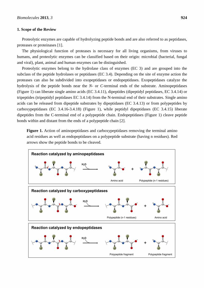
proteases can also be subdivided into exopeptidases or endopeptidases. Exopeptidases catalyze the
hydrolysis of the peptide bonds near the N- or C-terminal ends of the substrate. Aminopeptidases
(Figure 1) can liberate single amino acids (EC 3.4.11), dipeptides (dipeptidyl peptidases, EC 3.4.14) or
tripeptides (tripeptidyl peptidases EC 3.4.14) from the N-terminal end of their substrates. Single amino
acids can be released from dipeptide substrates by dipeptidases (EC 3.4.13) or from polypeptides by
carboxypeptidases (EC 3.4.16-3.4.18) (Figure 1), while peptidyl dipeptidases (EC 3.4.15) liberate
dipeptides from the C-terminal end of a polypeptide chain. Endopeptidases (Figure 1) cleave peptide
bonds within and distant from the ends of a polypeptide chain [2].
Figure 1. Action of aminopeptidases and carboxypeptidases removing the terminal amino
acid residues as well as endopeptidases on a polypeptide substrate (having n residues). Red
arrows show the peptide bonds to be cleaved.
Biomolecules 2013, 3 925
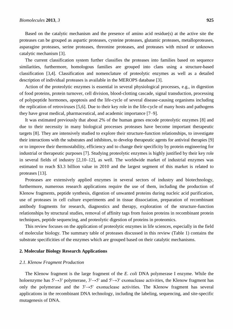
Based on the catalytic mechanism and the presence of amino acid residue(s) at the active site the
proteases can be grouped as aspartic proteases, cysteine proteases, glutamic proteases, metalloproteases,
asparagine proteases, serine proteases, threonine proteases, and proteases with mixed or unknown
catalytic mechanism [3].
The current classification system further classifies the proteases into families based on sequence
similarities, furthermore, homologous families are grouped into clans using a structure-based
classification [3,4]. Classification and nomenclature of proteolytic enzymes as well as a detailed
description of individual proteases is available in the MEROPS database [3].
Action of the proteolytic enzymes is essential in several physiological processes, e.g., in digestion
of food proteins, protein turnover, cell division, blood-clotting cascade, signal transduction, processing
of polypeptide hormones, apoptosis and the life-cycle of several disease-causing organisms including
the replication of retroviruses [5,6]. Due to their key role in the life-cycle of many hosts and pathogens
they have great medical, pharmaceutical, and academic importance [7–9].
It was estimated previously that about 2% of the human genes encode proteolytic enzymes [8] and
due to their necessity in many biological processes proteases have become important therapeutic
targets [8]. They are intensively studied to explore their structure-function relationships, to investigate
their interactions with the substrates and inhibitors, to develop therapeutic agents for antiviral therapies [9]
or to improve their thermostability, efficiency and to change their specificity by protein engineering for
industrial or therapeutic purposes [7]. Studying proteolytic enzymes is highly justified by their key role
in several fields of industry [2,10–12], as well. The worldwide market of industrial enzymes was
estimated to reach $3.3 billion value in 2010 and the largest segment of this market is related to
proteases [13].
Proteases are extensively applied enzymes in several sectors of industry and biotechnology,
furthermore, numerous research applications require the use of them, including the production of
Klenow fragments, peptide synthesis, digestion of unwanted proteins during nucleic acid purification,
use of proteases in cell culture experiments and in tissue dissociation, preparation of recombinant
antibody fragments for research, diagnostics and therapy, exploration of the structure-function
relationships by structural studies, removal of affinity tags from fusion proteins in recombinant protein
techniques, peptide sequencing, and proteolytic digestion of proteins in proteomics.
This review focuses on the application of proteolytic enzymes in life sciences, especially in the field
of molecular biology. The summary table of proteases discussed in this review (Table 1) contains the
substrate specificities of the enzymes which are grouped based on their catalytic mechanisms.
2. Molecular Biology Research Applications
2.1. Klenow Fragment Production
The Klenow fragment is the large fragment of the E. coli DNA polymerase I enzyme. While the
holoenzyme has 5'→3' polymerase, 3'→5' and 5'→3' exonuclease activities, the Klenow fragment has
only the polymerase and the 3'→5' exonuclease activities. The Klenow fragment has several
applications in the recombinant DNA technology, including the labeling, sequencing, and site-specific
mutagenesis of DNA.
Biomolecules 2013, 3 926
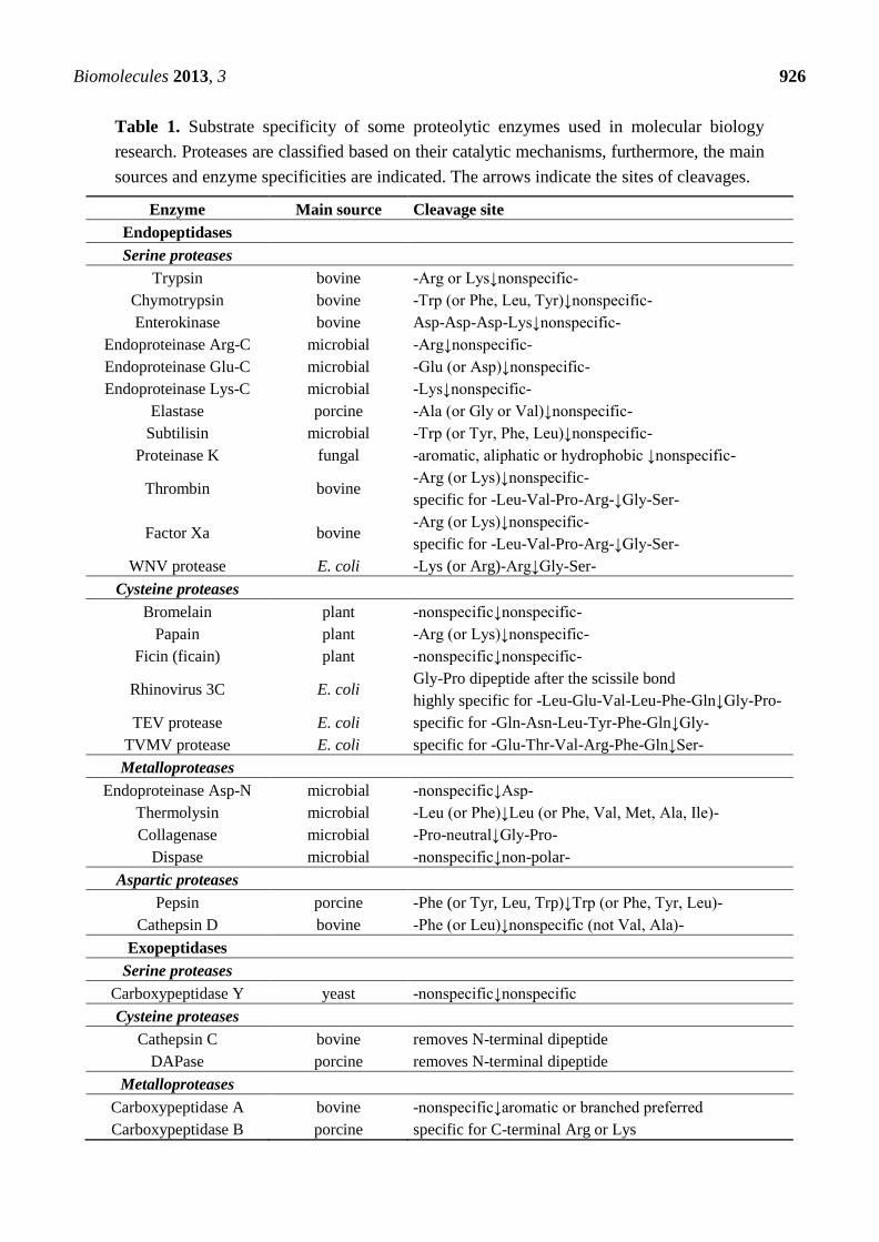
Table 1. Substrate specificity of some proteolytic enzymes used in molecular biology
research. Proteases are classified based on their catalytic mechanisms, furthermore, the main
sources and enzyme specificities are indicated. The arrows indicate the sites of cleavages.
Enzyme Main source Cleavage site
Endopeptidases
Serine proteases
Trypsin bovine -Arg or Lys↓nonspecific-
Chymotrypsin bovine -Trp (or Phe, Leu, Tyr)↓nonspecific-
Enterokinase bovine Asp-Asp-Asp-Lys↓nonspecific-
Endoproteinase Arg-C microbial -Arg↓nonspecific-
Endoproteinase Glu-C microbial -Glu (or Asp)↓nonspecific-
Endoproteinase Lys-C microbial -Lys↓nonspecific-
Elastase porcine -Ala (or Gly or Val)↓nonspecific-
Subtilisin microbial -Trp (or Tyr, Phe, Leu)↓nonspecific-
Proteinase K fungal -aromatic, aliphatic or hydrophobic ↓nonspecific-
Thrombin bovine -Arg (or Lys)↓nonspecific-
specific for -Leu-Val-Pro-Arg-↓Gly-Ser-
Factor Xa bovine -Arg (or Lys)↓nonspecific-
specific for -Leu-Val-Pro-Arg-↓Gly-Ser-
WNV protease E. coli -Lys (or Arg)-Arg↓Gly-Ser-
Cysteine proteases
Bromelain plant -nonspecific↓nonspecific-
Papain plant -Arg (or Lys)↓nonspecific-
Ficin (ficain) plant -nonspecific↓nonspecific-
Rhinovirus 3C E. coli Gly-Pro dipeptide after the scissile bond
highly specific for -Leu-Glu-Val-Leu-Phe-Gln↓Gly-Pro-
TEV protease E. coli specific for -Gln-Asn-Leu-Tyr-Phe-Gln↓Gly-
TVMV protease E. coli specific for -Glu-Thr-Val-Arg-Phe-Gln↓Ser-
Metalloproteases
Endoproteinase Asp-N microbial -nonspecific↓Asp-
Thermolysin microbial -Leu (or Phe)↓Leu (or Phe, Val, Met, Ala, Ile)-
Collagenase microbial -Pro-neutral↓Gly-Pro-
Dispase microbial -nonspecific↓non-polar-
Aspartic proteases
Pepsin porcine -Phe (or Tyr, Leu, Trp)↓Trp (or Phe, Tyr, Leu)-
Cathepsin D bovine -Phe (or Leu)↓nonspecific (not Val, Ala)-
Exopeptidases
Serine proteases
Carboxypeptidase Y yeast -nonspecific↓nonspecific
Cysteine proteases
Cathepsin C bovine removes N-terminal dipeptide
DAPase porcine removes N-terminal dipeptide
Metalloproteases
Carboxypeptidase A bovine -nonspecific↓aromatic or branched preferred
Carboxypeptidase B porcine specific for C-terminal Arg or Lys
Biomolecules 2013, 3 927
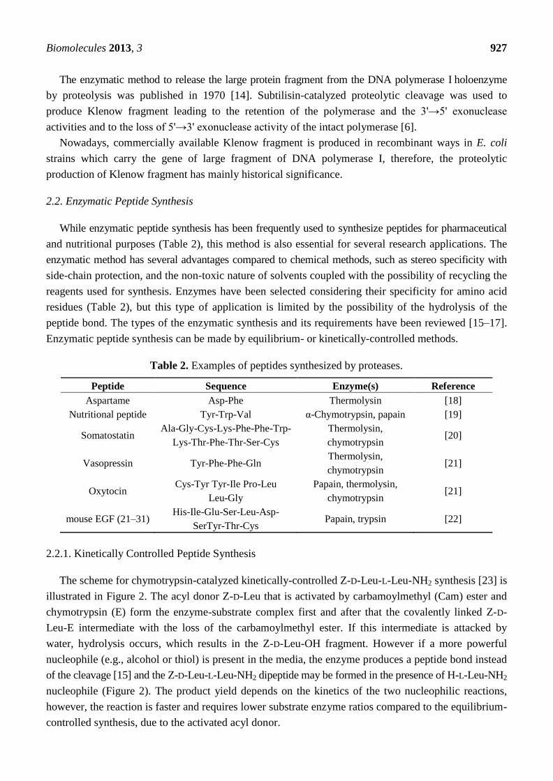
The enzymatic method to release the large protein fragment from the DNA polymerase I holoenzyme
by proteolysis was published in 1970 [14]. Subtilisin-catalyzed proteolytic cleavage was used to
produce Klenow fragment leading to the retention of the polymerase and the 3'→5' exonuclease
activities and to the loss of 5'→3' exonuclease activity of the intact polymerase [6].
Nowadays, commercially available Klenow fragment is produced in recombinant ways in E. coli
strains which carry the gene of large fragment of DNA polymerase I, therefore, the proteolytic
production of Klenow fragment has mainly historical significance.
2.2. Enzymatic Peptide Synthesis
While enzymatic peptide synthesis has been frequently used to synthesize peptides for pharmaceutical
and nutritional purposes (Table 2), this method is also essential for several research applications. The
enzymatic method has several advantages compared to chemical methods, such as stereo specificity with
side-chain protection, and the non-toxic nature of solvents coupled with the possibility of recycling the
reagents used for synthesis. Enzymes have been selected considering their specificity for amino acid
residues (Table 2), but this type of application is limited by the possibility of the hydrolysis of the
peptide bond. The types of the enzymatic synthesis and its requirements have been reviewed [15–17].
Enzymatic peptide synthesis can be made by equilibrium- or kinetically-controlled methods.
Table 2. Examples of peptides synthesized by proteases.
Peptide Sequence Enzyme(s) Reference
Aspartame Asp-Phe Thermolysin [18]
Nutritional peptide Tyr-Trp-Val α-Chymotrypsin, papain [19]
Somatostatin Ala-Gly-Cys-Lys-Phe-Phe-Trp-
Lys-Thr-Phe-Thr-Ser-Cys
Thermolysin,
chymotrypsin [20]
Vasopressin Tyr-Phe-Phe-Gln Thermolysin,
chymotrypsin [21]
Oxytocin Cys-Tyr Tyr-Ile Pro-Leu
Leu-Gly
Papain, thermolysin,
chymotrypsin [21]
mouse EGF (21–31) His-Ile-Glu-Ser-Leu-Asp-
SerTyr-Thr-Cys Papain, trypsin [22]
2.2.1. Kinetically Controlled Peptide Synthesis
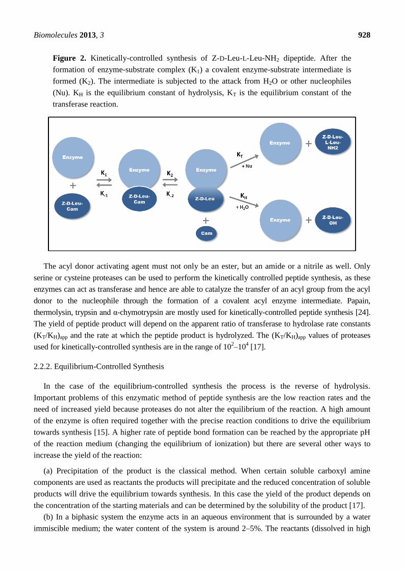
The scheme for chymotrypsin-catalyzed kinetically-controlled Z-D-Leu-L-Leu-NH2 synthesis [23] is
illustrated in Figure 2. The acyl donor Z-D-Leu that is activated by carbamoylmethyl (Cam) ester and
chymotrypsin (E) form the enzyme-substrate complex first and after that the covalently linked Z-D-
Leu-E intermediate with the loss of the carbamoylmethyl ester. If this intermediate is attacked by
water, hydrolysis occurs, which results in the Z-D-Leu-OH fragment. However if a more powerful
nucleophile (e.g., alcohol or thiol) is present in the media, the enzyme produces a peptide bond instead
of the cleavage [15] and the Z-D-Leu-L-Leu-NH2 dipeptide may be formed in the presence of H-L-Leu-NH2
nucleophile (Figure 2). The product yield depends on the kinetics of the two nucleophilic reactions,
however, the reaction is faster and requires lower substrate enzyme ratios compared to the equilibrium-
controlled synthesis, due to the activated acyl donor.
Biomolecules 2013, 3 928
Figure 2. Kinetically-controlled synthesis of Z-D-Leu-L-Leu-NH2 dipeptide. After the
formation of enzyme-substrate complex (K1) a covalent enzyme-substrate intermediate is
formed (K2). The intermediate is subjected to the attack from H2O or other nucleophiles
(Nu). KH is the equilibrium constant of hydrolysis, KT is the equilibrium constant of the
transferase reaction.
The acyl donor activating agent must not only be an ester, but an amide or a nitrile as well. Only
serine or cysteine proteases can be used to perform the kinetically controlled peptide synthesis, as these
enzymes can act as transferase and hence are able to catalyze the transfer of an acyl group from the acyl
donor to the nucleophile through the formation of a covalent acyl enzyme intermediate. Papain,
thermolysin, trypsin and α-chymotrypsin are mostly used for kinetically-controlled peptide synthesis [24].
The yield of peptide product will depend on the apparent ratio of transferase to hydrolase rate constants
(KT/KH)app and the rate at which the peptide product is hydrolyzed. The (KT/KH)app values of proteases
used for kinetically-controlled synthesis are in the range of 102–10
4 [17].
2.2.2. Equilibrium-Controlled Synthesis
In the case of the equilibrium-controlled synthesis the process is the reverse of hydrolysis.
Important problems of this enzymatic method of peptide synthesis are the low reaction rates and the
need of increased yield because proteases do not alter the equilibrium of the reaction. A high amount
of the enzyme is often required together with the precise reaction conditions to drive the equilibrium
towards synthesis [15]. A higher rate of peptide bond formation can be reached by the appropriate pH
of the reaction medium (changing the equilibrium of ionization) but there are several other ways to
increase the yield of the reaction:
(a) Precipitation of the product is the classical method. When certain soluble carboxyl amine
components are used as reactants the products will precipitate and the reduced concentration of soluble
products will drive the equilibrium towards synthesis. In this case the yield of the product depends on
the concentration of the starting materials and can be determined by the solubility of the product [17].
(b) In a biphasic system the enzyme acts in an aqueous environment that is surrounded by a water
immiscible medium; the water content of the system is around 2–5%. The reactants (dissolved in high
Biomolecules 2013, 3 929
concentrations) can diffuse from the organic phase into the water until the equilibrium is reached; the
enzyme-catalyzed synthesis is followed by the diffusion of the products back into the organic phase. The
organic phase reduces the dielectric constant of the medium and thus the acidity of the carboxyl group of
the acyl donor as well, which in turn promotes the synthesis of the peptide bond instead of hydrolysis [24].
This method is applicable only for the synthesis of water insoluble products.
(c) The dissolved state system can be used for the synthesis of water-soluble products (short
peptides, high molecular weight peptides and proteins). In this environment forcing the reaction
towards peptide synthesis requires the mass action, the addition of a water-miscible organic co-solvent
in high concentration or the excess of one reactant. In serine protease-catalyzed reaction in water, the
rate determining feature is the acylation of the enzyme while the product yield at equilibrium depends
on the partition coefficient and the ratio of the aqueous and organic volumes [17]. The chymotrypsin-
and subtilisin-catalyzed synthesis of N-Bz-L-Tyr-L-Leu-NH2 is more efficient in hydrophobic organic
solvents; adding water in sub-saturating concentration increases the yield of the chymotrypsin-
catalyzed peptide synthesis [25].
2.2.3. Strategies Used in Enzymatic Synthesis
The use of enzymes in organic solvents have several advantages compared to aqueous solvents
which have led to their widespread application: the thermodynamic equilibrium can be shifted towards
synthesis, the undesirable side reactions can be reduced, the nonpolar substrates are more soluble in
organic solvents, the separation process and enzyme recovery is more effective in a low water-containing
environment [26–29]. Many proteases, such as thermolysin, subtilisin and α-chymotrypsin [26,29] can
maintain their active conformation in organic solvents and show good functionality in the synthesis of
aspartame and demorphin derivatives.
However, the use of enzymes in organic solvent has also disadvantages such as the unfavorable
effects of the organic solvents on enzyme activity and stability. The modification of biocatalysts by
protein engineering [30,31] and/or chemical modification or the use of naturally solvent-tolerant
proteases [32] for peptide synthesis is a developing field. The driving force of this field is the aim of
making biocatalysts with proper features to suit them for the reactions under specific synthesis
conditions. Site-directed mutagenesis is a very effective tool and can be used by protein engineers to
screen mutants with enhanced stability, activity or specificity, furthermore, this method can be used to
explore structure-function relationships (rational design).
Subtilisin has been extensively studied and engineered via site-directed mutagenesis to make it
more capable of peptide bond formation in aqueous solution [33]. Single and multiple mutations have
been introduced into subtilisin to increase its stability and make it more resistant against oxidizing
agents, thermal denaturations and inactivation effects of polar solvents [28].
Thermolysin has a higher synthesis rate compared to the solvent stable PST-01 protease from
Pseudomonas aeruginosa. Considering the high structural similarity of these enzymes, the synthetic
activity of PST-01 protease was increased by the Y114F mutation [31], moreover, the Y114R and
Y114S mutations resulted in better activity enhancement.
Chemical modification is also an efficient method to modify the properties of enzymes used for
peptide synthesis. Thiol-subtilisin, in which the serine residue has been chemically changed to cysteine
Biomolecules 2013, 3 930
at the active site, shows an enhanced aminolysis to hydrolysis ratio in aqueous solution and in dimethyl
sulphoxide. The stability of proteases can also be increased by chemical modification e.g., a hydrophilic
carbohydrate-polyacrylate polymer coat can make the enzymes highly active and stable in polar
solvents and more resistant against thermal inactivation [28].
Immobilization of proteases is the most frequently applied method for the recovery of products
without great loss of the catalysts, which greatly decreases the cost of the synthesis. This approach also
ensures better operational stability of biocatalysts and control of the reaction. Enzyme immobilization
techniques can be divided into five groups: (a) covalent attachment to solid support; (b) absorption on
solid support; (c) entrapment in polymeric gel; (d) crosslinking with bifunctional reagents and (e)
encapsulation [17,34].
Substrate engineering means the manipulation of the leaving group and is a powerful tool to
increase the specificity of the proper enzyme and/or increase the rate and the yield of the reaction [24].
Protease-catalyzed synthesis of stereochemically modified peptides is also a preferable application
compared to chemical synthesis due to stereospecificity of the proteases.
Proteases can bind not only natural substrates, but also specifically designed substrate mimetics,
which are also very useful tools to increase the yield of peptide synthesis. Substrate mimetics can bind
to the active site of the enzyme and in this way proteases can be used for the synthesis of products
containing non-specific amino acids. The undesired cleavage of the newly synthesized peptide bonds
can be avoided using this method and it is not required to change the properties of the medium or the
enzyme [17].
The production of peptides with amides at their C-termini is a great challenge for enzymatic peptide
synthesis, but amidation may be required to retain biological activity.
2.3. Nucleic Acid Isolation
Generally, the first step of nucleic acid isolation protocols is the lysis of the biological material
containing the DNA or RNA of interest. Before the purification and concentration of nucleic acids the
contaminating proteins and other macromolecules have to be removed from the sample. Undamaged
nucleic acids can be isolated when the degradation of the DNA and RNA present in the sample is
avoided by the inhibition and removal of DNases and RNases. The nucleases can be inhibited by the
addition of chelators (e.g., EDTA) which bind the ions essential for their action. Besides the
inactivation of nucleases, proteolytic enzymes are applied during the nucleic acid isolation to remove
total protein content of the sample.
The most widely used proteolytic enzyme in nucleic acid purification is the Proteinase K, which
was described in 1974 [35]. Proteinase K is a non-specific serine endopeptidase which can catalyze the
cleavage of peptide bonds at the carboxylic side of aromatic, aliphatic, or hydrophobic amino acid
residues. Besides the digestion of unwanted proteins, Proteinase K also quickly inactivates the
nucleases which might degrade the nucleic acids present in the sample [36]. This proteolytic digestion
decreases the level of contaminants in the nucleic acid extract and prevents nucleic acids from
degradation leading to a higher yield of the DNA or RNA to be isolated.
Biomolecules 2013, 3 931
2.4. Cell Isolation and Tissue Dissociation
Cell biology studies frequently require the dissociation of primary tissues and the isolation of viable
cells for tissue culturing. The most common method for cell isolation is the enzymatic digestion of the
junctions connecting the cells and the components of the surrounding extracellular matrix, by which
the cells can be released from a wide variety of tissues. Several enzymes are available in the market for
the detachment of cultured cells, cell dissociation and cell component or membrane-associated protein
isolation [37,38]. Besides the polysaccharidases, nucleases and lipases, the proteases are the most
important enzymes used widely to dissociate cells from tissues.
The experimental conditions of cell isolation are functions of several parameters, including the type
of the tissue and the source of its origin. Cells with high viability can be isolated in high yield using a
suitable enzyme or the optimal combination of enzymes. As proteases differ in their specificities,
different enzymes are recommended to be used use for most effective tissue disruption, depending on
the origin and type of the tissue. We describe below the enzymes most commonly used for cell isolation.
The matrix metalloproteinase collagenase was first isolated in 1953 [39]. This endopeptidase can
digest the collagenous extracellular matrix in a zinc-dependent manner. Collagenase cleaves the peptide
bonds within the triple helices of native collagen, between a neutral amino acid and Gly within the
Pro-X-Gly-Pro sequence. This sequence can be found most frequently in the collagen; therefore collagenases
digest other proteins less efficiently. A commercially available collagenase (clostridiopeptidase A) is
produced by Clostridium histolyticum, and it is capable of digesting collagen fibers very effectively.
Solutions supplied for tissue dissociation contain collagenase and other additional proteinases which
can digest the components of the extracellular matrix [38,40]. The serine protease elastase is a unique
enzyme which can cleave the peptide bonds in elastin, therefore, it is generally used to dissociate
tissues containing a high amount of elastin connective fibers. Elastase cleaves peptide bonds next to
smaller neutral amino acids and besides its protease activity it also has esterase and amidase activities.
Papain is a cysteine peptidase of Carica papaya latex. Papain, similarly to elastase, also has amidase
and esterase activities and has a broad specificity. Papain has less damaging effects on tissues and
therefore it is typically applied for cell dissociation of neuronal tissues. Besides cell dissociation,
papain is also widely used for integral membrane protein solubilization and for digestion of
proteoglycans. The serine protease trypsin is a very specific proteinase cleaving the peptide bonds at
the C-terminal end of positively charged Lys and Arg side chains. Due to the high specificity of trypsin
the digestion of tissue proteins is less effective and it is generally used for tissue dissociation together
with other proteolytic enzymes. Serine protease chymotrypsin cleaves peptide bonds preferentially at
the carboxyl side of aromatic Tyr, Trp and Phe residues. Chymotrypsin is less widely used for tissue
dissociation; the use of other additional proteases is required for efficient digestion. The Zn-
metalloprotease dispase is also a neutral protease. This non-specific protease cleaves the peptide bonds
of proteins at the amino side of non-polar amino acid residues.
2.5. Cell Culturing
Cells isolated from a tissue can be cultured separately from the organism in cell culture flasks using
appropriate growth medium. Adherent cells grown in a cell culture flask are attached to the surface by
Biomolecules 2013, 3 932
protein bridges which have to be disrupted during passaging. The cells can be released from the cell
flask surface mechanically using a cell scraper or can be detached by a protease treatment using
trypsin solution.
Trypsinization means the process used for the detachment of adherent cells using trypsin solution to
digest the adhesion molecules by which the cells are attached to the surface of the culture flask.
Trypsin solutions generally contain EDTA to reduce the concentration of metal ions that might
inhibit trypsin.
Although trypsinization is the most commonly used method to detach adherent cells from cell
culture flasks, the effects of this protease treatment were only recently studied in detail. It was found
that trypsinization can affect the extracellular matrix surrounding the cells [41] and has physiological
effects on cells grown in cell cultures [42]. Trypsin treatment can lead to cleavage of membrane
proteins and receptors, which can cause significant changes in the expression level of different
proteins: level of growth- and metabolism-related protein expressions were found to be
down-regulated after trypsinization, while up-regulation of apoptosis-related protein expressions was
seen after the protease treatment [42,43]. This effect should be taken into account when trypsinization
is involved in experimental design.
2.6. Antibody Fragment Production
Antibody molecules are produced by the immune system against foreign substances and are
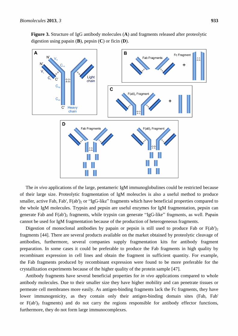
classified into the immunoglobulin superfamily of the proteins (Figure 3A). They consist of four
polypeptide chains, two identical heavy chains (H) and two identical light chains (L) which are
connected by disulfide bridges. Both the H and the L chains contain variable (VH and VL) and constant
(CH1, CH2, CH3 and CL) regions, respectively. The VH and VL chains, containing hypervariable
regions, are responsible for the antigen-antibody interactions and determine the antigen specificity [6].
Fragments of the monoclonal antibodies are widely used in diagnostics, therapeutics and in
biopharmaceutical research [44–46] having beneficial properties compared to the whole immunoglobulin
molecules due to their smaller size and lower immunogenicity [44]. Fragments of whole
immunoglobulin molecules can be produced using recombinant DNA technology or can be generated by
enzymatic digestion. Here we discuss the proteolytic antibody fragmentation method.
Generally, the papain, pepsin and ficin proteases are used for the specific digestion of IgG
molecules. Digestion of an antibody by the cysteine protease papain results in three fragments due to
the cleavage of peptide bonds in the hinge region between CH1 and CH2 domains: one Fc
(crystallizable) and two identical Fab (antigen binding) fragments are released (Figure 3B). While both
released Fab fragments carry one antigen-binding site, the Fc fragment does not have antigen-binding
ability. The aspartic acid protease pepsin cleaves the peptide bonds of the antibody near the disulfide
bonds connecting the H chains (Figure 3C). This digestion results in the release of the peptides of the
Fc region and one F(ab')2 fragment containing both antigen binding sites. The cysteine protease ficin
can release both F(ab')2 or Fab fragments (Figure 3D), depending on the cysteine concentration [44].
Biomolecules 2013, 3 933
Figure 3. Structure of IgG antibody molecules (A) and fragments released after proteolytic
digestion using papain (B), pepsin (C) or ficin (D).
The in vivo applications of the large, pentameric IgM immunoglobulines could be restricted because
of their large size. Proteolytic fragmentation of IgM moleucles is also a useful method to produce
smaller, active Fab, Fab', F(ab')2 or ―IgG-like‖ fragments which have beneficial properties compared to
the whole IgM molecules. Trypsin and pepsin are useful enzymes for IgM fragmentation, pepsin can
generate Fab and F(ab')2 fragments, while trypsin can generate ―IgG-like‖ fragments, as well. Papain
cannot be used for IgM fragmentation because of the production of heterogeneous fragments.
Digestion of monoclonal antibodies by papain or pepsin is still used to produce Fab or F(ab')2
fragments [44]. There are several products available on the market obtained by proteolytic cleavage of
antibodies, furthermore, several companies supply fragmentation kits for antibody fragment
preparation. In some cases it could be preferable to produce the Fab fragments in high quality by
recombinant expression in cell lines and obtain the fragment in sufficient quantity. For example,
the Fab fragments produced by recombinant expression were found to be more preferable for the
crystallization experiments because of the higher quality of the protein sample [47].
Antibody fragments have several beneficial properties for in vivo applications compared to whole
antibody molecules. Due to their smaller size they have higher mobility and can penetrate tissues or
permeate cell membranes more easily. As antigen-binding fragments lack the Fc fragments, they have
lower immunogenicity, as they contain only their antigen-binding domain sites (Fab, Fab'
or F(ab')2 fragments) and do not carry the regions responsible for antibody effector functions,
furthermore, they do not form large immunocomplexes.
Biomolecules 2013, 3 934
The antigen-binding fragments of antibodies have great importance from the viewpoint of clinical
and therapeutic applications. Antibody fragments could be administered to prevent the development of
a disease (e.g., restenosis), could be applied during the diagnosis (e.g., metastatic breast and colon
cancer) or for the treatment of some diseases (e.g., macular degeneration) or to detect toxins or
neutralize snake venoms [44,45]. The current number of antibody-based therapeutics approved by the
FDA is 35, while several other antibodies are in clinical trials. The relevance of antibody fragments in
structural studies is discussed in detail in the following paragraph. Further important biotechnological
applications of antibody fragments, e.g., as immunodetection, immunopurification and detoxification,
have been reviewed in the recent past [46].
2.7. Structural Studies
Crystallographers have great challenges in the structure determination of transmembrane proteins. It
is difficult to crystallize these proteins due to their high molecular flexibility, hydrophobic surfaces and
low solubility. Antibody fragments are very useful tools for solving these problems. Specific binding
of antibody fragments can increase the overall hydrophilicity of proteins and the solubility of the
transmembrane proteins, furthermore, they can decrease the flexibility and stabilize the conformation
of the molecule [48]. The structures of the membrane proteins co-crystallized with antibody fragments
can be determined at higher resolution because these crystals have a higher diffraction quality [49–51].
The proteases have great indirect significance in structure determination, because the antibody
fragments produced by proteolytic cleavage of whole immunoglobulins can be used to improve the
crystallization properties [52–54].
The proteases are not only tools for crystallographers but are also important target molecules for
structural biologists and have great relevance in antiviral therapies, drug and therapeutics development
from the viewpoint of structural biology. Besides interest in proteases with unknown structure, the
results of structural studies can help researchers evaluate the structure-function relationships more
efficiently; increasing knowledge on the structural organization of viral proteases can help to explore
their action, perform comparative studies by which we can better understand the structure-function and
evolutionary relationships and recognize general or specific features [55–57]. One of the main driving
forces of structure determination of proteases is the need for the development of efficient drugs for
antiviral therapies [7,9,58–60]. Both structural and enzymatic inhibition studies are required for the
structure-based drug development of protease inhibitors [61]. Structural data can also help protein
engineers to alter the specificity and to improve the enzymatic properties of proteases by structure-
guided mutagenesis [62–64] for several purposes.
2.8. Fusion Tag Removal
The proteins produced by recombinant techniques are typically linked with a fusion partner termed
a fusion tag. The introduction of a fusion tag means the fusion of an additional protein or peptide to the
recombinant protein. These fusion tags are extensively used from basic research to high-throughput
structural biology owing to the several advantages they provide in the expression of different
recombinant proteins [65,66]. The tags largely aid the detection and purification of proteins; moreover
they also could have a favorable effect on protein yield and/or solubility. Tags can prevent proteins
Biomolecules 2013, 3 935
from proteolytic digestion, can protect antigenicity or facilitate the folding of the fusion protein.
On the other hand, they can also negatively alter solubility, structural integrity and biological
activity [47,67,68] or may cause a disadvantage for further application of the protein, so the removal of
a tag can be crucial [66].
Commonly used affinity tags and fusion protein partners are the hexahistidine-tag (His6),
FLAG-tag, maltose binding protein (MBP), glutathione S-transferase (GST), thioredoxin (TRX), small
ubiquitin-like modifier (SUMO), ubiquitin (Ub) and green fluorescent protein (GFP) [69].
In some cases the tag can be removed by a chemical treatment but those methods are rather
unspecific compared to enzymatic cleavages and may lead to protein denaturation and/or side chain
modifications of amino acids in the target protein. The specificity and detergent sensitivity of common
proteases used for tag removal have been examined and reviewed previously [70–73]. Both endo- and
exo-proteases could be suitable for fusion tag removal.
2.8.1. Endoproteases
Serine proteases such as enterokinase (also referred to as enteropeptidase), factor Xa and thrombin
have been widely used for many years to remove N-terminal tags, but several cases have been reported
in which they cleaved not only at the desired cleavage site but also in the protein of interest. These
incidents led to the extensive application of viral proteases like human rhinovirus (HRV) 3C protease
and tobacco etch virus (TEV) protease. While sequences recognized by a cellular serine protease and
the viral proteases could be similar, the viral proteases cleave the protein substrates at the undesired
sites less efficiently due to their high specificity and low catalytic rate, moreover, recombinant viral
proteases can be produced in high quantities in E. coli [73]. These findings and the limited activity of
the generally used serine proteases in some detergents, which are needed to study the membrane
proteins, inspired the search for other viral proteases for tag removal, such as Tobacco Vein Mottling
Virus (TVMV) protease [74], West Nile Virus (WNV) protease [75] and some alphaviral proteases:
Venezuelan Equine Encephalitis Virus (VEEV) protease, Semliki Forest Virus (SFV) and Sindbis
Virus (SIN) protease [76].
2.8.2. Exopeptidases
Aminopeptidases and carboxypeptidases are not as widely used as endopeptidases, as they
frequently leave amino acid residues on the target protein, while it is easier to design cleavage sites
with endopeptidases not to leave extra residues after the cleavage. However, if is still desired to add a
tag onto the C-terminal of the target protein a carboxypeptidase may be used for its removal. Among
metallocarboxypeptidases, type A carboxypeptidases remove mostly aromatic or branched aliphatic
side chain containing amino acids, while type B carboxypeptidases prefer basic amino acids.
Carboxypeptidases can be utilized to remove a His6 tag from the C-terminal end of a protein [77].
Dipeptidyl aminopeptidase (DAPase) is a useful enzyme for the removal of N-terminal dipeptides.
Biomolecules 2013, 3 936
2.9. Proteomic Applications
Proteomic studies are made with the aim to identify, characterize, and quantify the required samples
and typically involve mass spectrometric (MS) analysis. Besides determination of the composition of
protein complexes, chemical properties, post-translational modifications and structural properties of
proteins can also be revealed by MS.
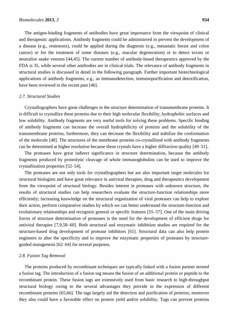
MS is a powerful analytical method to measure the mass of proteins or peptides by the analysis of
mass-to-charge ratio (m/z) ratio. Generally, the samples to be analyzed contain a mixture of various
proteins and/or polypeptides that have to be separated and digested into smaller fragments before the MS
analysis. Protein separation can be performed efficiently by polyacrylamide gel electrophoresis. The
separated proteins can be digested after the electrophoresis by chemical cleavage or by enzyme-catalyzed
digestion of peptide bonds. The process in which the bands or spots are cut out from the gel followed by
the addition of protease(s) to the gel containing the protein(s) of interest is called in-gel digestion [78].
Proteolysis of whole proteins leads to the release of smaller peptides with different molecular masses
which are suitable for MS analysis (Figure 4).
Figure 4. Steps of proteomic analysis using mass-spectrometry after separation and in-gel
digestion of proteins of interest.
Trypsin is the most widely used proteolytic enzyme for protein digestion in MS analysis. Due to its
high specificity it is easy to predict the cleavage sites and to compare the results of experimental
enzymatic and theoretical in-silico digestion. The Asp-N, Lys-C, Arg-C and Glu-C enyzmes are also
highly sequence-specific endoproteases but they are less active [78]. Chymotrypsin, pepsin, papain,
elastase, subtilisin, proteinase K, thrombin, factor Xa and some other proteases are also suitable
enzymes for fragmentation in MS analysis [79].
Several proteases are available for this fragmentation of which specificities are well established [79,80].
The number and length of released peptide fragments depends on the protease(s) applied for selective
Biomolecules 2013, 3 937
proteolysis of the targeted protein. Applications developed for in silico protein fragmentation are
useful to predict the proteolytic fragments and to choose the most proper enzyme for the most effective
digestion based on the enzyme specificities (http://prospector.ucsf.edu).
Generally, highly sequence-specific proteases are preferred for protein fragmentation instead of less
specific enzymes, as the latter ones produce a very complex mixture of fragments. In the case of the
efficient fragmentation the peptide, the fragments have a proper length and are released in high yield,
and the complete sequence of the whole protein can be covered by the analysis of the
proteolytic fragments.
3. Summary
Besides extended application for nutritional and pharmaceutical purposes, proteases from natural
sources are also widely used tools in molecular biology practice. Their degradative properties make
them useful for general protein digestion in tissue dissociation, cell isolation, and cell culturing. The
specificity and the predictability of cleavages by proteases enables their use for more specific tasks
such as antibody fragment production, the removal of affinity tags from recombinant proteins and
specific protein digestion in the proteomics field mainly for protein sequencing. Moreover, the already
mentioned specificity makes proteases—in a water restricted environment—able to synthesize the
peptide bonds instead of hydrolyzing them. This property combined with their enantioselectivity has
also promoted their use in peptide synthesis.
The expansion of knowledge has assisted the increase of applications of proteolytic enzymes for
several purposes, and the application fields are widening with the help of protein engineering
techniques and by chemical modification of the enzymes [7,62]. Studies made with the aim to better
understand the structure and function of existing proteolytic enzymes and to obtain new, engineered
proteases with altered properties for therapeutic, industrial or research fields require the use of the
applications discussed in this paper.
Acknowledgments
This work was supported by the TÁMOP 4.2.2.A-11/1/KONV-2012-0023 ―VÉD-ELEM‖ and
by the Hungarian Science and Research Fund (OTKA 101591) implemented through the New
Hungary Development Plan co-financed by the European Social Fund and the European Regional
Development Fund.
Conflicts of Interest
The authors declare no conflict of interest.
References
1. Barrett, A.J.; McDonald, J.K. Nomenclature: Protease, proteinase and peptidase. Biochem. J.
1986, 237, 935.
2. Rao, M.B.; Tanksale, A.M.; Ghatge, M.S.; Deshpande, V.V. Molecular and biotechnological
aspects of microbial proteases. Microbiol. Mol. Biol. Rev. 1998, 62, 597–635.
Biomolecules 2013, 3 938
3. Rawlings, N.D.; Barrett, A.J.; Bateman, A. MEROPS: The database of proteolytic enzymes, their
substrates and inhibitors. Nucleic Acids Res. 2012, 40, D343–D350.
4. Rawlings, N.D.; Barrett, A.J. Evolutionary families of peptidases. Biochem. J. 1993, 290,
205–218.
5. Neurath, H.; Walsh, K.A. Role of proteolytic enzymes in biological regulation (a review).
Proc. Natl. Acad. Sci. USA 1976, 73, 3825–3832.
6. Devlin, T.M. Textbook of Biochemistry with Clinical Correlations, 5th ed.; Wiley & Sons:
New York, NY, USA, 2002.
7. Li, Q.; Yi, L.; Marek, P.; Iverson, B.L. Commercial proteases: Present and future. FEBS Lett.
2013, 587, 1155–1163.
8. Craik, C.S.; Page, M.J.; Madison, E.L. Proteases as therapeutics. Biochem. J. 2011, 435, 1–16.
9. Antonelli, G.; Turriziani, O. Antiviral therapy: Old and current issues. Int. J. Antimicrob. Agents
2012, 40, 95–102.
10. Kirk, O.; Borchert, T.V.; Fuglsang, C.C. Industrial enzyme applications. Curr. Opin. Biotechnol.
2002, 13, 345–351.
11. Rani, K.; Rana, R.; Datt, S. Review on latest overview of proteases. Int. J. Curr. Life Sci. 2012, 2,
12–18.
12. Ray, A. Protease enzyme- potential industrial scope. Int. J. Technol. 2012, 2, 1–4.
13. Sarrouh, B.; Santos, T.M.; Miyoshi, A.; Dias, R.; Azevedo, V. Up-to-date insight on industrial
enzymes applications and global market. J. Bioprocess. Biotech. 2012, S4:002
14. Klenow, H.; Henningsen, I. Selective elimination of the exonuclease activity of the
deoxyribonucleic acid polymerase from Escherichia coli B by limited proteolysis. Proc. Natl.
Acad. Sci. USA 1970, 65, 168–175.
15. Morihara, K. Using proteases in peptide synthesis. Trends Biotechnol. 1987, 5, 164–170.
16. Bhalla, T.C.; Kumar, D.; Gajju, H.; Agrawal, H.O. Thermophilic bacterial proteases. J. Punjab
Acad. Sci. 1999, 1, 77–91
17. Kumar, D.; Bhalla, T.C. Microbial proteases in peptide synthesis: Approaches and applications.
Appl. Microbiol. Biotechnol. 2005, 68, 726–736.
18. Kühn, D.; Dürrschmidt, P.; Mansfeld, J.; Ulbrich-Hofmann, R. Biolysin and thermolysin in
dipeptide synthesis: A comparative study. Biotechnol. Appl. Biochem. 2002, 36, 71–76.
19. Kimura, Y.; Muraya, K.; Araki, Y.; Matsuoka, H.; Nakanishi, K.; Matsuno, R. Synthesis peptides
consisting of essential amino acids by a reactor system using three proteinases and an organic
solvent. Agric. Biol. Chem. 1990, 54, 3331–3333.
20. Bille, V.; Ripak, C.; van Aasche, I.; Forni, I.; Degelaen, L.; Searso, A. A Semi-Enzymatic
Synthesis of Somatostatin. In Proceedings of 21st European Peptide Symposium. ESCOM,
Leiden, The Netherlands, 1991; Giralt, E., Andreu, D., Eds.; pp. 253–254.
21. Rizo, J.; Gierarch, L.M. Constrained peptides: Models of bioactive peptides and protein
structures. Ann. Rev. Biochem. 1992, 61, 387–418.
22. Widmer, F.; Bayne, S.; Houen, G.; Rigby, R.B.; Whittaker, R.G.; Johansen, J.T. Use of
Proteolytic Enzymes for the Synthesis of Fragments of Mouse Epidermal Growth Factor.
In Proceedings of Peptides 1984, Almquist, Stockholm, 1985; Ragnurrson, U., Ed.; pp. 193–196.
Biomolecules 2013, 3 939
23. Salam, S.M.; Kagawa, K.; Kawashiro, K. Alpha-chymotrypsin-catalyzed peptide synthesis using
N-protected D-amino acid carbamoylmethyl esters as acyl donors. Biotechnol. Lett. 2005, 27,
1199–1203.
24. Guzmán, F.; Barberis, S.; Illanes, A. Peptide synthesis: Chemical or enzymatic.
Electron. J. Biotechnol. 2007, 10, 279–314.
25. Sergeeva, M.V.; Paradkar, V.M.; Dordick, J.S. Peptide synthesis using proteases dissolved in
organic solvents. Enzyme Microb. Technol. 1997, 20, 623–628.
26. Dordick, J.S. Enzymatic catalysis in monophasic organic solvents. Enzyme Microb. Technol.
1989, 11, 194–211.
27. Khemlnitski, Y.L.; Levashov, A.V.; Klyachko, N.L.; Martinek, K. Engineering biocatalytic
systems in organic media with low water content. Enzyme Microb. Technol. 1988, 10, 710–724.
28. Gill, I.; Fandino, R.L.; Jobra, X.; Vulfson, E.N. Biologically active peptides and enzymatic
approaches to their production. Enzyme Microb. Technol. 1996, 18, 162–183.
29. Shen, H.Y.; Tian, G.L.; Ye, Y.H. Synthesis of demorphin (1–4) derivatives catalysed by proteases
in organic solvents. J. Pept. Res. 2004, 65, 143–148.
30. Wells, J.A.; Estell, D.A. Subtilisin: An enzyme designed to be engineered. Trends Biochem. Sci.
1988, 13, 291–297.
31. Ogino, H.; Tsuchiyama, S.; Yasuda, M.; Doukyu, N. Enhancement of the aspartame precursor
synthetic activity of an organic solvent-stable protease. Protein Eng. Des. Sel. 2010, 23, 147–152.
32. Xu, J.X.; Jiang, M.; Sun, H.L.; He, B.F. An organic solvent-stable protease from organic
solvent-tolerant Bacillus cereus WQ9–2: Purification, biochemical properties, and potential
application in peptide synthesis. Bioresour. Technol. 2010, 101, 7991–7994.
33. Abrahmsén, L.; Tom, J.; Burnier, J.; Butcher, K.A.; Kossiakoff, A.; Wells, J.A. Engineering
subtilisin and its substrates for efficient ligation of peptide bonds in aqueous solution.
Biochemistry 1991, 30, 4151–4159.
34. Adamczak, M.; Krishna, S.H. Enzyme for efficient biocatalysis. Food Technol. Biotechnol. 2004,
42, 251–264.
35. Ebeling, W.; Hennrich, N.; Klockow, M.; Metz, H.; Orth, H.D.; Lang, H. Proteinase K from
Tritirachium album Limber. Eur. J. Biochem. 1974, 47, 91–97.
36. Carpi, F.M.; di Pietro, F.; Vincenzetti, S.; Mignini, F.; Napolioni, V. Human DNA extraction
methods: Patents and applications. Recent Pat. DNA Gene Seq. 2011, 5, 1–7.
37. Sigma-Aldrich Corporation. Enzymes for cell dissociation and lysis. Biofiles Life Sci. Res. 2006,
Avaiable online: http://www.sigmaaldrich.com/ etc/medialib/docs/Sigma/
General_Information/2/biofiles_issue2.Par.0001.File.tmp/biofiles_issue2.pdf (accessed on 20 July
2013).
38. Santangelo, C. Worthington biochemical online tissue dissociation guide. Worthington Biochem.
Corp. 2011. Avaiable online: http://www.worthington-biochem.com/ tissuedissociation/
default.html (accessed on 20 July 2013).
39. Mandl, I.; Maclennan, J.D.; Howes, E.L. Isolation and characterization of proteinase and
collagenase from Cl. histolyticum. J. Clin. Investig. 1953, 32, 1323–1329.
40. Kin, T.; Johnson, P.R.; Shapiro, A.M.; Lakey, J.R. Factors influencing the collagenase digestion
phase of human islet isolation. Transplantation 2007, 83, 7–12.
Biomolecules 2013, 3 940
41. Canavan, H.E.; Cheng, X.; Graham, D.J.; Ratner, B.D.; Castner, D.G. Cell sheet detachment
affects the extracellular matrix: A surface science study comparing thermal liftoff, enzymatic, and
mechanical methods. J. Biomed. Mater. Res. A 2005, 75, 1–13.
42. Huang, H.L.; Hsing, H.W.; Lai, T.C.; Chen, Y.W.; Lee, T.R.; Chan, H.T.; Lyu, P.C.; Wu, C.L.;
Lu, Y.C.; Lin, S.T.; et al. Trypsin-induced proteome alteration during cell subculture in
mammalian cells. J. Biomed. Sci. 2012, 17, doi:10.1186/1423-0127-17-36.
43. Danhier, P.; Copetti, T.; de Preter, G.; Leveque, P.; Feron, O.; Jordan, B.F.; Sonveaux, P.; Gallez, B.
Influence of cell detachment on the respiration rate of tumor and endothelial cells. PLoS One
2013, 8, e53324.
44. Rader, C. Overview on concepts and applications of Fab antibody fragments. Curr. Protoc.
Protein Sci. 2009, 55, 6.9.1–6.9.14.
45. Flanagan, R.J.; Jones, A.L. Fab antibody fragments: Some applications in clinical toxicology.
Drug Saf. 2004, 27, 1115–1133.
46. De Marco, A. Biotechnological applications of recombinant single-domain antibody fragments.
Microb. Cell Fact. 2011, 10, doi:10.1186/1475-2859-10-44.
47. Zhao, Y.; Gutshall, L.; Jiang, H.; Baker, A.; Beil, E.; Obmolova, G.; Carton, J.; Taudte, S.;
Amegadzie, B. Two routes for production and purification of Fab fragments in biopharmaceutical
discovery research: Papain digestion of mAb and transient expression in mammalian cells.
Protein Expr. Purif. 2009, 67, 182–189.
48. Hunte, C.; Michel, H. Crystallisation of membrane proteins mediated by antibody fragments.
Curr. Opin. Struct. Biol. 2002, 12, 503–508.
49. Kovari, L.C.; Momany, C.; Rossmann, M.G. The use of antibody fragments for crystallization and
structure determinations. Structure 1995, 3, 1291–1293.
50. Caffrey, M. Membrane protein crystallization. J. Struct. Biol. 2003, 42, 108–132.
51. Bill, R.M.; Henderson, P.J.; Iwata, S.; Kunji, E.R.; Michel, H.; Neutze, R.; Newstead, S.;
Poolman, B.; Tate, C.G.; Vogel, H. Overcoming barriers to membrane protein structure
determination. Nat. Biotechnol. 2011, 29, 335–340.
52. Zhou, Y.; Morais-Cabral, J.H.; Kaufman, A.; MacKinnon, R. Chemistry of ion coordination and
hydration revealed by a K+ channel-Fab complex at 2.0 A resolution. Nature 2001, 414, 43–48.
53. Rasmussen, S.G.F.; Choi, H.J.; Rosenbaum, D.M.; Kobilka, T.S.; Thian, F.S.; Edwards, P.C.;
Burghammer, M.; Ratnala, V.R.; Sanishvili, R.; Fischetti, R.F.; et al. Crystal structure of the
human beta2 adrenergic G-protein-coupled receptor. Nature 2007, 450, 383–387.
54. Kwong, P.D.; Wyatt, R.; Robinson, J.; Sweet, R.W.; Sodroski, J.; Hendrickson, W.A. Structure of
an HIV gp120 envelope glycoprotein in complex with the CD4 receptor and a neutralizing human
antibody. Nature 1998, 393, 648–659.
55. Bagossi, P.; Sperka, T.; Fehér, A.; Kádas, J.; Zahuczky, G.; Miklóssy, G.; Boross, P.; Tőzsér, J.
Amino acid preferences for a critical substrate binding subsite of retroviral proteases in type 1
cleavage sites. J. Virol. 2005, 79, 4213–4218.
56. Eizert, H.; Bander, P.; Bagossi, P.; Sperka, T.; Miklóssy, G.; Boross, P.; Weber, I.T.; Tőzsér, J.
Amino acid preferences of retroviral proteases for amino-terminal positions in a type 1 cleavage
site. J. Virol. 2008, 82, 10111–10117.
Biomolecules 2013, 3 941
57. Tőzsér, J. Comparative studies on retroviral proteases: Substrate specificity. Viruses 2010, 2,
147–165.
58. Patick, A.K.; Potts, K.E. Protease inhibitors as antiviral agents. Clin. Microbiol. Rev. 1998, 11,
614–627.
59. Wlodawer, A.; Gustchina, A. Structural and biochemical studies of retroviral proteases.
Biochim. Biophys. Acta 2000, 1477, 16–34.
60. De Clercq, E. Strategies in the design of antiviral drugs. Nat. Rev. Drug Discov. 2000, 1, 13–25.
61. Wlodawer, A. Structure-based design of AIDS drugs and the development of resistance. Vox Sang.
2002, 83 (Suppl. 1), 23–26.
62. Pogson, M.; Georgiou, G.; Iverson, B.L. Engineering next generation proteases.
Curr. Opin. Biotechnol. 2009, 20, 390–397.
63. Lim, E.J.; Sampath, S.; Coll-Rodriguez, J.; Schmidt, J.; Ray, K.; Rodgers, D.W. Swapping the
substrate specificities of the neuropeptidases neurolysin and thimet oligopeptidase. J. Biol. Chem.
2007, 282, 9722–9732.
64. Villa, J.P.; Bertenshaw, G.P.; Bond, J.S. Critical amino acids in the active site of meprin
metalloproteinases for substrate and peptide bond specificity. J. Biol. Chem. 2003, 278,
42545–42550.
65. Derewenda, Z.S. The use of recombinant methods and molecular engineering in protein
crystallization. Methods 2004, 34, 354–363.
66. Waugh, D.S. Making the most of affinity tags. Trends Biotechnol. 2005, 23, 316–320.
67. Renzi, F.; Panetta, G.; Vallone, B.; Brunori, M.; Arceci, M.; Bozzoni, I.; Laneve, P.; Caffarelli, E.
Large-scale purification and crystallization of the endoribonuclease XendoU: Troubleshooting
with His-tagged proteins. Acta Crystallogr. Sect. F Struct. Biol. Cryst. Commun. 2006, 62,
298–301.
68. Horchani, H.; Ouertani, S.; Gargouri, Y.; Sayari, A. The N-terminal His-tag and the
recombination process affect the biochemical properties of Staphylococcus aureus lipase produced
in Escherichia coli. J. Mol. Catal. B Enzym. 2009, 61, 194–201.
69. Terpe, K. Overview of tag protein fusions: From molecular and biochemical fundamentals to
commercial systems. Appl. Microbiol. Biotechnol. 2003, 60, 523–533.
70. Jenny, R.J.; Mann, K.G.; Lundblad, R.L. A critical review of the methods for cleavage of fusion
proteins with thrombin and factor Xa. Protein Expr. Purif. 2003, 31, 1–11.
71. Arnau, J.; Lauritzen, C.; Petersen, G.E.; Pedersen, J. Current strategies for the use of affinity tags
and tag removal for the purification of recombinant proteins. Protein Expr. Purif. 2005, 48, 1–13.
72. Vergis, J.M.; Wiener, M.C. The variable detergent sensitivity of proteases that are utilized for
recombinant protein affinity tag removal. Protein Expr. Purif. 2011, 78, 139–142.
73. Waugh, D.S. An overview of enzymatic reagents for the removal of affinity tags. Protein Expr. Purif.
2011, 80, 283–293.
74. Nallamsetty, S.; Kapust, R.B.; Tőzsér, J.; Cherry, S.; Tropea, J.E.; Copeland, T.D.; Waugh, D.S.
Efficient site-specific processing of fusion proteins by tobacco vein mottling virus protease in vivo
and in vitro. Protein Expr. Purif. 2004, 38, 108–115.
75. Huang, Q.; Li, Q.; Chen, A.S.; Kang, C. West Nile virus protease activity in detergent solutions
and application for affinity tag removal. Anal. Biochem. 2013, 435, 44–46.
Biomolecules 2013, 3 942
76. Zhang, D.; Tőzsér, J.; Waugh, D.S. Molecular cloning, overproduction, purification and
biochemical characterization of the p39 nsp2 protease domains encoded by three alphaviruses.
Protein Expr. Purif. 2009, 64, 89–97.
77. Austin, B.P.; Tőzsér, J.; Bagossi, P.; Tropea, J.E.; Waugh, D.S. The substrate specificity of
Metarhizium anisopliae and Bos taurus carboxypeptidases A: Insights into their use as tools for
the removal of affinity tags. Protein Expr. Purif. 2011, 77, 53–61.
78. Steen, H.; Mann, M. The ABC’s (and XYZ’s) of peptide sequencing. Nat. Rev. Mol. Cell Biol.
2004, 5, 699–711.
79. Granvogl, B.; Plöscher, M.; Eichacker, L.A. Sample preparation by in-gel digestion for mass
spectrometry-based proteomics. Anal. Bioanal. Chem. 2007, 389, 991–1002.
80. Meyers, A.; Trauger, S.; Webb, W.; Reisdorph, N.; Wranik, C.; Peters, E.; Siuzdak, G. Protein
identification and profiling with mass spectrometry. Spectroscopy 2003, 17, 1–15.
© 2013 by the authors; licensee MDPI, Basel, Switzerland. This article is an open access article
distributed under the terms and conditions of the Creative Commons Attribution license
(http://creativecommons.org/licenses/by/3.0/).