Embed Size (px)
Citation preview

Research ArticleAn Eleven-Year Retrospective Study of EndogenousBacterial Endophthalmitis
Takashi Nishida,1 Kyoko Ishida,2 Yoshiaki Niwa,3 Hideaki Kawakami,4
Kiyofumi Mochizuki,1 and Kiyofumi Ohkusu5
1Department of Ophthalmology, Gifu University Graduate School of Medicine, Gifu, Japan2Department of Ophthalmology, Toho University Ohashi Medical Center, 2-17-6 Ohashi, Meguro-ku, Tokyo 153-8515, Japan3Department of Ophthalmology, Gifu Prefectural General Medical Center, Gifu, Japan4Department of Ophthalmology, Gifu Municipal Hospital, Gifu, Japan5Department of Microbiology, Tokyo Medical University Graduate School of Medicine, Tokyo, Japan
Correspondence should be addressed to Kyoko Ishida; [email protected]
Received 28 November 2014; Revised 7 January 2015; Accepted 8 January 2015
Academic Editor: Timothy Y. Lai
Copyright © 2015 Takashi Nishida et al.This is an open access article distributed under theCreative CommonsAttribution License,which permits unrestricted use, distribution, and reproduction in any medium, provided the original work is properly cited.
Purpose. To determine the clinical features, microbial profiles, treatment outcomes, and prognostic factors for endogenous bacterialendophthalmitis (EBE). Methods. The medical records of 27 eyes of 21 patients diagnosed with EBE for 11 years were reviewed.Collected data included age, site of infection, visual acuities (VAs), microbial profiles, and treatment regimen. Results. The meanage was 68.5 years. Gram-positive organisms accounted for 76.2%, while gram-negative ones accounted for 19.0%. Staphylococcusaureus was the most common causative organism (52.3%) of which 72.7% wasmethicillin-resistant S. aureus. A final VA of ≥20/40was achieved in 44% and 20/200 or better was in 64%. Eyes with initial VA of ≥20/200 (𝑃 = 0.003) and focal involvements (𝑃 =0.011) had significantly better final VA. Initial VA (𝑃 = 0.001) and the interval between onset of ocular symptoms and intravitrealantibiotic injection (𝑃 = 0.097) were associated with final VA in eyes receiving intravitreal antibiotics. Conclusions. EBE is generallyassociatedwith poor visual outcome; however the prognosismay depend on initial VA, extent of ocular involvement, and an intervalbetween onset of ocular symptoms and intravitreal antibiotic injection. Early diagnosis and early intravitreal injection supplementto systemic antibiotics might lead to a relatively good visual outcome.
1. Introduction
Endogenous bacterial endophthalmitis (EBE) is rare andaccounts for 2% to 8% of all cases of endophthalmitis [1–3]. It is a damaging disease, and the visual prognosis is gen-erally poor with more than one-half of eyes becoming blinddespite treatment [1–4].The systemic pathological conditionspredisposing an eye to endophthalmitis include malignan-cies, alcoholism, cardiovascular diseases, diabetes mellitus,indwelling catheters, bone and joint diseases, intravenousdrug use, hemodialysis, trauma, and cirrhosis of the liver [1–3, 5, 6]. Endogenous bacterial endophthalmitis is both anocular and extraocular disease [1, 3].
The visual outcome of endogenous endophthalmitis ispoorer than that of exogenous endophthalmitis. The main
factors associated with the poor prognosis include the viru-lence of the causative organisms, compromised host condi-tions, and a delay in diagnosis and treatment [1–3, 6]. EBE canbe an unusual complication of systemic bacterial infectionswith severe consequences if left untreated. Patients at highrisk for bacteremia who present with ocular complaintsshould have a thorough funduscopic examination to rule outendophthalmitis [6, 7].
There are no standardized diagnosis or treatment guide-lines for endogenous bacterial endophthalmitis because of itslow incidence, inadequate data on the treatment regimens,and the lack of long-term follow-up results [2, 3, 8].
The purpose of this study is to evaluate the clinicalfeatures, microbiological spectrum, treatment regimens, andoutcomes of EBE in three centers in Japan during an 11-year
Hindawi Publishing CorporationJournal of OphthalmologyVolume 2015, Article ID 261310, 11 pageshttp://dx.doi.org/10.1155/2015/261310

2 Journal of Ophthalmology
period. We also aimed to assess associated factors for visualprognosis.
2. Patients and Methods
2.1. Patients. We reviewed the medical records of consec-utive patients diagnosed with EBE between January 2002and June 2013. All of the patients were referred to threehospitals of Gifu prefecture: Gifu University Graduate Schoolof Medicine, Gifu Municipal Hospital, and Gifu PrefecturalGeneral Medical Center. The patients were diagnosed withEBE by constitutional symptoms, decrease in visual acuity(VA), ocular pain, hypopyon, subretinal lesions, and anteriorand/or posterior uveitis. The uveitis was confirmed by cul-tures of the blood, vitreous, or aqueous humor in all cases.The findings in 3 of these patients (Patients 6, 7, and 12) werereported previously [9–11].
Patients were excluded if they had a history of oculartrauma or ocular surgery within 1 year of the onset of theinfection or evidence of a primary external ocular infectionsuch as infectious keratitis or filtering bleb infection [2, 12].
The medical records were reviewed. Collected dateincluded the age, sex, presenting complaints, underlyingsystemic infections, preexistingmedical conditions, source ofinfection, laterality, VA, microbial profiles, treatment meth-ods, and initial and final VAs. The main outcome measurewas the best-corrected VA at the final follow-up examination.In earlier reports [9, 12], a best-corrected VA of countingfingers (CF) or better was classified as being a good visualoutcome for the statistical analyses; however in this reportwe used 20/200 or better as a good visual outcome. Otheroutcome measures included the results of microbiologicalinvestigations and anatomical and clinical outcomes.
2.2. Statistical Analysis. Fisher’s exact test, Chi-square test,Kruskal-Wallis test, Mann-Whitney 𝑈 test, and Spearman’srank correlation coefficient were used to determine the sig-nificance of any differences. Logistic regression and multi-ple regression analysis were also used to detect associatedfactors for visual prognosis. The odds ratio was calculated,and the 95th percentile confidence intervals (95% CI) weredetermined. The decimal VA values were converted to thelogarithm of the minimum angle of resolution (logMAR)units. For a visual acuity less than CF the following arbitrarylogMAR values were used: CF = 2.00 logMAR units, handmotion= 2.30 logMARunits, light perception= 2.60 logMARunits, and no light perception = 2.90 logMAR units [13, 14].
A 𝑃 value of <0.05 was considered to be significant.All statistical analyses were performed using SPSS softwareversion 16.0 (SPSS Japan, Tokyo, Japan).
3. Results
3.1. Patient Characteristics and Systemic Features. The studywas approved by the institutional review boards. Twenty-seven eyes of 21 Japanese patientswere included in the currentstudy. Patient characteristics and systemic features are shownin Table 1. The mean age at presentation was 68.5 ± 9.7 years
(range, 41 to 80 years). The mean follow-up period was 76.3weeks with a range of 2 to 520 weeks. There were 14 (66.7%)men and 7 (33.3%) women. Thirteen (61.9%) patients hadcommunity-acquired infections, and the others (38.1%) hadhospital-acquired infections. The number of patients withEBE was 5 (23.8%) in the spring, 3 (14.3%) in the summer,8 (38.1%) in the autumn, and 5 (23.8%) in the winter.
All patients had one or more preexisting medical con-ditions that predisposed to the development of EBE. Themost common medical condition was diabetes mellitus (13patients, 61.9%), followed by hypertension (𝑛=6, 28.6%), gas-trointestinal disorders (𝑛 = 5, 28.8%), cardiac disease (𝑛 = 5,28.8%), malignancy (𝑛 = 5, 28.8%; 4 solid organmalignanciesand 1 hematologic malignancy), urological diseases (𝑛 = 5,28.8%), and hemodialysis (𝑛 = 2, 9.5%) (Table 1). Systemicsteroids were being used to treat an underlying illness in 5(28.8%) patients.
Extraocular infectious foci were identified in 17 (81.0%)patients, infective endocarditis was identified in 3 (14.3%)patients, pneumonia was identified in 2 (9.5%), soft tissue(skin and wound) infection was identified in 2 (9.5%), peri-tonitis was identified in 2 (9.5%), catheter-related infectionwas identified in 2 (9.5%), recent trauma (head and kidney)was identified in 2 (9.5%), liver abscess was identified in 1,urinary tract infection was identified in 1, burn was identifiedin 1, and psoas abscess was identified in one. The source ofinfection could not be identified in 4 patients.
Eighteen patients (85.7%) had an onset of systemic orocular symptoms within 7 days prior to their admission, and2 patients developed symptoms ≥4 weeks before admission(mean 6.0 days; range, <1 to 29 days). The most com-mon initial systemic symptom was fever including cold-likesymptoms in 16 (76.2%) patients, followed by diarrhea andvomiting (𝑛 = 1), anterior chest pain (𝑛 = 1), back pain (𝑛 = 1),myalgia (𝑛 = 1), and injection site abscess (𝑛 = 1).
At the first visit to a doctor, a high body temperature(>38∘C) was noted in 16 (76.2%), elevated C-reactive protein(range, 3.69–30.23mg/dL) in 16 (76.2%), and a high whiteblood cell count (12,630–24,560/𝜇L) in 11 (52.3%) of the 21patients. The plasma level of 𝛽-D-glucan was measured in14 patients and the range was 11.9–230 pg/mL, and it wasconsidered positive in 1 patient when the cutoff value was setat 20 pg/mL [15].
3.2. Ocular Features. Five (23.8%) presented with right eyeinvolvement, 10 (47.6%) with left eye involvement, and 6(28.6%) with bilateral involvement (Table 2). The first ocularsymptomwas decreased vision (14 patients, 66.7%), floaters (5patients, 23.8%), pain (2 patients, 9.5%), and eyelid swelling(1 patient, 4.8%). However, only 5 (23.8%) of 21 patientsinitially consulted with an ophthalmologist in a privateoffice or hospital, and 16 (76.2%) sought help from otherclinical departments, for example, internal medicine (𝑛 = 10),orthopedics (𝑛 = 2), emergency (𝑛 = 2), and surgery (𝑛 =2). The mean interval between the onset of ocular signs orsymptoms and the first visit to an ophthalmologist was 8.4days (range, 1 to 30 days). A correct initial ocular diagnosisof bacterial endophthalmitis wasmade by ophthalmologist in

Journal of Ophthalmology 3
Table 1: Patient characteristics and systemic features.
Number Age(years) Sex Underlying medical
conditionPresumed
infection sourceCRP
(mg/dL)WBC(/𝜇L)
𝛽-D-glucan(pg/mL)
Systemicsteroid
Days from onset ofsymptoms to initial
examination(ophthalmologist)
1 72 M HT, cardiac disease Liver abscess 8.18 13100 ND − 4 (same day)2 60 F DM Psoas abscess 21.42 15890 ND − 0 (6)3 72 F DM, uterine cancer Peritonitis 6.8 18100 ND − 3 (same day)4† 76 M DM Pneumonia 30.23 14230 <5.0 − 29 (same day)5† 80 M Gastric cancer Catheter-related 9.98 12700 ND − 6 (same day)
6 73 MAutoimmune
hemolytic anemia,DM, HT
Unknown (manyorgans) 0.34 12630 230 + 2 (2)‡
7 74 F Cardiac disease Infectiveendocarditis 10.65 11850 <5.0 − 5 (14)
8† 77 M Colon cancer,intestinal perforation Peritonitis 2.69 9390 6.3 + 28 (same day)
9 60 M
DM, HT,hemodialysis, gastric
ulcer, infectiousspondylitis
Soft tissueinfection 7.68 4490 15.4 − 6 (same day)
10 70 F Lung disease, HT Pneumonia 10.93 14200 6.6 − 2 (2)‡
11 58 M DM, cardiac disease Infectiveendocarditis 18.71 12050 5.3 − 1 (8)
12 73 M Urological disease Urinary tractinfection 16.15 19320 ND − 4 (same day)
13† 41 F Adult onset Still’sdisease Unknown 0.8 24560 <5.0 + 2 (30)
14† 59 M DM, hemodialysis Burn 9.32 9400 ND − 13 (same day)
15 71 FDM, HT, cardiac
disease, rheumatoidarthritis
Infectiveendocarditis 18.96 15500 ND − 1 (same day)
16 79 M DM, colon cancer Unknown 10.09 8230 <5.0 − 1 (1)‡
17† 71 M
DM, HT, renal failure,liver cirrhosis,
rupture of umbilicalhernia
Catheter-related 12.25 8470 9.1 − 6 (7)
18 78 M Renal failure Unknown 0.62 7300 11.9 + 5 (same day)19† 55 M DM, HT Head trauma 14.49 23210 <5.0 − 2 (2)‡
20 68 F DM Leg abscess 0.09 11610 <5.0 + 3 (3)‡
21† 72 M DM, cardiac disease Renal trauma 3.69 9770 19.6 − 2 (same day)†Hospital-acquired patient; CRP, C-reactive protein; WBC, white blood cell; M, male; F, female; DM, diabetes mellitus; HT, hypertension; ND, not done;‡initially consulted an ophthalmologist.
15 (71.4%) patients.Other initial ocular diagnoseswere uveitis(𝑛= 4, 19.0%), fungal endophthalmitis (𝑛= 2, 9.5%), choroidaltumor (𝑛 = 1), and glaucoma (𝑛 = 1).
The principal sites of EBE [6] for the 27 eyes were diffuseposterior endophthalmitis in 10 (37.0%) eyes, posterior focalendophthalmitis in 13 (48.1%) eyes, and panophthalmitis in 4(14.8%) eyes (Table 2).
3.3.Microbiology. Theorganism causing the endophthalmitiswas identified by a positive culture from at least one bodyfluid source in 19 (90.5%) of the 21 patients (Table 2). In two
of 21 patients, the pathogenic organism was not determined.Funguswas not detected in any of the samples.The organismsisolated from the blood, intraocular, and other cultures areshown in Table 3. Twenty-two organisms were identified intotal. The blood was the highest source of a positive cultureand 17 organisms were detected from 15 of 20 patients. Apositive vitreous culture was obtained in 6 of 10 patientsand aqueous humor in 2 of 6 patients. Two patients hadpositive central venous catheter tip cultures and a positivesoft tissue culture including psoas. Diagnostic vitrectomywas performed in 1 patient (Patient 6) at 49 days after

4 Journal of Ophthalmology
Table2:Ocularfeatures,microbiologicalfin
ding
s,andtre
atments.
Num
ber
Eye
Type
ofEB
En1
Organism
Cultu
reAntibiotic
sSurgeryn
3InitialVA
Outcome
(follo
w-upin
weeks)
Bloo
dVitre
ous
Other
positive
cultu
res
Syste
mic
Intravitrealn2
1OD
Pan
K.pn
eumoniae−
+ASP
CNon
eEn
ucleation(7)
CFNLP
(17)
2OS
PDS.aureus
+ND
Psoas
(S.aureus)
PAPM
/PM,V
CM,
CLDM,C
TRX
CAZ(<1)
—20/25
20/16
(520)
3OD
PF(N
-L-T)
S.agalactia
e,CN
S+
+IPM/CS,ITCZ
VCM
+CA
Z#(1)
Vit+
PEA(1)
20/600
20/20(35)
4†OS
PDMRS
A+−(A
Q−)
Urin
e(M
RSA)
PAPM
/BP,VC
MVC
M+CA
Z#(1)
Vit+
PEA+s/o
(1)
CF20/200
(3)
5†OS
PDS.aureus,
S.agalactia
e+
ND
CEZ,
ST,M
EPM,
F-FL
CZ,C
DTR
-PI
Non
e—
20/600
20/25(78)
6OS
PF(Pos)
Nocardia
farcinica
+−n4
MCF
G,F-FLC
Z,MEP
M,IPM
/CS,ST
Non
e—
20/50
20/40(313)§1
7OS
Pan
S.equisim
ilis
++(A
Q+)
PCG,C
TRX,
GM,
TAZ/PIPC
VCM
+MEP
M(<1)
—LP
Phthisis(57)
8†OD
PDMRS
AND
−IV
cann
ula
(MRS
A)
CPR,
VCM
VCM
+CA
Z#(9)
Vit+
PEA(37)
LPPh
thisis(2)
9OD
PF(Pos)
MRS
A+
ND
CAZ,
BIPM
,CEZ
,CTM
Non
e—
20/20
20/25(35)
10OD OS
PD PDS.pn
eumoniae
+− −
CPR,
TAZ/PIPC
,PIPC,
FMOX,
CFPN
-PI
VCM
+CA
Z#(28)
VCM
+CA
Z#(8
&15)
Vit+
PEA+s/o
(30)
Vit+
PEA(10)
LP HM
LP(19
6)HM
(196)
11OD OS
PF PFUnk
nown
−ND
Sputum
(MRS
A)
FOM,C
ZOP,CT
RX,
GM,V
CM,LZD
Non
eNon
e—
20/32
20/28
20/16
(113)
20/16
(113)
12OS
Pan
K.pn
eumoniae−
+(A
Q+)
CPR,
CEZ,
IPM/CS.
CTRX
Non
eEn
ucleation(9)
LPNLP
(4)
13†
OD OS
PF(T)
PDMRS
A+
ND
CFDN,M
EPM,M
INO,
MCF
G,F-FLC
Z,BIPM
,LV
FX,TAZ/PIPC
,VCM
,LZ
D
Non
eNon
e—
ND
Death
(3)
14†
OD OS
PF(N
)PF
(N)
MRS
A+
(AQ−)
(AQ−)
VCM,T
EIC
VCM
+CA
Z(8
&15)
VCM
+CA
Z(15)
—20/500
20/500
20/250
(14)
20/200
(14)
15OS
PF(N
)S.agalactia
e+
ND
AZM
,IPM
/CS,GM
Non
e—
CF20/20(33)
16OD
PDUnk
nown
−(A
Q−)
CZOP
VCM
+CA
Z(4)
—HM
HM
(15)
17†
OS
PDMRS
A+
(AQ−)
VCM,F-FLC
ZVC
M(<1&
2)—
HM
20/60(7)
18OS
PDNe
isseriasp.+‡
S.aureus
+VC
M,M
EPM,C
EZ,
MCF
G,FLC
ZVC
M+CA
Z#(22)
Vit(22)
20/25
20/20(7)
19†
OD OS
PF(Sup
N)
Pan
S.aureus
++
MIN
O,D
RPM,LZD
,CE
ZVC
M+CA
Z(<1)
Non
e—
Enucleation(23)
20/20
NLP
20/100§2(67)
NLP
(67)

Journal of Ophthalmology 5
Table2:Con
tinued.
Num
ber
Eye
Type
ofEB
En1
Organism
Cultu
reAntibiotic
sSurgeryn
3InitialVA
Outcome
(follo
w-upin
weeks)
Bloo
dVitre
ous
Other
positive
cultu
res
Syste
mic
Intravitrealn2
20OS
PF(Sup
N)
MRS
A−
ND
Leg
abscess
(MRS
A)
ST,C
TRX,
LZD
Non
e—
20/20
20/20(44)
21†
OD OS
PF(T)
PF(T)
MRS
A,
E.clo
acae
+ND
IVcann
ula
(MRS
A)
CFPM
,F-FLC
Z,LZ
D,
DAP,RF
P,ST
VCM
(9)
VCM
(16)
—20/10
020/40
20/20(39)
20/50§3(39)
†
Hospital-a
cquiredpatie
nt,n
1 site
ofsubretinalabscessinparentheses,
n2nu
mberinparenthesesrepresentsd
aysfrom
diagno
sisof
endo
phthalmitis,
n3nu
mberinparenthesesrepresentsd
aysfrom
onseto
focular
symptom
s,and
n4diagno
sticvitre
ctom
y;OD,right
eye;OS,lefteye;EB
E,endo
geno
usbacterialend
ophthalm
itis;Pan,
pano
phthalmitis;PD
,posterio
rdiffuse;PF,p
osterio
rfocal:N
,nasal;T
,tem
poral;L,
lower;
Pos,po
sterio
r;SupT
,sup
eriortem
poral;SupN
,sup
eriorn
asal;C
NS,coagulase-negativ
eStaphylo
coccus;M
RSA,m
ethicillin-resis
tant
Staphylococcus
aureus;A
Q,aqu
eous;N
D,n
otdo
ne;IV,
intravenou
s;VA
,visu
alacuity;V
it,vitre
ctom
y;PE
A,p
hacoem
ulsifi
catio
nandaspiratio
n;s/o,
silicon
eoil;NLP,n
olight
perceptio
n;CF
,cou
ntingfin
gers;L
P,light
perceptio
n;HM,h
andmotion;
§1cataract
andepire
tinal
mem
brane;
§2vitre
oush
emorrhage;
§3cataract;#vitre
ctom
yandintravitrealinjectio
n;‡
notd
etectedNe
isseria
sp.from
bloo
dcultu
re;T
AZ/PIPC
,tazob
actam/piperacillin;P
IPC,
piperacillin;
PCG,b
enzylp
enicillin;A
SPC,
aspo
xicillin;CT
RX,ceft
riaxone;C
EZ,cefazolin;C
PR,cefpirome;CZ
OP,cefozopran;C
DTR
-PI,cefdito
renpivoxil;CT
M,cefotiam;C
FDN,cefdinir;CA
Z,cefta
zidime;CF
PM,cefepim
e;FM
OX,
flomoxef;C
FPN-PI,
cefcapenep
ivoxil;IPM/CS,im
ipenem
/cilastatin;M
EPM,m
erop
enem
;BIPM,biapenem;PAPM
/BP,panipenem/betam
ipron;DRP
M,doripenem
;GM,gentamicin;A
ZM,azithromycin;C
LDM,clin
damycin;M
INO,
minocyclin
e;LZ
D,linezolid;V
CM,vancomycin;T
EIC,
teicop
lanin;
DAP,daptom
ycin;LVFX
,levofl
oxacin;R
FP,rifampicin;
ST,sulfametho
xazole;FOM,fosfomycin;F-FLC
Z,fosflucon
azole;MCF
G,m
icafun
gin.
6 Journal of Ophthalmology
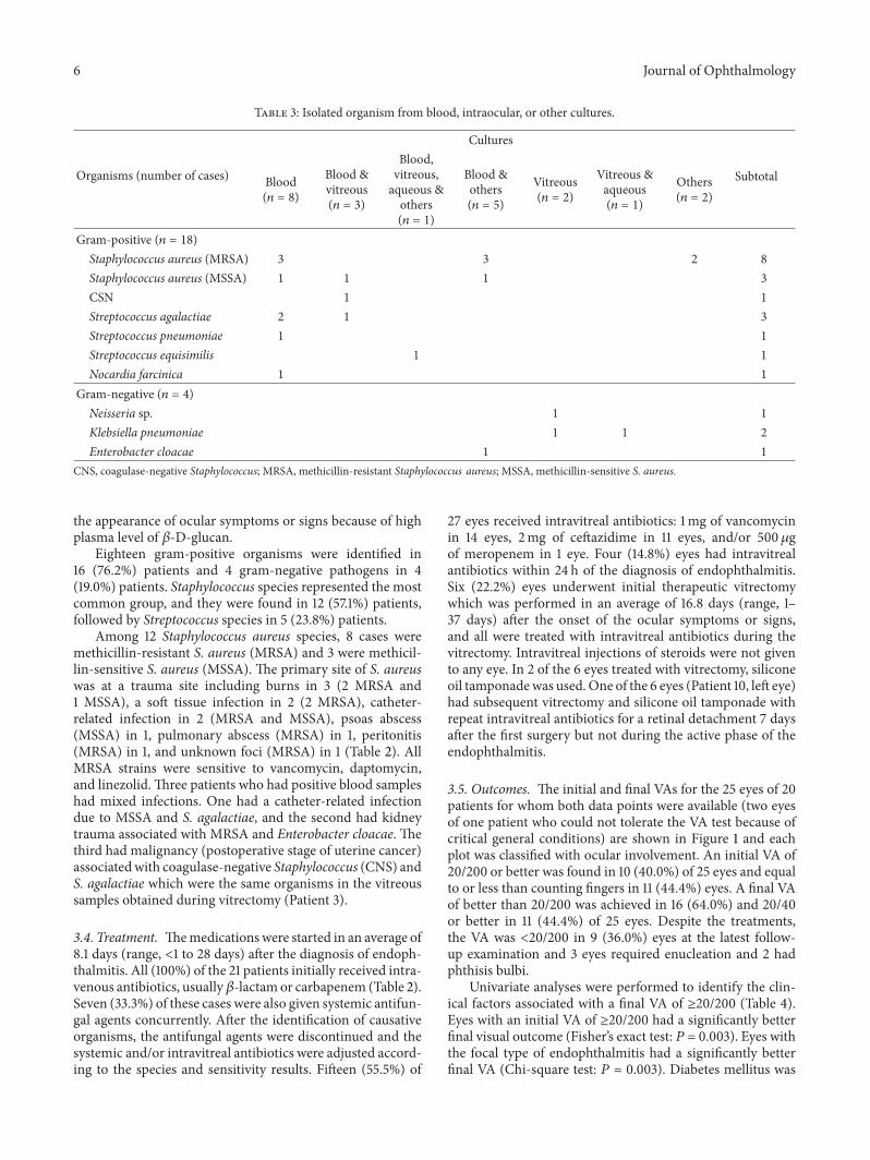
Table 3: Isolated organism from blood, intraocular, or other cultures.
Organisms (number of cases)
Cultures
SubtotalBlood(𝑛 = 8)
Blood &vitreous(𝑛 = 3)
Blood,vitreous,aqueous &others(𝑛 = 1)
Blood &others(𝑛 = 5)
Vitreous(𝑛 = 2)
Vitreous &aqueous(𝑛 = 1)
Others(𝑛 = 2)
Gram-positive (𝑛 = 18)Staphylococcus aureus (MRSA) 3 3 2 8Staphylococcus aureus (MSSA) 1 1 1 3CSN 1 1Streptococcus agalactiae 2 1 3Streptococcus pneumoniae 1 1Streptococcus equisimilis 1 1Nocardia farcinica 1 1
Gram-negative (𝑛 = 4)Neisseria sp. 1 1Klebsiella pneumoniae 1 1 2Enterobacter cloacae 1 1
CNS, coagulase-negative Staphylococcus; MRSA, methicillin-resistant Staphylococcus aureus; MSSA, methicillin-sensitive S. aureus.
the appearance of ocular symptoms or signs because of highplasma level of 𝛽-D-glucan.
Eighteen gram-positive organisms were identified in16 (76.2%) patients and 4 gram-negative pathogens in 4(19.0%) patients. Staphylococcus species represented the mostcommon group, and they were found in 12 (57.1%) patients,followed by Streptococcus species in 5 (23.8%) patients.
Among 12 Staphylococcus aureus species, 8 cases weremethicillin-resistant S. aureus (MRSA) and 3 were methicil-lin-sensitive S. aureus (MSSA). The primary site of S. aureuswas at a trauma site including burns in 3 (2 MRSA and1 MSSA), a soft tissue infection in 2 (2 MRSA), catheter-related infection in 2 (MRSA and MSSA), psoas abscess(MSSA) in 1, pulmonary abscess (MRSA) in 1, peritonitis(MRSA) in 1, and unknown foci (MRSA) in 1 (Table 2). AllMRSA strains were sensitive to vancomycin, daptomycin,and linezolid.Three patients who had positive blood sampleshad mixed infections. One had a catheter-related infectiondue to MSSA and S. agalactiae, and the second had kidneytrauma associated with MRSA and Enterobacter cloacae. Thethird had malignancy (postoperative stage of uterine cancer)associatedwith coagulase-negative Staphylococcus (CNS) andS. agalactiae which were the same organisms in the vitreoussamples obtained during vitrectomy (Patient 3).
3.4. Treatment. Themedications were started in an average of8.1 days (range, <1 to 28 days) after the diagnosis of endoph-thalmitis. All (100%) of the 21 patients initially received intra-venous antibiotics, usually𝛽-lactamor carbapenem (Table 2).Seven (33.3%) of these cases were also given systemic antifun-gal agents concurrently. After the identification of causativeorganisms, the antifungal agents were discontinued and thesystemic and/or intravitreal antibiotics were adjusted accord-ing to the species and sensitivity results. Fifteen (55.5%) of
27 eyes received intravitreal antibiotics: 1mg of vancomycinin 14 eyes, 2mg of ceftazidime in 11 eyes, and/or 500 𝜇gof meropenem in 1 eye. Four (14.8%) eyes had intravitrealantibiotics within 24 h of the diagnosis of endophthalmitis.Six (22.2%) eyes underwent initial therapeutic vitrectomywhich was performed in an average of 16.8 days (range, 1–37 days) after the onset of the ocular symptoms or signs,and all were treated with intravitreal antibiotics during thevitrectomy. Intravitreal injections of steroids were not givento any eye. In 2 of the 6 eyes treated with vitrectomy, siliconeoil tamponadewas used.One of the 6 eyes (Patient 10, left eye)had subsequent vitrectomy and silicone oil tamponade withrepeat intravitreal antibiotics for a retinal detachment 7 daysafter the first surgery but not during the active phase of theendophthalmitis.
3.5. Outcomes. The initial and final VAs for the 25 eyes of 20patients for whom both data points were available (two eyesof one patient who could not tolerate the VA test because ofcritical general conditions) are shown in Figure 1 and eachplot was classified with ocular involvement. An initial VA of20/200 or better was found in 10 (40.0%) of 25 eyes and equalto or less than counting fingers in 11 (44.4%) eyes. A final VAof better than 20/200 was achieved in 16 (64.0%) and 20/40or better in 11 (44.4%) of 25 eyes. Despite the treatments,the VA was <20/200 in 9 (36.0%) eyes at the latest follow-up examination and 3 eyes required enucleation and 2 hadphthisis bulbi.
Univariate analyses were performed to identify the clin-ical factors associated with a final VA of ≥20/200 (Table 4).Eyes with an initial VA of ≥20/200 had a significantly betterfinal visual outcome (Fisher’s exact test: 𝑃 = 0.003). Eyes withthe focal type of endophthalmitis had a significantly betterfinal VA (Chi-square test: 𝑃 = 0.003). Diabetes mellitus was

Journal of Ophthalmology 7
Table 4: Prognostic factors associated with good visual outcomes.
Factors Final visual outcome Odds ratio (95% CI) 𝑃
≥20/200 <20/200Sex
Male 12 6 1.50 (0.25–8.98) 1.00Female 4 3
EyeRight 5 5 0.36 (0.07–1.97) 0.397Left 11 4
Initial visual acuity≥20/200 10 0 — 0.003<20/200 6 9
Extent of ocular involvementPanophthalmitis 0 4 — 0.003#
Posterior focal 5 4Posterior diffuse 11 1
Diabetic mellitusYes 14 3 14.00 (1.84–106.47) 0.010No 2 6
HypertensionYes 5 2 1.59 (0.24–10.57) 1.00No 11 7
Systemic steroid treatmentYes 3 1 1.85 (0.16–20.94) 1.00No 13 8
Infection placeCommunity-acquired endophthalmitis 7 6 0.39 (0.08–2.01) 0.411Hospital-acquired endophthalmitis 9 3
Causative organismsGram stain positive 11 6 1.44 (0.19–11.04) 1.00Gram stain negative 3 2
K. pneumoniaYes 0 2 0.0 (0.0–1.00) 0.121No (others) 14 6
Intravitreal antibioticsYes 9 6 0.64 (0.13–43.32) 0.691No 7 3
Interval between diagnosis and intravitrealantibiotics≤1 day 5 1 6.25 (0.50–77.50) 0.287>1 day 4 5
VitrectomyYes 3 3 0.46 (0.08–2.66) 0.630No 13 6
Fisher’s exact test.#Chi-square test.
significantly associated with final VA of ≥20/200 (Fisher’sexact test: 𝑃 = 0.010). The sex, laterality, hypertension,systemic steroid treatments for underlying illness, infec-tion place (community-acquired/hospital-acquired endoph-thalmitis), causative organs, and treatment with or withoutintravitreal antibiotics or vitrectomy were not significantly
associated with final VA of ≥20/200 (Fisher’s exact test: all𝑃 values > 0.05). To identify the associated factors for visualoutcome, logistic regression analysis was used employingfinal VA of ≥20/200 as the outcome value and several param-eters as explanatory variables. The latter included age, sex,laterality, initial visual acuity, extent of ocular involvement,

8 Journal of Ophthalmology
PanPF
PD
Fina
l Sne
llen
equi
vale
nts
20/20
20/200
HM
LP
NLP20/2020/200CFHMLPNLP
Initial Snellen equivalents
Figure 1: Visual outcome of endogenous bacterial endophthalmitis.CF, counting fingers; HM, hand motion; LP, light perception; NLP,no light perception (including enucleation); Pan, panophthalmitis;PD, posterior diffuse; PF, posterior focal.
presence or absence of diabetes mellitus or hypertension,infection place, causative organs, treatment with or withoutintravitreal antibiotics or vitrectomy, interval between onsetof ocular symptom and ophthalmology consultation, andfollow-up period. None was detected as an associated factorfor final VA of ≥20/200 by logistic regression analysis.
Using the actual values of final logMAR VA, eyes withinitial VA of ≥20/200 (Mann-Whitney 𝑈 test: 𝑃 = 0.003),focal type of endophthalmitis (Kruskal-Wallis test:𝑃=0.004),and diabetes mellitus (Mann-Whitney 𝑈 test: 𝑃 = 0.014)were also significantly associated with final logMAR VA. Anintravitreal injection and vitrectomywere not associated withgood visual outcomes (Mann-Whitney 𝑈 test: 𝑃 = 0.462, 𝑃 =0.947, resp.). There was no significant difference in the vis-ual outcomes between gram-positive and gram-negativeinfections and between MRSA and MSSA infections (Mann-Whitney 𝑈 test; 𝑃 = 0.969, 𝑃 = 0.758, resp.). A longer timebetween the onset of ocular symptoms and intravitreal antibi-otic injection was weakly correlated with worse visual out-comes (Spearman: 𝑃 = 0.056). However, the interval betweenthe diagnosis of endophthalmitis and intravitreal injectionwas not significantly correlated with the final visual outcome(Spearman: 𝑃 = 0.616). The correlations between the finalvisual outcome and age, interval between the onset of ocularsymptoms and initial examination by an ophthalmologistand vitrectomy were not significant (Spearman: 𝑃 = 0.275,𝑃 = 0.176, and 𝑃 = 0.216, resp.). Among continuous valuesincluding age, initial logMAR VA, follow-up period, andinterval between onset of ocular symptoms and intravitrealantibiotic injection,multiple regression analysis detected that
initial logMAR VA was significantly associated with finallogMARVA in eyes with intravitreal antibiotic injection (𝑃 =0.001, 95% CI: 0.437–1.344). The interval between onset ofocular symptoms and intravitreal antibiotic injection wasweakly associated with final logMAR VA (𝑃 = 0.097, 95%CI: −0.006–0.064) by multiple regression analysis. In eyeswithout intravitreal antibiotic injection, initial logMAR VA(𝑃 = 0.022, 95% CI: 0.198–1.798) was detected as a relatedfactor for final logMAR VA by multiple regression analysis.
The two eyes infected with K. pneumoniae were enu-cleated (Patients 1 and 12). One patient (Patient 13) diedbefore discharge. This patient had adult onset Still’s diseasewith administration of high-dose steroids, but disseminatedMRSA ultimately developed.
4. Discussion
EBE is a rare form of infection which occurs when organismreaches the eye via the bloodstream and then crosses theblood-ocular barrier. Although the visual prognosis wasreported to be very poor [1], the current study revealed thatit mainly depended on initial VA.
In our 27 eyes of 21 case series, extraocular infectious fociwere identified in 81.0% and infective endocarditis (14.3%)was the common extraocular infection. It has been reportedthat the extraocular sites of bacterial endophthalmitis werethe endocardium, liver, lung, central nervous system, and therenal and urinary tracts [3]. Okada et al. and Yonekawa etal. reported that the most common site of EBE was the heartwith infectious endocarditis [2, 8]. Infectious endocarditis isconsidered to be one of the most serious infections in theWestern world, and the causative organisms were most oftenstaphylococci, streptococci, and enterococci [16]. Kuriyan etal. reported that, in spite of prompt vancomycin treatment,most patients with ocular Streptococcus infections had poorVA [17]. In our study, EBE with infective endocarditis wascaused by S. equisimilis, a subspecies of Group G Streptococ-cus, S. agalactiae, a subspecies of Group B Streptococcus, andan unidentified organism in one patient each and the visualoutcomes varied from phthisis to 20/16. Despite intravitrealvancomycin injection, 1 patient with initial VA of lightprojection ended in phthisis.
Gram-positive organisms are more common in NorthAmerican and European cases of EBE [2, 3, 18]. In thecurrent study, we also found that gram-positive organismswere the most common bacterial pathogens, especially S.aureus which was isolated from 11 (MRSA in 8 and MSSAin 3) patients. Visual outcomes are generally poor in EBEcaused by MRSA [19]. Ho et al. reported a high incidenceof retinal detachments in eyes with endogenous MRSAendophthalmitis, and almost one-half of the affected patientseventually required enucleation or evisceration [5]. In our9 eyes (7 patients) with MRSA in whom we could examineVA measurement, 7 eyes were treated with both systemicand intravitreal antibiotics. Eight eyes had a final VA betterthan 20/200, and that is better than that reported earlier[5, 19]. One eye with initial VA of light perception endedin phthisis. No retinal detachments developed and none had

Journal of Ophthalmology 9
to be enucleated during the follow-up period. There was nosignificant difference in the visual outcomes between MRSAand MSSA infections in the current study.
Gram-negative microorganisms have been reported tobe the main causative pathogens of endogenous endoph-thalmitis in East Asians [4, 20]. Among the gram-negativemicroorganisms, there has been an increase in K. pneumoniaas the causative organism in endogenous endophthalmitis.Thus, it has become an important pathogen in recent yearsin Asian countries [1, 3, 4, 20–25], and the incidence ofinfections byK. pneumonia in eyes with endogenous endoph-thalmitis was 50% to 61% [4, 26]. A national clinical study in19 hospitals on bacterial and fungal endophthalmitis duringthe past-5-to-20-year period (until 1988) in Japan showedthat the incidence of endogenous endophthalmitis causedby K. pneumonia was 25% (5 of 20 cases) [27]. Torisakiet al. performed a literature search on EBE caused by K.pneumoniae published between 1989 and 1998 in Japan [28].A total of 26 references (single case and small case series) on30 patients (41 eyes) were found and the visual outcome ofall except one was poor with 30 of 41 eyes having a final VAof no light perception [28]. In our study, K. pneumoniae wasfound in only 2 patients (9.5%), one with liver abscess andone with epididymitis. Initial VAs were counting finger andlight perception, respectively, and both lost light perceptionduring follow-up. It has been documented that Klebsiellasp. has worse prognosis [4, 21, 22]. Although initial VAin 2 patients with K. pneumoniae was poor in our study,K. pneumoniae infection itself may have worse prognosis.Recent experimental models focused on EBE caused by K.pneumoniae concluded thatK. pneumoniae’s ability to disruptretinal function and generate an inflammatory response, thuscausing further damage, can be attributed to its capsule.Moreover, they demonstrated that bacterial products, such asendotoxin, present on the surface of K. pneumonia, furtherstimulate the host inflammatory response [29, 30].The reasonfor the high incidence of this infection in Asia compared withnon-Asia has not been determined [4, 26].
The most common systemic condition associated withbacterial endophthalmitis was diabetes mellitus followed byhypertension, cardiac disease, gastrointestinal disorders, andurological diseases [2]. Jackson et al. also reported that themost common predisposing medical condition was diabetesmellitus (62%) including type II diabetes (42%) in a literaturereview and its presence was significantly associated with poorVA [3]. Although the most common predisposing medicalcondition was diabetes mellitus (61.9%) in the current study,patients with diabetes mellitus had significantly better finallogMAR VA (Mann-Whitney 𝑈 test: 𝑃 = 0.014). Thosepatients also had significantly better initial logMAR VA(Mann-Whitney 𝑈 test: 𝑃 = 0.022) and 12 of 17 eyes withdiabetes mellitus were the focal type of endophthalmitis.
In patients in whom we could examine VA, the incidenceof the posterior focal type endophthalmitis was 48.0% (12eyes) and eyes with the focal type of endophthalmitis weremore likely to have a final visual acuity of ≥20/200 than thosewith posterior diffuse or panophthalmitis (Fisher’s exact test:𝑃 = 0.011). Similar to our study, Greenwald et al. reportedthe posterior focal type of EBE might have a better visual
outcome than the posterior diffuse or panophthalmitis withvitreal involvement [6]. Focal subretinal abscess may expandto panendophthalmitis, if not treated. However, our resultmay not necessarily represent the natural course of EBE (fromfocal to diffuse posterior then to panophthalmitis). Naturalcourse of intraocular EBE expansion must be elucidated.
EBE develops when organisms from systemic or localinfections disseminate through the blood and enter the intra-ocular spaces through the blood-ocular barrier [22]. Thus,an early detection of the infection site and treatment ofthe causative organism are very important [12]. Systemicantibiotics treat the distant foci of infection and preventcontinued bacteremia, thereby reducing the chances of aninvasion of the eye [20]. Some patients were treated withintravitreal antibiotics as well as with systemic antibiotics,while others were treated with intravitreal antibiotics andvitrectomy as the initial treatment modality in previous [1, 4,12] and our studies. Yonekawa et al. recommended intravit-real antibiotics administered within 24 h to supplement thesystemic antibiotics [8]. In our patients, the interval betweenthe onset of ocular symptoms and the intravitreal antibioticinjection was weakly correlated with the final visual outcome(Spearman: 𝑃 = 0.056 and multiple regression analysis: 𝑃 =0.097). For 15 (55.5%) of 27 eyes, intravitreal antibiotics wereused. None (0%) of 9 eyes receiving intravitreal antibiotics(with systemic antibiotic, but not vitrectomy) underwentenucleation, compared with 3 (25%) of 12 eyes who hadsystemic treatment alone (no vitrectomy). Recent review [1]also mentioned that intravitreal antibiotics may be associatedwith a trend for fewer enucleation surgeries. Although thesedata suggested an association between intravitreal antibioticsand preservation of the eye, they cannot establish a causallink.
Romero et al. recommended surgical intervention forpatients infected with especially virulent organisms, or visualacuity of ≤20/400, or severe vitreous involvement as inadvanced stages, for example, posterior diffuse endoph-thalmitis or panophthalmitis [7]. In the current study, 5eyes with initial VA of ≤20/400 (4 with posterior diffusetype infection and 1 with focal type infection) and 1 eyewith posterior diffuse type infection caused by Neisseria sp.underwent vitrectomy. Final VAs varied from NLP (phthisis)to 20/20. Three eyes with initial VA of hand motion or lightperception and with posterior diffuse endophthalmitis endedin final VA of less than CF. Surgical interventions for EBE aredifficult because most patients are in poor general conditionand are a high risk for general anesthesia [12]. In our cases,4 of 6 eyes underwent vitrectomy more than 7 days after theonset of the ocular symptoms, and the visual acuity outcomeswere poor except for one eye (Patient 18). Because of patientcondition, the timing of surgery was delayed. On the otherhand, Wong et al. reported that the final visual outcome wasunrelated to the use of vitrectomy in the management andonly the virulence of the organism predicted the outcome[20]. At present, the efficacy of immediate ocular therapiesincluding vitrectomy and intravitreal antibiotics against EBEis still controversial [22].
The most common systemic finding was fever includ-ing cold-like symptoms (76.2% in our study and 57.7% in

10 Journal of Ophthalmology
the systemic review) [1]. At the first visit to a doctor, ahigh body temperature (>38∘C) (76.2%), elevated C-reactiveprotein (76.2%), and a high white blood cell count (52.3%)were observed in the current study. Diabetes mellitus was themost common medical condition in our study (61.9%) andthe systemic review (33%) [1]. A funduscopic examinationshould be performed as soon as possible after the onset ofsymptoms in those with elevated CRP, high WBC, unknownorigin of fever, and/or a history of diabetic mellitus.Themostcommon ocular symptoms were decreased vision (66.7%),floaters (23.8%), and pain (9.5%) in our study and those aresimilar to others [1].
In the current study, a final VA of 20/200 or better wasachieved in 64% and the median final VA was 20/40. Visualoutcomes seems to be better than a recent review [1] of 342cases of EBE (initial VAs were not mentioned), in which themedian final VA was 20/100, with 44% worse than 20/200.The reasons for better visual outcome in our study mightbe as follows. There were only two eyes with Klebsiella sp.whichwas reported to beworse prognosis [4, 21, 22]. In Japan,physicians routinely send patients to an ophthalmologistfor funduscopic examination when patients have diabetesmellitus. Patients were also referred to an ophthalmologistfrom physicians according to the Guidelines forManagementof Deep-Seated Mycoses 2007 in Japan, when endogenousendophthalmitis was suspected. As a result, most patientswere referred to an ophthalmologist earlier and early systemicand/or intravitreal antibiotics could be instituted. The num-ber of eyes with initial VA of ≥20/200 was 10 (40.0%) andfinal VA of ≥20/200 was 16 (64.0%) and 20/40 or better was 11(44.4%) of 25 eyes in our study. InWu’s study [4], the numberof eyes with initial VA of ≥20/200 was 0 (0%) of 15 eyes withEBE and final VA of ≥20/200 was 3 (20.0%). Moreover ourstatistical analysis clearly showed initial VA as a related factorfor visual outcome. However, it has been reported that earlydiagnosis alone usually did not result in good visual outcome[2]. Other than a delay in diagnosis and treatment, factorsassociated with the poor prognosis may include the virulenceof the causative organisms and compromised host condi-tions.
The limitations of this study are those inherent in ret-rospective studies, including lack of controls and uniformprotocol. However large randomized control trial for this raredisease is not practical; observational case series will be animportant source for developing future treatment guidelines.
5. Conclusions
EBE is a rare but often devastating ocular and systemicdisorder. In contrast with previous studies, our study showedbetter visual outcome. The prognosis may depend on initialVA, extent of ocular involvement, and an interval betweenthe onset of ocular symptoms and intravitreal antibioticinjection.Thus, early diagnosis and early intravitreal injectionof antibiotics supplement to immediate systemic antibioticsmight lead to a favorable visual outcome. A higher index ofsuspicion should be maintained by clinicians. All patientswith bacteremia, including suspected cases, should have at
least one dilated retinal examination early in the course oftherapy preferably performed by an ophthalmologist.
Conflict of Interests
The authors declare that there is no conflict of interestsregarding the publication of this paper.
References
[1] T. L. Jackson, T. Paraskevopoulos, and I. Georgalas, “Systematicreview of 342 cases of endogenous bacterial endophthalmitis,”Survey of Ophthalmology, vol. 59, no. 6, pp. 627–635, 2014.
[2] A. A. Okada, R. P. Johnson, W. C. Liles, D. J. D’Amico, and A. S.Baker, “Endogenous bacterial endophthalmitis: report of a ten-year retrospective study,”Ophthalmology, vol. 101, no. 5, pp. 832–838, 1994.
[3] T. L. Jackson, S. J. Eykyn, E. M. Graham, and M. R. Stanford,“Endogenous bacterial endophthalmitis: a 17-year prospectiveseries and review of 267 reported cases,” Survey of Ophthalmol-ogy, vol. 48, no. 4, pp. 403–423, 2003.
[4] Z. H. Y. Wu, R. P. S. Chan, F. O. J. Luk et al., “Review of clinicalfeatures, microbiological spectrum, and treatment outcomes ofendogenous endophthalmitis over an 8-year period,” Journal ofOphthalmology, vol. 2012, Article ID 265078, 5 pages, 2012.
[5] V. Ho, L. Y. Ho, T. M. Ranchod, K. A. Drenser, G. A. Williams,and B. R. Garretson, “Endogenous methicillin-resistant Staphy-lococcus aureus endophthalmitis,” Retina, vol. 31, no. 3, pp. 596–601, 2011.
[6] M. J. Greenwald, L. G. Wohl, and C. H. Sell, “Metastaticbacterial endophthalmitis: a contemporary reappraisal,” Surveyof Ophthalmology, vol. 31, no. 2, pp. 81–101, 1986.
[7] C. F. Romero, M. K. Rai, C. Y. Lowder, and K. A. Adal,“Endogenous endophthalmitis: case report and brief review,”American Family Physician, vol. 60, no. 2, pp. 510–514, 1999.
[8] Y. Yonekawa, R. V. P. Chan, A. K. Reddy, C. G. Pieroni, T. C. Lee,and S. Lee, “Early intravitreal treatment of endogenous bacterialendophthalmitis,” Clinical & Experimental Ophthalmology, vol.39, no. 8, pp. 771–778, 2011.
[9] H. Kawakami, A. Sawada, K. Mochizuki, K. Takahashi, T.Muto, andK.Ohkusu, “EndogenousNocardia farcinica endoph-thalmitis,” Japanese Journal of Ophthalmology, vol. 54, no. 2, pp.164–166, 2010.
[10] S. Suemori, A. Sawada, S. Komori, K. Mochizuki, K. Ohkusu,andH.Takemura, “Case of endogenous endophthalmitis causedby Streptococcus equisimilis,” Clinical Ophthalmology, vol. 4, no.1, pp. 917–918, 2010.
[11] A. Sawada, S. Komori, K. Udo et al., “Case of endogenousendophthalmitis caused by Klebsiella pneumoniae with magAand rmpA genes in an immunocompetent patient,” Journal ofInfection and Chemotherapy, vol. 19, no. 2, pp. 326–329, 2013.
[12] S. Lee, T. Um, S. G. Joe et al., “Changes in the clinical featuresand prognostic factors of endogenous endophthalmitis: fifteenyears of clinical experience in Korea,” Retina, vol. 32, no. 5, pp.977–984, 2012.
[13] J. Kennerdell, A. Tyutyunikov, R. Edwards et al., “The ischemicoptic neuropathy decompression trial (IONDT): design andmethods,” Controlled Clinical Trials, vol. 19, no. 3, pp. 276–296,1998.
[14] V. A. Deramo, T. A. Cox, A. B. Syed, P. P. Lee, and S. Fekrat,“Vision-related quality of life in people with central retinal

Journal of Ophthalmology 11
vein occlusion using the 25-item National Eye Institute VisualFunction Questionnaire,” Archives of Ophthalmology, vol. 121,no. 9, pp. 1297–1302, 2003.
[15] M. Tanaka, Y. Kobayashi, H. Takebayashi, M. Kiyokawa, and H.Qiu, “Analysis of predisposing clinical and laboratory findingsfor the development of endogenous fungal endophthalmitis. Aretrospective 12-year study of 79 eyes of 46 patients,”Retina, vol.21, no. 3, pp. 203–209, 2001.
[16] D. R. Murdoch, R. G. Corey, B. Hoen et al., “Clinical presen-tation, etiology, and outcome of infective endocarditis in the21st century: the International Collaboration on Endocarditis-Prospective Cohort Study,” Archives of Internal Medicine, vol.169, no. 5, pp. 463–473, 2009.
[17] A. E. Kuriyan, K. D.Weiss, H.W. Flynn et al., “Endophthalmitiscaused by streptococcal species: clinical settings, microbiology,management, and outcomes,” American Journal of Ophthalmol-ogy, vol. 157, no. 4, pp. 774–780, 2014.
[18] T. Ness, K. Pelz, and L. L. Hansen, “Endogenous endoph-thalmitis: microorganisms, disposition and prognosis,” ActaOphthalmologica Scandinavica, vol. 85, no. 8, pp. 852–856, 2007.
[19] T. Ness andC. Schneider, “Endogenous endophthalmitis causedbymethicillin-resistant Staphylococcus aureus (MRSA),”Retina,vol. 29, no. 6, pp. 831–834, 2009.
[20] J.-S.Wong, T.-K. Chan,H.-M. Lee, and S.-P. Chee, “Endogenousbacterial endophthalmitis: an East Asian experience and areappraisal of a severe ocular affliction,”Ophthalmology, vol. 107,no. 8, pp. 1483–1491, 2000.
[21] C. C. A. Sng, A. Jap, Y. H. Chan, and S. P. Chee, “Riskfactors for endogenous Klebsiella endophthalmitis in patientswith Klebsiella bacteremia: a case-control study,” British Journalof Ophthalmology, vol. 92, no. 5, pp. 673–677, 2008.
[22] P. P. Connell, E. C. O’Neill, D. Fabinyi et al., “Endogenousendophthalmitis: 10-year experience at a tertiary referral cen-tre,” Eye, vol. 25, no. 1, pp. 66–72, 2011.
[23] M. Ang, A. Jap, and S.-P. Chee, “Prognostic factors and out-comes in endogenous Klebsiella pneumoniae endophthalmitis,”American Journal of Ophthalmology, vol. 151, no. 2, pp. 338–e2,2011.
[24] K. S. Chung, Y. K. Kim, Y. G. Song et al., “Clinical reviewof endogenous endophthalmitis in Korea: a 14-year review ofculture positive cases of two large hospitals,” Yonsei MedicalJournal, vol. 52, no. 4, pp. 630–634, 2011.
[25] H. W. Lim, J. W. Shin, H. Y. Cho et al., “Endogenous endoph-thalmitis in the korean population: a six-year retrospectivestudy,” Retina, vol. 34, no. 3, pp. 592–602, 2014.
[26] A. H. Kashani and D. Eliott, “The emergence of Klebsiellapneumoniae endogenous endophthalmitis in the USA: basicand clinical advances,” Journal of Ophthalmic Inflammation andInfection, vol. 3, no. 1, article 28, 2013.
[27] H. Hatano, K. Inoue, H. Matoba et al., “Endophthalmitis inJapan—a nationwide study with reference to type and etiology,”Nihon Ganka Gakkai Zasshi, vol. 95, no. 4, pp. 369–376, 1991(Japanese).
[28] M. Torisaki, N. Sakai, K. Yonewaki, and H. Funada, “Two casesof endophthalmitis caused by Klebsiella pneumoniaemetastaticfrom pyogenic liver abscess,” Journal of Eye, vol. 17, no. 3, pp.395–401, 2000 (Japanese).
[29] J. J. Hunt, J.-T. Wang, and M. C. Callegan, “Contribution ofmucoviscosity-associated gene A (magA) to virulence in exper-imental Klebsiella pneumoniae endophthalmitis,” InvestigativeOphthalmology andVisual Science, vol. 52, no. 9, pp. 6860–6866,2011.
[30] B. J. Wiskur, J. J. Hunt, andM. C. Callegan, “Hypermucoviscos-ity as a virulence factor in experimental Klebsiella pneumoniaeendophthalmitis,” InvestigativeOphthalmology&Visual Science,vol. 49, no. 11, pp. 4931–4938, 2008.

Submit your manuscripts athttp://www.hindawi.com
Stem CellsInternational
Hindawi Publishing Corporationhttp://www.hindawi.com Volume 2014
Hindawi Publishing Corporationhttp://www.hindawi.com Volume 2014
MEDIATORSINFLAMMATION
of
Hindawi Publishing Corporationhttp://www.hindawi.com Volume 2014
Behavioural Neurology
EndocrinologyInternational Journal of
Hindawi Publishing Corporationhttp://www.hindawi.com Volume 2014
Hindawi Publishing Corporationhttp://www.hindawi.com Volume 2014
Disease Markers
Hindawi Publishing Corporationhttp://www.hindawi.com Volume 2014
BioMed Research International
OncologyJournal of
Hindawi Publishing Corporationhttp://www.hindawi.com Volume 2014
Hindawi Publishing Corporationhttp://www.hindawi.com Volume 2014
Oxidative Medicine and Cellular Longevity
Hindawi Publishing Corporationhttp://www.hindawi.com Volume 2014
PPAR Research
The Scientific World JournalHindawi Publishing Corporation http://www.hindawi.com Volume 2014
Immunology ResearchHindawi Publishing Corporationhttp://www.hindawi.com Volume 2014
Journal of
ObesityJournal of
Hindawi Publishing Corporationhttp://www.hindawi.com Volume 2014
Hindawi Publishing Corporationhttp://www.hindawi.com Volume 2014
Computational and Mathematical Methods in Medicine
OphthalmologyJournal of
Hindawi Publishing Corporationhttp://www.hindawi.com Volume 2014
Diabetes ResearchJournal of
Hindawi Publishing Corporationhttp://www.hindawi.com Volume 2014
Hindawi Publishing Corporationhttp://www.hindawi.com Volume 2014
Research and TreatmentAIDS
Hindawi Publishing Corporationhttp://www.hindawi.com Volume 2014
Gastroenterology Research and Practice
Hindawi Publishing Corporationhttp://www.hindawi.com Volume 2014
Parkinson’s Disease
Evidence-Based Complementary and Alternative Medicine
Volume 2014Hindawi Publishing Corporationhttp://www.hindawi.com