Embed Size (px)
Citation preview
Md. Ishtiaque Ahmad et al., Asian Journal of Pharmaceutical Technology & Innovation, 03 (15); 2015; 91 - 106
www.asianpharmtech.com 91
Asian Journal of Pharmaceutical Technology & Innovation ISSN: 2347-8810
Research Article
Received on: 02-12-2015 Accepted on: 11-12-2015 Published on: 15-12-2015
Hospital Acquired Infections in Different
Wards of Patna Medical College & Hospital
Corresponding Author: Md. Ishtiaque Ahmad1*, Shankar Prakash2 * Dr. Md. Ishtiaque Ahmad, M.D. Microbiology Assistant, Narayan Medical College & Hospital, Sasaram, Jamuhar, Bihar, India.
.
ABSTRACT
Nosocomial infection - also called “hospital acquired infection” this
includes infections acquired in the hospital but appearing after
discharge, and also occupational infections among staff of the facility.
The most common types of Nosocomial infections that could occur in a
hospital are surgical wound and other soft tissue infections, Urinary
tract infections, Respiratory infections, Gastroenteritis, Meningitis.
Naturally this work was undertaken with a view to study the problems
of postoperative sepsis and other types of infection during
hospitalization period and to express more knowledge over this subject.
The samples of Pus, Urine, Sputum and Swab Samples from the
different parts of the hospitals were collected.
These were then cultured into the different media. After the specific
duration the cultural and morphological characters were noted.
The organism were identified on the basis of characters of the colony,
Gram staining, Motility test, Biochemical reactions & coagulase tests.
In Burn wound infection it was observed that most sensitive antibiotics
against all above mention organism were Piperacillin, Gentamysin,
Amikacin.
In Noscomial urinary tract infection E. coli was the most common
microorganism isolated and showed most sensitivity to Ceftazidime.
Staphylococcus aureus was the most common organism isolated from
surgical wards and it was most sensitive with the Cefotaxime.
From respiratory tract infection most common organism was
Staphylococcus aureus and most sensitive antibiotics was Imipenem.
*Email Id- [email protected] Key-words: Nosocomial infection, hospital acquired infection, Patna
Medical College & Hospital, hospital infection, antibiotics, resistance.
Cite this article as: Md. Ishtiaque Ahmad, Shankar Prakash, Hospital Acquired infections in different wards of Patna Medical College & Hospital, Asian Journal of Pharmaceutical Technology & Innovation, 03 (15); 2015. www.asianpharmtech.com 1 M.D., Microbiology Assistant , Narayan Medical College & Hospital, Sasaram, Jamuhar, Bihar, India. 2 Professor & Head, Department of Microbiology, Patna Medical College, Patna, Bihar, India

Md. Ishtiaque Ahmad et al., Asian Journal of Pharmaceutical Technology & Innovation, 03 (15); 2015; 91 - 106
www.asianpharmtech.com 92
INTRODUCTION Nosocomial(“nosus”- diseas, “komeion”- to take care of) Nosocomial infection - also called “hospital acquired
infection” can be defined as:
An infection acquired in hospital by a patient who was admitted for a reason other than that infection (1). An in-
fection occurring in a patient in a hospital or other health care facility in whom the infection was not present or
incubating at the time of admission. This includes infections acquired in the hospital but appearing after
discharge, and also occupational infections among staff of the facility (2).
Patient care is provided in facilities which range from highly equipped clinics and technologically advanced
university hospitals to front-line units with only basic facilities. Despite progress in public health and hospital
care, infections continue to develop in hospitalized patients, and may also affect hospital staff. Many factors
promote infection among hospitalized patients: decreased immunity among patients; the increasing variety of
medical procedures and invasive techniques creating potential routes of infection; and the transmission of
drug-resistant bacteria among crowded hospital populations, where poor infection control practices may
facilitate trans-
Frequency of infection
Nosocomial infections occur worldwide and affect both developed and resource-poor countries. Infections
acquired in health care settings are among the major causes of death and increased morbidity among
hospitalized patients. They are a significant burden both for the patient and for public health. A prevalence
survey conducted under the auspices of WHO in 55 hospitals of 14 countries representing 4 WHO Regions
(Europe, Eastern Mediterranean, South-East Asia and Western Pacific) showed an average of 8.7% of hospital
patients had nosocomial infections. At any time, over 1.4 million people worldwide suffer from infectious
complications acquired in hospital (3). The highest frequencies of nosocomial infections were reported from
hospitals in the Eastern Mediterranean and South-East Asia Regions (11.8 and 10.0% respectively), with a
prevalence of 7.7 and 9.0% respectively in the European and Western Pacific Regions (4).
The most frequent nosocomial infections are infections of surgical wounds, urinary tract infections and lower
respiratory tract infections. The WHO study, and others, have also shown that the highest prevalence of
nosocomial infections occurs in intensive care units and in acute surgical and orthopaedic wards. Infection
rates are higher among patients with increased susceptibility because of old age, underlying disease, or
chemotherapy (5-7).
Sources of Hospital Infections (10-12):
For an infection to occur in the hospital the prerequisites are:
A susceptible host.
A microbe capable of producing an infection.
An environment that is congenial for the multiplication of the microbe. It is the delicate interplay of these 3
components that ultimately culminates in the occurrence of an infection.
Also, various combinations of four main factors influence the nature and frequency of infections. These are:
(i) Low resistance of the patients
(ii) Contact with infectious persons
(iii) Contaminated environmental sites
(iv) Drug resistance of endemic organisms
The source of the infecting organism may be exogenous - from another patient or a member of the hospital
staff, or from the inanimate environment in the hospital; or it may be endogenous - from the patients own flora
which at the time of infection may include organisms brought into the hospital at admission and certain others
acquired subsequently. In either case, the infecting organisms may spontaneously invade the tissues of the

Md. Ishtiaque Ahmad et al., Asian Journal of Pharmaceutical Technology & Innovation, 03 (15); 2015; 91 - 106
www.asianpharmtech.com 93
patient or may be introduced into them by surgical procedures, instrumental manipulation or nursing
procedures.
The inanimate environment of the hospital that acts as an important source comprises of:
i. Contaminated air, water, food and medicaments
ii. Used equipment’s and instruments
iii. Soiled linen
iv. Hospital waste (Bio medical waste)
Types of Hospital-Acquired Infections (13-14)
The most common types of Nosocomial infections that could occur in a hospital set up are: -
1. Surgical wound and other soft tissue infections.
2. Urinary tract infections
3. Respiratory infections
4. Gastroenteritis
5. Meningitis
Of these, surgical infections special importance for the surgeon and is dealt with in brief in the following
paragraphs.
Surgical Wound Infections (16-20)
Staphylococcus aureus remains the dominant species in surgical wound infection, followed by the
enterobacteria. Bacteroides sp. along with other gut bacteria, very often in mixed growth is found typically in
wounds after a colonized viscus has been entered. Although S. Aureus may occur in all types of wound, it is the
typical cause of the less frequent wound infection in "clean" surgery. Most commonly, infection of surgical
wounds occurs at the time of surgery. Again, in the great majority of cases, the origin of the bacteria appears to
be the patient's own body flora (endogenous infection). Much less often it is from a member of the surgical
team. However, in any instances the origin is obscure. The usual and common routes are direct spread from the
incised organs and intraoperative contamination of instruments and of surgeons' gloves and clothing.
Contamination from various types of apparatus has occasionally been described. Although the air-borne route
is important in the implantation of prostheses, it occurs only in rare episodes in general surgery.
The mode of spread of infections is hospital occurs mainly by the following 2 methods: (21-26) -
1. Aerial
2. Contact
"Aerial" transmission could be from the nose/mouth of the person or from inanimate sources like the air-
conditioning plants, respiratory apparatus etc. a variety of infections including measles, small pox, tuberculosis,
sepsis by Staphylococcus aureus and Streptococcus pyogenes, meningococcal infections, respiratory diseases
associated with Streptococcus pneumoniae, Streptococcus pyogenes. From inanimate sources aerial spread
could result in respiratory infections by Enterobacteria, Pseudomonas aeruginosa and Legionella (27).
'"Contact" could be either from other patients, doctors, nurses and other staff or from independent
environmental sources. While any of these could lead to respiratory infection, sepsis or diarrhoea, direct
contact into tissue or wounds or mucous membranes by infected needles, surgical instruments or by blood
and/or blood products could result in serious infections like hepatitis or AIDS (29).
Hospital acquired infection particularly in surgical specialties is the single important factor that affects hospital
productivity and performances and therefore deserves the greatest attention (30).
It is worth remembering that man is the main reservoir of staphylococcus aureus in the animal kingdom and
the most susceptible to staphylococcus infection. It is known that new born infants who are staphylococcus free
a birth are colonized by organisms within two days (Torrey G.G. and Reese, M.K. 1945)
Naturally this work was undertaken with a view to study the problems of postoperative sepsis and other types
of infection during hospitalization period and to express more knowledge over this subject (30-38).

Md. Ishtiaque Ahmad et al., Asian Journal of Pharmaceutical Technology & Innovation, 03 (15); 2015; 91 - 106
www.asianpharmtech.com 94
MATERIALS (39-45)
PUS: The pus (postoperative discharge) from infected wound in hospital of patients were collected aseptically
from different surgical units & pus and discharge from burn wound in the burn-ward (plastic surgery) ward of
Patna Medical College & Hospital (PMCH) for the purpose of studying the incidence and bacteriology.
Urine: Urine sample from different wards urine from the patient who developed symptoms of UTI 48 hour
after admissions with or without catheter collected for bacteriological study.
Sputum: Sputum sample from the patient who developed symptoms of RTI productive cough with
expectoration 48 hours after admissions in different wards.
Sample from OT & Wards Environment: Swab Samples from different sites from floor, wall, OT table, beds
linen, beds, nasal catheter, air settle plates kept at different sites in different OT collected.
Media and reagents: The following media and reagents were prepared and used:
Thioglycollate broth.
Blood agar media.
Mac Conkey’s agar media.
Nutrient agar media.
Hugh and Leifson media.
Christensen’s media.
Citrate utillsation test.
Peptone water.
Sugar media (Peptone water bases).
Methyl red test (Glucose phosphate peptone water media).
Voger-Proskaur test.
Indole test.
Phenylalanine deaminase test.
Test for hydrogen sulphide production.
Oxidase test.
Coagulase test.
Blood Agar Media: Sterile blood was added to sterile nutrient agar that has been melted and cooled at 50°C and plates were poured. MacConkey’s agar media: The peptone and taurocholate was dissolved in the water by heating. The agar was added and dissolved in the steamer. It was then cleared by filtration. The pH was adjusted to 7.5. The well-shaken solution of lactose and neutral red was added and mixed. It was heated in the autoclave with free stream (100°C) for one hour and then at 115°C for 15 minutes. Then the plates were poured. Nutrient agar media: It was heated in steam sterilizer for one hour so that it was fully dissolved. It was cleared with the aid of white of egg. pH was adjusted to 7.5 and filtered through Whatman No. 1 filter paper. It was sterilized with 5 lbs. Pressure for half an hour and then poured in sterilized petri-discs with aseptic precaution.
Hugh and Leifson Media: The pH was adjusted to 7.1 before adding the bromothymol blue and the media was autoclaved in a flask at 121°C for 30 minutes. The glucose to be added was sterilized separately and added to give a final concentration of 1 per cent. The media were then tube. Duplicate tubes of media were inoculated by stabbing. One tube was promptly covered with a layer of sterile liquid paraffin to a depth of 5-10 mm. And both will be incubated for 24 hours.
Christensen’s media: With final pH 6.5 to 6.9 this agar base was sterilized in autoclave at 120°C in flasks containing 80 ml. 20 percent solution of urea was prepared, sterilized by filtering through sterile bacteriological filter. This solution in a final concentration of 20 percent was added to the flask of agar base
Md. Ishtiaque Ahmad et al., Asian Journal of Pharmaceutical Technology & Innovation, 03 (15); 2015; 91 - 106
www.asianpharmtech.com 95
melted at temperature of 50°C. they were mixed well and distributed aseptically into sterile test tubes each containing 6.8 ml and kept in slanting position and used for urease formation.
Simmon’s citrate media: It is a modification of Koser’s medium with agar and an indicator. Dispensed, autoclaved at 121°C for 15 minutes and allowed to set as slopes. A Peotone water suspension of the organisms to be tested was inoculated. Incubated for 24 hours at 37°C. Results were indicated as below:
Positive – Blue colour and streak of growth. Negative – Original green colour and no growth.
Peptone water: Peptone,Sodium chloride were dissolved by steaming and then filtered. The reaction was adjusted to pH 7.4. Peptone water was then tubes in 5 ml amount in sterilized test tubes and sterilized by autoclaving at 15 lbs. Pressure for 20 minutes. Sugar media (Peptone water base): The pH medium was adjusted to 7.2-7.3 and sterilized solution of the glucose, lactose, sucrose, maltose and mannitol were added in the proportion of 1 percent. Andrade’s indicator was made by adding in NaOH to a 0.5 percent solution of acid fuchsine until the colour just became yellow. It is used at a final concentration of 1 percent in the medium and it turns dark reddish pink if acid is produced. Peptone water with Andrade’s indicator was tube in 5 ml amount; the Durham fermentation tubes inserted and the test tubes were stoppered with cotton wool. They were then sterilized in the autoclave at 121°C for 15 minutes. The sugars were prepared separately in 10 percent solution in distilled water and were sterilized in the steamer. The sterile sugars were kept in screw-capped bottles. Methyl red test (Glucose phosphate peptone water media): Peptone and phosphate were dissolved and pH was adjusted to 7.6. The solution was filtered and dispensed in 5 ml amount. It was sterilized at 121°C for 15 minutes. Glucose solution was sterilized by filter-action and 0.25 ml of the solution was added to each tube. Final concentration was made 0.5 per cent. Methyl red indicator solution: Glucose phosphate medium was inoculated and incubated at 37°C for 24 hours. After incubation, 5 drops of methyl red reagent was added. Positive tests were bright red and negative were yellow. Glucose phosphate peptone water as for methyl red test. Inoculated the organism in glucose phosphate medium and incubated at 37°C for 24 hours. 1 ml of 40 percent potassium hydroxide and 3 ml of 5 percent solution of alpha-naphthol in absolute alcohol was added. A positive reaction was seen by the development of a pink colour in 2-5 minutes. Principle: Due to oxidation, acetyl methyl carbinol is formed which reacts with the guanidine residue in the peptone giving a pink colour of the medium. Phenylalanine deaminase test:
This test indicates the ability of an organism to deaminate phenylalanine with the production of phenylpyruvic acid, which will react with ferric salts to give a green colour. PH was adjusted to 7.4, sterilized by autoclaving at 121°C for 15 minutes. Allowed to solidify in tubes as long slopes. A fairly heavy inoculum was inoculated and incubated for 48 hours at 37°C. A few drops of a 10% solution of ferric chloride were run down over the growth on the slope. Positive test will show: Development of green colour in the fluid and in the slope. Test for hydrogen sulphide production: 10 percent lead acetate solution in distilled water was prepared and filter paper was cut into small pieces and it was soaked in 10 percent lead acetate solution. Excess of lead acetate was allowed to drain out and filter paper pieces were allowed to dry. It was then kept in screw-capped bottle for use. A piece of filter paper was wrapped around the cotton wool plugging the peptone water, which was inoculated with the organism. It was incubated at 37°C for 24 hours and after 24 hours there will be blackening of the filter paper, if the bacteria is hydrogen sulphide producing. Oxidase test: Dry filter paper method:
Strips of Whatman’s No.1 filter paper were soaked in a freshly prepared 1 percent solution of tetramethyl-p-phenylene-diamine dihydrochloride. After draining for about 30 seconds the strips were freeze-dried and stored in a dark bottle tightly sealed with a screw cap. The papers had light purple tint and kept at

Md. Ishtiaque Ahmad et al., Asian Journal of Pharmaceutical Technology & Innovation, 03 (15); 2015; 91 - 106
www.asianpharmtech.com 96
room temperature. For use a strip was removed and moistened with deistilled water. The colony to be tested was picked up with loop and smeared over the moist area. Positive reaction: Indicated by an intense deep-purple hue, appearing within 5-10 seconds.
Coagulase test (tube method): 0.1 ml of culture suspension of the organism grown over night in nutrient broth was added to 0.5 ml of
citrated, human plasma (diluted in 1 in 10 with saline) in a narrow test tube. Diluted plasma alone in a similar tube serves as the control. Tubes were incubated at 37°C for six hours. If positive the plasma clots and does not flow when the tube is inverted. Antibiotic Disc for doing sensitivity test: Following antibiotic discs were used for the sensitivity test. Suitable drug content in microgram for sensitivity test disc are given below:
Pus swabs or frank pus were taken at the time of stitch removal or on 7th day after operation. The condition of the surgical wound at the time of stitch removal or on 7th day after operation in relation to the degree of wound infection was judged as follow: In case of Burn wound inschange & pus, which appear often 48 hours noted were taken. Mild: There was redness around the stitch or wound margin or slight discharge but no us from either of the two. Moderate: Definite signs of inflammation around the stitch or wound margin, with thin purulent discharge not much in almost on applying pressure or spontaneously stitch abscesses. Severe: There were frank profuse pus discharge with or without gaping of superficial wound margins. Method of collection of specimen: 1. By means of well sterilized all glass syringe pus was aspirated from the site of wound and then it was
brought in the bacteriological laboratory, Patna Medical College for culture and identification of microorganisms.
2. Where frank pus was not available, exudates was collected from the site of wound in the sterilized cotton wool swab and brought to the laboratory for culture and identification of microorganisms.
All pus swabs ad frank pus were inoculated in the Thioglycollate broth, Blood agar media and MacConkey’s media and incubated for 24 hours at 37°C temperature. Next day, cultural and morphological characters were studied. A smear was prepared from the colonies and gram staining was done. Then, on the Blood agar plate haemolysis was noted and on the MacConkey’s plate microorganisms were differentiated between lactose fermenting and non-lactose fermenting biochemical test done. Method of Urine Culture Patient Who developed symptoms of UTI > 48 hours of Admission who were with catheter or without catheter became symptomatic 48 hours other catheterization or 48 hour after admission urine was collected. Specimens of urine-collected from patient who was without catheter midstream urine was collected whereas who were with catheter as collected through draining portal of urinary catheter using aseptic precautions. Collected urine sends immediately to lab for culture with in ½ an hour as well as grams staining of centrifugal part urine was done to detect scanty bacteria or cells. An inoculating loop of standard dimensions is used to take up a small, approximately fixed and known volume of mixed un-centrifugal urine and inoculated over a plate of agar culture media, macConkey’s agar and Blood agar and incubated at 37°C for 24 hour. Next day cultural and morphological character was studied. A swab was prepaired from the colonies and gram staining was done. Then a Blood agar plate haemolysis was noted and the macConkeey’s plate microorganism differentiated between lactose fermenting and non-lactose fermenting. Final identification of microorganisms was done by biochemical and other tests and sensitivity and resistant pattern of microorganisms were performed on nutrient agar media by Kirby Bauer method. Method For Pus Culture: Nichrome wire was sterilized by heating over Bunsen’s flame and then allowed to be cooled and then one loopful of pus was taken from sterilized container and then inoculated in the upper most portion of Thioglycollate broth. Again, one loopful of pus was taken after resterilisation and recooling of the nichrone wire and it was inoculated on the MacConkey and Blood agar plate. Then the plates were incubated for 24 hours at 37°C temperature.
Md. Ishtiaque Ahmad et al., Asian Journal of Pharmaceutical Technology & Innovation, 03 (15); 2015; 91 - 106
www.asianpharmtech.com 97
Identification of Organisms: After the colony count, identification of bacteria was done on the basis of following studies: 1. Character of the colony: Shape and size, colour, dry or moist, mucoid, surface and margin, hemolytic property and whether lactose fermenting or non-lactose fermenting, pigment production. 2. Gram’s staining: Method: i) A drop of culture material was put over a clean dry slide by Nichrone wire loop, spread thinly and was
dried by shaking the slide in the air and was fixed by keeping quite high above the Bunsen’s flame. ii) The smear was then covered with 1% aqueous crystal violet for one minute. iii) The stain was then tipped off and then the slide was flushed freely with iodine solution for two minutes. iv) Whole slide was washed gently with tap water. v) Then Acetone (100%) was poured over the slide and allowed to act for few seconds and then drained off. vi) The slide was blotted out and dried in air. vii) 2% safranine in aqueous solution was poured over the slide and kept for ten seconds. viii) Again the slide was flushed with tap water, blotted out and dried in air.
3. Motility test: Motility test was done by hanging drop. 4. Biochemical reactions: i) Indole production test. ii) Methyl red reaction test. iii) Voges-Proskauer reaction test. iv) Citrate utilization reaction. v) Hydrogen sulphide production test. vi) Oxidase test. vii) P.P.A. reaction.
5. Coagulase test: Sensitivity Test: Sensitivity of the microorganisms isolate was done by Kirby Bauer technique. The microorganisms cultured from the specimen difference specie from different ward were identified, first of all by their staining reaction (Gram’s staining) and then by studying their morphological characters under microscope but identity was confirmed by noting their cultural characters and biochemical activities. Sputum: Patient who developed cough purulent expectoration with or without fever more than 48 hours after admission sputum’s from them collected, specimen which on grams stained smear of homogenized specimen shows less than 10 polymorphs to every squamous epithelium cells and the patient is not leucopenic are material probably consisting mainly saliva that kind of sputum rejected. Sputum’s was collected before starting of Antibiotic preferably usually during morning, first cough on waking. Patient was instructed to wait until he feels material coughed into his throat and then spit it directly into the opened container without spilling over rim. Procedure sputum was diluted lin2 homogenized sputum further lin 100 in sterile broth and inocuteed a 0.005ml loopful of the dilution on to each culture plate on Blood agar, macionvegs ogar & chocholote agor, incubated at 37°C for 18.24 hours plus 5-10% CO2 . Collected specimen immediately brought to microbiology department. First grams staining done examined by oil Immersion lens, slides which had less than 10 polymorphs/mm3. That specimen was rejected. If polymorphs is more than 10 pt is probably derived from an infected site in the lower respiratory tract. Patient who had difficulty in coughing sputum into mouth postural drainage and appropriate physiotherapy, which often causes exudates to move into bronchi and stimulated productive cough. Organism isolated according colony character on plate & according to their biochemical reactions.

Md. Ishtiaque Ahmad et al., Asian Journal of Pharmaceutical Technology & Innovation, 03 (15); 2015; 91 - 106
www.asianpharmtech.com 98
Pus: In this study Staph Aureus (32) is most common organism isolated from post surgical wound infection, followed by klebsiella, Pseudomonas (18), E-coli (15) & Proteus (5) isolated from the wound:
Table – 1 Type of organism isolated from Different wards
Type of Organism Medicines Surgery Orthopedics Gyneclogy & Obs. Paediatricts
E-coli 0 10 3 2 - Klebsiella 3 15 7 5 -
Staphylococcus 2 10 11 9 -
Pseudomonas 1 5 5 7 -
Proteus 2 1 0 2 -
In this study mixed infection of organism Isolated in surgery (staphylococcus + Pseudomonas) 6.6% and pure
93.99% where as mixed colony also isolated from Gynoecologylus mixed (staphylococcus + Proteus) 9.01% &
Pus 90.99% where of Pus culture of organism in other wards isolated:
Table 2 : Pus & mixed Infection isolated from different word
Type of Organism
No.of Organism and their Percentage
Pure Mixed
No. % No. % No. % E-coli 15 16.16 15 100 - -
Klebsiellasppp 20 22.22 20 100 - -
Staphylococcus 32 35.55 30 93.75 2 6.25
Pseudomonas 18 20 18 100 - -
Proteus spp 5 5.55 3 60 2 40
Total 90 100
Table: 3 Age & No. of infected Patients in Elective & Emergency Ward
Age
Elective Emergency
T.N. Cases No of infected and there percentage T.N. Cases
No of infected and there percentage
No. % No. % 0 - 20 17 10 3 30 4 2 50
21 - 40 35 28 4 14.28 5 2 40 41 - 60 38 24 6 25 11 6 54.54 Total 82 62 13 20.96 20 10 50
Table – 4 Showing Infected cases in Elective & Emergency operation in Different sex
Type of operation
No. of Case
Male Female Infected % Infected %
Elective 62 39 62.90 23 37.10 Emergency 20 11 55.00 9 45.00
In this study it is showing that Post operative wound infection is more is Poor health whereas obese people
have little less rate of infection than poor health patient wound infection is least in Healthy patient.
Table 5: Showing incident of infection ascending to Health Status:
Health Status Case No of infected and there percentage
No. %
Poor 37 25 67.56
Obese 35 20 57.14
Healthy 18 7 38.88
Total 90 52 57.77

Md. Ishtiaque Ahmad et al., Asian Journal of Pharmaceutical Technology & Innovation, 03 (15); 2015; 91 - 106
www.asianpharmtech.com 99
Ward Environment
In this study it was observed that Staph aureus was the most common organism followed by E-coli &
Pseudomonas. Showing organism isolated from different wards environment:
Table 6: Showing organism isolated from different wards environment
WARD No.of
Sample G-Positive G-Negative
Staph E-coli Pseudomonas
Wall 20 2 1 0 Floor 20 1 1 1 Air settle plates 20 1 0 0 Beds 20 2 0 0 Nasal cathet 20 0 0 0 Bed linen 20 1 1 1 Total 120 7 3 2
G No. % Positive 7 5.83 Negative 5 4.16
Burn
In this study it is found that Pseudomonas aurginosa (37.5%) was the most common organis causing Burn
wound infection followed by Staph Aureus (27.5%), Stapepidea (12.5%) Proteus Sp. (12.5%), E-coli (2.5%),
Klebsialla (7.5%).
Table: 7 Age Vs Sex with No. of Wound Swab culture
Age (Year) Male Female No. of wound
swab culture +ve No. %
0 – 10 2 3 5 12.5 11 – 20 3 5 8 20 21 – 30 4 13 17 42.5 31 – 40 1 4 5 12.5 41 – 50 1 2 3 7.5
>=51 0 2 2 5 Total 40
Type of Sex Type of organism
Sex Total Gram
staining Total
No % No %
Male 11 27.5 Negative 24 60.0
Female 29 72.5 Possitive 16 40.0
Table 8: Type of Bacteria isolated from Different site of Burn wound
Organism Isolated
Chest, Shoulder
Arms
Abdomens Head & Neck
Back, Buttock lower
extremities
Total %
Staph + 6 2 2 1 11 27.5
Staph + 2 1 1 1 5 12.5
Ps. Aerugin 5 3 1 6 15 37.5
Proteus 1 0 1 3 5 12.5
E-coli 0 0 0 1 1 2.5
Kiebsiella 1 1 0 1 3 7.5

Md. Ishtiaque Ahmad et al., Asian Journal of Pharmaceutical Technology & Innovation, 03 (15); 2015; 91 - 106
www.asianpharmtech.com 100
Table 9: Type of culture
Total no of burn wound swab taken
Positive culture Negative culture
No. & No. %
92 40 43.48 52 56.52
Urines
In this study showing No of Isolates from Different wards Surgery Ortho & Obs. Gynac have maximum No. of
Infected cases.
Table 10: Wards & Organism Isolated
Depts. Wards Organism Isolated
No. %
Medicine 23 19.17
Surgery 30 25
Orthopaedics 27 22.5
Obs & Gynae 30 25
Paediatrics 10 8.33
In this study showing E-coli is the most common organism isolated (47.5%) on which 96.49% were Pure,
where as Klebsiella was (42.5%) in total isolate (120), with 100% Pure culture, where as staphylococcus
comprises 3.33% e 50% Pure culture where as pseudomonas (4.16%) & Proteus (2.28%) in all Isolates,
Proteus was (66.66%) Pure.
Table 11 : Type of organism isolated from Urine
Type of organism isolated from Urine
No. of organism Pure Cultue No. % No. %
E-coli 57 47.50 55 96.49
Klebsiella 51 42.50 51 100
Staphylococcus 4 3.33 2 50
Pseudomonas 5 4.17 5 100
Proteus 3 2.50 2 66.67
Table 12 : Showing Different organism in different wards in urinary tract infection.
Type of organism isolated from Urine
Medicine Surgery Orthopaedics Obs & Gynaec Paediatrics No. % No. % No. % No. % No. %
E-coli 9 15.79 15 26.32 13 22.81 13 22.81 7 12.28
Kiebsiella 10 19.61 10 19.61 14 27.45 14 27.45 3 5.88
Staphylococcus 0 0 3 75.00 0 0 1 25.00 0 0
Pseudomonas 2 40.00 2 40.00 0 0 1 20.00 0 0
Proteus 2 66.67 0 0.00 0 0 1 33.33 0 0
In this study showing UTI in & surgery Dept. Highest with E-coli (26.31%) Followed by: Orthopedics & Obs-
Gynec.
Table 13 Showing of Incidence of UTI in Different Ward in Catheterized & Non-Catheterized
Medicine Surgery Orthopaedics Obs & Gynae Paediatrics Total = 120
No. %
Catheterized 19 25 22 18 10 94 78.33 Non-
Catheterized 4 5 5 12 0 26 21.67
Md. Ishtiaque Ahmad et al., Asian Journal of Pharmaceutical Technology & Innovation, 03 (15); 2015; 91 - 106
www.asianpharmtech.com 101
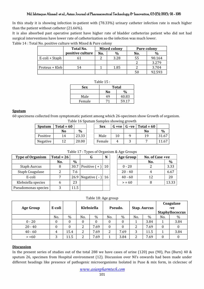
In this study it is showing infection in-patient with (78.33%) urinary catheter infection rate is much higher
than the patient without catheter (21.66%).
It is also absorbed past operative patient have higher rate of bladder catheterize patient who did not had
surgical interventions have lower rate of catheterization so the infection was much lower.
Table 14 : Total No. positive culture with Mixed & Pure colony
Total No. positive culture
Mixed colony Pure colony No. % No. %
E-coli + Staph 61 2 3.28 55 90.164 2 3.279
Proteus + Kleb 54 1 1.85 2 3.704 50 92.593
Table 15 :
Sex Total No %
Male 49 40.83 Female 71 59.17
Sputum
60 specimens collected from symptomatic patient among which 26-specimen show Growth of organism.
Table 16 Sputum Samples showing growth
Sputum Total = 60 Sex G +ve G –ve Total = 60 No % No %
Positive 14 23.33 Male 10 9 19 31.67
Negative 12 20.00 Female 4 3 7 11.67
Table 17 : Types of Organism & Age Groups
Type of Organism Total = 26 G N Age Group No. of Case +ve No. % No. %
Staph Aurcus 8 30.7 Positive ( + ) 10 0 - 20 2 3.33
Staph Coagulase 2 7.6 20 - 40 4 6.67
E-coli 7 26.9 Negative ( - ) 16 40 - 60 12 20
Klebsiella species 6 23 > = 60 8 13.33
Pseudomonas species 3 11.5
Table 18: Age group
Age Group E-coli Klebsiella Pseudo. Stap. Aurcus Coagulase
-ve Staphyllococcus
No. % No. % No. % No. % No. % 0 - 20 0 0 0 0 0 0 1 3.84 1 3.84
20 - 40 0 0 2 7.69 0 0 2 7.69 0 0
40 - 60 4 15.4 2 7.69 2 7.69 3 11.5 1 3.84
> =60 3 11.5 2 7.69 1 3.84 2 7.69 0 0
Discussion
In the present series of studies out of the total 288 we have cases of urine (120) pus (90), Pus (Burn) 40 &
sputum 26, specimen from Hospital environment (12). Discussion over NI’s onwards had been made under
different headings like presence of pathogenic microorganisms Isolated in Puse & mix form, in ciclecnec of
Md. Ishtiaque Ahmad et al., Asian Journal of Pharmaceutical Technology & Innovation, 03 (15); 2015; 91 - 106
www.asianpharmtech.com 102
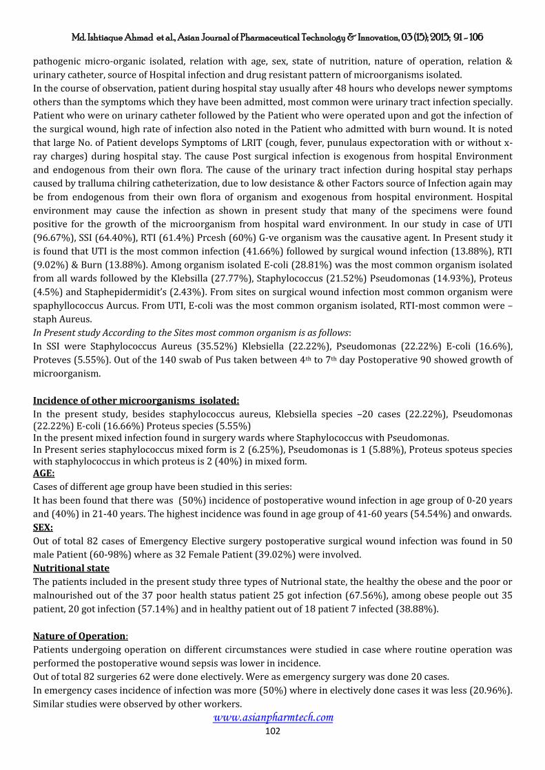
pathogenic micro-organic isolated, relation with age, sex, state of nutrition, nature of operation, relation &
urinary catheter, source of Hospital infection and drug resistant pattern of microorganisms isolated.
In the course of observation, patient during hospital stay usually after 48 hours who develops newer symptoms
others than the symptoms which they have been admitted, most common were urinary tract infection specially.
Patient who were on urinary catheter followed by the Patient who were operated upon and got the infection of
the surgical wound, high rate of infection also noted in the Patient who admitted with burn wound. It is noted
that large No. of Patient develops Symptoms of LRIT (cough, fever, punulaus expectoration with or without x-
ray charges) during hospital stay. The cause Post surgical infection is exogenous from hospital Environment
and endogenous from their own flora. The cause of the urinary tract infection during hospital stay perhaps
caused by tralluma chilring catheterization, due to low desistance & other Factors source of Infection again may
be from endogenous from their own flora of organism and exogenous from hospital environment. Hospital
environment may cause the infection as shown in present study that many of the specimens were found
positive for the growth of the microorganism from hospital ward environment. In our study in case of UTI
(96.67%), SSI (64.40%), RTI (61.4%) Prcesh (60%) G-ve organism was the causative agent. In Present study it
is found that UTI is the most common infection (41.66%) followed by surgical wound infection (13.88%), RTI
(9.02%) & Burn (13.88%). Among organism isolated E-coli (28.81%) was the most common organism isolated
from all wards followed by the Klebsilla (27.77%), Staphylococcus (21.52%) Pseudomonas (14.93%), Proteus
(4.5%) and Staphepidermidit’s (2.43%). From sites on surgical wound infection most common organism were
spaphyllococcus Aurcus. From UTI, E-coli was the most common organism isolated, RTI-most common were –
staph Aureus.
In Present study According to the Sites most common organism is as follows:
In SSI were Staphylococcus Aureus (35.52%) Klebsiella (22.22%), Pseudomonas (22.22%) E-coli (16.6%),
Proteves (5.55%). Out of the 140 swab of Pus taken between 4th to 7th day Postoperative 90 showed growth of
microorganism.
Incidence of other microorganisms isolated:
In the present study, besides staphylococcus aureus, Klebsiella species –20 cases (22.22%), Pseudomonas (22.22%) E-coli (16.66%) Proteus species (5.55%) In the present mixed infection found in surgery wards where Staphylococcus with Pseudomonas. In Present series staphylococcus mixed form is 2 (6.25%), Pseudomonas is 1 (5.88%), Proteus spoteus species with staphylococcus in which proteus is 2 (40%) in mixed form. AGE:
Cases of different age group have been studied in this series:
It has been found that there was (50%) incidence of postoperative wound infection in age group of 0-20 years
and (40%) in 21-40 years. The highest incidence was found in age group of 41-60 years (54.54%) and onwards.
SEX:
Out of total 82 cases of Emergency Elective surgery postoperative surgical wound infection was found in 50
male Patient (60-98%) where as 32 Female Patient (39.02%) were involved.
Nutritional state
The patients included in the present study three types of Nutrional state, the healthy the obese and the poor or
malnourished out of the 37 poor health status patient 25 got infection (67.56%), among obese people out 35
patient, 20 got infection (57.14%) and in healthy patient out of 18 patient 7 infected (38.88%).
Nature of Operation:
Patients undergoing operation on different circumstances were studied in case where routine operation was
performed the postoperative wound sepsis was lower in incidence.
Out of total 82 surgeries 62 were done electively. Were as emergency surgery was done 20 cases.
In emergency cases incidence of infection was more (50%) where in electively done cases it was less (20.96%).
Similar studies were observed by other workers.

Md. Ishtiaque Ahmad et al., Asian Journal of Pharmaceutical Technology & Innovation, 03 (15); 2015; 91 - 106
www.asianpharmtech.com 103
Nosocomial UTI Study:
In the present studies in UTI organism isolated askle as follows total 120 cases were studied. Organism isolated
E-coli 57(47.5%) Klebsilla 51 (42.5%), Staphylococcus 4 (3.33%), Pseudomonas 15 (4.16%) Proteus 3 (2.25%).
In present study E-coli 47.5%, Klebsiella (41.5%), Pseudomonas (4.46%) was isolated.
In total 120 cases of UTI studied maximum No of cases Isolated from surgery (30) & Obs (30) & Gyhaccong
Followed by orthopedics (27), medicine (33) paediatrics (10).
Catheter related Nosocomial UTI:
In total 120 positive culture catheterized and Non-catheterized isolated organism were mostly from the
catheterized patient in which 94 (78.33%) Positive culture found where as in catheterized Patient it was 26
(21.66%) so it is clear catheterized patient had higher rate of infection.
Pure and mixed culture:
Mixed culture of E-coli e staphyloceus isolated in 2 cases in total 61 isolatye percentage of E.coli + staph were
3.27% were as mixed colony of proteus & Kleb found in one case out of 54 isolate of proteus & klebsiella
percentage of mixed colony of with these organism were 1.85%
Among pure culture E-coli was 96.49%, Klebsiella (3.92%), staphylococcus (50%), pseudomonas (100%) &
Protens was 66.66%) pure.
Sex incidence in Nosolomial UTI:
Out of 120 studied cases 71 (59.26%) were female and 49 male (40.80%).
NRTI (Nosocomial Respiration tract infection):
In this study among 60 Symptomatic Patient of RTI sputum collected in which 26 cases showed positive culture.
Among isolated organism Staph aurues (30.7%) was the most common organism isolated followed by E-coli
(26.9%), Klebsiella (23.0%) Pseudomonas (11.5%), coagulase Negative staphylococcus aureus (7.6%).
Burn:
In present study 40 cases were studied in which gram positive bacteria was (16.0%) where as gram negative
bacterial isolated were more (24.0%) sample was collected was collected between 4-10 days post burn period.
Sex & age relation with burn wound infection:
In these study 0-10yr age group has 5 case isolated organism (12.5%) in Burn most of the burn were accidental
during play & work. In the age group of 11-20 year 8 cases reported (20.0%) in which Female were 5 and Male
3. In the age group of 21-30 years which show maximum no of burn wound infection 42.5% in which Female
(13) Male (4) out of 17 Positive culture cases. In the age group of 41-50 years burn wound infection were 7.5%.
In the age group above 51 year of age only 5% burn wound infection found. In the present series of study there
are 29 Female burn Patients and 11 Male Patient who got Nococomial burn wound infection. In the present
series it is observed that mostly the isolateds in the burn of chest, shoulder, Arms in staph aureus,
pseudomonas aeruginosa, Staphylococous Epidermidis & Proteus sp. Where as positive wound swab around
pelvis, thigh buttock, back & lower abdomen, mostly the were gram-negative bacteria.
Bacterial isolates from ward Environment:
Out of the 120 samples from different sites of hospital 12 sites had culture positive among which
Staphylococcus aureus was the most common it was 7 out of 12, second most common isolate was E-coli 3 out
of 12 isolates followed by Pseudomonas 2 out of 12 isolates. Settle plates had growth of microorganism.
Two kinds of bacteria carrying particles found in the air are small particles that remained suspended in the air
for long periods, and large particles which fall on the ground with in about an hour in a still atmosphere.

Md. Ishtiaque Ahmad et al., Asian Journal of Pharmaceutical Technology & Innovation, 03 (15); 2015; 91 - 106
www.asianpharmtech.com 104
Infection caused by bacteria, which gain entry by inhalation can either by large or by small particles the large
particles were collected by allowing them to fall on the surface on an exposed plate of blood agar medium.
A pair of blood agar plates at vaious sites in the ward was kept to find out the degree of aerial contamination.
Exposing the plate’s in the ward each may appear to give more accurate information. A general relationship
between total air count in the operation theatre and risk of infection had been established when the counts are
in the range of 700-1800 particles pmq there significant risk where they are below 180 pmq.the risk is
probably slight.
Unlike air, which is freely mixed and there fore uniformly contaminated surface tend to be irregularly
contaminated and the results of bacteriological investigations on them were difficult to evaluate.
Sensitivity pattern of Burn wound infection:
Out of 60 isolate of Burn wound infection among 32 isolates of staphylococcus aureus most sensitive antibiotic
were Cefotaxime (30.63%) with resistance (9.38%), staphylococcus aureus was also sensitive with Ofloxacin
(87.50%), Ceftazidime (78.13%), Piperacillin (78.13%), Amikacin (78.13%). It showed most resistance with
Ampicillin (40.63%), Ceftriaxone (37.50%).
Among, E-coli isolated from burn wound it was most sensitive with Piperacillin (86.67%) Ofloxacin (93.33%),
Ampicillin (86.67%), Gentamysin (86.67%). Most resistance showed against cetotaxim (46.67%), Nitillimycin
(46.67%), Ampicillin (33.33%).
Isolated Klebsiella sp. Showed most sensitivity against Ceftazidime (80%) Norfloxacin (70%), Imipenem (65%).
While most resistance showed against ciprofloxacin (60%), Amikacin (5.5%).
Against Pseudomonas sp. most sensitive were cefotaxim (90.63%) ofloxacin (87.50%), Imipenem (81.25%),
Piperacillin (78.13%).
Against protens isolates most sensitive were Imipenem (80%), ofloxacin (80%), Piperacillin (60%)
ciprofloxacin (60%) and most resistance against Amikacin (60%), Netillimycin (60%), Norfloxacin (60%).
It was observed in this study that most sensitive antibiotics against all above mention organism most sensitive
were Piperacillin (93.33% to 60%), Gentamysin (86.67% to 60%), Amikacin (86.67% to 40%).
Sensitivity Pattern of Noscomial urinary tract infection:
E-coli was the most common organisms isolated from urine they showed most sensitivity with Ceftazidime
(91.23%), Amikacin (78.95%), Netillimycin (68.42%), Gentamysin (66.67%), Ampicillin (64.91%). It is
Resistant against Ceftriaxone (42.11%), Cefotaxime (56.14%), Norflox (49.12%), Piperacillin (35.09%).
Klebsiella showed sensitivity with Ceftazidime (90.20%), Imipenem (90%), Amikacin (88.24%), Gentamycin
(82.35%) and Resistance against Cefotaxim (41.18%), Norfloxacin (31.37%).
Staphylococcus showed maximum sensitivity with Ceftazidime (100%), Imipenem (100%), Piperacillin
(100%), Ceftriaxone (75%). And Resistance against Ampicillin (50%), Ciprofloxacin (50%), Gentamysin (25%).
Pseudomonas species showed sensitivity with Ceftazidime (80%), Imipenem (80%), Gentamysin (80%), and
Resistance against Ciprofloxacin (40%), Amikacin (40%), Ofloxacin (40%).
Proteus species showed sensitivity with Ceftazidime (100%), Imipenem (100%), Piperacillin (100%),
Gentamysin (100%). And it is Resistance against Ampicillin (33.33%), Cefotaxim (33.33%), Netillimycin
(33.33%).
It was observed that sensitivity of Ceftazidime against all organism varies from (90.20% to 100%), sensitivity
with Imipenem between (87.72% to 100%) with Ampicillin (64.91% to 78.43%), Gentamysin (82.35% to
66.57%).
Sensitivity Pattern of Nosomical Surgical site infection:
Staphylococcus aureus was the most common organism isolated from wards. It was most sensitive with the
Cefotaxime (90.63%), Ofloxacin (87.50%), Piperacillin (78.13%), Imipenem (81.25%), It was most resistant
with ampicillin (43.6%), Gentamycin (28.13%), Ceftriaxone (37.50%).

Md. Ishtiaque Ahmad et al., Asian Journal of Pharmaceutical Technology & Innovation, 03 (15); 2015; 91 - 106
www.asianpharmtech.com 105
Klebsiella species was the second most common organism isolated. It was most sensitive with Ceftazidime
(80%), Norfloxacin (65%), Piperacillin (65%), Imipenem (65%). Most resistance with Ciprofloxacin (60%),
Amikacin (55%), Ceftriaxone (50%), Cefotaxim (45%), Piperacillin (35%), Ceftazidime (35%).
Among E-coli isolated from PUS it was sensitive with Piperacillin (93.33%), Ofloxacin (93.33%),
Norfloxacin(100%), Gentamysin (86.67%). It was resistant with Cefotaxim (46.67%), Netillimycin (46.67%),
Ciprofloxacin (26.67%), Imipenem (26.67%), Ceftazidime (26.67%).
Pseudomonas aeruginosa isolated from Pus was most sensitive with Ceftazidime (77.78%), Imipenem
(77.78%), Amikacin (77.78%), Ciprofloxacin (77.78%), Cefotaxim (33.33%).
Isolated Proteus species showed most sensitivity with Imipenem (80%), Ofloxacin (80%), Piperacillin (60%),
Ceftriaxone (60%). Most resistant with Norfloxacin (60%), Cefotaxim (60%), Ampicillin (60%).
According to above findings most organism showed sensitivity with Piperacillin (93.33% to 60%) Ofloxacin
(93.33% to 65%), Norfloxacin (100% to 40%), Ceftazidime (80% to 40%),.
Nosocomial Respiratory Tract infection Sensitivity Pattern:
From respiratory tract infection most common organism was Staphylococcus aureus, which was most sensitive
to Piperacillin (87.50%), Imipenem (75%), Cefotaxim (75%), Ceftriaxone (75%), Ampicillin (75%). And Most
resistant with Gentamysin (75%), Amikacin (62%), Ciprofloxacin (50%), Ofloxacin (37.5%).
E-coli was most sensitivity with Ceftazidime (85.71%), Ciprofloxacin (71.43%), Ofloxacin (71.42%),
Norfloxacin (71.43%).
Klebsiella was most sensitive with Ceftazidime (83.33%), Imipenem (100%), Netillimycin (83.33%),
Ciprofloxacin (83.33%), Ceftriaxone (66.67%), Gentamysin (66.67%), Amikacin (66.67%).
Pseudomonas was most sensitive with Ceftazidime (100%), Imipenem, Gentamysin (66.66%), Amikacin
(66.67%).
Streptococcus epidermatdis was most sensitive with Imipenem (100%), Piperacillin (100%), Gentamysin
(50%), Amikacin (50%).
Among all antibiotics most sensitive antibiotics was Imipenem (100% to 62.5%), Ceftazidime (100% to 75%),
Most restance shown against Ampicillin (25% to 100%), Gentamysin (66.67% to 28.87%).
References:
1. Ayliffe GAJ, Nosocomial infection - the irreducible minimum. Infect COntrol 1986; 7 , Suppl.: 24.
2. Ayliffe GAJ, Rev Infect Dis 1991; 13 Suppl 10 : S 800 - 4.
3. Ayliffe GAJ, Lowbury EJL. Airborn infection in hospital, Hospital infect 982; 3 ; 217 - 240.
4. Bennett JV, Branchman PS. Hospital infections, 3rd edn, Little Brown, Boston, 1992.
5. Bonten MJM, Hayden MK, Nathan C, Van Voorhis J, Matushek M, Slaughter S, et al. Lancet 1996; 348; 1615-9.
6. Bauer, A.W. Kirby, W.W.W. Sherris, J.C. and Truck, M. Amer. J. Clin. Path., 493,1996.
7. Centre of Disease Control. Nosocomial enterococci resistant to vancomycin in the United States, 1989-1993. Morbid
Mortal Wkly Rep 1993; 42: 597-599.
8. Chaudhuri AK. Infection Control in hospitals: has its quality-enhancing and cos-effect role been appreciated? J Hosp
Infect 1993: 25: 1-6.
9. Centres for Disease Control and Prevention, Hospital Infections Program. (NNIS) report, data summary from October
1986-April 1996, issued May 1996.
10. Combined Working Party of the British Society for Antimicrobial Chemotherapy and the Hospital infection Society,
1996, Guidelines on the control of methicillin resistant Staphylococcus aureus in the community. J Hosp Infect 1995:
31: 1-12.
11. Dinnen, Surg. Gynae & Obst. 113,91.
12. Dineed et al , Surg. Gynae & Obst. 106,453,1953.
13. Emmerson AM, Enstone JE, et al.the Second National prevalence survey of Infection in Hospitals – overview of the
results. J Hosp Infect 1996: 32: 175-190.
14. Emori TG, Gaynes RP. An overview of nosocomial infections, including the role of the microbiology laboratory. Cli
Microbio Rev 1993: 6:428-42.

Md. Ishtiaque Ahmad et al., Asian Journal of Pharmaceutical Technology & Innovation, 03 (15); 2015; 91 - 106
www.asianpharmtech.com 106
15. Finegold DS. Hospital acquired infections. N. Engl J Med 1970: 283: 1348-1391.
16. George RC, Utteley AHC et al High level vancomycin resistant enterococci causing hospital infection. Epidemiol infect
1989:103:173-81.
17. Glenister HM, How to we collct data for survelliance of wound infection? J Hosp infec 1993:24:283-289.
18. Goldman DA. Nosocomial infection control in the united states of America, J Hosp Infec 1986: 8: 116-28.
19. Hospital Infection control Practices Advisory Committee (HICPAC), 1995, Recommendations for preventing the spread
of vancomycin resistance. Infect control Hosp Epidemiol 1995: 16: 105-113.
20. Hospital Acquired Infections: guidelines to laboratory methods, edited by M. T. Parker WHO Regional Publications,
European Series No.4, 1978.
21. Haley RW, Culver DH, White J, Morgan WM, Amber TG,Mann VP, et al. Am J Epidemiol 1985:121:182-205.
22. Haley RW, Culver DH, Whit JW, Morgan WM, Emori TG, AM J Epidemiol 1985:121:159-67.
23. Hiramatsu K, Aritaka N, Hanaki H, Kawasaki S, Hosoda Y, Hori S, et al . AM J Epidemiol 1985:121:182.
24. Hiramatsu K, Hanaki H, Ino T, Yabuta K, Oguzi T, Tenover FC. J Antimicrob Chemother 1997; 40; 135-136.
25. Handerson, Brit. Jour. Of Sug. 54, 756,1967.
26. Howard, Ann. Of Surg.-160;33,1964.
27. Howe, New Eng. Jour. Of Med.-251, 411, 1954.
28. Howe, Amer. Jour. Of Surg. – 107, 696, 1964.
29. Jines, R.J., Jakson, D.M. and Lowbwry, E.J.L. 1966, Brit. J. Plast. Surg.19.43.
30. Krishna Prakash S., Nosocomial infections- 2000, 132-136.
31. Larson E. A casual link between hand washing and mode of infection. Exam. Of the evidence, Infect Control 1988;9;28-
36.
32. Livorenese LL. Dial S, Samel C. Hospital acquired infection with vancomycine resistant Enterococcus faecium
transmitted by electronic thermometer. Ann Intern Med 1992; 117; 112-116.
33. Millar MR, Brown NM, et al. Outbreak of infection with penicillin resistant Streptococcus pneumniae in a Hospital for
elderly, J Hosp infect 1994;1994;27;99-104.
34. Miles, A.A., Miles, E, M. Bruke, J., Brit. J. Exp. Path., 38, 79, 1957.
35. Miles et al, Brit. J. of Exp. Path. 31, 73, 1950.
36. McNeill et al, B.M. J. – 2-279-1961.
37. NNIS System. Am J Infect Control 1996; 24; 380-8.
38. Okesola, A.O., Kehinde. A.O., 2004, Annals of Burns and Fire disasters Vol XVII-n-1 March.
39. Parker MT. Hospital acquired Infections: Guidelines to Laboratory Methods, WHO regional publications European
Series No. 4, WHO, Cophagen, 1978.
40. Phipott Howard J, Casewel M. Hospital Infection Control Policies and Practical Procedure, WB, Saunders, London, 1994.
41. Slaughter S, Hayden MK, Nathan C, Hu TC, Rice T, Van Voorhis J, et al. Ann Intern Med 1996; 125; 448-56.
42. Warren JW, Platt R Thomas KJ. Antibiotic irrigation and catheter associated urinary tract infections. N Eng J Med 1987;
299; 570-573.
43. Williams REO, Blowers R et al. Hospital infection. Caused and Prevention, Lloyd-Luke London, 30, 1960.
44. Weinstein RS. Epideiology and Control of Nosocomial infections in adult intensive care units. Amer J Med 1991; 91;
179-84.
45. Williams et al, Lancet – 2, 659, 1960.
46. AIRHH: International Association for Research in Hospital Hygine (Monaco): www.monaco.mc/assoc/airhh.
47. APIC: Association for Professionals in Infection Control and Epidemiology; http://www.apic.org/
48. APSI: Associazione Controllo Infezioni (Italy) www.apsi.it
49. Health Canada: Division of Nosoco,al and Occupational Infections www.hc-sc.gc.ca/hpb/lcdc/bid/nosocom/index.html
50. HELICS: Hospital in Eurpose Link for Infection Control through Surveillance www.helics.univ-lyon1.fr
51. Infection Control Nurses Association (UK) www.icna.co.uk
52. IFIC – International Federation Of Infection Control http://theific.org/
53. NNIS: National Nosocomial Infections Surveillance (USA) www’ A.cdc.gov/ncidod/hip/nnis/@nnis/htm
54. Centers for Disease Control and Prevention http://www.cdc.gov/hai/
55. SFHH - French Society of Hospital Hygiene and quality of care sfhh.univ-lyon1.fr
56. SHEA: Society for Healthcare Epidemiology of America www.shea-online.org