Embed Size (px)
Citation preview
Hindawi Publishing CorporationEvidence-Based Complementary and Alternative MedicineVolume 2013 Article ID 461478 9 pageshttpdxdoiorg1011552013461478
Research ArticleHepatoprotective Potential of Chestnut Bee Pollen on CarbonTetrachloride-Induced Hepatic Damages in Rats
Oktay YJldJz1 Zehra Can2 Oumlzlem Saral23 Esin YuluL4 Ferhat Oumlztuumlrk5
Rezzan AliyazJcJoLlu6 Sinan Canpolat7 and Sevgi KolaylJ2
1 Macka Vocational School Karadeniz Technical University 61750 Trabzon Turkey2Department of Chemistry Faculty of Sciences Karadeniz Technical University61080 Trabzon Turkey
3 Department of Chemistry Artvin Coruh University 08000 Artvin Turkey4Department of Histology and Embryology Faculty of Medicine Karadeniz Technical UniversityTrabzon Turkey
5 Department of Molecular Biology and Genetics Faculty of Sciences Canik Basari University55080 Samsun Turkey
6 Faculty of Pharmacy Karadeniz Technical University 61080 Trabzon Turkey7Department of Physiology Faculty of Medicine Karadeniz Technical University Trabzon Turkey
Correspondence should be addressed to Sevgi Kolaylı skolayli61yahoocom
Received 11 May 2013 Accepted 24 August 2013
Academic Editor Mohammad Ahmad Al-Shatouri
Copyright copy 2013 Oktay Yıldız et al This is an open access article distributed under the Creative Commons Attribution Licensewhich permits unrestricted use distribution and reproduction in any medium provided the original work is properly cited
Bee pollen has been used as an apitherapy agent for several centuries to treat burns wounds gastrointestinal disorders andvarious other diseases The aim of our study was to investigate the hepatoprotective effects of chestnut bee pollen against carbontetrachloride (CCI
4)-induced liver damage Total phenolic content flavonoid ferric reducingantioxidant power andDPPH radical
activity measurements were used as antioxidant capacity determinants of the pollen The study was conducted in rats as sevengroups Two different concentrations of chestnut bee pollens (200 and 400mgkgday) were given orally and one group wasadministered with silibinin (50mgkgday ip) for seven days to the rats following the CCI
4treatment The protective effect
of the bee pollen was monitored by aspartate transaminase (AST) and alanine transaminase (AST) activities histopathologicalimaging and antioxidant parameters from the blood and liver samples of the rats The results were compared with the silibinin-treated and untreated groups We detected that CCI
4treatment induced liver damage and both the bee pollen and silibinin-treated
groups reversed the damage however silibinin caused significant weight loss and mortality due severe diarrhea in the rats Thechestnut pollen had showed 2887mg GAEg DW of total phenolic substance 807mg QUEg DW of total flavonoid 9271mgCyn-3-glukg DW of total anthocyanins and 9mg 120573-carotene100 g DW of total carotenoid and substantial amount of antioxidantpower according to FRAP and DPPH activity The results demonstrated that the chestnut bee pollen protects the hepatocytes fromthe oxidative stress and promotes the healing of the liver damage induced by CCI
4toxicity Our findings suggest that chestnut bee
pollen can be used as a safe alternative to the silibinin in the treatment of liver injuries
1 Introduction
Apitherapy has been used in folk medicine since the earlyages of human beings and in the recent years its appli-cation in the treatment of burns wounds gastrointestinaldisorders ulcers and carcinogenesis has been increasing
Bee pollen is one of the richest and purest natural foodsthat way ever discovered the tremendous nutritional andmedicinal value of the pollen has been used for centuriesBee pollen is a perfectly balanced food and is rich inamino acids proteins hormones enzymes carbohydratesminerals fats a considerable amount of vitamins phenolic
2 Evidence-Based Complementary and Alternative Medicine
substances phytochemicals and significant quantities ofantioxidant agents [1ndash3] The chemical composition of thebee pollen depends on its botanical and geographical prop-erties Pollen contains about 1ndash5 total phenolic substanceswhich include different subtypes such as flavonoids phenolicacids anthocyanins and tannins They exhibit a wide rangeof biological activities including antioxidant antimicrobialanti-inflammatory antiatherogenic anticarcinogenic andantithrombotic activities [1ndash4] Phenolic compounds areconsidered to be beneficial for human health since theydecrease the risk of degenerative diseases caused by oxidativestress Many researchers have demonstrated that the phenoliccompounds within the pollens inhibit the occurrence anddevelopment of numerous degenerative disorders [2]
Carbon tetrachloride (CCI4) is a hepatotoxic agent that
enhances the formation of free radicals which cause lipidperoxidation of cellular and organelle membranes [5] thusone of the most widely used toxic agents to induce liver dis-eases in animal models CCI
4is metabolized by cytochrome
p450 system to highly reactive trichloromethyl free radicalsand reactive oxygen species which initiate lipid peroxidationandnecrosis In addition the toxic agent causes inflammatoryresponse initiated by the activated hepatic macrophagesmainly Kupffer cells [6] Many researchers have pointed outthat CCI
4exposure could cause significant biochemical dis-
orders such as fatty liver hepatitis and cirrhosis in laboratoryanimals [7 8]
It is recognized that hepatocyte damage is one of theserious pathological disorders for human To measure aspar-tate transaminase (AST) and alanine transaminase (ALT)activities in the serum or plasma is the simplest methodto diagnose hepatocyte injuries [3 9ndash11] Oxidative stresswas shown as one of the major causes of liver injury [11]Silibinin or silybin is the active component of silymarin andcommonly used for the treatment of various liver damagesSilymarin is obtained through the extraction of milk thistleseeds (Silybum marianum) which is rich in flavonoidsand used as a hepatoprotective drug for several decades Ithas been shown that the bee pollen which contains manyphenolic substances has similar effect as silibinin in termsof hepatoprotection [1 2 12] For this purpose many naturalextracts and honey bee products such as honey and pollenwere used to treat hepatic disorders in laboratory animals[3 4 9 10]
In this study we determined the therapeutic effects ofchestnut bee pollen on the CCI
4-induced liver damage in
the rat model We analyzed the chemical and antioxidantproperties of the chestnut pollen and determined that pollensupplementation recovered the body weight AST and ALTenzyme levels malondialdehyde (MDA) and superoxidedismutase (SOD) levels as well as decreased the histologicaldamage and apoptosis at the hepatocytes following theCCI4treatment We detected that both the bee pollen- and
silibinin-treated groups reversed the CCI4-induced hepatic
damage however silibinin caused significant weight loss andmortality due to severe diarrhea in the rats Our resultssuggest that chestnut pollen can be used as a safe alternativefor the treatment of liver damaging diseases
2 Material and Methods
21 Reagents CCI4 silibinin gallic acid quercetin ethyl
alcohol methanol Trolox (6-hydroxy-2578-tetramethyl-chroman-2-carboxylic acid) 246-tripyridyl-s-triazine(TPTZ) Folin-Ciocalteursquos phenol reagent 22-diphenyl-1-picrylhydrazyl (DPPH) cyanidin-3-O-glucoside TBA 1133-tetramethoxypropane 120573-carotene nitroblue tetrazoliumxanthine and xanthine oxidase were purchased from SigmaChemical Co (St Louis MO USA) AST andALT diagnostickits were also purchased from Sigma Olive oil was obtainedfrom KOMILI Sızma Company (Izmir Turkey)
22 Bee Pollen Samples Pollen samples were obtained fromthe expert beekeepers of Zonguldak Turkey (Western BlackSea area) in 2008 flowering season The samples were driedat 40∘C oven and palynological identification was done byDr Sibel Silici of Erciyes University Turkey Nine familiesof pollen pellets were found in the sample Fabaceae (Med-icago spp Trifolium spp) Fagaceae (Castanea sativa L)Asteraceae (Aster spp Cirsium spp Carduus spp) Apiaceae(Apium spp) Caryophyllaceae (Dianthus spp) Poaceae (Zeamay) Rosaceae (Malus spp) Myrtaceae (Myrtus communis)and Rhamnaceae (Rhamnus cathartica) Chestnut sativa wasdominant (gt45) in the pollen mixture
For the analysis of antioxidant potential of the pollenthe samples were prepared by mixing 1 g of dried powderof pollen sample with 10mL methanol in a flask attachedcondenser then sonicated in a sonicator apparatus (ElmaTranssonic Digital Germany) After 3 h sonication theextract was used for antioxidant tests Although we dissolvedthe pollen in sterile H
2O to feed the animals by gavage it
has been shown that the antioxidant potential of the pollenis revealed with better percentage when it is extracted usingmethanol [13 14]
Chemical analysis of the bee pollen was performedaccording to the method described at AOAC [15] and thevalues were calculated as per dried pollen weight (DW)In addition the following potential antioxidant propertieswere measured according to the references mentioned totalphenolic content [16] total flavonoids [17] and total antho-cyanins [18] total carotenoids [17] total antioxidant activityaccording to the ferric reducing antioxidant power (FRAP)[19] and to free radical scavenging activity of 22-diphenyl-1-picrylhydrazyl (DPPH) [20] FRAP values were expressed asTrolox equivalent antioxidant power and radical scavengingactivity of DPPHwas expressed as SC
50 which represents the
concentration of the extract (mgmL) required to inhibit the50 of the free radical scavenging activity The lower SC
50
value indicates the higher antioxidant activity
23 Animals and Experimental Procedure Forty-nineSprague-Dawley rats were studied which were 12 weeks oldand of 250ndash300 g approximate weight Animals were fedwith standard rat feed and allowed to drink water Animalswere kept in temperature controlled (20ndash25∘C) cages with12 h dark and 12 h light cycles Food was withdrawn for 12 hbefore the experiments
Evidence-Based Complementary and Alternative Medicine 3
Animals were divided into seven groups (see Supplemen-tary Table 1 in Supplementary Material available online athttpdxdoiorg1011552013461478) The first group (G1ndashG3) was used as controls and other experimental groupsThecontrol groups received 08mL of saline (09 vv) in water[21] olive oil (08 vv) and ethanol (02 vv) once per dayfor the entire period (7 days) by ip injection respectivelyThe rats in G4 to G7 were administered ip with CCI
4
dissolved in olive oil at a dose of 085mLkgday body weightG5 was fed with silibinin dissolved in ethanol at a dose of50mgkgday G6 and G7 were fed with pollen at 200mgkgand 400mgkg once per day by gavage respectively Inthis study dried pollen samples were dissolved in deionizedwater This study was approved by the Animal Care andUse Committee from the Faculty of Medicine in KaradenizTechnical University (KTU)
24 Determination of Liver Enzyme Activities and ProteinsPlasma was obtained from the whole blood samples of thetreated rats through centrifugation at 2000timesg for 10min andstored at minus20∘C until analysis AST and ALT activities weremeasured by a Roche Diagnostics Modular Analyzer usingthe manufacturerrsquos commercial kits according to the instruc-tions (Roche Diagnostics GmbH D-68298 and MannheimGermany)
Reduction of nitroblue tetrazolium by xanthine-xanthineoxidase system was used to measure superoxide dismutase(SOD) activity in erythrocyte hemolysate of the rat bloodsamples Formazan formation was examined at 560 nm usingthe spectrophotometer (Beckman-coulter DU 530) Theenzyme activity that causes 50 inhibition was regarded asone unit using bovine erythrocytes SOD as standard and theresults were read as Ug Hb [22]
MDA levels were measured with a colorimetric testwith thiobarbituric acid (TBA) which is used to assessendogenous lipid [23] Fresh tissue samples obtained fromthe treated rats were kept at minus80∘C until the analysis Livertissues were weighed and homogenized in ice-cold 115KCl The homogenate was centrifuged at 2000timesg for 10minThe breakdown product of 1133-tetramethoxypropane wasused as standard and tissue MDA levels were calculated asnmolmL plasma or g tissue Total protein of the liver extractswas analyzed using Lowry et al [24] method with bovineserum albumin as the standard The values were achieved byinterpolation on a calibration standard curve at 650 nm
25 Histological Preparation and Analysis For histologicalanalysis liver tissue samples were fixed immediately in10 buffered formaldehyde dehydrated with ethanol seriescleaned with xylene embedded in paraffin and sectionedas 5 120583m Tissue sections were stained with hematoxylinand eosin (HampE) then examined under a light microscope(Olympus BX-51 Olympus Optical Co Ltd Tokyo Japan)All liver tissue slides were examined at high magnificationand images were recorded by an independent histologist Fivehigh-power fields were selected by random sampling and thefollowing criteria were followed in semiquantification of theliver injuries hepatocyte degeneration vascular congestion
sinusoidal dilatation congestion in enlarged sinusoids andfatty degeneration Each specimen was marked using a scaleof 0 to 3 (0 = none 1 mild 2 moderate and 3 severe) Themean histologic score was calculated for each group
26 TUNEL Analysis For the detection of apoptotic cellswithin our groups (G1ndashG7) 4120583m thick serial sections wereprepared from the paraffin-embedded liver samples ForTUNEL analysis In situ Cell Death Detection Kit (RocheMannheim Germany) was used in accordance with themanufacturerrsquos instructions to detect the fragmented DNAassociatedwith apoptotic cells and clearly visible nuclear frag-ments or sharp and condensed chromatic masses or crescentin the nuclei The stained sections were evaluated under alight microscope (Olympus BX51 microscope Tokyo Japan)at times400 magnification One hundred cells were countedper liver slide in five microscopic fields The percentage ofTUNEL-positive apoptotic cells was calculated and this rep-resented the apoptotic index (AI) An independent histologistexamined the stained specimens in a blinded fashion [11]
27 Statistical Analysis The results were presented as meanvalues and standard deviations Data and regression analyseswere performed via Microsoft Office Excel 2003 (MicrosoftCorporation Redmond WA) Data were tested using SPSS(version 90 for Windows 98 SPSS Inc) Statistical analysesof the results were based on Kruskal-Wallis test and Pearsoncorrelation analysis which is a nonparametric test Thesignificance of the differences was statistically considered atthe level of 119875 lt 005
3 Results and Discussion
This study investigated the hepatoprotective potential ofaquatic extracts of chestnut bee pollen on CCI
4-induced hep-
atic damages in rats Before the treatments some chemicaland antioxidant properties of the pollen were investigatedwhich are summarized in Table 1 The main componentsof the pollen were protein starch fat minerals and waterThe chestnut pollen has approximately 2887mg GAEg oftotal phenolic compounds (TPC) 807mg QUEg of totalflavonoids 9271mg Cyn-3-glukg of total anthocyanins and29mg 120573-carotene100 g of total carotenoids These com-pounds are well established for their responsibility of theantioxidant activities of the natural honeybee products [1 25]The chestnut pollen also demonstrated substantial antiox-idant activity which is measured through ferric reducingantioxidant power (FRAP) and DPPH radical scavengingactivity (Table 1) Although there are other honeybee prod-ucts with higher bioactivity such as royal jelly and propolischestnut pollen was used in this study due to its watersolubility and relatively higher antioxidant capacity Cheng etal [26] recently studied the antioxidant and hepatoprotectiveeffects of Schisandra chinensis pollen extract (SCPE) onCCl
4-
induced acute liver damage in mice and found that SCPE hadstrong antioxidant activities and significant protective effectagainst the acute hepatotoxicity induced by CCl
4 which was
also supported by the evaluation of liver histopathology in
4 Evidence-Based Complementary and Alternative Medicine
Table 1 Chemical and antioxidant properties of the investigated honeybee pollen of chestnut
(a)
Chemical propertiesMoistures Ash Protein Starch Fat 1442 plusmn 042 223 plusmn 021 2367 plusmn 208 683 plusmn 683 587 plusmn 239
(b)
Antioxidant propertiesTotal phenolic content(mg GAEg DW)
Total flavonoid(mg QUEg DW)
Total anthocyanins(mg Cyn-3-glukg DW)
Total carotenoid(mg 120573-carotene100 g DW)
FRAP(mM Troloxg DW)
DPPHSC50 (mgmL)
2887 plusmn 248 807 plusmn 083 9271 plusmn 2521 29 plusmn 90 8231 plusmn 241 062 plusmn 010
mice Furthermore in another recent study Tohamy et al[27] studied the antioxidant capacities of the water extractsof the Egyptian bee pollen (WEBP) on cisplatin (CDDP)-induced damages in multiple organs and found that WEBPwas more potent in the recovery of hepatic and testicledamages
The phenolic compounds of the pollens vary dependingon the geographic characteristics and the flora of the region[1] Recently Ulusoy andKolayli [13] studied theAnzer pollenfrom the Black Sea Region of Turkey and found that TPCof Anzer pollen ranged from 4407 to 12410mg GAEgwhich is higher than the chestnut pollen The TPC of thebee pollen was also studied by Marghitas et al [28] usingtwelve types of pollen collected from Romania In this studythe TPC ranged from 640 to 164mg GAEg dry matter ofpollen and Salix type of pollen showed the highest contentof total phenolic and antioxidant activity In another studythe phenolic content (polyphenolics flavonols flavones andflavonones) and the antioxidant activity in six varieties of beepollen from Sonoran Desert were studied [14] They foundthat the phenolic content ranged between 1591 and 3485mgGAEg pollen and a great variation was observed among thepollen samplesThere was no previous study found about thephenolic content and antioxidant activity of chestnut pollentomake comparisonwith our results however there are a fewstudies about chestnut honey that contains crucial amountsof phenolic substances that are related to biological activitiessuch as antioxidant antimicrobial and anti-inflammatoryfeatures [29 30]
We measured the initial weight of the 49 rats that westudied in 7 groups (G1ndashG7) and compared their weights atthe end of the 7th day (Table 2 Supplementary Table 1)Whilethe rats in the first two control groups of physiological saline(G2) and olive oil (G3) increased inweight (07) the ethanolgroup (G3) decreased in weight (55) Meanwhile weightloss was detected in all of the CCl
4-injected groups (G4ndash
G7) the untreated CCl4control group (G4) and silibinin-
treated group (G5) showed the highest weight loss withminus95and minus82 respectively (Table 2) In addition the significantweight loss in the silibinin group was due to severe diarrheaand caused mortality of 1 rat (out of 7) within the group(G5) Meanwhile the weight loss was the least amount inthe pollen-treated groups (G6-G7) The more the amount ofpollen fed the less the weight loss in the rats This suggests
that the bee pollen acted as an efficient nutrition supplementin the CCl
4-exposed rats without any substantial side effect
and mortalityThe activities of plasma AST and ALT enzymes were
significantly elevated in rats treated with CCI4due to toxic
effect (Table 3) Although the enzyme activities were notchanged in the control groups (G1ndashG3) a significant increasein the CCI
4-treated groups (G4ndashG7) was observed AST and
ALT enzyme activities were markedly increased in all theCCI4administered groups especially 4-5 times increased
in the G4 (CCI4-only group) than the control groups (G1ndash
G3) In case of the intake of silibinin and the bee pollenwere reduced both enzyme activities ALT and AST enzymesare specific to liver damage they are used routinely forthe determination of liver injuries [31] When the beepollen was administered at higher doses of 400mgkgdayAST and ALT levels were significantly decreased In thepresent study the silibinin-treated (50mgkgday) rats (G5)showed better improvement than the low-dose pollen-treated(200mgkgday) group (G6) however as a critical side effectsevere diarrhea andweight loss were detected in the silibinin-treated group Furthermore the improvement of AST andALT levels within the pollen-treated groups (G6 and G7)correlated with the concentration of the pollen given to therats
Among the apitherapy studies related to the preventionand protection of liver damage and toxicity Turkez et al[10] showed that propolis extracts inhibited the liver damageinduced by AlCl
3treatment in the mouse model Similarly
Liu et al (2012) [9] reported that quercetin an antioxidantsubstance found in honeybee products prevented liver injuryfrom lead-induced oxidation Kolankaya et al [32] showedthat the propolis of Castanea sativa showed a protectiveeffect against alcohol-induced liver damage El-Denshary etal [33] indicated that dietary honey consumption reduced thehepatotoxicity against CCI
4-induced liver damage Recently
there are some animal model studies that showed that manyhoneybee products exert curative potential for the protectionof hepatocytes from oxidative damages [3 10 31 32]
Malondialdehyde (MDA) a lipid peroxidation productwas measured in plasma erythrocyte and liver tissues of thetreated animals to analyze the damage caused by oxidativestress induced via CCI
4treatment The amount of MDA
in liver tissue serves as an indicator of lipid peroxidation
Evidence-Based Complementary and Alternative Medicine 5
Table2Ex
perim
entalresultsof
transaminasee
nzym
esandantio
xidant
parametersinthep
lasm
a(119899=7)D
ataw
ereg
iven
asmeanplusmnSD
Group
sWeightg
ain
(1)
Plasma
Erythrocytes
Liver
AST
ALT
MDA
SOD
MDA
SOD
MDA
SOD
UL
UL
nmolm
Lplasma
Um
gprotein
nMg
Hb
Ug
Hb
nmolg
tissue
Ug
liver
G1(control)
+07(plusmn10
7)b17157plusmn2174a8586plusmn1431a
050plusmn002
a72930plusmn30
c055plusmn03a6293plusmn315
bc485plusmn02a
54010plusmn103
c
G2(con
trol)
+07(plusmn10
7)b20714plusmn2059a
8657plusmn890
a051plusmn001
a75010plusmn25
c053plusmn03a8093plusmn681
c582plusmn04a
52020plusmn192
c
G3(con
trol)
minus54(plusmn0946)
a14550plusmn2145a4450plusmn1147a
053plusmn003
ab70065plusmn45
c055plusmn01a4612plusmn195
ab595plusmn05a
51515plusmn231
c
G4(C
CI4)
minus95
(plusmn0905)
a89257plusmn2911d47486plusmn13476
d057plusmn008
b26025plusmn15
a096plusmn13a1542plusmn217
a2262plusmn27e26050plusmn421
a
G5(silibinin)
minus82(plusmn0918)
a51880plusmn122b21880plusmn7325b
053plusmn002
ab30590plusmn40
b067plusmn03a1526plusmn141
a830plusmn08b
29060plusmn232
b
G6(lo
wpo
llen)minus67(plusmn0933)
a69083plusmn149c38340plusmn9302c
053plusmn003
ab26530plusmn30
a049plusmn03a1474plusmn66a1482plusmn15d27020plusmn123
bc
G7(highpo
llen)minus16
(plusmn0984)
ab4640plusmn6902b27700plusmn4962b
052plusmn001
a27025plusmn20
ab059plusmn05a1772plusmn87a
1094plusmn09c31010plusmn144
b
(1)Bo
dyweight(BW)changesd
uringthee
xperim
ents(m
eanplusmnSD
)ap
ositive
(+)v
alue
show
sanincrease
oftheb
odyof
ther
atwhereas
anegativev
alue
(minus)sho
wsd
ecreaseo
fthe
body
ofther
at
a Valuesa
resig
nificantly
different
from
G4G5G6andG7(119875lt005)
b Valuesa
resig
nificantly
different
from
thoseo
fcon
trols(G
1ndashG3)(119875lt005)
c Valuesa
resig
nificantly
different
from
thoseo
fsilibinin(G
5)(119875lt005)
d Valuesa
resig
nificantly
different
from
G7(400
mgpo
llenkg
BW)(119875lt005)
6 Evidence-Based Complementary and Alternative Medicine
Table 3 Histologic analysis and scoring of liver sections of the treated and untreated rats
Groups Hepatocytedegeneration Vascular congestion Sinusoidal dilatation Congestion in
enlarged sinusoidsFatty
degenerationApoptosisindex (AI)
G1 (control) 043 plusmn 0535a 057 plusmn 0535ab 029 plusmn 0488a 029 plusmn 0488a 00 plusmn 00a 357 plusmn 215a
G2 (control) 043 plusmn 0535a 071 plusmn 0488abc 086 plusmn 0378ab 029 plusmn 0488a 00 plusmn 00a 471 plusmn 170a
G3 (control) 029 plusmn 0488a 043 plusmn 0535a 071 plusmn 0488ab 029 plusmn 0488a 00 plusmn 00a 386 plusmn 168a
G4 (CCI4) 271 plusmn 0488c 071 plusmn 0488abc 157 plusmn 0535c 086 plusmn 0378b 300 plusmn 00c 3557 plusmn 635d
G5 (silibinin) 200 plusmn 0577b 114 plusmn 069bcd 129 plusmn 0756bc 071 plusmn 0488ab 186 plusmn 069b 200 plusmn 316c
G6 (low pollen) 214 plusmn 0378b 143 plusmn 0535d 129 plusmn 0756bc 086 plusmn 0378b 214 plusmn 069b 2157 plusmn 404c
G7 (high pollen) 171 plusmn 0488b 129 plusmn 0488cd 129 plusmn 0488bc 071 plusmn 0488ab 171 plusmn 0756b 1586 plusmn 329b
The histological score was calculated using a scale from 0 to 3 0 none 1 mild 2 moderate and 3 severeaValues are significantly different from G4 G5 G6 and G7 (119875 lt 005)bValues are significantly different from those of controls (G1ndashG3) (119875 lt 005)cValues are significantly different from those of silibinin (G5) (119875 lt 005)dValues are significantly different from G7 (400mg pollenkg BW) (119875 lt 005)
(a) (b)
(c) (d)
Figure 1 Histopathological analysis of treated and untreated liver sections (a) Normal hepatocytes (uarr) and sinusoids (998771) in the controlgroup untreated CCI
4(HampE times100) (b) Destroyed group with CCI
4(G4) increased fatty degeneration (uarr) (c) Pollen-treated group (G6)
with 200mgkg decreased fatty degeneration and regeneration in hepatocytes (uarr) (d) Pollen-treated group (G7) with 400mgkg pollenfatty degeneration markedly decreased (uarr)
which is a well-known occurrence in the liver injury due togeneration of reactive species [34] Within the liver tissuesthere was not a significant alteration detected in the MDAlevels of the control groups (G1ndashG3) whereas the MDA levelwas significantly increased in the sick group (G4) which wasalmost 5 times more than G1ndashG3 A substantial decrease wasobserved in the MDA levels of the rats which were exposed
to silibinin and pollen groups (G5ndashG7) (119875 lt 005) As aresult of CCI
4treatment MDA levels were increased in the
plasma erythrocytes and liver which have demonstratedthat CCI
4caused lipid peroxidation Liver plays a major role
in themetabolism of xenobiotic thus it is vulnerable tomanycompounds which are either toxic as themselves or producemetabolites that can cause liver damage [9 34]The efficiency
Evidence-Based Complementary and Alternative Medicine 7
(a) (b)
(c) (d)
Figure 2 Results on apoptosis analysis (TUNEL) of CCI4-induced apoptosis in primary rat hepatocytes apoptotic hepatocytes (uarr) and
normal hepatocytes (998771) (times400) (a) Control group untreated with CCI4(G1) brown labeled apoptotic hepatocytes (uarr) and blue labeled
normal hepatocytes (998771) (b) Destroyed with CCI4(G4) apoptotic hepatocytes (uarr) (c) Pollen-treated group (G6) with 200mgKg (d) Pollen-
treated group (G7) with 400mgKg
of silibinin against oxidative stress in liver was reported inthe previous studies [12] Our results clearly showed that boththe silibinin and the bee pollen are effective in the preventionof lipid peroxidation as well as oxidative damage which wasinduced by CCI
4 However silibinin caused mortality due to
severe diarrhea in our rat modelSuperoxide dismutase (SOD) is an important antioxidant
enzyme that protects the organism from the harmful effectsof superoxide radicals formed as a result of oxidative stress[4] In our study there was no significant difference detectedin SOD activities in plasma liver and erythrocyte among thecontrol groups (G1ndashG3) whereas SOD activity was decreasedin the CCI
4-exposed groups The lowest SOD activity was
detected in the rats exposed to CCI4(G4) and the activity
slightly increased in the groups that were silibinin- andpollen-administered groups (G5ndashG7) SOD is physiologicallysynthesized in the liver cells similar to other antioxidantenzymes thus the protein synthesis is negatively affected as aresult of the hepatocellular injury induced by CCI
4 However
SOD activity was increased in pollen- and silibinin-treatedgroups possibly owing to the healing of hepatocellulardamage Similar to our results it has been shown that SODactivity was decreased notably in the mice treated withtrichlorfon which is an organopesticide but significantlyincreased in the groups which were given pine honey [3]The results of our present study are in agreement with the
previous studies in terms of alterations in oxidative stressmarkers in response to apitherapy [3 31]
Microscopic examination showed liver parenchyma andsinusoids of the hepatocytes were healthy in the controlgroups (G1ndashG3) (Figure 1(a)) Meanwhile obvious damageswere observed in the liver sections of the CCI
4-exposed
group Common degeneration and noticeable fatty vac-uolization were observed in the hepatocytes from the sickgroup (G4) There was vascular congestion in the sinusoids(Figure 1(b)) A slight degree of vacuolar degeneration andfatty vacuolization were observed in the hepatocytes of G5(silibinin) G6 and G7 (pollen-administered groups) how-ever the level of vacuolar degeneration of these groups wasless than that ofG4And also histological improvement ofG7(400mg pollenkgday) was better than that of G6 (200mgpollenkgday) (Figures 1(c) and 1(d)) The improvementwas significantly relevant with the bee pollen amount givento the animals Histological damage scoring and statisticalevaluation of groups were shown in Table 3
Hepatocyte apoptosiswas analyzed using terminal deoxy-nucleotidyl transferase-mediated deoxyuridine triphosphatenick end-labeling (TUNEL) assay The number of apoptoticnuclei was much higher in sick group (G4) than that ofthe control groups (Figures 2(a) and 2(b)) however thenumber of apoptotic nuclei of G5ndashG7 (silibinin- and pollen-administered groups) was much reduced compared to the
8 Evidence-Based Complementary and Alternative Medicine
sick group (G4) (Figures 2(c) and 2(d)) The apoptosis valuesin the liver and the results of the statistical analysis areshown in Table 3 Histopathological damage scoring andTUNEL analysis demonstrated that tissue damage in the liverwas higher in the groups exposed to CCI
4and found that
the tissue damage was significantly reduced in the groupstreated with pollen following CCI
4exposure The apoptosis
index (AI) was significantly increased in the group that wasexposed to CCI
4without any treatment (G4) compared to
the treatment groups (G5ndashG7) (Table 3) However the AIsof these treatment groups were at least five times more thanthe control groups which were not exposed to CCl
4(G1ndashG3)
This suggests that the initial exposure of the cells to CCI4
massively increased the apoptosis of the hepatocytes thus thetreatments of the rats with either silibinin or pollen could notcompletely reverse the devastating impact of CCI
4
Experimental liver damage model is mostly achievedby exposing rats to CCI
4 It has been shown that lipid
peroxidation increases and free oxygen radicals are releasedin the rats exposed to CCI
4[7 8] Free oxygen radicals
are known to be the mediators of tissue damage Reac-tive oxygen radicals cause oxidative damage to cellularmacromolecules such as nucleic acids proteins and lipidswhich eventually lead to apoptosis Free oxygen radicalscause lipid peroxidation several metabolic disorders andfunctional abnormalities through inducing membrane andDNA damages [35] Lipid peroxidation is believed to bean important cause of destruction and damage to the cellmembranes which leads to changes inmembrane fluidity andpermeability Moreover lipid peroxidation also enhances therate of protein degradation which initiates the eventual lysisof the cells [11] Our results indicated that the water pollenextracts inhibited the CCI
4-induced apoptotic cell death and
hepatotoxicity It has been reported that free oxygen radicalsplay important roles in CCI
4-induced cell injury [7] When
the amount of free oxygen radicals in the cell are increasedthey overpower the defense systems and cause oxidative stressor cell injury leading to development of various diseasesThus CCI
4-induced apoptosis is related to oxidative stress
in hepatocytes and intracellular antioxidants may protecthepatocytes against cell apoptosis induced by CCI
4[9]
4 Conclusion
CCI4is a hepatotoxic agent that enhances the formation of
free radicals which cause lipid peroxidation of cellular andorgan damages The chestnut bee pollen contains substantialnutrients and possesses many phenolic compounds whichare the factors of high antioxidant properties Our studyclearly shows that the chestnut bee pollen exerts highlybeneficial biological activities in the protection of hepatocytesfrom oxidative stress and toxicity induced by CCI
4exposure
Therefore we conclude that the chestnut bee pollen could besafely included in the daily human diet as a food additivewhich will enhance the inhibition of the oxidative stressChestnut bee pollen could be used as a suitable alternativeto silibinin in the treatment of hepatocellular pathologies
Acknowledgments
This study was supported by Research Fund of KaradenizTechnical University (Project no 20091110025) Two of theauthors Oktay Yildiz and Huseyin Sahin were funded byTUBITAK-BIDEB for their graduate studies
References
[1] S Bogdanov ldquoQuality and standards of pollen and beeswaxrdquoApiacta vol 38 pp 334ndash341 2004
[2] A GHegazi ldquoMedical importance of bee productsrdquoUludag BeeJournal vol 12 pp 136ndash146 2012
[3] G Eraslan M Kanbur S Silici and M Karabacak ldquoBeneficialeffect of pine honey on trichlorfon induced some biochemicalalterations in micerdquo Ecotoxicology and Environmental Safetyvol 73 no 5 pp 1084ndash1091 2010
[4] G Eraslan M Kanbur and S Silici ldquoEffect of carbaryl onsome biochemical changes in rats the ameliorative effect of beepollenrdquo Food and Chemical Toxicology vol 47 no 1 pp 86ndash912009
[5] G A Clawson ldquoMechanisms of carbon tetrachloride hepato-toxicityrdquo Pathology and Immunopathology Research vol 8 no2 pp 104ndash112 1989
[6] W Peng C Zhang H Lv et al ldquoComparative evaluation ofthe protective potentials of human paraoxonase 1 and 3 againstCCl4-induced liver injuryrdquo Toxicology Letters vol 193 no 2 pp
159ndash166 2010[7] S Basu ldquoCarbon tetrachloride-induced lipid peroxidation
eicosanoid formation and their regulation by antioxidant nutri-entsrdquo Toxicology vol 189 no 1-2 pp 113ndash127 2003
[8] H Dashti B Jeppsson I Hagerstrand et al ldquoThioacetamide-and carbon tetrachloride-induced liver cirrhosisrdquo EuropeanSurgical Research vol 21 no 2 pp 83ndash91 1989
[9] C-M Liu J-Q Ma and Y-Z Sun ldquoPuerarin protects the ratliver against oxidative stress-mediated DNA damage and apop-tosis induced by leadrdquo Experimental and Toxicologic Pathologyvol 64 pp 575ndash582 2012
[10] H Turkez M I Yousef and F Geyikoglu ldquoPropolis preventsaluminium-induced genetic and hepatic damages in rat liverrdquoFood and Chemical Toxicology vol 48 no 10 pp 2741ndash27462010
[11] E Yulug C Tekinbas H Ulusoy et al ldquoThe effects of oxidativestress on the liver and ileum in rats caused by one-lungventilationrdquo Journal of Surgical Research vol 139 no 2 pp 253ndash260 2007
[12] V DrsquoAndrea L M Perez and E J Sanchez Pozzi ldquoInhibitionof rat liver UDP-glucuronosyltransferase by silymarin and themetabolite silibinin-glucuroniderdquo Life Sciences vol 77 no 6 pp683ndash692 2005
[13] E Ulusoy and S Kolayli ldquoPhenolic composition and antioxi-dant properties of Anzer Bee pollenrdquo Journal of Food Biochem-istry 2013
[14] B W LeBlanc O K Davis S Boue A DeLucca and TDeeby ldquoAntioxidant activity of Sonoran Desert bee pollenrdquoFood Chemistry vol 115 no 4 pp 1299ndash1305 2009
[15] W Horwitz Ed Official Methods of Analysis of AOAC Interna-tional Association of Official Analytical Chemists 17th edition2000
[16] V L Singleton and J Rossi ldquoColorimetry of total phenolics withphosphomolybdic-phosphotungstic acid reagentsrdquo AmericanJournal of Enology and Viticulture vol 16 pp 144ndash158 1965
Evidence-Based Complementary and Alternative Medicine 9
[17] M Morais L Moreira X Feas and L M EstevinholdquoHoneybee-collected pollen from five Portuguese NaturalParks palynological origin phenolic content antioxidant prop-erties and antimicrobial activityrdquoFood andChemical Toxicologyvol 49 no 5 pp 1096ndash1101 2011
[18] M M Giusti and R E Wrolstad ldquoCharacterization andmeasurement of anthocyanins by UV-visible spectroscopyrdquo inCurrent Protocols in Food Analytical Chemistry John Wiley ampSons 2001
[19] I F F Benzie and J J Strain ldquoFerric reducingantioxidant powerassay direct measure of total antioxidant activity of biologicalfluids and modified version for simultaneous measurementof total antioxidant power and ascorbic acid concentrationrdquoMethods in Enzymology vol 299 pp 15ndash27 1999
[20] P Molyneux ldquoThe use of the stable free radical diphenylpicryl-hyrazyl (DPPH) for estimating antioxidant activityrdquo Songk-lanakarin Journal of Science and Technology vol 26 pp 211ndash2192004
[21] E C Schlorff K Husain and S M Somani ldquoDose and timedependent effects of ethanol on antioxidant system in rat testesrdquoAlcohol vol 18 no 2-3 pp 203ndash214 1999
[22] Y Sun L W Oberley and Y Li ldquoA simple method for clinicalassay of superoxide dismutaserdquo Clinical Chemistry vol 34 no3 pp 497ndash500 1988
[23] H Ohkawa N Ohishi and K Yagi ldquoAssay for lipid peroxidesin animal tissues by thiobarbituric acid reactionrdquo AnalyticalBiochemistry vol 95 no 2 pp 351ndash358 1979
[24] O H Lowry N J Rosebrough A L Farr and R J RandallldquoProtein measurement with the Folin phenol reagentrdquo TheJournal of Biological Chemistry vol 193 no 1 pp 265ndash275 1951
[25] R Aliyazıcıoglu H Sahin O Erturk E Ulusoy and S KolaylildquoProperties of phenolic composition and biological activity ofpropolis from Turkeyrdquo International Journal of Food Propertiesvol 16 pp 277ndash287 2011
[26] N Cheng N Ren H Gao X Lei J Zheng and W CaoldquoAntioxidant and hepatoprotective effects of Schisandra chinen-sis pollen extract on CCl
4-induced acute liver damage in micerdquo
Food and Chemical Toxicology vol 55 pp 234ndash240 2013[27] A A Tohamy E M Abdella R R Ahmed and Y K
Ahmed ldquoAssessment of anti-mutagenic anti-histopathologicand antioxidant capacities of Egyptian bee pollen and propolisextractsrdquo Cytotechnology 2013
[28] L A Marghitas O G Stanciu D S Dezmirean et al ldquoIn vitroantioxidant capacity of honeybee-collected pollen of selectedfloral origin harvested from Romaniardquo Food Chemistry vol 115no 3 pp 878ndash883 2009
[29] C Nasuti R Gabbianelli G Falcioni and F CantalamessaldquoAntioxidative and gastroprotective activities of anti-inflammatory formulations derived from chestnut honeyin ratsrdquo Nutrition Research vol 26 no 3 pp 130ndash137 2006
[30] M Kucuk S Kolaylı Karaoglu S E Ulusoy C Baltacı andF Candan ldquoBiological activities and chemical composition ofthree honeys of different types from Anatoliardquo Food Chemistryvol 100 pp 526ndash534 2007
[31] M Kanbur G Eraslan L Beyaz et al ldquoThe effects of royal jellyon liver damage induced by paracetamol inmicerdquo Experimentaland Toxicologic Pathology vol 61 no 2 pp 123ndash132 2009
[32] D Kolankaya G Selmanoglu K Sorkun and B Salih ldquoProtec-tive effects of Turkish propolis on alcohol-induced serum lipidchanges and liver injury in male ratsrdquo Food Chemistry vol 78no 2 pp 213ndash217 2002
[33] E S El Denshary M A Al-Gahazali F A Mannaa H ASalem N S Hassan and M A Abdel-Wahhab ldquoDietary honeyand ginseng protect against carbon tetrachloride-induced hep-atonephrotoxicity in ratsrdquo Experimental and Toxicologic Pathol-ogy vol 64 pp 753ndash760 2012
[34] S R Dalton S M L Lee R N King et al ldquoCar-bon tetrachloride-induced liver damage in asialoglycoproteinreceptor-deficient micerdquo Biochemical Pharmacology vol 77 no7 pp 1283ndash1290 2009
[35] A F Ahmed M F Mahmoud M A Ouf and E A El-FathaahldquoAminoguanidine potentiates the hepatoprotective effect ofsilymarin in CCL
4treated ratsrdquo Annals of Hepatology vol 10
no 2 pp 207ndash215 2011
Submit your manuscripts athttpwwwhindawicom
Stem CellsInternational
Hindawi Publishing Corporationhttpwwwhindawicom Volume 2014
Hindawi Publishing Corporationhttpwwwhindawicom Volume 2014
MEDIATORSINFLAMMATION
of
Hindawi Publishing Corporationhttpwwwhindawicom Volume 2014
Behavioural Neurology
EndocrinologyInternational Journal of
Hindawi Publishing Corporationhttpwwwhindawicom Volume 2014
Hindawi Publishing Corporationhttpwwwhindawicom Volume 2014
Disease Markers
Hindawi Publishing Corporationhttpwwwhindawicom Volume 2014
BioMed Research International
OncologyJournal of
Hindawi Publishing Corporationhttpwwwhindawicom Volume 2014
Hindawi Publishing Corporationhttpwwwhindawicom Volume 2014
Oxidative Medicine and Cellular Longevity
Hindawi Publishing Corporationhttpwwwhindawicom Volume 2014
PPAR Research
The Scientific World JournalHindawi Publishing Corporation httpwwwhindawicom Volume 2014
Immunology ResearchHindawi Publishing Corporationhttpwwwhindawicom Volume 2014
Journal of
ObesityJournal of
Hindawi Publishing Corporationhttpwwwhindawicom Volume 2014
Hindawi Publishing Corporationhttpwwwhindawicom Volume 2014
Computational and Mathematical Methods in Medicine
OphthalmologyJournal of
Hindawi Publishing Corporationhttpwwwhindawicom Volume 2014
Diabetes ResearchJournal of
Hindawi Publishing Corporationhttpwwwhindawicom Volume 2014
Hindawi Publishing Corporationhttpwwwhindawicom Volume 2014
Research and TreatmentAIDS
Hindawi Publishing Corporationhttpwwwhindawicom Volume 2014
Gastroenterology Research and Practice
Hindawi Publishing Corporationhttpwwwhindawicom Volume 2014
Parkinsonrsquos Disease
Evidence-Based Complementary and Alternative Medicine
Volume 2014Hindawi Publishing Corporationhttpwwwhindawicom
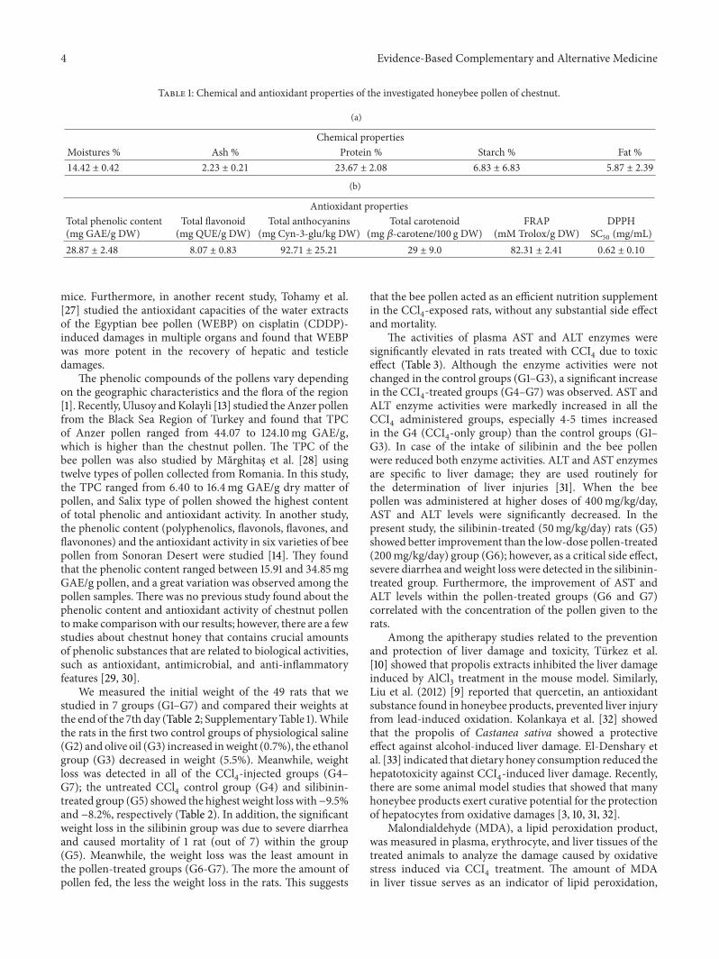
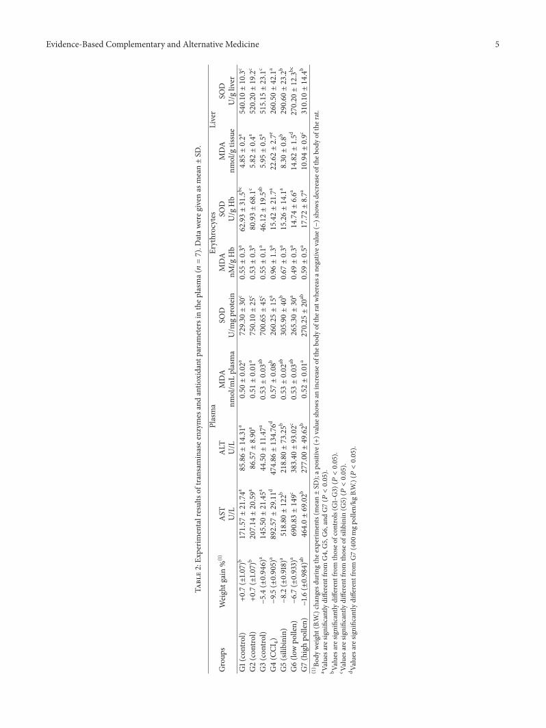