Embed Size (px)
Citation preview
Iranian Journal of Fisheries Sciences 20(2) 572-589 2021
DOI: 10.22092/ijfs.2021.123905
Research Article
Molecular identification and toxicity effects of cyanobacteria
species isolated from the Khoor-e-Khooran mangrove forest,
Persian Gulf
Zaheri A.1,2
; Bahador N.2*
; Yousefzadi M.3; Arman M.
4
Received: March 2020 Accepted: May 2020
Abstract
The increasing incidence of the harmful cyanobacterial blooms in mangrove ecosystem is a
potential threat for aquatic organisms and their consumers. In the present study, we have
evaluated the biodiversity of cyanobacteria and monitored their cyanotoxins. We isolated
120 bacterial isolates using BG11 medium from water and sediment samples collected from
10 stations throughout of the Khoor-e-Khooran mangrove forest at august 2018.
Biodiversity and distribution pattern based on morphological characteristics showed that 10
cyanobacterial genera were spread over the studied area. Phormidium, Oscillatoria,
Spirulina and Nostoc genera were dominated with frequency percentages of 25%, 20%,
10%, and 10% respectively. Analysis of 16S rRNA sequences showed that the strains have
high similarity with type strains in NCBI GenBank ranged from 98% to 100%.
Phylogenetic analysis proposed the non-indigenous origin of Microcystis strains because of
their phylogenetic divergence. We detected microcystin gene in Microcystis sp. strain KH
3, Microcystis sp. strain KH 4 and Microcystis sp. strain KH 11, while nodularin and
cylindrospermopsin gene were not detected in all isolated cyanobacteria. The extracted
metabolites from KH 3 and KH 4 strains showed cytotoxicity with LC50 of 139.3 and 225.8
µg/mL against Artemia salina respectively. Their LC50 were 231.3 and 211.2 µg/mL
against shrimp larvae respectively. They inhibited the proliferation of HUVEC cell lines
with IC50 of 11.13 and 13.29 µg/mL and HEPG2 with IC50 of 15.49 and 12.51 µg/mL,
respectively. Our results represented diversity and distribution pattern of cyanobacteria and
demonstrated the incidence of microcystin in the Khoor-e-Khooran mangrove forest.
Keywords: Marine cyanobacteria, Cyanotoxins, Mangrove forest, Persian Gulf,
Shrimp
1- Department of Microbiology, College of Science, Science and Research Branch, Islamic
Azad University, Fars, Iran
2- Department of Microbiology, College of Science, Agriculture and Modern Technology,
Shiraz Branch, Islamic Azad University, Shiraz, Iran
3- Department of Marine Biology, Faculty of Marine Sciences and Technology, University of
Hormozgan, Bandar Abbas, Iran.
4- Department of Biology, Payame Noor University, PO BOX 19395-3697, Tehran, Iran.
* Corresponding author's Email: [email protected]
[ D
OR
: 20.
1001
.1.1
5622
916.
2021
.20.
2.14
.5 ]
[
Dow
nloa
ded
from
jifr
o.ir
on
2022
-02-
10 ]
1 / 18
573 Zaheri et al., Molecular identification and toxicity effects of cyanobacteria species isolated from ...
Introduction
Cyanobacteria are photoautotrophic
oxygenic bacteria that have acquired
special characteristics during evolution
to adapt to biogeochemical and
ecological changes (Huisman et al.,
2018; Barzkar et al., 2021a). The
ecological flexibility of cyanobacteria
enables them to grow in diverse
ecosystems and environmental
conditions. However, their diversity
pattern could be influenced by
anthropogenic and climatic-induced
changes (Paerl, 2018). The increasing
occurrence of harmful cyanobacterial
blooms (HCBs) is a consequence of
these global environmental variations
(Visser et al., 2016). Increases in HCBs
occurrences in susceptible ecosystems
like mangrove habitats could lead to
catastrophic outcomes for their fauna,
flora and human health (Weber et al.,,
2020).
Mangrove forests are coastal
ecosystems that are located in some
tropical and subtropical areas (Gozari et
al., 2021). However, their plant
diversity is poor, but they support a
high diversity of animal species such as
a variety of birds, mammals,
amphibians, arthropods, fish and
shellfish species (Friess et al., 2019).
This habitat plays a significant role in
accumulation of carbon and nutrients,
recovery of marine biomass and also
functions as a nursery place for larval
growth of some animals such as
commercial fish and shellfish species
(Malik et al., 2017; Carrasquilla-Henao
et al., 2019; Pourmozaffar et al.,
2019b). Because of these natural
functions, this ecosystem have
ecological and economical importance
(Anneboina and Kumar, 2017). The
interactions of plants, animals and
microorganisms are necessary to
guaranty balanced food chains and
survival of the mangrove ecosystem
(Nelson et al., 2019; Pourmozaffar et
al., 2019a).
Mangrove-inhabiting cyanobacteria
tolerate to fluctuation of environmental
conditions such as temperature, salinity,
light intensity, osmotic pressure, ionic
concentration and weathering in this
ecosystem (Alvarenga et al., 2015).
Therefore, mangrove-inhabiting
cyanobacteria are considered a
biodiversity hotspot (Mariani et al.,
2015). However, the biodiversity of
mangrove-inhabiting cyanobacteria is
underestimated and a large number of
them has not properly characterized
(Barzkar et al., 2021b; Nabout et al.,
2013). This is mainly because of the use
of morphological features compared to
molecular methods, which provided
considerable accurate data for
identification. Understanding of
cyanobacterial diversity in mangrove
forests can facilitate monitoring
program and bloom modeling to control
harmful cyanobacterial blooms (Song
2017; Ralston and Moore 2020).
Several studies showed that mangrove-
inhabiting cyanobacteria dominantly
belong to Oscillatoria, Phormidium,
Spirulina, Lyngbya, Nostoc and
Chroococcus genera (Sakthivel and
Kathiresan 2013; Ram and Shamina
2017; Gaysina et al., 2019).
[ D
OR
: 20.
1001
.1.1
5622
916.
2021
.20.
2.14
.5 ]
[
Dow
nloa
ded
from
jifr
o.ir
on
2022
-02-
10 ]
2 / 18
Iranian Journal of Fisheries Sciences 20(2) 2021 574
The most challenging properties of
cyanobacteria are their potential to
produce toxic metabolites nominated as
cyanotoxins. Cyanotoxins categorized
into hepatotoxins, dermatotoxins and
neurotoxins (Meriluoto et al., 2017).
The deleterious effects of cyanotoxins
on aquatic organisms and their potential
for bioaccumulation and transferring
through the food web can endanger
biodiversity and aquatic consumer‟s
health (McQuaid and Lee, 2019).
Cyanotoxins have acute effects on
aquatic animals such as reduce their
survivorship, inhibit their feeding and
cause paralysis. The most important
chronic effects of cyanotoxins on
aquatic animals are reduction of growth
and fecundity, behavioral alterations
and biochemical alterations in activity
of important enzymes like
phosphatases, glutathione-S-
transferases, acetylcholinesterase and
proteases (Ferrão-Filho and
Kozlowsky-Suzuki, 2011). For
instance, microcystins, which classified
in hepatotoxins group, produce by
Microcystis species. This genus is a
bloom-forming cyanobacterium in
estuarine and mangrove ecosystems and
responsible for intoxication of aquatic
organisms and their consumers (Pham
and Utsumi, 2018). Concludingly,
monitoring of cyanotoxins especially in
mangrove ecosystems is essential for
better understanding and predicting the
emergence of HCBs and to ensure
public safety (Jaramillo and O'Shea
2019; Roy-Lachapelle et al., 2019).
Detection of toxic genes and toxicity
assays are robust approaches to monitor
cyanotoxins.
Khoor-e-Khooran mangrove forests
is a natural protected area that located
near the Bandar-e-Khamir city on the
northern coast of the Persian Gulf
(Milani, 2018). This habitat harbors
high density of mangrove plants and
provided a focal point for the
propagation of fish and shellfish species
in the region (Tamadoni Jahromi et al.,
2021a and b). The aims of the present
study were to monitor the toxigenic
cyanobacteria in the Khoor-e-Khooran
mangrove forests and to investigate
cyanobacterial diversity in this natural
protected area.
Materials and methods
Sample collection
Sampling was conducted in August
2018 in the Khoor-e-Khooran
mangrove wetland located on the
northern coast of the Persian Gulf,
Bandar-e-Khamir, Iran. Water samples
were collected from 10 selected stations
from 30 cm below the surface in a 250
mL dark glass bottle. The sediment
samples were collected from same
stations by a grab sampler (Gozari et
al., 2019c). Sampling process was done
in triplicate and all samples were taken
to the laboratory immediately within 3
hours (Authority GBRMP , 2019).
Isolation of cyanobacteria
One hundred µL of each sample was
inoculated on the BG 11 medium,
which was prepared with natural
seawater. The inoculated media were
incubated at 28˚C under 12 hours light
[ D
OR
: 20.
1001
.1.1
5622
916.
2021
.20.
2.14
.5 ]
[
Dow
nloa
ded
from
jifr
o.ir
on
2022
-02-
10 ]
3 / 18
575 Zaheri et al., Molecular identification and toxicity effects of cyanobacteria species isolated from ...
and 12 hours darkness with light
intensity 1500-2000 lux. After 2 weeks,
the appeared colonies were investigated
for belonging to cyanobacteria. Distinct
colonies were selected according to
morphological characteristic and were
purified by subculturing on to BG11
medium (Ferris and Hirsch, 1991).
Characterization of cyanobacteria
The purified isolates were identified
according to morphological
characteristics based on the
identification key in Standard Methods
for Examination of Water and
Wastewater and WHO guidelines
(Lawton et al., 1999; Carranzo 2012)
.The micromorphological characters
were observed with a light microscope
(Nikon, Japan) with 40X magnification.
Detection of cyanotoxins genes
For genomic DNA extraction the
purified isolates were inoculated in BG
11 medium and incubated in the
appropriate condition, which was
previously mentioned. The sufficient
biomass (50-100 mg) of the isolates
were harvested and DNA extraction
was carried out by DNGTM
– Plus kit
(Iranian Gene Fanavar Company). After
DNA extraction, the cyanotoxins
biomarkers, including microcystin,
nodularine and cylindrospermopsin
were detected using the
Microcystin/nodularine gene PCR
detection and Cylindrospermopsin gene
PCR detection kits (Iranian Gene
Fanavar Company) respectively
according to company instruction. The
results of experiments were detected by
agarose gel electrophoresis (Emtyazjoo
et al., 2019)
Extraction of cyanotoxins
After the cultivation period, cells were
harvested by centrifugation. Harvested
cells were briefly washed with distilled
water to remove salts. The
cyanobacterial extracts were obtained
by homogenization using methanol:
chloroform (1:1 v/v) in the ratio of 1:10
(biomass: solvent) and kept at 25 C for
24 h in a photo-incubator. Solutions
were centrifuged at 10 000 rpm for 10
min, the supernatant recovered, and
subsequently dried and stored at -20 C
(Nazemi et al., 2017). A total of 50 mg
of the cyanobacterial crude extracts was
dissolved in 1mL of dimethyl sulfoxide
(DMSO) for bioassays.
(Maruthanayagam et al., 2013).
Brine shrimp cytotoxicity assay
Toxicity of extracted metabolites from
cyanobacterial isolates was assayed by
Brine-shrimp microwell cytotoxicity
method. One gram of the commercially
available (INVETM
) of brine shrimp
cysts Artemia salina cultivated in 3% of
saline water at 22-29 C under white
light for 48h hours. For cytotoxicity
assay, 100 µL nauplii suspension
containing 15 nauplii per 100 µL was
transferred to 100 µL cyanobacterial
extract (125, 250,500 and 1000 µL/mL)
in a 96-well microplate and incubated at
25 C for 24 hours (Gozari et al., 2018;
Gozari et al., 2019a). The percent of
mortality was recorded. The cytotoxic
activity was calculated by the following
formula and presented as LC50.
[ D
OR
: 20.
1001
.1.1
5622
916.
2021
.20.
2.14
.5 ]
[
Dow
nloa
ded
from
jifr
o.ir
on
2022
-02-
10 ]
4 / 18
Iranian Journal of Fisheries Sciences 20(2) 2021 576
Toxicity = (Ncontrol - Ntest/Ncontrol) ×100%
Ncontrol is the number of live nauplii in
untreated well and Ntest is the number of
live nauplii in treated well.
Toxicity against shrimp larvae
Toxicity of extracted metabolites
against Penaeus vanammei larvae was
investigated and reported as LC50 value
(Gozari et al., 2016). The LC50 value is
a concentration of an extract that could
kill 50% of the whole treated larvae.
The shrimp larvae at post larval stage 9
were provided from the hatchery center
of the Persian Gulf and Oman Sea
Ecological Research Institute. Then 10
larvae were transferred to 250 mL
Erlenmeyer flask containing 200 mL
seawater. The extracted metabolites
were added to each flask at final
concentrations 1000, 500, 250, 125
μg/mL. The mortality rates were
recorded up to 6 days and LC50 value
were calculated by following equation:
Toxicity = (Ncontrol - Ntest/Ncontrol)
×100%
Ncontrol is the number of live larvae in
untreated flask and Ntest is the number
of live larvae in treated flask.
Cytotoxicity against human cell lines
The cytotoxicity of cyanobacterial
extracted metabolites was evaluated in
HUVECs (human umbilical vein
endothelial cells) and HEPG2
(hepatocellular carcinoma) cell lines by
MTT cell proliferation assay. The One
hundred microliters of the HUVECs or
HEPG2 cell suspensions in DMEM or
RPMI media were transferred in 96-
well Microplates. The cell density was
subjected at 104 cells per well. After
incubation at 37°C for 24 hours in CO2
incubator, cell lines were treated with
100 μL of each extract at certain final
concentrations (100, 50, 25, 12.5
μg/mL). After 36 hours additional
incubation, 50 μL of the prepared MTT
solution (5 mg/mL) was injected in to
each well and kept in incubator for 4
hours. The MTT solution was removed
and 100 μL of DMSO/ethanol solution
(4:1) was added to each well. Then the
96- well plate was kept in a shaker to
dissolve the formazan dye. Finally, the
absorbance of each well was recorded
at wavelength of 550 nm by microplate
reader (Gozari et al., 2019b). The
survival rate of the cell lines was
calculated by following formula:
Cell viability (%) = [(ODt e s t) − (ODBlank) / (ODcontrol) − (ODBlank)] × 100
Molecular Identification of
cyanobacteria
After the genomic DNA extraction that
was previously described, the specific
genes were detected by Cyanobacteria
Specific PCR Detection Kit (Iranian
Gene Fanavar Company). The PCR
reaction was performed according to
company„s instruction. Amplification
and visualization of the 487 bp
fragment on agarose gel confirmed the
cyanobacterial identity. For
[ D
OR
: 20.
1001
.1.1
5622
916.
2021
.20.
2.14
.5 ]
[
Dow
nloa
ded
from
jifr
o.ir
on
2022
-02-
10 ]
5 / 18
577 Zaheri et al., Molecular identification and toxicity effects of cyanobacteria species isolated from ...
phylogenetic analysis, we amplified the
16S rRNA gene using PCR reaction by
universal primers including 9F and
1541R as described by Heuer (1997).
The PCR products were purified with
the Roche PCR Purification kit (Roche
Applied Science) and sequenced by
Macrogen Company (Seoul, Korea).
The 16S rRNA gene sequences were
analyzed by BLAST program at NCBI
(National Centre for Biotechnology
Information) (Madden, 2013) and
submitted to the GenBank database
with following accession numbers:
MN864652, MN864653, MN864654,
MN864655, MN864656, MN864657,
MN864658, MN864659, MN837908,
MN837909. Phylogenetic tree was
constructed based on neighbor joining
algorithm (Saitou and Nei, 1987) using
MEGA X program (Kumar et al.,
2018).
Statistical analysis
All of the experiments were performed
in triplicates. The statistical significance
of the data was analyzed with one-way
ANOVA followed by LSD using SPSS
program (Version 24) and the
significance level was set at p<0.05.
The results of biodiversity of
cyanobacteria were reported as
percentage. The cytotoxicity results
expressed as mean LC50±standard error
(SE). The LC50 values of the extracts
were calculated using the linear
regression between the final
concentration of the extracts and
respective cytotoxicity percent
calculated from the previously
mentioned equation by the software
Graph Pad PRISM version 6 (Graph
Pad Software, San Diego, CA). The
statistical significance of resultant tree
topology was evaluated by bootstrap
analysis (Felsenstein, 1985) based on
1000 replicates.
Results
Isolation and Characterization of
Cyanobacteria
One hundred isolates of cyanobacteria
were obtained from the collected
samples. Morphological properties
showed that the isolated strains
belonged to 10 different genera of
cyanobacteria. Phormidium,
Oscillatoria, Spirulina and Nostoc
genera were dominated in collecting
samples with 25%, 20%, 10%, and 10%
respectively (Fig. 1). Molecular
identification of 24 distinct isolates with
specific primers confirmed that 10
strains belonged to cyanobacteria (Fig.
2).
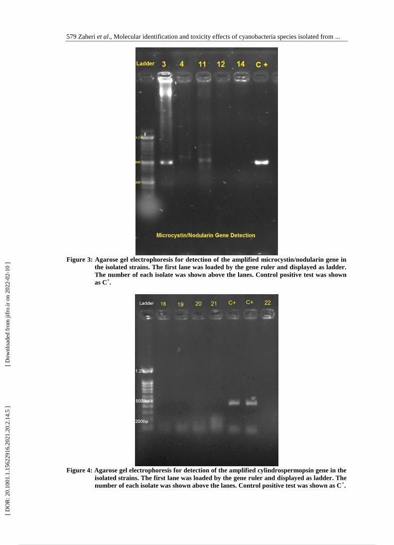
Detection of cyanotoxins genes
Screening of microcystin, nodularin and
cylindrospermopsin genes showed that
microcystin/nodularin genes were
existed in genomic DNA of the 3 out of
10 isolated strains (Fig. 3). While
cylindrospermopsin genes were absent
in all strains (Fig. 4).
[ D
OR
: 20.
1001
.1.1
5622
916.
2021
.20.
2.14
.5 ]
[
Dow
nloa
ded
from
jifr
o.ir
on
2022
-02-
10 ]
6 / 18
Iranian Journal of Fisheries Sciences 20(2) 2021 578
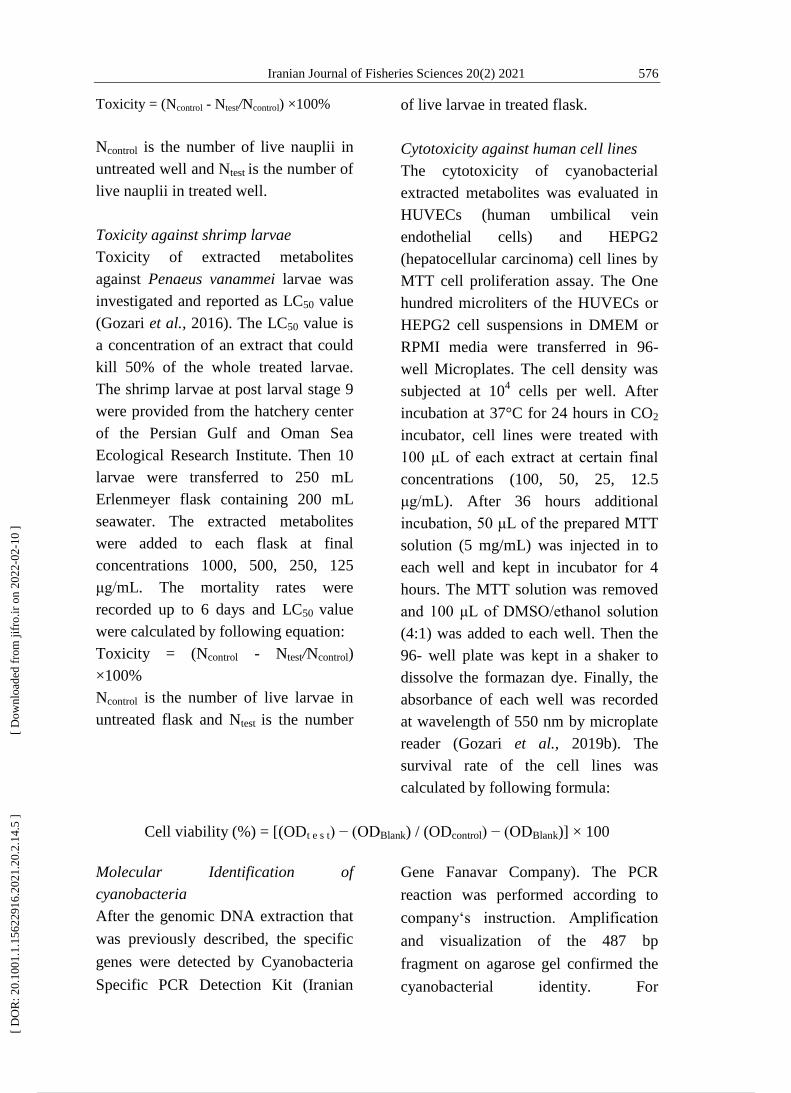
Figure 1: Biodiversity pattern of cyanobacteria in collected samples from the Khoor-e-Khooran
wetland. The frequency of each genus was represented by different colors. Some genera
were absent in some stations.
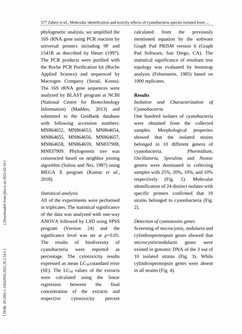
Figure 2: Agarose gel electrophoresis of the amplified cyanobacterial specific genes in the isolated
strains. The first lane in each series was loaded by the gene ruler and displayed as
ladder. The number of each isolate was shown above the lanes.
[ D
OR
: 20.
1001
.1.1
5622
916.
2021
.20.
2.14
.5 ]
[
Dow
nloa
ded
from
jifr
o.ir
on
2022
-02-
10 ]
7 / 18
579 Zaheri et al., Molecular identification and toxicity effects of cyanobacteria species isolated from ...
Figure 3: Agarose gel electrophoresis for detection of the amplified microcystin/nodularin gene in
the isolated strains. The first lane was loaded by the gene ruler and displayed as ladder.
The number of each isolate was shown above the lanes. Control positive test was shown
as C+.
Figure 4: Agarose gel electrophoresis for detection of the amplified cylindrospermopsin gene in the
isolated strains. The first lane was loaded by the gene ruler and displayed as ladder. The
number of each isolate was shown above the lanes. Control positive test was shown as C+.
[ D
OR
: 20.
1001
.1.1
5622
916.
2021
.20.
2.14
.5 ]
[
Dow
nloa
ded
from
jifr
o.ir
on
2022
-02-
10 ]
8 / 18
Iranian Journal of Fisheries Sciences 20(2) 2021 580
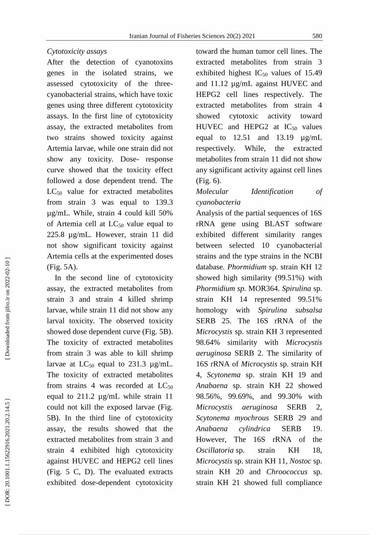
Cytotoxicity assays
After the detection of cyanotoxins
genes in the isolated strains, we
assessed cytotoxicity of the three-
cyanobacterial strains, which have toxic
genes using three different cytotoxicity
assays. In the first line of cytotoxicity
assay, the extracted metabolites from
two strains showed toxicity against
Artemia larvae, while one strain did not
show any toxicity. Dose- response
curve showed that the toxicity effect
followed a dose dependent trend. The
LC50 value for extracted metabolites
from strain 3 was equal to 139.3
µg/mL. While, strain 4 could kill 50%
of Artemia cell at LC50 value equal to
225.8 µg/mL. However, strain 11 did
not show significant toxicity against
Artemia cells at the experimented doses
(Fig. 5A).
In the second line of cytotoxicity
assay, the extracted metabolites from
strain 3 and strain 4 killed shrimp
larvae, while strain 11 did not show any
larval toxicity. The observed toxicity
showed dose dependent curve (Fig. 5B).
The toxicity of extracted metabolites
from strain 3 was able to kill shrimp
larvae at LC50 equal to 231.3 µg/mL.
The toxicity of extracted metabolites
from strains 4 was recorded at LC50
equal to 211.2 µg/mL while strain 11
could not kill the exposed larvae (Fig.
5B). In the third line of cytotoxicity
assay, the results showed that the
extracted metabolites from strain 3 and
strain 4 exhibited high cytotoxicity
against HUVEC and HEPG2 cell lines
(Fig. 5 C, D). The evaluated extracts
exhibited dose-dependent cytotoxicity
toward the human tumor cell lines. The
extracted metabolites from strain 3
exhibited highest IC50 values of 15.49
and 11.12 µg/mL against HUVEC and
HEPG2 cell lines respectively. The
extracted metabolites from strain 4
showed cytotoxic activity toward
HUVEC and HEPG2 at IC50 values
equal to 12.51 and 13.19 µg/mL
respectively. While, the extracted
metabolites from strain 11 did not show
any significant activity against cell lines
(Fig. 6).
Molecular Identification of
cyanobacteria
Analysis of the partial sequences of 16S
rRNA gene using BLAST software
exhibited different similarity ranges
between selected 10 cyanobacterial
strains and the type strains in the NCBI
database. Phormidium sp. strain KH 12
showed high similarity (99.51%) with
Phormidium sp. MOR364. Spirulina sp.
strain KH 14 represented 99.51%
homology with Spirulina subsalsa
SERB 25. The 16S rRNA of the
Microcystis sp. strain KH 3 represented
98.64% similarity with Microcystis
aeruginosa SERB 2. The similarity of
16S rRNA of Microcystis sp. strain KH
4, Scytonema sp. strain KH 19 and
Anabaena sp. strain KH 22 showed
98.56%, 99.69%, and 99.30% with
Microcystis aeruginosa SERB 2,
Scytonema myochrous SERB 29 and
Anabaena cylindrica SERB 19.
However, The 16S rRNA of the
Oscillatoria sp. strain KH 18,
Microcystis sp. strain KH 11, Nostoc sp.
strain KH 20 and Chroococcus sp.
strain KH 21 showed full compliance
[ D
OR
: 20.
1001
.1.1
5622
916.
2021
.20.
2.14
.5 ]
[
Dow
nloa
ded
from
jifr
o.ir
on
2022
-02-
10 ]
9 / 18
581 Zaheri et al., Molecular identification and toxicity effects of cyanobacteria species isolated from ...
with Oscillatoria acuminata PCC
6304, Microcystis aeruginosa SERB 2,
Nostoc sp. USMNA and Chroococcus
turgidus SERB 26, respectively.
Figure 5: Cytotoxicity of extracted metabolites from the isolated cyanobacteria. A. cytotoxicity
against Artemia salina, B. against Penaeus vanammei Larvae, C. against HepG2 cell line, D.
against HUVEC cell line. 3: Microcystis sp. strain KH 3, 4: Microcystis sp. strain KH4, 11:
Microcystis sp. strain KH 11.
Figure 6: Cytotoxic activity of extracting metabolites against human cell lines. A. HUVEC cells in
control well. B. HUVEC cells after treatment with cyanobacterial extract. C. HEPG2 cells
in control well. D. HEPG2 cells after treatment with cyanobacterial extract.
[ D
OR
: 20.
1001
.1.1
5622
916.
2021
.20.
2.14
.5 ]
[
Dow
nloa
ded
from
jifr
o.ir
on
2022
-02-
10 ]
10 / 18
Iranian Journal of Fisheries Sciences 20(2) 2021 582
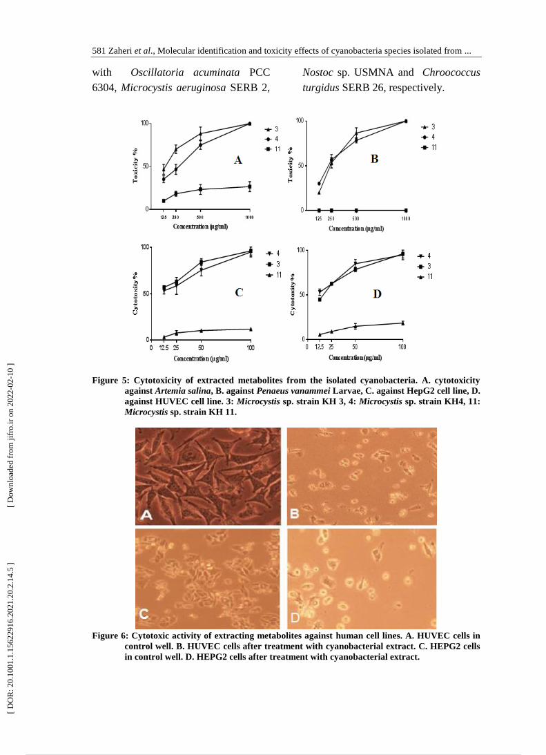
Interpretation of the constructed
phylogenetic tree based on 16S rRNA
gene demonstrated that the
cyanobacterial strains positioned in two
different clades. Microcystis strains
were located together into the same
cluster although they were merged in
subclades. Phormidium and
Oscillatoria were located in a same
clade while Chroococcus and Spirulina
strains located in other clade but they
have been derived from a common
ancestor (Fig. 7). Other strains
developed different lineages in a same
clade. All isolated strains located with
their closest type strains in a same
cluster.
Figure 7: Phylogenetic tree based on 16S rRNA gene sequence analysis, reconstructed from
evolutionary distances by using the neighbor-joining method, showing the phylogenetic
position of the isolated cyanobacterial strains (Bold font) and type strain in NCBI
GenBank. Bootstrap values are indicated at the relevant branching points. Numbers of
branch node are bootstrap values based on 1000 resampling. Bar 0.02 substitutions per
nucleotide position. Streptomyces sp. was used as an out-group.
Discussion
Monitoring of toxicogenic
cyanobacteria in mangrove ecosystems
is a necessary requirement because of
the increasing occurrence of harmful
cyanobacterial blooms due to climate
change (Ramos et al., 2017). We
isolated 120 presumptive cyanobacterial
isolates from water and sediment
samples throughout the mangrove
ecosystem in Khoor-e-Khooran
wetland. The biodiversity pattern
showed that Phormidium, Spirulina,
Oscillatoria genera were dominated in
all sample stations. Other studies also
reported the dominance of these genera
in mangrove ecosystems. For example,
17 species of cyanobacteria, which
[ D
OR
: 20.
1001
.1.1
5622
916.
2021
.20.
2.14
.5 ]
[
Dow
nloa
ded
from
jifr
o.ir
on
2022
-02-
10 ]
11 / 18
583 Zaheri et al., Molecular identification and toxicity effects of cyanobacteria species isolated from ...
dominantly belonged to Oscillatoria
were isolated from Cardoso Island,
Brazil (Branco et al., 1994). In another
study, Ram and colleagues (2017)
showed that Oscillatoria salina,
Oscillatoria ornata and Oscillatoria
vizagapatensis dominated in seven
mangrove ecosystems in the India.
Mohamed and Al-Shehri (2015) , also
identified 15 genera and 5 families of
cyanobacteria from microbial mats of 3
mangrove ecosystems in the Red Sea.
The isolated cyanobacteria dominantly
belonged to Oscillatoria and
Phormidium genera.
Several possible factors could
determine the diversity pattern of
cyanobacteria in the Khoor-e-Khooran
wetland. In mangrove areas at Sao
Paulo state, south-east Brazil, the first
factor is the fluctuation of
physicochemical conditions, including
temperature, salinity, oxygen and light
intensity (Alvarenga et al., 2015). The
second influencing factor is the urban
and industrial wastewaters that flow up
to the mangrove areas at south east
coast of India (Balasubramanian et al.,
2011). The ballast water of commercial
cargo ships, which contained non-
indigenous species might be another
determining factor. Interestingly, an
international sea route is passing near
the Khoor-e-Khooran wetland.
Molecular identification of distinct
isolates confirmed the morphological
characterization of eight isolates, while
the identities of two isolates were not
confirmed. These results revealed that
14 isolates that we previously identified
as cyanobacteria based on
morphological properties did not belong
to them. Inconsistency between
morphological and molecular
identification mainly because of their
basic principles were reported in other
studies. For instance the isolated
cyanobacteria from the Sao Paulo were
characterized as Nostoc and
Leptolyngbya genera based on
morphological properties, whereas
molecular identification showed that
these isolates were new taxa (Silva et
al., 2014). Comparison of 16S rRNA
sequences of the cyanobacterial strains
with closest strains in NCBI GenBank
showed different homology ranged
from 98 to 100 %. Mohamed and Al-
Shehri (2015) also reported the high
similarity of isolated cyanobacteria with
their closest strain ranged from 67.6 to
97.7% (Mohamed and Al-Shehri,
2015). The phylogenetic analysis
showed that Microcystis strains located
in a different cluster and
formed a different lineage during
evolution. The Microcystis strains
located in a same cluster with
Microcystis aeruginosa SERB 2 that
was isolated from the Andaman Island,
India. Consequently, it could be
hypothesized that the toxigenic
Microcystis strains might be originated
from non-indigenous species.
Detection of toxic genes showed that
the microcystin/nodularine genes were
existed in isolated cyanobacteria while
cylindrospermopsin gene was not
detected. Cyanotoxins genes also were
investigated in other studies. For
example Zare et al. (2015) detected the
saxitoxin genes (sxtAF, sxtAR) in
[ D
OR
: 20.
1001
.1.1
5622
916.
2021
.20.
2.14
.5 ]
[
Dow
nloa
ded
from
jifr
o.ir
on
2022
-02-
10 ]
12 / 18
Iranian Journal of Fisheries Sciences 20(2) 2021 584
isolated cyanobacteria from the Kor
river. They proposed that molecular
detection of toxins has been presented
an alternative method for chemical
analysis. Among the isolated strains,
Microcystis sp. strain KH3 and
Microcystis sp. strain KH4 exhibited
high cytotoxic activity against Artemia
salina, shrimp larvae and human cell
lines while Microcystis sp. strain KH11
did not show any significant toxicity.
The cytotoxicity value of the extracted
metabolite from Microcystis sp. strain
KH3 was recorded as 139.3, 231.3,
11.13 and 15.49 µg/mL against
Artemia, shrimp larvae, HEPG2 and
HUVEC cell lines respectively. These
values were 225.8, 211.2, 13.19 and
12.51 µg/mL for the extracted
metabolites from Microcystis sp. strain
KH4. Mohamed and Al-Shehri (2015),
reported that the extracted cyanotoxins
from 9 cyanobacterial strains showed
cytotoxicity against Artemia salina
ranged from 0.3 to 5.1 mg/mL.
Maruthanayagam et al. (2013) showed
that the extracted cyanotoxins from
Geitlerinema sp. CNP 1019 exhibited
cytotoxic activity against MCF7 cell
line with GI50 value equal to 25.7
µg/mL. Introduction of microcystin into
food chains well studied in marine
ecosystems. Studies showed that
accumulation of microcystin in
phytoplanktivorous fishes is 3.5 times
more than zooplanktivorous fish. In
addition, accumulation of microcystin
in liver of Osmerus eperlanus was
recorded as 874 µg/mL (Ibelings et al.,
2005). Filter-feeder coelenterates
especially eatable ones such as oysters
can also accumulate cyanotoxins and
consequently be considered as potential
danger to the consumer‟s health
(Ferrão-Filho and Kozlowsky-Suzuki,
2011; Tamadoni Jahromi et al., 2020).
Fish cage-cultured farms also can be
influenced from the cyanobacterial
blooms in the Khoor-e-Khooran
wetland. In conclusion,
the existence of microcystins in Khoor-
e-Khooran wetland, which is a major
hatchery for shrimp‟s populations in the
region, requires more monitoring
efforts. These results also represented a
new understanding of cyanobacterial
diversity in the Khoor-e-Khooran
mangrove wetland.
Acknowledgments
This research article is a part of the PhD
thesis entitled “Evaluation of biological
diversity pattern, genetic detection and
cytotoxic activity of cyanotoxins from
cyanobacteria of Bandar-e-Khamir„s
mangrove forests”. The authors are
thankful to Dr. Mohsen Gozari at the
Persian Gulf and Oman Sea Ecology
Research Center for his assistance in
this project.
References
Alvarenga, D.O., Rigonato, J.,
Branco, L.H.Z. and Fiore, M.F.,
2015. Cyanobacteria in mangrove
ecosystems. Biodiversity and
Conservation, 24, 799-817. DOI:
10.1007/s10531-015-0871-2
Anneboina, L.R. and Kumar, K.K.,
2017. Economic analysis of
mangrove and marine fishery
linkages in India. Ecosystem
[ D
OR
: 20.
1001
.1.1
5622
916.
2021
.20.
2.14
.5 ]
[
Dow
nloa
ded
from
jifr
o.ir
on
2022
-02-
10 ]
13 / 18
585 Zaheri et al., Molecular identification and toxicity effects of cyanobacteria species isolated from ...
Services, 24, 114-123. DOI:
10.1016/j.ecoser.2017.02.004
Authority GBRMP., 2019. Marine
Monitoring Program: Quality
assurance and quality control manual
2017-18., Great Barrier Reef Marine
Park Authority. pp. 10-27.
Balasubramanian, L., Subramanian,
G., Nazeer, T.T., Simpson, H.S.,
Rahuman, S.T. and Raju, P., 2011.
Cyanobacteria cultivation in
industrial wastewaters and biodiesel
production from their biomass: A
review. Biotechnology and Applied
Biochemistry, 58, 220-225. DOI:
doi.org/10.1002/bab.31
Barzkar, N., Khan, Z., Jahromi, S.T.,
Poormozaffar, S., Gozari, M. and
Nahavandi, R., 2021a. A critical
review on marine serine protease and
its inhibitors: A new wave of drugs?
International Journal of Biological
Macromolecules, 170, 674-687.
DOI:10.1016/j.ijbiomac.2020.12.134
Barzkar, N., Sohail, M., Jahromi,
S.T., Gozari, M., Poormozaffar, S.,
Nahavandi, R. and Hafezieh, M.,
2021b. Marine Bacterial Esterases:
Emerging Biocatalysts for Industrial
Applications. Applied Biochemistry
and Biotechnology, 1-28. DOI:
10.1007/s12010-020-03483-8
Branco, L.H.Z., Silva, S.M.F. and
Santanna, C.L., 1994. Stichosiphon
mangle sp. nova, a new cyanophyte
from mangrove environments.
Archiv fur Hydrobiologie, 10, 1-7.
DOI: ESP142010200001
Carranzo IV Standard Methods for
examination of water and
wastewater. In: Anales De
Hidrología Médica, 2012.
Universidad Complutense de
Madrid, 5, 100-185. DOI: 978-
087553-013-0
Carrasquilla-Henao, M., Ban, N.,
Rueda, M. and Juanes, F., 2019.
The mangrove-fishery relationship:
A local ecological knowledge
perspective. Marine Policy, 108,
103656. DOI:
10.1016/j.marpol.2019.103656
Emtyazjoo, M., Abbaszadeh, A. and
Hassan, M., 2019. Diagnosis
toxogenic Saxitoxin Cyanobacteria
by molecular method in Amir
Kalayeh Lagoon–Iran. Research in
Marine Sciences, 4, 528 – 538.
Felsenstein, J., 1985. Confidence limits
on phylogenies: an approach using
the bootstrap. Evolution, 39, 783-
791. DOI: 10.1111/j.1558-
5646.1985.tb00420.x
Ferrão-Filho, A.D.S. and Kozlowsky-
Suzuki, B., 2011. Cyanotoxins:
bioaccumulation and effects on
aquatic animals. Marine drugs, 9,
2729-2772. DOI:
10.3390/md9122729
Ferris, M.J. and Hirsch, C., 1991.
Method for isolation and purification
of cyanobacteria. Appl Environ
Microbiol, 57, 1448-1452. DOI:
16348486
Friess, D.A., Rogers, K., Lovelock,
C.E., Krauss, K.W., Hamilton,
S.E., Lee, S.Y., Lucas, R.,
Primavera, J., Rajkaran, A. and
Shi, S., 2019. The State of the
World's Mangrove Forests: Past,
Present, and Future. Annual Review
of Environment and Resources, 44,
[ D
OR
: 20.
1001
.1.1
5622
916.
2021
.20.
2.14
.5 ]
[
Dow
nloa
ded
from
jifr
o.ir
on
2022
-02-
10 ]
14 / 18
Iranian Journal of Fisheries Sciences 20(2) 2021 586
89-115. DOI: 10.1146/annurev-
environ-101718-033302
Gaysina, L.A., Saraf, A. and Singh,
P., 2019. Cyanobacteria in Diverse
Habitats. In: Cyanobacteria.
Elsevier, pp. 1-28.
Gozari, M., Mortazavi, M., Bahador,
N. and Rabbaniha, M., 2016.
Isolation and screening of
antibacterial and enzyme producing
marine actinobacteria to approach
probiotics against some pathogenic
vibrios in shrimp Litopenaeus
vannamei. Iranian Journal of
Fisheries Sciences, 15, 630-644.
Gozari, M., Bahador, N., Jassbi, A.R.,
Mortazavi, M. and Eftekhar, E.,
2018. Antioxidant and cytotoxic
activities of metabolites produced by
a new marine Streptomyces sp.
isolated from the sea cucumber
Holothuria leucospilota. Iranian
Journal of Fisheries Sciences, 17,
413-426. DOI:
10.22092/IJFS.2018.116076
Gozari, M., Bahador, N., Jassbi, A.R.,
Mortazavi, M.S., Hamzehei, S. and
Eftekhar, E., 2019a. Isolation,
distribution and evaluation of
cytotoxic and antioxidant activity of
cultivable actinobacteria from the
Oman Sea sediments. Acta
Oceanologica Sinica, 38, 84-90.
DOI: 10.1007/s13131-019-1515-2
Gozari, M., Bahador, N., Mortazavi,
M.S., Eftekhar, E. and Jassbi,
A.R., 2019b. An “olivomycin A”
derivative from a sponge-associated
Streptomyces sp. strain SP 85. 3
Biotech, 9, 439-451. DOI:
10.1007/s13131-019-1515-2
Gozari, M., Zaheri, A., Jahromi, S.T.,
Gozari, M. and Karimzadeh, R.,
2019c. Screening and
characterization of marine
actinomycetes from the northern
Oman Sea sediments for cytotoxic
and antimicrobial activity.
International Microbiology, 22, 521-
530. DOI: 10.1007/s10123-019-
00083-3
Gozari, M., Alborz, M., El-Seedi,
H.R. and Jassbi, A.R., 2021.
Chemistry, Biosynthesis and
Biological Activity of Terpenoids
and Meroterpenoids in Bacteria and
Fungi Isolated from Different
Marine Habitats. European Journal
of Medicinal Chemistry, 210,
112957. DOI:
doi.org/10.1016/j.ejmech.2020.1129
57
Heuer, H., Krsek, M., Baker, P.,
Smalla, K. and Wellington, E.,
1997. Analysis of actinomycete
communities by specific
amplification of genes encoding 16S
rRNA and gel-electrophoretic
separation in denaturing gradients.
Appl Environ Microbiol, 63, 3233-
3241. PMID: 9251210
Huisman, J., Codd, G.A., Paerl,
H.W., Ibelings, B.W., Verspagen,
J.M. and Visser, P.M., 2018.
Cyanobacterial blooms. Nature
Reviews Microbiology, 16, 471-483.
DOI: 10.1038/s41579-018-0040-1
Ibelings, B.W., Bruning, K., De
Jonge, J., Wolfstein, K., Pires,
L.D., Postma, J. and Burger, T.,
2005. Distribution of microcystins in
a lake foodweb: no evidence for
[ D
OR
: 20.
1001
.1.1
5622
916.
2021
.20.
2.14
.5 ]
[
Dow
nloa
ded
from
jifr
o.ir
on
2022
-02-
10 ]
15 / 18
587 Zaheri et al., Molecular identification and toxicity effects of cyanobacteria species isolated from ...
biomagnification. Microbial
Ecology, 49, 487-500. DOI:
10.1007/s00248-004-0014-x
Jaramillo, M. and O'Shea, K.E.,
2019. Analytical methods for
assessment of cyanotoxin
contamination in drinking water
sources. Current Opinion in
Environmental Science and Health,
7, 45-51. DOI:
10.1016/j.coesh.2018.10.003
Kumar, S., Stecher, G., Li, M.,
Knyaz, C. and Tamura, K., 2018.
MEGA X: molecular evolutionary
genetics analysis across computing
platforms. Molecular biology and
evolution, 35, 1547-1549. DOI:
10.1093/molbev/msy096
Lawton, L., Marsalek, B., Padisák, J.
and Chorus, I., 1999. Determination
of cyanobacteria in the laboratory.
Toxic cyanobacteria in water: A
guide to their public health
consequences, monitoring and
management. World Health
Organization. pp. 1-28.
Madden, T., 2013. The BLAST
sequence analysis tool. The NCBI
Handbook [Internet]. Bethesda, MD:
National Library of Medicine (US),
National Center for Biotechnology
Information.
Malik, A., Mertz, O. and Fensholt,
R., 2017. Mangrove forest decline:
consequences for livelihoods and
environment in South Sulawesi.
Regional Environmental Change, 17,
157-169. DOI: 10.1007/s10113-016-
0989-0
Mariani, M.A., Padedda, B.M.,
Kaštovský, J., Buscarinu, P.,
Sechi, N., Virdis, T. and Lugliè, A.,
2015. Effects of trophic status on
microcystin production and the
dominance of cyanobacteria in the
phytoplankton assemblage of
Mediterranean reservoirs. Scientific
Reports, 5, 1-16. DOI:
10.1038/srep17964
Maruthanayagam, V., Nagarajan, M.
and Sundararaman, M., 2013.
Cytotoxicity assessment of
cultivable marine cyanobacterial
extracts in Artemia salina (brine
shrimp) larvae and cancer cell lines.
Toxin Reviews, 32, 1-9. DOI:
10.3109/15569543.2012.754772
McQuaid, M. and Lee, A., 2019. The
Bioaccumulation of Cyanotoxins in
Aquatic Food Webs.
Meriluoto, J., Spoof, L. and Codd,
G.A., 2017. Handbook of
cyanobacterial monitoring and
cyanotoxin analysis. John Wiley and
Sons. pp. 55-87.
Milani, A.S., 2018. Mangrove Forests
of the Persian Gulf and the Gulf of
Oman. In: Threats to Mangrove
Forests. Springer, pp. 53-75.
Mohamed, Z.A. and Al-Shehri, A.M.,
2015. Biodiversity and toxin
production of cyanobacteria in
mangrove swamps in the Red Sea off
the southern coast of Saudi Arabia.
Botanica Marina, 58, 23-34. DOI:
10.1515/bot-2014-0055
Nabout, J.C., da Silva Rocha, B.,
Carneiro, F.M. and Sant’Anna,
C.L., 2013. How many species of
Cyanobacteria are there? Using a
discovery curve to predict the
species number. Biodiversity and
[ D
OR
: 20.
1001
.1.1
5622
916.
2021
.20.
2.14
.5 ]
[
Dow
nloa
ded
from
jifr
o.ir
on
2022
-02-
10 ]
16 / 18
Iranian Journal of Fisheries Sciences 20(2) 2021 588
Conservation, 22, 2907-2918. DOI:
10.1007/s10531-013-0561-x
Nazemi, M., Moradi, Y., Rezvani,
Gilkolai, F., Ahmaditaba, M.,
Gozari, M. and Salari, Z., 2017.
Antimicrobial activities of semi
polar-nonpolar and polar secondary
metabolites of sponge Dysidea
pallescens from Hengam Island,
Persian Gulf. Iranian Journal of
Fisheries Sciences, 16, 200-209.
Nelson, J.A., Lesser, J., James, W.R.,
Behringer, D.P., Furka, V. and
Doerr, J.C., 2019. Food web
response to foundation species
change in a coastal ecosystem. Food
Webs, 21, e00125.
Paerl, H.W., 2018. Mitigating toxic
planktonic cyanobacterial blooms in
aquatic ecosystems facing increasing
anthropogenic and climatic
pressures. Toxins, 10, 1-16. DOI:
10.3390/toxins10020076
Pham, T.L. and Utsumi, M., 2018. An
overview of the accumulation of
microcystins in aquatic ecosystems.
Journal of environmental
management, 213, 520-529. DOI:
10.1016/j.jenvman.2018.01.077
Pourmozaffar, S., Jahromi, S.T.,
Rameshi, H. and Gozari, M.,
2019a. Evaluation of some
haemolymph biochemical properties
and F‐cell prevalence in
hepatopancreas of white leg shrimp
(Litopenaeus vanammei) after fed
diets containing apple cider vinegar
and propionic acid. Aquaculture
Research, 50, 3435-3443. DOI:
10.1111/are.14303
Pourmozaffar, S., Tamadoni
Jahromi, S., Rameshi, H., Sadeghi,
A., Bagheri, T., Behzadi, S.,
Gozari, M., Zahedi, M.R. and
Abrari Lazarjani, S., 2019b. The
role of salinity in physiological
responses of bivalves. Reviews in
Aquaculture, 12, 1548-1566. DOI:
DOI: 10.1111/raq.12397
Ralston, D.K. and Moore, S.K., 2020.
Modeling harmful algal blooms in a
changing climate. Harmful Algae,
91, 101729. DOI:
10.1016/j.hal.2019.101729
Ram, A.T. and Shamina, M., 2017.
Cyanobacterial diversity from seven
mangrove environments of Kerala,
India. World News of Natural
Sciences, 9, 91-97. DOI: 2543-5426
Ramos, V., Moreira, C., Mankiewicz‐
Boczek, J. and Vasconcelos, V.,
2017. Application of Molecular
Tools in Monitoring Cyanobacteria
and Their Potential Toxin
Production. Molecular Tools for the
Detection and Quantification of
Toxigenic Cyanobacteria, ProQuest
LLC. pp. 301-333.
Roy-Lachapelle, A., Duy, S.V.,
Munoz, G., Dinh, Q.T., Bahl, E.,
Simon, D.F. and Sauvé, S., 2019.
Analysis of multiclass cyanotoxins
(microcystins, anabaenopeptins,
cylindrospermopsin and anatoxins)
in lake waters using on-line SPE
liquid chromatography high-
resolution Orbitrap mass
spectrometry. Analytical Methods,
11, 5289-5300. DOI:
10.1039/C9AY01132C
[ D
OR
: 20.
1001
.1.1
5622
916.
2021
.20.
2.14
.5 ]
[
Dow
nloa
ded
from
jifr
o.ir
on
2022
-02-
10 ]
17 / 18

589 Zaheri et al., Molecular identification and toxicity effects of cyanobacteria species isolated from ...
Saitou, N. and Nei, M., 1987. The
neighbor-joining method: a new
method for reconstructing
phylogenetic trees. Molecular
Biology and Evolution, 4, 406-425.
DOI:10.1093/oxfordjournals.molbev
.a040454
Sakthivel, K. and Kathiresan, K.,
2013. Cyanobacterial diversity from
mangrove sediment of south east
coast of India. Asian Journal of
Biodiversity, 4, 1-14. DOI:
20153296725
Song, L., 2017. A multiomics approach
to study the microbiome response to
phytoplankton blooms. Applied
Microbiology and Biotechnology,
101, 4863-4870. DOI:
10.1007/s00253-017-8330-5
Tamadoni Jahromi, S.,
Pourmozaffar, S., Rameshi, H.,
Gozari, M. and Nahavandi, R.,
2020. Evaluation of hemolymph
biochemical properties, clearance
rate, bacterial microbiota and
expression of HSP genes of gulf
pearl oyster Pinctada radiata in
response to salinity changes.
Fisheries Science, 86, 1-11. DOI:
10.1111/raq.12397
Tamadoni Jahromi S, Othman A,
Rosazlina R, Pourmozaffar S,
Gozari M. 2021a. Population
genetics of Penaeus semisulcatus
from Persian Gulf and Oman Sea
using newly developed DNA
microsatellite markers. IJFS, 20(1),
157-178. DOI:
10.22092/ijfs.2021.350914.0
Tamadoni Jahromi, S.,
Pourmozaffar, S., Jahanbakhshi,
A., Rameshi, H., Gozari, M.,
Khodadadi, M., Sohrabipour, J.,
Behzadi, S., Bazrkar, N. and
Nahavandi, R., 2021b. Effect of
different levels of dietary Sargassum
cristaefolium on growth
performance, hematological
parameters, histological structure of
hepatopancreas and intestinal
microbiota of Litopenaeus vannamei.
Aquaculture, 533, 736130.
DOI:10.1016/j.aquaculture.2020.736
130
Visser, P.M., Verspagen, J.M.,
Sandrini, G., Stal, L.J., Matthijs,
H.C., Davis, T.W., Paerl, H.W.
and Huisman, J., 2016. How rising
CO2 and global warming may
stimulate harmful cyanobacterial
blooms. Harmful Algae, 54, 145-
159. DOI: 10.1016/j.hal.2015.12.006
Weber, S.J., Mishra, D.R., Wilde,
S.B. and Kramer, E., 2020. Risks
for cyanobacterial harmful algal
blooms due to land management and
climate interactions. Science of the
Total Environment, 703, 134608.
DOI:10.1016/j.scitotenv.2019.13460
8
Zare, M., Bahador, N. and Salehi,
M.B., 2015. Isolation of
Cyanobacteria Producing Saxitoxin
from Kor River Located in
Marvdasht, Fars Province, Iran.
International Journal of Life
Sciences, 9, 54-57. DOI:
10.1016/j.scitotenv.2019.13460
[ D
OR
: 20.
1001
.1.1
5622
916.
2021
.20.
2.14
.5 ]
[
Dow
nloa
ded
from
jifr
o.ir
on
2022
-02-
10 ]
Powered by TCPDF (www.tcpdf.org)
18 / 18