Embed Size (px)
Citation preview

Caberlotto and Nguyen BMC Systems Biology 2014, 8:65http://www.biomedcentral.com/1752-0509/8/65
RESEARCH ARTICLE Open Access
A systems biology investigation ofneurodegenerative dementia reveals a pivotalrole of autophagyLaura Caberlotto* and Thanh-Phuong Nguyen
Abstract
Background: Neurodegenerative dementia comprises chronic and progressive illnesses with major clinical featuresrepresented by progressive and permanent loss of cognitive and mental performance, including impairment ofmemory and brain functions. Many different forms of neurodegenerative dementia exist, but they are allcharacterized by death of specific subpopulation of neurons and accumulation of proteins in the brain. Weincorporated data from OMIM and primary molecular targets of drugs in the different phases of the drug discoveryprocess to try to reveal possible hidden mechanism in neurodegenerative dementia. In the present study, a systemsbiology approach was used to investigate the molecular connections among seemingly distinct complex diseaseswith the shared clinical symptoms of dementia that could suggest related disease mechanisms.
Results: Network analysis was applied to characterize an interaction network of disease proteins and drug targets,revealing a major role of metabolism and, predominantly, of autophagy process in dementia and, particularly, intauopathies. Different phases of the autophagy molecular pathway appear to be implicated in the individualdisease pathophysiology and specific drug targets associated to autophagy modulation could be considered forpharmacological intervention. In particular, in view of their centrality and of the direct association to autophagyproteins in the network, PP2A subunits could be suggested as a suitable molecular target for the development ofnovel drugs.
Conclusion: The present systems biology investigation identifies the autophagy pathway as a central dis-regulatedprocess in neurodegenerative dementia with a prevalent involvement in diseases characterized by tau inclusion andindicates the disease-specific molecules in the pathway that could be considered for therapy.
Keywords: GSK-3β, AMPK, Frontotemporal dementia, Alzheimer’s disease, Lewy bodies disease, Progressivesupranuclear palsy, Corticobasal dementia, Pick’s disease, Amyotrophic lateral sclerosis-Parkinsonism/dementiacomplex
BackgroundDementia is a clinical syndrome that characterizes manydifferent etiologies, including neurodegenerative, meta-bolic, vascular, and infectious diseases defined by a clusterof symptoms and signs manifested by difficulties in me-mory, disturbances in language, psychological and psy-chiatric alterations, and impairments in daily livingactivities. The neurodegenerative dementias can be causedby a multiplicity of conditions or diseases that lead to the
* Correspondence: [email protected] Microsoft Research, University of Trento Centre for ComputationalSystems Biology (COSBI), Piazza Manifattura 1, 38068 Rovereto, Italy
© 2014 Caberlotto and Nguyen; licensee BioMCreative Commons Attribution License (http:/distribution, and reproduction in any mediumDomain Dedication waiver (http://creativecomarticle, unless otherwise stated.
progressive and irreversible degeneration of specific popu-lations of neurons and their connections. The most com-mon cause of neurodegenerative dementia is Alzheimer'sdisease, less frequent causes include, among other, Lewybody dementia, frontotemporal dementia, and Priondisease.Individual neurodegenerative dementia diseases are
characterized histologically by varying grades of neuronalloss, gliosis, and abnormal accumulation of proteins. Thenature of protein deposition defines the histological classi-fication of each neurodegenerative dementia in two majorgroups: tauopathies and synucleinopathies, associated withthe pathological aggregation of tau or alpha-synuclein
ed Central Ltd. This is an Open Access article distributed under the terms of the/creativecommons.org/licenses/by/2.0), which permits unrestricted use,, provided the original work is properly credited. The Creative Commons Publicmons.org/publicdomain/zero/1.0/) applies to the data made available in this

Caberlotto and Nguyen BMC Systems Biology 2014, 8:65 Page 2 of 15http://www.biomedcentral.com/1752-0509/8/65
proteins in the brain, respectively [1,2]. Despite increasingglobal prevalence, the precise neurobiological basis andterms for objective diagnosis of neurodegenerative demen-tias remain controversial, and comprehensive understan-ding of the neurobiology basis of the diseases remainslacking. Moreover, heterogeneous clinical presentations ofthe same molecular pathology, comorbidity or unexpectedpathologies which characterize the aging brain and astrong clinical and pathological overlap between dis-tinct neuropathological diagnoses render insights in thedifferent disorders extremely important for diagnosticpurposes.The molecular background of the phenotypic variability
in neurodegenerative dementia has been investigated anda spectrum of relations between clinical syndromes andmolecular features has been identified. Although someproteins have emerged as important players in the me-chanism of neurodegeneration, the precise molecular ma-chinery involved in neurodegeneration remains largelyunknown.Systems biology has been paving the way to the explo-
ration of complex associations of diseases and, thus, to theinference of the pathogenic mechanism of a particulardisease by considering disease-related components in alarge-scale network [3-5]. Although systems biology ap-proach could be limited by its deterministic view of genesas influencing various phenotypes, and by the lack ofappreciation of physiological regulation and of culturaland environmental aspects, it could, however, give advan-tages over the narrow view of what constitutes ‘traditionalbiology’. Molecular networks, particularly protein-proteininteraction networks (PIN), are extraordinarily informativebecause it is well-known that most cellular componentsdo not solely perform the biological functionality, butinterplay with other cellular components in an intricateinteraction network [4-8]. Human PIN has been a valuabledata resource to study molecular pathogenesis for a widerange of diseases [6-13]. Among those, numerous studieshave been carried out to deeply understand the molecularnetworks related to neurodegenerative diseases (NDs),proposing different methodological approaches includingnetwork analysis to study Alzheimer’s disease based onPIN and data integration [14], inference of overlappingregulators of NDs in different organisms [15], pathway-based method to uncover the direct commonality amongNDs [16], or reconstruction of NDs network based on PPInetworks, regulatory networks, and Boolean networks[17]. In addition to disease genes study, systems biologyapproach has been also applied to drug target elucidation[18]. Yu et al. proposed a systematic approach that used ofRandom Forest and Support Vector Machine for pre-dicting drug targets by combining the chemical, genomic,and pharmacological information [19]. In Emig et al. [20],a disease gene expression signature and a high-quality
interaction network were integrated using network-basedapproach to prioritize the list of drug targets. Thus, sys-tems biology application in pharmacology hold promisesfor drug discovery.The aim of the present study was to identify key molecu-
lar hubs relevant for neurodegenerative dementia using anetwork-based approach in a context of protein-proteininteraction. The diseases studied were: Frontotemporal de-mentia (FTD), Alzheimer disease (AD), Lewy bodies disease(LBD), Progressive supranuclear palsy (PSP), Corticobasaldementia (CBD), Pick’s disease, Prion disease, Hungtington’sdisease and Amyotrophic lateral sclerosis-Parkinsonism/dementia complex. Both Tauopathies and synucleinopa-thies were included to try to uncover any molecular altera-tion characterizing these subgroups of dementia-relateddiseases. This is the first attempt of application of systemsbiology methodology to reveal the molecular complexityof this subgroup of neurodegenerative disorders integra-ting not only the current knowledge on the specific dis-eases (OMIM), but also the drug targets, representing thebroadest coverage of the genes that has been consideredrelevant for the treatment of dementia-associated symp-toms. This integration of network analysis in biomedicalresearch has uncovered hidden molecular pathways thatare mutual between distinct diseases sharing the commonsymptoms of dementia and provided further support tothe hypothesis of alteration in autophagy as the molecularbasis of these groups of neurodegenerative disorders, par-ticularly tauopathies.
MethodsThe study pipeline is presented in Figure 1 and it is com-posed by three major steps: (1) Reconstruction of theinteraction network by integrating OMIM disease genes,drug target genes and PIN, (2) Analysis of the interactionnetwork of disease genes and drug targets, and (3)Functional annotations analysis of disease genes and drugtargets. The three steps are described in details in the fol-lowing subsections.
Reconstructing integrating networks of disease genes anddrug targetsThree datasets were used to construct the network:OMIM disease genes, drug target genes and proteininteraction network.
OMIM disease seed genesDisease genes and genetic phenotypes of the differentneurodegenerative dementia diseases (Table 1) were ex-tracted from the OMIM database [21], a comprehensive,authoritative compendium of human genes and geneticphenotypes [22]. Keywords that are most relevant forthe disorders were defined, such as the official diseasenames and alternative names. A text mining process was

Figure 1 Schematic representation of the network analysis workflow. Seed genes provide information on neurodegenerative dementiadiseases-associated genes in the OMIM database and on primary molecular drug targets in the different phases of the drug discovery process.Following reconstruction of the protein-protein interaction (PPI) network derived from the integration of seed genes (disease genes and drugtargets) and I2D database, we characterized the functional enrichment of the protein in the network by testing over-represented Gene Ontologybiological process terms.
Caberlotto and Nguyen BMC Systems Biology 2014, 8:65 Page 3 of 15http://www.biomedcentral.com/1752-0509/8/65
performed to extract genes related to the dementia key-words in the OMIM database.
Drug target seed genesDrug molecular targets were obtained by collecting infor-mation from different pharmaceutical company websites,from a clinical trial database (www.clinicaltrial.gov) andfrom the Drug Bank (www.drugbank.ca) (Additional file 1:Table S1). Drugs for the treatment of dementia in allphases of the drug discovery process, from preclinical tomarketed drugs, were included. This approach allowedobtaining the broadest coverage of the genes of interestfor pharmaceutical drug development to identify the over-all key molecular targets of interest for the treatment ofdementia. Only primary targets were considered as seedgenes for network analysis.
Protein-protein interaction networkThe protein interaction network integrating dementiagenes and drug target was extracted from the InterologousInteraction Database (i2d) [23], which is an integrateddatabase of the majority of all known experimental andpredicted human protein interaction data sets (includingHRPD, BIND, BioGrid, etc.). The database consisted of846,116 interactions in total, with 173,338 homo sapiens-related. To construct a PIN related to dementia, we firstlyextracted the corresponding product proteins of the seedgenes (diseases and drug target). We used the ID mapping
scheme provided by the Uniprot database to map the seedgene symbols to the Uniprot protein accessions. Conse-quently, two sets of proteins were obtained, the set of dis-ease proteins SDG corresponding to the OMIM diseaseseed genes, and the set of drug target proteins SDT corre-sponding to drug target seed genes. Based on these twosets SDG and SDT, we then extracted PIN by processingraw data of protein-protein interactions (PPI) in the i2ddatabase. All the homologous predicted protein inte-ractions in the i2d database were excluded to increase thereliability of the protein interaction data. The final inter-action network contained the nodes representing diseaseproteins and drug targets, and the edges representing theirprotein interactions (Figure 2). We took into account onlydirect interactions (i.e., one-step neighbors). The networkwas undirected and unweighted because we consideredthe binary interactions.
Network analysisTo gain information on the network and their participa-ting proteins, we evaluated the centrality of proteins in thenetwork. In view of the fact that the functional importanceof proteins might be inferred from their central roles inthe network [24-26], we computed the degree index foreach protein, one of the most applied indices to evaluatethe centrality in the network.A graph G(E,V) consists of a set of vertices (V) and a
set of edges (E) between them. An edge eij connects

Table 1 Neurodegenerative dementia diseases with theirrelative disease proteins and protein marker
Disease Diseaseprotein
Officialgene
symbol
Proteinmarker
Alzheimer P05067 APP Tau
Alzheimer P01023 A2M Tau
Alzheimer P49768 PSEN1 Tau
Alzheimer Q6ZW49 PAXIP1 Tau
Alzheimer P49810 PSEN2 Tau
Alzheimer P29474 NOS3 Tau
Alzheimer P05164 MPO Tau
Alzheimer Q92870 APBB2 Tau
Alzheimer P02649 APOE Tau
Alzheimer P00749 PLAU Tau
Alzheimer Q30201 HFE Tau
Alzheimer Q92673 SORL1 Tau
Alzheimer P12821 ACE Tau
Alzheimer Q13867 BLMH Tau
Amyotrophic lateralsclerosis-Parkinsonism/Dementia complex
Q99497 PARK7 Tau
Dementia, familial,non-specific
Q9UQN3 CHMP2B Tau
Dystonia-Parkinsonism P21675 TAF1 Tau
Frontotemporal Dementia Q13148 TARDBP Tau
Frontotemporal Dementia P10636 MAPT Tau
Frontotemporal Dementia P28799 GRN Tau
Supranuclear palsy P10636 MAPT Tau
Prion P04156 PRNP Prion
Prion P54259 ATN1 Prion
Prion Q99574 SERPINI1 Prion
Prion P01920 HLA-DQB1 Prion
Prion, Huntington disease-like 1 P04156 PRNP Prion/Hungtintin
Huntington Disease P42858 HTT Hungtintin
Huntington disease-like 1 P04156 PRNP Hungtintin
Huntington disease-like 2 Q8WXH2 JPH3 Hungtintin
Huntington disease-like-4 P20226 TPB Hungtintin
Dementia, Lewy body P37840 SNCA Alpha-synuclein
Dementia, Lewy body Q16143 SNCB Alpha-synuclein
List of neurodegenerative dementia disease, proteins (Uniprot ID) associatedto the disease as obtained from OMIM database and relative Official GeneSymbol and protein markers related to the diseases.
Caberlotto and Nguyen BMC Systems Biology 2014, 8:65 Page 4 of 15http://www.biomedcentral.com/1752-0509/8/65
vertex vi with vertex vj. Here, an undirected graph is in-vestigated since our studied protein interaction networksare undirected. An undirected graph has the propertythat eij and eji are considered identical. Therefore, the
neighbourhood Ni for a vertex vi is defined as its directconnected neighbours by Equation (1):
Ni ¼ vj : eij∈E� � ð1Þ
The degree Di of a vertex is defined as the number ofvertices |Ni|, in its neighbourhood Ni.We then computed different network measures to
comprehend the topological properties of the con-structed network (Table 2).
� Number of connected components: A connectedcomponent is a group of all nodes that are pairwiseconnected. The number of connected componentsindicates the connectivity of a network – a lowernumber of connected components suggest astronger connectivity.
� Measures to shortest paths: The length of a path isthe number of edges forming it. There may bemultiple paths connecting two given nodes. Theshortest path length, also called distance, betweentwo nodes n and m is denoted by L (n,m).
� Network diameter: the largest distance betweentwo nodes. If a network is disconnected, itsdiameter is the maximum of all diameters of itsconnected components.
� Network radius: the smallest distance betweentwo nodes
� Average shortest path length: also known as thecharacteristic path length, gives the expecteddistance between two connected nodes
� Average number of neighbors: indicates the averageconnectivity of a node in the network
� Network density: a normalized value of the averagenumber of neighbors
� Network centralization: a simple and widely usedindex of the connectivity distribution. Networkswhose topologies resemble a star have acentralization close to 1, whereas decentralizednetworks are characterized by having acentralization close to 0
� Network heterogeneity: reflects the tendency of anetwork to contain hub nodes
� Clustering coefficients: In undirected networks, theclustering coefficient Cn of a node n is defined asCn = 2en/(kn(kn-1)), where kn is the number ofneighbors of n and en is the number of connectedpairs between all neighbors of n. The clusteringcoefficient is a ratio N / M, where N is the numberof edges between the neighbors of n, and M is themaximum number of edges that could possibly existbetween the neighbors of n. The network clusteringcoefficient is the average of the clusteringcoefficients for all nodes in the network.

DISEASE PROTEINS
DRUG TARGETS
MEDIATOR
Figure 2 Overview of the PPI network created using disease and drug targets seed proteins and showing their common/shared directinteracting proteins (mediators).
Caberlotto and Nguyen BMC Systems Biology 2014, 8:65 Page 5 of 15http://www.biomedcentral.com/1752-0509/8/65
To investigate the proteins potentially related to de-mentias, we determined mediator proteins, which aredefined as proteins that have direct interactions withboth proteins in the set of SDG and of SDT. First, basedon the PIN extracted, we searched the direct neighboursvj of all proteins vi where vj ∈ SDG, denoted IDG = {vj}.Similarly, we obtained the set IDT = {vk}, where vk isdirect neighbours of proteins vi and vi ∈ SDT. Then theset of mediator proteins is the intersection set of the twosets IDG and IDT, denoted M by Equation (2).
M ¼ IDG∩IDT ð2Þ
Table 2 Network measures calculated for the integratednetwork
Statistics measures Value
Number of connected components 6
Network diameter 9
Network radius 1
Average shortest path length 3.851
Average number of neighbors 4.209
Network density 0.001
Network centralization 0.120
Network heterogeneity 3.897
Clustering coefficients 0.116
Network measures show the topological properties of the network withdifferent criteria. The first column is the type of measures and the secondcolumn is the corresponding values.
List of mediator proteins is shown in Additional file 1:Table S1.
Functional annotation analysisThe complete lists of mediator proteins and the sub-listrelated to tauopathies (Table 1) were used to extract themost representative GO biological process terms (i.e., theones that are over-represented, but that do not refer tomost general biological processes). For identifying andvisualizing enriched GO terms, we used GOrilla andREVIGO tools [27,28]. Hypergeometric distribution wasapplied to test GO term enrichment, and a p-value thresh-old of 0.001 was selected. The output graphs were ob-tained from REVIGO, a web server that considers longlists of Gene Ontology terms and summarizes them by re-moving redundant GO terms. These terms can be vi-sualized in semantic similarity-based scatterplots and thisgraph-based visualization accurately renders the sub-divisions and the semantic relationships in the data. Eachof the GO terms is a node in the graph, and 3% of thestrongest GO term pairwise similarities are designated asedges in the graph (Figures 3, 4, and Additional file 2:Figure S2).In depth analysis of specific GO terms-associated genes
was performed. In particular, among the metabolic-relatedGO terms indicated by the functional enrichment analysisof the complete list of mediators and of the tauopathies-associated sub-list (Figures 3 and 4), autophagy was se-lected for further analysis. Thus, the proteins list relatedto GO terms associated to autophagy (GO:0010506 and

Immune response&
Inflammation
Cell surface receptor signaling pathway
Metabolic ProcessFigure 3 Summary of statistically significant Gene Ontology biological processes functional annotation corresponding to proteins inthe mediator list as obtained from REVIGO [28]. Nodes are GO terms and edges represent the strongest GO terms pairwise similarity. Colorsrepresent the p-values (low values in green, high in red). Only significant GO terms are shown (P < 0.001).
Caberlotto and Nguyen BMC Systems Biology 2014, 8:65 Page 6 of 15http://www.biomedcentral.com/1752-0509/8/65
GO:0006914; Additional file 1: Table S1) were studied and,in addition, for a complete coverage of the autophagy asso-ciated genes in the mediator list, other mediator proteinsthat have been demonstrated to be involved in autophagyprocess as described in Uniprot (keyword: autophagy) wereincluded in the analysis. In addition to the GO-enrichmentanalysis, we explored the human autophagy network [29]to investigate the centrality of our mediators in the contextof an experimentally validated human autophagy network[29]. Behrends et al. used a modified version of the Com-parative Proteomics Analysis Software Suite to identifythe autophagy interaction network (AIN) of 409 non-redundant high-confidence candidate interaction proteins(HCIPs), making 751 interactions. They then employedhierarchical clustering in AIN to model ten functional sub-networks. We carried out two different analyses, first onthe complete AIN and, second, on the functional clusterednetwork. Firstly we computed the intersection of the medi-ator list obtained by our method and the list of interactingproteins in the complete AIN, to study the coverage andthe topological roles of the mediators. The degree cen-trality and articulation position were calculated for allmediators based on the AIN. A node is considered anarticulation point (or cut vertex) if, and only if, by remo-ving it (and edges through it) we disconnect the graph.Subsequently, to discover the functional roles of mediatorsrelated to autophagy, we compared the mediator list with
the list of 32 primary baits and 33 secondary baits inthe functional sub-networks described in Behrends et al.paper [29].
ResultsWe obtained the integrated network consisting of 3,450proteins and 7,367 interactions. Table 2 shows the sta-tistics of the integrated network. There are 6 connectedcomponents and, among them, there exists a giant com-ponent (the largest connected components) consisting of3,435 proteins (99.57% of the total number of proteins)and 7,251 interactions (98.43% of the total number of in-teractions). Thus, the network is well-connected andcomprehensive for network analysis. The shortest pathlength and neighborhood measures showed that the net-work is centralized in a number of hubs, and proteins inthe network are close to each other and easily reachedthrough short paths. Using the degree index, highly-ranked proteins were extracted as shown in Table 3. Thefunctional annotation analysis of the highly ranked pro-teins revealed a predominant role of metabolic processesincluding regulation of energy homeostasis, glucoseand lipid metabolism (Additional file 2: Figure S2 andAdditional file 1: Table S1). The proteins associated tothese metabolic-related GO terms are mainly AMPKsubunits (PRKAA1 and PRKAA2) and NF-kB. Cell re-ceptor signaling pathways with terms associate to TRK

Immune response&
Inflammation
Metabolic Process
Cell surface receptor signaling pathwayCognition
Cell death
Figure 4 Representation of statistically significant Gene Ontology biological processes functional annotation corresponding toproteins in the mediator list corresponding to neurodegenerative dementia diseases characterized by tau inclusions as obtained fromREVIGO [28]. Nodes are GO terms and edges represent the strongest GO terms pairwise similarity. Colors represent the p-values (low values ingreen, high in red). Only significant GO terms are shown (P < 0.001).
Caberlotto and Nguyen BMC Systems Biology 2014, 8:65 Page 7 of 15http://www.biomedcentral.com/1752-0509/8/65
receptor and Wnt receptor pathways were also signifi-cantly enriched (Additional file 1: Table S1).Functional enrichment analyses of GO biological
process terms were also performed for the complete list ofmediators, showing metabolic related terms, immuneresponse and inflammatory processes, and cell surface re-ceptor signaling pathways (Figure 3 and Additional file 1:Table S1). Considering mediator protein associated toTauopathies, similar functional annotations were pre-sented with the addition of terms related to cell death andcognition-related terms, such as synaptic transmissionand learning and memory (Figure 4 and Additional file 1:Table S1). Based on the increasing interest of the role ofmetabolism in dementia, a major focus was dedicated tothe metabolic processes associated terms, in particular toautophagy. As described in Table 4, 27 mediators are in-volved in autophagy processes or in the regulation ofautophagy.
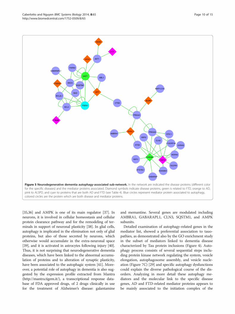
The disease genes associated to the autophagy media-tors are shown in Figure 5 and listed in Table 4, while inFigure 6, the drug targets directly associated to theautophagy mediators are represented. A prevalence ofsubunits of Protein Phosphatase 2A (PP2A), was evidentand highlighted in the figure.By investigating the overlaps between the mediator list
obtained and the human autophagy network as describedin Behrends et al. paper [29], 45 mediators were found inthe network (Additional file 3: Table S3). Eight mediatorswere found in top 10 central nodes in the network byranking the degree centrality. Moreover, 24/45 mediatorswere the articulation nodes that are of high interest asthe important nodes to prevent network fragmentation(Additional file 3: Table S3). The centrality and crucialpositions in the autophagy interaction network of themediators highlighted their relevant role in the autophagyprocess (Figure 7A). By analyzing the 10 functional

Table 3 List of high-ranked proteins in the dementia network using the degree index
Uniprot ID Official gene symbol Gene name Degree index
Q13131 PRKAA1 Protein kinase, AMP-activated, alpha 1 catalytic subunit 419
P54646 PRKAA2 Protein kinase, AMP-activated, alpha 2 catalytic subunit 392
P03372 ESR1 Estrogen receptor 1 250
P19838 NFKB1 Nuclear factor of kappa light polypeptide gene enhancer in B-cells 1 247
Q13547 HDAC1 Histone deacetylase 1 218
Q00005 PPP2R2B Protein phosphatase 2 (formerly 2A), regulatory subunit B, beta isoform 201
P17252 PRKCA Protein kinase C, alpha 194
P49841 GSK3B Glycogen synthase kinase 3 beta 170
P06493 CDK11 Cell division cycle 2, G1 to S and G2 to M 163
P04150 NR3C1 Nuclear receptor subfamily 3, group C, member 1 (glucocorticoid receptor) 156
Q92769 HDAC2 Histone deacetylase 2 138
P05067 APP Amyloid beta (A4) precursor protein 136
P19438 TNFRSF1A Tumor necrosis factor receptor superfamily, member 1A 133
P67775 PPP2CA Protein phosphatase 2 (formerly 2A), catalytic subunit, alpha isoform 127
P20226 TBP TATA box binding protein 126
P24941 Cdk2 Cyclin-dependent kinase 2 125
P30153 PPP2R1A Protein phosphatase 2 (formerly 2A), regulatory subunit A, alpha isoform 121
P54259 ATN1 Atrophin 1 109
P50750 CDK9 Cyclin-dependent kinase 9 102
In bold is the mediator which is also disease proteins, having direct interactions with drug targets and other disease genes.
Caberlotto and Nguyen BMC Systems Biology 2014, 8:65 Page 8 of 15http://www.biomedcentral.com/1752-0509/8/65
network clustered in the AIN, we found 25 mediatorproteins in the network. The autophagy-related mediatorproteins were not predominantly belonging to any ofthe sub-networks described by Behrends and collabo-rators, but they are present in almost all sub-networks(Figure 7B). FTD-associated mediator proteins (Table 4)were found in protein kinase network, vesicle elongationand autophagosome assembly and vesicle nucleation auto-phagy phases, while AD-associated proteins were seenonly in the protein kinase network, vesicle elongation andautophagosome assembly stages (Figure 7C).
DiscussionIn the present study, network analysis was used to explorefrom the systems biology perspective, the molecular con-nections among multifactorial complex diseases with theshared clinical symptoms of dementia, which could sug-gest related disease mechanisms. A number of diseaseswere considered, both common (e.g. Alzheimer’s disease)and rare disorders (e.g. amyotropic lateral sclerosis withparkinsonism and dementia) that have as a common andmajor symptom a progressive and permanent loss of cog-nitive and mental performance (Table 1).While previous systems biology studies on disease
focus on the disease gene network or drug target net-work, separately, the method proposed in the currentstudy presented an integrated methodology that can take
advantage of both these data, providing further insightinto the interactome related to dementia.Among the most connected proteins (with more than
100 interactions in the network; Table 3) the first 2 pro-teins in the list were PRKAA1 and PRKAA2, subunits ofAMP-regulated kinase (AMPK). AMPK is a central regu-lator of energy homeostasis controlling neuronal main-tenance in response to metabolic stress. Latest researchsupport an involvement of AMPK in Alzheimer [30,31]and, in our previous study on Alzheimer’s disease, on aseparate set of data and with a very different systems biol-ogy methodological approach, AMPK-related genes werealso found to be strongly associated to the disease [14].Moreover, abnormal neuronal accumulation of activatedAMPK (pAMPK) has been described in different tauopa-thies including PSP, AD, Pick’s disease, and CBD [32].Thus, the present findings support once more the pro-posed hypothesis of an alteration of metabolic functionsand energy regulation in dementia.Considering the complete list of mediator proteins, Gene
ontology (GO) enrichment analysis confirmed a signifi-cant involvement of metabolic signaling regulating energyhomeostasis, lipid and glucose metabolism (Figure 3).Metabolic disturbances have been strongly associated to orconsidered a predisposing factor in AD and a metabolic/signal transduction hypothesis for AD and other tauo-pathies has been suggested by Iqbal et al. [33]. Amongst

Table 4 Autophagy-related proteins and association with dementia disease proteins
Disease Disease protein Disease protein Mediator autophagy Mediator autophagy
Uniprot ID Official gene symbol Uniprot ID Official gene symbol
AD P02649 APOE P10636 MAPT
AD P05067 APP P00519 ABL1
AD P05067 APP P49768 PSEN1
AD P05067 APP P10636 MAPT
AD P05067 APP P07339 CTSD
AD Q13867 BLMH Q13131 PRKAA1
AD Q13867 BLMH Q9BXW4 MAP1LC3C
AD P29474 NOS3 P31749 AKT1
AD P49810 PSEN2 P49768 PSEN1
AD Q92673 SORL1 Q9H1Y0 ATG5
AD Q92673 SORL1 Q9C0C7 AMBRA1
AD Q92673 SORL1 Q13131 PRKAA1
FTD Q13148 TARDBP Q6ZNE5 ATG14/KIAA0831
FTD Q13148 TARDBP O95166 GABARAP
FTD Q13148 TARDBP P54646 PRKAA2
FTD Q13148 TARDBP Q13286 CLN3
FTD Q13148 TARDBP Q5MNZ9 WIPI1
FTD Q13148 TARDBP Q676U5 ATG16L1
FTD Q13148 TARDBP Q9BSB4 C12orf44/ATG101
FTD Q13148 TARDBP Q9H1Y0 ATG5
FTD Q13148 TARDBP Q9Y484 WDR45
FTD Q13148 TARDBP Q9Y4P8 WIPI2
FTD P49768 PSEN1 P10415 BCL2
FTD P49768 PSEN1 Q07817 BCL2L1
FTD/PSNP P10636 MAPT O60260 PARK2
FTD/PSNP P10636 MAPT P00519 ABL1
FTD/PSNP P10636 MAPT P31749 AKT1
FTD/PSNP P10636 MAPT P49768 PSEN1
FTD/PSNP P10636 MAPT Q13501 SQSTM1
ALSPD Q99497 PARK7 Q6ZNE5 ATG14/ KIAA0831
ALSPD Q99497 PARK7 Q9Y484 WDR45
ALSPD Q99497 PARK7 Q13286 CLN3
ALSPD Q99497 PARK7 Q9H1Y0 ATG5
ALSPD Q99497 PARK7 Q9Y4P8 WIPI2
ALSPD Q99497 PARK7 Q15831 STK11
ALSPD Q99497 PARK7 Q9P2Y5 UVRAG
ALSPD Q99497 PARK7 Q7Z6L1 TECPR1
LBD P37840 SNCA Q9H0R8 GABARAPL1,3/ATG8
LBD P37840 SNCA Q92934 BAD
In bold are the mediators protein which are also disease proteins.
Caberlotto and Nguyen BMC Systems Biology 2014, 8:65 Page 9 of 15http://www.biomedcentral.com/1752-0509/8/65
the metabolic-related terms, a role for autophagy andregulation of autophagy was highlighted (Figures 3 and 4).Although autophagy has been known for decades, its
relevance in neurons and glial physiology has been demon-strated only recently [34]. Autophagy is involved in theintracellular turnover of proteins and cell organelles
APP
HTT
ABL1
AKT1
NOS3
C12ORF44ATG5
STK11
CLN3
UVRAG
WIPI2
GABARAP
PARK7
TECPR1
WDR45
ATG16L1
WIPI1TARDBP
KIAA0831
APOE
MAPT
PARK2
SQSTM1SNCA
BAD
PSEN1BCL2
BLMH
SORL1
CTSD
AMBRA1
MAP1LC3C
PRKAA1
PRKAA2
ITM2B
GABARAPL1
PSEN2
BCL2L1
Figure 5 Neurodegenerative dementia autophagy-associated sub-network. In the network are indicated the disease proteins (different colorfor the specific diseases) and the mediator proteins associated. Diamond symbols indicate disease proteins, green is related to FTD, orange to AD,pink to ALSPD, and cyan to proteins that are both AD and FTD (see Table 4). Blue circles represent mediator protein associated to autophagy,colored circles are the protein which are both disease and mediator proteins.
Caberlotto and Nguyen BMC Systems Biology 2014, 8:65 Page 10 of 15http://www.biomedcentral.com/1752-0509/8/65
[35,36] and AMPK is one of its main regulator [37]. Inneurons, it is involved in cellular homeostasis and cellularprotein clearance pathway and for the remodeling of ter-minals in support of neuronal plasticity [38]. In glial cells,autophagy is implicated in the elimination not only of glialproteins, but also of those secreted by neurons, whichotherwise would accumulate in the extra-neuronal space[39], and it is activated in astrocytes following injury [40].Thus, it is not surprising that neurodegenerative dementiadiseases, which have been linked to the abnormal accumu-lation of proteins and to alteration of synaptic plasticity,have been associated to the autophagic system [41]. More-over, a potential role of autophagy in dementia is also sug-gested by the expression profile extracted from Mantra(http://mantra.tigem.it/), a transcriptional response data-base of FDA approved drugs, of 2 drugs clinically in usefor the treatment of Alzheimer’s disease: galantamine
and memantine. Several genes are modulated includingAMBRA1, GABARAPL1, CLN3, SQSTM1, and AMPKsubunits.Detailed examination of autophagy-related genes in the
mediator list, showed a preferential association to tauo-pathies, as demonstrated also by the GO enrichment studyin the subset of mediators linked to dementia diseasecharacterized by Tau protein inclusions (Figure 4). Auto-phagy process consists of several sequential steps inclu-ding protein kinase network regulating the system, vesicleelongation, autophagosome assembly, and vesicle nucle-ation (Figure 7C) [29] and specific autophagy dysfunctionscould explain the diverse pathological course of the dis-orders. Analyzing in more detail these autophagy me-diators and the molecular link to the specific diseasegenes, AD and FTD-related mediator proteins appears tobe mainly associated to the initiation complex of the

GABRG2
HDAC5
RRM1 GABRG3
GABRG1
GABARAP
FGFR1
GABARAPL1
PTDSS1
TNFRSF1A
GRIA3
GRIA1
AMBRA1
QPCT
KIAA0831
MAP1LC3C
ATG5
WDR45
HDAC1
SOD1
SLC5A6
PPP2R2A
C12ORF44
NFKB1
PPP2R2B
SCARB1
WIPI2
BAD
GABRR2
GABRR1
GRIA2
SQSTM1
PPP2R3B
GCLN3
CDK4
SIGMAR1
PRKCI
ATG16L1PPP2R1B
PDE4A
STK11
PPP2R1A
FNTA
UVRAG
PPP2R4
BCL2
GSK3BMAPT
CDK2
PRKCA
PPP2CB
PRKAA1
PPP2R5A
CTSD
CTSB
GSK3A
HMOX1ESR2
NOS3
NR3C1
WIPI1
ESR1
AKT1
PARK2
PSENEN APH1B
PSEN2
HDAC4
CDK5
GRIN2B
BACE1
BCL2L1
LRRK2
PSEN1
TECPR1
HDAC3PPP2CA
PRKAA2
CDK1
PPP3CA
APH1AA
APP
ABL1
GRIN2D
CSF1
NTRK1
KITNCSTN
Figure 6 Autophagy-associated drug target sub-network. In the network are shown the drug targets (yellow squares) and the associatedautophagy-related mediator proteins (blue circles). In red are highlighted the PP2A proteins subunits.
Caberlotto and Nguyen BMC Systems Biology 2014, 8:65 Page 11 of 15http://www.biomedcentral.com/1752-0509/8/65
macroautophagy cascade, involving mainly beclin 1 inter-actome: B-cell CLL/lymphoma 2 (BCL2), BCL2-like 1(BCL2L1), and Atg14 (Figure 6). Beclin 1 interactomecontains stimulating and suppressive components regulat-ing the initiation of the autophagosome formation and, re-cently, Beclin 1 has been found to be down-regulated inAD brain. Moreover, suppression of Beclin 1 in culturedneurons and transgenic mice induces the deposition ofamyloid-β peptides, whereas its overexpression reduces itsaccumulation [42]. Beclin 1 protein also assembles twocore complexes, Atg14L or UVRAG complexes, and withAtg14L protein induces the phagophore formation and,thus, stimulates autophagocytosis, whereas the UVRAG/Beclin 1 complex controls other Beclin 1-dependentfunctions, e.g. phagocytosis. The subcellular compart-mentation of Beclin 1 is regulated by different moleculesincluding BCL-2 [43] and mTOR [44] which are
represented in the mediator list. Several proteins can con-trol the activation of beclin complex including two proteinkinases included in our mediator list: CKD2 and CDK5[45] (Additional file 1: Table S1). Atg5 protein, a mediatorrelated to both AD and FTD disease genes (Figures 5 and6), is also essential for the autophagy process and, in aconjugated form with Atg12 and Atg8 (LC3), is in-volved in the early stages of autophagosome formation(Figure 7C). Taken together these results suggest impair-ment in the early stages of autophagy complex essentialfor autophagosome formation [46,47], including proteinkinase network regulating autophagosome assembling.The hypothesis that autophagy regulation and, in particu-lar, its induction could contribute to AD pathology is alsosupported by recent evidence suggesting that the synthesisof components of the lysosome is up-regulated at thetranscriptional and translational levels in the AD brain

FTD
AD
FTD/AD
C
BA
ALSPD
Figure 7 Integration of neurodegenerative dementia mediator proteins with human autophagy network (modified from [29]). In A)mediator proteins of the dementia network are charted in the human autophagy interaction network, in B) mediator proteins are mapped in thehuman autophagy sub-networks and in C) mediator proteins are highlighted in the functional integration of the autophagy interaction network.
Caberlotto and Nguyen BMC Systems Biology 2014, 8:65 Page 12 of 15http://www.biomedcentral.com/1752-0509/8/65
and AD mouse models [48-54]. Moreover, Lipinski et al.[55] recently reported an up-regulation of the trans-cription of genes stimulating autophagy and a down-regulation of the negative regulators of autophagy in thebrains of AD affected subjects. In the dementia network,this interactome is connected to presenilin (Figure 5) whosemutation underlies the majority of familial Alzheimer’sdisease cases [56-58] and whose role in autophagy hasbeen shown to be central [59], presenilin 1 being essen-tial for lysosome acidification, and proteolysis duringautophagy [60].Frontotemporal dementia-related mediator proteins
seem to be involved not only in the vesicle elongation andautophagosome assembling process, but also, and exclu-sively, to vesicle nucleation procedure (Figure 7C). Thisprocess includes WIPI proteins (WD-repeat protein in-teracting with phosphoinosides), WIPI-1 and WIPI-2,
evolved from the yeast ancestral autophagy protein Atg18(Proikas-Cezanne T, 2004; Polson HE, 2010) as membranecomponents of autophagosomes (Mauthe 2011, [61]).Both WIPI-1 and WIPI-2 specifically bind PtdIns(3)P andlocalize at autophagosomal membranes (phagophore) [62].TARDPB (TDP-43) appears to be a central protein in
our autophagy-related sub-network (Figure 5). TDP-43 isa DNA/RNA-binding protein with multicellular functions.Several mutations of its gene have been reported in casesof frontotemporal lobar degeneration (FTLD) [63]. Itis processed and degraded by both autophagy and theubiquitin-proteasome systems [64]. Activation of auto-phagy by rapamycin plays an active role in the clearanceof TDP-43 deficits in mouse model with proteinopathiesof the TAR DNA-binding protein 43 [65]. Depletion ofTDP-43 induces a down-regulation of the major auto-phagy component Atg7, causes impairment of autophagy

Caberlotto and Nguyen BMC Systems Biology 2014, 8:65 Page 13 of 15http://www.biomedcentral.com/1752-0509/8/65
and facilitates the accumulation of polyubiquitinated pro-teins which could be rescued by overexpression of theprotein, with a feedback regulatory loop between TDP-43and autophagy [64]. In our network, TDP-43 is linked toAMPK subunit PRKAA2 and a functional link betweenthese two proteins has been suggested in pathological con-ditions showing that activated AMPK adversely affectsmutant TDP-43-induced motor neurons diseases [66]. Inaddition, it is related to other central autophagy proteinssuch as ATG5 and ATG16L, which create a multimericcomplex playing an essential role in autophagosome for-mation, a system highly conserved in all eukaryotes [67].The other proteins linked to TDP-43 are WIPI1 and 2(Figure 5). Thus, these findings could suggest a thera-peutic modulation of autophagy involving approaches thatfunctionally target WIPI proteins and ATG5-ATG16 com-plex for the treatment of FTD and other diseases involvingmutations in TDP-43 gene.Apart from the metabolic-associated biological pro-
cesses terms, cell surface receptor signaling pathway-related terms were also highly significant enriched, inparticular in proteins associated to the Wnt pathway(Figures 3 and 4). Several proteins in the mediator listare represented in the pathway (see Additional file 4:Figure S4) including GSK-3β, a tau kinase that wasalso included in the most connected mediator proteinslist (Table 3) and in the autophagy-related proteins(Additional file 3: Table S3). Several preclinical and clinicaldata strongly link GSK-3β to dementia: different inhibitorsof GSK3B activity block neurodegeneration in vitro, andGSK-3β -mediated Wnt signaling can mediate amyloidpeptide toxicity in vitro [68,69]. Finally, in human post-mortem brain, this protein is physically associated withneurofibrillary tangles, one of the pathologic hallmarks ofAD [70]. WNT pathway has also been recently linked toautophagy. In fact, autophagy negatively regulates Wntsignalling by promoting Dishevelled (Dvl) degradation,with a role for Von Hippel–Lindau protein-mediated ubi-quitylation [71], both of them present in the dementia net-work mediator list.In our dementia network, among the drug targets asso-
ciated to the autophagy-related mediators, the highest rep-resented proteins are subunits of the Protein phosphatase2A (PP2A; Figure 5), a serine/threonine-specific proteinphosphatase consisting of A, B and C subunits that playsmultiple roles in different signaling pathways and regu-lates diverse cellular processes. Among the six PP2A pro-teins, three proteins (PPP2R2B, PPP2CA, and PPP2R1A)are also listed in the highly ranked proteins in the de-mentia network (Table 3), demonstrating their centrality.A recent study confirms that PP2A blockade inhibitsautophagy potentially through activation of AMPK [72]. Arole of PP2A in dementia is further demonstrated by theevidence that okadaic acid and calyculin A, two potent
PP2A inhibitors [73], are able to induce tauopathy andcognitive deficiency in rats [74,75]. Thus, PP2A subunitscould be considered as a potential therapeutic targetfor AD.In our drug targets list related to autophagy mediators
(Figure 6), other molecular targets could be consideredsuitable for therapeutic intervention including AMPK-related proteins, a highly ranked protein in our network(Table 3 and Additional file 3: Table S3) and a targetwhich has been already considered for the treatment ofAlzheimer’s disease. In fact, pioglitazone, an antidiabeticdrug which acts also by activating AMPK [76], has beenproven to reverse pathological conditions in an animalmodel of the disease [77] and it is in clinical trial forAlzheimer’s disease (www.clincaltrial.gov).In more general terms, a direct action on the regu-
lation of autophagy, potentially an activation of theautophagic process should be considered to the deve-lopment of optimal therapeutics, although autophagycan function both as a cytoprotective mechanism, but italso has the capacity to cause cell death.
ConclusionThis network analysis considering the established know-ledge on different neurodegenerative dementia diseaserepresented by OMIM data and the drug targets in thedifferent phases of the drug discovery process, identifiesthe autophagy process as a central dis-regulated pathwayin these sub-group of neurodegenerative disorders. Wecould hypothesize that different mutation or alterationat the genomic level could affect different phases of theautophagy process and thus therapeutic modulationcould involve approaches that functionally target thespecific proteins. Exploring the molecular mechanismsof autophagy opens an avenue for development of noveldrugs and particularly, these results could suggest thepotentiality of drug targeting specific PP2A subunits forthe treatment of dementia.
Additional files
Additional file 1: Table S1. List of Drug targets, list of mediatorproteins in the dementia network, and gene ontology biologicalfunctions enrichment analysis results of all mediators, mediator related totauopathies and highly ranked protein in the dementia network.
Additional file 2: Figure S2. Summary of statistically significant GeneOntology biological processes functional annotation corresponding toproteins in the highly ranked proteins as obtained from REVIGO [28].Nodes are GO terms and edges represent the strongest GO termspairwise similarity. Colors represent the p-values (low values in green,high in red). Only significant GO terms are shown (P < 0.001).
Additional file 3: Table S3. List of 45 mediators (with their degreecentrality related to dementia and autophagy [29] networks) found in theautophagy interaction network [29]. In bold are proteins that play a roleas the articulation points in the human autophagy network.

Caberlotto and Nguyen BMC Systems Biology 2014, 8:65 Page 14 of 15http://www.biomedcentral.com/1752-0509/8/65
Additional file 4: Figure S4. Schematic Figure representing the WNTpathway as described in the Biocarta database. In red are labeled thedementia network mediator proteins.
AbbreviationsFTD: Frontotemporal dementia; AD: Alzheimer disease; LBD: Lewy bodiesdisease; PSP: Progressive supranuclear palsy; CBD: Corticobasal dementia;HD: Hungtington’s disease; ALSPD: Amyotrophic lateral sclerosis-Parkinsonism/dementia complex; PIN: Protein-protein interaction network; GSK-3β: Glycogensynthase kinase beta; AMPK: AMP-regulated kinase; TDP-43: TAR DNA-bindingprotein 43; PP2A: Protein phosphatase 2A.
Competing interestsThe authors declare that there is no competing interest in relation to thepublication of this article.
Authors’ contributionsLC and TPN conceived and designed the study, collected and analyzed thedata and wrote the paper. Both authors read and approved the finalmanuscript.
AcknowledgmentsWe are grateful to Bianca Baldacci for the graphic design contribution and toCorrado Priami and Mario Lauria for valuable discussions.
Received: 14 January 2014 Accepted: 20 May 2014Published: 7 June 2014
References1. Hickey C, Chisholm T, Passmore MJ, O’Brien JD, Johnston J: Differentiating
the dementias: revisiting synucleinopathies and tauopathies. CurrAlzheimer Res 2008, 5:52–60.
2. Galpern WR, Lang AE: Interface between tauopathies andsynucleinopathies: a tale of two proteins. Ann Neurol 2006, 59:449–458.
3. Loscalzo J, Barabasi A-L: Systems biology and the future of medicine.Wiley Interdiscip Rev Syst Biol Med 2011, 3:619–627.
4. Barabasi A-L, Gulbahce N, Loscalzo J: Network medicine: a network-basedapproach to human disease. Nat Rev Genet 2011, 12:56–68.
5. Vidal M, E Cusick M, Barabási A-L: Interactome networks and humandisease. Cell 2011, 144:986–998.
6. Oti M, Snel B, Huynen MA, Brunner HG: Predicting disease genes usingprotein-protein interactions. J Med Genet 2006, 43:691–698.
7. Kann MG: Protein interactions and disease: computational approaches touncover the etiology of diseases. Brief Bioinform 2007, 8:333–346.
8. Schuster-Böckler B, Bateman A: Protein interactions in human geneticdiseases. Genome Biol 2008, 9:R9.
9. Navlakha S, Kingsford C: The power of protein interaction networks forassociating genes with diseases. Bioinformatics 2010, 26:1057–1063.
10. Nguyen T-P, Ho T-B: Detecting disease genes based on semi-supervisedlearning and protein-protein interaction networks. Artif Intell Med 2012,54:63–71.
11. Jordán F, Nguyen T-P, Liu W-C: Studying protein-protein interactionnetworks: a systems view on diseases. Brief Funct Genomics 2012,11:497–504.
12. Caberlotto L, Lauria M, Nguyen T-P, Priami C: The central role ofAMP-kinase and energy homeostasis impairment in Alzheimer’sdisease: a multifactor network analysis. Plos One 2013, 8(11):e78919.
13. Thanh-Phuong N, Laura C, Morine CP MJ: Network analysis ofneurodegenerative disease highlights a role of toll-like receptorsignaling. Biomed Res Int 2014, 2014:686505.
14. Caberlotto L, Lauria M, Nguyen T-P, Scotti M: The central role of AMP-kinase and energy homeostasis impairment in Alzheimer’s disease:a multifactor network analysis. PLoS One 2013, 8:e78919.
15. Chen X, Burgoyne RD: Identification of common genetic modifiers ofneurodegenerative diseases from an integrative analysis of diversegenetic screens in model organisms. BMC Genomics 2012, 13:71.
16. Limviphuvadh V, Tanaka S, Goto S, Ueda K, Kanehisa M: The commonalityof protein interaction networks determined in neurodegenerativedisorders (NDDs). Bioinformatics 2007, 23:2129–2138.
17. Vasaikar SV, Padhi AK, Jayaram B, Gomes J: NeuroDNet - an open sourceplatform for constructing and analyzing neurodegenerative diseasenetworks. BMC Neurosci 2013, 14:3.
18. Gad SC: Pharmaceutical Sciences Encyclopedia. Hoboken, NJ, USA: John Wiley& Sons, Inc.; 2010.
19. Yu H, Chen J, Xu X, Li Y, Zhao H, Fang Y, Li X, Zhou W, Wang W, Wang Y:A systematic prediction of multiple drug-target interactions fromchemical, genomic, and pharmacological data. PLoS One 2012,7:e37608.
20. Emig D, Ivliev A, Pustovalova O, Lancashire L, Bureeva S, Nikolsky Y,Bessarabova M: Drug target prediction and repositioning using anintegrated network-based approach. PLoS One 2013, 8:e60618.
21. Hamosh A, Scott AF, Amberger JS, Bocchini CA, McKusick VA: OnlineMendelian Inheritance in Man (OMIM), a knowledgebase of humangenes and genetic disorders. Nucleic Acids Res 2005, 33(Database issue):D514–D517.
22. Baxevanis AD: Searching Online Mendelian Inheritance in Man (OMIM)for information for genetic loci involved in human disease. Curr ProtocHum Genet 2003, Chapter 9:Unit9.13.
23. Brown KR, Jurisica I: Online predicted human interaction database.Bioinformatics 2005, 21:2076–2082.
24. Zotenko E, Mestre J, O’Leary DP, Przytycka TM: Why do hubs in the yeastprotein interaction network tend to be essential: reexamining theconnection between the network topology and essentiality. PLoS ComputBiol 2008, 4:e1000140.
25. Zhang S, Jin G, Zhang X-S, Chen L: Discovering functions and revealingmechanisms at molecular level from biological networks. Proteomics2007, 7:2856–2869.
26. Yook S-H, Oltvai ZN, Barabási A-L: Functional and topologicalcharacterization of protein interaction networks. Proteomics 2004,4:928–942.
27. Eden E, Navon R, Steinfeld I, Lipson D, Yakhini Z: GOrilla: a tool fordiscovery and visualization of enriched GO terms in ranked gene lists.BMC Bioinformatics 2009, 10:48.
28. Supek F, Bošnjak M, Škunca N, Šmuc T: REVIGO summarizes and visualizeslong lists of gene ontology terms. PLoS One 2011, 6:e21800.
29. Behrends C, Sowa ME, Gygi SP, Harper JW: Network organization of thehuman autophagy system. Nature 2010, 466:68–76.
30. Salminen A, Kaarniranta K, Haapasalo A, Soininen H, Hiltunen M: AMP-activated protein kinase: a potential player in Alzheimer’s disease.J Neurochem 2011, 118:460–474.
31. Cai Z, Yan L-J, Li K, Quazi SH, Zhao B: Roles of AMP-activated proteinkinase in Alzheimer’s disease. Neuromolecular Med 2012, 14:1–14.
32. Vingtdeux V, Davies P, Dickson DW, Marambaud P: AMPK isabnormally activated in tangle- and pre-tangle-bearing neuronsin Alzheimer’s disease and other tauopathies. Acta Neuropathol 2011,121:337–349.
33. Iqbal K, Grundke-Iqbal I: Metabolic/signal transduction hypothesis ofAlzheimer’s disease and other tauopathies. Acta Neuropathol 2005,109:25–31.
34. Mizushima N, Levine B, Cuervo AM, Klionsky DJ: Autophagy fights diseasethrough cellular self-digestion. Nature 2008, 451:1069–1075.
35. Klionsky DJ, Emr SD: Autophagy as a regulated pathway of cellulardegradation. Science 2000, 290:1717–1721.
36. Levine B, Yuan J: Autophagy in cell death: an innocent convict? J ClinInvest 2005, 115:2679–2688.
37. Roach PJ: AMPK - > ULK1 - > autophagy. Mol Cell Biol 2011, 31:3082–3084.38. Komatsu M, Kominami E, Tanaka K: Autophagy and neurodegeneration.
Autophagy 2006, 2:315–317.39. Martin A, Joseph JA, Cuervo AM: Stimulatory effect of vitamin C on
autophagy in glial cells. J Neurochem 2002, 82:538–549.40. Qin A-P, Liu C-F, Qin Y-Y, Hong L-Z, Xu M, Yang L, Liu J, Qin Z-H, Zhang H-L:
Autophagy was activated in injured astrocytes and mildly decreased cellsurvival following glucose and oxygen deprivation and focal cerebralischemia. Autophagy 2010, 6:738–753.
41. Kragh CL, Ubhi K, Wyss-Coray T, Wyss-Corey T, Masliah E: Autophagy indementias. Brain Pathol 2012, 22:99–109.
42. Salminen A, Kaarniranta K, Kauppinen A, Ojala J, Haapasalo A, Soininen H,Hiltunen M: Impaired autophagy and APP processing in Alzheimer’sdisease: The potential role of Beclin 1 interactome. Prog Neurobiol 2013,106–107:33–54.

Caberlotto and Nguyen BMC Systems Biology 2014, 8:65 Page 15 of 15http://www.biomedcentral.com/1752-0509/8/65
43. Pattingre S, Tassa A, Qu X, Garuti R, Liang XH, Mizushima N, Packer M,Schneider MD, Levine B: Bcl-2 antiapoptotic proteins inhibit Beclin1-dependent autophagy. Cell 2005, 122:927–939.
44. Kanazawa T, Taneike I, Akaishi R, Yoshizawa F, Furuya N, Fujimura S,Kadowaki M: Amino acids and insulin control autophagic proteolysisthrough different signaling pathways in relation to mTOR in isolated rathepatocytes. J Biol Chem 2004, 279:8452–8459.
45. Furuya T, Kim M, Lipinski M, Li J, Kim D, Lu T, Shen Y, Rameh L, Yankner B,Tsai L-H, Yuan J: Negative regulation of Vps34 by Cdk mediatedphosphorylation. Mol Cell 2010, 38:500–511.
46. Funderburk SF, Wang QJ, Yue Z: The Beclin 1–VPS34 complex–at thecrossroads of autophagy and beyond. Trends Cell Biol 2010, 20:355–362.
47. He C, Levine B: The Beclin 1 interactome. Curr Opin Cell Biol 2010,22:140–149.
48. Cataldo AM, Peterhoff CM, Schmidt SD, Terio NB, Duff K, Beard M, MathewsPM, Nixon RA: Presenilin mutations in familial Alzheimer disease andtransgenic mouse models accelerate neuronal lysosomal pathology.J Neuropathol Exp Neurol 2004, 63:821–830.
49. Cataldo AM, Barnett JL, Mann DM, Nixon RA: Colocalization of lysosomalhydrolase and beta-amyloid in diffuse plaques of the cerebellum andstriatum in Alzheimer’s disease and Down's syndrome. J Neuropathol ExpNeurol 1996, 55:704–715.
50. Cataldo AM, Hamilton DJ, Nixon RA: Lysosomal abnormalities indegenerating neurons link neuronal compromise to senile plaquedevelopment in Alzheimer disease. Brain Res 1994, 640:68–80.
51. Cataldo AM, Hamilton DJ, Barnett JL, Paskevich PA, Nixon RA: Properties ofthe endosomal-lysosomal system in the human central nervous system:disturbances mark most neurons in populations at risk to degenerate inAlzheimer’s disease. J Neurosci 1996, 16:186–199.
52. Mufson EJ, Counts SE, Ginsberg SD: Gene expression profiles ofcholinergic nucleus basalis neurons in Alzheimer’s disease. NeurochemRes 2002, 27:1035–1048.
53. Ginsberg SD, Alldred MJ, Counts SE, Cataldo AM, Neve RL, Jiang Y,Wuu J, Chao MV, Mufson EJ, Nixon RA, Che S: Microarray analysis ofhippocampal CA1 neurons implicates early endosomal dysfunctionduring Alzheimer’s disease progression. Biol Psychiatry 2010,68:885–893.
54. Nixon RA, Cataldo AM: Lysosomal system pathways: genes toneurodegeneration in Alzheimer’s disease. J Alzheimers Dis 2006,9(3 Suppl):277–289.
55. Lipinski MM: Towards the global understanding of the autophagyregulatory network. Autophagy 2010, 6:1218–1220.
56. Barton AJ, Crook BW, Karran EH, Brown F, Dewar D, Mann DM, Pearson RC,Graham DI, Hardy J, Hutton M, Duff K, Goate AM, Clark RF, Roberts GW:Alteration in brain presenilin 1 mRNA expression in early onset familialAlzheimer’s disease. Neurodegeneration 1996, 5:213–218.
57. Borchelt DR, Ratovitski T, van Lare J, Lee MK, Gonzales V, Jenkins NA,Copeland NG, Price DL, Sisodia SS: Accelerated amyloid deposition in thebrains of transgenic mice coexpressing mutant presenilin 1 and amyloidprecursor proteins. Neuron 1997, 19:939–945.
58. Gómez-Isla T, Growdon WB, McNamara MJ, Nochlin D, Bird TD, Arango JC,Lopera F, Kosik KS, Lantos PL, Cairns NJ, Hyman BT: The impact of differentpresenilin 1 andpresenilin 2 mutations on amyloid deposition,neurofibrillary changes and neuronal loss in the familial Alzheimer’sdisease brain: evidence for other phenotype-modifying factors. Brain1999, 122(Pt 9):1709–1719.
59. Neely KM, Green KN: Presenilins mediate efficient proteolysis via theautophagosome-lysosome system. Autophagy 2011, 7:664–665.
60. Lee J-H, Yu WH, Kumar A, Lee S, Mohan PS, Peterhoff CM, Wolfe DM,Martinez-Vicente M, Massey AC, Sovak G, Uchiyama Y, Westaway D, CuervoAM, Nixon RA: Lysosomal proteolysis and autophagy require presenilin 1and are disrupted by Alzheimer-related PS1 mutations. Cell 2010,141:1146–1158.
61. Proikas-Cezanne T, Robenek H: Freeze-fracture replica immunolabelling revealshuman WIPI-1 and WIPI-2 as membrane proteins of autophagosomes. J CellMol Med 2011, 15:2007–2010.
62. Vergne I, Roberts E, Elmaoued RA, Tosch V, Delgado MA, Proikas-Cezanne T,Laporte J, Deretic V: Control of autophagy initiation by phosphoinositide3-phosphatase Jumpy. EMBO J 2009, 28:2244–2258.
63. Borroni B, Archetti S, Del Bo R, Papetti A, Buratti E, Bonvicini C, Agosti C,Cosseddu M, Turla M, Di Lorenzo D, Pietro Comi G, Gennarelli M, Padovani
A: TARDBP mutations in frontotemporal lobar degeneration: frequency,clinical features, and disease course. Rejuvenation Res 2010, 13:509–517.
64. Bose JK, Huang C-C, Shen C-KJ: Regulation of autophagy by neuropathologicalprotein TDP-43. J Biol Chem 2011, 286:44441–44448.
65. Wang I-F, Tsai K-J, Shen C-KJ: Autophagy activation ameliorates neuronalpathogenesis of FTLD-U mice: a new light for treatment of TARDBP/TDP-43 proteinopathies. Autophagy 2013, 9:239–240.
66. Ng C-H, Guan MSH, Koh C, Ouyang X, Yu F, Tan E-K, O’Neill SP, Zhang X, ChungJ, Lim K-L: AMP kinase activation mitigates dopaminergic dysfunction andmitochondrial abnormalities in Drosophila models of Parkinson’s disease.J Neurosci 2012, 32:14311–14317.
67. Matsushita M, Suzuki NN, Obara K, Fujioka Y, Ohsumi Y, Inagaki F: Structureof Atg5.Atg16, a complex essential for autophagy. J Biol Chem 2007,282:6763–6772.
68. Noh M-Y, Koh S-H, Kim Y, Kim HY, Cho GW, Kim SH: Neuroprotectiveeffects of donepezil through inhibition of GSK-3 activity inamyloid-beta-induced neuronal cell death. J Neurochem 2009,108:1116–1125.
69. Martinez A, Gil C, Perez DI: Glycogen synthase kinase 3 inhibitors in the nexthorizon for Alzheimer’s disease treatment. Int J Alzheimers Dis 2011,2011:280502.
70. Pei JJ, Braak E, Braak H, Grundke-Iqbal I, Iqbal K, Winblad B, Cowburn RF:Distribution of active glycogen synthase kinase 3beta (GSK-3beta) in brainsstaged for Alzheimer disease neurofibrillary changes. J Neuropathol ExpNeurol 1999, 58:1010–1019.
71. Gao C, Cao W, Bao L, Zuo W, Xie G, Cai T, Fu W, Zhang J, Wu W, Zhang X,Chen Y-G: Autophagy negatively regulates Wnt signalling by promotingDishevelled degradation. Nat Cell Biol 2010, 12:781–790.
72. Magnaudeix A, Wilson CM, Page G, Bauvy C, Codogno P, Lévêque P,Labrousse F, Corre-Delage M, Yardin C, Terro F: PP2A blockade inhibitsautophagy and causes intraneuronal accumulation of ubiquitinatedproteins. Neurobiol Aging 2013, 34:770–790.
73. Haystead TA, Sim AT, Carling D, Honnor RC, Tsukitani Y, Cohen P, HardieDG: Effects of the tumour promoter okadaic acid on intracellular proteinphosphorylation and metabolism. Nature 1989, 337:78–81.
74. Zhang Z, Simpkins JW: An okadaic acid-induced model of tauopathy andcognitive deficiency. Brain Res 2010, 1359:233–246.
75. Yang X, Yang Y, Fu Z, Li Y, Feng J, Luo J, Zhang Q, Wang Q, Tian Q:Melatonin ameliorates Alzheimer-like pathological changes and spatialmemory retention impairment induced by calyculin A. J Psychopharmacol2011, 25:1118–1125.
76. Saha AK, Avilucea PR, Ye J-M, Assifi MM, Kraegen EW, Ruderman NB:Pioglitazone treatment activates AMP-activated protein kinase in ratliver and adipose tissue in vivo. Biochem Biophys Res Commun 2004,314:580–585.
77. Searcy JL, Phelps JT, Pancani T, Kadish I, Popovic J, Anderson KL, Beckett TL,Murphy MP, Chen K-C, Blalock EM, Landfield PW, Porter NM, Thibault O:Long-term pioglitazone treatment improves learning and attenuatespathological markers in a mouse model of Alzheimer’s disease.J Alzheimers Dis 2012, 30:943–961.
doi:10.1186/1752-0509-8-65Cite this article as: Caberlotto and Nguyen: A systems biologyinvestigation of neurodegenerative dementia reveals a pivotal role ofautophagy. BMC Systems Biology 2014 8:65.
Submit your next manuscript to BioMed Centraland take full advantage of:
• Convenient online submission
• Thorough peer review
• No space constraints or color figure charges
• Immediate publication on acceptance
• Inclusion in PubMed, CAS, Scopus and Google Scholar
• Research which is freely available for redistribution
Submit your manuscript at www.biomedcentral.com/submit