Embed Size (px)
Citation preview
Staehr et al. BMC Research Notes 2012, 5:383http://www.biomedcentral.com/1756-0500/5/383
brought to you by COREView metadata, citation and similar papers at core.ac.uk
provided by Springer - Publisher Connector
RESEARCH ARTICLE Open Access
Influence of perioperative oxygen fraction onpulmonary function after abdominal surgery:a randomized controlled trialAnne K Staehr1*, Christian S Meyhoff1, Steen W Henneberg2, Poul L Christensen3 and Lars S Rasmussen1
Abstract
Background: A high perioperative inspiratory oxygen fraction (FiO2) may reduce the frequency of surgical siteinfection. Perioperative atelectasis is caused by absorption, compression and reduced function of surfactant. It iswell accepted, that ventilation with 100% oxygen for only a few minutes is associated with significant formation ofatelectasis. However, it is still not clear if a longer period of 80% oxygen results in more atelectasis compared to alow FiO2.Our aim was to assess if a high FiO2 is associated with impaired oxygenation and decreased pulmonary functionalresidual capacity (FRC).
Methods: Thirty-five patients scheduled for laparotomy for ovarian cancer were randomized to receive either 30%oxygen (n = 15) or 80% oxygen (n = 20) during and for 2 h after surgery. The oxygenation index (PaO2/FiO2) wasmeasured every 30 min during anesthesia and 90 min after extubation. FRC was measured the day before surgeryand 2 h after extubation by a rebreathing method using the inert gas SF6.
Results: Five min after intubation, the median PaO2/FiO2 was 69 kPa [53-71] in the 30%-group vs. 60 kPa [47-69] inthe 80%-group (P= 0.25). At the end of anesthesia, the PaO2/FiO2 was 58 kPa [40-70] vs. 57 kPa [46-67] in the 30%-and 80%-group, respectively (P= 0.10). The median FRC was 1993 mL [1610-2240] vs. 1875 mL [1545-2048] atbaseline and 1615 mL [1375-2318] vs. 1633 mL [1343-1948] postoperatively in the 30%- and 80%-group, respectively(P= 0.70).
Conclusion: We found no significant difference in oxygenation index or functional residual capacity betweenpatients given 80% and 30% oxygen for a period of approximately 5 hours.
Trial registration: ClinicalTrials.gov Identifier: NCT00637936.
Keywords: Pulmonary gas exchange, Pulmonary atelectasis, Functional residual capacity, Pulmonary function,Anesthesia, General, Oxygen, Partial pressure, Surgery, Gynaecological
BackgroundAtelectasis is a common perioperative complication [1].It is observed in more than 90% of all anesthesitizedpatients with an average of 3-4% collapsed lung area and10-15% collapsed lung tissue.Main mechanisms underlying the atelectasis formation
are compression, loss of surfactant or impared surfactant
* Correspondence: [email protected] of Anaesthesia, Centre of Head and Orthopaedics, CopenhagenUniversity Hospital, Rigshospitalet, Blegdamsvej 9, DK-2100, Copenhagen �,DenmarkFull list of author information is available at the end of the article
© 2012 Staehr et al.; licensee BioMed CentralCommons Attribution License (http://creativecreproduction in any medium, provided the or
function and absorption of gas (oxygen) from alveoli be-hind closed or intermittently closed airways [2,3]. Venti-lation for only a few minutes with 100% oxygen causes asignificant increase in atelectasis shortly after anesthesiainduction compared to ventilation with lower oxygenconcentration [4]. However, no clear association betweenatelectasis formation and oxygen concentration duringmaintenance of anesthesia has yet been established [3],and a number of other harms and benefits still need fur-ther investigation [5].A FiO2 of 0.80 during and for the first few h after
elective colorectal surgery has been associated with a
Ltd. This is an Open Access article distributed under the terms of the Creativeommons.org/licenses/by/2.0), which permits unrestricted use, distribution, andiginal work is properly cited.
Staehr et al. BMC Research Notes 2012, 5:383 Page 2 of 8http://www.biomedcentral.com/1756-0500/5/383
reduced frequency of surgical site infection [6,7]. It hasbeen investigated in thirty patients how this high oxygenconcentration for a prolonged time affects postoperativepulmonary function compared to a FiO2 of 0.30 [8].That study found no significant changes in forced vitalcapacity (FVC), forced expiratory volume (FEV1.0), arter-ial oxygen tension (PaO2), alveolar-arterial oxygen differ-ence ((Aa)DO2) or frequency and severity of atelectasisas measured first postoperative day. Atelectasis deter-mined by computed tomography (CT) was seen in 94%of the patients given 0.80 FiO2 compared to 64% ofpatients given 0.30 FiO2 (P= 0.12).Atelectasis causes a reduction in gas exchange cap-
acity, and this can be estimated by measuring the func-tional residual capacity (FRC) [9]. Atelectasis leads tointrapulmonary right to left shunts [10], which can beassessed by a decrease in the ratio between the arterialoxygen tension and the inspired oxygen concentration,the oxygenation index (PaO2/FiO2) [11]. An increase inPaO2/FiO2 was found in patients given 0.40 FiO2 com-pared to patients given 1.0 FiO2 during laparoscopiccholecystectomy [12]. However, the perioperativechanges in PaO2/FiO2 and the FRC have not yet beeninvestigated in patients given 0.80 FiO2 or 0.30 FiO2.The aim of this study was to compare the changes in
perioperative PaO2/FiO2 and FRC in patients given 0.80FiO2 or 0.30 FiO2 during and for 2 h after surgery forovarian cancer. We hypothesized that a FiO2 of 0.80would result in a larger reduction in PaO2/FiO2 at theend of anesthesia than a FiO2 of 0.30.
MethodsDanish Medicines Agency and Local Ethics Committeeapproved the study (NCT00637936), and a writteninformed consent was obtained from all subjects.The study was conducted from March 2008 to July
2008. Eligible patients were aged 18 years or older,scheduled for explorative laparotomy for ovarian cancer,defined as a Risk of Malignancy Index (RMI)≥ 200 [13].Exclusion criteria were: inability to give informed con-sent, inability to keep arterial oxygen saturation (SpO2)above 90% without supplemental oxygen, chemotherapywithin 3 months, and surgery within 30 days (exceptsurgery under local anesthesia or dilation and curettageunder general anaesthesia). The study was a part of amulticenter trial investigating the influence of FiO2 onsurgical site infection, the PROXI-trial [14,15]. However,all patients in this study were recruited in one centre.Patients were randomized by a central interactive
voice-response system at the Copenhagen Trial Unit toeither FiO2 = 0.30 (30%-group) or FiO2 = 0.80 (80%-group) with stratification for center, diabetes mellitus,acute or elective surgery and body mass index (< 30 kg/m2
vs. ≥ 30 kg/m2).
All patients received paracetamol 1 g and diclofenac50 mg orally preoperatively and pain intensity was moni-tored in the postanesthesia care unit using the VisualAnalog Scale (VAS, scale 0–100). Analgesics were admi-nistered if VAS exceeded 30 using opioids, non-steroidanti-inflammatory drugs or local anesthesia epidurally ifapplicable.Patients were preoxygenated with 100% oxygen for
5 min. Anesthesia was induced with intravenous (IV) ad-ministration of propofol 2 mg/kg together with fentanylor remifentanil, and rocuronium 0.6 mg/kg was given tofacilitate endotracheal intubation. Anesthesia was main-tained with propofol or sevoflurane, and remifentanil.After intubation, the lungs were ventilated with an
adjusted fraction of inspired oxygen of 1.0 for 5 minuntil the first arterial blood sample was obtained. Subse-quently, they received the allocated FiO2 until immedi-ately before extubation, when an adjusted fraction ofinspired oxygen of 1.0 was given.The level of positive end-expiratory pressure (PEEP)
was kept at 5 cmH2O. The lungs were ventilated by vol-ume control ventilation with a tidal volume of 8–10 mL/kgand a respiratory frequency of 10–12 per min, aimingat an end-tidal carbon dioxide concentration of 4.5 to6.0 kPa.Fluid was given only to replace measured or calculated
deficits (no third space loss) aiming at a body weight in-crease less than 1 kg [14]. Peroperative blood loss wasreplaced 1:1 with colloids, not exceeding 500 mL morethan estimated blood loss [14]. Ephedrine, metaoxedrineor dopamine-infusion was used to keep the systolic arterialpressure> 90 mmHg. PEEP was primarily increased, ifhypoxaemia was detected or suspected in order to keepthe SpO2 above 94% and the PaO2 above 9 kPa. If this didnot improve oxygenation, FiO2 was increased. Lung re-cruitment manoeuvres were not allowed in the periodfrom preoxygenation to 2 h after extubation. Neuromus-cular block was assessed in all patients using train-of-four(TOF) monitoring with the TOF-watchW (Danmeter APS,Odense, Denmark). Patients were extubated, when theywere fully awake and TOF-ratio≥ 0.90.After arrival to the postanesthesia care unit, patients
were given the allocated FiO2 for 2 h by a non-rebreathingfacemask with a reservoir (High Concentration OxygenMask, Intersurgical Ltd, Wokingham, UK). Patients in the30%-group were given a mixture of 2 litres of oxygen and14 litres of air per minute and patients in the 80%-groupwere given a mixture of 14 litres of oxygen and 2 litres ofair per minute. Subsequently, oxygen was given only at thephysician’s discretion according to usual clinical practice,which in our institution will be to administer supplemen-tal oxygen to keep SpO2 above 94% in a lung-healthy pa-tient. The physician was blinded to group-allocation.Patients were monitored with continous pulse oximetry
Staehr et al. BMC Research Notes 2012, 5:383 Page 3 of 8http://www.biomedcentral.com/1756-0500/5/383
and no chest physical therapy was given in the posta-nesthesia care unit.Arterial blood samples were obtained with 2-mL syr-
inges containing heparin from a radial arterial catheter,transported in iced water and analysed with ABL System777 blood gas analyser (Radiometer, Copenhagen, Den-mark) within 10 min. Samples were obtained: five minafter intubation, every half h during surgery, and 90 minafter extubation. Five samples were drawn at each meas-urement and PaO2 was calculated as the average of threevalues after omitting the highest and the lowest values.FRC was measured the day before surgery and 2 hours
after extubation using the inert gas-rebreathing methodand the Innocor-system (Innovision, Copenhagen, Den-mark) [16,17]. The Innocor-system measured the changein the concentration of SF6 after 30 sec of rebreathingwith the patient in the supine position and the upperbody elevated 45°. The test was repeated and FRC calcu-lated as the average of the two measurements. The qual-ity of the data was evaluated on the basis of predefinedcriteria [18,19] by an investigator blinded to allocationand if of inadequate quality, they were excluded fromfurther analysis.Patients were seen daily by a study investigator, and
were examined according to routine clinical practice bythe attending physician, if she presented with symptomsof pulmonary complications, including chest radiographsor CT, when relevant. The radiologist was specificallyinstructed to evaluate the severity of atelectasis withJoyce et al. modification [20] of Wilcox severity scoring[21]: 0 = no atelectasis; 1 = plate atelectasis; 2 = segmentalatelectasis; 3 = partial lobar atelectasis; 4 = complete atel-ectasis of one lung lobe; and 5 = complete atelectasis ofone lung lobe in addition to any of the above.To maintain blinding perioperative FiO2, flow of oxy-
gen and air and PaO2 were collected on separate sheetsand placed in sealed, opaque envelopes until data ana-lysis. The patients, surgical staff, the radiologists and thespecialist, who evaluated the FRC data, were notinformed about group allocation.The primary outcome was the change in PaO2/FiO2 at
end of anesthesia. The secondary outcomes were changein PaO2/FiO2 90 min after extubation, change in FRC2 h after extubation, incidence and severity of atelectasiswithin 14 days, and SpO2 2 h and 3 days after surgery.Major violations to the protocol were defined as: lung
recruitment manoeuvre, FiO2 above 0.60 in the 30%-group, PEEP ≥ 10 cmH2O for more than 1 h, and failureto use the oxygen mask for more than 1 h.
StatisticsData are reported with mean±SD or median [interquartilerange]. Data were compared with the Mann–Whitney testand categorical data with the χ2 test using SAS for
Windows, version 9.1 (SAS Institute Inc., Cary, NC, USA).P< 0.05 was considered statistically significant.We considered a 4 kPa difference in PaO2/FiO2 to be
clinically relevant and estimated a SD of 4 kPa based ondata from a previous study [12]. We calculated that atotal sample size of 30 patients would allow us to detectthis difference with a power of 80% and a significantlevel of 0.05.
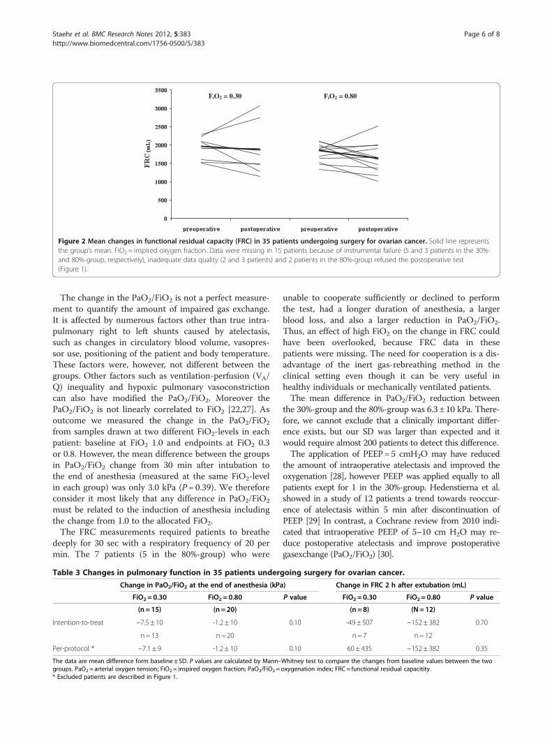
ResultsWe included 35 patients in this study (Figure 1, Table 1).Five min after intubation, the median PaO2/FiO2 was
69 kPa [53-71] in the 30%-group and 60 kPa [47-69] inthe 80%-group (P= 0.25). At end of anesthesia, thePaO2/FiO2 was 58 kPa [40-70] vs. 57 kPa [46-67] in the30%- and 80%-group, respectively (P= 0.10, Table 2).Ninety min after extubation, the PaO2/FiO2 was reducedto 56 kPa [37-60] in the 30%-group and 50 kPa [42-57]in the 80%-group (P= 0.66).Data on FRC with adequate quality were collected in
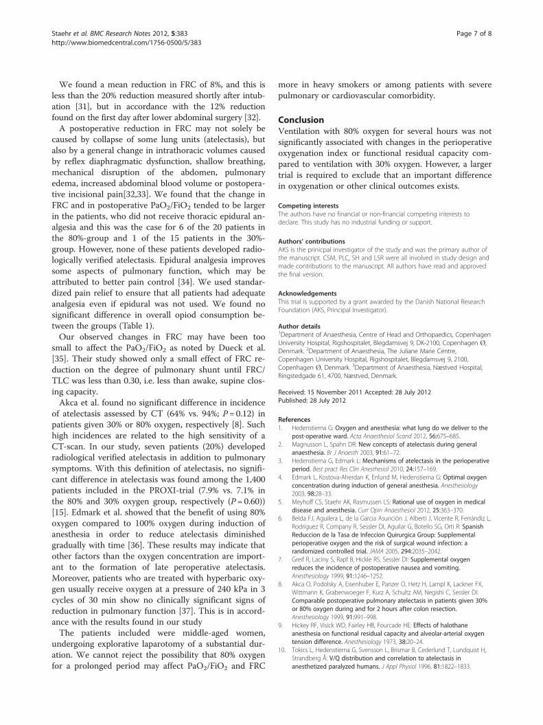
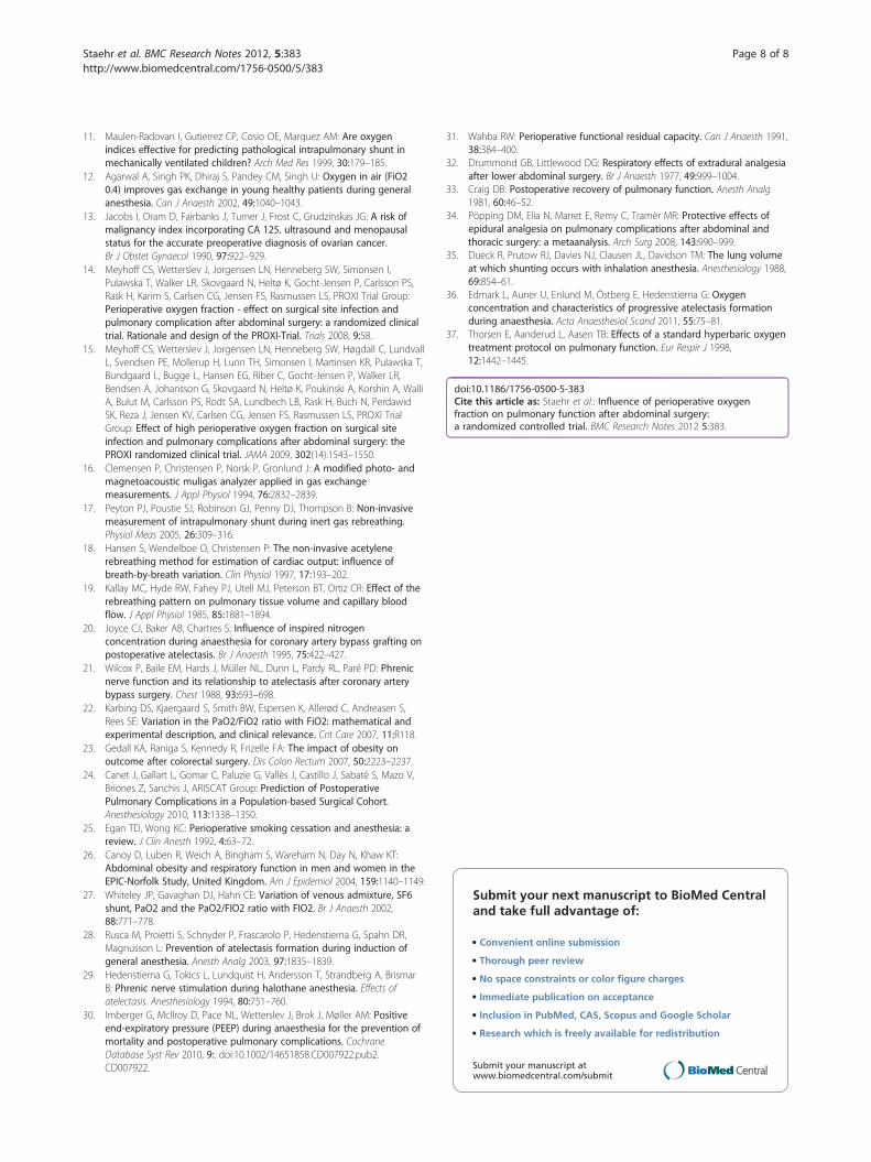
20 patients (Figure 1). The median FRC were 1993 mL[1610-2240] vs. 1875 mL [1545-2048] at baseline and1615 mL [1375-2318] vs. 1633 mL [1343-1948] post-operatively in the 30%- and 80%-group, respectively(P= 0.70) (Table 2, Figure 2).Changes in PaO2/FiO2 and FRC are reported in Table 3.
No significant difference was found between the groups.Five patients (25%) in the 80%-group developed radio-
logically verified atelectasis compared to 2 patients(13%) in the 30%-group (P= 0.51), and there were nosignificant differences in postoperative SpO2 (Table 2).Three patients had major protocol violations (Figure 1),
but the exclusion of these patients did not change theresults significantly (Table 3). In order to keep PaO2
above 9 kPa additionally one patient in the 30%-groupreceived 58% oxygen for one hour and one patient in the80%-group had PEEP adjusted to 7 cmH2O. In the post-anesthesia care unit one patient in the 30%-group wasgiven the allocated mixture of 2 litres of oxygen and 14litres of air per minute until the last arterial blood sam-ple had been drawn. Thereafter, the patient received amixture of 4 litres of oxygen and 12 litres of air per mi-nute until measurement of FRC and SpO2. SpO2 wasmeasured with the patient breathing air.A post hoc analysis of the alveolar-arteriel oxygen dif-
ference (AaO2 gradient) showed no significant baselinedifference between the two groups. Mean AaO2 gradient5 min after intubation was 31 ± 16 kPa in the 80%-groupand 27 ± 15 kPa in the 30%-group (P= 0.31).
DiscussionContrary to our primary hypothesis, we did not find a sig-nificant difference in PaO2/FiO2 at end of anesthesia be-tween patients given a high perioperative oxygen fraction
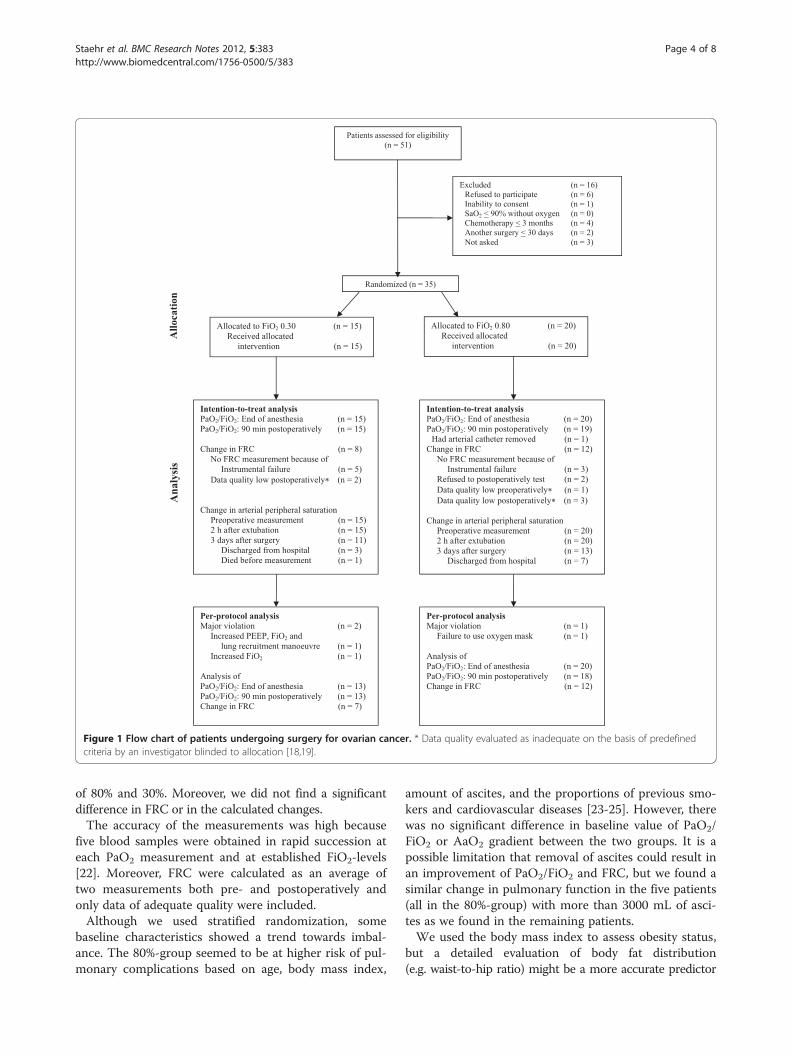
Figure 1 Flow chart of patients undergoing surgery for ovarian cancer. * Data quality evaluated as inadequate on the basis of predefinedcriteria by an investigator blinded to allocation [18,19].
Staehr et al. BMC Research Notes 2012, 5:383 Page 4 of 8http://www.biomedcentral.com/1756-0500/5/383
of 80% and 30%. Moreover, we did not find a significantdifference in FRC or in the calculated changes.The accuracy of the measurements was high because
five blood samples were obtained in rapid succession ateach PaO2 measurement and at established FiO2-levels[22]. Moreover, FRC were calculated as an average oftwo measurements both pre- and postoperatively andonly data of adequate quality were included.Although we used stratified randomization, some
baseline characteristics showed a trend towards imbal-ance. The 80%-group seemed to be at higher risk of pul-monary complications based on age, body mass index,
amount of ascites, and the proportions of previous smo-kers and cardiovascular diseases [23-25]. However, therewas no significant difference in baseline value of PaO2/FiO2 or AaO2 gradient between the two groups. It is apossible limitation that removal of ascites could result inan improvement of PaO2/FiO2 and FRC, but we found asimilar change in pulmonary function in the five patients(all in the 80%-group) with more than 3000 mL of asci-tes as we found in the remaining patients.We used the body mass index to assess obesity status,
but a detailed evaluation of body fat distribution(e.g. waist-to-hip ratio) might be a more accurate predictor
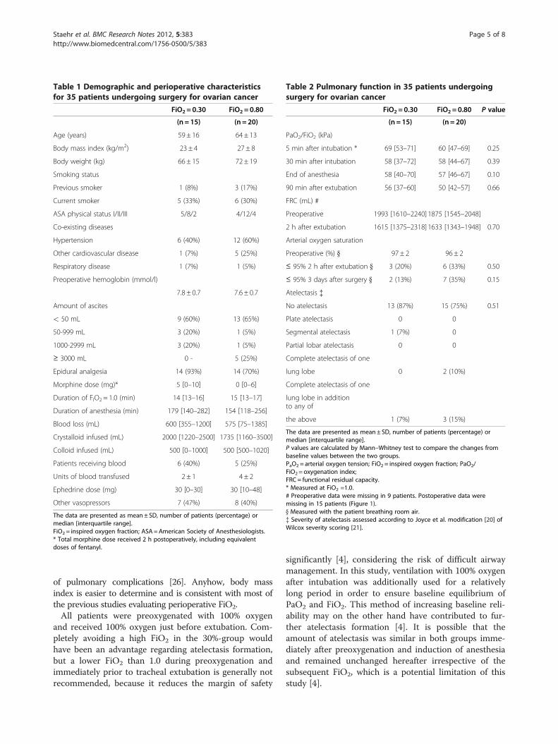
Table 1 Demographic and perioperative characteristicsfor 35 patients undergoing surgery for ovarian cancer
FiO2 = 0.30 FiO2 = 0.80
(n = 15) (n = 20)
Age (years) 59 ± 16 64 ± 13
Body mass index (kg/m2) 23 ± 4 27 ± 8
Body weight (kg) 66 ± 15 72 ± 19
Smoking status
Previous smoker 1 (8%) 3 (17%)
Current smoker 5 (33%) 6 (30%)
ASA physical status I/II/III 5/8/2 4/12/4
Co-existing diseases
Hypertension 6 (40%) 12 (60%)
Other cardiovascular disease 1 (7%) 5 (25%)
Respiratory disease 1 (7%) 1 (5%)
Preoperative hemoglobin (mmol/l)
7.8 ± 0.7 7.6 ± 0.7
Amount of ascites
< 50 mL 9 (60%) 13 (65%)
50-999 mL 3 (20%) 1 (5%)
1000-2999 mL 3 (20%) 1 (5%)
≥ 3000 mL 0 - 5 (25%)
Epidural analgesia 14 (93%) 14 (70%)
Morphine dose (mg)* 5 [0–10] 0 [0–6]
Duration of FIO2 = 1.0 (min) 14 [13–16] 15 [13–17]
Duration of anesthesia (min) 179 [140–282] 154 [118–256]
Blood loss (mL) 600 [355–1200] 575 [75–1385]
Crystalloid infused (mL) 2000 [1220–2500] 1735 [1160–3500]
Colloid infused (mL) 500 [0–1000] 500 [500–1020]
Patients receiving blood 6 (40%) 5 (25%)
Units of blood transfused 2 ± 1 4 ± 2
Ephedrine dose (mg) 30 [0–30] 30 [10–48]
Other vasopressors 7 (47%) 8 (40%)
The data are presented as mean ± SD, number of patients (percentage) ormedian [interquartile range].FiO2 = inspired oxygen fraction; ASA =American Society of Anesthesiologists.* Total morphine dose received 2 h postoperatively, including equivalentdoses of fentanyl.
Table 2 Pulmonary function in 35 patients undergoingsurgery for ovarian cancer
FiO2 = 0.30 FiO2 = 0.80 P value
(n = 15) (n = 20)
PaO2/FiO2 (kPa)
5 min after intubation * 69 [53–71] 60 [47–69] 0.25
30 min after intubation 58 [37–72] 58 [44–67] 0.39
End of anesthesia 58 [40–70] 57 [46–67] 0.10
90 min after extubation 56 [37–60] 50 [42–57] 0.66
FRC (mL) #
Preoperative 1993 [1610–2240]1875 [1545–2048]
2 h after extubation 1615 [1375–2318]1633 [1343–1948] 0.70
Arterial oxygen saturation
Preoperative (%) } 97 ± 2 96 ± 2
≤ 95% 2 h after extubation } 3 (20%) 6 (33%) 0.50
≤ 95% 3 days after surgery } 2 (13%) 7 (35%) 0.15
Atelectasis {
No atelectasis 13 (87%) 15 (75%) 0.51
Plate atelectasis 0 0
Segmental atelectasis 1 (7%) 0
Partial lobar atelectasis 0 0
Complete atelectasis of one
lung lobe 0 2 (10%)
Complete atelectasis of one
lung lobe in additionto any of
the above 1 (7%) 3 (15%)
The data are presented as mean ± SD, number of patients (percentage) ormedian [interquartile range].P values are calculated by Mann–Whitney test to compare the changes frombaseline values between the two groups.PaO2 = arterial oxygen tension; FiO2 = inspired oxygen fraction; PaO2/FiO2 = oxygenation index;FRC = functional residual capacity.* Measured at FiO2 =1.0.# Preoperative data were missing in 9 patients. Postoperative data weremissing in 15 patients (Figure 1).} Measured with the patient breathing room air.{ Severity of atelectasis assessed according to Joyce et al. modification [20] ofWilcox severity scoring [21].
Staehr et al. BMC Research Notes 2012, 5:383 Page 5 of 8http://www.biomedcentral.com/1756-0500/5/383
of pulmonary complications [26]. Anyhow, body massindex is easier to determine and is consistent with most ofthe previous studies evaluating perioperative FiO2.All patients were preoxygenated with 100% oxygen
and received 100% oxygen just before extubation. Com-pletely avoiding a high FiO2 in the 30%-group wouldhave been an advantage regarding atelectasis formation,but a lower FiO2 than 1.0 during preoxygenation andimmediately prior to tracheal extubation is generally notrecommended, because it reduces the margin of safety
significantly [4], considering the risk of difficult airwaymanagement. In this study, ventilation with 100% oxygenafter intubation was additionally used for a relativelylong period in order to ensure baseline equilibrium ofPaO2 and FiO2. This method of increasing baseline reli-ability may on the other hand have contributed to fur-ther atelectasis formation [4]. It is possible that theamount of atelectasis was similar in both groups imme-diately after preoxygenation and induction of anesthesiaand remained unchanged hereafter irrespective of thesubsequent FiO2, which is a potential limitation of thisstudy [4].
FiO2 = 0.30 FiO2 = 0.80
Figure 2 Mean changes in functional residual capacity (FRC) in 35 patients undergoing surgery for ovarian cancer. Solid line representsthe group’s mean. FiO2 = inspired oxygen fraction. Data were missing in 15 patients because of instrumental failure (5 and 3 patients in the 30%-and 80%-group, respectively), inadequate data quality (2 and 3 patients) and 2 patients in the 80%-group refused the postoperative test(Figure 1).
Staehr et al. BMC Research Notes 2012, 5:383 Page 6 of 8http://www.biomedcentral.com/1756-0500/5/383
The change in the PaO2/FiO2 is not a perfect measure-ment to quantify the amount of impaired gas exchange.It is affected by numerous factors other than true intra-pulmonary right to left shunts caused by atelectasis,such as changes in circulatory blood volume, vasopres-sor use, positioning of the patient and body temperature.These factors were, however, not different between thegroups. Other factors such as ventilation-perfusion (VA/Q) inequality and hypoxic pulmonary vasoconstrictioncan also have modified the PaO2/FiO2. Moreover thePaO2/FiO2 is not linearly correlated to FiO2 [22,27]. Asoutcome we measured the change in the PaO2/FiO2
from samples drawn at two different FiO2-levels in eachpatient: baseline at FiO2 1.0 and endpoints at FiO2 0.3or 0.8. However, the mean difference between the groupsin PaO2/FiO2 change from 30 min after intubation tothe end of anesthesia (measured at the same FiO2-levelin each group) was only 3.0 kPa (P= 0.39). We thereforeconsider it most likely that any difference in PaO2/FiO2
must be related to the induction of anesthesia includingthe change from 1.0 to the allocated FiO2.The FRC measurements required patients to breathe
deeply for 30 sec with a respiratory frequency of 20 permin. The 7 patients (5 in the 80%-group) who were
Table 3 Changes in pulmonary function in 35 patients underg
Change in PaO2/FiO2 at the end of anesthesia (kPa
FiO2 = 0.30 FiO2 = 0.80
(n = 15) (n = 20)
Intention-to-treat −7.5 ± 10 -1.2 ± 10
n= 13 n= 20
Per-protocol * −7.1 ± 9 -1.2 ± 10
The data are mean difference form baseline ± SD. P values are calculated by Mann–groups. PaO2 = arterial oxygen tension; FiO2 = inspired oxygen fraction; PaO2/FiO2 =* Excluded patients are described in Figure 1.
unable to cooperate sufficiently or declined to performthe test, had a longer duration of anesthesia, a largerblood loss, and also a larger reduction in PaO2/FiO2.Thus, an effect of high FiO2 on the change in FRC couldhave been overlooked, because FRC data in thesepatients were missing. The need for cooperation is a dis-advantage of the inert gas-rebreathing method in theclinical setting even though it can be very useful inhealthy individuals or mechanically ventilated patients.The mean difference in PaO2/FiO2 reduction between
the 30%-group and the 80%-group was 6.3 ± 10 kPa. There-fore, we cannot exclude that a clinically important differ-ence exists, but our SD was larger than expected and itwould require almost 200 patients to detect this difference.The application of PEEP=5 cmH2O may have reduced
the amount of intraoperative atelectasis and improved theoxygenation [28], however PEEP was applied equally to allpatients exept for 1 in the 30%-group. Hedenstierna et al.showed in a study of 12 patients a trend towards reoccur-ence of atelectasis within 5 min after discontinuation ofPEEP [29] In contrast, a Cochrane review from 2010 indi-cated that intraoperative PEEP of 5–10 cm H2O may re-duce postoperative atelectasis and improve postoperativegasexchange (PaO2/FiO2) [30].
oing surgery for ovarian cancer.
) Change in FRC 2 h after extubation (mL)
P value FiO2 = 0.30 FiO2 = 0.80 P value
(n = 8) (N = 12)
0.10 -49 ± 507 −152± 382 0.70
n = 7 n= 12
0.10 60 ± 435 −152± 382 0.35
Whitney test to compare the changes from baseline values between the twooxygenation index; FRC = functional residual capacitity.
Staehr et al. BMC Research Notes 2012, 5:383 Page 7 of 8http://www.biomedcentral.com/1756-0500/5/383
We found a mean reduction in FRC of 8%, and this isless than the 20% reduction measured shortly after intub-ation [31], but in accordance with the 12% reductionfound on the first day after lower abdominal surgery [32].A postoperative reduction in FRC may not solely be
caused by collapse of some lung units (atelectasis), butalso by a general change in intrathoracic volumes causedby reflex diaphragmatic dysfunction, shallow breathing,mechanical disruption of the abdomen, pulmonaryedema, increased abdominal blood volume or postopera-tive incisional pain[32,33]. We found that the change inFRC and in postoperative PaO2/FiO2 tended to be largerin the patients, who did not receive thoracic epidural an-algesia and this was the case for 6 of the 20 patients inthe 80%-group and 1 of the 15 patients in the 30%-group. However, none of these patients developed radio-logically verified atelectasis. Epidural analgesia improvessome aspects of pulmonary function, which may beattributed to better pain control [34]. We used standar-dized pain relief to ensure that all patients had adequateanalgesia even if epidural was not used. We found nosignificant difference in overall opiod consumption be-tween the groups (Table 1).Our observed changes in FRC may have been too
small to affect the PaO2/FiO2 as noted by Dueck et al.[35]. Their study showed only a small effect of FRC re-duction on the degree of pulmonary shunt until FRC/TLC was less than 0.30, i.e. less than awake, supine clos-ing capacity.Akca et al. found no significant difference in incidence
of atelectasis assessed by CT (64% vs. 94%; P= 0.12) inpatients given 30% or 80% oxygen, respectively [8]. Suchhigh incidences are related to the high sensitivity of aCT-scan. In our study, seven patients (20%) developedradiological verified atelectasis in addition to pulmonarysymptoms. With this definition of atelectasis, no signifi-cant difference in atelectasis was found among the 1,400patients included in the PROXI-trial (7.9% vs. 7.1% inthe 80% and 30% oxygen group, respectively (P= 0.60))[15]. Edmark et al. showed that the benefit of using 80%oxygen compared to 100% oxygen during induction ofanesthesia in order to reduce atelectasis diminishedgradually with time [36]. These results may indicate thatother factors than the oxygen concentration are import-ant to the formation of late peroperative atelectasis.Moreover, patients who are treated with hyperbaric oxy-gen usually receive oxygen at a pressure of 240 kPa in 3cycles of 30 min show no clinically significant signs ofreduction in pulmonary function [37]. This is in accord-ance with the results found in our studyThe patients included were middle-aged women,
undergoing explorative laparotomy of a substantial dur-ation. We cannot reject the possibility that 80% oxygenfor a prolonged period may affect PaO2/FiO2 and FRC
more in heavy smokers or among patients with severepulmonary or cardiovascular comorbidity.
ConclusionVentilation with 80% oxygen for several hours was notsignificantly associated with changes in the perioperativeoxygenation index or functional residual capacity com-pared to ventilation with 30% oxygen. However, a largertrial is required to exclude that an important differencein oxygenation or other clinical outcomes exists.
Competing interestsThe authors have no financial or non-financial competing interests todeclare. This study has no industrial funding or support.
Authors’ contributionsAKS is the prinicpal investigator of the study and was the primary author ofthe manuscript. CSM, PLC, SH and LSR were all involved in study design andmade contributions to the manuscript. All authors have read and approvedthe final version.
AcknowledgementsThis trial is supported by a grant awarded by the Danish National ResearchFoundation (AKS, Principal Investigator).
Author details1Department of Anaesthesia, Centre of Head and Orthopaedics, CopenhagenUniversity Hospital, Rigshospitalet, Blegdamsvej 9, DK-2100, Copenhagen �,Denmark. 2Department of Anaesthesia, The Juliane Marie Centre,Copenhagen University Hospital, Rigshospitalet, Blegdamsvej 9, 2100,Copenhagen �, Denmark. 3Department of Anaesthesia, Næstved Hospital,Ringstedgade 61, 4700, Næstved, Denmark.
Received: 15 November 2011 Accepted: 28 July 2012Published: 28 July 2012
References1. Hedenstierna G: Oxygen and anesthesia: what lung do we deliver to the
post-operative ward. Acta Anaesthesiol Scand 2012, 56:675–685.2. Magnusson L, Spahn DR: New concepts of atelectasis during general
anaesthesia. Br J Anaesth 2003, 91:61–72.3. Hedenstierna G, Edmark L: Mechanisms of atelectasis in the perioperative
period. Best pract Res Clin Anesthesiol 2010, 24:157–169.4. Edmark L, Kostova-Aherdan K, Enlund M, Hedenstierna G: Optimal oxygen
concentration during induction of general anesthesia. Anesthesiology2003, 98:28–33.
5. Meyhoff CS, Staehr AK, Rasmussen LS: Rational use of oxygen in medicaldisease and anesthesia. Curr Opin Anaesthesiol 2012, 25:363–370.
6. Belda FJ, Aguilera L, de la Garcia Asunción J, Alberti J, Vicente R, Ferrándiz L,Rodriguez R, Company R, Sessler DI, Aguilar G, Botello SG, Orti R: SpanishReduccion de la Tasa de Infeccion Quirurgica Group: Supplementalperioperative oxygen and the risk of surgical wound infection: arandomized controlled trial. JAMA 2005, 294:2035–2042.
7. Greif R, Laciny S, Rapf B, Hickle RS, Sessler DI: Supplemental oxygenreduces the incidence of postoperative nausea and vomiting.Anesthesiology 1999, 91:1246–1252.
8. Akca O, Podolsky A, Eisenhuber E, Panzer O, Hetz H, Lampl K, Lackner FX,Wittmann K, Grabenwoeger F, Kurz A, Schultz AM, Negishi C, Sessler DI:Comparable postoperative pulmonary atelectasis in patients given 30%or 80% oxygen during and for 2 hours after colon resection.Anesthesiology 1999, 91:991–998.
9. Hickey RF, Visick WD, Fairley HB, Fourcade HE: Effects of halothaneanesthesia on functional residual capacity and alveolar-arterial oxygentension difference. Anesthesiology 1973, 38:20–24.
10. Tokics L, Hedenstierna G, Svensson L, Brismar B, Cederlund T, Lundquist H,Strandberg Å: V/Q distribution and correlation to atelectasis inanesthetized paralyzed humans. J Appl Physiol 1996, 81:1822–1833.
Staehr et al. BMC Research Notes 2012, 5:383 Page 8 of 8http://www.biomedcentral.com/1756-0500/5/383
11. Maulen-Radovan I, Gutierrez CP, Cosio OE, Marquez AM: Are oxygenindices effective for predicting pathological intrapulmonary shunt inmechanically ventilated children? Arch Med Res 1999, 30:179–185.
12. Agarwal A, Singh PK, Dhiraj S, Pandey CM, Singh U: Oxygen in air (FiO20.4) improves gas exchange in young healthy patients during generalanesthesia. Can J Anaesth 2002, 49:1040–1043.
13. Jacobs I, Oram D, Fairbanks J, Turner J, Frost C, Grudzinskas JG: A risk ofmalignancy index incorporating CA 125, ultrasound and menopausalstatus for the accurate preoperative diagnosis of ovarian cancer.Br J Obstet Gynaecol 1990, 97:922–929.
14. Meyhoff CS, Wetterslev J, Jorgensen LN, Henneberg SW, Simonsen I,Pulawska T, Walker LR, Skovgaard N, Heltø K, Gocht-Jensen P, Carlsson PS,Rask H, Karim S, Carlsen CG, Jensen FS, Rasmussen LS, PROXI Trial Group:Perioperative oxygen fraction - effect on surgical site infection andpulmonary complication after abdominal surgery: a randomized clinicaltrial. Rationale and design of the PROXI-Trial. Trials 2008, 9:58.
15. Meyhoff CS, Wetterslev J, Jorgensen LN, Henneberg SW, Høgdall C, LundvallL, Svendsen PE, Mollerup H, Lunn TH, Simonsen I, Martinsen KR, Pulawska T,Bundgaard L, Bugge L, Hansen EG, Riber C, Gocht-Jensen P, Walker LR,Bendsen A, Johansson G, Skovgaard N, Heltø K, Poukinski A, Korshin A, WalliA, Bulut M, Carlsson PS, Rodt SA, Lundbech LB, Rask H, Buch N, PerdawidSK, Reza J, Jensen KV, Carlsen CG, Jensen FS, Rasmussen LS, PROXI TrialGroup: Effect of high perioperative oxygen fraction on surgical siteinfection and pulmonary complications after abdominal surgery: thePROXI randomized clinical trial. JAMA 2009, 302(14):1543–1550.
16. Clemensen P, Christensen P, Norsk P, Gronlund J: A modified photo- andmagnetoacoustic muligas analyzer applied in gas exchangemeasurements. J Appl Physiol 1994, 76:2832–2839.
17. Peyton PJ, Poustie SJ, Robinson GJ, Penny DJ, Thompson B: Non-invasivemeasurement of intrapulmonary shunt during inert gas rebreathing.Physiol Meas 2005, 26:309–316.
18. Hansen S, Wendelboe O, Christensen P: The non-invasive acetylenerebreathing method for estimation of cardiac output: influence ofbreath-by-breath variation. Clin Physiol 1997, 17:193–202.
19. Kallay MC, Hyde RW, Fahey PJ, Utell MJ, Peterson BT, Ortiz CR: Effect of therebreathing pattern on pulmonary tissue volume and capillary bloodflow. J Appl Physiol 1985, 85:1881–1894.
20. Joyce CJ, Baker AB, Chartres S: Influence of inspired nitrogenconcentration during anaesthesia for coronary artery bypass grafting onpostoperative atelectasis. Br J Anaesth 1995, 75:422–427.
21. Wilcox P, Baile EM, Hards J, Müller NL, Dunn L, Pardy RL, Paré PD: Phrenicnerve function and its relationship to atelectasis after coronary arterybypass surgery. Chest 1988, 93:693–698.
22. Karbing DS, Kjaergaard S, Smith BW, Espersen K, Allerød C, Andreasen S,Rees SE: Variation in the PaO2/FiO2 ratio with FiO2: mathematical andexperimental description, and clinical relevance. Crit Care 2007, 11:R118.
23. Gedall KA, Raniga S, Kennedy R, Frizelle FA: The impact of obesity onoutcome after colorectal surgery. Dis Colon Rectum 2007, 50:2223–2237.
24. Canet J, Gallart L, Gomar C, Paluzie G, Vallès J, Castillo J, Sabaté S, Mazo V,Briones Z, Sanchis J, ARISCAT Group: Prediction of PostoperativePulmonary Complications in a Population-based Surgical Cohort.Anesthesiology 2010, 113:1338–1350.
25. Egan TD, Wong KC: Perioperative smoking cessation and anesthesia: areview. J Clin Anesth 1992, 4:63–72.
26. Canoy D, Luben R, Weich A, Bingham S, Wareham N, Day N, Khaw KT:Abdominal obesity and respiratory function in men and women in theEPIC-Norfolk Study, United Kingdom. Am J Epidemiol 2004, 159:1140–1149.
27. Whiteley JP, Gavaghan DJ, Hahn CE: Variation of venous admixture, SF6shunt, PaO2 and the PaO2/FIO2 ratio with FIO2. Br J Anaesth 2002,88:771–778.
28. Rusca M, Proietti S, Schnyder P, Frascarolo P, Hedenstierna G, Spahn DR,Magnusson L: Prevention of atelectasis formation during induction ofgeneral anesthesia. Anesth Analg 2003, 97:1835–1839.
29. Hedenstierna G, Tokics L, Lundquist H, Andersson T, Strandberg A, BrismarB: Phrenic nerve stimulation during halothane anesthesia. Effects ofatelectasis. Anesthesiology 1994, 80:751–760.
30. Imberger G, McIlroy D, Pace NL, Wetterslev J, Brok J, Møller AM: Positiveend-expiratory pressure (PEEP) during anaesthesia for the prevention ofmortality and postoperative pulmonary complications. CochraneDatabase Syst Rev 2010, 9:. doi:10.1002/14651858.CD007922.pub2.CD007922.
31. Wahba RW: Perioperative functional residual capacity. Can J Anaesth 1991,38:384–400.
32. Drummond GB, Littlewood DG: Respiratory effects of extradural analgesiaafter lower abdominal surgery. Br J Anaesth 1977, 49:999–1004.
33. Craig DB: Postoperative recovery of pulmonary function. Anesth Analg1981, 60:46–52.
34. Pöpping DM, Elia N, Marret E, Remy C, Tramèr MR: Protective effects ofepidural analgesia on pulmonary complications after abdominal andthoracic surgery: a metaanalysis. Arch Surg 2008, 143:990–999.
35. Dueck R, Prutow RJ, Davies NJ, Clausen JL, Davidson TM: The lung volumeat which shunting occurs with inhalation anesthesia. Anesthesiology 1988,69:854–61.
36. Edmark L, Auner U, Enlund M, Östberg E, Hedenstierna G: Oxygenconcentration and characteristics of progressive atelectasis formationduring anaesthesia. Acta Anaesthesiol Scand 2011, 55:75–81.
37. Thorsen E, Aanderud L, Aasen TB: Effects of a standard hyperbaric oxygentreatment protocol on pulmonary function. Eur Respir J 1998,12:1442–1445.
doi:10.1186/1756-0500-5-383Cite this article as: Staehr et al.: Influence of perioperative oxygenfraction on pulmonary function after abdominal surgery:a randomized controlled trial. BMC Research Notes 2012 5:383.
Submit your next manuscript to BioMed Centraland take full advantage of:
• Convenient online submission
• Thorough peer review
• No space constraints or color figure charges
• Immediate publication on acceptance
• Inclusion in PubMed, CAS, Scopus and Google Scholar
• Research which is freely available for redistribution
Submit your manuscript at www.biomedcentral.com/submit