Embed Size (px)
Citation preview

Li et al. BMC Plant Biology (2015) 15:143 DOI 10.1186/s12870-015-0536-z
RESEARCH ARTICLE Open Access
Tomato Sl3-MMP, a member of the Matrixmetalloproteinase family, is required fordisease resistance against Botrytis cinerea andPseudomonas syringae pv. tomato DC3000
Dayong Li, Huijuan Zhang, Qiuming Song, Lu Wang, Shixia Liu, Yongbo Hong, Lei Huang and Fengming Song*Abstract
Background: Matrix metalloproteinases (MMPs) are a family of zinc-dependent endopeptidases. MMPs have beencharacterized in detail in mammals and shown to play key roles in many physiological and pathological processes.Although MMPs in some plant species have been identified, the function of MMPs in biotic stress responses remainselusive.
Results: A total of five MMP genes were identified in tomato genome. qRT-PCR analysis revealed that expression ofSl-MMP genes was induced with distinct patterns by infection of Botrytis cinerea and Pseudomonas syringae pv. tomato(Pst) DC3000 and by treatment with defense-related hormones such as salicylic acid, jasmonic acid and ethyleneprecursor 1-amino cyclopropane-1-carboxylic acid. Virus-induced gene silencing (VIGS)-based knockdown of individualSl-MMPs and disease assays indicated that silencing of Sl3-MMP resulted in reduced resistance to B. cinerea and PstDC3000, whereas silencing of other four Sl-MMPs did not affect the disease resistance against these two pathogens.The Sl3-MMP-silenced tomato plants responded with increased accumulation of reactive oxygen species and alertedexpression of defense genes after infection of B. cinerea. Transient expression of Sl3-MMP in leaves of Nicotianabenthamiana led to an enhanced resistance to B. cinerea and upregulated expression of defense-related genes.Biochemical assays revealed that the recombinant mature Sl3-MMP protein had proteolytic activities in vitro withdistinct preferences for specificity of cleavage sites. The Sl3-MMP protein was targeted onto the plasma membrane ofplant cells when transiently expressed in onion epidermal cells.
Conclusion: VIGS-based knockdown of Sl3-MMP expression in tomato and gain-of-function transient expression ofSl3-MMP in N. benthamiana demonstrate that Sl3-MMP functions as a positive regulator of defense response against B.cinerea and Pst DC3000.
Keywords: Tomato (Solanum lycopersicum), Matrix metalloproteinases, Botrytis cinerea, Pseudomonas syringae pv.tomato DC3000, Disease resistance, Proteolysis
* Correspondence: [email protected] Key Laboratory for Rice Biology, Institute of Biotechnology, ZhejiangUniversity, Hangzhou, Zhejiang 310058, China
© 2015 Li et al. This is an Open Access article distributed under the terms of the Creative Commons Attribution License(http://creativecommons.org/licenses/by/4.0), which permits unrestricted use, distribution, and reproduction in any medium,provided the original work is properly credited. The Creative Commons Public Domain Dedication waiver (http://creativecommons.org/publicdomain/zero/1.0/) applies to the data made available in this article, unless otherwise stated.

Li et al. BMC Plant Biology (2015) 15:143 Page 2 of 18
BackgroundProteases play key roles in the regulation of a variety ofbiological processes [1]. Matrix metalloproteinases(MMPs) are a family of zinc- and calcium-dependentproteases belonging to the metzincin clan of metalloen-dopeptidases, EC subclass 3.4.24, MA (M) clan accord-ing to the MEROPS database [2, 3]. The MMP family ischaracterized by the presence of a highly conserved cata-lytic domain containing an HEXXHXXGXX(H/D) zinc-binding sequence followed by a conserved methioninethat forms a tight 1,4-β turn called Met-turn [4]. Membersof this family have mainly been studied in mammals, buthave also been found in simpler animals and plants [5]. Inhuman, 23 MMP genes have been identified to encodeproteins with similar structure, e.g., an N-terminal signalpeptide for the secretory pathway, a prodomain that regu-lates the latency of the enzyme and a catalytic domainwith the active zinc-binding site [6]. In addition, most ofthe human MMP proteins contain a C-terminal hemo-pexin (HPX)-like domain, which is believed to be import-ant in regulating the activity and specificity of the catalyticdomain [7, 8]. It has been shown that human MMPs playkey roles in many physiological and pathological processes[9, 10].Members of the MMP family have been identified in
higher plants, but only few of them have been studied todate [2]. Similar to the human MMPs, the predicted pri-mary structures of plant MMPs contain a signal peptide,a prodomain with the cysteine-switch motif and a cata-lytic domain containing the active zinc-binding sequenceand structural zinc- and calcium-binding site followedby the conserved Met-turn [11–13]. Activation of MMPsrequires physical delocalization of the prodomain fromthe catalytic site by proteolytic or nonproteolytic mecha-nisms [14]. It is believed that all plant MMPs are synthe-sized as inactive forms and are localized either in theplasma membrane or in the extracellular space. How-ever, it was found that Arabidopsis At4-MMP contains apredicted non-cleavable N-terminal signal peptide andtobacco Nt1-MMP was inserted into the plasma mem-brane [15].The biological function of MMP proteases in higher
plants is largely unknown. Based on the expression pat-terns, it is proposed that the plant MMPs may be in-volved in remodeling of the extracellular matrix (ECM)during plant growth and development [2]. The first plantMMP was identified as an ethylenediaminetetraaceticacid (EDTA)-sensitive Azocoll-degrading enzyme in soy-bean [16]. In cucumber, Cs1-MMP was found to be as-sociated with senescence and cell death in cotyledondevelopment [17]. In Arabidopsis, 5 MMP genes wereidentified and were found to be differentially expressedin roots, leaves, stems and flowers [15]. The At2-MMPmutant plants exhibited altered growth in association
with late flowering and early senescence, supporting aphysiological and developmental role for plant MMPs[18]. In Medicago truncatula, expression of Mt1-MMPwas induced in young nodules, specifically in associationwith Sinorhizobium meliloti infection [13]. An Mt1-MMP RNAi mutant in M. truncatula showed noduleswith enlarged infection threads and substantial increasein the number of bacterial colonies; whereas an ectopicoverexpression of Mt1-MMP in roots led to a significantdecrease in nodule number [13]. On the other hand, sev-eral lines of evidence also indicate that MMPs may beinvolved in biotic and abiotic stress responses in plants.In soybean, Gm2-MMP was isolated as a pathogen-induced gene [19]. Expression of Gm2-MMP wasinduced rapidly in compatible and incompatible interac-tions with pathogens, but not by salicylic acid (SA) andjasmonic acid (JA), two classical pathogen response sig-naling molecules [19]. In the tobacco suspension lineBY-2, Nt1-MMP was expressed at low level but was in-duced immediately after treatment with Pseudomonassyringae [11]. In Arabidopsis, distinct expression pat-terns for each MMP in response to various abiotic andbiotic stresses were described in the Genevestigator ana-lysis [20]. At3-MMP showed significant changes in tran-script levels under stress conditions, while other MMPsdisplayed minimal transcript changes [20]. The expressionof At2-MMP is tightly controlled in a tissue-responsiveway during stress conditions. At2-MMP in 4-week-oldplants was induced in leaves by cadmium or methyljasmonate and in roots by sodium chloride; however, cad-mium inhibited the expression of At2-MMP in inflores-cence and leaves of 10-week-old plants [18].In the present study, we characterized the MMP family
in tomato and performed functional analyses for theirroles in disease resistance. A total of five MMP geneswere identified in tomato and their expression wasinduced with distinct patterns in response to pathogeninfection and treatments with defense-related hormones.Silencing of Sl3-MMP in tomato resulted in reduced re-sistance to Botrytis cinerea and Pseudomonas syringaepv. tomato (Pst) DC3000 whereas transient expression ofSl3-MMP in Nicotiana benthamiana led to an enhancedresistance to B. cinerea. Our data demonstrate thatmember of the MMP family may participate in the regu-lation of defense response in plants against pathogeninfection.
ResultsIdentification of the Sl-MMP family in tomatoTo identify members of the MMP family in tomato,HMM and Blastp searches using MMP proteins previ-ously reported from Arabidopsis and other plant speciesas queries against the recently published tomato genomesequences (Release Version ITAG2.40) were performed.

Li et al. BMC Plant Biology (2015) 15:143 Page 3 of 18
Five significant hits corresponding to non-redundantputative Sl-MMP genes were identified (Table 1). Allfive Sl-MMPs are intronless genes, which is consistent withstructural features of genes for MMPs in Arabidopsis, soy-bean, cucumber andMedicago truncatula [13, 15, 17, 19, 21].Full-length cDNAs for Sl2-MMP and Sl3-MMP wereidentified in the NCBI and SOL databases while no full-length cDNA was found for other three members (Table 1).We amplified and cloned all 5 Sl-MMP genes using gene-specific primers and confirmed by sequencing. These se-quences were submitted to GenBank for deposition andpresented in Additional file 1.The Sl-MMP proteins are approximately 360 amino
acids with molecular weight of ~40 kDa (Table 1). TheSl-MMP proteins shared conserved structural features,e.g., a signal sequence at N terminus, a propeptide do-main, a catalytic domain, a transmembrane domain at Cterminus (Fig. 1a). Characteristic motifs including aPRCGxxD motif, which is characteristic of the cysteineswitch mechanism of activation [22], in the propeptidedomain and a HExGHxxGxxH zinc-binding region anda conserved methionine residue in the Met-turn in thecatalytic domain are present in the Sl-MMP proteins(Fig. 1b). In addition, each of Sl-MMPs contains aninvariant DLESV motif on the N-terminal side of thezinc-binding region (Fig. 1b), which is thought to be aplant-specific motif with unknown function [15]. Thismotif is replaced by a distinct consensus sequence ofNLFLV in human and insect MMPs [4, 23] but is notpresent in single-celled green algae MMPs [20, 24]. Interm of secondary structure feature, Sl-MMPs have 2 β-strands, 4 β-sheets and 3 α-helices and they all containthree active site histidines and a catalytic glutamate resi-due in the zinc-binding region (Fig. 1a). Putative con-served structural ligands for binding zinc and calciumincluding 3 histidine (H), 2 aspartic acid (D) and 1 glu-tamic acid residues are present in Sl-MMPs (Fig. 1a).However, like MMPs from other plants, Sl-MMPs donot contain a C-terminal hemopexin domain, which ispresent in most human MMPs [8].Phylogenetic tree analysis of Sl-MMPs with previously
identified MMPs from other plant species clearly distin-guished different groups, which had distinct featureslinked to plant species or specific functions. Groups Iand II can be further separated into two subgroups,
Table 1 Characterization of tomato Sl-MMP genes and proteins
Genes Locus in SOL Proteins in NCBI Size (aa)
Sl1-MMP Solyc08g078550 XP_010325488 356
Sl2-MMP Solyc04g005040 CCH68443 363
Sl3-MMP Solyc04g005050 NP_001266203 367
Sl4-MMP Solyc05g006360 XP_010320628 357
Sl5-MMP Solyc10g018750 XP_004248606 357
representing MMP branches from dicots and monocots.Sl2-MMP, Sl3-MMP and Sl4-MMP are assigned in GroupI, which contain Nt1-MMP, At2-MMP and At3-MMPthat are known to be pathogen-responsive [11, 18, 20].Sl1-MMP and Sl5-MMP belong to Group II, whosemembers have been proposed to be involved in plantgrowth and development [20]. Group III only hostsMMPs from legumes such as Glycine max and Medicagotruncatula and thus seem to be legume-specific.
Expression of Sl-MMPs in response to pathogens anddefense signaling-related hormonesTo explore the possible involvement of Sl-MMPs indefense response against pathogen infection, we firstanalyzed the expression changes of Sl-MMPs after infec-tion with B. cinerea. As shown in Fig. 2a, expression ofthe Sl-MMP genes were induced upon infection of B.cinerea but showed distinct expression patterns. Gener-ally, the expression of Sl1-MMP, Sl3-MMP, Sl4-MMPand Sl5-MMP was significantly induced with peaks at48 h whereas the expression of Sl2-MMP was inducedsignificantly with peaks at 24 h after infection with B.cinerea, as compared with those in the mock-inoculatedplants (Fig. 2a). Specifically, the expression levels of Sl1-MMP, Sl3-MMP and Sl5-MMP in B. cinerea-infectedplants showed >5 folds of increases over those in themock-inoculated plants at 48 h after inoculation (Fig. 2a).The expressions of Sl1-MMP and Sl3-MMP exhibited 4-5 folds of increases at 24 h after infection of B. cinerea.It was noted that the expression of Sl1-MMP, Sl4-MMPand Sl5-MMP was induced only at 48 h after infectionof B. cinerea (Fig. 2a). These results indicate that the Sl-MMP genes respond with different dynamics and magni-tude of expression after infection of B. cinerea.We next analyzed the expression changes of Sl-MMPs
after infection with Pst DC3000. As shown in Fig. 3b,only Sl1-MMP, Sl2-MMP and Sl3-MMP were inducedupon infection of Pst DC3000 and again showed differ-ent expression patterns. Generally, the expression of Sl1-MMP and Sl3-MMP was induced significantly withpeaks at 24 h while the expression of Sl2-MMP was in-duced significantly with peaks at 12 h after infectionwith Pst DC3000, as compared with those in the mock-inoculated plants (Fig. 2b). Specifically, the expression ofSl1-MMP and Sl3-MMP in Pst DC3000-infected plants
MW (kD) pI cDNAs in NCBI/SOL
40.17 5.20 –
40.00 5.46 AK328733/AK327627/ SGN-U573509
40.21 5.08 AK322919/ SGN-U573510
40.50 6.00 –
40.80 7.83 –

Fig. 1 Sequence alignment and phylogenetic tree analysis of Sl-MMP with other plant MMP proteins. a Predicted domains of Sl-MMP proteins. TM,transmembrane domain. b Alignment of Sl-MMPs. Numbers on the right indicate amino acid positions of the Sl-MMP proteins. The cysteine switchmotif, zinc-binding sequence and DLESV sequence are boxed in red. Secondary structure features such as β-strand, β-sheet and α-helix are indicatedabove the aligned sequences. The active site histidine and the catalytic glutamate residues are indicated in red. Ligands of the conserved structural zincand calcium are colored in green and yellow, respectively. The hydrophobic base forming the methionine residue of the Met-turn is highlighted inblue. c Phylogenetic tree analysis of Sl-MMPs with other plant MMPs. Phylogenetic tree was constructed by Neighbor-joining method using MEGAprogram. Plant MMPs used and their GenBank accessions are as follows: Arabidopsis thaliana At1-MMP (NP_193397), At2-MMP (NP_177174), At3-MMP(NP_173824), At4-MMP (NP_182030), At5-MMP (NP_176205), Glycine max Gm2-MMP (AAL27029), Gm-ACU24527 (ACU24527), Gm-Slti114 (ABW96008),Hordeum vulgare HvMMP1 (BAJ94792), HvMMP2 (BAJ93963), HvMMP3 (BAJ94176), HvMMP4 (BAJ90264), Medicago truncatula Mt1-MMP (CAA77093),Nicotiana tabacum NtMMP1 (ABF58910), Solanum lycopersicum Sl1-MMP (XP_010325488), Sl2-MMP (CCH68443), Sl3-MMP (NP_001266203), Sl4-MMP(XP_010320628), Sl5-MMP (XP_004248606), Zea mays ZmMMP1 (NP_001151749), ZmMMP2 (NP_001142095), Oryza sativa Os1-MMP (NP_001048075),Os2-MMP (NP_001057259), Os3-MMP (NP_001065361). Bootstrap values from 100 replicates are indicated at each node. Bar represents the number ofamino acid differences per site
Li et al. BMC Plant Biology (2015) 15:143 Page 4 of 18

Fig. 2 Expression patterns of Sl-MMPs in response to B. cinerea or P. syringae pv. tomato DC3000 treatment. Tomato plants were inoculated by sporesuspension (2 × 10⁵ spores/ml) of B. cinerea or buffer solution as a mock-inoculation control (a) and by vacuum infiltration with P. syringae pv. tomatoDC3000 (OD600 = 0.0002) or sterilized 10 mM MgCl2 solution as a mock-inoculation control (b). Leaf samples were collected at indicated time pointsand gene expression was analyzed by qRT-PCR. Relative expression levels were calculated by comparing with the corresponding values at 0 h (as acontrol) after inoculation and shown as folds of the actin transcript values. Data presented are the means ± SD from three independent experimentsand different letters above the columns indicate significant differences at p < 0.05 level
Li et al. BMC Plant Biology (2015) 15:143 Page 5 of 18
showed >5 folds of increases over those in the mock-inoculated plants at 24 h, whereas the expression of Sl2-MMP was induced significantly with peaks at 12 and24 h after infection with Pst DC3000 (Fig. 2b). Interest-ingly, the expression of Sl5-MMP was down-regulated at12 h (Fig. 2b). These results indicate that the expressionof Sl1-MMP, Sl2-MMP and Sl3-MMP was induced byPst DC3000.We also examined the dynamics of Sl-MMPs expres-
sions in tomato plants after treatments with SA, methyljasmonate (MeJA) and 1-amino cyclopropane-1-carboxylicacid (ACC) [a precursor of ethylene (ET)], three defensesignaling-related hormones. As shown in Fig. 4, differentexpression patterns for Sl-MMPs were observed in re-sponse to these defense signaling-related hormones. InSA-treated plants, expression of Sl3-MMP and Sl4-MMPwas significantly increased by 2-3 folds over that in thecontrol plants, while expressions of Sl1-MMP, Sl2-MMPand Sl3-MMP were not affected (Fig. 3a). In JA- or ACC-treated plants, expression of Sl4-MMP was stronglyinduced, reaching 3-4 folds of increased at 6 h aftertreatment (Fig. 3b and c). Besides Sl4-MMP, the expres-sion of Sl1-MMP, Sl2-MMP and Sl3-MMP was also
induced by JA, showing an increase of 2-3 folds at12 h (Fig. 3b). Except Sl4-MMP, expression of otherfour Sl-MMPs was not affected by ACC (Fig. 3c).Interestingly, the expression of Sl5-MMP was not af-fected by both JA and ACC during the experimentalperiod (Fig. 3b and c). These data indicate that the to-mato Sl-MMPs respond with different expression pat-terns to SA, JA and ET, three well-known defensesignaling-related hormones.
Silencing of Sl3-MMP resulted in reduced resistance to B.cinerea and Pst DC3000To examine the possible involvement of Sl-MMPs in dis-ease resistance, we performed functional analyses byvirus-induced gene silencing (VIGS) approach throughcomparing the disease phenotype between individual Sl-MMP-silenced plants and non-silenced control plants.For this purpose, specific fragment for each Sl-MMPgene (Additional file 2) was chosen to generate VIGSconstruct and standard VIGS procedure with a phytoenedesaturase (PDS) construct as an indicative for VIGS ef-ficiency of each experiment was performed on 2-week-old plants [25, 26]. Under our experiment conditions, >90 %

Fig. 3 Expression patterns of Sl-MMPs in response to defense signaling hormones. Tomato plants were treated by foliar spraying of 1 mM SA(a), 100 μM MeJA (b), 100 μM ACC (c) or equal volume of solution as a control and leaf samples were collected at indicated time points. Geneexpression was analyzed by qRT-PCR and relative expression levels were calculated by comparing with the corresponding values at 0 h(as a control) after treatment. Relative expression was shown as folds of the actin transcript values. Data presented are the means ± SD fromthree independent experiments and different letters above the columns indicate significant differences at p < 0.05 level
Li et al. BMC Plant Biology (2015) 15:143 Page 6 of 18
of the pTRV2-PDS-infiltrated plants showed bleachingphenotype (data not shown). The silencing efficiency andspecificity for each Sl-MMP gene was examined by qRT-PCR analyzing the transcript level of the target Sl-MMPgene and other four Sl-MMP genes in the pTRV2-target Sl-MMP-infiltrated plants. When compared with those in thepTRV2-GUS-infiltrated plants, the transcript level of the tar-get Sl-MMP gene was significantly reduced whereas thetranscript levels of the other Sl-MMP genes were compar-able in the pTRV2-target Sl-MMP-silenced plants (Fig. 4).Overall, the silencing efficiency for a target Sl-MMP genewas approximately 70 % (Fig. 4). The efficiencies and specifi-city of silencing for each individual Sl-MMP gene were satis-fied for further experiments and all the subsequentexperiments were performed only on those pTRV2-Sl-MMPs-infiltrated plants with high levels of silencingefficiency (>70 %).
We first examined the possible involvement of Sl-MMPs in resistance against B. cinerea by challenging thepTRV2-Sl-MMPs-infiltrated plants with spore suspen-sion of B. cinerea and comparing the disease severityand in planta fungal growth with those in pTRV-GUS-infiltrated non-silenced plants. In our detached leaf as-says, B. cinerea-caused lesions on detached leaves fromthe pTRV2-Sl1-MMP-, pTRV2-Sl2-MMP-, pTRV2-Sl4-MMP- and pTRV2-Sl5-MMP-infiltrated plants weresimilar to those on the detached leaves from pTRV2-GUS-infilrtratd plants (Fig. 5a), suggesting that Sl1-MMP, Sl2-MMP, Sl4-MMP and Sl51-MMP may not beinvolved in resistance against B. cinerea. However, B.cinerea-caused lesions on detached leaves from thepTRV2-Sl3-MMP-infiltrated plans were significantly lar-ger and developed faster, merging into large necroticareas, as compared with those on leaves from the

Fig. 4 Silencing efficiency and specificity for target genes in silenced plants. Two-week-old tomato seedlings were infiltrated with agrobacteriacarrying pTRV2-Sl-MMPs or pTRV2-GUS and leaf samples were collected at 4 weeks after agroinfiltration. Expression levels of each Sl-MMP genesin targeted and nontargeted Sl-MMP-silenced and non-silenced plants were analyzed by qRT-PCR and data obtained were normalized with actintranscript values. Data presented are the means ± SD from three independent experiments and different letters above the columns indicatesignificant differences at p < 0.05 level
Li et al. BMC Plant Biology (2015) 15:143 Page 7 of 18
pTRV2-GUS-infiltrated plants (Fig. 5a), at 3 days afterinoculation (dpi), showing an approximately 60 % of in-crease in lesion size over those on leaves from thepTRV2-GUS-infiltrated control plants (Fig. 5c). We fur-ther analyzed and compared the disease severity and inplanta fungal growth in the pTRV2-Sl3-MMP- andpTRV2-GUS-infiltrated plants after inoculation by foliarspraying with spore suspension of B. cinerea in wholeplant inoculation experiments. As shown in Fig. 5b, thepTRV2-GUS-infiltrated control plants displayed slightdisease symptoms, whereas the pTRV2-Sl3-MMP-infil-trated plants showed severe diseases symptoms, showinglarge necrotic areas and maceration or wilting of fullleaves at 5 dpi. Analysis of the transcript for the B.cinerea actin gene BcActin revealed that growth of B.cinerea in leaf tissues of the pTRV2-Sl3-MMP-infiltratedplants had 3 times higher than those in the pTRV2-GUS-infiltrated control plants at 24 and 48 h after in-oculation (Fig. 5d). These data indicate that silencing ofthe Sl3-MMP resulted in reduced resistance to B.cinerea, demonstrating the requirement of Sl3-MMP forresistance to B. cinerea.As mentioned above that silencing of Sl3-MMP re-
sulted in a clear phenotype in change of disease resist-ance to B. cinerea, we subsequently focused our effortson the functions in resistance to other diseases, mechan-ism and biochemical activity of Sl3-MMP. We examined
whether Sl3-MMP is also involved in resistance againstPst DC3000, which is a hemibiotrophic bacterial patho-gen that has different infection style from that of B.cinerea. In our experiments, necrotic lesions were ob-served in the inoculated leaves of the pTRV2-Sl3-MMP-and pTRV2-GUS- infiltrated plants; however, the lesionson leaves of the pTRV2-Sl3-MMP-infiltrated plants werelarger and denser than those in the pTRV2-GUS-infiltrated plants (Fig. 6a). At 2 and 4 dpi, the bacterialpopulation in the inoculated leaves of the pTRV2-Sl3-MMP-infiltrated plants showed approximately 10 and 25folds higher over those in the pTRV2-GUS-infiltratedplants, respectively (Fig. 6b). These results indicate thatsilencing of Sl3-MMP resulted in reduced resistance toPst DC3000, implying the requirement of Sl3-MMP forresistance against Pst DC3000.
Silencing of Sl3-MMP attenuated defense responseagainst B. cinereaTo elucidate the possible mechanism involved in the re-duced resistance in Sl3-MMP-silenced plants, we ana-lyzed and compared the accumulation of reactive oxygenspecies (ROS), cell-death response and expression ofdefense genes before and after infection with B. cinereabetween the Sl3-MMP-silenced plants and the controlplants. No difference in accumulation of H2O2, as de-tected by 3, 3-diaminobenzidine (DAB) staining, was

Fig. 5 Silencing of Sl3-MMP resulted in reduced resistance to B. cinerea. Two-week-old seedlings were infiltrated with agrobacteria carryingpTRV2-Sl-MMP or pTRV2-GUS and were inoculated at 4 weeks after VIGS infiltration by dropping spore suspension (1 × 10⁵ spores/mL) ondetached leaves or foliar spraying with spore suspension (2 × 10⁵ spores/mL) onto leaves of whole plants. a and b Disease phenotype and lesionsizes in leaves of the pTRV2-Sl-MMPs- and pTRV2-GUS-infiltrated plants in detached leaf inoculation assays. Lesion sizes were measured at 3 daysafter inoculation on a minimum of 20 leaves in each experiment. c and d Disease phenotype on and fungal growth in the pTRV2-Sl3-MMP- and pTRV2-GUS-infiltrated plants in whole plant inoculation assays. Fungal growth in planta was estimated by analyzing the transcript levels of BcActin gene by qRT-PCR using SlActin as an internal control at the indicated time points after inoculation. Data presented in b and d are the means ± SD from threeindependent experiments and different letters above the columns indicate significant differences at p < 0.05 level
Li et al. BMC Plant Biology (2015) 15:143 Page 8 of 18
observed in leaves of pTRV2-Sl3-MMP- and pTRV2-GUS-infiltrated plants without infection of B. cinerea(Fig. 7a), indicating that silencing of Sl3-MMP itself didnot affect the generation and accumulation of H2O2 in to-mato plants. After infection with B. cinerea, significant ac-cumulation of H2O2, shown as brown precipitates inleaves, was detected in leaves of pTRV2-Sl3-MMP- andpTRV2-GUS-infiltrated plants (Fig. 7a). However, theleaves from pTRV2-Sl3-MMP-infiltrated plants showedconsistent increase in intensity of the stained areas(Fig. 7a), showing increases of 85 % at 12 h and 24 % 24 h,when compared with those in pTRV2-GUS-infiltrated
plants after infection of B. cinerea (Fig. 7b). On the otherhand, levels of cell death, as detected by Trypan bluestaining, and electrolyte leakage, as estimated by ion con-ductivity, were comparable in leaves of pTRV2-Sl3-MMP-and pTRV2-GUS-infiltrated plants without infection of B.cinerea but significantly increased after infection with B.cinerea (Fig. 7c and d). Notably, the levels of cell deathand electrolyte leakage in leaves of pTRV2-Sl3-MMP-infil-trated plants were significantly higher than those in leavesof pTRV2-GUS-infiltrated plants after infection of B.cinerea (Fig. 7c), leading to 38 % increases for electrolyteleakage at 24 h after infection (Fig. 7d). These data

Fig. 6 Silencing of Sl3-MMP resulted in reduced resistance to P.syringae pv. tomato DC3000. Two-week-old seedlings were infiltratedwith agrobacteria carrying pTRV2-Sl3-MMP or pTRV2-GUS and wereinoculated by infiltration with Pst DC3000 4 weeks after VIGS infiltration.a Representative disease phenotype. b Bacterial population. Datapresented in b are the means ± SD from three independentexperiments and different letters above the columns indicate significantdifferences at p< 0.05 level
Fig. 7 Increased accumulation of H2O2 and cell death in Sl3-MMP-silenced planwith agrobacteria carrying pTRV2-Sl3-MMP or pTRV2-GUS and were inoculatedinoculation control at 4 weeks after VIGS infiltration. Leaves from six individual pdeath and H2O2 accumulation. a and b Accumulation of H2O2 by DAB stainingblue staining. d Electrolyte leakage. Data presented in b and d are the means ±columns indicate significant differences at p< 0.05 level
Li et al. BMC Plant Biology (2015) 15:143 Page 9 of 18
indicate that silencing of Sl3-MMP resulted in increasedROS accumulation of H2O2 and excessive cell death inpTRV2-Sl3-MMP-infiltrated plants upon infection of B.cinerea.To explore the possible mechanism for the increased
accumulation of H2O2 in the Sl3-MMP-silenced plants,we analyzed and compared the expression of genes en-coding for NADPH oxidases, glutathione reductase(GR), catalases (CAT), superoxide dismutases (SOD) andascorbate peroxidases (APX) in the pTRV2-Sl3-MMP-infiltrated plants. As shown in Fig. 8a, no significant dif-ference in the expression levels of these selected geneswas observed between the pTRV2-GUS-infiltrated andpTRV2-Sl3-MMP-infiltrated plants without infection ofB. cinerea. By contrast, the expression levels of Rboh1and Wfi1, two genes for NADPH oxidases, in thepTRV2-Sl3-MMP-infiltrated plants were significantlyelevated upon Botrytis infection, showing ~5-fold in-creases over those in the pTRV2-GUS-infiltrated plants.Similarly, the expression levels of APX and GR in thepTRV2-Sl3-MMP-infiltrated plants were also increasedas compared with those in the pTRV2-GUS-infiltratedplants (Fig. 8a). By contrast, no significant difference
ts after infection with B. cinerea. Two-week-old seedlings were infiltratedwith spore suspension of B. cinerea or with buffer solution as a mock-lants were collected at 24 h after inoculation and used for analysis of celland quantification method respectively. c Cell death detected by trypanSD from three independent experiments and different letters above the
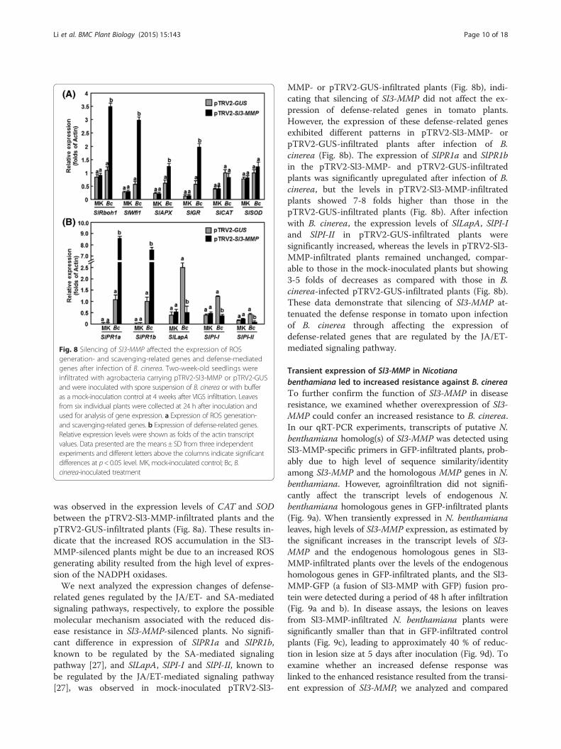
Fig. 8 Silencing of Sl3-MMP affected the expression of ROSgeneration- and scavenging-related genes and defense-mediatedgenes after infection of B. cinerea. Two-week-old seedlings wereinfiltrated with agrobacteria carrying pTRV2-Sl3-MMP or pTRV2-GUSand were inoculated with spore suspension of B. cinerea or with bufferas a mock-inoculation control at 4 weeks after VIGS infiltration. Leavesfrom six individual plants were collected at 24 h after inoculation andused for analysis of gene expression. a Expression of ROS generation-and scavenging-related genes. b Expression of defense-related genes.Relative expression levels were shown as folds of the actin transcriptvalues. Data presented are the means ± SD from three independentexperiments and different letters above the columns indicate significantdifferences at p< 0.05 level. MK, mock-inoculated control; Bc, B.cinerea-inoculated treatment
Li et al. BMC Plant Biology (2015) 15:143 Page 10 of 18
was observed in the expression levels of CAT and SODbetween the pTRV2-Sl3-MMP-infiltrated plants and thepTRV2-GUS-infiltrated plants (Fig. 8a). These results in-dicate that the increased ROS accumulation in the Sl3-MMP-silenced plants might be due to an increased ROSgenerating ability resulted from the high level of expres-sion of the NADPH oxidases.We next analyzed the expression changes of defense-
related genes regulated by the JA/ET- and SA-mediatedsignaling pathways, respectively, to explore the possiblemolecular mechanism associated with the reduced dis-ease resistance in Sl3-MMP-silenced plants. No signifi-cant difference in expression of SlPR1a and SlPR1b,known to be regulated by the SA-mediated signalingpathway [27], and SlLapA, SlPI-I and SlPI-II, known tobe regulated by the JA/ET-mediated signaling pathway[27], was observed in mock-inoculated pTRV2-Sl3-
MMP- or pTRV2-GUS-infiltrated plants (Fig. 8b), indi-cating that silencing of Sl3-MMP did not affect the ex-pression of defense-related genes in tomato plants.However, the expression of these defense-related genesexhibited different patterns in pTRV2-Sl3-MMP- orpTRV2-GUS-infiltrated plants after infection of B.cinerea (Fig. 8b). The expression of SlPR1a and SlPR1bin the pTRV2-Sl3-MMP- and pTRV2-GUS-infiltratedplants was significantly upregulated after infection of B.cinerea, but the levels in pTRV2-Sl3-MMP-infiltratedplants showed 7-8 folds higher than those in thepTRV2-GUS-infiltrated plants (Fig. 8b). After infectionwith B. cinerea, the expression levels of SlLapA, SlPI-Iand SlPI-II in pTRV2-GUS-infiltrated plants weresignificantly increased, whereas the levels in pTRV2-Sl3-MMP-infiltrated plants remained unchanged, compar-able to those in the mock-inoculated plants but showing3-5 folds of decreases as compared with those in B.cinerea-infected pTRV2-GUS-infiltrated plants (Fig. 8b).These data demonstrate that silencing of Sl3-MMP at-tenuated the defense response in tomato upon infectionof B. cinerea through affecting the expression ofdefense-related genes that are regulated by the JA/ET-mediated signaling pathway.
Transient expression of Sl3-MMP in Nicotianabenthamiana led to increased resistance against B. cinereaTo further confirm the function of Sl3-MMP in diseaseresistance, we examined whether overexpression of Sl3-MMP could confer an increased resistance to B. cinerea.In our qRT-PCR experiments, transcripts of putative N.benthamiana homolog(s) of Sl3-MMP was detected usingSl3-MMP-specific primers in GFP-infiltrated plants, prob-ably due to high level of sequence similarity/identityamong Sl3-MMP and the homologous MMP genes in N.benthamiana. However, agroinfiltration did not signifi-cantly affect the transcript levels of endogenous N.benthamiana homologous genes in GFP-infiltrated plants(Fig. 9a). When transiently expressed in N. benthamianaleaves, high levels of Sl3-MMP expression, as estimated bythe significant increases in the transcript levels of Sl3-MMP and the endogenous homologous genes in Sl3-MMP-infiltrated plants over the levels of the endogenoushomologous genes in GFP-infiltrated plants, and the Sl3-MMP-GFP (a fusion of Sl3-MMP with GFP) fusion pro-tein were detected during a period of 48 h after infiltration(Fig. 9a and b). In disease assays, the lesions on leavesfrom Sl3-MMP-infiltrated N. benthamiana plants weresignificantly smaller than that in GFP-infiltrated controlplants (Fig. 9c), leading to approximately 40 % of reduc-tion in lesion size at 5 days after inoculation (Fig. 9d). Toexamine whether an increased defense response waslinked to the enhanced resistance resulted from the transi-ent expression of Sl3-MMP, we analyzed and compared

Fig. 9 Transient expression of Sl3-MMP in N. benthamiana conferredan increased resistance to B. cinerea. a Expression of Sl3-MMP.Agrobacteria carrying pFGC-Sl3-MMP or pFGC-eGFP were infiltratedinto leaves of N. benthamiana and expression of Sl3-MMP was analyzedby qRT-PCR. Relative expression levels were calculated by comparingwith the corresponding values at 0 h (as a control) after infiltration.b Immunoblot analysis of Sl3-MMP-GFP fusion proteins in N. benthamianaleaves at 48 h after agroinfiltration. A GFP-specific antibody was usedfor detection of GFP-fusion protein. Equal loading of total proteins wasexamined by Ponceau staining. c and d Disease symptom and lesionsize. e Expression of defense-related genes. Opposite part of the leavesinfiltrated with Sl3-MMP-GFP or pFGC-eGFP was inoculated by droppingspore suspension (2 × 10⁵ spores/mL) of B. cinerea and lesion sizes weremeasured at 5 days after inoculation. Data presented in d are themeans ± SD from a minimum of 60 lesions. Data presented in a and eare the means ± SD from three independent experiments and differentletters above the columns indicate significant differences at p< 0.05 level
Fig. 10 Mature Sl3-MMP protein had proteolytic activity with differentcleavage site specificity. a Purification of recombinant Sl3-MMPm. M,marker for protein molecular weight; lane 1, total cell proteins fromnon-induced bacteria carrying pGEX-Sl3-MMPm; lane 2, total cellprotein from induced bacteria carrying pGEX-Sl3-MMPm; lane 3,purified protein from non-induced bacteria carrying pGEX-Sl3-MMPm;lane 4, purified Sl3-MMP protein. b Proteolytic activity of Sl3-MMPm onMBP in the absence (-) or presence (+) of 1 mM EDTA. Reactions withdifferent component combinations were incubated with 5 μg of MBPat 37 °C and the products were resolved by 16 % SDS-PAGE. Lane 1, anegative control containing MBP only; Lane 2, a negative control withGST tag; Lane 3, a reaction containing Sl3-MMPm and 1 mM EDTA;lane 4, a reaction containing Sl3-MMPm. * indicates the productdegraded from MBP by Sl3-MMPm. c Cleavage site specificity of Sl3-MMPm on synthetic quenched peptide substrates. Enzymatic activityof Sl3-MMPm on synthetic quenched peptides QF24, QF35 and QF75was measured as relative fluorescence in arbitrary units over 90 min at1 min intervals. Values are the means of triplicate measurements
Li et al. BMC Plant Biology (2015) 15:143 Page 11 of 18
the expression of some selected defense-related genes inleaves of the GFP- and Sl3-MMP-infiltrated N. benthami-ana plants. As shown in Fig. 9e, the expression of PR1,PR2, PR3 and PR4 in Sl3-MMP-infiltrated plants were sig-nificantly upregulated at 24 h after infiltration, showing 5-24 folds of increases over those in GFP-infiltrated plants,whereas no significant difference in the levels of thesedefense-related genes was observed between GFP-infiltrated
plants at 0 h and 24 h and between GFP- and Sl3-MMP-infiltrated plants at 0 h after infiltration (Fig. 9e). Thesedata demonstrate that transient expression of Sl3-MMP inN. benthamiana plants conferred an increased resistanceagainst B. cinerea through an activated defense responseresulted from the upregulated expression of defense-related genes.
Proteolytic activity and subcellular localization of Sl3-MMPTo delineate the biochemical activity of Sl3-MMP, weexpressed the putative mature form of Sl3-MMP(Gly155-Ser340), in which the N-terminal propeptidedomain as well as the C-terminal predicted transmem-brane domains were deleted, in E. coli, and purified therecombinant mature Sl3-MMP protein (Sl3-MMPm) tohomogeneity as examined on a SDS-PAGE gel (Fig. 10a).The activity of the recombinant Sl3-MMPm was exam-ined for its ability to cleave a general protease substratebovine myelin basic protein (MBP). As shown in Fig. 10b,

Fig. 11 Sl3-MMP was targeted to plasma membrane in onionepidermal cells. Plasmids pFGC-Sl3-MMP and pFGC-eGFP alone wereintroduced into onion epidermal cells by particle bombardment andfluorescence was observed before (a) and after (b) plasmolysis.Plasmolysis was achieved by treating the epidermal cells with 0.8 Mmannitol. Same cells were viewed simultaneously under fluorescentfield for GFP and bright field for the intact cells. Merged images ofconfocal GFP fluorescence and bright field are also presented. Bluearrows indicate the cells undergoing plasmolysis and red arrows
Li et al. BMC Plant Biology (2015) 15:143 Page 12 of 18
MBP was present as a single band without any degrad-ation in absence of Sl3-MMPm and the GST tag alonedid not cause degradation of MBP. However, significantdegradation of MBP was detected in the presence ofSl3-MMPm, showing an additional band with small mo-lecular weight (Fig. 10b). Meanwhile, addition of 1 mMEDTA, an inhibitor of MMP [2], in the Sl3-MMPm-MBPreaction completely abolished the degradation of MBP bySl3-MMPm (Fig. 10b). These results demonstrate that therecombinant Sl3-MMPm had proteolytic activity on MBP.To determine the cleavage site specificity, we examinedwhether Sl3-MMPm was able to hydrolyze quenchedfluorescent (QF) peptide substrates QF24, QF35 andQF75. As shown in Fig. 10c, the recombinant Sl3-MMPmcleaved the QF24 most efficiently, followed by QF 35 andthen QF75, indicating a cleavage site specificity of Sl3-MMP towards different substrates.Subcellular localization of Sl3-MMP was examined by
transient expression of a GFP-tagged Sl3-MMP con-struct in onion epidermal cells. To exclude the possibil-ity that Sl3-MMP-GFP is secreted into the apoplast andnot associated with the plasma membrane, the GFP-tagged Sl3-MMP construct was introduced into onionepidermal cells by particle bombardment and GFP wasobserved before and after plasmolysis, which was in-duced by rinsing the cells with 0.8 M mannitol solutionfor 10 min. The GFP protein alone accumulated in thecells without specific localization; however, GFP fromthe GFP-tagged Sl3-MMP protein was clearly localizedon the plasma membrane before and after plasmolysisand no GFP was detected in the space between plasmamembrane and cell wall (Fig. 11). These observations in-dicate that Sl3-MMP was targeted to plasma membraneof cells but not secreted into the apoplast.
indicate the GFP signals on plasma membrane or in cellsafter plasmolysis
DiscussionMMPs are a family of zinc-dependent endopeptidaseswidely distributed in all organisms. However, only a few ofplant MMPs have been studied for their biological func-tions so far [2]. In the present study, we characterized thetomato MMP family, analyzed the expression patterns ofSl-MMP genes in response to pathogen infection andtreatments of defense-related signaling hormones, andperformed VIGS- and transient expression-based func-tional analyses to explore the involvement of Sl-MMPs indisease resistance. Our results demonstrate that Sl3-MMPact as a positive regulator of defense response against B.cinerea and Pst DC3000 in tomato, providing new insightsinto the biological function of plant MMPs.The significance of the proteases is generally related to
their substrates and the physiological consequences ofthese actions. No physiological substrate has been identi-fied for higher plant MMPs. However, activity of plant
MMPs was detected in vitro on general animal proteasesubstrates [11, 12, 15, 17, 19, 28, 29]. It was also foundthat recombinant mature MMPs, lacking the N-terminalpropeptide domains showed higher proteolytic activitythan the full-length MMPs [15, 19, 28]. In the presentstudy, we demonstrated that Sl3-MMPm, lacking the N-terminal propeptide domain, had proteolytic activitytoward MBP in vitro (Fig. 10b). Similar proteolytic activ-ity on MBP was verified for At1-MMP, At2-MMP, At5-MMP [15, 30], Gm2-MMP [19] and Pta1-MMP [12].However, proteolytic activities of plant MMPs on othersubstrates were also detected, e.g., Cs1-MMPand Nt1-MMP on gelatin [17, 28] and At3-MMP and Nt1-MMPon β-casein [11, 28, 30]. Therefore, it is likely that mem-bers of plant MMP family may have different substratesin plants and such substrate specificity may determinetheir biological functions. On the other hand, previous

Li et al. BMC Plant Biology (2015) 15:143 Page 13 of 18
reports have shown that both human and plant MMPsdiffer in their cleavage site preferences [30]. Syntheticpeptides, designed for vertebrate MMPs, have been usedto characterize the proteolytic activity of SMEP1/Gm1-MMP [29], At1-MMP-At5-MMP [15, 30], Cs1-MMP[17] and Nt1-MMP [28]. Among these synthetic pep-tides, QF24 is a substrate for all MMPs [31], QF35 is thestromelysin substrate [32] and QF75 was designed tomimic the activation cleavage site in human MMP-2 andhas an amino acid sequence different from the typicalMMP cleavage motif [30]. In the present study, wefound that Sl3-MMPm, lacking the N-terminal propep-tide domain, cleaved the QF24 with the highest effi-ciency, followed by QF35 and QF75 (Fig. 10c). Thus,Sl3-MMP is able to efficiently cleave general MMP sub-strates. This is in agreement with the differences in thecleavage site specificities observed in Arabidopsis MMPs[30]. For example, At1-MMP was found to cleave effi-ciently QF24 and QF35 while only At5-MMP was foundto be able to cleave QF75 [15, 30].Most human MMPs are secreted into the extracellular
matrix, but six membrane-type MMPs maintain contactwith the cell surface through either transmembrane do-mains or glycosylphosphatidylinositol (GPI) anchors[33]. Like some plant MMPs such as GmMMP2, At2-MMP, At4-MMP and At5-MMP [20], Sl3-MMP containsa signal peptide (1-21 aa) at the N-terminus and a puta-tive GPI-anchor modification site or a transmembranedomain at the C-terminus and was predicted to belocated in the extracellular space by WoLF PSORT andTargetP programs. Our experimental evidence fromtransient expression of the GFP-tagged Sl3-MMP inonion epidermal cells clearly demonstrated that Sl3-MMP is targeted to plasma membrane but not secretedinto the apoplast (Fig. 11). This is consistent with theprevious observations that both of soybean Slti114 and to-bacco Nt1-MMP, which are phylogenetically closestMMPs to Sl3-MMP, localized in the plasma membrane[11, 34], but differs from the soybean SMEP1, which wasreported to be tightly bound to the cell wall [21]. AnimalMMPs have been recognized as a class of enzymes thatplays a critical role in ECM turnover and remodelingbased on their ability to hydrolyze the major protein com-ponents of the ECM [35]. However, whether Sl3-MMP ex-erts its biological function through degradation of plantECM components needs to be further investigated.Direct evidence supporting the involvement of MMPs
in plant disease resistance is lacking although membersof the MMP family have been shown to be induced bypathogen infection and defense signaling hormones[2, 20]. In the present study, we observed that the expres-sion of Sl-MMPs could be induced by both Pst DC3000and B. cinerea (Figs. 3 and 4). This is similar to the ob-servations that the expression of Gm2-MMP in soybean
[19] and Nt1-MMP in tobacco suspension cells [11] wasinduced by different fungal, oomycetes or bacterial path-ogens. However, pathogen-induced expression patternsvaried among the Sl-MMPs, e.g., all Sl-MMPs, especiallyfor Sl1-MMP, Sl3-MMP and Sl5-MMP, exhibited upregu-lated expression patterns in response to B. cinerea whileonly Sl1-MMP, Sl2-MMP and Sl3-MMP showed upregu-lated patterns in response to Pst DC3000 (Fig. 2). Inaddition, the expression of most of the Sl-MMPs was alsoinduced by defense signaling hormones such as SA, JAand ACC (Fig. 3). This is consistent with At2-MMPwhose expression was induced rapidly by MeJA [18] butdiffers from Gm2-MMP, which was not induced by SAand MeJA in soybean suspension cells [19]. Therefore, itis likely that the expression of Sl-MMPs is precisely con-trolled by complex mechanisms in response to infectionfrom different pathogens and/or defense signalinghormones.Our VIGS- and transient expression-based functional
analyses led to the identification of Sl3-MMP as a posi-tive regulator of defense response against B. cinerea.Firstly, silencing of Sl3-MMP but not other four Sl-MMPs resulted in reduced resistance against B. cinereaand Pst DC3000, as the Sl3-MMP-silenced plants exhib-ited severe disease symptoms and supported more inplanta pathogen growth (Figs. 6 and 7). Accompanyingwith the reduced resistance in Sl3-MMP-silenced plantswas attenuated defense responses upon infection of B.cinerea, e.g., increased ROS accumulation and cell deathand downregulated expression of JA/ET-mediated sig-naling responsive defense-related genes (Figs. 8 and 9).B. cinerea can induce the generation of ROS in plants tobenefit its infection [36–38]; however, ROS may functionin different ways in the interaction between tomato andB. cinerea [39]. It is generally accepted that ROS accu-mulated during the late stage directly benefits the estab-lishment of infection by B. cinerea [39] and thatsustained production of ROS as a facilitator of cell deathmay promote susceptibility [40]. In the present study, theSl3-MMP-silenced plants accumulated larger amount ofH2O2 at 24 h than the control plants after inoculation(Fig. 7a and b). This increase in ROS accumulation in theSl3-MMP-silenced plants was mainly due to an acceler-ated ROS generation rather than a reduced ROS scaven-ging ability because the expression levels of SlRboh1 andSlWfi1, coding for NADPH oxidases that are plasmamembrane-localized ROS generating enzymes [41], weresignificantly higher but the expression levels of SlCAT andSlSOD, coding enzymes capable of scavenging ROS [42]were similar to those in the control plants after infectionof B. cinerea (Fig. 8a). Increased expression levels of SlAPXand SlGR in the Sl3-MMP-silenced plants might be re-sponses to the change in cellular redox status causedby the excessive ROS accumulated after infection of B.

Li et al. BMC Plant Biology (2015) 15:143 Page 14 of 18
cinerea. Furthermore, excessive ROS accumulation in theSl3-MMP-silenced plants thus resulted in an increased levelof cell death, as revealed by Trypan blue staining and meas-urement of electrolyte leakage (Fig. 7c and d), which shouldfavor the growth of B. cinerea (Fig. 5d). It is therefore likelythat accelerated ROS accumulation in B. cinerea-infectedSl3-MMP-silenced plants may be one of the mechanismsleading to a reduced resistance to B. cinerea. On the otherhand, B. cinerea-induced expressions of SlPR1a andSlPR1b, regulated by the SA-mediated signaling pathway[27], and SlLapA, SlPI-I, and SlPI-II, regulated by the JA/ET-mediated signaling pathway [27], were significantly in-creased and suppressed, respectively, in Sl3-MMP-silencedplants (Fig. 9b), indicating that silencing of Sl3-MMP mayaffect the efficiency of the JA/ET-mediated signaling path-way in regulating expression of defense-related genes. Sec-ondly, transient expression of Sl3-MMP in N. benthamianaplants conferred an enhanced resistance to B. cinerea andupregulated expression of defense genes (Fig. 9), furthersupporting the hypothesis that Sl3-MMP acts as a positiveregulator of defense response against B. cinerea. This isconsistent with the observations that ectopic overexpres-sion of MtMMPL1 led to numerous abortive infections andan overall decrease in the number of nodules upon Sinorhi-zobium meliloti infection [13]. Additionally, silencing ofSl3-MMP also led to a reduced resistance to Pst DC3000, ahemibiotrophic bacterial pathogen that has different lifestyle from that of B. cinerea, indicating a broad involvementof Sl3-MMP in regulation of disease resistance against dif-ferent pathogens.
ConclusionTomato genome encodes five Sl-MMP genes and all ofthem exhibited differentially expression patterns in re-sponse to pathogens and defense signaling hormones.The present study focused on the characterization of Sl-MMPs in disease resistance and data from VIGS- andtransient expression-based analyses demonstrated thatSl3-MMP functions as a positive regulator of defenseresponse against B. cinerea and Pst DC3000 in tomato.Further biochemical studies indicate that Sl3-MMP pos-sess an in vitro enzymatic activity with different cleavagesite specificities and is targeted on the plasma mem-brane. However, the biological functions of other fourSl-MMPs need to be investigated. Further identificationof endogenous substrates for Sl3-MMPs will be helpfulin elucidation of the biochemical mechanism of Sl3-MMP in disease resistance.
MethodsPlant growth, treatment and disease assaysTomato (Solanum lycopersicum L.) cv. Suhong 2003 wasused for all experiments. Plants were grown in a mixtureof perlite: vermiculite: plant ash (1:6:2) in a growth room
under fluorescent light (200 μE m2 s−1) at 22-24 °C with60 % relative humidity and a 14 h light/10 h dark cycle.Pathogen inoculation, disease assays and measurementof pathogen in planta growth were performed basicallyaccording to previously described protocols [43, 44]. Foranalysis of gene expression in responding to pathogeninfection, mock-inoculation controls were set by treatingthe plants with buffer solution (B. cinerea) or 10 mMMgCl2 (Pst DC3000). For analysis of gene expression inresponse to defense signaling hormones, 4-week-old to-mato plants were treated by foliar spraying with 100 μMMeJA, 100 μM ACC or 100 μM SA in 0.1 % ethanol andequal volume of 0.1 % ethanol solution as control. Leafsamples were collected at indicated time points aftertreatment or inoculation and stored at -80 °C until use.
Identification of tomato Sl-MMPs and bioinformaticsanalysesArabidopsis AtMMPs were used as queries to performmultiple database searches against the proteome and gen-ome files downloaded from the SOL Genomics Network(SGN, http://solgenomics.net) [45]. BlastP and TBlastN atNCBI (http://blast.ncbi.nlm.nih.gov) were performed withan e-value cutoff set to 1e − 003 [46]. All protein se-quences were compared with known MMP sequencesusing ClustalX (http://www.clustal.org/) to verify the se-quences were candidate MMPs. The obtained MMP se-quences were examined by domain analysis programsPFAM (http://pfam.sanger.ac.uk/) and SMART (http://smart.embl-heidelberg.de/) with the default cutoff param-eters [47, 48]. The isoelectric points and molecularweights were predicted with the help of the proteomicsand sequence analysis tools on the ExPASy ProteomicsServer (http://expasy.org/). Sequence alignment was car-ried out by the ClustalX program [49]. Putative signal pep-tides and transmembrane domains were predicted bySignalP 4.1 (http://www.cbs.dtu.dk/services/SignalP/) andTMpred (http://ch.embnet.org/software/TMPRED_form.html), respectively. Phylogenetic trees including the to-mato MMP protein sequences were constructed using theneighbor-joining (NJ) method of the MEGA6 programwith the p-distance and complete deletion option parame-ters [50]. The reliability of the obtained trees was testedusing a bootstrapping method with 1000 replicates.
Cloning of the Sl-MMP genesThe coding sequence of the Sl3-MMP gene was ampli-fied using a pair of gene-specific primers (Additional file 3)from tomato cDNAs and cloned into pMD19-T vector,yielding plasmids pMD19-Sl-MMPs. After confirmationby sequencing, the plasmid pMD19-Sl3-MMP was usedfor further experiments.

Li et al. BMC Plant Biology (2015) 15:143 Page 15 of 18
Purification of recombinant Sl3-MMP protein and proteolyticactivity assaysThe coding sequence for the mature form of Sl3-MMPprotein (Sl3-MMPm) was amplified from pMD19-Sl3-MMP with a pair of primers (Additional file 3) andcloned into pGEX-4 T-3 vector at EcoRI/XhoI sites,which was fused to glutathione-S-tranferase (GST) at itsN-terminal. The recombinant plasmid pGEX-Sl3-MMPm and empty vector were introduced into the E.coli strain Rosetta DE3 and expression of Sl3-MMPm fu-sion and GST tag in E. coli cells was induced by 1 mMisopropyl-D-thiogalactoside (IPTG) at 20 °C overnight.The GST-tagged Sl3-MMPm fusion protein and GSTtag were purified using the Bug-Buster GST-Bind purifi-cation kit following the manufacturer’s protocols(Merck, Darmstadt, Germany). Protein concentrationwas determined using Bio-Rad protein assay kit (Bio-Rad, CA, USA) following the recommended method.For proteolytic activity assays, purified Sl3-MMPm or
GST (a negative control) was incubated with 5 μg ofMBP (Sigma-Aldrich, St. Louis, MO, USA) in the ab-sence (-) or presence (+) of 1 mM EDTA in 200 mMTris–HCl, pH7.5, containing 10 mM CaCl2, 0.1 % (w/v)Brij35 and 1× EDTA-free protease inhibitor cocktail(Roche Diagnostics, Mannheim, Germany) at 37 °C over-night. Products were analyzed by 16 % Tricine–SDS-PAGE and stained in Colloidal Coomassie [51].Proteolytic activity of Sl3-MMP were measured using the
synthetic fluorescent substrates Mca-Pro-Leu-Gly-Leu-Dpa-Ala-Arg-NH2 (QF-24), Mca-Pro-Leu-Ala-Nva-Dpa-Ala-Arg-NH2 (QF-35) and Mca-KESAbuNLFVLKDpaR-NH2 (QF-75) (synthesized by Invitrogen Life Technologies,Inc.). Purified recombinant Sl3-MMPm was incubated to afinal concentration of 1 μM in a total volume of 100 μL of50 mM Hepes, pH7.5, 5 mM CaCl2, and 10 μM ZnCl2.Synthetic quenched fluorescent peptide substrates wereadded from a 100x stock in DMSO to a final concentrationof 1 μM. The excitation and emission wavelengths were setat 320 and 405 nm, respectively and the fluorescence wasmeasured at one minute intervals for 90 min.
Subcellular localizationThe coding sequence of Sl3-MMP was amplified frompMD19-Sl3-MMP using a pair of primers Sl3-MMP-GFP-F and Sl3-MMP-GFP-R (Additional file 3) andinserted into pFGC-Egfp at BamHI/XbaI sites. The re-combinant plasmid pFGC-Sl3-MMP and the empty vec-tor pFGC-Egfp were introduced into onion epidermalcells by particle bombardment method. Particle bom-bardment was performed with a PDS-1000 (Bio-Rad,Hercules, CA, USA) according to the manufacturer’s in-structions. GFP was detected 24 h after bombardment.Plasmolysis was achieved by treating the bombardedonion epidermal cells with 0.8 M mannitol for 10 min.
Microscopic observation was performed using a ZeissLSM 780 confocal laser scanning microscope (Carl Zeiss,Germany) and representative photographs were taken.
VIGS in tomato and transient expression in N.benthamianaFor VIGS constructs, fragments of 300-400 bp(Additional file 2) for Sl-MMPs were amplified from to-mato cDNAs using gene-specific primers (Additional file 3)and cloned into pTRV2 vector [26], yielding pTRV2-Sl1-MMP-pTRV2-Sl5-MMP. The recombinant plasmidswith pTRV2-GUS (as control) were then introduced intoAgrobacterium tumefaciens strain GV3101 by electropor-ation using GENE PULSER II Electroporation System(Bio-Rad Laboratories, Hercules, CA, USA). Agrobac-teria carrying pTRV2-GUS (control) or pTRV2-Sl-MMPplasmids were grown in YEP medium (50 μg/ml rifampi-cin, 50 μg/ml kanamycin and 25 μg/ml gentamicin) for24 h with continuous shaking at 28 °C. Cells were centri-fuged and resuspended in infiltration buffer (10 mMMgCl2, 10 mM MES, 200 μM acetosyringone, pH5.7).Agrobacteria carrying pTRV2-GUS or pTRV2-Sl-MMPwere mixed with agrobacteria carrying pTRV1 in a ratioof 1:1 and adjusted to OD600 = 1.5. The mixed agrobac-teria suspension was infiltrated into the abaxial surfaceof 2-week-old seedlings using a 1 ml needleless syringe.Efficiency of the silencing protocol was examined usingphytoene desaturase (PDS) gene as a marker of silencingin tomato plants according to the protocol describedpreviously [26]. The VIGS-infiltrated plants were allowedto grow for three weeks under same condition as men-tioned above and then used for all experiments.For transient expression in N. benthamiana, agrobac-
teria carrying pFGC-Sl3-MMP or pFGC-eGFP emptyvector were infiltrated into leaves of 4-week-old plantsusing 1 ml needleless syringes. Leaf samples were col-lected 2 days after agroinfiltration for analyzing the ex-pression level of Sl3-MMP and were used for diseaseassays and physiological, biochemical and molecularanalyses.
qRT-PCR analysis of gene expressionTotal RNA was extracted by Trizol regent (TaKaRa,Dalian, China) according to the manufacturer’s instruc-tions. RNA was treated with RNase-free DNase and thenreverse-transcribed into cDNA using the PrimeScript RTregent kit (TaKaRa, Dalian, China). The obtained cDNAswere used for gene expression analysis with real timequantitative PCR. Each qPCR reaction contained 12.5 μLSYBR Premix Ex Taq (TaKaRa, Dalian, China), 0.1 μgcDNA and 7.5 pmol of each gene-specific primer(Additional file 3) in a final volume of 25 μL, and hadthree independent biological replicates. The qPCR wasperformed in a CFX96 real-time PCR detection system

Li et al. BMC Plant Biology (2015) 15:143 Page 16 of 18
(BioRad, Hercules, CA, USA). Relative gene expressionlevel was calculated using 2–△△CT method as described[52].
Western blot analysisLeaf discs were ground into 200 μl lysis buffer (50 mMTris–HCl, pH7.4, 150 mM NaCl, 1 mM EDTA, 1 mMDDT, 0.1 % (v/v) Triton X-100, and 1× protease inhibi-tor cocktail from Sigma plus 1 mM PMSF), followed byaddition of 100 μl loading buffer. The samples wereboiled for 5 min and subsequently centrifuged at10,000× g for 10 min at 4 °C. Proteins in 20 μL of thesupernatant were separated on a 12 % SDS-PAGE gel andtransferred onto PVDF membrane by semi-dry transfer.Detection of GFP was performed using a polyclonal rabbitanti-GFP antibody (1:5000 dilution; GenScript, Nanjing,China) and a Horseradish peroxidase-conjugated anti-rabbit antibody (1:10,000 dilution; GenScript, Nanjing,China) according to the manufacturer’s instructions. Pro-teins on PVDF membranes were detected by SuperSignalWest Pico Chemiluminescent Substrate (Thermo Scientific,Rockford, IL, USA).
Histochemical assays and measurement of electrolyteleakageDetection of H2O2 was performed by DAB staining [53].Leaf samples were collected from inoculated tomatoplants at 24 h after inoculation or N. benthamianaplants at 48 h after infiltration for transient expression.Leaves were dipped into DAB solution (1 mg/ml, pH3.8)and incubated for 8 h in dark at room temperature. TheDAB-treated leaves were removed, placed into aceticacid/glycerol/ethanol (1:1:1, vol/vol/vol), and boiled for5 min in a water bath, followed by several changes of thesolution. Subsequently, the leaves were maintained in60 % glycerol. Accumulation of H2O2 was visualizedusing a digital camera and quantified using ImageJ soft-ware (National Institutes of Health) from DAB image.Trypan blue staining to visualize cell death and B.cinerea hypha were performed as previously described[44]. Fresh tissue was harvested, stained, and boiledfor 30 s in lactophenol (10 mL of lactic acid, 10 mL ofglycerol, 10 mL of liquid phenol, and 10 mL of dis-tilled water) containing 10 mg of trypan blue. Tissuewas rapidly transferred and boiled in alcoholic lacto-phenol (2:1 95 % ethanol:lactophenol) for 1 min,washed in 50 % ethanol at room temperature for2 min, and stored in water. The stained leaves wereexamined using a digital camera.Electrolyte leakage was measured as previously de-
scribed [54]. Leaf discs (0.5 cm diameter) were washed insterile double-distilled water for 30 min, followed by incu-bation for 2 h at room temperature with gentle agitation.Electrolyte leakage from the leaf samples was evaluated by
measuring ion conductivity using a conductivity meter(FE30, Mettler-Toledo Group, Switzerland).
Statistical analysisAll experiments were repeated independently threetimes. Data obtained from three independent experi-ments were subjected to statistical analysis according tothe Student’s t-test and the probability values of p < 0.05were considered as significant difference.
Accession numbers for Sl-MMPs and the defense-relatedgenesThe Sl-MMP sequences were deposited in GenBankdatabase under the following accession numbers: Sl1-MMP, KR081423; Sl2-MMP, KR081424; Sl3-MMP,KR081425; Sl4-MMP, KR081426; Sl5-MMP, KR081427.The defense-related genes used in this study and theirGenBank accession numbers are as follows: SlRboh1,respiratory burst oxidase homolog 1 (NM_001247197);SlWfi1, whitefly-induced 1 (NM_001247342); SlAPX,ascorbate peroxidase (XM_006366063); SlGR, glutathi-one reductase (XM_010328522); SlCAT, catalase (XM_004238382); SlSOD, superoxide dismutase (AF527880);PR1a, pathogenesis-related 1a (NM_001247869); PR1b,pathogenesis-related 1b (NM_001247385); SlLapA, leu-cine aminopeptidase (AK319505); SlPI-I, proteinase in-hibitors I (M13938); SlPI-II, proteinase inhibitors II(JN091682).
Availability of supporting dataThe cDNA and protein sequences of Sl-MMPs and theVIGS fragments for Sl-MMP genes used in this studyare included in Additional files 1 and 2.Phylogenetic datasets are available for download atDryad (http://datadryad.org/; doi: 10.5061/dryad.7qh1r).
Additional files
Additional file 1: Sequences of cDNAs and proteins of Sl-MMPs.
Additional file 2: Sequences of the VIGS fragments for Sl-MMPs.
Additional file 3: Table S1. Primers used in this study for differentpurposes.
AbbreviationsACC: 1-amino cyclopropane-1-carboxylic acid; APX: Ascorbate peroxidases; B.cinerea: Botrytis cinerea; CAT: Catalases; DAB: 3, 3-diaminobenzidine; dpi: Dayspost-inoculation; ECM: Extracellular matrix; EDTA: Ethylenediaminetetraaceticacid; GR: Glutathione reductase; MeJA: Methyl jasmonate; MMP: Matrixmetalloproteinase; PDS: Phytoene desaturase; Pst: Pseudomonas syringae pv.tomato; QF: Quenched fluorescent; ROS: Reactive oxygen species;qRT-PCR: Quantitative reverse transcription PCR; SA: Salicylic acid;SOD: Superoxide dismutase; VIGS: Virus-induced gene silencing.
Competing interestsThe authors declare that they have no competing interests.

Li et al. BMC Plant Biology (2015) 15:143 Page 17 of 18
Author’s contributionsDL and FS designed the experiments. DL, HZ, QS, LW, SL, YH and LH carriedout most of the experiments. DL performed bioinformatics analysis. FS andDL wrote the paper. All authors read and approved the final manuscript.
AcknowledgementsThis work was supported by the National High-Tech R & D Program (No.2012AA101504), the National Basic Research Program of China(2009CB119005), the National Key Technology R & D Program of China(2011BAD12B04) and the Research Fund for the Doctoral Program of HigherEducation of China (20120101110070).
Received: 7 April 2015 Accepted: 29 May 2015
References1. Van Der Hoorn RAL. Plant proteases: From phenotypes to molecular
mechanisms. Annu Rev Plant Biol. 2008;59:191–223.2. Marino G, Funk C. Matrix metalloproteinases in plants: a brief overview.
Physiol Plant. 2012;145(1):196–202.3. Rawlings ND, Barrett AJ, Bateman A. MEROPS: The peptidase database.
Nucleic Acids Res. 2010;38:D227–33.4. Rawlings ND, Barrett AJ. Introduction: Metallopeptidases and their clans.
Handbook of Proteolytic Enzymes. 2004;1:231–68.5. Massova I, Kotra LP, Fridman R, Mobashery S. Matrix metalloproteinases:
Structures, evolution, and diversification. FASEB J. 1998;12(12):1075–95.6. Gomis-Rüth FX. Structural aspects of the metzincin clan of
metalloendopeptidases. Appl Biochem Biotechnol-Part B Mol Biotechnol.2003;24(2):157–202.
7. Cauwe B, Opdenakker G. Intracellular substrate cleavage: A novel dimensionin the biochemistry, biology and pathology of matrix metalloproteinases.Crit Rev Biochem Mol Biol. 2010;45(5):351–423.
8. Piccard H, Van Den Steen PE, Opdenakker G. Hemopexin domains asmultifunctional liganding modules in matrix metalloproteinases and otherproteins. J Leukoc Biol. 2007;81(4):870–92.
9. Page-McCaw A, Ewald AJ, Werb Z. Matrix metalloproteinases and theregulation of tissue remodelling. Nat Rev Mol Cell Biol. 2007;8(3):221–33.
10. Murphy G, Nagase H. Progress in matrix metalloproteinase research. MolAspects Med. 2008;29(5):290–308.
11. Schiermeyer A, Hartenstein H, Mandal MK, Otte B, Wahner V, Schillberg S. Amembrane-bound matrix-metalloproteinase from Nicotiana tabacum cv.BY-2 is induced by bacterial pathogens. BMC Plant Biol. 2009;9:83
12. Ratnaparkhe SM, Egertsdotter EMU, Flinn BS. Identification andcharacterization of a matrix metalloproteinase (Pta1-MMP) expressed duringLoblolly pine (Pinus taeda) seed development, germination completion, andearly seedling establishment. Planta. 2009;230(2):339–54.
13. Combier JP, Vernié T, De Billy F, El Yahyaoui F, Mathis R, Gamas P. TheMtMMPL1 early nodulin is a novel member of the matrixmetalloendoproteinase family with a role in Medicago truncatula infectionby Sinorhizobium meliloti. Plant Physiol. 2007;144(2):703–16.
14. Hadler-Olsen E, Fadnes B, Sylte I, Uhlin-Hansen L, Winberg JO. Regulation ofmatrix metalloproteinase activity in health and disease. FEBS J.2011;278(1):28–45.
15. Maidment JM, Moore D, Murphy GP, Murphy G, Clark IM. Matrixmetalloproteinase homologues from Arabidopsis thaliana. Expression andactivity. J Biol Chem. 1999;274(49):34706–10.
16. Ragster L, Chrispeels MJ. Azocoll-digesting proteinases in soybean leaves:Characteristics and changes during leaf maturation. Plant Physiol.1979;64:857–62.
17. Delorme VGR, McCabe PF, Kim DJ, Leaver CJ. A matrix metalloproteinasegene is expressed at the boundary of senescence and programmed celldeath in cucumber. Plant Physiol. 2000;123(3):917–27.
18. Golldack D, Popova OV, Dietz KJ. Mutation of the matrix metalloproteinaseAt2-MMP inhibits growth and causes late flowering and early senescence inArabidopsis. J Biol Chem. 2002;277(7):5541–7.
19. Liu Y, Dammann C, Bhattacharyya MK. The matrix metalloproteinase geneGmMMP2 is activated in response to pathogenic infections in soybean.Plant Physiol. 2001;127(4):1788–97.
20. Flinn BS. Plant extracellular matrix metalloproteinases. Funct Plant Biol.2008;35(12):1183–93.
21. Pak JH, Liu CY, Huangpu J, Graham JS. Construction and characterization ofthe soybean leaf metalloproteinase cDNA. FEBS Lett. 1997;404(2-3):283–8.
22. Van Wart HE, Birkedal-Hansen H. The cysteine switch: a principle of regulationof metalloproteinase activity with potential applicability to the entire matrixmetalloproteinase gene family. Proc Natl Acad Sci U S A. 1990;87(14):5578–82.
23. Sekhon BS. Matrix metalloproteinases-an overview. Res Rep Biol. 2010;1:1–20.24. Kubo T, Saito T, Fukuzawa H, Matsuda Y. Two tandemly-located matrix
metalloprotease genes with different expression patterns in theChlamydomonas sexual cell cycle. Curr Genet. 2001;40(2):136–43.
25. Kumagai MH, Donson J, Della-Cioppa G, Harvey D, Hanley K, Grill LK.Cytoplasmic inhibition of carotenoid biosynthesis with virus-derived RNA.Proc Natl Acad Sci USA. 1995;92(5):1679–83.
26. Liu Y, Schiff M, Dinesh-Kumar SP. Virus-induced gene silencing in tomato.Plant J. 2002;31(6):777–86.
27. Kawazu K, Mochizuki A, Sato Y, Sugeno W, Murata M, Seo S, et al. Differentexpression profiles of jasmonic acid and salicylic acid inducible genes in thetomato plant against herbivores with various feeding modes. Arthropod-PlantInteractions. 2012;6(2):221–30.
28. Mandal MK, Fischer R, Schillberg S, Schiermeyer A. Biochemical properties ofthe matrix metalloproteinase NtMMP1 from Nicotiana tabacum cv. BY-2suspension cells. Planta. 2010;232(4):899–910.
29. McGeehan G, Burkhart W, Anderegg R, Becherer JD, Gillikin JW, Graham JS.Sequencing and characterization of the soybean leaf metalloproteinase:Structural and functional similarity to the matrix metalloproteinase family.Plant Physiol. 1992;99(3):1179–83.
30. Marino G, Huesgen PF, Eckhard U, Overall CM, Schroder WP, Funk C. Family-wide characterization of matrix metalloproteinases from Arabidopsis thalianareveals their distinct proteolytic activity and cleavage site specificity. Biochem J.2014;457(2):335–46.
31. Knight CG, Willenbrock F, Murphy G. A novel coumarin-labelled peptide forsensitive continuous assays of the matrix metalloproteinases. FEBS Lett.1992;296(3):263–6.
32. Murphy G, Nguyen Q, Cockett MI, Atkinson SJ, Allan JA, Knight CG, et al.Assessment of the role of the fibronectin-like domain of gelatinase A byanalysis of a deletion mutant. J Biol Chem.1994;269(9):6632–6.
33. Nagase H, Visse R, Murphy G. Structure and function of matrixmetalloproteinases and TIMPs. Cardiovasc Res. 2006;69(3):562–73.
34. Cho CW, Chung E, Kim K, Soh HA, Jeong YK, Lee SW, et al. Plasmamembrane localization of soybean matrix metalloproteinase differentiallyinduced by senescence and abiotic stress. Biol Plant. 2009;53(3):461–7.
35. Nagase H. Substrate Specificity of MMPs. In: Clendeninn N, Appelt K, editors.Matrix Metalloproteinase Inhibitors in Cancer Therapy. Cancer DrugDiscovery and Development: Humana Press; 2001. p. 39–66.
36. Temme N, Tudzynski P. Does Botrytis cinerea ignore H2O2-induced oxidativestress during infection? Characterization of Botrytis activator protein 1. MolPlant Microbe Interact. 2009;22(8):987–98.
37. Govrin EM, Rachmilevitch S, Tiwari BS, Solomon M, Levine A. An elicitorfrom Botrytis cinerea induces the hypersensitive response in Arabidopsisthaliana and other plants and promotes the gray mold disease.Phytopathology. 2006;96(3):299–307.
38. Govrin EM, Levine A. The hypersensitive response facilitates plant infectionby the necrotrophic pathogen Botrytis cinerea. Curr Biol. 2000;10(13):751–7.
39. Mengiste T. Plant immunity to necrotrophs. Annu Rev Phytopathol.2012;50:267–94.
40. Asselbergh B, Curvers K, Franca SC, Audenaert K, Vuylsteke M, VanBreusegem F, et al. Resistance to Botrytis cinerea in sitiens, an abscisic acid-deficient tomato mutant, involves timely production of hydrogen peroxideand cell wall modifications in the epidermis. Plant Physiol.2007;144(4):1863–77.
41. Marino D, Dunand C, Puppo A, Pauly N. A burst of plant NADPH oxidases.Trends Plant Sci. 2012;17(1):9–15.
42. Kużniak E, Skłodowska M. Fungal pathogen-induced changes in the antioxidantsystems of leaf peroxisomes from infected tomato plants. Planta.2005;222(1):192–200.
43. Abuqamar S, Chai MF, Luo H, Song F, Mengiste T. Tomato protein kinase 1bmediates signaling of plant responses to necrotrophic fungi and insectherbivory. Plant Cell. 2008;20(7):1964–83.
44. Li X, Zhang Y, Huang L, Ouyang Z, Hong Y, Zhang H, et al. Tomato SlMKK2and SlMKK4 contribute to disease resistance against Botrytis cinerea. BMCPlant Biol. 2014;14:166.

Li et al. BMC Plant Biology (2015) 15:143 Page 18 of 18
45. Fernandez-Pozo N, Menda N, Edwards JD, Saha S, Tecle IY, Strickler SR, et al.The Sol Genomics Network (SGN)-from genotype to phenotype tobreeding. Nucleic Acids Res. 2015;43:D1036–41.
46. Altschul SF, Gish W, Miller W, Myers EW, Lipman DJ. Basic local alignmentsearch tool. J Mol Biol. 1990;215(3):403–10.
47. Letunic I, Doerks T, Bork P. SMART 7: recent updates to the protein domainannotation resource. Nucleic Acids Res. 2012;40:D302–5.
48. Finn RD, Bateman A, Clements J, Coggill P, Eberhardt RY, Eddy SR, et al.Pfam: the protein families database. Nucleic Acids Res. 2013;42:D222–30.
49. Thompson JD, Gibson TJ, Plewniak F, Jeanmougin F, Higgins DG. TheCLUSTAL_X windows interface: flexible strategies for multiple sequencealignment aided by quality analysis tools. Nucleic Acids Res.1997;25(24):4876–82.
50. Tamura K, Stecher G, Peterson D, Filipski A, Kumar S. MEGA6: Molecularevolutionary genetics analysis version 6.0. Mol Biol Evol. 2013;30(12):2725–9.
51. Candiano G, Bruschi M, Musante L, Santucci L, Ghiggeri GM, Carnemolla B,et al. Blue silver: A very sensitive colloidal Coomassie G-250 staining forproteome analysis. Electrophoresis. 2004;25(9):1327–33.
52. Livak KJ, Schmittgen TD. Analysis of relative gene expression data usingreal-time quantitative PCR and the 2(-Delta Delta C(T)) Method. Methods.2001;25(4):402–8.
53. ThordalChristensen H, Zhang ZG, Wei YD, Collinge DB. Subcellularlocalization of H2O2 in plants. H2O2 accumulation in papillae andhypersensitive response during the barley-powdery mildew interaction.Plant J. 1997;11(6):1187–94.
54. Choi DS, Hwang IS, Hwang BK. Requirement of the cytosolic interactionbetween PATHOGENESIS-RELATED PROTEIN10 and LEUCINE-RICH REPEATPROTEIN1 for cell death and defense signaling in pepper. Plant Cell.2012;24(4):1675–90.
Submit your next manuscript to BioMed Centraland take full advantage of:
• Convenient online submission
• Thorough peer review
• No space constraints or color figure charges
• Immediate publication on acceptance
• Inclusion in PubMed, CAS, Scopus and Google Scholar
• Research which is freely available for redistribution
Submit your manuscript at www.biomedcentral.com/submit