Embed Size (px)
Citation preview

Capuani Journal of Cardiothoracic Surgery 2014, 9:71http://www.cardiothoracicsurgery.org/content/9/1/71
RESEARCH ARTICLE Open Access
The trabecula septomarginalis (Leonardo’s cord)in abnormal ventriculo-arterial connections:anatomic and morphogenetic implicationsAthos Capuani
Abstract
Background: The abnormal ventriculo-arterial connections in atrio-ventricular concordance and situs solitus withtwo well developed ventricles include the range from tetralogy of Fallot throughout the different forms of doubleoutlet right ventricle to transposition of great arteries.The infundibular septum and the trabecula septomarginalis are the fundamental anatomical landmarks for thesegmental analysis.In these abnormalities there is a pathological progressive counter-clockwise rotation of the infundibular septumwhich divorces from the antero-superior limb of the trabecula septomarginalis and achieves his identity. Is thereany anatomical evidence of a simultaneous abnormal counter-clockwise rotation of the trabecula septomarginalis?
Methods: Malposition of great arteries is a generic term since all relationships have to be expected.We present specimens with anatomical evidence of a progressive counter-clockwise rotation from 0° to about 180°of the plane passing throughout the trabecula septomarginalis’s limbs.
Results: We can observe sequentially:
1. Malformations in which the posterior limb of the trabecula septomarginalis is committed to the ventriculoinfundibular fold: (tetralogy of Fallot, double outlet right ventricle with sub-aortic ventricular septal defect,truncus arteriosus and doubly committed ventricular septal defect);2. Malformations in which the posterior limb of the trabecula septomarginalis is committed to the infundibularseptum (double outlet right ventricle with sub-pulmonary ventricular septal defect, transposition of great arteries).
Conclusions:
1. The sequential-segmental analysis identify all the morphologies.2. The trabecula septomarginalis plane presents a progressive counter-clockwise twist on the long axis.3. Since the trabeculated portions of the ventricles are the oldest developmental components, our observationssupport the hypothesis that the abnormal ventriculo-arterial connections could be in relation with a pathologicalmyocardial process during early cardio-genesis.
We are promoting new studies to investigate our anatomical observations.
Keywords: Ventriculo-arterial connections, Trabecula septomarginalis, Infundibular septum, Ventricular septum,Ventriculo-infundibular fold
Correspondence: [email protected] Hospital Gatien de Clocheville CHRU Tours, Paediatric CardiacSurgery, 49 Boulevard Béranger, 37044 Tours cedex 9, France
© 2014 Capuani; licensee BioMed Central Ltd. This is an Open Access article distributed under the terms of the CreativeCommons Attribution License (http://creativecommons.org/licenses/by/2.0), which permits unrestricted use, distribution, andreproduction in any medium, provided the original work is properly credited. The Creative Commons Public DomainDedication waiver (http://creativecommons.org/publicdomain/zero/1.0/) applies to the data made available in this article,unless otherwise stated.
Capuani Journal of Cardiothoracic Surgery 2014, 9:71 Page 2 of 10http://www.cardiothoracicsurgery.org/content/9/1/71
BackgroundDuring embryogenesis the junction of the myocardial out-flow tract with the great arteries undergoes remodelling [1].A counter-clockwise rotation of the infundibular septum
(IS) [2] from tetralogy of Fallot (TF) to different forms ofdouble outlet right ventricle (DORV) to transposition ofgreat arteries (TGA) has been described [3-5].Is there any anatomical evidence of a sequential counter-
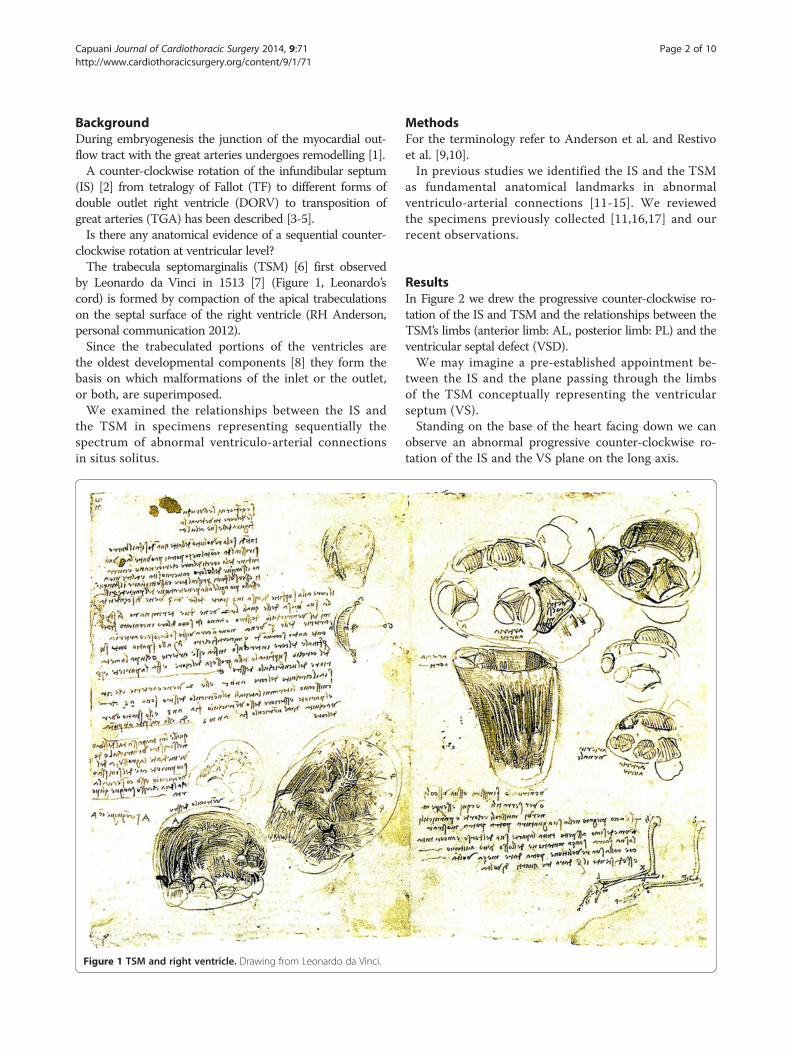
clockwise rotation at ventricular level?The trabecula septomarginalis (TSM) [6] first observed
by Leonardo da Vinci in 1513 [7] (Figure 1, Leonardo’scord) is formed by compaction of the apical trabeculationson the septal surface of the right ventricle (RH Anderson,personal communication 2012).Since the trabeculated portions of the ventricles are
the oldest developmental components [8] they form thebasis on which malformations of the inlet or the outlet,or both, are superimposed.We examined the relationships between the IS and
the TSM in specimens representing sequentially thespectrum of abnormal ventriculo-arterial connectionsin situs solitus.
Figure 1 TSM and right ventricle. Drawing from Leonardo da Vinci.
MethodsFor the terminology refer to Anderson et al. and Restivoet al. [9,10].In previous studies we identified the IS and the TSM
as fundamental anatomical landmarks in abnormalventriculo-arterial connections [11-15]. We reviewedthe specimens previously collected [11,16,17] and ourrecent observations.
ResultsIn Figure 2 we drew the progressive counter-clockwise ro-tation of the IS and TSM and the relationships between theTSM’s limbs (anterior limb: AL, posterior limb: PL) and theventricular septal defect (VSD).We may imagine a pre-established appointment be-
tween the IS and the plane passing through the limbsof the TSM conceptually representing the ventricularseptum (VS).Standing on the base of the heart facing down we can
observe an abnormal progressive counter-clockwise ro-tation of the IS and the VS plane on the long axis.

Figure 2 Conceptual relationships between IS and VIF in abnormal ventriculo-arterial connections in situs solitus and atrio-ventricularconcordance. 1A Normal relationships. 1B TF. Anterior deviation/displacement of the IS (compare to Figure 4). 1C DORV with sub-aortic VSD. The ALblends or is committed to the counter-clockwise twisted IS. The PL blends or is committed to the VIF (compare to Figure 5). 1D Taussig-Bing (DORV withsub-pulmonary VSD). The PL blends or is committed to a more counter-clockwise twisted IS (compare to Figure 8). 1E TGA. The IS inserts abnormally tothe TSM forming a totally displaced infundibulum (compare to Figure 9). 1F and 1G Absent IS in truncus arteriosus and doubly committed juxta-arterialVSD. The PL still blends or is committed to the VIF of the single outlet or of the aorta (compare to Figure 7 and 6).
Capuani Journal of Cardiothoracic Surgery 2014, 9:71 Page 3 of 10http://www.cardiothoracicsurgery.org/content/9/1/71
In the normal heart the IS and the VS are well aligned:the AL blends with the IS (Figure 2A, compare withFigure 3).In the spectrum of abnormal ventriculo-arterial mal-
formations the IS progressively divorces from the TSMcreating an angle at the insertion to the AL.With an angle from 0° to about 90° the aorta be-
comes dextro-posed and more anterior. We observesequentially dextro-position of the aorta, TF (Figure 2B,compare with Figure 4), DORV with sub-aortic VSD(Figure 2C, compare with Figure 5).The VSD represents a malalignment gap between VS
and IS, is in sub-aortic position and is cradled betweenthe limbs of the TSM.The VSD may also be doubly committed juxta-arterial
(Figure 2G, compare with Figure 6) in absence of IS or
in the settings of truncus arteriosus (Figure 2F, comparewith Figure 7).In these sequence the AL blends or is committed to
the deviated IS.As the counter-clockwise rotation of the VS plane in-
creases to about 180°, is the PL which blends or is commit-ted to the IS, the VSD becomes sub-pulmonary (Figure 2D,compare with Figure 8) and we observe the Taussig-Bingspectrum.At the end of the VS rotation, at about 180°, there is
alignment of the IS with the VS as in the normal heartbut the IS is abnormally inserted to the TSM.The aorta arises from the right ventricle with a sub-
aortic infundibulum and there is pulmonary-mitral fi-brous continuity normally present in classic TGA withintact septum. (Figure 2E, compare with Figure 9).
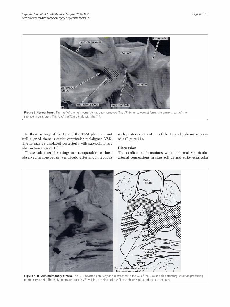
Figure 3 Normal heart. The roof of the right ventricle has been removed. The VIF (inner curvature) forms the greatest part of thesupraventricular crest. The PL of the TSM blends with the VIF.
Capuani Journal of Cardiothoracic Surgery 2014, 9:71 Page 4 of 10http://www.cardiothoracicsurgery.org/content/9/1/71
In these settings if the IS and the TSM plane are notwell aligned there is outlet-ventricular malaligned VSD.The IS may be displaced posteriorly with sub-pulmonaryobstruction (Figure 10).These sub-arterial settings are comparable to those
observed in concordant ventriculo-arterial connections
Figure 4 TF with pulmonary atresia. The IS is deviated anteriorly and ispulmonary atresia. The PL is committed to the VIF which stops short of the
with posterior deviation of the IS and sub-aortic sten-osis (Figure 11).
DiscussionThe cardiac malformations with abnormal ventriculo-arterial connections in situs solitus and atrio-ventricular
attached to the AL of the TSM as a free standing structure producingPL and there is tricuspid-aortic continuity.
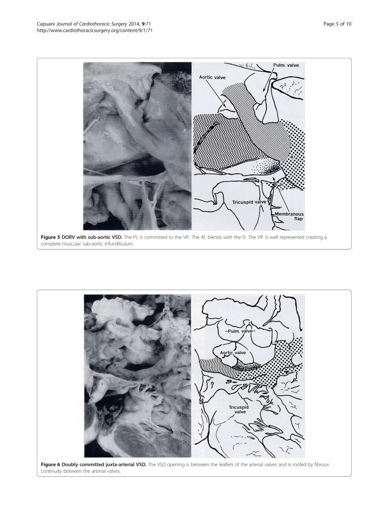
Figure 5 DORV with sub-aortic VSD. The PL is committed to the VIF. The AL blends with the IS. The VIF is well represented creating acomplete muscular sub-aortic infundibulum.
Figure 6 Doubly committed juxta-arterial VSD. The VSD opening is between the leaflets of the arterial valves and is roofed by fibrouscontinuity between the arterial valves.
Capuani Journal of Cardiothoracic Surgery 2014, 9:71 Page 5 of 10http://www.cardiothoracicsurgery.org/content/9/1/71
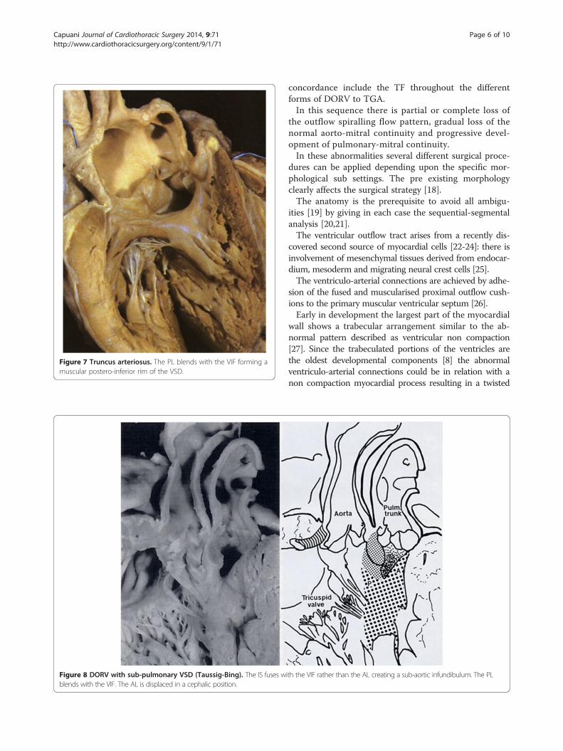
Figure 7 Truncus arteriosus. The PL blends with the VIF forming amuscular postero-inferior rim of the VSD.
Figure 8 DORV with sub-pulmonary VSD (Taussig-Bing). The IS fuses wblends with the VIF. The AL is displaced in a cephalic position.
Capuani Journal of Cardiothoracic Surgery 2014, 9:71 Page 6 of 10http://www.cardiothoracicsurgery.org/content/9/1/71
concordance include the TF throughout the differentforms of DORV to TGA.In this sequence there is partial or complete loss of
the outflow spiralling flow pattern, gradual loss of thenormal aorto-mitral continuity and progressive devel-opment of pulmonary-mitral continuity.In these abnormalities several different surgical proce-
dures can be applied depending upon the specific mor-phological sub settings. The pre existing morphologyclearly affects the surgical strategy [18].The anatomy is the prerequisite to avoid all ambigu-
ities [19] by giving in each case the sequential-segmentalanalysis [20,21].The ventricular outflow tract arises from a recently dis-
covered second source of myocardial cells [22-24]: there isinvolvement of mesenchymal tissues derived from endocar-dium, mesoderm and migrating neural crest cells [25].The ventriculo-arterial connections are achieved by adhe-
sion of the fused and muscularised proximal outflow cush-ions to the primary muscular ventricular septum [26].Early in development the largest part of the myocardial
wall shows a trabecular arrangement similar to the ab-normal pattern described as ventricular non compaction[27]. Since the trabeculated portions of the ventricles arethe oldest developmental components [8] the abnormalventriculo-arterial connections could be in relation with anon compaction myocardial process resulting in a twisted
ith the VIF rather than the AL creating a sub-aortic infundibulum. The PL

Figure 10 TGA with sub-pulmonary obstruction due to posteriordeviation of the IS.
Figure 9 TGA. Sub-aortic infundibulum. The limbs of the TSM areabout 180° twisted in a cephalic position.
Capuani Journal of Cardiothoracic Surgery 2014, 9:71 Page 7 of 10http://www.cardiothoracicsurgery.org/content/9/1/71
VS and finally in the spectrum of malformations we areobserving.To describe the different morphologies malposition of
the great arteries is a generic term since the variability ofthe arterial relationships is considerable and all relationsfrom normal to side by side to antero posterior have tobe expected [15].It is better to analyze the outflow in terms of three
components: the intra pericardial trunks, the arterialvalves, and the ventricular tracts [28].In the normal heart the sub-pulmonary supra-ventricular
crest is made up of the muscularised proximal outflowcushions (RH Anderson personal communication 2012).Most of this muscle becomes the free-standing sub-pulmonary infundibulum and a small part persists as realmuscular IS but in the normal heart cannot be recognised(Figure 3).The key feature of the morphology in abnormal
ventriculo-arterial connections is the location and insertionof the IS relative to the remainder of the VS [11].The TSM, first drawn by Leonardo da Vinci in 1513
(Figure 1), is an extensive septal trabeculation of theright ventricle formed by compaction of the apical mus-cular trabeculations on the septal surface (RH Andersonpersonal communication 2012).
Figure 11 Posterior deviation of the IS in a heart with concordantventriculo-arterial connections and interruption of the aortic arch.
Capuani Journal of Cardiothoracic Surgery 2014, 9:71 Page 8 of 10http://www.cardiothoracicsurgery.org/content/9/1/71
The TSM is formed by an antero-superior limb and aninfero-posterior limb and the plane passing throughout thelimbs conceptually may represent the plane of the VS.At Carnegies stages 15-19 the outflow tract makes a
marked counter clockwise rotation [1] and in mousemodels by transgenic studies in mutant embryos withcono-truncal defects has been reported a counter clock-wise rotation of the outflow suggesting a myocardial per-turbation [29].Indeed in the spectrum of abnormal ventriculo-arterial
connections from TF to DORV to TGA we can observea sequential right-left counter clockwise rotation of theIS facing down the ventricles standing on the base of theheart and a simultaneous progressive twisting right-leftof the VS plane on the longitudinal axis.In TF (Figure 4) there is dextro-anterior deviation of
the IS [30] and the angle between the IS and the VShas been reported from 60° up to 130°. In the majorityof cases is about 90° [2]. The PL committed to the ven-triculo infundibular fold (VIF) follows the dextroposi-tion of the aorta and the AL blends with the IS twistingthe TSM.If the IS is absent the VSD becomes doubly committed
juxta arterial: the PL still blends with the VIF (Figure 6).A similar disposition of the limbs can be found in the
common arterial trunk (Figure 7).In this morphology the PL may form a muscular postero-
inferior rim on the VSD or there may be a fibrous continu-ity tricuspid-truncal valve.In DORV the counter-clockwise rotation is more ac-
centuated (between 90° and 180°) [31-33].The VSD represents a malalignment gap between the
VS and the IS and may be committed or not committedto the great arteries. It can also be in relation with onegreat artery, however not directly committed owing toan extensive VIF or an extreme dextroposition of theaorta or aberrant chordae tendinee [34].According to some investigators [2] the diagnosis of
DORV should be reserved for hearts with a bilateralsub-arterial infundibulum. In contrast others formu-lated the concept that DORV is a malformation inwhich both great arteries arise completely or almostfrom the right ventricle with or without mitro-aortic ormitro-pulmonary continuity [31].These different views reflect the fact that the term
DORV in reality identifies different morphologies insidea spectrum of malformations.In the settings of DORV with sub-aortic VSD the AL
is still committed to the IS (Figure 5).In DORV with sub-pulmonary VSD it is the PL which
blends or is committed to the IS and the AL is displacedantero-superiorly (Figure 8).There is a almost complete twisting of the TSM on the
long axis.
We consider the overall spectrum of hearts with sub-pulmonary VSD to represent the Taussig-Bing.This malformation has the names of the authors who
described the pathology for the first time.In the original paper the two vessels are side by side,
the pulmonary artery being in its normal place [33],however as pointed out the relationships of the great ar-teries are variable.There are three patterns of the angle between the IS
and the remainder of the muscular VS plane: right angle(90°: great arteries side by side), acute angle (90° < 180°:aorta dextroposed and anterior), parallel (about 180°: greatarteries antero-posterior) [35].The commonest coronary pattern in the parallel pos-
ition is comparable to one found in complete transpos-ition of great arteries, what we actually expect being inthese cases about at the end of VS 180° rotation.The following morphology in the progressive TSM
twist is represented by the TGA [2,36-43].When more than half of the circumference of the pul-
monary valve is supported by the left ventricle theventriculo-arterial connections are considered discord-ant rather than double outlet.The second cardiac lineage rotates as enters the heart be-
fore the outflow tract cushions are formed. In the normalcardio-genesis the cushions are spiral and the myocardiumretracts as they fuse. In the settings of TGA the cushionsare initially formed in straight fashion and the difference isfound in the way that the distal ends of the cushions fusewith a protrusion from the dorsal wall of the aortic sac (RHAnderson personal communication 2012).In classical TGA there is mitro-pulmonary continuity,
the aorta is anterior and right sided and the aortic valveis supported by a muscular infundibulum, however, insome cases the aorta may be left sided (S, D, L transpos-ition) [41] or posterior and right sided (normal relations)[42] and in some hearts there is a muscular sub-arterialinfundibulum in both ventricles.In TGA with intact septum the IS inserts abnormally
to the TSM forming a totally displaced infundibulum(Figure 9). There is no correlation between the anteriorposition of the aorta and the length of the infundibulumand the typical VSD is outlet ventricular [38] howeverother VSD’s types as trabecular or inlet of different ori-gin may be associated.In transposition with aorta to the left (S, D, L) the IS was
found deviated posteriorly and leftward squeezing the sub-pulmonary outflow tract with an abnormal cono-septalangulation varying from 71+/−44 degrees [41], what weexpect considering that the VS rotation on the long axis ac-cording the sequence of our observations is at or close to180° (Figure 10).Indeed this morphology is similar to the one observed
in posterior deviation of the IS in concordant ventriculo-

Capuani Journal of Cardiothoracic Surgery 2014, 9:71 Page 9 of 10http://www.cardiothoracicsurgery.org/content/9/1/71
arterial connections with VSD and sub-aortic narrowing inwhich obviously the VS developed normally (Figure 11).
Conclusions
I. The counter-clockwise rotation of the IS and thesimultaneous twist of the TSM characterise all sub-settings of abnormal ventriculo-arterial connections insitus solitus and atrio-ventricular concordance. This isparticularly important for the diagnosis and correctivesurgery in this complex spectrum of malformations.
II. The presented anatomical observations support thehypothesis that the abnormal ventriculo-arterialconnections may be successive stages of the sameembryo-genetic process at ventricular level and thatmay arise from an abnormal myocardial rotation inaddition to an abnormal outflow tract septation.
III.The abnormal ventriculo-arterial connections could bein relation with a pathological myocardial compactionprocess. We promote further investigations.
Limitations of the studyThis study relates to abnormal ventriculo-arterial con-nections in situs solitus atrio-ventricular concordanceand two well developed ventricles.A further spectrum of malformations can be seen ex-
tending towards double outlet left ventricle.
AbbreviationsDORV: Double outlet right ventricle; TF: Tetralogy of Fallot; TGA: Transposition ofgreat arteries; IS: Infundibular septum; VS: Ventricular septum; TSM: Trabeculaseptomarginalis; PL: Posterior limb of the TSM; AL: Anterior limb of the TSM;VIF: Ventriculo infundibular fold; VSD: Ventricular septal defect.
Competing interestsThe Author has no financial or no financial competing interests.This study received no specific grant from any funding agency,commercial or not-for-profit sectors.The Author transfers the copyright to the publisher.
AcknowledgementsWe are grateful to Prof. Robert H Anderson for the invaluable help anddiscussions during this study and to Mrs Marianna AS Capuani for herprecious secretarial assistance and translation support.Figures 3, 4, 5, 6 and 8 have been reproduced from The Society of ThoracicSurgeons, Annals of Thoracic Surgery 1995; 59: 352–360 by permission of theAuthors.Figures 7 and 9, 10, 11 have been reproduced from “The Heart Structure inHealth and Disease” 1992 Gower Medical Publishing by permission of Prof.Robert H Anderson.The convention of sliding is used in similar fashion throughout theillustrations: heavy stipples TSM, fine stipples IS, wavy lines VIF.
Received: 30 November 2013 Accepted: 31 March 2014Published: 21 April 2014
References1. Müller F, O’Rahilly R: Developmental stages in human embryos. Washington
DC: Carnegie Institution of Washington, Publication 63T; 1987.2. Goor DA, Lillehei CW: Congenital malformations of the heart. New York:
Grune & Stratton Inc; 1975.3. Goor DA, Edwards JE: The transition from double outlet right ventricle to
complete transposition. A pathological study. Am J Cardiol 1972, 29:267.
4. Lomonico MP, Moore GW, Hutchins GM: Rotation of the junction of theoutflow tract and great arteries in the embryonic human heart. Anat Rec1986, 216:544–546.
5. Bostrom MPG, Hutchins GM: Arrested rotation of the outflow tract mayexplain double-outlet right ventricle. Circulation 1988, 77:1258–1265.
6. Tandler J: Anatomie des Herzens. In Karl von Bardeleben’s Handbuch derAnatomie des Menschen. Jena Germany: Verlag von Gustav Fisher; 1913.
7. Da Vinci L: Anatomical drawing RL 19118-19v. In Anatomical Drawings(1485-1515) from the Royal Library Collection. Windsor Castle.
8. De la Cruz MV, Sánchez Gómez C, Cayre R: The developmentalcomponents of the ventricles: their significance in congenitalcardiacmalformations. Cardiol Young 1991, 1:123–128.
9. Anderson RH, Becker AE, Van Mierop LHS:What should we call the “crista “?Br Heart J 1977, 39:856–859.
10. Restivo A, Smith A, Wilkinson JL, Anderson RH: The medial papillarymuscle complex audits related septomarginal trabeculation. A normalanatomical study on human hearts. J Anat 1989, 163:231–242.
11. Capuani A, Uemura H, Yen Ho S, Anderson RH: Anatomic spectrum ofabnormal ventriculo arterial connections: surgical implications. Ann ThoracSurg 1995, 59:352–360.
12. Capuani A: Trabécule Septomarginalis, Septum Infundibulaire et ConnexionVentriculo-Arterielle [Abstract]. In Proceedings of the Journées d’Automne de laSociété Française de Chirurgie Thoracique et Cardiovasculaire. Edited by SociétéFrançaise de Chirurgie Thoracique et Cardiovasculaire. Paris; 1995.
13. Capuani A: Morphologie et Morphogenèse des malformationsconotroncales [Abstract]. In Proceedings of the Neuvième Journèe de laSociété Française de Foetopathologie: les Cardiopathies Conotroncales. Editedby Société Française de Foetopathologie. Paris; 1998.
14. Capuani A: Abnormal ventriculo-arterial connections in situs solitus and atrio ven-tricular concordance: relations between outlet septum, trabecula septomarginalisand ventriculo infundibular fold. Journal de Chirurgie Thoracique et Cardiovasculaire.Edited by Société Française de Chirurgie Thoracique et Cardiovasculaire 2008, 12:35–45.
15. Capuani A, Soulé N, Meot M, Vaillant MC, Poinsot J, Lefort B, Chantepie A,Neville P: Anatomical landmarks for abnormal ventriculo-arterial connectionswith usual arrangement [Abstract]. J Cardiothorac Surg 2013, 8(Suppl 1):s 0251.
16. Anderson RH, Becher AE: The heart: structure in health and disease. NewYork: Gower Medical Publishing; 1992.
17. Wilcox BR, Anderson RH: Surgical anatomy of the heart. 2nd edition. London:Gower Medical Publishing; 1992:8.26–8.27.
18. Mavroudis C, Backer C, Idriss RF: Pediatric cardiac surgery. 4th edition.Blackwell Publishing Ltd; 2013.
19. Anderson RH, Wilcox BR: Herbert sloan lecture. Understanding cardiacanatomy: the prerequisite for optimal cardiac surgery. Ann Thorac Surg1995, 59:1366–1375.
20. Van Praagh R: The segmental approach to diagnosis in congenital heartdisease. In Birth defects original article series n.5. Edited by Bergsma.Baltimore: Williams & Wilkins; 1972:4–23.
21. Shinebourne EA, Macartney FJ, Anderson RH: Sequential chamber localization.Logical approach to diagnosis in congenital heart disease. Br Heart J 1976,38:327–340.
22. Waldo KL, Kumiski DH, Wallis KT, Stadt HA, Hutson MR, Platt DH, Kirby ML:Conotruncal myocardium arises from a secondary heart field. Development2001, 128:3179–3188.
23. Kelly RG, Brown NA, Buckingham ME: The arterial pole of the mouse heartforms from Fgf10-expressing cells in pharyngeal mesoderm. Dev Cell2001, 1:435–440.
24. Mjaatvedt CH, Nakaoka T, Moreno-Rodriguez R: The outflow tract of the heart isrecruited from a novel heart-forming field. Dev Biol 2001, 238:97–109.
25. Sizarov A, Lamers WH, Mohun TJ, Brown NA, Anderson RH, Moorman AFM:Three-dimensional and molecular analysis of the arterial pole of thedeveloping human heart. J Anat 2012, 220(4):336–349.
26. Anderson RH, Chaudhry B, Mohun TJ, Bamforth SD, Hoyland D, Pillips HM,Webb S, Moorman AFM, Brown NA, Henderson DY: Normal and abnormaldevelopment of the intrapericardial arterial trunks in humans and mice.Cardiovasc Res 2012, 95:108–115.
27. Freedom RM, Yoo SJ, Perrin D, Taylor G, Petersen S, Anderson RH: Themorphological spectrum of ventricular non compaction. Cardiol Young2005, 15:345–364.
28. Anderson RH, Webb S, Brown NA, Lamers W, Moorman A: Developmnet ofthe heart: (3) formation of the ventricular outflow tracts, arterial valves,and intrapericardial arterial truncus. Heart 2003, 89:1110–1118.

Capuani Journal of Cardiothoracic Surgery 2014, 9:71 Page 10 of 10http://www.cardiothoracicsurgery.org/content/9/1/71
29. Bajolle F, Zaffran S, Kelly RG, Hadchovel J, Bonnet D, Brown NA, BuckinghamME: Rotation of the myocardial wall of the outflow tract is implicated inthe normal positioning of the great arteries. Circ Res 2006, 98:421–428.
30. Anderson RH, Tynan M: Tetralogy of Fallot - a centennial review. Int JCardiol 1988, 21:219–232.
31. Lev M, Bharati S, Meng L, Liberthson RR, Paul MH, Idriss F: A concept ofdouble-outlet right ventricle. J Thorac Cardiovasc Surg 1972, 64:271–281.
32. Wilcox BR, Yen Ho S, Macartney FJ, Becker AE, Gerlis LM, Anderson RH:Surgical anatomy of double-outlet right ventricle with Situs Solitus andAtrioventricular Concordance. J Thorac Cardiovasc Surg 1981, 82:405–417.
33. Van Praagh R: What is the Taussig-Bing malformation? Circulation 1968,38:445–449.
34. Beekman RP, Bartelings MM, Hazecamp MG, Gittenberger-de Groot AC,Ottenkamp J: The morphologic nature of noncommitted ventricularseptal defects in specimens with double-outlet right ventricle. J ThoracCardiovasc Surg 2002, 124:984–990.
35. Uemura H, Yagihara T, Kawashima Y, Nishigaki K, Kamiya T, Yen Ho S,Anderson RH: Coronary arterial anatomy in double-outlet right ventriclewith subpulmonary VSD. Ann Thorac Surg 1995, 59:591–597.
36. Becker AE, Anderson RH: How should we describe hearts in which theaorta is connected to the right ventricle and the pulmonary trunk tothe left ventricle? A matter for reason and logic. Am J Cardiol 1968,51:911–912.
37. Paul MH, Van Praagh R, Van Praagh S: Transposition of great arteries. InPediatric Cardiology. Edited by Watson H. Saint Louis: CV Mosby Co; 1968.
38. Thiene G, Razzolini R, Dalla-Volta S: Aorto-pulmonary relationships,arterio-ventricular alignment, and ventricular septal defects incomplete transposition of the great arteries. Eur J Cardiol 1976,4(1):13–24.
39. Anderson RH, Tynan M: Complete transposition. The significance ofdescribing separately connexions, arterial relationships and infundibularmorphology. Int J Cardiol 1984, 5:19–22.
40. Kurosawa H, Van Mierop LHS: Surgical anatomy of the infundibularseptum in transposition of the great arteries with ventricular septaldefect. J Thorac Cardiovasc Surg 1986, 91:123–132.
41. Hoyel L, Van Praagh R, Lacour-Gayet F, Serraf A, Petit J, Bruniaux J, PlanchéC: Transposition of Great Arteries (SDL). Pathologic anatomy, diagnosis,and surgical management of a newly recognised complex. J ThoracCardiovasc Surg 1995, 110:613–624.
42. Beuren A: Differential diagnosis of the Taussig-Bing heart from completetransposition of the great vessels with posteriorly overriding pulmonaryartery. Circulation 2002, 21:1071–1087.
43. Kirby ML: Embryogenesis of transposition of the great arteries. A lessonfrom the heart. Circ Res 2002, 9:87–89.
doi:10.1186/1749-8090-9-71Cite this article as: Capuani: The trabecula septomarginalis (Leonardo’scord) in abnormal ventriculo-arterial connections: anatomic andmorphogenetic implications. Journal of Cardiothoracic Surgery 2014 9:71.
Submit your next manuscript to BioMed Centraland take full advantage of:
• Convenient online submission
• Thorough peer review
• No space constraints or color figure charges
• Immediate publication on acceptance
• Inclusion in PubMed, CAS, Scopus and Google Scholar
• Research which is freely available for redistribution
Submit your manuscript at www.biomedcentral.com/submit