Embed Size (px)
Citation preview
RESEARCH ARTICLE
Phase 1 repolarization rate defines Ca2+ dynamicsand contractility on intact mouse heartsMarıa Micaela López Alarcón1, Ainhoa Rodrıguez de Yurre1, Juan Ignacio Felice2, Emiliano Medei1, and Ariel L. Escobar3
In the heart, Ca2+ influx through L-type Ca2+ channels triggers Ca2+ release from the sarcoplasmic reticulum. In mostmammals, this influx occurs during the ventricular action potential (AP) plateau phase 2. However, in murine models, the influxthrough L-type Ca2+ channels happens in early repolarizing phase 1. The aim of this work is to assess if changes in the openprobability of 4-aminopyridine (4-AP)–sensitive Kv channels defining the outward K+ current during phase 1 can modulate Ca2+
currents, Ca2+ transients, and systolic pressure during the cardiac cycle in intact perfused beating hearts. Pulsed local-fieldfluorescence microscopy and loose-patch photolysis were used to test the hypothesis that a decrease in a transient K+ current(Ito) will enhance Ca2+ influx and promote a larger Ca2+ transient. Simultaneous recordings of Ca2+ transients and APs by pulsedlocal-field fluorescence microscopy and loose-patch photolysis showed that a reduction in the phase 1 repolarization rateincreases the amplitude of Ca2+ transients due to an increase in Ca2+ influx through L-type Ca2+ channels. Moreover,4-AP induced an increase in the time required for AP to reach 30% repolarization, and the amplitude of Ca2+ transients waslarger in epicardium than endocardium. On the other hand, the activation of Ito with NS5806 resulted in a reduction of Ca2+
current amplitude that led to a reduction of the amplitude of Ca2+ transients. Finally, the 4-AP effect on AP phase 1 wassignificantly smaller when the L-type Ca2+ current was partially blocked with nifedipine, indicating that the phase 1 rate ofrepolarization is defined by the competition between an outward K+ current and an inward Ca2+ current.
IntroductionThe cardiac ventricular action potential (AP) is the earliestphysiological event that controls contraction during the cardiaccycle. Typically, the prolonged duration of the ventricular APallows an increase in the L-type Ca2+ channel open probability,leading to a sustained influx of Ca2+ into the myocyte. In smallmammals, the duration of the AP needs to be short enough to copewith a higher heart rate. In a mouse, the heart rate can be as highas 600 beats/min, setting the total duration of the AP to valuesshorter than 100 ms. This fact led researchers to assume that thetime course of a mouse ventricular AP lacks a plateau phase(phase 2; Nerbonne and Kass, 2005; Dilly et al., 2006). However,our laboratory recently demonstrated that when APs are recordedin situ at the intact organ level, the mouse ventricular AP displaysa phase 2 (Ferreiro et al., 2012; Ramos-Franco et al., 2016). Ad-ditionally, we found that this phase 2 was more hyperpolarizedthan in larger mammals (Kornyeyev et al., 2010; Valverde et al.,2010; Ferreiro et al., 2012) and was driven by an influx of Na+
through the Na+-Ca2+ exchanger (NCX; Ramos-Franco et al.,2016). Moreover, we demonstrated that in the intact heart, theinflux of Ca2+ that triggers Ca2+ transients occurs during the
deactivation of L-type Ca2+ channels in AP phase 1 (Ramos-Francoet al., 2016). Although other groups have proposed this idea fordifferent mammalian species such as dog (Zygmunt et al., 1997;Banyasz et al., 2003; Cordeiro et al., 2004), rat (Bouchard et al.,1995; Sah et al., 2002; Cooper et al., 2010), and mouse (Dilly et al.,2006; Kondo et al., 2006), those experiments were performedmostly in isolated ventricular myocytes under voltage-clampconditions. Even though that approach has been extremelyhelpful, the “exact” ventricular region and layer from which thetested dissociated myocytes originated was not precisely defined.Furthermore, the electric, metabolic, and mechanical couplingbetween cells at different layers is lost when cells are isolated.Moreover, at the intact heart level there is a transmural electricalgradient that defines the excitation-contraction coupling prop-erties; this electrical gradient is also missing when the myocytesare isolated. Finally, ionic currents in isolated myocytes areusually recorded at room temperature, at bradycardic pacingrates, and in the presence of exogenous intracellular Ca2+ buffersto help with cell viability, a condition that also changes theproperties of Ca2+ dynamics during diastole and systole.
.............................................................................................................................................................................1Instituto de Biofısica Carlos Chagas Filho, Universidade Federal do Rio de Janeiro, Brazil; 2Centro de Investigaciones Cardiovasculares, Universidad Nacional de La Plata, LaPlata, Argentina; 3Department of Bioengineering, School of Engineering, University of California, Merced, CA.
Correspondence to Ariel L. Escobar: [email protected].
© 2019 López-Alarcón et al. This article is distributed under the terms of an Attribution–Noncommercial–Share Alike–No Mirror Sites license for the first six months afterthe publication date (see http://www.rupress.org/terms/). After six months it is available under a Creative Commons License (Attribution–Noncommercial–Share Alike 4.0International license, as described at https://creativecommons.org/licenses/by-nc-sa/4.0/).
Rockefeller University Press https://doi.org/10.1085/jgp.201812269 771
J. Gen. Physiol. 2019 Vol. 151 No. 6 771–785
Dow
nloaded from http://rupress.org/jgp/article-pdf/151/6/771/1237463/jgp_201812269.pdf by guest on 03 April 2022
In the present article, we propose to gauge the role of a key K+
conductance activated during phase 1 repolarization in definingthe transmural Ca2+ signaling. An integrated and simultaneousassessment of the mechanical activity, AP kinetics, electro-cardiograms (ECGs), Ca2+ transients, and Ca2+ currents wasperformed on intact perfused hearts when Kv channels wereinhibited by 4-aminopyridine (4-AP) or activated with 1-[2,4-dibromo-6-(1H-tetrazol-5-yl)-phenyl]-3-(3,5-bis-trifluoromethyl-phenyl)-urea (NS5806).We think that understanding the kineticsof phase 1 repolarization in different ventricular layers will helpus to understand not only transmural electrical repolarization butalso mechanical dysfunctions. Furthermore, the knowledge ob-tained from the experiments presented here will help us under-stand pathophysiological mechanisms related to systolic heartfailure and define novel pharmacological interventions to treatthis set of clinical conditions.
Material and methodsEthical approvalAll animal experiments were performed on adult mice (CharlesRiver Laboratories) in accordance with the Institutional AnimalCare and Use Committee of the University of California Merced.Animals were maintained by the National Institutes of HealthGuide for the Care and Use of Laboratory Animals (NationalInstitutes of Health Publication No. 85-23, Revised 1996). TheInstitutional Animal Care and Use Committee of the Universityof California Merced approved the euthanasia method used(#2008–201).
Whole-heart preparationHearts were obtained from 8-wk-old Balb/C male mice (CharlesRiver Laboratories). Animals were injected intraperitoneally withNa+-heparin and euthanized by cervical dislocation 15 min afterthe injection. The heart was rapidly removed, and the aorta wascannulated onto a standard horizontal (Zimmer, 1998; Mejıa-Alvarez et al., 2003) Langendorff apparatus for continuous per-fusion with normal Tyrode's solution containing, in mM, NaCl140, KCl 5.3, CaCl2 2, MgCl2 1, NaPO4H2 0.33, HEPES 10, andglucose 10. The osmolarity of the solution was 295 mOsm/L, andthe pH, 7.4. After spontaneous cardiac contractility became reg-ular, hearts were loaded with either Ca2+-sensitive or voltage-sensitive fluorescent indicators. Specifically, Rhod-2AM or Di-8-ANEPPSwere perfused for 30min after the hearts were stabilizedin the Langendorff setup. Di-8-ANEPPS (10 µg) was preparedwith 20 µl of 20% Pluronic (Biotium) in 5 ml of Tyrode’s solution.Rhod-2AM (50 µg) was diluted with 20 µl of 20% Pluronic in500 µl of Tyrode’s solution and perfused at room temperatureusing peristaltic pumps. Stepwise details of the preparation ofthese dyes have been published by our group (Aguilar-Sanchezet al., 2017). A Peltier unit was used to set the temperature of theTyrode’s solution in the horizontal chamber to 33°C.
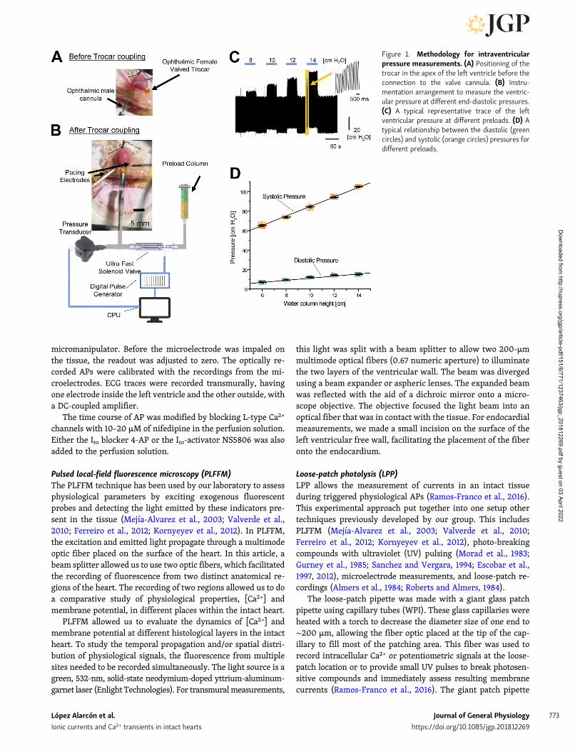
Experimental setupPressure measurementsWe developed a novel experimental approach to study cardiacmechanics. The Langendorff perfused heart is a retroperfused
preparation. Consequently, the pressure cannot be measured inthe aorta. The usual approach to overcome this problem is tointroduce a small balloon through the mitral valve and measurethe pressure using a transducer. Here, we present a novelmethod in which we introduce a tiny vitrectomy ophthalmic-valved trocar (Alcon) in the apex of the left ventricle. Thepressure at the outlet of the valve was measured with a solid-state integrated pressure transducer (Honeywell). A scheme ofthe methodology is presented in Fig. 1. Fig. 1 A shows how theophthalmic-valved trocar is inserted though the apex of the leftventricle. Fig. 1 B illustrates the general arrangement used tomeasure the left intraventricular pressure. The trocar is con-nected through a valved coupler and is directly connected to thepressure transducer. The end-diastolic volume and the end-diastolic pressure can be imposed to the heart with the help ofa column connected to the valved coupler through a fast elec-trovalve. Consequently, the height of the column will set theend-diastolic pressure when the electrovalve is open. The end-diastolic pressure can be modified by changing the height of thewater column. The electrovalve will define the type of pressuremeasurement: if the electrovalve is closed, the measurementwill be isovolumetric, and if it is open, it will be isotonic atconstant load condition. Fig. 1 C depicts typical recordings of theisotonic pressure when end-diastolic pressure was modified.The pressures were calibrated before each experiment by plot-ting the pressure transducer output as a function of the columnheight. The inset illustrates how fast the developed pressurechanges when the end-diastolic pressure is increased from aresting condition. Fig. 1 D shows a typical Frank–Starling re-sponse when the end-diastolic volume was increased. The in-crease in the end-diastolic volume produces an increase in theend-diastolic pressure (green dots). Interestingly, the developedsystolic pressure (orange dots) presents a much larger increasethan the diastolic pressure (green dots). This result is consistentwith an increase in the diastolic pressure as function of thepreload. Finally, measurements of the ventricular pressure us-ing a plastic or latex balloon are usually obtained under isovol-umetric conditions. In this article, we decided to evaluate thedeveloped systolic pressure when the volume of the ventricle ischanging (isotonic contraction). Compared with an isovolu-metric contraction, this condition is closer to what happens withthe ventricular function when blood is ejected from the ven-tricle. Finally, constant load implies that the height of the water(Tyrode’s) column against which the heart is pumping out theventricular volume and the output resistance are constant.
Electrical measurements using sharp microelectrodes and ECGSharp glass microelectrodes, filled with 3 M KCl, were used torecord electrical signals during APs. An electrometric amplifier(WPI) was used to record the membrane potential. Glass mi-croelectrodes were pulled with a Micropipette Puller (Flaming/Brown; Sutter Instrument Co.) and kept overnight in 3 M KCl toremove any air bubbles that could cause additional resistance ornoise. These microelectrodes had a resistance of 10–20 MΩ.Microelectrodes were connected to a high input impedancedifferential amplifier (Duo 773 Electrometer; WPI) and posi-tioned at the surface of the heart using a manual mechanical
López Alarcón et al. Journal of General Physiology 772
Ionic currents and Ca2+ transients in intact hearts https://doi.org/10.1085/jgp.201812269
Dow
nloaded from http://rupress.org/jgp/article-pdf/151/6/771/1237463/jgp_201812269.pdf by guest on 03 April 2022
micromanipulator. Before the microelectrode was impaled onthe tissue, the readout was adjusted to zero. The optically re-corded APs were calibrated with the recordings from the mi-croelectrodes. ECG traces were recorded transmurally, havingone electrode inside the left ventricle and the other outside, witha DC-coupled amplifier.
The time course of AP was modified by blocking L-type Ca2+
channels with 10–20 µM of nifedipine in the perfusion solution.Either the Ito blocker 4-AP or the Ito-activator NS5806 was alsoadded to the perfusion solution.
Pulsed local-field fluorescence microscopy (PLFFM)The PLFFM technique has been used by our laboratory to assessphysiological parameters by exciting exogenous fluorescentprobes and detecting the light emitted by these indicators pre-sent in the tissue (Mejıa-Alvarez et al., 2003; Valverde et al.,2010; Ferreiro et al., 2012; Kornyeyev et al., 2012). In PLFFM,the excitation and emitted light propagate through a multimodeoptic fiber placed on the surface of the heart. In this article, abeam splitter allowed us to use two optic fibers, which facilitatedthe recording of fluorescence from two distinct anatomical re-gions of the heart. The recording of two regions allowed us to doa comparative study of physiological properties, [Ca2+] andmembrane potential, in different places within the intact heart.
PLFFM allowed us to evaluate the dynamics of [Ca2+] andmembrane potential at different histological layers in the intactheart. To study the temporal propagation and/or spatial distri-bution of physiological signals, the fluorescence from multiplesites needed to be recorded simultaneously. The light source is agreen, 532-nm, solid-state neodymium-doped yttrium-aluminum-garnet laser (Enlight Technologies). For transmuralmeasurements,
this light was split with a beam splitter to allow two 200-µmmultimode optical fibers (0.67 numeric aperture) to illuminatethe two layers of the ventricular wall. The beam was divergedusing a beam expander or aspheric lenses. The expanded beamwas reflected with the aid of a dichroic mirror onto a micro-scope objective. The objective focused the light beam into anoptical fiber that was in contact with the tissue. For endocardialmeasurements, we made a small incision on the surface of theleft ventricular free wall, facilitating the placement of the fiberonto the endocardium.
Loose-patch photolysis (LPP)LPP allows the measurement of currents in an intact tissueduring triggered physiological APs (Ramos-Franco et al., 2016).This experimental approach put together into one setup othertechniques previously developed by our group. This includesPLFFM (Mejıa-Alvarez et al., 2003; Valverde et al., 2010;Ferreiro et al., 2012; Kornyeyev et al., 2012), photo-breakingcompounds with ultraviolet (UV) pulsing (Morad et al., 1983;Gurney et al., 1985; Sanchez and Vergara, 1994; Escobar et al.,1997, 2012), microelectrode measurements, and loose-patch re-cordings (Almers et al., 1984; Roberts and Almers, 1984).
The loose-patch pipette was made with a giant glass patchpipette using capillary tubes (WPI). These glass capillaries wereheated with a torch to decrease the diameter size of one end to∼200 µm, allowing the fiber optic placed at the tip of the cap-illary to fill most of the patching area. This fiber was used torecord intracellular Ca2+ or potentiometric signals at the loose-patch location or to provide small UV pulses to break photosen-sitive compounds and immediately assess resulting membranecurrents (Ramos-Franco et al., 2016). The giant patch pipette
Figure 1. Methodology for intraventricularpressure measurements. (A) Positioning of thetrocar in the apex of the left ventricle before theconnection to the valve cannula. (B) Instru-mentation arrangement to measure the ventric-ular pressure at different end-diastolic pressures.(C) A typical representative trace of the leftventricular pressure at different preloads. (D) Atypical relationship between the diastolic (greencircles) and systolic (orange circles) pressures fordifferent preloads.
López Alarcón et al. Journal of General Physiology 773
Ionic currents and Ca2+ transients in intact hearts https://doi.org/10.1085/jgp.201812269
Dow
nloaded from http://rupress.org/jgp/article-pdf/151/6/771/1237463/jgp_201812269.pdf by guest on 03 April 2022
was filled with Tyrode’s solution and held by a microelectrodeholder half-cell (WPI). A micromanipulator was used to placethe patch pipette on the surface of the ventricle. The interior ofthe giant glass patch pipette was voltage clamped to the samepotential of the surrounding bath. A flash photolysis system letus fractionally change a specific ionic current (i.e., Ca2+ cur-rents by photolyzing nifedipine) under the loose-patch pipette.Nifedipine was locally photoinactivated by UV illuminationgenerated by a diode-pumped solid-state UV laser (355 nm). UVlight was optomechanically shuttered for 1–50 ms and appliedthrough an external quartz multimode fiber-optic or by a fiberpositioned inside the patch pipette. The basic idea is to recordthe total membrane current with a macro patch pipette underconditions in which we can photolytically activate a conduc-tance. The difference between the total current recorded in thepresence and the absence of the drug allows us to reveal thespecific current that was pharmacologically blocked. As thearea under the pipette, where the current is recorded, is muchsmaller than the space constant, the neighboring tissue imposesan electrotonic coupling. This electrotonic coupling will act asan electric sink and impedes the activation of the photoliticallyactivated current from producing any changes in the local APbeneath the recording pipette. This situation, in which a changein the transmembrane current there does not produce a changeof the membrane potential, mimics what happens under avoltage-clamp condition, but in this case, the ventricular syn-cytium is acting as a spatial clamp. Finally, hearts were paced at4–6 Hz and 33°C.
Statistical analysisThe physiological recordings of the APs, ECG, Ca2+ transients,Ca2+ currents, and ventricular pressure were evaluated based onwell-established parameters in the field of cardiac electrophys-iology. To compare between recordings before and after drugretroperfusion, the APs and Ca2+ transient traces were normal-ized to the mean value of the control signals.
The AP trace for each set of experiments was evaluated atcertain repolarization times. Specifically, the time it takes forthe AP to reach 30% or 90% repolarization (APD30 or APD90)was assessed. The repolarization times between control andnoncontrol experiments were then evaluated and normalized tothe control values for each heart. After this normalization, val-ues from five experiments (n = 5 hearts, or otherwise noted)were compiled. Statistical analysis was performed with Origin 8using one-way ANOVA.
Several parameters of Ca2+ transient kinetics were measured.Specifically, we measured the fractional Ca2+ transient amplitudewith respect to a control trace; the rise time of the Ca2+ transients,measured as the time it takes for the Ca2+ transient to go from10% to 90% of the maximum amplitude; and the half relaxationtime, measured as the time it takes for the Ca2+ transient to relaxto half of its maximum amplitude. Each of these parameters re-corded in control and noncontrol experiments was evaluated andnormalized to the control values for each heart used. The datafrom all the Ca2+ transients obtained from five experiments (n = 5hearts unless stated otherwise) were compiled, and statisticalanalysis was performed with Origin 8 and one-way ANOVA.
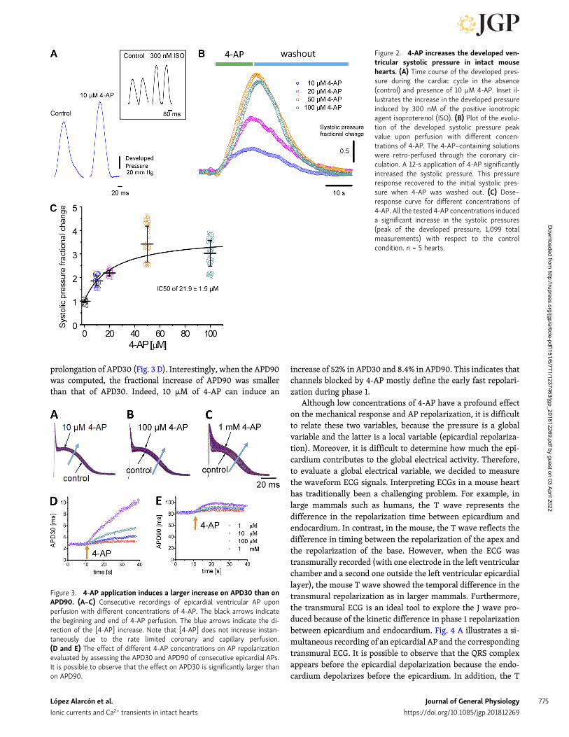
Results4-AP–sensitive Kv channels define mechanical andelectrophysiological properties on an intact beatingmouse heartIn mice, Ito has a central role in repolarizing the AP. Thus, wedecided to test the hypothesis that alterations in this current willhave a dramatic effect on cardiac contractility at the intact heartlevel. To test this idea, we performed experiments measuringthe time course of the developed pressure in the left ventriclewhen Ito is blocked with increasing concentrations of the Kvblocker 4-AP.
Fig. 2 illustrates a typical experiment in which Ito was im-paired with different concentrations of 4-AP. As we show inFig. 2 A, even a low concentration of 4-AP (10 µM) can produce asignificant increase in the amplitude of the ventricular devel-oped pressure under an isotonic condition at a constant after-load. The inset in Fig. 2 A illustrates the effect of 300 nM ofisoproterenol on the pressure measured with the newmethod inwhich a vitrectomy ophthalmic-valved trocar was introduced inthe left ventricle. This indicates that this new method can trackincreases in the developed pressure under different experi-mental conditions that promote an increase in contractility. InFig. 2 B, we can observe the change in the amplitude of thesystolic pressure upon retroperfusion with increasing concen-trations of 4-AP. Specifically, a short 12-s bolus of 4-AP dissolvedin Tyrode’s solution was applied through the coronary vascu-lature. At all concentrations, we can observe a significant in-crease in systolic pressure that decreases to values similar to thecontrol condition when 4-AP is washed out. This protocol al-lowed us to construct a dose–response curve (Fig. 2 C) showingthat there were significant increases of the measured pressurefor each concentration of 4-AP (n = 5 hearts) and that the effectof 4-AP on contraction has an 50% inhibitory concentration of21.9 ± 1.5 µM.
Although the reported affinities for the blocking of Kv 4.2 andKv 4.3 by 4-AP are ∼1 mM, in the experiments presented inFig. 2, a much lower 4-AP concentration showed a large effect onthe developed pressure, suggesting that other Kv channels cancontrol contractility. Consequently, to assess if low concen-trations of 4-AP have an impact on ventricular electrical activity,we decided to evaluate the effect of 4-AP in the repolarization ofthe epicardial AP.
Recently, we demonstrated that although the mouse epicar-dial AP displays a fast phase 1, it also shows a significant phase2 at a more hyperpolarized (more negative) membrane potentialcompared with othermammals when recordings are made at theintact heart level under physiological conditions. Interestingly,phase 2 is not directly driven by an influx of Ca2+ through L-typeCa2+ channels, but by the influx of Na+ through the NCX. Fig. 3shows the effect of 4-AP on the repolarization of the epicardialAP. Fig. 3 (A–C) depicts the action of the blocker when a heartwas perfused with increasing concentrations of 4-AP. Inthe three cases (Fig. 3, A–C), the AP shows a typical epicardialspike-and-dome behavior. Additionally, it is possible to observethat the application of 4-AP slows phase 1 repolarization andprolongs AP duration. The APD30 was evaluated during theapplication of increasing concentrations of 4-AP, inducing
López Alarcón et al. Journal of General Physiology 774
Ionic currents and Ca2+ transients in intact hearts https://doi.org/10.1085/jgp.201812269
Dow
nloaded from http://rupress.org/jgp/article-pdf/151/6/771/1237463/jgp_201812269.pdf by guest on 03 April 2022
prolongation of APD30 (Fig. 3 D). Interestingly, when the APD90was computed, the fractional increase of APD90 was smallerthan that of APD30. Indeed, 10 µM of 4-AP can induce an
increase of 52% in APD30 and 8.4% in APD90. This indicates thatchannels blocked by 4-AP mostly define the early fast repolari-zation during phase 1.
Although low concentrations of 4-AP have a profound effecton the mechanical response and AP repolarization, it is difficultto relate these two variables, because the pressure is a globalvariable and the latter is a local variable (epicardial repolariza-tion). Moreover, it is difficult to determine how much the epi-cardium contributes to the global electrical activity. Therefore,to evaluate a global electrical variable, we decided to measurethe waveform ECG signals. Interpreting ECGs in a mouse hearthas traditionally been a challenging problem. For example, inlarge mammals such as humans, the T wave represents thedifference in the repolarization time between epicardium andendocardium. In contrast, in the mouse, the T wave reflects thedifference in timing between the repolarization of the apex andthe repolarization of the base. However, when the ECG wastransmurally recorded (with one electrode in the left ventricularchamber and a second one outside the left ventricular epicardiallayer), the mouse T wave showed the temporal difference in thetransmural repolarization as in larger mammals. Furthermore,the transmural ECG is an ideal tool to explore the J wave pro-duced because of the kinetic difference in phase 1 repolarizationbetween epicardium and endocardium. Fig. 4 A illustrates a si-multaneous recording of an epicardial AP and the correspondingtransmural ECG. It is possible to observe that the QRS complexappears before the epicardial depolarization because the endo-cardium depolarizes before the epicardium. In addition, the T
Figure 2. 4-AP increases the developed ven-tricular systolic pressure in intact mousehearts. (A) Time course of the developed pres-sure during the cardiac cycle in the absence(control) and presence of 10 µM 4-AP. Inset il-lustrates the increase in the developed pressureinduced by 300 nM of the positive ionotropicagent isoproterenol (ISO). (B) Plot of the evolu-tion of the developed systolic pressure peakvalue upon perfusion with different concen-trations of 4-AP. The 4-AP–containing solutionswere retro-perfused through the coronary cir-culation. A 12-s application of 4-AP significantlyincreased the systolic pressure. This pressureresponse recovered to the initial systolic pres-sure when 4-AP was washed out. (C) Dose–response curve for different concentrations of4-AP. All the tested 4-AP concentrations induceda significant increase in the systolic pressures(peak of the developed pressure, 1,099 totalmeasurements) with respect to the controlcondition. n = 5 hearts.
Figure 3. 4-AP application induces a larger increase on APD30 than onAPD90. (A–C) Consecutive recordings of epicardial ventricular AP uponperfusion with different concentrations of 4-AP. The black arrows indicatethe beginning and end of 4-AP perfusion. The blue arrows indicate the di-rection of the [4-AP] increase. Note that [4-AP] does not increase instan-taneously due to the rate limited coronary and capillary perfusion.(D and E) The effect of different 4-AP concentrations on AP repolarizationevaluated by assessing the APD30 and APD90 of consecutive epicardial APs.It is possible to observe that the effect on APD30 is significantly larger thanon APD90.
López Alarcón et al. Journal of General Physiology 775
Ionic currents and Ca2+ transients in intact hearts https://doi.org/10.1085/jgp.201812269
Dow
nloaded from http://rupress.org/jgp/article-pdf/151/6/771/1237463/jgp_201812269.pdf by guest on 03 April 2022
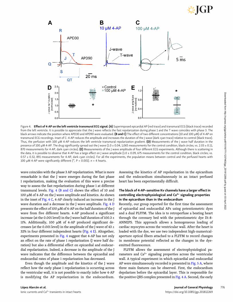
wave coincides with the phase 3 AP repolarization.What is moreremarkable is that the J wave emerges during the fast phase1 repolarization, making the evaluation of this wave a preciseway to assess the fast repolarization during phase 1 at differenttransmural levels. Fig. 4 (B and C) shows the effect of 10 and100 µM of 4-AP on the J wave amplitude and kinetics. As shownin the inset of Fig. 4 C, 4-AP clearly induced an increase in the Jwave duration and a decrease in the J wave amplitude. Fig. 4 Dpresents the effect of 100 µM of 4-AP on the half duration of the Jwave from five different hearts. 4-AP produced a significantincrease (at the 0.001 level) in the J wave half duration of 100.3 ±11%. Additionally, 100 µM of 4-AP produced significant de-creases (at the 0.001 level) in the amplitude of the J wave of 43 ±32% in four different independent hearts (Fig. 4 E). Altogether,experiments presented in Fig. 4 suggest that 4-AP has not onlyan effect on the rate of phase 1 repolarization (J wave half du-ration) but also a differential effect on epicardial and endocar-dial repolarization. Indeed, a decrease in the amplitude of the Jwave indicates that the difference between the epicardial andendocardial rates of phase 1 repolarization has decreased.
Even though the amplitude and the kinetics of the J wavereflect how the early phase 1 repolarization is occurring acrossthe ventricular wall, it is not possible to exactly infer how 4-APis modifying the AP repolarization in the endocardium.
Assessing the kinetics of AP repolarization in the epicardiumand the endocardium simultaneously in an intact perfusedheart has been experimentally difficult.
The block of 4-AP–sensitive Kv channels have a larger effect incontrolling electrophysiological and Ca2+ signaling propertiesin the epicardium than in the endocardiumRecently, our group reported for the first time the assessmentof epicardial and endocardial APs using potentiometric dyesand a dual PLFFM. The idea is to retroperfuse a beating heartthrough the coronary bed with the potentiometric dye Di-8-ANNEPS. This approach allows a homogeneous loading ofcardiac myocytes across the ventricular wall. After the heart isloaded with the dye, we use two independent high-numerical-aperture optical fibers attached to a PLFFM to record changesin membrane potential reflected as the changes in the dye-emitted fluorescence.
PLFFM allows the assessment of electrophysiological pa-rameters and Ca2+ signaling properties across the ventricularwall. A typical experiment in which epicardial and endocardialAP were simultaneously recorded is presented in Fig. 5 A, wherethree main features can be observed. First, the endocardiumdepolarizes before the epicardial layer. This is responsible forthe positive QRS complex presented in Fig. 4 A. Second, the early
Figure 4. Effect of 4-AP on the left ventricle transmural ECG signal. (A) Superimposed epicardial AP (red trace) and transmural ECG (black trace) recordedfrom the left ventricle. It is possible to appreciate that the J wave reflects the fast repolarization during phase 1 and the T wave coincides with phase 3. Theblack arrows indicate the position where APD30 and APD90 were evaluated. (B and C) The effect of two different concentrations (10 and 100 µM) of 4-AP ontransmural ECG recordings. Inset of C: 4-AP reduces the amplitude and increases the duration of the J wave (dark cyan trace) relative to control (black trace).Thus, the perfusion with 100 µM 4-AP reduces the left ventricle transmural repolarization gradient. (D) Measurements of the J wave half duration in thepresence of 100 µM 4-AP. The drug significantly spread out the J wave (1.0 ± 0.04, 1,065 measurements for the control condition, black circles; vs. 2.03 ± 0.11,870 measurements for 4-AP, dark cyan circles). (E) Measurements of the J wave amplitude of four different ECG experiments. Although there is scattering inthe data, it is possible to observe that 4-AP has a large effect on J wave amplitude (1.0 ± 0.09, 675 measurements for the control condition, black circles; vs.0.57 ± 0.32, 891 measurements for 4-AP, dark cyan circles). For all the experiments, the population means between control and the perfused hearts with100 µM 4-AP were significantly different (*, P < 0.001). n = 4 hearts.
López Alarcón et al. Journal of General Physiology 776
Ionic currents and Ca2+ transients in intact hearts https://doi.org/10.1085/jgp.201812269
Dow
nloaded from http://rupress.org/jgp/article-pdf/151/6/771/1237463/jgp_201812269.pdf by guest on 03 April 2022
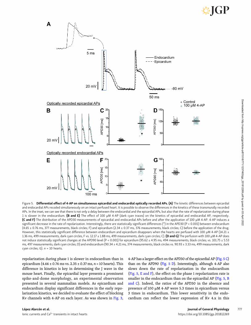
repolarization during phase 1 is slower in endocardium than inepicardium (4.66 ± 0.76ms vs. 2.35 ± 0.37ms, n = 10 hearts). Thisdifference in kinetics is key in determining the J wave in themouse heart. Finally, the epicardial layer presents a prominentspike-and-dome morphology, an experimental observationpresented in several mammalian models. As epicardium andendocardium display significant differences in the early repo-larization kinetics, we decided to evaluate the effect of blockingKv channels with 4-AP on each layer. As was shown in Fig. 3,
4-AP has a larger effect on the APD30 of the epicardial AP (Fig. 5 C)than on the APD90 (Fig. 5 D). Interestingly, although 4-AP alsoslows down the rate of repolarization in the endocardium(Fig. 5, E and F), the effect on the phase 1 repolarization rate issmaller in the endocardium than on the epicardial AP (Fig. 5, Band C). Indeed, the ratios of the APD30 in the absence andpresence of 100 µM 4-AP were 5.3 times in epicardium versus3 times in endocardium. This lower sensitivity in the endo-cardium can reflect the lower expression of Kv 4.x in this
Figure 5. Differential effect of 4-AP on simultaneous epicardial and endocardial optically recorded APs. (A) The kinetic differences between epicardialand endocardial APs recoded simultaneously on an intact perfused heart. It is possible to observe the differences in the kinetics of these transmurally recordedAPs. In the inset, we can see that there is not only a delay between the endocardial and the epicardial APs, but also that the rate of repolarization during phase1 is slower in the endocardium. (B and E) The effect of 100 µM 4-AP (dark cyan traces) on the kinetics of epicardial and endocardial AP, respectively.(C and F) The distribution of the APD30 measurements of epicardial and endocardial APs before and after the application of 100 µM 4-AP. 4-AP induces asignificant decrease in the rate of repolarization. Interestingly, there are statistically significant differences (*) in the APD30 (P < 0.001) between endocardium(4.65 ± 0.76 ms, 377 measurements, black circles; F) and epicardium (2.34 ± 0.37 ms, 376 measurements, black circles; C) before the application of the drug.However, this statistically significant difference between endocardium and epicardium disappears when the hearts are perfused with 100 µM 4-AP (14.21 ±1.36 ms, 499 measurements, dark cyan circles, F vs. 12.17 ± 1.88 ms, 499 measurements, dark cyan circles; C). (D and G) The perfusion with 100 µM 4-AP doesnot induce statistically significant changes at the APD90 level (P < 0.001) for epicardium (95.62 ± 4.95 ms, 494 measurements, black circles; vs. 101.75 ± 5.53ms, 497 measurements, dark cyan circles; D) and endocardium (90.34 ± 4.21 ms, 374 measurements, black circles vs. 90.93 ± 3.33 ms, 499 measurements, darkcyan circles; G). n = 10 hearts.
López Alarcón et al. Journal of General Physiology 777
Ionic currents and Ca2+ transients in intact hearts https://doi.org/10.1085/jgp.201812269
Dow
nloaded from http://rupress.org/jgp/article-pdf/151/6/771/1237463/jgp_201812269.pdf by guest on 03 April 2022
internal ventricular layer (Mattiazzi et al., 2015; Aguilar-Sanchez et al., 2017).
Undoubtedly, the perfusion of the hearts with 4-AP has adramatic effect on both the mechanical and electrical propertiesof the mouse heart. However, none of the experiments pre-sented above define the relationship between excitability andcontractility. Therefore, we designed experiments to directlyevaluate how changes in the AP waveform can modify the sys-tolic Ca2+. Themost likely scenario is that 4-AP will enhance Ca2+
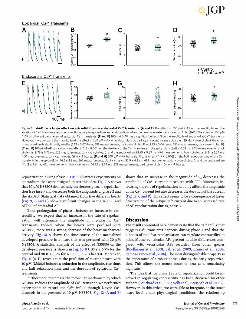
release from the SR by changing the amplitude and the kineticsof the L-type Ca2+ currents. In addition, if 4-AP has an effect onthe amplitude of Ca2+ transients, we expect that 4-AP will have alarger effect on the epicardium than in the endocardium. Toassess the effect of 4-AP on the amplitude of the Ca2+ transientsin the epicardium and the endocardium, we performed experi-ments to measure simultaneously Ca2+ transients in both layers.The Ca2+ transient recordings were obtained by perfusing theheart with the Ca2+ indicator Rhod-2 and by simultaneouslyrecording fluorescence signals with two independent opticalfibers coupled to a PLFFM. Experiments presented in Fig. 6 il-lustrate the effect produced by the perfusion of the heart with100 µM 4-AP on the amplitude and the kinetics of epicardial andendocardial Ca2+ transients (n = 4 hearts). Fig. 6 (A–D) showsthat 4-AP induces an increase in the amplitude, rise time, andhalf relaxation time of the epicardial Ca2+ transients. Even more,although 4-AP also induces an augmentation of the amplitudeand the duration of the endocardial Ca2+ transients (Fig. 6, E–H),the effect on this innermost layer is smaller than in the epi-cardium. This is consistent with the idea that the epicardial ef-fects of 4-AP are larger due to the heterogeneous expression ofKv 4.x channels across the ventricular wall.
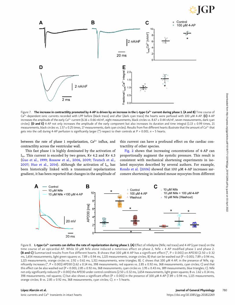
Clearly, the results presented in Fig. 6 demonstrate that theincrease in the systolic pressure with 4-AP observed in Fig. 2is due to an increase in the amplitude and the kinetics of theCa2+ transient. However, we still miss the link between thedecreased rate of repolarization during phase 1 and the in-crease in cytosolic Ca2+ (contractility). As we already stated,the most likely possibility is that changes in the AP phase1 repolarization will induce an increase in the amplitude of theL-type Ca2+ current and the total amount of Ca2+ that thistransport mechanism brings into the cells. Recently, our lab-oratory was able to measure for the first time Ca2+-dependentcurrents during a triggered AP at the intact heart level usingthe novel LPP technique (Ramos-Franco et al., 2016). To assessif changes during the repolarization of phase 1 can define thekinetics of Ca2+ influx into the cell, we performed LPP ex-periments in hearts exposed to 4-AP. A typical experiment isshown in Fig. 7 (A and B), which illustrates epicardial currentsrecorded in the absence and presence of 100 µM 4-AP, re-spectively. The LPP recording shows two components, onefast early current and a late slower current. As we alreadydemonstrated in a previous article (Ramos-Franco et al.,2016), the current during the early phase is carried by Ca2+
ions permeating through L-type Ca2+ channels, and the latecomponent is mediated by the influx of Na+ through the NCXacting in its forward mode. Furthermore, in Ramos-Francoet al. (2016), we demonstrated that this Ca2+ influx was
terminating by voltage-dependent deactivation and not byCa2+-dependent inactivation.
This late Na+ current through the NCX is activated by Ca2+
released from the SR. Remarkably, the perfusion of the heartwith 4-AP not only induces an increase in the amplitude of thefast inward component of the current but also increases its meanduration. Fig. 7 (C and D) reinforces this observation: currentsrecorded from five different animals show that the increase inthe current amplitude and the total charge carried through theL-type Ca2+ currents during phase 1 of the AP is significantlylarger in hearts perfused with 4-AP (57% for the total charge)than in control hearts.
One critical aspect of LPP experiments is to record simulta-neously the transmembrane current and the epicardial AP. Thisallows us to verify if the photolytic activation of the inwardcurrent is changing the properties of the AP.
As we already demonstrated, perfusion of the heart withnifedipine induces a reduction in the amplitude of phase2 (Ferreiro et al., 2012). In addition, we found that in the pres-ence of nifedipine, the effect of 4-AP on the phase 1 repolariza-tion rate was much smaller. Fig. 8 A illustrates the effect of 4-APin the presence of nifedipine in the AP. Additionally, data ob-tained from five different hearts show that in the presence ofnifedipine, 4-AP’s effect was 4 times smaller (Fig. 8, B and C).This result suggests that the repolarization kinetics during phase1 is defined by a competition between a K+ efflux and a Ca2+
influx (Gómez et al., 1997). Therefore, 4-AP will have a dual rolein phase 1 repolarization. First, it will block Kv channels, slowingthe rate of repolarization, and second, the slower repolarizationwill keep the membrane potential depolarized for a longer timeduring phase 1. This will reduce the deactivation of the L-typeCa2+ channel. This “prolonged activation” (increase in the openprobability) not only will bring more Ca2+ into the cell to in-crease contractility but also will modify the AP repolarization.
Activation of Kv 4.x channels reduces the mechanical responseby increasing the rate of repolarization during phase 1; thisfinally leads to an attenuation in the L-type Ca2+ and theCa2+ transientResults presented in this article indicate that the blockage of Kvchannels increases the systolic force because a slower AP re-polarization phase 1 increases the Ca2+ influx through L-typechannels, activating a larger Ca2+ transient with the concomi-tant increase in contractility. A final proof of concept supportingthis idea is the assessment of contractility (i.e., Ca2+) when theKv channels are activated instead of being inhibited. Kv chan-nels are tetramers in which each α subunit has six transmem-brane segments. Although this is the main structure for K+
permeation, other regulatory subunits can modify the level ofexpression and also can regulate the kinetics of the channels.Moreover, a central regulatory subunit, K+ channel-interactingprotein (KChIP), has a central role in regulating the Kv 4.xchannels. Furthermore, some drugs can interact with Kv 4.xchannels when KChIP is also expressed in the plasma mem-brane. Interestingly, NS5806 can slow the rate of inactivation ofKv 4.x when the α subunit interacts with KChIP (Gonzalez et al.,2014). Thus, we expect that NS5806 will accelerate the rate of
López Alarcón et al. Journal of General Physiology 778
Ionic currents and Ca2+ transients in intact hearts https://doi.org/10.1085/jgp.201812269
Dow
nloaded from http://rupress.org/jgp/article-pdf/151/6/771/1237463/jgp_201812269.pdf by guest on 03 April 2022
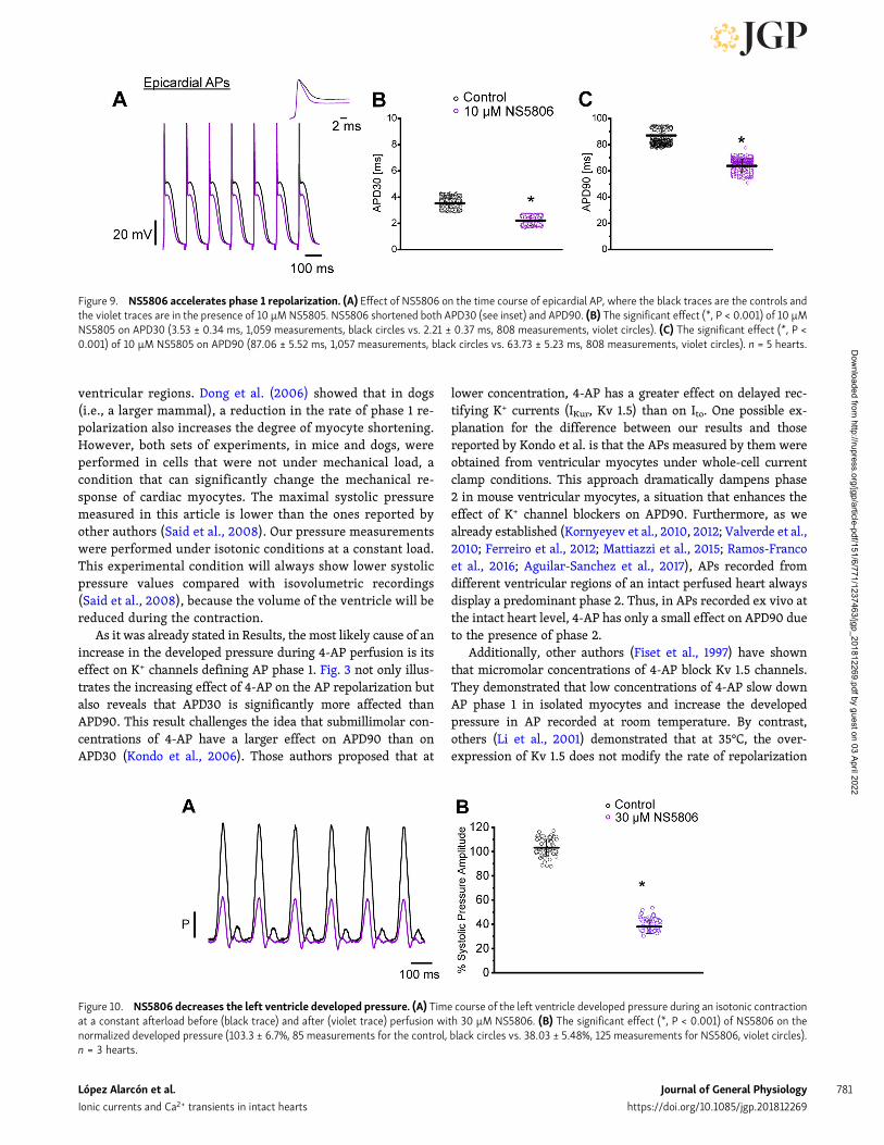
repolarization during phase 1. Fig. 9 illustrates experiments onepicardium that were designed to test this idea. Fig. 9 A showsthat 10 µM NS5806 dramatically accelerates phase 1 repolariza-tion (see inset) and decreases both the amplitude of phase 2 andthe APD90. Statistical data obtained from five different hearts(Fig. 9, B and C) show significant changes in the APD30 andAPD90 of epicardial AP.
If the prolongation of phase 1 induces an increase in con-tractility, we expect that an increase in the rate of repolari-zation will attenuate the amplitude of myoplasmic Ca2+
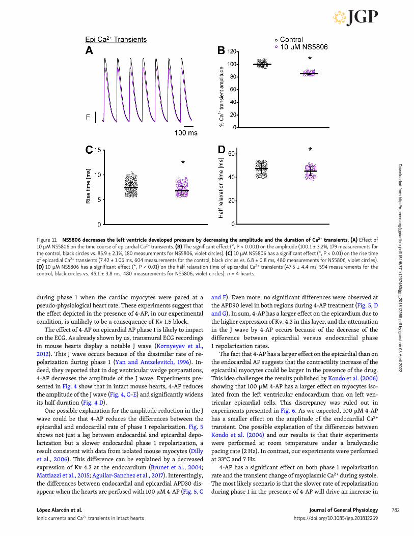
transients. Indeed, when the hearts were perfused withNS5806, there was a strong decrease of the heart mechanicalactivity. Fig. 10 A shows the time course of the normalizeddeveloped pressure in a heart that was perfused with 30 µMNS5806. A statistical analysis of the effect of NS5806 on thedeveloped pressure is shown in Fig. 10 B (103.2 ± 6.7% for thecontrol and 38.0 ± 5.5% for NS5806; n = 3 hearts). Moreover,Fig. 11 (A–D) reveals that the perfusion of murine hearts with10 µM NS5806 induces a reduction in the amplitude, rise time,and half relaxation time and the duration of epicardial Ca2+
transients.Furthermore, to unmask the molecular mechanism by which
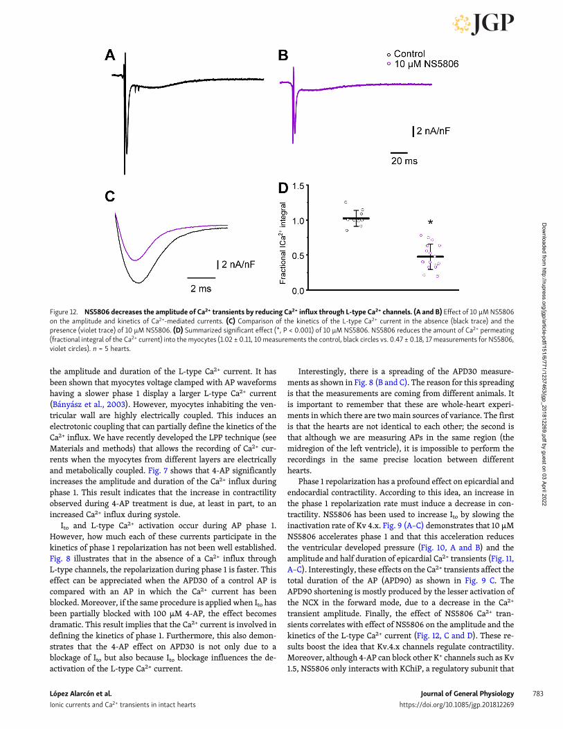
NS5806 reduces the amplitude of Ca2+ transient, we performedexperiments to record the Ca2+ influx through L-type Ca2+
channels in the presence of 10 µM NS5806. Fig. 12 (A and B)
shows that an increase in the magnitude of Ito decreases theamplitude of Ca2+ currents measured with LPP. Moreover, in-creasing the rate of repolarization not only affects the amplitudeof the Ca2+ current but also decreases the duration of the current(Fig. 12, C and D). This effect seems to be a consequence of fasterdeactivation of the L-type Ca2+ current due to an increased rateof AP repolarization during phase 1.
DiscussionThe results presented here demonstrate that the Ca2+ influx thattriggers Ca2+ transients happens during phase 1 and that thekinetics of this fast repolarization can regulate contractility inmice. Mouse ventricular APs present notable differences com-pared with ventricular APs recorded from other species(Knollmann et al., 2001; Sah et al., 2003; Brunet et al., 2004;Ramos-Franco et al., 2016). The most distinguishable property isthe appearance of a robust phase 1 during the early repolariza-tion. This allows the mouse heart to beat at a remarkablyhigh rate.
The idea that the phase 1 rate of repolarization could be in-volved in regulating contractility has been discussed by otherauthors (Bouchard et al., 1995; Volk et al., 1999; Sah et al., 2002).However, in this article, we were able to integrate, at the intactheart level under physiological conditions, the relationship
Figure 6. 4-AP has a larger effect on epicardial than on endocardial Ca2+ transients. (A and E) The effect of 100 µM 4-AP on the amplitude and thekinetics of Ca2+ transients recorded simultaneously in epicardium and endocardium when the heart was externally paced at 7 Hz. (B–D) The effect of 100 µM4-AP on different parameters of epicardial Ca2+ transients. (E and F) 100 µM 4-AP has a significant effect (*) on the amplitude of endocardial Ca2+ transients.However, if we compare the magnitude of the effect of 100 µM 4-AP on endocardium (F; dark cyan circles) versus epicardium (B; dark cyan circles), the effectin endocardium is significantly smaller (1.13 ± 0.07 times, 588measurements, dark cyan circles; E vs. 1.33 ± 0.04 times, 557 measurements, dark cyan circles; B).(C and G) 100 µM 4-AP has a significant effect (*, P < 0.001) on the rise time of the Ca2+ transients in the epicardium (8.42 ± 0.58ms, 662 measurements, blackcircles vs. 8.78 ± 0.72 ms, 623 measurements, dark cyan circles; C) and the endocardium (8.79 ± 0.89 ms, 474 measurements, black circles vs. 9.24 ± 1.34 ms,400 measurements, dark cyan circles; G). n = 4 hearts. (D and H) 100 µM 4-AP has a significant effect (*, P < 0.001) on the half relaxation time of the Ca2+
transients in the epicardium (46.5 ± 3.9 ms, 662 measurements, black circles vs. 52.9 ± 4.3 ms, 663 measurements, dark cyan circles; D) and the endocardium(41.11 ± 3.6 ms, 451 measurements, black circles vs. 46.92 ± 3.24 ms, 623 measurements, dark cyan circles; D). n = 4 hearts.
López Alarcón et al. Journal of General Physiology 779
Ionic currents and Ca2+ transients in intact hearts https://doi.org/10.1085/jgp.201812269
Dow
nloaded from http://rupress.org/jgp/article-pdf/151/6/771/1237463/jgp_201812269.pdf by guest on 03 April 2022
between the rate of phase 1 repolarization, Ca2+ influx, andcontractility across the ventricular wall.
This fast phase 1 is highly dominated by the activation ofIto. This current is encoded by two genes, Kv 4.2 and Kv 4.3(Guo et al., 1999; Rossow et al., 2006, 2009; Teutsch et al.,2007; Huo et al., 2014). Although the activation of Ito hasbeen historically linked with a transmural repolarizationgradient, it has been reported that changes in the amplitude of
this current can have a profound effect on the cardiac con-tractility of other species.
Fig. 2 shows that increasing concentrations of 4-AP canproportionally augment the systolic pressure. This result isconsistent with mechanical shortening experiments in iso-lated myocytes described by several authors. For example,Kondo et al. (2006) showed that 100 µM 4-AP increases sar-comere shortening in isolated mouse myocytes from different
Figure 7. The increase in contractility promoted by 4-AP is driven by an increase in the L-type Ca2+ current during phase 1. (A and B) Time course ofCa2+-dependent ionic currents recorded with LPP before (black trace) and after (dark cyan trace) the hearts were perfused with 100 µM 4-AP. (C) 4-APincreases the amplitude of the early Ca2+ current (6.16 ± 0.66 nA/nF, eight measurements, black circles vs. 8.67 ± 0.44 nA/nF, seven measurements, dark cyancircles). (D and E) 4-AP not only increases the amplitude of the early component but also increases its duration and time integral (1.13 ± 0.99 times, 21measurements, black circles vs. 1.57 ± 0.25 times, 17 measurements, dark cyan circles). Results from five different hearts illustrate that the amount of Ca2+ thatgets into the cell during 4-AP perfusion is significantly larger (*) respect to their controls at P > 0.001. n = 5 hearts.
Figure 8. L-type Ca2+ currents can define the rate of repolarization during phase 1. (A) Effect of nifedipine (Nife; red trace) and 4-AP (cyan trace) on thetime course of an epicardial AP. While 10 µM Nife alone induced a notorious effect on phase 2, Nife + 4-AP modified phase 1 and phase 2.(B and C) Summarized results from five different hearts. B shows that 100 µM 4-AP has a significant effect (*, P < 0.001) on APD30 (2.50 ± 0.32ms, 1,654 measurements, light green squares vs. 7.89 ± 0.94 ms, 1,115 measurements, orange circles; B) that can be washed out (P < 0.001; 7.89 ± 0.94 ms,1,115 measurements, orange circles vs. 2.92 ± 0.61 ms, 1,311 measurements, wine triangles; B). C shows that 100 µM 4-AP, in the presence of Nife, sig-nificantly increases (*, P < 0.001) APD30 (1.62 ± 0.14 ms, 398 measurements, red squares vs. 2.85 ± 0.92 ms, 368 measurements, cyan circles; C) and thatthis effect can be also washed out (P < 0.001; 2.85 ± 0.92 ms, 368 measurements, cyan circles vs. 1.95 ± 0.43 ms, 389 measurements, blue triangles; C). Nifenot only significantly reduces (P < 0.001) the APD30 under control conditions (2.50 ± 0.32 ms, 1,654 measurements, light green squares; B vs. 1.62 ± 0.14 ms,398 measurements, red squares; C) but also shows a significant effect (P < 0.001) in the presence of 100 µM 4-AP (7.89 ± 0.94 ms, 1,115 measurements,orange circles; B vs. 2.85 ± 0.92 ms, 368 measurements, cyan circles; C). n = 5 hearts.
López Alarcón et al. Journal of General Physiology 780
Ionic currents and Ca2+ transients in intact hearts https://doi.org/10.1085/jgp.201812269
Dow
nloaded from http://rupress.org/jgp/article-pdf/151/6/771/1237463/jgp_201812269.pdf by guest on 03 April 2022
ventricular regions. Dong et al. (2006) showed that in dogs(i.e., a larger mammal), a reduction in the rate of phase 1 re-polarization also increases the degree of myocyte shortening.However, both sets of experiments, in mice and dogs, wereperformed in cells that were not under mechanical load, acondition that can significantly change the mechanical re-sponse of cardiac myocytes. The maximal systolic pressuremeasured in this article is lower than the ones reported byother authors (Said et al., 2008). Our pressure measurementswere performed under isotonic conditions at a constant load.This experimental condition will always show lower systolicpressure values compared with isovolumetric recordings(Said et al., 2008), because the volume of the ventricle will bereduced during the contraction.
As it was already stated in Results, the most likely cause of anincrease in the developed pressure during 4-AP perfusion is itseffect on K+ channels defining AP phase 1. Fig. 3 not only illus-trates the increasing effect of 4-AP on the AP repolarization butalso reveals that APD30 is significantly more affected thanAPD90. This result challenges the idea that submillimolar con-centrations of 4-AP have a larger effect on APD90 than onAPD30 (Kondo et al., 2006). Those authors proposed that at
lower concentration, 4-AP has a greater effect on delayed rec-tifying K+ currents (IKur, Kv 1.5) than on Ito. One possible ex-planation for the difference between our results and thosereported by Kondo et al. is that the APs measured by them wereobtained from ventricular myocytes under whole-cell currentclamp conditions. This approach dramatically dampens phase2 in mouse ventricular myocytes, a situation that enhances theeffect of K+ channel blockers on APD90. Furthermore, as wealready established (Kornyeyev et al., 2010, 2012; Valverde et al.,2010; Ferreiro et al., 2012; Mattiazzi et al., 2015; Ramos-Francoet al., 2016; Aguilar-Sanchez et al., 2017), APs recorded fromdifferent ventricular regions of an intact perfused heart alwaysdisplay a predominant phase 2. Thus, in APs recorded ex vivo atthe intact heart level, 4-AP has only a small effect on APD90 dueto the presence of phase 2.
Additionally, other authors (Fiset et al., 1997) have shownthat micromolar concentrations of 4-AP block Kv 1.5 channels.They demonstrated that low concentrations of 4-AP slow downAP phase 1 in isolated myocytes and increase the developedpressure in AP recorded at room temperature. By contrast,others (Li et al., 2001) demonstrated that at 35°C, the over-expression of Kv 1.5 does not modify the rate of repolarization
Figure 9. NS5806 accelerates phase 1 repolarization. (A) Effect of NS5806 on the time course of epicardial AP, where the black traces are the controls andthe violet traces are in the presence of 10 µM NS5805. NS5806 shortened both APD30 (see inset) and APD90. (B) The significant effect (*, P < 0.001) of 10 µMNS5805 on APD30 (3.53 ± 0.34 ms, 1,059 measurements, black circles vs. 2.21 ± 0.37 ms, 808 measurements, violet circles). (C) The significant effect (*, P <0.001) of 10 µM NS5805 on APD90 (87.06 ± 5.52 ms, 1,057 measurements, black circles vs. 63.73 ± 5.23 ms, 808 measurements, violet circles). n = 5 hearts.
Figure 10. NS5806 decreases the left ventricle developed pressure. (A) Time course of the left ventricle developed pressure during an isotonic contractionat a constant afterload before (black trace) and after (violet trace) perfusion with 30 µM NS5806. (B) The significant effect (*, P < 0.001) of NS5806 on thenormalized developed pressure (103.3 ± 6.7%, 85 measurements for the control, black circles vs. 38.03 ± 5.48%, 125 measurements for NS5806, violet circles).n = 3 hearts.
López Alarcón et al. Journal of General Physiology 781
Ionic currents and Ca2+ transients in intact hearts https://doi.org/10.1085/jgp.201812269
Dow
nloaded from http://rupress.org/jgp/article-pdf/151/6/771/1237463/jgp_201812269.pdf by guest on 03 April 2022
during phase 1 when the cardiac myocytes were paced at apseudo-physiological heart rate. These experiments suggest thatthe effect depicted in the presence of 4-AP, in our experimentalcondition, is unlikely to be a consequence of Kv 1.5 block.
The effect of 4-AP on epicardial AP phase 1 is likely to impacton the ECG. As already shown by us, transmural ECG recordingsin mouse hearts display a notable J wave (Kornyeyev et al.,2012). This J wave occurs because of the dissimilar rate of re-polarization during phase 1 (Yan and Antzelevitch, 1996). In-deed, they reported that in dog ventricular wedge preparations,4-AP decreases the amplitude of the J wave. Experiments pre-sented in Fig. 4 show that in intact mouse hearts, 4-AP reducesthe amplitude of the J wave (Fig. 4, C–E) and significantly widensits half duration (Fig. 4 D).
One possible explanation for the amplitude reduction in the Jwave could be that 4-AP reduces the differences between theepicardial and endocardial rate of phase 1 repolarization. Fig. 5shows not just a lag between endocardial and epicardial depo-larization but a slower endocardial phase 1 repolarization, aresult consistent with data from isolated mouse myocytes (Dillyet al., 2006). This difference can be explained by a decreasedexpression of Kv 4.3 at the endocardium (Brunet et al., 2004;Mattiazzi et al., 2015; Aguilar-Sanchez et al., 2017). Interestingly,the differences between endocardial and epicardial APD30 dis-appear when the hearts are perfused with 100 µM 4-AP (Fig. 5, C
and F). Even more, no significant differences were observed atthe APD90 level in both regions during 4-AP treatment (Fig. 5, Dand G). In sum, 4-AP has a larger effect on the epicardium due tothe higher expression of Kv. 4.3 in this layer, and the attenuationin the J wave by 4-AP occurs because of the decrease of thedifference between epicardial versus endocardial phase1 repolarization rates.
The fact that 4-AP has a larger effect on the epicardial than onthe endocardial AP suggests that the contractility increase of theepicardial myocytes could be larger in the presence of the drug.This idea challenges the results published by Kondo et al. (2006)showing that 100 µM 4-AP has a larger effect on myocytes iso-lated from the left ventricular endocardium than on left ven-tricular epicardial cells. This discrepancy was ruled out inexperiments presented in Fig. 6. As we expected, 100 µM 4-APhas a smaller effect on the amplitude of the endocardial Ca2+
transient. One possible explanation of the differences betweenKondo et al. (2006) and our results is that their experimentswere performed at room temperature under a bradycardicpacing rate (2 Hz). In contrast, our experiments were performedat 33°C and 7 Hz.
4-AP has a significant effect on both phase 1 repolarizationrate and the transient change of myoplasmic Ca2+ during systole.The most likely scenario is that the slower rate of repolarizationduring phase 1 in the presence of 4-AP will drive an increase in
Figure 11. NS5806 decreases the left ventricle developed pressure by decreasing the amplitude and the duration of Ca2+ transients. (A) Effect of10 µM NS5806 on the time course of epicardial Ca2+ transients. (B) The significant effect (*, P < 0.001) on the amplitude (100.1 ± 3.2%, 179 measurements forthe control, black circles vs. 85.9 ± 2.1%, 180 measurements for NS5806, violet circles). (C) 10 µM NS5806 has a significant effect (*, P < 0.01) on the rise timeof epicardial Ca2+ transients (7.42 ± 1.06 ms, 604 measurements for the control, black circles vs. 6.8 ± 0.8 ms, 480 measurements for NS5806, violet circles).(D) 10 µM NS5806 has a significant effect (*, P < 0.01) on the half relaxation time of epicardial Ca2+ transients (47.5 ± 4.4 ms, 594 measurements for thecontrol, black circles vs. 45.1 ± 3.8 ms, 480 measurements for NS5806, violet circles). n = 4 hearts.
López Alarcón et al. Journal of General Physiology 782
Ionic currents and Ca2+ transients in intact hearts https://doi.org/10.1085/jgp.201812269
Dow
nloaded from http://rupress.org/jgp/article-pdf/151/6/771/1237463/jgp_201812269.pdf by guest on 03 April 2022
the amplitude and duration of the L-type Ca2+ current. It hasbeen shown that myocytes voltage clamped with AP waveformshaving a slower phase 1 display a larger L-type Ca2+ current(Banyasz et al., 2003). However, myocytes inhabiting the ven-tricular wall are highly electrically coupled. This induces anelectrotonic coupling that can partially define the kinetics of theCa2+ influx. We have recently developed the LPP technique (seeMaterials and methods) that allows the recording of Ca2+ cur-rents when the myocytes from different layers are electricallyand metabolically coupled. Fig. 7 shows that 4-AP significantlyincreases the amplitude and duration of the Ca2+ influx duringphase 1. This result indicates that the increase in contractilityobserved during 4-AP treatment is due, at least in part, to anincreased Ca2+ influx during systole.
Ito and L-type Ca2+ activation occur during AP phase 1.However, how much each of these currents participate in thekinetics of phase 1 repolarization has not been well established.Fig. 8 illustrates that in the absence of a Ca2+ influx throughL-type channels, the repolarization during phase 1 is faster. Thiseffect can be appreciated when the APD30 of a control AP iscompared with an AP in which the Ca2+ current has beenblocked. Moreover, if the same procedure is applied when Ito hasbeen partially blocked with 100 µM 4-AP, the effect becomesdramatic. This result implies that the Ca2+ current is involved indefining the kinetics of phase 1. Furthermore, this also demon-strates that the 4-AP effect on APD30 is not only due to ablockage of Ito but also because Ito blockage influences the de-activation of the L-type Ca2+ current.
Interestingly, there is a spreading of the APD30 measure-ments as shown in Fig. 8 (B and C). The reason for this spreadingis that the measurements are coming from different animals. Itis important to remember that these are whole-heart experi-ments in which there are twomain sources of variance. The firstis that the hearts are not identical to each other; the second isthat although we are measuring APs in the same region (themidregion of the left ventricle), it is impossible to perform therecordings in the same precise location between differenthearts.
Phase 1 repolarization has a profound effect on epicardial andendocardial contractility. According to this idea, an increase inthe phase 1 repolarization rate must induce a decrease in con-tractility. NS5806 has been used to increase Ito by slowing theinactivation rate of Kv 4.x. Fig. 9 (A–C) demonstrates that 10 µMNS5806 accelerates phase 1 and that this acceleration reducesthe ventricular developed pressure (Fig. 10, A and B) and theamplitude and half duration of epicardial Ca2+ transients (Fig. 11,A–C). Interestingly, these effects on the Ca2+ transients affect thetotal duration of the AP (APD90) as shown in Fig. 9 C. TheAPD90 shortening is mostly produced by the lesser activation ofthe NCX in the forward mode, due to a decrease in the Ca2+
transient amplitude. Finally, the effect of NS5806 Ca2+ tran-sients correlates with effect of NS5806 on the amplitude and thekinetics of the L-type Ca2+ current (Fig. 12, C and D). These re-sults boost the idea that Kv.4.x channels regulate contractility.Moreover, although 4-AP can block other K+ channels such as Kv1.5, NS5806 only interacts with KChiP, a regulatory subunit that
Figure 12. NS5806 decreases the amplitude of Ca2+ transients by reducing Ca2+ influx through L-type Ca2+ channels. (A and B) Effect of 10 µMNS5806on the amplitude and kinetics of Ca2+-mediated currents. (C) Comparison of the kinetics of the L-type Ca2+ current in the absence (black trace) and thepresence (violet trace) of 10 µM NS5806. (D) Summarized significant effect (*, P < 0.001) of 10 µM NS5806. NS5806 reduces the amount of Ca2+ permeating(fractional integral of the Ca2+ current) into the myocytes (1.02 ± 0.11, 10 measurements the control, black circles vs. 0.47 ± 0.18, 17 measurements for NS5806,violet circles). n = 5 hearts.
López Alarcón et al. Journal of General Physiology 783
Ionic currents and Ca2+ transients in intact hearts https://doi.org/10.1085/jgp.201812269
Dow
nloaded from http://rupress.org/jgp/article-pdf/151/6/771/1237463/jgp_201812269.pdf by guest on 03 April 2022
interacts with Kv. 4.x channels and not with Kv 1.5 (Li et al.,2005).
Taken together, the results presented in this article demon-strate for the first time the role of Ito in defining cardiac con-tractility during the cardiac cycle of intact hearts. Furthermore,ventricular myocytes are electrically coupled in an environmenthaving a step repolarization gradient. Thus, this step repolari-zation gradient can induce a graded contractility response acrossthe ventricular wall. Finally, this suggests that the transmuraldifferences in Ca2+ signaling are critical in defining the con-tractile properties of the free ventricular wall.
AcknowledgmentsWe thank Drs. Alicia Mattiazzi, Josefina Ramos-Franco, JulioCopello, Guillermo Perez, and Maria Zogbhi for their valuablediscussions.
This study was supported by Fundação de Amparo a Pesquisado Rio de Janeiro 219901 to M.M. López Alarcón; Coordenação deAperfeiçoamento de Pessoal de Nıvel Superior Fellowship to A.Rodrıguez de Yurre; a postdoctoral fellowship from ConsejoNacional de Investigaciones Cientıficas y Tecnicas Argentina toJ.I. Felice; and Proyectos de Investigación Cientıfica y Tecno-lógica Raices 2524, University of California, Castle FacilityUC-Castle-20095-442167, and National Institutes of Health1R01GM132753-01 to A.L. Escobar.
The authors declare no competing financial interests.Author contributions: A.L. Escobar conceived the project and
designed the experiments. M.M. López Alarcón, A. Rodrıguez deYurre, J.I. Felice, E. Medei, and A.L. Escobar contributed to thedata acquisition, analysis, and interpretation. A.L. Escobar and A.Rodrıguez de Yurre wrote the manuscript, which was approvedby all the authors. All the authors agree to be accountable for allaspects of the work, all those designated as authors qualify forauthorship, and all those who qualify for authorship are listed.
Eduardo Rıos served as editor.
Submitted: 1 October 2018Revised: 15 February 2019Accepted: 15 March 2019
ReferencesAguilar-Sanchez, Y., D. Fainstein, R. Mejia-Alvarez, and A.L. Escobar. 2017.
Local Field Fluorescence Microscopy: Imaging Cellular Signals in IntactHearts. J. Vis. Exp. (121). https://doi.org/10.3791/55202
Almers, W., W.M. Roberts, and R.L. Ruff. 1984. Voltage clamp of rat andhuman skeletal muscle: measurements with an improved loose-patchtechnique. J. Physiol. 347:751–768. https://doi.org/10.1113/jphysiol.1984.sp015094
Banyasz, T., L. Fülop, J. Magyar, N. Szentandrassy, A. Varró, and P.P. Nanasi.2003. Endocardial versus epicardial differences in L-type calciumcurrent in canine ventricular myocytes studied by action potentialvoltage clamp. Cardiovasc. Res. 58:66–75. https://doi.org/10.1016/S0008-6363(02)00853-2
Bouchard, R.A., R.B. Clark, and W.R. Giles. 1995. Effects of action potentialduration on excitation-contraction coupling in rat ventricular my-ocytes. Action potential voltage-clamp measurements. Circ. Res. 76:790–801. https://doi.org/10.1161/01.RES.76.5.790
Brunet, S., F. Aimond, H. Li, W. Guo, J. Eldstrom, D. Fedida, K.A. Yamada, andJ.M. Nerbonne. 2004. Heterogeneous expression of repolarizing,
voltage-gated K+ currents in adult mouse ventricles. J. Physiol. 559:103–120. https://doi.org/10.1113/jphysiol.2004.063347
Cooper, P.J., C. Soeller, and M.B. Cannell. 2010. Excitation-contraction cou-pling in human heart failure examined by action potential clamp in ratcardiac myocytes. J. Mol. Cell. Cardiol. 49:911–917. https://doi.org/10.1016/j.yjmcc.2010.04.012
Cordeiro, J.M., L. Greene, C. Heilmann, D. Antzelevitch, and C. Antzelevitch.2004. Transmural heterogeneity of calcium activity and mechanicalfunction in the canine left ventricle. Am. J. Physiol. Heart Circ. Physiol.286:H1471–H1479. https://doi.org/10.1152/ajpheart.00748.2003
Dilly, K.W., C.F. Rossow, V.S. Votaw, J.S. Meabon, J.L. Cabarrus, and L.F.Santana. 2006. Mechanisms underlying variations in excitation-contraction coupling across the mouse left ventricular free wall.J. Physiol. 572:227–241. https://doi.org/10.1113/jphysiol.2005.102020
Dong, M., X. Sun, A.A. Prinz, and H.-S. Wang. 2006. Effect of simulated I(to)on guinea pig and canine ventricular action potential morphology. Am.J. Physiol. Heart Circ. Physiol. 291:H631–H637. https://doi.org/10.1152/ajpheart.00084.2006
Escobar, A.L., P. Velez, A.M. Kim, F. Cifuentes, M. Fill, and J.L. Vergara. 1997.Kinetic properties of DM-nitrophen and calcium indicators: rapidtransient response to flash photolysis. Pflugers Arch. 434:615–631.https://doi.org/10.1007/s004240050444
Escobar, A.L., C.G. Perez, M.E. Reyes, S.G. Lucero, D. Kornyeyev, R. Mejıa-Al-varez, and J. Ramos-Franco. 2012. Role of inositol 1,4,5-trisphosphate inthe regulation of ventricular Ca(2+) signaling in intactmouse heart. J. Mol.Cell. Cardiol. 53:768–779. https://doi.org/10.1016/j.yjmcc.2012.08.019
Ferreiro, M., A.D. Petrosky, and A.L. Escobar. 2012. Intracellular Ca2+ releaseunderlies the development of phase 2 in mouse ventricular action po-tentials. Am. J. Physiol. Heart Circ. Physiol. 302:H1160–H1172. https://doi.org/10.1152/ajpheart.00524.2011
Fiset, C., R.B. Clark, T.S. Larsen, and W.R. Giles. 1997. A rapidly activatingsustained K+ current modulates repolarization and excitation-contraction coupling in adult mouse ventricle. J. Physiol. 504:557–563.https://doi.org/10.1111/j.1469-7793.1997.557bd.x
Gómez, A.M., J.P. Benitah, D. Henzel, A. Vinet, P. Lorente, and C. Delgado.1997. Modulation of electrical heterogeneity by compensated hyper-trophy in rat left ventricle. Am. J. Physiol. 272:H1078–H1086. https://doi.org/10.1152/ajpheart.1997.272.3.H1078
Gonzalez, W.G., K. Pham, and J. Miksovska. 2014. Modulation of the voltage-gated potassium channel (Kv4.3) and the auxiliary protein (KChIP3)interactions by the current activator NS5806. J. Biol. Chem. 289:32201–32213. https://doi.org/10.1074/jbc.M114.577528
Guo, W., H. Xu, B. London, and J.M. Nerbonne. 1999. Molecular basis oftransient outward K+ current diversity in mouse ventricular myocytes.J. Physiol. 521:587–599. https://doi.org/10.1111/j.1469-7793.1999.00587.x
Gurney, A.M., J.M. Nerbonne, and H.A. Lester. 1985. Photoinduced removalof nifedipine reveals mechanisms of calcium antagonist action on singleheart cells. J. Gen. Physiol. 86:353–379. https://doi.org/10.1085/jgp.86.3.353
Huo, R., Y. Sheng, W.-T. Guo, and D.-L. Dong. 2014. The potential role ofKv4.3 K+ channel in heart hypertrophy. Channels (Austin). 8:203–209.https://doi.org/10.4161/chan.28972
Knollmann, B.C., A.N. Katchman, and M.R. Franz. 2001. Monophasic actionpotential recordings from intact mouse heart: validation, regional het-erogeneity, and relation to refractoriness. J. Cardiovasc. Electrophysiol.12:1286–1294. https://doi.org/10.1046/j.1540-8167.2001.01286.x
Kondo, R.P., D.A. Dederko, C. Teutsch, J. Chrast, D. Catalucci, K.R. Chien, andW.R. Giles. 2006. Comparison of contraction and calcium handlingbetween right and left ventricular myocytes from adult mouse heart: arole for repolarization waveform. J. Physiol. 571:131–146. https://doi.org/10.1113/jphysiol.2005.101428
Kornyeyev, D., M. Reyes, and A.L. Escobar. 2010. Luminal Ca(2+) contentregulates intracellular Ca(2+) release in subepicardial myocytes of intactbeating mouse hearts: effect of exogenous buffers. Am. J. Physiol. HeartCirc. Physiol. 298:H2138–H2153. https://doi.org/10.1152/ajpheart.00885.2009
Kornyeyev, D., A.D. Petrosky, B. Zepeda, M. Ferreiro, B. Knollmann, and A.L.Escobar. 2012. Calsequestrin 2 deletion shortens the refractoriness ofCa2+ release and reduces rate-dependent Ca2+-alternans in intact mousehearts. J. Mol. Cell. Cardiol. 52:21–31. https://doi.org/10.1016/j.yjmcc.2011.09.020
Li, H., W. Guo, H. Xu, R. Hood, A.T. Benedict, and J.M. Nerbonne. 2001.Functional expression of a GFP-tagged Kv1.5 alpha-subunit in mouseventricle. Am. J. Physiol. Heart Circ. Physiol. 281:H1955–H1967. https://doi.org/10.1152/ajpheart.2001.281.5.H1955
López Alarcón et al. Journal of General Physiology 784
Ionic currents and Ca2+ transients in intact hearts https://doi.org/10.1085/jgp.201812269
Dow
nloaded from http://rupress.org/jgp/article-pdf/151/6/771/1237463/jgp_201812269.pdf by guest on 03 April 2022
Li, H., W. Guo, R.L. Mellor, and J.M. Nerbonne. 2005. KChIP2 modulates thecell surface expression of Kv 1.5-encoded K(+) channels. J. Mol. Cell.Cardiol. 39:121–132. https://doi.org/10.1016/j.yjmcc.2005.03.013
Mattiazzi, A., M. Argenziano, Y. Aguilar-Sanchez, G. Mazzocchi, and A.L.Escobar. 2015. Ca2+ Sparks and Ca2+ waves are the subcellular eventsunderlying Ca2+ overload during ischemia and reperfusion in perfusedintact hearts. J. Mol. Cell. Cardiol. 79:69–78. https://doi.org/10.1016/j.yjmcc.2014.10.011
Mejıa-Alvarez, R., C. Manno, C.A. Villalba-Galea, L. del Valle Fernandez, R.R.Costa, M. Fill, T. Gharbi, and A.L. Escobar. 2003. Pulsed local-fieldfluorescence microscopy: a new approach for measuring cellular sig-nals in the beating heart. Pflugers Arch. 445:747–758. https://doi.org/10.1007/s00424-002-0963-1
Morad, M., Y.E. Goldman, and D.R. Trentham. 1983. Rapid photochemicalinactivation of Ca2+-antagonists shows that Ca2+ entry directly acti-vates contraction in frog heart. Nature. 304:635–638. https://doi.org/10.1038/304635a0
Nerbonne, J.M., and R.S. Kass. 2005. Molecular physiology of cardiac repo-larization. Physiol. Rev. 85:1205–1253. https://doi.org/10.1152/physrev.00002.2005
Ramos-Franco, J., Y. Aguilar-Sanchez, and A.L. Escobar. 2016. Intact HeartLoose Patch Photolysis Reveals Ionic Current Kinetics During Ventric-ular Action Potentials. Circ. Res. 118:203–215. https://doi.org/10.1161/CIRCRESAHA.115.307399
Roberts, W.M., and W. Almers. 1984. An improved loose patch voltage clampmethod using concentric pipettes. Pflugers Arch. 402:190–196. https://doi.org/10.1007/BF00583334
Rossow, C.F., K.W. Dilly, and L.F. Santana. 2006. Differential calcineurin/NFATc3 activity contributes to the Ito transmural gradient in the mouseheart. Circ. Res. 98:1306–1313. https://doi.org/10.1161/01.RES.0000222028.92993.10
Rossow, C.F., K.W. Dilly, C. Yuan, M. Nieves-Cintrón, J.L. Cabarrus, and L.F.Santana. 2009. NFATc3-dependent loss of I(to) gradient across the leftventricular wall during chronic β adrenergic stimulation. J. Mol. Cell.Cardiol. 46:249–256. https://doi.org/10.1016/j.yjmcc.2008.10.016
Sah, R., R.J. Ramirez, and P.H. Backx. 2002. Modulation of Ca(2+) release incardiac myocytes by changes in repolarization rate: role of phase-
1 action potential repolarization in excitation-contraction coupling.Circ. Res. 90:165–173. https://doi.org/10.1161/hh0202.103315
Sah, R., R.J. Ramirez, G.Y. Oudit, D. Gidrewicz, M.G. Trivieri, C. Zobel, andP.H. Backx. 2003. Regulation of cardiac excitation-contraction couplingby action potential repolarization: role of the transient outward po-tassium current (I(to)). J. Physiol. 546:5–18. https://doi.org/10.1113/jphysiol.2002.026468
Said, M., R. Becerra, J. Palomeque, G. Rinaldi, M.A. Kaetzel, P.L. Diaz-Syl-vester, J.A. Copello, J.R. Dedman, C. Mundiña-Weilenmann, L. Vittone,and A. Mattiazzi. 2008. Increased intracellular Ca2+ and SR Ca2+ loadcontribute to arrhythmias after acidosis in rat heart. Role of Ca2+/cal-modulin-dependent protein kinase II. Am. J. Physiol. Heart Circ. Physiol.295:H1669–H1683. https://doi.org/10.1152/ajpheart.00010.2008
Sanchez, J.A., and J. Vergara. 1994. Modulation of Ca2+ transients by photo-release of caged nucleotides in frog skeletal muscle fibers. Am. J. Physiol.266:C1291–C1300. https://doi.org/10.1152/ajpcell.1994.266.5.C1291
Teutsch, C., R.P. Kondo, D.A. Dederko, J. Chrast, K.R. Chien, and W.R. Giles.2007. Spatial distributions of Kv4 channels and KChip2 isoforms in themurine heart based on laser capture microdissection. Cardiovasc. Res.73:739–749. https://doi.org/10.1016/j.cardiores.2006.11.034
Valverde, C.A., D. Kornyeyev, M. Ferreiro, A.D. Petrosky, A. Mattiazzi, andA.L. Escobar. 2010. Transient Ca2+ depletion of the sarcoplasmic re-ticulum at the onset of reperfusion. Cardiovasc. Res. 85:671–680. https://doi.org/10.1093/cvr/cvp371
Volk, T., T.H. Nguyen, J.H. Schultz, and H. Ehmke. 1999. Relationship be-tween transient outward K+ current and Ca2+ influx in rat cardiacmyocytes of endo- and epicardial origin. J. Physiol. 519:841–850. https://doi.org/10.1111/j.1469-7793.1999.0841n.x
Yan, G.X., and C. Antzelevitch. 1996. Cellular basis for the electrocardio-graphic J wave. Circulation. 93:372–379. https://doi.org/10.1161/01.CIR.93.2.372
Zimmer, H.-G. 1998. The Isolated Perfused Heart and Its Pioneers. NewsPhysiol. Sci. 13:203–210.
Zygmunt, A.C., D.C. Robitelle, and G.T. Eddlestone. 1997. Ito1 dictates be-havior of ICl(Ca) during early repolarization of canine ventricle. Am.J. Physiol. 273:H1096–H1106. https://doi.org/10.1152/ajpheart.1997.273.3.H1096
López Alarcón et al. Journal of General Physiology 785
Ionic currents and Ca2+ transients in intact hearts https://doi.org/10.1085/jgp.201812269
Dow
nloaded from http://rupress.org/jgp/article-pdf/151/6/771/1237463/jgp_201812269.pdf by guest on 03 April 2022