Embed Size (px)
Citation preview
Hindawi Publishing CorporationEvidence-Based Complementary and Alternative MedicineVolume 2013, Article ID 172537, 13 pageshttp://dx.doi.org/10.1155/2013/172537
Research ArticlePopulus balsamifera Extract and Its Active ComponentSalicortin Reduce Obesity and Attenuate InsulinResistance in a Diet-Induced Obese Mouse Model
Despina Harbilas,1,2,3,4 Diane Vallerand,1,2,3,4 Antoine Brault,1,2,3,4 Ammar Saleem,4,5
John T. Arnason,4,5 Lina Musallam,1,2,3,4 and Pierre S. Haddad1,2,3,4
1 Canadian Institutes of Health Research (CIHR) Team in Aboriginal Antidiabetic Medicines, Universite de Montreal, P.O. Box 6128,Downtown Station, Montreal, QC, Canada H3C 3J7
2Natural Health Products and Metabolic Diseases Laboratory, Department of Pharmacology, Universite de Montreal, P.O. Box 6128,Downtown Station, Montreal, QC, Canada H3C 3J7
3 Institute of Nutrition and Functional Foods, Universite Laval, Quebec City, QC, Canada G1V 0A64Montreal Diabetes Research Center, Centre de Recherche du Centre Hospitalier de l’Universite de Montreal,Montreal, QC, Canada H1W 4A4
5Department of Biology and Center for Research in Biopharmaceuticals and Biotechnology, University of Ottawa, Ottawa,ON, Canada K1N 6N5
Correspondence should be addressed to Lina Musallam; [email protected] andPierre S. Haddad; [email protected]
Received 13 February 2013; Accepted 8 April 2013
Academic Editor: Ravirajsinh N. Jadeja
Copyright © 2013 Despina Harbilas et al. This is an open access article distributed under the Creative Commons AttributionLicense, which permits unrestricted use, distribution, and reproduction in any medium, provided the original work is properlycited.
Populus balsamifera L. (BP) is a medicinal plant stemming from the traditional pharmacopoeia of the Cree of Eeyou Istchee (CEI—Northern Quebec). In vitro screening studies revealed that it strongly inhibited adipogenesis in 3T3-L1 adipocytes, suggestingpotential antiobesity activity. Salicortin was identified, through bioassay-guided fractionation, as the active component responsiblefor BP’s activity.The present study aimed to assess the potential of BP and salicortin at reducing obesity and features of themetabolicsyndrome, in diet-induced obese C57Bl/6 mice. Mice were subjected to high fat diet (HFD) for sixteen weeks, with BP (125 or250mg/kg) or salicortin (12.5mg/kg) introduced in the HFD for the last eight of the sixteen weeks. BP and salicortin effectivelyreducedwhole body and retroperitoneal fat padweights, as well as hepatic triglyceride accumulation. Glycemia, insulinemia, leptin,and adiponectin levels were also improved. This was accompanied by a small yet significant reduction in food intake in animalstreated with BP. BP and salicortin (slightly) alsomodulated key components in signaling pathways involved with glucose regulationand lipid oxidation in the liver,muscle, and adipose tissue.These results confirm the validity of theCEI pharmacopoeia as alternativeand complementary antiobesity and antidiabetic therapies.
1. IntroductionObesity results from a variety of risk factors, including unh-ealthy dietary habits and a sedentary lifestyle, resulting inhigher energy input than output [1]. It also increases the riskfor other chronic illnesses such as type 2 diabetes (T2D) andinsulin resistance (IR) [1]. Insulin resistance is characterizedby a decreased ability of insulin sensitive tissues to respondto insulin action. Skeletal muscle is the principal tissueinvolved in glucose metabolism through insulin-dependent
or exercise-sensitive glucose transport (Glut4) [2] implicatingthe Akt [3] and AMPK [4] pathways, respectively. Thesepathways are also implicated in glucose metabolism in theliver [5–7] and adipose tissue [3, 8–12].The liver is consideredto be the principal tissue involved in glucose storage andproduction [13]. Adipose tissue synthesizes and stores fattyacids and is recognized as an endocrine organ; releasingadipokines (leptin, adiponectin) that are implicated in glu-cose and lipid metabolism [14–20]. Obesity not only leads to
2 Evidence-Based Complementary and Alternative Medicine
excessive fat storage in adipose tissue, but also to ectopic fatstorage in other insulin sensitive tissues such as the muscleand liver (nonalcoholic fatty liver disease; NAFLD). This,in part, contributes to the development of insulin resistance[21, 22].
Several metabolic and signaling pathways are involved inperpetrating the disturbances of obesity and insulin resis-tance in the three main insulin-sensitive tissues. PPAR𝛾 isinvolved in the differentiation of adipose tissue; inducinglipid accumulation [23]. Other pathways are involved inlipid entry (FAT/CD36, FABP4) [24–27], lipid metabolism(SREBP-1c and FAS) [28, 29], and oxidation (ACC, CPT-1,PPAR𝛼, UCP pathways) [30]. The ERK pathway, involvedin cell proliferation and differentiation, also seems to playan important role in both the liver (leads to NAFLD) [31]and adipose tissue [32, 33]. The IKK𝛼𝛽 pathway is involvedin the inflammatory response characteristic of obesity andindirectly mediates insulin resistance [27].
In Canada, the Cree of Eeyou Istchee (CEI) of EasternJames Bay have a prevalence of obesity and T2D that is,respectively, at least 1.5 [34, 35] and 4 times higher [36]than the general Canadian population. This may be theconsequence of major lifestyle changes (decreased physicalactivity and gradual adoption of nontraditional diets), as wellas cultural difficulty to comply with modern T2D treatments.Our team has been working with the CEI to identify plantsstemming from their traditional pharmacopoeia that couldoffer culturally adapted complementary and alternative treat-ments for obesity and T2D. As part of an ethnobotanicalsurvey, Populus balsamifera L. (Salicaceae) (balsam poplar)was identified as a plant used by the CEI to treat a variety ofsymptoms associatedwith T2D. As part of an in vitro bioassayplatform used to screen for the antidiabetic potential of CEIplants, the 3T3-L1 cell line was selected to assess glitazone-like activity and stimulation of adipogenesis. Populus bal-samifera L., also known as balsam poplar, unexpectedly andpotently inhibited the accumulation of intracellular triglyc-erides [37–39], suggesting potential antiobesity activity. Thisplant extract contains a number of active components,namely, salicin, salicortin, salireposide, and populoside [37].In subsequent studies conducted in the same cell line, abioassay-guided fractionation approach identified salicortin,a salicylate glycoside, as the principal active component ofP. balsamifera responsible for the observed inhibition ofadipogenesis [39]. Salicortin is abundant in poplar, willowbark, and throughout the Salicaceae family [39]. Althoughsalicylates are well known for having anti-inflammatoryproperties, improving insulin sensitivity [40–42], and evenhaving antiproliferative effects [43], antiadipogenic activityhad never been ascribed prior to the studies conducted by ourteam. We thus introduced P. balsamifera extract, alongside ahigh-fat diet (HFD), to study the plant’s ability to mitigatethe development of obesity using the in vivo diet-inducedobese (DIO) C57BL/6 mouse model. The results clearlydemonstrated that the plant extract substantially attenuatedweight gain and the development of insulin resistance [44].
In the present studies, we sought to evaluate the effective-ness of P. balsamifera as well as its active principle salicortinat treating obesity and insulin resistance once they have
been established in the same model [45, 46]. As previouslydescribed by other researchers [47] and discussed furtherbelow, DIO mice respond in a stratified manner to the HFD;some animals being resistant to the HFD (low responders—LR) while others show the clearcut profile of metabolicdisease (high responders—HR). P. balsamifera and salicortinwere thus administered to the latter DIO mice in order todetermine their potential effectiveness in countering obesityand insulin resistance.
2. Materials and Methods
2.1. Plant Extracts. Specimens of Populus balsamifera L.(Salicaceae) were collected on CEI territory (Eastern JamesBay, Quebec, Canada). Dr. Alain Cuerrier, taxonomist at theMontreal Botanical Garden, confirmed that the botanicalidentity and voucher specimens were deposited in theMarie-Victorin herbarium of the Montreal Botanical Garden inMontreal, Canada (Mis03-49). A crude 80% ethanolic extractof P. balsamifera was prepared as previously described [37].Salicortin, the active principle of P. balsamifera, was pro-duced through fractionation, isolation, andpurification of thecrude plant extract as previously described [39].The structureof the purified compoundwas identified and confirmed by 1Hand 13C NMR and by comparison with previously reporteddata. 1D- and 2D-NMR spectra were generated using anAvance 400 spectrometer (Bruker Biospin Corporation) [39].
2.2. Animals and Diets. Four-week-old male nondiabeticC57BL/6 mice (Charles River Laboratories, Saint-Constant,QC, Canada) were housed in individual cages, maintainedon a 12 h light-dark cycle in a temperature and humidity-controlled animal room, and given free access to food andwater. Following acclimatization, the mice were dividedinto groups of approximately 12 mice each. Chow controlsreceived a standard diet (SD; 18% protein content, 4.5% crudefat; Charles River Animal rodent diet) for 16 weeks. Othergroups were fed a high fat diet (HFD; Bio-Serv Diet #F3282;60% energy from fat) for eight weeks. P. balsamifera at 125or 250mg/kg, and salicortin at 12.5mg/kg were incorporatedin the HFD and treatments continued for an additional 8weeks (DIO controls receiving only HFD). Balsam poplarextract studies were initiated first while the active compoundwas being identified, isolated, or purified. Since, salicortincomposes 10% of the whole plant extract, and that the mostefficient dose of balsam poplar was 125mg/kg, salicortin wasadministered at 12.5mg/kg. Hence, experimental groups ofplant extract and salicortin are compared to distinct CHOWnonobese and DIO controls. However, both experimentalprotocols were conducted in an identical manner, and DIOcontrols reacted in a fully comparable fashion relative tononobese Chow congeners in both studies. Body weight,food and water intake, as well as glycemia were measured3 times/week during the entire study. Glycemia was mea-sured by pricking the tail vein and by using a commercialglucometer (Accu-Check Roche, Montreal, QC, Canada).Measurements were always performed at the same time/day,in the same order and by the same person. All experimental
Evidence-Based Complementary and Alternative Medicine 3
protocols were approved by the animal experimentationethics committee of the Universite de Montreal and werecarried out in full respect of the guidelines from theCanadianCouncil for the Care and Protection of Animals.
2.3. Data and Animal Segregation. The area under the curve(AUC) was calculated for parameters measured in a contin-uous manner throughout the study. The total AUC was thenseparated into two parts: fraction 1 (F1), representing AUCbetweenweek 0 and 4 (firstmonth of treatment), and fraction2 (F2) corresponding to the AUC between week 4 and 8 ofplant extract administration (second month of treatment).This segregation served to determine the plant extract’stemporal course of action, that is, whether early in onset (first4 weeks), later (last 4 weeks), or present throughout the study.Once the experimental feeding protocol had been carried out,we became aware of the studies of Peyot and collaborators[47] discriminating low responders (LR) and high responders(HR) in the DIOmousemodel. As discussed by these authorsand observed in our own studies, pooling animals withdifferent characteristics, such as lowweight gain, weak IR andnear-normal glycemia, with animals with high weight gain,frank IR, and hyperglycemia, can yield misleading results[47]. Therefore, we segregated the DIO animals based onthese published criteria and analyzed our data accordingly.As expected, low responder animals exhibited a near normalmetabolic profile and treatment with the plant extract or itsactive component essentially had little if any effect. This ispositive in the sense that P. balsamifera extract and salicortinmay have a desirable safety margin by being active only inmetabolically compromised animals. This also confirms thevalidity of the segregation. Hence, data are presented for theeffects of plant extract and active principle in HR animalsonly. This segregation did however reduce our sample size,hence contributing to data variability.
2.4. Surgical Procedure. At the end of the experimentalprotocol, mice were anesthetized with an intraperitonealinjection of 50mg/kg pentobarbital and sacrificed by exsan-guination. Livers were flushed with physiological saline,dissected, immediately placed in liquid nitrogen, and storeduntil further use at −80∘C. Soleus skeletal muscle, whiteadipose tissue (WAT; epididymal and retroperitoneal fatpads), subscapular brown adipose tissue (BAT), and kidneyswere also collected, placed in liquid nitrogen, and stored at−80∘C until further use.
2.5. Blood Parameters. Plasma insulin, adiponectin, andleptin were assessed by radioimmunoassay (RIA: LincoResearch, St.Charles, MO, USA). To avoid interruptingdietary plant treatment and disturbing the HFD feeding pat-tern (hence affecting the DIO model), mice were not fastingwhen blood parameters were measured. Plasma levels ofAST, ALT, LDH, creatinine, alkaline phosphatase, and circu-lating lipids (triglycerides, total cholesterol, LDL-cholesteroland HDL-cholesterol) were measured by standard clinicalbiochemistry assays at the Department of Biochemistry ofSainte-Justine’s Children Hospital (Montreal, QC, Canada).
2.6. Tissue Triglyceride Measurement. Part of the frozenliver and muscle sections (around 100mg of each sam-ple) were ground into powder under liquid nitrogen andextracted using Folch’s chloroform/methanol (2 : 1) method[48]. Triglyceride content was quantified using a commercialkit (Randox Laboratories Ltd., UK).
2.7. Western Blot Analysis. Western blot analysis was per-formed on frozen liver, muscle, andWAT using the followingantibodies: p-Akt (Ser 473), Akt, p-AMPK (Thr 172), AMPK,Glut4, p-ACC, ACC, FAS, FABP4, phospho p44/42 MAPK,p44/42 MAPK, p-IKK𝛼𝛽, and 𝛽-actin (each at 1 : 1000 inblocking buffer incubated overnight at 4∘C; Cell SignalingTech Inc., Danvers, MA, USA). PPAR𝛼, PPAR𝛾, CPT-1,CD36, UCP-2, and SREBP1-c were measured using a 1 : 200dilution in blocking buffer and incubated either at 1 h roomtemperature (RT) or overnight (Santa Cruz BiotechnologyInc., Santa Cruz, CA, USA). The following HRP-conjugatedsecondary antibodies were used: anti-rabbit (1 : 10000; Jack-son Immunoresearch Laboratories Inc., West Grove, PA,USA), anti-mouse (1 : 4000; Cell SignalingTech Inc., Danvers,MA, USA), or anti-goat (1 : 5000; Santa Cruz Biotechnol-ogy inc., Santa Cruz, CA, USA). Immunoreactive proteinswere detected by enhanced chemiluminescence method (GEHealthcare, Baie d’Urfe, QC,Canada).Densitometric analysiswas performed using NIH Image J software (version 1.42q,NIH, USA).
2.8. Statistical Analysis. Data were analyzed by one-wayanalysis of variance (ANOVA) with Bonferroni post hocanalysis, or by unpaired Student’s t test (Sigma Stat software,Jandel Scientific, San Rafael, CA, USA), as appropriate. Areasunder the curve (AUC) were calculated with PRISM software(GraphPad, San Diego, CA, USA). Data are expressed as themean ± SEM of the indicated number of determinations.Statistical significance was set at 𝑃 < 0.05.
3. Results
3.1. Metabolic Profile of Responders to the High Fat Diet (DIOControls). As anticipated in relation to recent data by Peyot etal. [47], roughly half of the mice consuming the HFD becameobese and insulin resistant. Compared toChow controls, suchDIO control animals gained body weight, increased liver,BAT and WAT weights (Table 1, 𝑃 < 0.05), and displayedhyperlipidemia (total cholesterol, LDL, HDL). These micealso exhibited increased plasma glucose and insulin levels,an enhanced leptin/adiponectin ratio (Table 2, 𝑃 < 0.05) aswell as elevated hepatic and muscle triglyceride (TG) levels(Table 3; 𝑃 < 0.05 compared to Chow), thus confirming thepresence of an insulin resistant state. Only mice displayingan altered metabolic profile at 8 weeks of HFD feeding wereselected for the present study and randomized to receiveP. balsamifera or its active component, salicortin, for anadditional 8 weeks.
3.2. P. balsamifera and Salicortin Decrease Body Weight,Liver Weight, and Steatosis in DIO Mice. Treatment with
4 Evidence-Based Complementary and Alternative Medicine
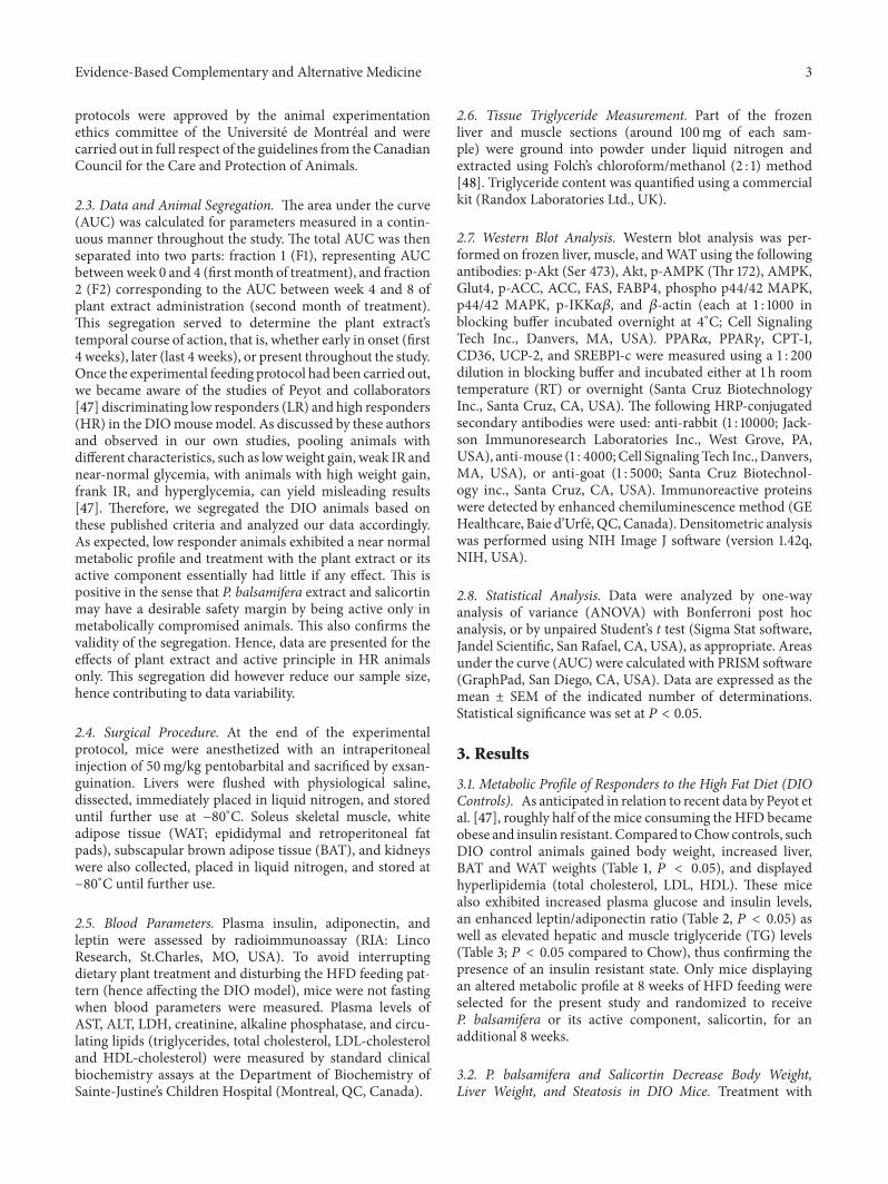
Table 1: Effects of obesity, P. balsamifera, and salicortin treatments on body and organ weights at sacrifice.
DIO P. balsamifera125mg/kg
P. balsamifera250mg/kg DIO Salicortin
12.5mg/kgBody Weight 138 ± 1† 120 ± 7∗ 131 ± 3 142 ± 2† 130 ± 3
§
Retroperitoneal Fat Pad 229 ± 12† 190 ± 28 209 ± 20 242 ± 13† 218 ± 10Epididymal Fat Pad 77 ± 3† 98 ± 17 103 ± 5∗ 97 ± 2 149 ± 11
§
Brown Adipose tissue 189 ± 14† 136 ± 22𝑃 = 0.057
168 ± 14 200 ± 6† 170 ± 11§
Liver Weight 167 ± 6† 108 ± 12∗ 122 ± 10∗ 166 ± 9† 118 ± 7§
Total Kidney 111 ± 4† 93 ± 3∗ 104 ± 4 104 ± 2 101 ± 3Measurements were obtained after 16 weeks of treatment with either standard diet (Chow), HFD (DIO), and for the last 8 of the 16 weeks with HFD incombination with P. balsamifera at 125 or 250mg/kg, or with the active salicortin at 12.5mg/kg. All values are expressed as a percentage of respective Chowcontrols (reference set at 100%) and represent the mean ± SEM. The number of animals for each group for the P. balsamifera protocol was: CHOW (𝑛 = 12);DIO (𝑛 = 8); P. balsamifera 125 (𝑛 = 5); P. balsamifera 250 (𝑛 = 7); and for the salicortin protocol: CHOW (𝑛 = 12); DIO (𝑛 = 7); salicortin (𝑛 = 9). †denotesDIO significantly different as compared to Chow (unpaired Student’s t test; P < 0.05). ∗denotes significantly different as compared to respective DIO (one wayANOVA, Bonferroni post hoc test; P < 0.05). §denotes significantly different as compared to respective DIO (unpaired Student’s t test; P < 0.05).
Table 2: Effects of obesity, P. balsamifera, and salicortin treatments on systemic parameters at sacrifice.
DIO P. balsamifera125mg/kg
P. balsamifera250mg/kg DIO Salicortin
12.5mg/kgGlucose (mmol/L) 135 ± 14† 105 ± 8 121 ± 10 121 ± 6† 114 ± 6
Insulin (ng/mL) 3056 ± 1074† 450 ± 238∗ 832 ± 423(P = 0.052) 1035 ± 150† 272 ± 62
§
Leptin (ng/mL) 211 ± 28† 108 ± 18∗ 145 ± 9(P = 0.051) 246 ± 19† 197 ± 13
§
Adiponectin (𝜇g/mL) 70 ± 3† 78 ± 10 82 ± 5(P = 0.054) 97 ± 5 101 ± 6
Leptin/adiponectin ratio 304 ± 37† 138 ± 16∗ 181 ± 16∗ 248 ± 12† 196 ± 18§
TG (mmol/L) 99 ± 8 80 ± 10 82 ± 8 118 ± 10 106 ± 14LDL (mmol/L) 391 ± 31† 355 ± 37 307 ± 42 344 ± 20† 207 ± 22
§
HDL (mmol/L) 141 ± 8† 112 ± 17 125 ± 8 157 ± 10† 137 ± 7Total cholesterol (mmol/L) 180 ± 9† 152 ± 14 151 ± 12 203 ± 11† 151 ± 7
§
ALT (U/L) 281 ± 39† 229 ± 47 271 ± 77 341 ± 123† 157 ± 27AST (U/L) 172 ± 30† 153 ± 22 137 ± 22 163 ± 27† 86 ± 5
§
Creatinine (U/L) 184 ± 50 491 ± 160 557 ± 138 276 ± 113 136 ± 30Alkaline phosphatase (U/L) 115 ± 20 85 ± 15 100 ± 9 106 ± 9 88 ± 17LDH (U/L) 341 ± 83† 137 ± 16
§ 193 ± 61 376 ± 156† 154 ± 38Measurements were obtained after 16 weeks of treatment with either standard diet (Chow), HFD (DIO), and for the last 8 of the 16 weeks with HFD incombination with P. balsamifera at 125 or 250mg/kg, or with the active salicortin at 12.5mg/kg. All values are expressed as a percentage of their respectiveChow controls (reference set at 100%) and represent the mean ± SEM. The number of animals for each group for the P. balsamifera protocol was: CHOW(𝑛 = 12); DIO (𝑛 = 8); P. balsamifera 125 (𝑛 = 5); P. balsamifera 250 (𝑛 = 7); and for the salicortin protocol: CHOW (𝑛 = 12); DIO (𝑛 = 7); salicortin (𝑛 = 9).†denotes DIO significantly different as compared to Chow (unpaired Student’s t test; P < 0.05). ∗denotes significantly different as compared to respective DIO(one way ANOVA, Bonferroni post hoc test; P < 0.05). §denotes significantly different as compared to respective DIO (unpaired Student’s t test; P < 0.05).
Table 3: Effects of obesity, P. balsamifera, and salicortin treatments on hepatic and muscular triglyceride accumulation.
DIO P. balsamifera 125mg/kg P. balsamifera 250mg/kg DIO Salicortin 12.5mg/kgLiver TG Levels (mg/g total liver) 930 ± 65† 436 ± 146∗ 521 ± 116∗ 1084 ± 180† 559 ± 93
§
Muscle TG levels (𝜇g/mg) 223 ± 54† 342 ± 81 267 ± 38 230 ± 32† 219 ± 24The colorimetric dosage of TG levels in both the liver and muscle was determined using a commercial kit (Randox Laboratories ltd). Measurements wereobtained after 16 weeks of treatment with either standard diet (Chow), HFD (DIO), and for the last 8 of the 16 weeks with HFD in combination with P.balsamifera at 125 or 250mg/kg, or with the active salicortin at 12.5mg/kg. All values are expressed as percentage of respective Chow (reference set at 100%)and represent the mean ± SEM. The number of animals for each group for the P. balsamifera protocol was: CHOW (𝑛 = 12); DIO (𝑛 = 8); P. balsamifera 125(𝑛 = 5); P. balsamifera 250 (𝑛 = 7); and for the salicortin protocol: CHOW (𝑛 = 12); DIO (𝑛 = 7); salicortin (𝑛 = 9). †denotes DIO significantly different ascompared to Chow (unpaired Student’s t test; P < 0.05). ∗denotes significantly different as compared to respective DIO (one way ANOVA, Bonferroni post hoctest; P < 0.05). §denotes significantly different as compared to respective DIO (unpaired Student’s t test; P < 0.05).
Evidence-Based Complementary and Alternative Medicine 5
Cum
ulat
ive c
hang
e in
body
wei
ght (
g)
†
∗
DIO
ChowDIO
P. balsamifera 125 mg/kgP. balsamifera 250 mg/kg
DIO + P. balsamifera
36
32
28
24
20
16
12
8
4
0
0 10 20 30 40 50 60 70 80 90 100 110 120
Day (s)
(a)
†
§
DIO DIO + Salicortin
Cum
ulat
ive c
hang
e in
body
wei
ght (
g) 36
32
28
24
20
16
12
8
4
0
0 10 20 30 40 50 60 70 80 90 100 110 120
Day (s)
ChowDIO
Salicortin 12.5 mg/kg
(b)250
200
150
100
50
0AUC
from
cum
ulat
ive
chan
gein
bod
y w
eigh
t(C
CBW
)% C
how
DIO
DIO
Salic
ortin
12.5
††
§ §
P. ba
lsam
ifera
125
P. ba
lsam
ifera
250
(c)
250
200
150
100
50
0AUC
from
cum
ulat
ive
chan
gein
bod
y w
eigh
t(C
CBW
)% C
how
DIO
DIO
Salic
ortin
12.5
† †
§
P. ba
lsam
ifera
125
P. ba
lsam
ifera
250
(d)250
200
150
100
50
0AUC
from
cum
ulat
ive
chan
gein
bod
y w
eigh
t(C
CBW
)% C
how ††
∗ §
DIO
DIO
Salic
ortin
12.5
P. ba
lsam
ifera
125
P. ba
lsam
ifera
250
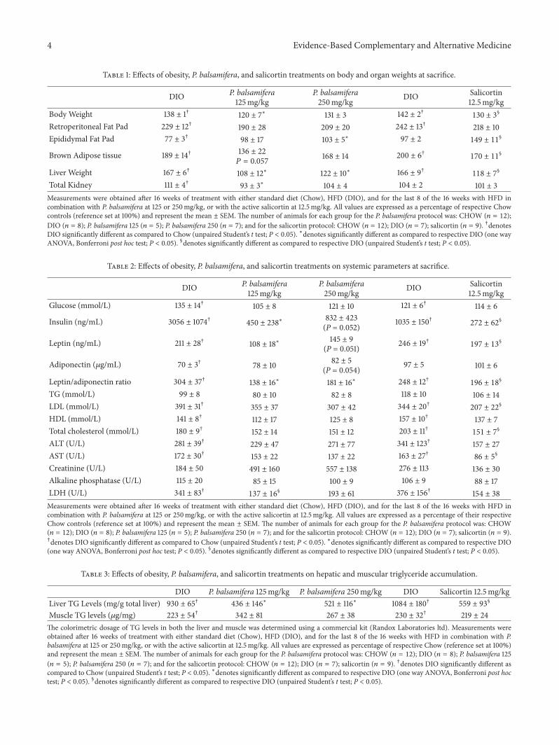
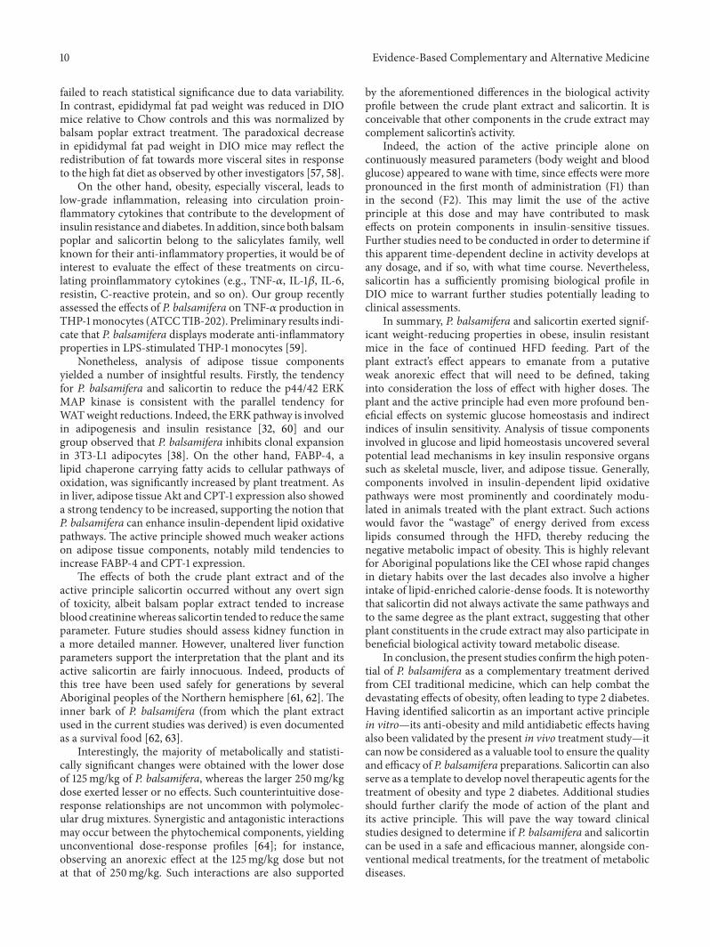
(e)Figure 1: Cumulative changes in body weight (CCBW) in C57BL/6 mice treated with either standard diet (Chow), HFD (DIO), and (a) HFDin combination with P. balsamifera at 125 or 250mg/kg, or (b) HFD in combination with salicortin. Area under the curve (AUC) of CCBWfor (c) total 8-week treatment period, (d) first 4 weeks of treatment (F1), and (e) second 4 weeks of treatment (F2). As mentioned, C57BL/6mice were administered either standard diet (Chow), HFD (DIO), and for the last 8 of the 16 weeks a HFD in combination with P. balsamiferaat 125 or 250mg/kg, or with Salicortin 12.5mg/kg. All values are mean ± SEM. Fraction 1 (F1) consists in the AUC between week 0 and 4, andfraction 2 (F2) corresponds to the AUC between week 4 and 8 of administration of the plant extract. The number of animals for the crudeplant extract protocol was CHOW (𝑛 = 12), DIO (𝑛 = 8), P. balsamifera 125 (𝑛 = 5), and P. balsamifera 250 (𝑛 = 7); and for the salicortinprotocol: CHOW (𝑛 = 12), DIO (𝑛 = 7), salicortin (𝑛 = 9). †denotes DIO significantly different as compared to Chow (unpaired Student’s ttest; 𝑃 < 0.05). ∗denotes significantly different as compared to respective DIO (one way ANOVA; 𝑃 < 0.05). §denotes significantly differentas compared to respective DIO (unpaired Student’s t test; 𝑃 < 0.05).
P. balsamifera (at 125mg/kg) significantly decreased bodyweight.This decrease reached 13% at sacrificewhen comparedtoDIO controls (𝑃 < 0.05; Table 1).When taking into accountcontinuous measurements of cumulative changes in bodyweight (CCBW; Figures 1(a) and 1(b)), the area under the
curve (AUC) was lowered by 8% (𝑃 < 0.05; Figure 1(c)) with125mg/kg ofP. balsamifera.This effectwas gradual, beginningwithin the first month (𝐹1 = 6% reduction; N.S.; Figure 1(d)),but becoming more pronounced in the second month of thetreatment (𝐹2 = 10% decrease; 𝑃 < 0.05; Figure 1(e)).
6 Evidence-Based Complementary and Alternative Medicine
200
150
100
50
0AUC
from
cum
ulat
ive
chan
ge in
blo
od g
luco
sele
vels
% C
how
DIO
DIO
††
∗ ∗ §
Salic
ortin
12.5
P. ba
lsam
ifera
125
P. ba
lsam
ifera
250
(a)
200
150
100
50
0AUC
from
cum
ulat
ive
chan
ge in
blo
od g
luco
sele
vels
% C
how †
†∗ ∗ §
DIO
DIO
Salic
ortin
12.5
P. ba
lsam
ifera
125
P. ba
lsam
ifera
250
(b)
200
150
100
50
0AUC
from
cum
ulat
ive
chan
ge in
blo
od g
luco
sele
vels
% C
how
††
∗ ∗
DIO
DIO
Salic
ortin
12.5
P. ba
lsam
ifera
125
P. ba
lsam
ifera
250
(c)
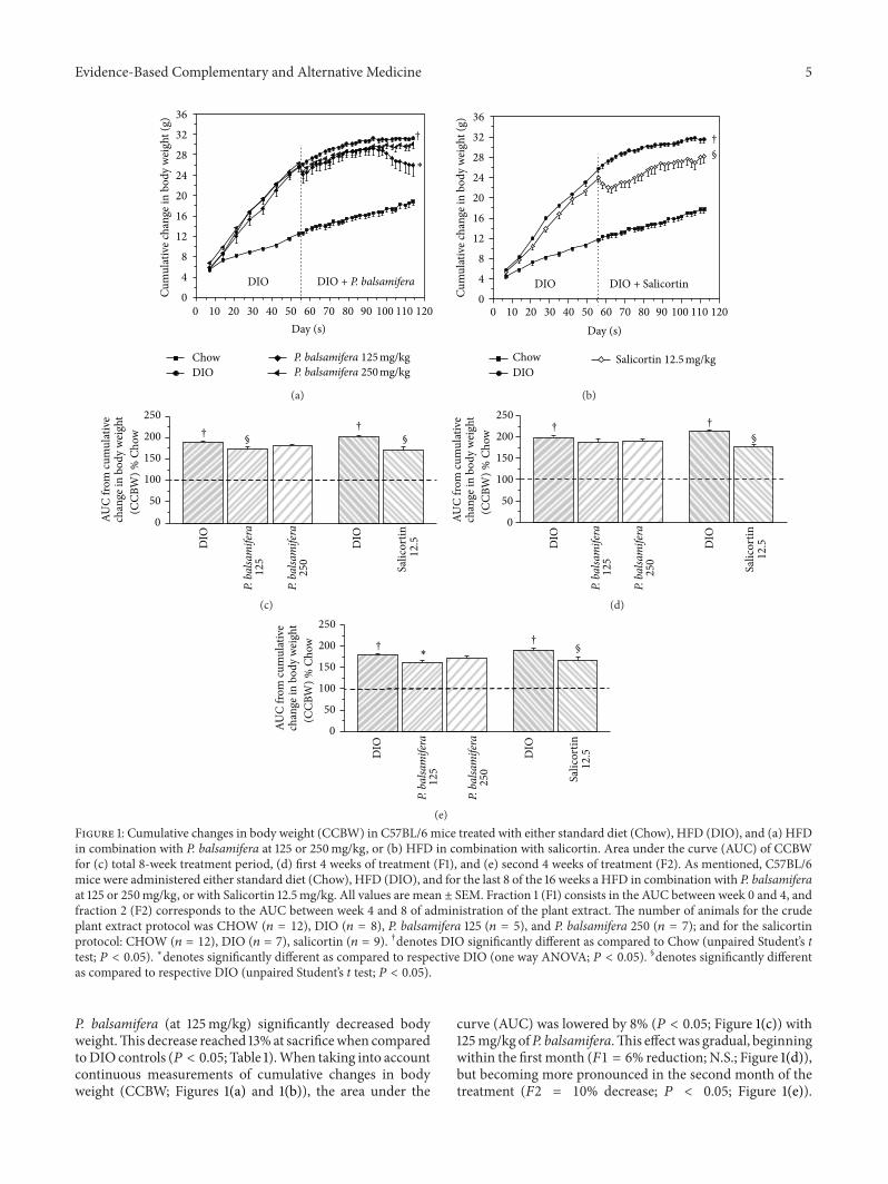
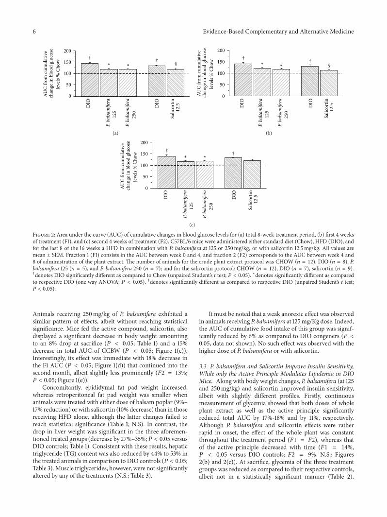
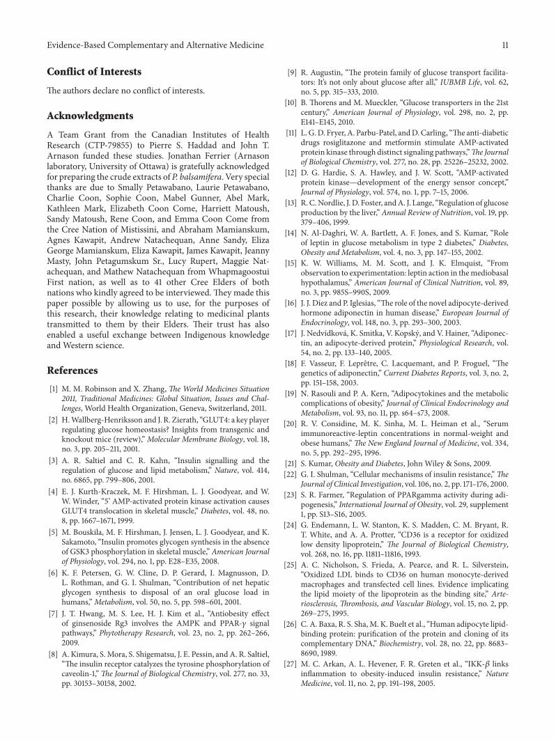
Figure 2: Area under the curve (AUC) of cumulative changes in blood glucose levels for (a) total 8-week treatment period, (b) first 4 weeksof treatment (F1), and (c) second 4 weeks of treatment (F2). C57BL/6 mice were administered either standard diet (Chow), HFD (DIO), andfor the last 8 of the 16 weeks a HFD in combination with P. balsamifera at 125 or 250mg/kg, or with salicortin 12.5mg/kg. All values aremean ± SEM. Fraction 1 (F1) consists in the AUC between week 0 and 4, and fraction 2 (F2) corresponds to the AUC between week 4 and8 of administration of the plant extract. The number of animals for the crude plant extract protocol was CHOW (𝑛 = 12), DIO (𝑛 = 8), P.balsamifera 125 (𝑛 = 5), and P. balsamifera 250 (𝑛 = 7); and for the salicortin protocol: CHOW (𝑛 = 12), DIO (𝑛 = 7), salicortin (𝑛 = 9).†denotes DIO significantly different as compared to Chow (unpaired Student’s t test; 𝑃 < 0.05). ∗denotes significantly different as comparedto respective DIO (one way ANOVA; 𝑃 < 0.05). §denotes significantly different as compared to respective DIO (unpaired Student’s t test;𝑃 < 0.05).
Animals receiving 250mg/kg of P. balsamifera exhibited asimilar pattern of effects, albeit without reaching statisticalsignificance. Mice fed the active compound, salicortin, alsodisplayed a significant decrease in body weight amountingto an 8% drop at sacrifice (𝑃 < 0.05; Table 1) and a 15%decrease in total AUC of CCBW (𝑃 < 0.05; Figure 1(c)).Interestingly, its effect was immediate with 18% decrease inthe F1 AUC (𝑃 < 0.05; Figure 1(d)) that continued into thesecond month, albeit slightly less prominently (𝐹2 = 13%;𝑃 < 0.05; Figure 1(e)).
Concomitantly, epididymal fat pad weight increased,whereas retroperitoneal fat pad weight was smaller whenanimals were treated with either dose of balsam poplar (9%–17% reduction) orwith salicortin (10%decrease) than in thosereceiving HFD alone, although the latter changes failed toreach statistical significance (Table 1; N.S). In contrast, thedrop in liver weight was significant in the three aforemen-tioned treated groups (decrease by 27%–35%;𝑃 < 0.05 versusDIO controls; Table 1). Consistent with these results, hepatictriglyceride (TG) content was also reduced by 44% to 53% inthe treated animals in comparison to DIO controls (𝑃 < 0.05;Table 3). Muscle triglycerides, however, were not significantlyaltered by any of the treatments (N.S.; Table 3).
It must be noted that a weak anorexic effect was observedin animals receiving P. balsamifera at 125mg/Kg dose. Indeed,the AUC of cumulative food intake of this group was signif-icantly reduced by 6% as compared to DIO congeners (𝑃 <0.05, data not shown). No such effect was observed with thehigher dose of P. balsamifera or with salicortin.
3.3. P. balsamifera and Salicortin Improve Insulin Sensitivity,While only the Active Principle Modulates Lipidemia in DIOMice. Along with body weight changes, P. balsamifera (at 125and 250mg/kg) and salicortin improved insulin sensitivity,albeit with slightly different profiles. Firstly, continuousmeasurement of glycemia showed that both doses of wholeplant extract as well as the active principle significantlyreduced total AUC by 17%-18% and by 11%, respectively.Although P. balsamifera and salicortin effects were ratherrapid in onset, the effect of the whole plant was constantthroughout the treatment period (𝐹1 = 𝐹2), whereas thatof the active principle decreased with time (𝐹1 = 14%,𝑃 < 0.05 versus DIO controls; 𝐹2 = 9%, N.S.; Figures2(b) and 2(c)). At sacrifice, glycemia of the three treatmentgroups was reduced as compared to their respective controls,albeit not in a statistically significant manner (Table 2).
Evidence-Based Complementary and Alternative Medicine 7
Secondly, insulinemia diminished by 85%with balsam poplarat 125mg/Kg (𝑃 < 0.05) and by 73% with 250mg/Kg(𝑃 = 0.052) as well as with salicortin (𝑃 < 0.05) incomparison to DIO controls (Table 2). Thirdly, the two dosesof P. balsamifera decreased leptin/adiponectin ratio by 41%–54% as compared to congeners receiving HFD alone (𝑃 <0.05; Table 2). Salicortin also significantly decreased this ratio,although to a lesser extent (by 21%; 𝑃 < 0.05; Table 2).In terms of the circulating lipid profile, only the salicortintreated group exhibited significantly lowered total plasmacholesterol and LDL levels, which were reduced by 25%and 40%, respectively (Table 2; 𝑃 < 0.05) as comparedto the DIO controls. Altogether, these findings illustrate animprovement in insulin sensitivity when balsam poplar or itsactive principle are added to the HFD.
Finally, P. balsamifera and salicortin tended to normalizeseveral systemic parameters of toxicity, although this did notreach statistical significance, except in the case of AST levelsfor salicortin (𝑃 < 0.05; Table 2) and LDH levels for P.balsamifera at 125mg/Kg (𝑃 < 0.05; Table 2).
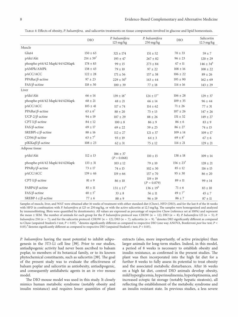
3.4. P. balsamifera Tends to Increase Skeletal Muscle Glut4 andImproves Components Related toMuscle Lipid Oxidation with-out Affecting the Akt and AMPKPathways,Whereas SalicortinTends to Increase Akt Phosphorylation and Activates p44/42MAPK. Despite the significant reduction of overall glycemiaexerted by the plant extract and its active principle, analysis ofprotein components involved in muscle glucose homeostasisdid not exhibit any statistically significant changes. Therewas a tendency for Glut4 expression to increase in animalstreated with P. balsamifera at 125mg/kg, (Table 4; N.S.balsampoplar versus correspondingDIO controls). Similarly,insulin-dependent Akt phosphorylation tended to increasein animals fed with salicortin, although data variabilityprecluded any definitive interpretation of these results. Theinsulin-independent AMPK pathway remained more clearlyunchanged.
In contrast, components involved in muscle lipid home-ostasis showed evidence of improvement with balsam poplartreatment. Indeed, P. balsamifera at 125mg/kg more thandoubled muscle PPAR𝛼 expression levels (Table 4; 137%increase compared to DIO 𝑃 < 0.05). When looking atcomponents involved in muscle fatty acid oxidation andsynthesis, again only the plant extract seemed to act onsuch pathways, by tending to increase phosphorylated ACClevels and to normalize FAS levels back down to Chowlevels (Table 4; N.S. compared to DIO controls). The p44/42MAPK pathway linked to exercise and insulin stimulationwas significantly activated with salicortin (Table 4, 𝑃 < 0.05),and showed a tendency to do so with the plant extract at250mg/kg (Table 4, N.S.).
3.5. The Effects of P. balsamifera and Salicortin on LiverComponents of Glucose and Lipid Homeostasis. Both doses ofP. balsamifera significantly increased hepatic phosphorylatedAkt in HFD-fed animals (Table 4; increases by 111% and87% for 125 and 250mg/kg groups, respectively; 𝑃 < 0.05compared toDIO controls), while the active principle showedonly a slight tendency to do so (22% increase). A number of
parameters related to hepatic lipid homeostasis or inflamma-tion showed interesting tendencies, but none of these effectsreached statistical significance. In all cases, tendencies weremore pronounced with the 125 than the 250mg/kg dose ofP. balsamifera. Notably, PPAR𝛼 appeared to be increased byboth balsam poplar and the active principle, while CPT-1seemed to be increased only by the plant extract (Table 4,N.S.). As for IKK𝛼𝛽 it appeared to be affected only by theplant extract, exhibiting a decrease of 43% and 30% with 125and 250mg/kg doses, respectively (Table 4; N.S.).
3.6. The Effect of P. balsamifera and Salicortin on AdiposeTissue Components of Glucose and Lipid Homeostasis. P. bal-samifera at 125mg/kg showed a strong tendency to increasephosphorylated Akt levels in adipose tissue (Table 4; increaseby 65%, 𝑃 = 0.068 compared to DIO controls). Likewise,CPT-1 expression in animals treated with the plant extractat 250mg/kg exhibited a strong tendency to be enhanced(Table 4; increase by 47%, 𝑃 = 0.079 compared to DIO),whereas the active principle had a similar albeit much weakereffect on this parameter (Table 4; 11% increase; N.S.). Incontrast, FABP4 was clearly and significantly increased byP. balsamifera at both doses (54% and 60% at 125 and250mg/kg, respectively, Table 4, 𝑃 < 0.05 compared to DIOcontrols), while salicortin showed only a slight tendency to doso (16%, N.S., Table 4;). Salicortin and balsam poplar showeda tendency to normalize PPAR𝛾 and phosphorylated p44/42MAPK to levels similar to those observed in Chow animals(Table 4). Other components failed to show any significantchanges in plant or active-principle treated treated animalscompared to their respective DIO controls.
4. Discussion
According to the World Health Organization (WHO), 75%of the world population still relies on traditional medicinefor primary health care needs and this often involves crudepreparations of medicinal plants [49]. In the Canadianprovince of Quebec, regional health authorities assigned tothe CEI are currently considering the usefulness of Creetraditional medicine, notably its associated pharmacopoeia,to deal with several health concerns such as type 2 diabetes;a condition that has reached epidemic proportions in theregion [36]. Our group has been working since 2003 withcommunities and health authorities in CEI to build thescientific evidence base in support of this initiative. Anethnobotanical study was conducted in collaboration withCEI Elders and healers that identified several plants usedto treat diabetes symptoms [37, 50, 51]. One of these wasPopulus balsamifera L. (Salicaceae) or balsam poplar. Theplant did not demonstrate much antidiabetic potential in invitro bioassays; for instance, it had little effect on muscleglucose uptake [37]. However, the plant caught our attentionby its complete inhibition of triglyceride accumulation andadipogenesis in the 3T3-L1 adipocyte cell line [37], suggestingpotential therapeutic usefulness against obesity. Salicortin,a salicylate glycoside, abundant in poplar, willow bark, aswell as throughout the Salicaceae family, was identifiedthrough bioassay-guided fractionation as the constituent of
8 Evidence-Based Complementary and Alternative Medicine
Table 4: Effects of obesity, P. balsamifera, and salicortin treatments on tissue components involved in glucose and lipid homeostasis.
DIO P. balsamifera125mg/kg
P. balsamifera250mg/kg DIO Salicortin
12.5mg/kgMuscle
Glut4 150 ± 63 321 ± 174 151 ± 52 70 ± 33 59 ± 7pAkt/Akt 214 ± 59† 195 ± 47 267 ± 82 96 ± 23 120 ± 29phospho p44/42 Mapk/44/42Mapk 178 ± 83 99 ± 15 273 ± 84 47 ± 11 146 ± 34
§
pAMPk/AMPk 138 ± 43 79 ± 10 97 ± 22 108 ± 16 108 ± 22pACC/ACC 122 ± 28 171 ± 56 157 ± 38 106 ± 22 89 ± 26PPAR𝛼/𝛽-actine 97 ± 23 229 ± 50
§ 143 ± 44 195 ± 90 162 ± 69FAS/𝛽-actine 118 ± 30 100 ± 39 77 ± 18 114 ± 16 143 ± 29
LiverpAkt/Akt 66 ± 14 139 ± 18∗ 124 ± 17∗ 106 ± 28 129 ± 37phospho p44/42 Mapk/44/42Mapk 68 ± 21 48 ± 21 66 ± 14 109 ± 35 96 ± 44pACC/ACC 105 ± 41 117 ± 74 114 ± 62 71 ± 26 77 ± 31PPAR𝛼/𝛽-actine 63 ± 6† 88 ± 20 75 ± 13 107 ± 26 147 ± 26UCP-2/𝛽-actine 94 ± 19 107 ± 29 88 ± 26 151 ± 32 149 ± 27CPT-l/𝛽-actine 84 ± 12 100 ± 8 86 ± 9 86 ± 6 83 ± 9FAS/𝛽-actine 69 ± 17 69 ± 22 59 ± 25 86 ± 27 74 ± 15SREBPl-c/𝛽-actine 88 ± 16 112 ± 27 121 ± 37 109 ± 14 109 ± 17CD36/𝛽-actine 63 ± 7† 93 ± 19 64 ± 3 69 ± 8† 67 ± 6pIKK𝛼𝛽/𝛽-actine 108 ± 23 62 ± 31 75 ± 12 114 ± 21 129 ± 21
Adipose tissue
pAkt/Akt 112 ± 13 186 ± 37(P = 0.068) 110 ± 15 138 ± 18 109 ± 14
phospho p44/42 Mapk/44/42Mapk 133 ± 31 103 ± 12 79 ± 10 156 ± 23† 128 ± 21PPAR𝛾/𝛽-actine 73 ± 17 74 ± 15 102 ± 30 85 ± 12 116 ± 15pACC/ACC 139 ± 46 119 ± 66 157 ± 70 95 ± 30 86 ± 20
CPT-l/𝛽-actine 81 ± 9 86 ± 10 119 ± 19(𝑃 = 0.079) 89 ± 11 99 ± 14
FABP4/𝛽-actine 85 ± 11 131 ± 11§
136 ± 19§ 71 ± 6 83 ± 10
FAS/𝛽-actine 40 ± 5† 33 ± 8 56 ± 11 49 ± 7† 45 ± 7SREBP-1 c/𝛽-actine 77 ± 6 88 ± 9 96 ± 19 86 ± 7 87 ± 11
Samples of muscle, liver, and WAT were obtained after 16 weeks of treatment with either standard diet (Chow), HFD (DIO), and for the last 8 of the 16 weekswith HFD in combination with P. balsamifera at 125 or 250mg/kg, or with the active salicortin at 12.5mg/kg. The samples were homogenized and analyzedby immunoblotting. Blots were quantified by densitometry. All values are expressed as percentage of respective Chow (reference set at 100%) and representthe mean ± SEM. The number of animals for each group for the P. balsamifera protocol was: CHOW (𝑛 = 12); DIO (𝑛 = 8); P. balsamifera 125 (𝑛 = 5); P.balsamifera 250 (𝑛 = 7); and for the salicortin protocol: CHOW (𝑛 = 12); DIO (𝑛 = 7); salicortin (𝑛 = 9). †denotes DIO significantly different as comparedto Chow (unpaired Student’s t test; P < 0.05). ∗denotes significantly different as compared to respective DIO (one way ANOVA, Bonferroni post hoc test; P <0.05).§denotes significantly different as compared to respective DIO (unpaired Student’s t test; P < 0.05).
P. balsamifera having the most potential to inhibit adipo-genesis in the 3T3-L1 cell line [39]. Prior to our studies,antiadipogenic activity had never been ascribed to balsampoplar, to members of its botanical family, or to its knownphytochemical constituents, such as salicortin [39]. The goalof the present study was to evaluate the effectiveness ofbalsam poplar and salicortin as antiobesity, antiadipogenic,and consequently antidiabetic agents in an in vivo mousemodel.
The DIO mouse model was used in this study. It closelymimics human metabolic syndrome (notably obesity andinsulin resistance) and requires lesser quantities of plant
extracts (also, more importantly, of active principles) thanlarger animals for long-term studies. Indeed, in this model,a period of 8 weeks is necessary to establish obesity andinsulin resistance, as confirmed in the present studies. Theplant was then incorporated into the high fat diet for afurther 8 weeks to fully assess its potential to treat obesityand the associated metabolic disturbances. After 16 weekson a high fat diet, control DIO animals develop obesity,mild hyperglycemia, hyperinsulinemia, hyperleptinemia, andincreased ectopic fat storage (notably hepatic steatosis), allreflecting the establishment of the metabolic syndrome andan insulin resistant state. In previous studies, a less severe
Evidence-Based Complementary and Alternative Medicine 9
model was used whereby animals were subjected to a HFDfor only 8 weeks; P. balsamifera being administered from theonset of the HFD feeding in order to evaluate its potentialto prevent obesity and its associated insulin resistant state[44]. The plant extract effectively reduced body weight gain,retroperitoneal fat pad weight, liver lipid content, as well ascirculating glucose, insulin and leptin levels. It also activatedpathways that were involved with glucose and lipid oxidation,as well as thermoregulation. The onset of action of the plantextract was immediate and sustained throughout its course ofadministration.
The results of the current study clearly demonstratethat in mice subjected to a continuous hypercaloric fat-laden diet, P. balsamifera significantly reduced body weight,whereas its active salicortin prevented further weight gain.The plant’s effect was more potent and statistically significantat 125mg/kg than 250mg/kg. Several anthropomorphic, sys-temic and tissue parameters were thus examined to circum-scribe the possible mechanisms of action of the plant extractand its active principle, salicortin.
A first potentially important lead came from data oncumulative food intake. Indeed, the plant extract at 125mg/kgslightly but significantly reduced energy intake, and thiswas visible in the second month of treatment (F2; datanot shown). This correlated well with the plant’s temporalaction on body weight. An initial reduction in body weightwas observed upon introduction of the plant extract in thediet and may have resulted from a behavioral response tothe food change. However, body weight rapidly resumedits course such that cumulative weight gain in the firstmonth period (F1) was not significantly different amongtreatment groups. This contrasts with the reduced energyintake in the second month period (F2) that coincided witha significant decrease in weight gain. Such results suggestedthat the plant may exhibit slight appetite-modifying effects.Interestingly, these putative anorexic effects were seen onlywith the 125mg/kg dose and indicate an unconventionaldose-response relationship, as discussed further below. Nev-ertheless, such anorexic effects warrant further investigation.Notably, appetite-related hormones, such as leptin, as wellas gut-brain appetite control mechanisms will need to beexamined.
However, the reduction in caloric intake was weakerthan the weight loss measured, roughly half to two-thirdsas important when considering total or F2 AUC measure-ments of cumulative weight changes, respectively. In contrast,the active salicortin decreased the overall AUC of CCBWwithout affecting cumulative energy intake. This not onlysuggests different profiles of biological activity between theactive principle and the plant extract, but also that otherphytochemical components present within balsam poplar arecontributing to its appetite-modifying effect.
On the other hand, although obesity was only partlycountered by P. balsamifera and salicortin, systemic glucosehomeostasis was more significantly improved. Indeed, con-tinuous glycemia measurements showed that the plant andits active principle had an overall effect to reduce bloodglucose variations toward normal values observed in Chow-fed controls. Even more telling was the dramatic decrease
of insulinemia seen with P. balsamifera at 125mg/kg andwith salicortin. Likewise, the leptin-to-adiponectin ratio, alsoreflective of insulin resistance, was essentially halved withthe plant extract and decreased by 1.5-fold with the activeprinciple. Interestingly, salicortin also significantly improvedthe blood lipid profile by decreasing LDL and total cholesterollevels, whereas the plant extract had no significant impact onsystemic parameters of lipid homeostasis. This again pointsto variations in biological activity between the crude extractand the purified active principle.
Further analysis of the major insulin responsive tissues,notably skeletal muscle, liver and adipose tissue, yieldeddata that highlights potential mechanisms at several levelsof metabolic control. Firstly, excessive skeletal muscle TGaccumulationwas not corrected byP. balsamifera or salicortintreatment. In fact, if anything, balsam poplar extract at125mg/kg tended to increase this parameter, albeit largevariations in the data preclude any definite interpretations.One possibility is that the two-month treatment was notsufficient to significantly affect muscle TG accumulation, yetimprovements in muscle lipid and glucose metabolism couldhave been initiated. Indeed, the crude plant extract did morethan double the expression of PPAR𝛼, which could lead toincreased fatty acid oxidation [30] and improved muscleinsulin sensitivity [52]. In animals receiving 125mg/kg of theplant, muscle Glut4 expression tended to increase and thisis consistent with enhanced insulin sensitivity. In contrast,salicortin treatment only significantly affectedmuscle p44/42MAPK activation, again hinting at different actions of theplant extract and its active principle.
In contrast, in the liver, P. balsamifera and salicortin treat-ment more than halved the elevated levels of accumulatedTGs. Since hepatic steatosis is increasingly recognized asa major contributor to systemic insulin resistance [53, 54],this action may have played a significant role in improvingsystemic glucose homeostasis and insulin sensitivity. Indeed,analysis of key tissue proteins indicated that P. balsamiferatreatment induced a doubling of liver Akt phosphorylation.Since Akt is a major component of the insulin-signalingcascade, part of the effect of balsam poplar could involveimproved hepatic insulin sensitivity. Indeed, Akt inhibitsglucose production and promotes glycogen deposition inthe liver [5, 6, 13]. In hepatic cell lines, our group recentlyfound that P. balsamifera inhibits glucose-6-phosphatase[55]. Other components also tended to be modulated byP. balsamifera in the liver and suggested that the plantmay favor salvaging lipid metabolism. Indeed, PPAR𝛼 levelswere increased by treatment with the plant and its activeprinciple, this transcription factor being known to enhancefatty acid oxidation [30]. The tendency for a reduction ofIKK𝛼𝛽 by the plant treatment, on the other hand, points to apotential improvement of inflammatory components knownto be involved in nonalcoholic fatty liver disease and ensuingmetabolic disturbances [27, 56]. Such effects of P. balsamiferaand salicortin on liver lipid homeostasis and inflammationwill require confirmation in future studies.
Despite large reductions in retroperitoneal fat pad weightat sacrifice, consistent with the significant reduction in bodyweight, such changes induced by P. balsamifera and salicortin
10 Evidence-Based Complementary and Alternative Medicine
failed to reach statistical significance due to data variability.In contrast, epididymal fat pad weight was reduced in DIOmice relative to Chow controls and this was normalized bybalsam poplar extract treatment. The paradoxical decreasein epididymal fat pad weight in DIO mice may reflect theredistribution of fat towards more visceral sites in responseto the high fat diet as observed by other investigators [57, 58].
On the other hand, obesity, especially visceral, leads tolow-grade inflammation, releasing into circulation proin-flammatory cytokines that contribute to the development ofinsulin resistance anddiabetes. In addition, since both balsampoplar and salicortin belong to the salicylates family, wellknown for their anti-inflammatory properties, it would be ofinterest to evaluate the effect of these treatments on circu-lating proinflammatory cytokines (e.g., TNF-𝛼, IL-1𝛽, IL-6,resistin, C-reactive protein, and so on). Our group recentlyassessed the effects of P. balsamifera on TNF-𝛼 production inTHP-1monocytes (ATCCTIB-202). Preliminary results indi-cate that P. balsamifera displays moderate anti-inflammatoryproperties in LPS-stimulated THP-1 monocytes [59].
Nonetheless, analysis of adipose tissue componentsyielded a number of insightful results. Firstly, the tendencyfor P. balsamifera and salicortin to reduce the p44/42 ERKMAP kinase is consistent with the parallel tendency forWATweight reductions. Indeed, the ERKpathway is involvedin adipogenesis and insulin resistance [32, 60] and ourgroup observed that P. balsamifera inhibits clonal expansionin 3T3-L1 adipocytes [38]. On the other hand, FABP-4, alipid chaperone carrying fatty acids to cellular pathways ofoxidation, was significantly increased by plant treatment. Asin liver, adipose tissue Akt and CPT-1 expression also showeda strong tendency to be increased, supporting the notion thatP. balsamifera can enhance insulin-dependent lipid oxidativepathways. The active principle showed much weaker actionson adipose tissue components, notably mild tendencies toincrease FABP-4 and CPT-1 expression.
The effects of both the crude plant extract and of theactive principle salicortin occurred without any overt signof toxicity, albeit balsam poplar extract tended to increaseblood creatininewhereas salicortin tended to reduce the sameparameter. Future studies should assess kidney function ina more detailed manner. However, unaltered liver functionparameters support the interpretation that the plant and itsactive salicortin are fairly innocuous. Indeed, products ofthis tree have been used safely for generations by severalAboriginal peoples of the Northern hemisphere [61, 62]. Theinner bark of P. balsamifera (from which the plant extractused in the current studies was derived) is even documentedas a survival food [62, 63].
Interestingly, the majority of metabolically and statisti-cally significant changes were obtained with the lower doseof 125mg/kg of P. balsamifera, whereas the larger 250mg/kgdose exerted lesser or no effects. Such counterintuitive dose-response relationships are not uncommon with polymolec-ular drug mixtures. Synergistic and antagonistic interactionsmay occur between the phytochemical components, yieldingunconventional dose-response profiles [64]; for instance,observing an anorexic effect at the 125mg/kg dose but notat that of 250mg/kg. Such interactions are also supported
by the aforementioned differences in the biological activityprofile between the crude plant extract and salicortin. It isconceivable that other components in the crude extract maycomplement salicortin’s activity.
Indeed, the action of the active principle alone oncontinuously measured parameters (body weight and bloodglucose) appeared to wane with time, since effects were morepronounced in the first month of administration (F1) thanin the second (F2). This may limit the use of the activeprinciple at this dose and may have contributed to maskeffects on protein components in insulin-sensitive tissues.Further studies need to be conducted in order to determine ifthis apparent time-dependent decline in activity develops atany dosage, and if so, with what time course. Nevertheless,salicortin has a sufficiently promising biological profile inDIO mice to warrant further studies potentially leading toclinical assessments.
In summary, P. balsamifera and salicortin exerted signif-icant weight-reducing properties in obese, insulin resistantmice in the face of continued HFD feeding. Part of theplant extract’s effect appears to emanate from a putativeweak anorexic effect that will need to be defined, takinginto consideration the loss of effect with higher doses. Theplant and the active principle had even more profound ben-eficial effects on systemic glucose homeostasis and indirectindices of insulin sensitivity. Analysis of tissue componentsinvolved in glucose and lipid homeostasis uncovered severalpotential lead mechanisms in key insulin responsive organssuch as skeletal muscle, liver, and adipose tissue. Generally,components involved in insulin-dependent lipid oxidativepathways were most prominently and coordinately modu-lated in animals treated with the plant extract. Such actionswould favor the “wastage” of energy derived from excesslipids consumed through the HFD, thereby reducing thenegative metabolic impact of obesity. This is highly relevantfor Aboriginal populations like the CEI whose rapid changesin dietary habits over the last decades also involve a higherintake of lipid-enriched calorie-dense foods. It is noteworthythat salicortin did not always activate the same pathways andto the same degree as the plant extract, suggesting that otherplant constituents in the crude extract may also participate inbeneficial biological activity toward metabolic disease.
In conclusion, the present studies confirm the high poten-tial of P. balsamifera as a complementary treatment derivedfrom CEI traditional medicine, which can help combat thedevastating effects of obesity, often leading to type 2 diabetes.Having identified salicortin as an important active principlein vitro—its anti-obesity and mild antidiabetic effects havingalso been validated by the present in vivo treatment study—itcan now be considered as a valuable tool to ensure the qualityand efficacy of P. balsamifera preparations. Salicortin can alsoserve as a template to develop novel therapeutic agents for thetreatment of obesity and type 2 diabetes. Additional studiesshould further clarify the mode of action of the plant andits active principle. This will pave the way toward clinicalstudies designed to determine if P. balsamifera and salicortincan be used in a safe and efficacious manner, alongside con-ventional medical treatments, for the treatment of metabolicdiseases.
Evidence-Based Complementary and Alternative Medicine 11
Conflict of Interests
The authors declare no conflict of interests.
Acknowledgments
A Team Grant from the Canadian Institutes of HealthResearch (CTP-79855) to Pierre S. Haddad and John T.Arnason funded these studies. Jonathan Ferrier (Arnasonlaboratory, University of Ottawa) is gratefully acknowledgedfor preparing the crude extracts of P. balsamifera. Very specialthanks are due to Smally Petawabano, Laurie Petawabano,Charlie Coon, Sophie Coon, Mabel Gunner, Abel Mark,Kathleen Mark, Elizabeth Coon Come, Harriett Matoush,Sandy Matoush, Rene Coon, and Emma Coon Come fromthe Cree Nation of Mistissini, and Abraham Mamianskum,Agnes Kawapit, Andrew Natachequan, Anne Sandy, ElizaGeorge Mamianskum, Eliza Kawapit, James Kawapit, JeannyMasty, John Petagumskum Sr., Lucy Rupert, Maggie Nat-achequan, and Mathew Natachequan from WhapmagoostuiFirst nation, as well as to 41 other Cree Elders of bothnations who kindly agreed to be interviewed.They made thispaper possible by allowing us to use, for the purposes ofthis research, their knowledge relating to medicinal plantstransmitted to them by their Elders. Their trust has alsoenabled a useful exchange between Indigenous knowledgeand Western science.
References
[1] M. M. Robinson and X. Zhang, The World Medicines Situation2011, Traditional Medicines: Global Situation, Issues and Chal-lenges, World Health Organization, Geneva, Switzerland, 2011.
[2] H.Wallberg-Henriksson and J. R. Zierath, “GLUT4: a key playerregulating glucose homeostasis? Insights from transgenic andknockout mice (review),”Molecular Membrane Biology, vol. 18,no. 3, pp. 205–211, 2001.
[3] A. R. Saltiel and C. R. Kahn, “Insulin signalling and theregulation of glucose and lipid metabolism,” Nature, vol. 414,no. 6865, pp. 799–806, 2001.
[4] E. J. Kurth-Kraczek, M. F. Hirshman, L. J. Goodyear, and W.W. Winder, “5’ AMP-activated protein kinase activation causesGLUT4 translocation in skeletal muscle,” Diabetes, vol. 48, no.8, pp. 1667–1671, 1999.
[5] M. Bouskila, M. F. Hirshman, J. Jensen, L. J. Goodyear, and K.Sakamoto, “Insulin promotes glycogen synthesis in the absenceof GSK3 phosphorylation in skeletal muscle,” American Journalof Physiology, vol. 294, no. 1, pp. E28–E35, 2008.
[6] K. F. Petersen, G. W. Cline, D. P. Gerard, I. Magnusson, D.L. Rothman, and G. I. Shulman, “Contribution of net hepaticglycogen synthesis to disposal of an oral glucose load inhumans,”Metabolism, vol. 50, no. 5, pp. 598–601, 2001.
[7] J. T. Hwang, M. S. Lee, H. J. Kim et al., “Antiobesity effectof ginsenoside Rg3 involves the AMPK and PPAR-𝛾 signalpathways,” Phytotherapy Research, vol. 23, no. 2, pp. 262–266,2009.
[8] A. Kimura, S. Mora, S. Shigematsu, J. E. Pessin, and A. R. Saltiel,“The insulin receptor catalyzes the tyrosine phosphorylation ofcaveolin-1,”The Journal of Biological Chemistry, vol. 277, no. 33,pp. 30153–30158, 2002.
[9] R. Augustin, “The protein family of glucose transport facilita-tors: It’s not only about glucose after all,” IUBMB Life, vol. 62,no. 5, pp. 315–333, 2010.
[10] B. Thorens and M. Mueckler, “Glucose transporters in the 21stcentury,” American Journal of Physiology, vol. 298, no. 2, pp.E141–E145, 2010.
[11] L. G.D. Fryer, A. Parbu-Patel, andD.Carling, “The anti-diabeticdrugs rosiglitazone and metformin stimulate AMP-activatedprotein kinase through distinct signaling pathways,”The Journalof Biological Chemistry, vol. 277, no. 28, pp. 25226–25232, 2002.
[12] D. G. Hardie, S. A. Hawley, and J. W. Scott, “AMP-activatedprotein kinase—development of the energy sensor concept,”Journal of Physiology, vol. 574, no. 1, pp. 7–15, 2006.
[13] R.C.Nordlie, J. D. Foster, andA. J. Lange, “Regulation of glucoseproduction by the liver,” Annual Review of Nutrition, vol. 19, pp.379–406, 1999.
[14] N. Al-Daghri, W. A. Bartlett, A. F. Jones, and S. Kumar, “Roleof leptin in glucose metabolism in type 2 diabetes,” Diabetes,Obesity and Metabolism, vol. 4, no. 3, pp. 147–155, 2002.
[15] K. W. Williams, M. M. Scott, and J. K. Elmquist, “Fromobservation to experimentation: leptin action in themediobasalhypothalamus,” American Journal of Clinical Nutrition, vol. 89,no. 3, pp. 985S–990S, 2009.
[16] J. J. Dıez and P. Iglesias, “The role of the novel adipocyte-derivedhormone adiponectin in human disease,” European Journal ofEndocrinology, vol. 148, no. 3, pp. 293–300, 2003.
[17] J. Nedvıdkova, K. Smitka, V. Kopsky, and V. Hainer, “Adiponec-tin, an adipocyte-derived protein,” Physiological Research, vol.54, no. 2, pp. 133–140, 2005.
[18] F. Vasseur, F. Lepretre, C. Lacquemant, and P. Froguel, “Thegenetics of adiponectin,” Current Diabetes Reports, vol. 3, no. 2,pp. 151–158, 2003.
[19] N. Rasouli and P. A. Kern, “Adipocytokines and the metaboliccomplications of obesity,” Journal of Clinical Endocrinology andMetabolism, vol. 93, no. 11, pp. s64–s73, 2008.
[20] R. V. Considine, M. K. Sinha, M. L. Heiman et al., “Serumimmunoreactive-leptin concentrations in normal-weight andobese humans,”The New England Journal of Medicine, vol. 334,no. 5, pp. 292–295, 1996.
[21] S. Kumar, Obesity and Diabetes, John Wiley & Sons, 2009.[22] G. I. Shulman, “Cellular mechanisms of insulin resistance,”The
Journal of Clinical Investigation, vol. 106, no. 2, pp. 171–176, 2000.[23] S. R. Farmer, “Regulation of PPARgamma activity during adi-
pogenesis,” International Journal of Obesity, vol. 29, supplement1, pp. S13–S16, 2005.
[24] G. Endemann, L. W. Stanton, K. S. Madden, C. M. Bryant, R.T. White, and A. A. Protter, “CD36 is a receptor for oxidizedlow density lipoprotein,” The Journal of Biological Chemistry,vol. 268, no. 16, pp. 11811–11816, 1993.
[25] A. C. Nicholson, S. Frieda, A. Pearce, and R. L. Silverstein,“Oxidized LDL binds to CD36 on human monocyte-derivedmacrophages and transfected cell lines. Evidence implicatingthe lipid moiety of the lipoprotein as the binding site,” Arte-riosclerosis, Thrombosis, and Vascular Biology, vol. 15, no. 2, pp.269–275, 1995.
[26] C. A. Baxa, R. S. Sha,M. K. Buelt et al., “Human adipocyte lipid-binding protein: purification of the protein and cloning of itscomplementary DNA,” Biochemistry, vol. 28, no. 22, pp. 8683–8690, 1989.
[27] M. C. Arkan, A. L. Hevener, F. R. Greten et al., “IKK-𝛽 linksinflammation to obesity-induced insulin resistance,” NatureMedicine, vol. 11, no. 2, pp. 191–198, 2005.
12 Evidence-Based Complementary and Alternative Medicine
[28] T. Porstmann, B. Griffiths, Y. L. Chung et al., “PKB/Akt inducestranscription of enzymes involved in cholesterol and fatty acidbiosynthesis via activation of SREBP,”Oncogene, vol. 24, no. 43,pp. 6465–6481, 2005.
[29] J. M. Ntambi, M. Miyazaki, J. P. Stoehr et al., “Loss of stearoyl-CoA desaturase-1 function protects mice against adiposity,”Proceedings of the National Academy of Sciences of the UnitedStates of America, vol. 99, no. 17, pp. 11482–11486, 2002.
[30] N. Chen, R. Bezzina, E. Hinch et al., “Green tea, black tea, andepigallocatechin modify body composition, improve glucosetolerance, and differentially alter metabolic gene expression inrats fed a high-fat diet,” Nutrition Research, vol. 29, no. 11, pp.784–793, 2009.
[31] N. C. Chavez-Tapia, N. Mendez-Sanchez, and M. Uribe, “Roleof nonalcoholic fatty liver disease in hepatocellular carcinoma,”Annals of Hepatology, vol. 8, supplement 1, pp. S34–S39, 2009.
[32] F. Bost, M. Aouadi, L. Caron, and B. Binetruy, “The role ofMAPKs in adipocyte differentiation and obesity,”Biochimie, vol.87, no. 1, pp. 51–56, 2005.
[33] E. Donzelli, C. Lucchini, E. Ballarini et al., “ERK1 and ERK2 areinvolved in recruitment and maturation of human mesenchy-mal stem cells induced to adipogenic differentiation,” Journal ofMolecular Cell Biology, vol. 3, no. 2, pp. 123–131, 2011.
[34] WHO, Obesity Prevalence in the Aboriginal Canadian Popula-tion, 2004.
[35] WHO, Obesity Prevalence in the Canadian Population, 2004.[36] D. Dannenbaum and E. Kuzmina, “J. T. clinical management
of diabetes in Eeyou Istchee—2009,” in Internal Report ForHealthcare Workers, Bay rBoHaSSoJ, Ed., Public Health ReportSeries 3 on Diabetes, Quebec, Canada, 2010.
[37] D. Harbilas, L. C. Martineau, C. S. Harris et al., “Evaluation ofthe antidiabetic potential of selected medicinal plant extractsfrom the Canadian boreal forest used to treat symptoms ofdiabetes: part II,” Canadian Journal of Physiology and Pharma-cology, vol. 87, no. 6, pp. 479–492, 2009.
[38] L. C. Martineau, J. Herve, A. Muhamad et al., “Anti-adipogenicactivities of Alnus incana and Populus balsamifera barkextracts, part I: sites and mechanisms of action,” Planta Medica,vol. 76, no. 13, pp. 1439–1446, 2010.
[39] L. C. Martineau, A. Muhammad, A. Saleem et al., “Anti-adipogenic activities of alnus incana and populus balsamiferabark extracts, part II: bioassay-guided identification of activessalicortin and oregonin,” PlantaMedica, vol. 76, no. 14, pp. 1519–1524, 2010.
[40] J. K. Kim, Y. J. Kim, J. J. Fillmore et al., “Prevention of fat-induced insulin resistance by salicylate,”The Journal of ClinicalInvestigation, vol. 108, no. 3, pp. 437–446, 2001.
[41] M. Yuan, N. Konstantopoulos, J. Lee et al., “Reversal of obesity-and diet-induced insulin resistance with salicylates or targeteddisruption of Ikk𝛽,” Science, vol. 293, no. 5535, pp. 1673–1677,2001.
[42] L. Zheng, S. J. Howell, D. A. Hatala, K. Huang, and T. S. Kern,“Salicylate-based anti-inflammatory drugs inhibit the earlylesion of diabetic retinopathy,” Diabetes, vol. 56, pp. 337–345,2007.
[43] B. Subramanian, A. Nakeff, K. Tenney, P. Crews, L. Gunatilaka,and F. Valeriote, “A new paradigm for the development of anti-cancer agents from natural products,” Journal of ExperimentalTherapeutics and Oncology, vol. 5, no. 3, pp. 195–204, 2006.
[44] D. Harbilas, A. Brault, D. Vallerand et al., “Populus balsam-ifera L. (Salicaceae) mitigates the development of obesity and
improves insulin sensitivity in a diet-induced obese mousemodel,” Journal of Ethnopharmacology, vol. 141, pp. 1012–1020,2012.
[45] R. Buettner, J. Scholmerich, and L. C. Bollheimer, “High-fatdiets: modeling the metabolic disorders of human obesity inrodents,” Obesity, vol. 15, no. 4, pp. 798–808, 2007.
[46] S. Collins, T. L. Martin, R. S. Surwit, and J. Robidoux, “Geneticvulnerability to diet-induced obesity in the C57BL/6J mouse:physiological and molecular characteristics,” Physiology andBehavior, vol. 81, no. 2, pp. 243–248, 2004.
[47] M. L. Peyot, E. Pepin, J. Lamontagne et al., “𝛽-cell failure in diet-induced obese mice stratified according to body weight gain:secretory dysfunction and altered islet lipidmetabolismwithoutsteatosis or reduced 𝛽-cell mass,” Diabetes, vol. 59, no. 9, pp.2178–2187, 2010.
[48] J. Folch, M. Lees, and G. H. Sloane Stanley, “A simple methodfor the isolation and purification of total lipides from animaltissues,” The Journal of Biological Chemistry, vol. 226, no. 1, pp.497–509, 1957.
[49] C. D. Egan, “Addressing use of herbal medicine in the primarycare setting,” Journal of the American Academy of Nurse Practi-tioners, vol. 14, no. 4, pp. 166–171, 2002.
[50] C. Leduc, J. Coonishish, P. Haddad, and A. Cuerrier, “Plantsused by the Cree Nation of Eeyou Istchee (Quebec, Canada)for the treatment of diabetes: a novel approach in quantitativeethnobotany,” Journal of Ethnopharmacology, vol. 105, no. 1-2,pp. 55–63, 2006.
[51] M. H. Fraser, A. Cuerrier, P. S. Haddad, J. T. Arnason, P. L.Owen, and T. Johns, “Medicinal plants of cree communities(Quebec, Canada): antioxidant activity of plants used to treattype 2 diabetes symptoms,” Canadian Journal of Physiology andPharmacology, vol. 85, no. 11, pp. 1200–1214, 2007.
[52] J. M. Ye, P. J. Doyle, M. A. Iglesias, D. G. Watson, G. J. Cooney,and E. W. Kraegen, “Peroxisome proliferator-activated receptor(PPAR)-𝛼 activation lowers muscle lipids and improves insulinsensitivity in high fat-fed rats. Comparison with PPAR-𝛾 acti-vation,” Diabetes, vol. 50, no. 2, pp. 411–417, 2001.
[53] J. Girard and M. Lafontan, “Impact of visceral adipose tissueon liver metabolism and insulin resistance. Part II: visceraladipose tissue production and liver metabolism,” Diabetes andMetabolism, vol. 34, no. 5, pp. 439–445, 2008.
[54] M. den Boer, P. J. Voshol, F. Kuipers, L. M. Havekes, and J. A.Romijn, “Hepatic steatosis: a mediator of the metabolic syn-drome. Lessons from animal models,” Arteriosclerosis, Throm-bosis, and Vascular Biology, vol. 24, no. 4, pp. 644–649, 2004.
[55] A. Nachar, A. Saleem, D. Vallerand et al., “Beneficial effectsin the liver of antidiabetic plants used in traditional medicineby the Cree of Bay James in Canada,” in Proceedings of the10th Annual Oxford International Conference on the Science ofBotanicals, Planta Medica, Mississipi, Miss, USA, 2011.
[56] H. Tilg and A. R. Moschen, “Evolution of inflammationin nonalcoholic fatty liver disease: the multiple parallel hitshypothesis,” Hepatology, vol. 52, no. 5, pp. 1836–1846, 2010.
[57] J. Luther, F. Driessler, M. Megges et al., “Elevated Fra-1 expres-sion causes severe lipodystrophy,” Journal of Cell Science, vol.124, no. 9, pp. 1465–1476, 2011.
[58] M. C. Stanton, S. C. Chen, J. V. Jackson et al., “Inflammatorysignals shift from adipose to liver during high fat feeding andinfluence the development of steatohepatitis in mice,” Journalof Inflammation, vol. 8, article 8, 2011.
[59] B.Walshe-Roussel, A. Saleem, C. Cieniak et al., “Phytochemicalprofiling and immunomodulatory activity of water and ethanol
Evidence-Based Complementary and Alternative Medicine 13
extracts from Cree of Eeyou Istchee anti-diabetic botanicals,”in Joint Meeting with American Society of Pharmacognosy-Phytochemical Society of North America (ASP-PSNA ’10),Florida, Fla, USA, 2010.
[60] Y. Zick, “Insulin resistance: a phosphorylation-based uncou-pling of insulin signaling,” Trends in Cell Biology, vol. 11, no. 11,pp. 437–441, 2001.
[61] J. T. Arnason, R. J. Hebda, and T. Johns, “Use of plants for foodand medicine by native peoples of Eastern Canada,” CanadianJournal of Botany, vol. 59, pp. 2189–2325, 1981.
[62] R. J. Marles, C. Clavelle, L. Monteleone, N. Tays, and D. Burns,Aboriginal Plant Use in Canada’s Northwest Boreal Forest, UBCPress, Vancouver, Canada, 2000.
[63] A. L. Leighton,Wild Plant Use By the Woods Cree (Nihithawak)of East-Central Saskatchewan, National Museums of Canada,Ottawa, Canada, 1985.
[64] T. Efferth and E. Koch, “Complex interactions between phyto-chemicals. The multi-target therapeutic concept of phytother-apy,” Current Drug Targets, vol. 12, no. 1, pp. 122–132, 2011.

Submit your manuscripts athttp://www.hindawi.com
Stem CellsInternational
Hindawi Publishing Corporationhttp://www.hindawi.com Volume 2014
Hindawi Publishing Corporationhttp://www.hindawi.com Volume 2014
MEDIATORSINFLAMMATION
of
Hindawi Publishing Corporationhttp://www.hindawi.com Volume 2014
Behavioural Neurology
EndocrinologyInternational Journal of
Hindawi Publishing Corporationhttp://www.hindawi.com Volume 2014
Hindawi Publishing Corporationhttp://www.hindawi.com Volume 2014
Disease Markers
Hindawi Publishing Corporationhttp://www.hindawi.com Volume 2014
BioMed Research International
OncologyJournal of
Hindawi Publishing Corporationhttp://www.hindawi.com Volume 2014
Hindawi Publishing Corporationhttp://www.hindawi.com Volume 2014
Oxidative Medicine and Cellular Longevity
Hindawi Publishing Corporationhttp://www.hindawi.com Volume 2014
PPAR Research
The Scientific World JournalHindawi Publishing Corporation http://www.hindawi.com Volume 2014
Immunology ResearchHindawi Publishing Corporationhttp://www.hindawi.com Volume 2014
Journal of
ObesityJournal of
Hindawi Publishing Corporationhttp://www.hindawi.com Volume 2014
Hindawi Publishing Corporationhttp://www.hindawi.com Volume 2014
Computational and Mathematical Methods in Medicine
OphthalmologyJournal of
Hindawi Publishing Corporationhttp://www.hindawi.com Volume 2014
Diabetes ResearchJournal of
Hindawi Publishing Corporationhttp://www.hindawi.com Volume 2014
Hindawi Publishing Corporationhttp://www.hindawi.com Volume 2014
Research and TreatmentAIDS
Hindawi Publishing Corporationhttp://www.hindawi.com Volume 2014
Gastroenterology Research and Practice
Hindawi Publishing Corporationhttp://www.hindawi.com Volume 2014
Parkinson’s Disease
Evidence-Based Complementary and Alternative Medicine
Volume 2014Hindawi Publishing Corporationhttp://www.hindawi.com