Embed Size (px)
Citation preview
RESEARCH ARTICLE
International Journal of Life Sciences 10 (2) : 2016; 173 - 188
ISSN: 2091-0525
Proteomics of the Japanese Traditional Mushroom Shiitake -
,Kiyotaka Horie¹, Masami Yonekura¹, Junko Shibato² ³,
,Hyung Wook Nam⁴ ⁵, Yoko Ogawa⁶, Takako Furusawa¹, ,Yoshiaki Kouzuma¹, Yoshinori Masuo² ⁷, Yasukazu Yoshida⁶,
,Ganesh Kumar Agrawal⁸ ⁹, Carlo F. Moro¹, Seiji Shioda³,
, , , , *Randeep Rakwal² ³ ⁸ ⁹ ¹⁰1 Laboratory of Molecular Food Functionality, College of Agriculture, Ibaraki
University, Ami, Ibaraki 300-0393, Japan 2 Health Technology Research Center
(HTRC), National Institute of Advanced Industrial Science and Technology
(AIST) West, Tsukuba, Ibaraki 305-8569, Japan 3 Global Research Center for
Innovative Life Sciences, Hoshi University School of Pharmacy and
Pharmaceutical Sciences, 2-4-41 Ebara, Shinagawa, Tokyo 142-8501, Japan 4
Protein Network Research Center, Yonsei University, Seoul 120-749, South
Korea 5 Department of Pharmacology, Toxicology and Neuroscience,
Louisiana State University Health Science Center, Shreveport, LA 71130, USA 6
Health Technology Research Center (HTRC), National Institute of Advanced
Industrial Science and Technology (AIST), Midorigaoka, Ikeda 563-8577
Osaka, Japan 7 Laboratory of Neuroscience, Department of Biology, Faculty of
Science, Toho University, 2-2-1 Miyama, Funabashi, Chiba 274-8510, Japan 8
Research Laboratory for Biotechnology and Biochemistry (RLABB), GPO
13265, Kathmandu, Nepal 9 GRADE (Global Research Arch for Developing
Education) Academy Pvt., Ltd., Adarsh Nagar-13, Birgunj, Nepal 10 Faculty of
Health and Sport Sciences, & Tsukuba International Academy for Sport
Studies (TIAS), University of Tsukuba, 1-1-1 Tennodai, Tsukuba, Ibaraki 305-
8574, Japan
*Corresponding Author: Prof. Randeep Rakwal, Health Technology
Research Center (HTRC), National Institute of Advanced Industrial Science
and Technology (AIST) West, Tsukuba, Ibaraki 305-8569, Japan
Email: [email protected]
Abstract
Lentinula edodes (L. edodes) or shiitake is one of the most popular mushrooms in Japan. Here, we report proteome of the fruit body of L. edodes by one- and two-dimensional gel electrophoresis (1-DGE and 2-DGE) based complementary proteomics approaches in conjunction with tandem mass spectrometry (LC-MS/MS). 1-DGE mass spectrometry (MS) identi�ed 91 (84 nonredundant proteins). 2-DGE analysis revealed 780 colloidal coomassie brilliant blue-stained protein spots. Of 103 selected protein spots from 2D gel and post-transfer onto a polyvinyldifuoride (PVDF) membrane, 35 nonredundant proteins were identi�ed by LC-MS/MS and N-terminal amino acid sequencing. In total, 110 nonoverlapping and nonredundant proteins were identi�ed belonging to 17 functional categories. Notably, several signal transduction-related protein such as 14-3-3 protein, guanine nucleotide binding protein beta subunit, small G protein Ras and protein related with trehalose metabolism were identi�ed. We also report potentially post-translationally modi�ed (phosphorylation and glycosylation) proteins based on Pro-Q diamond/emerald phospho-protein/glycoprotein gel staining.
Keywords: Fruit body; Shiitake; Phenol extraction; Protein pro�les and identi�cation; Mushroom proteomics
Citation: Horie, K., Yonekura, M., Shibato, J.,
Nam, H. W., Ogawa, Y., Furusawa, T., Kouzuma, Y.,
Masuo, Y., Yoshida, Y., Agrawal, G. K., Moro, C. F.,
Shioda, S., Rakwal, R. (2016). Proteomics of the
Japanese Traditional Mushroom Shiitake -
Lentinula edodes. International Journal of Life
Sciences 10(2): e173-e188.
Received: November 17, 2016
Accepted: November 20, 2016
Published: November 30, 2016
Copyright: © 2016 International Journal of Life
Sciences.
Data Availability Statement: The primary data
used for this analysis was provided by
.................................................. Further information can
be obtained from ..............................................................
Funding: Research reported in this publication
was supported by .............................................................
Open Access
International Journal of Life Sciences 10 (2) : 2016 www.ijlsonline.org
Lentinula edodes

Page 174International Journal of Life Sciences 10 (2) : 2016
Mushrooms are de�ned as macrofungi with a distinctive and visible fruiting body that may be visible above the ground or lie under the ground. A large percentage of fungi belonging to class Basidiomycetes (and some Ascomycetes) are classi�ed as mushrooms, i.e., with fruiting bodies. Mushroom life cycle consists of the spore and mycelium, and a fruit body [1, 2]. The fruit body is the most valued as food resource. Previous researches have revealed that certain environmental factors like irradiation, low temperature stimulation, and nutrient starvation are necessary for formation of the fruit body [ ]. These experimental results are 3-5practically applied to the arti�cial cultivation of mushrooms enabling growers to expand mushroom productivity. However, the mechanism on how these macrofungi receive environmental factors, transmit signal to nucleus, affect gene transcription and protein expression, to vertically construct fruit body from mycelium, is still unknown. Among the thousands of mushrooms strains worldwide, around twenty to thirty strains are only known to be arti�cially cultivable in Japan [ ]. Therefore, it is not only from the aspect of cultivation 6that mushroom science and research is important, but also elucidation of molecular mechanism of fruit body formation must be linked to efficient production of mushrooms and expansion of arti�cially cultivable strains.
In Japan, many mushrooms are cultivated and eaten as part of normal daily diet, such as the bunashimeji (Hypsizygus marmoreus), enokitake (Flammulina velutipes), eryngii (Pleurotus eryngii), hiratake (Pleurotus ostreatus), maitake (Grifola frondosa), matsutake (Tricholoma matsutake), nameko (Pholiota nameko), shimeji (Lyophyllum shimeji), and shiitake (Lentinula
thedodes (Berk.) Sing. [ ]. A focus of this study is the shiitake mushroom, which till the 20 6century remained the most cultivable and consumed mushroom, but which production-wise, in Japan has been overtaken by the enokitake mushroom in 2000 [ ]. The most recent 6
thdata as of December 27 , 2014 showed that production-wise the bunashimeji is now number 2, followed by shiitake (Ministry of Agriculture, Forestry and Fisheries; http://www.rinya.maff.go.jp/index.html). Nevertheless, shiitake remains the most popular mushroom – as it is considered not only as a �avorful food but also “the elixir of life” by the Japanese people.
Shiitake, whose Japanese name translates to SHII=oak, and TAKE=mushroom is variously called around the world as Japanese mushroom, black forest mushroom, golden oak mushroom, Oakwood mushroom, and in China, “xiang gu” or fragrant mushroom. Globally, Shiitake mushroom is the second largest produced in the world after the button mushroom, Agraicus bisporus (J. Lge.) Imbach [ ]. For example, in 1997, the Shiitake production 2,7,8worldwide was more than 1,564,000 metric tonnes, of which 88.8% was produced in China [ ]. To note, till 1983, Japan accounted for more than 82% of the total world production, but 2which had dropped to 7.3% in 1997 [ ]. Some reasons include the cultivation and 2consumption of a diverse variety of mushrooms in Japan, and the rise of new cultivation technologies for other mushroom species, thereby diminishing the consumption of shiitake [ ]. The growing production of Shiitake globally also may also be another factor [ ]. In Japan, 6 2approximately 67 thousands ton of Shiitake mushroom is produced annually (Ministry of Agriculture Forestry and Fisheries; http://www.rinya.maff.go.jp/index.html).
Shiitake production has a long history and primitive methods of its cultivation are reported thfrom the mid-17 century that represents the origin of mushroom cultivation in Japan [ ]. It 6
was only in 1943, that Dr. Kisaku Mori established the arti�cial cultivation methodology for growing this delicious mushroom. Brie�y, he developed a new inoculation method using wooden dowels or plugs of colonized mycelia inserted into drilled holes in the logs of broadleaf trees, usually Quercus and Castanopsis [ ; Mori & Company, Gumma, Japan, 6http://www.drmori.co.jp/company.html]. Other than food, mushroom in general yields numerous bene�cial compounds for human health [ ]. What do we know speci�cally about 9the shiitake? As early as 1969, the polysaccharide lentinan, having antitumor effect was discovered from the L. edodes fruit body and has been utilized as a medicine [ ]. The 10, 11
Introduction
www.ijlsonline.org
Proteomics of the Japanese Traditional Mushroom Shiitake - Lentinula edodes
Page 175International Journal of Life Sciences 10 (2) : 2016
fruit body also contains low-molecular-weight organic compounds, eritadenine, which exhibits hypocholesterolemic action, ergosterol, which is precursor of vitamin D, and guanylic acid, a rich source of the umami �avor [ ]. Hence, fruit body of L. edodes is 12-14valuable as not only an important food but also as a medicine resource. Recently, with a view to the emerging market of shiitake in the USA, and its potential as a functional food for promoting human health, Brauer and co-workers reported on the management practices for good yield and high-molecular weight polysaccharide, including lentinan contents [ ].15
Shishido and coworkers showed that the activity of adenylate cyclase and concentration of cAMP are increased in the process of fruit body formation [ ]. Following this report, 16numerous genes related with formation of fruit body have been discovered by various researchers. Recently the L. edodes genome draft sequences were shown by the University of Tokyo Institute of Technology and Forestry and Forest Product Research Institute (http://www.ffpri.affrc.go.jp/pubs/kikan/kikan-11.html). It is encouraging to see an increased level of genomics research for L. edodes, and which will have an impact on proteomics research too. To note, Coprinopsis cinerea genome was completed in the year 2003 by Pukkila and coworkers at University of North Carolina (USA) [ ]. The C. cinerea is 17, 18cultivable on simple media and complete its life cycle (2 weeks) in the laboratory. For these reasons, many researchers have selected this fungus and actively conducted extensive (reverse) genetic analysis for elucidating the mechanism of fruit body formation.
Compared to advances in genomics there is a general lack of protein-based analyses in relation to the identi�cation of mushrooms proteins and our present study is a step forward in that direction. Differing from human, mushroom has cell wall and uses carbohydrates (not protein) as frame for supporting itself. Therefore it is considered that proteins play an important role as functional molecules in mushroom growth and development. Though, studying genes is efficient, we cannot know whether that gene is translated into a protein that is stable and has function in the cell. Thus, studying proteins is essential to our understanding the fungal biology, and proteomics approaches are a tool toward that goal [ ]. At the laboratory of Prof. Masami Yonekura, in collaboration with the group of Randeep 19Rakwal, the focus has been on studying mushrooms to speci�cally screen their protein components with an to identify functional proteins/peptides. In 2008, our group published the �rst mushroom proteome study on two newly cultivable mushrooms Sparassis crispa and Hericium erinaceum in the Japanese market [ ], thereby paving a way for proteome-20wide approach for the mushroom fruit body proteome. In this study, and building on our past experience [ ], we analyzed the proteome of the mature fruit body of shiitake using a 20combination of one-/two-dimensional gel electrophoresis (1-/2-DGE) along with tandem mass spectrometry for protein identi�cation as �rst step for elucidating mechanism of formation of fruit body (Figure 1).
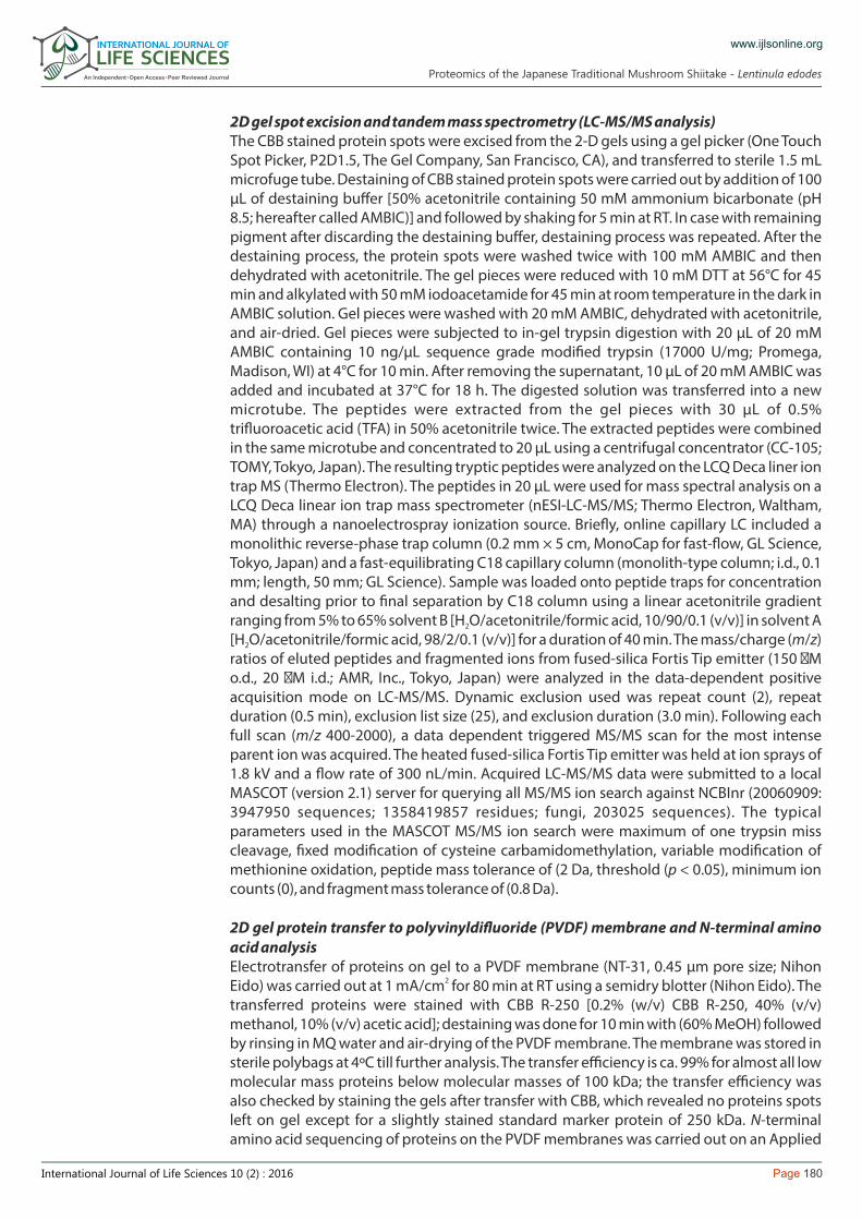
Mushroom used in this studyThe mature fruiting bodies of L. edodes were obtained from a farmer (Hitachiomiya, Ibaraki, Japan), who used the L. edodes sawdust spawn (Kinko 697) from Kinko Shiitake cooperative, Tottori, Japan. The wood log cultivation method is illustrated in Figure 2. In late autumn (November to December), trees of Quercus that are 5 to 20 cm in diameter with partially green leaves were felled, and then left on the felling site till cross section of the trees started cracking up. During this process, water content decreases by transpiration through the green leaves and viable cells of tree are gradually lost; such conditions are favorable for growth of mycelium. The dried trees were cut into logs of about 90 cm in length, and then piled up to less than 100 cm high. Prior to inoculation, holes (diameter: 8 mm, depth: 2 cm, 30 to 60 holes per one log) were drilled at 15 cm intervals along the longitudinal direction with rows 3 – 4 cm apart, alternately using an electric drill. Sawdust spawn covered with foam polystyrene, protecting mycelium from air exposure, was pressed into the holes by
www.ijlsonline.org
Materials andMethods
Proteomics of the Japanese Traditional Mushroom Shiitake - Lentinula edodes
Page 176International Journal of Life Sciences 10 (2) : 2016
www.ijlsonline.org
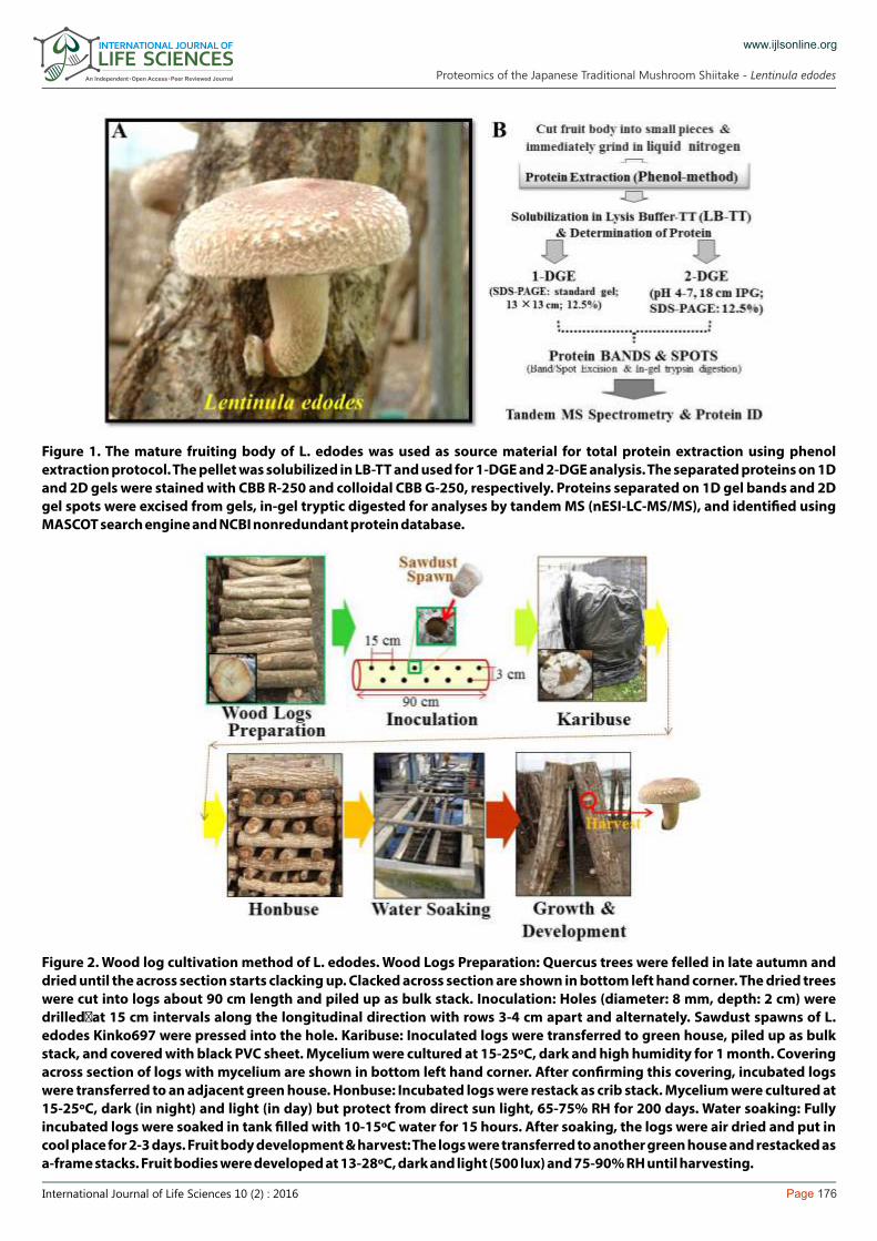
Figure 1. The mature fruiting body of L. edodes was used as source material for total protein extraction using phenol extraction protocol. The pellet was solubilized in LB-TT and used for 1-DGE and 2-DGE analysis. The separated proteins on 1D and 2D gels were stained with CBB R-250 and colloidal CBB G-250, respectively. Proteins separated on 1D gel bands and 2D gel spots were excised from gels, in-gel tryptic digested for analyses by tandem MS (nESI-LC-MS/MS), and identi�ed using MASCOT search engine and NCBI nonredundant protein database.
Figure 2. Wood log cultivation method of L. edodes. Wood Logs Preparation: Quercus trees were felled in late autumn and dried until the across section starts clacking up. Clacked across section are shown in bottom left hand corner. The dried trees were cut into logs about 90 cm length and piled up as bulk stack. Inoculation: Holes (diameter: 8 mm, depth: 2 cm) were drilled�at 15 cm intervals along the longitudinal direction with rows 3-4 cm apart and alternately. Sawdust spawns of L. edodes Kinko697 were pressed into the hole. Karibuse: Inoculated logs were transferred to green house, piled up as bulk stack, and covered with black PVC sheet. Mycelium were cultured at 15-25ºC, dark and high humidity for 1 month. Covering across section of logs with mycelium are shown in bottom left hand corner. After con�rming this covering, incubated logs were transferred to an adjacent green house. Honbuse: Incubated logs were restack as crib stack. Mycelium were cultured at 15-25ºC, dark (in night) and light (in day) but protect from direct sun light, 65-75% RH for 200 days. Water soaking: Fully incubated logs were soaked in tank �lled with 10-15ºC water for 15 hours. After soaking, the logs were air dried and put in cool place for 2-3 days. Fruit body development & harvest: The logs were transferred to another green house and restacked as a-frame stacks. Fruit bodies were developed at 13-28ºC, dark and light (500 lux) and 75-90% RH until harvesting.
Proteomics of the Japanese Traditional Mushroom Shiitake - Lentinula edodes

Page 177International Journal of Life Sciences 10 (2) : 2016
hand. Inoculated logs were transferred to a green house, piled up as bulk stack and covered with black PVC sheets for protecting from direct sun light, and maintained at optimum temperature and humidity appropriate for the growing mycelium. The logs were occasionally watered for keeping high humidity. During the �rst time of culture term called “Karibuse”, culture condition was maintained at 15 – 25ºC, dark and 85% relative humidity (RH) for 1 to 2 months. After con�rming covered cross-section of log with mycelia, the incubated logs were transferred to another green house, stacked as crib stacks but not covered by PVC sheets. In the second time of culture term called “Honbuse”, the culture conditions were maintained at 15 – 25ºC, dark (in night) and light (in day) but protected from direct sun light and 65 – 75% RH for 200 – 250 days. Fully incubated logs were soaked in tank �lled with water maintained at 10 – 15ºC temperature for about 15 hours. This sudden change of environment is required to trigger the formation of fruit body. After water soaking, the logs were naturally dried by leaning and gathering each other in cool (18ºC) environment for 2 – 3 days, and then transferred to a third green house. Finally, logs were stacked as frame stacks until harvesting of the fruit bodies. Fruit body development condition was maintained at 13 - 28ºC, dark (at night) and light (in day-time, 500 lux) and 75 – 90% RH. Five days were required for harvest from stacking. Harvested fruit bodies were immediately frozen in dry ice, and then stocked at -80ºC in the laboratory.
Extraction of total proteinsFor extraction of total protein, three mature fruiting bodies were used. Deep-frozen fruiting bodies were immediately ground to a very �ne powder in liquid nitrogen (N ) using liquid N2 2
chilled mortar and pestle. The sample powder from each fruit body was stored in 50 mL falcon tubes at -80ºC. For extraction of total protein, equal amounts (~ 30 mg) of powder were mixed from the three replicates in a 2 mL Eppendorf microfuge tube and placed in an ice-box �lled with liquid N . Proteins were extracted using the Phenol method [ ] as 2 20, 21described hereon. Tris-buffered phenol (hereafter referred to as TBP) was prepared as described in Figure 3. Proteins were extracted from tissue powder (100 mg) by addition of 500 µL of TBP and extraction media [0.9 M sucrose, 0.1 M Tris (pH 8.8), 10 mM EDTA, and 0.4% (v/v) 2-mercaptoethanol (2-ME) in distilled (MQ) water], followed by mixing using an invert shaker at RT for 30 min. The suspensions were centrifuged at 15000 rpm for 20 min at 4ºC. After centrifugation, the top phenol phase was transferred to a new microtube, followed by addition of 500 µL of TBP and same amount of extraction media to back-extract aqueous phase, followed by centrifugation at 15000 rpm for 20 min at 4ºC. The top phenol phase was transferred into the �rst extraction and vortexed. Solubilized proteins in phenol were precipitated by addition of �ve volumes of 0.1 M ammonium acetate in 100% methanol, vortexed, and incubated at -20ºC overnight. The suspension was centrifuged at 15000 rpm for 20 min at 4ºC to obtain the protein pellet. The pellet was washed twice with 1 mL of 0.1 M
www.ijlsonline.org
Figure 3. Preparation of the buffers
Proteomics of the Japanese Traditional Mushroom Shiitake - Lentinula edodes

Page 178International Journal of Life Sciences 10 (2) : 2016
ammonium acetate in 100% methanol, with 1 mL of 80% ice-cold acetone, and �nally once with 70% ethanol. Following centrifugation (at 15000 rpm for 20 min at 4ºC), the supernatant was decanted and the pellet was dried at 37ºC for 10-15 min. Proteins were solubilized in 200 µL of lysis buffer [7 M (w/v) urea, 2 M (w/v) thiourea, 4% (w/v) CHAPS, 18 mM (w/v) Tris-HCl (pH 8.0), 14 mM (w/v) Trizma base, 0.2% (v/v) Triton X-100, 50 mM (w/v) DTT, 1% (v/v) pH 3-10 ampholyte, and EDTA-free proteinase inhibitor (Roche) tablets in a total volume of 100 mL; see Figure 3 for preparation of LB-TT], followed by centrifugation at 15000 rpm for 20 min at 20ºC. The supernatant was stored in aliquots at -80°C. Protein
TMconcentration was determined with a Coomassie Plus Protein Assay Kit (PIERCE, Rockford, IL, USA) using bovine serum albumin (BSA) as a standard and a NanoDrop 1000 spectrophotometer (Thermo Scienti�c, Wilmington, DE, USA).
One-dimensional gel electrophoresis (1-DGE)The total protein obtained above was precipitated using a ProteoExtract Protein Precipitation Kit (Calbiochem, Darmstadt, Germany). The pellet was resolubilized in homogenization buffer [0.2 M Tris-HCl buffer, pH 7.8, containing 5 mM EDTA 2Na, 14 mM 2-ME, 10% (v/v) glycerol, and 2 EDTA-free proteinase inhibitor tablets (Roche) per 100 mL of buffer solution in distilled water]. To effectively solubilize the protein pellet, sodium dodecyl sulfate (SDS)-sample buffer [2.5X, 62 mM Tris (pH 6.8) containing 10% (v/v) glycerol, 2.5% (w/v) SDS, and 5% (v/v) 2-ME, pH 6.8] was added to the mixture, followed by vortexing, sonication (water bath), and centrifugation of the sample at 15000 rpm for 15 min (4ºC). The supernatant was used for protein quanti�cation as described above. Just before electrophoresis, a drop of bromophenol blue (BPB) was added to the protein samples and the mixture boiled for 1 min at 95ºC. Fifty micrograms of protein was loaded into three well replications for 1-DGE using the Nihon Eido (Tokyo, Japan) standard vertical electrophoresis unit. The 12.5% SDS-PAGE (4%T, 2.6%C stacking gels, pH 6.8 and 12.5%T, 2.6%C separating gels, pH 8.8) was carried out at constant current of 35 mA for ca. 3 h. The electrophoresis running buffer (ERB, 10X) is as follows: 250 mM Tris, 30.3 g; 1.92 M glycine, 144 g; and 1% (w/v) SDS, 10 g; 1X diluted ERB was used. Five microliters of the commercially available
TM ready-to-use molecular mass standard (DualColor PrecisionPlus Protein Standard, Bio-Rad, Hercules, CA, USA) was loaded in the well adjacent to the samples. The gel was stained with Coomassie brilliant blue (CBB) R-250 [0.1% (w/v) CBB R-250, 50% (v/v) methanol, 10% (v/v) acetic acid] for 30 min. Destaining was carried out with destaining solution [40% (v/v) methanol, 10% (v/v) acetic acid]. Destaining solution was occasionally replaced, until clear background of the gel and clear bands were visible.
Mass spectrometry analysis (1-D shotgun approach)Prior to MS analysis using the shotgun approach [ ], each lane was sliced into eight pieces 20(Figure 4) of gel matrix and digested with 1 µg of trypsin at 37 °C for 18 h. Brie�y, the tryptic peptides samples were separated by C-18 reverse-phase column and analyzed on a nanoelectrospray ionization mass spectrometer (nESI-LC-MS/MS). The ultimate nanoLC systems, combined with the FAMOS autosampler and Switchos column switching valve (LC-Packings, Amsterdam, Netherlands) was used. Samples were loaded onto a precolumn (2 cm × 200 µm i.d.; Zorbax 300SB-C18, 5 µM, Agilent, CA), and washed with the loading solvent (H O/0.1% formic acid; �ow rate, 4 µL/min.) for 10 min to remove salts. Subsequently, 2
a Switchos II column switching device transferred �ow paths to the analytical column (15 cm × 75 µm i.d.; Zorbax 300SBC18, 5 µM, Agilent). The nano�ow eluted at a �ow rate of 200 nL/min using a 110 min gradient elution from 0% solvent A to 32% solvent B, where solvent A was 0.1% formic acid with 5% acetonitrile and solvent B was 0.1% formic acid with 90% acetonitrile. The column outlet was coupled directly to the high voltage ESI source, which was interfaced to the QSTAR mass spectrometer (Applied Biosystems, Foster City, CA). The nanospray voltage was typically 2.3 kV in the nESI-LC-MS/MS mode. The nESI-LC-MS/MS running on the QSTAR instrument was acquired in Information Dependent Acquisition mode, which allows the user to acquire MS/MS spectra based on an inclusion mass list and
www.ijlsonline.org
Proteomics of the Japanese Traditional Mushroom Shiitake - Lentinula edodes

Page 179International Journal of Life Sciences 10 (2) : 2016
dynamic assessment of relative ion intensity. The data acquisition time was set to 3 s per spectrum over m/z range of 400-1500 Da. Acquired data were searched against the National Center for Biotechnology Information (NCBI) nonredundant (NR) protein database (20080409: 2821560 sequences; 968325167 residues; fungi, 2821560 sequences) using the MASCOT software package (Version 2.1, Matrix Sciences, U.K.; www.matrixscience.com). The peptide mass and MS/MS tolerance were 0.6 and 1.0 Da, respectively. The peptides have the allowance of two tryptic mis-cleavages and also partially modi�ed with oxidation (M) within two charge states (+2, +3).
Two-dimensional gel electrophoresis (2-DGE)2-DGE was carried out using precast IPG strips (18 cm, pH 4-7) on an IPGphor unit (GE Healthcare Bio-Sciences AB, Uppsala, Sweden) followed by 12.5% SDS-PAGE on a vertical electrophoresis unit (Nihon Eido). The volume carrying 750 µg of total protein was mixed with LB-TT containing 0.5% (v/v) pH 4-7 IPG buffer to bring to a �nal volume of 340 µL. A trace of BPB was added and the mixture centrifuged at 15000 rpm for 15 min followed by pipetting into 18 cm strip holder tray placed into the IPGphor unit. IPG strips were carefully placed onto the protein samples avoiding air bubbles between the sample and the gel strip. The IPG strips were allowed to passively rehydrate with the protein samples for 1.5 h, followed by overlaying the strips with cover �uid (mineral oil), and this was directly linked to a �ve-step active rehydration and focusing protocol (18 cm strip) as described previously [ ]. Isoelectric focusing (IEF) was carried out at 20°C, and a total of 68902 Vh was used for 20the 18 cm strip. Following IEF, the IPG strips were immediately used for the second dimension (SDS-PAGE). The IPG strips were incubated in equilibration buffer (50 mM Tris-HCl (pH 8.8), 6 M urea, 30% (v/v) glycerol, and 2% (w/v) SDS) containing 2% (w/v) DTT for 10 min (twice) with gentle agitation, followed by incubation in the same equilibration buffer supplemented with 2.5% (w/v) iodoacetamide for the same time periods as above at RT. SDS-PAGE (35 mA/gel) was performed for 3.5 h using 1X ERB. For each sample, a minimum of three IPG strips and corresponding SDS-PAGE was used under the same conditions. To visualize the separated protein spots, the 2D gels were stained with colloidal CBB G-250 [0.08% (w/v) CBB G-250, 20% (v/v) ethanol, 1.6% (v/v) phosphoric acid, 8% (w/v) ammonium sulfate] for 72 h; destaining of the gel is not required, only a rinse in MQ water for 3 times is necessary. Protein patterns in the gels were recorded as digitalized images (24 bit color, resolution 300 dpi) using the ProteomeScan2000 digital scanner (System Biotics KK), and saved as TIFF �les. ImageMaster 2D Platinum software ver. 5.0 (GE Healthcare) was used for downstream analysis of detected protein spots on 2D gels.
www.ijlsonline.org
Figure 4. One dimensional gel pro�les of L. edodes mature fruit body protein. Total proteins were separated on 12.5% SDS-PAGE followed by stained using CBB R-250. Gel was divided eight fractions (I through VIII) and sliced along red dotted lines. In-gel digested peptides were subjected to nLC-ESI MS/MS analysis. Proteins were identi�ed using the MASCOT search engine and NCBInr protein database. The molecular weight markers are indicated on the left-hand side of gel.
Proteomics of the Japanese Traditional Mushroom Shiitake - Lentinula edodes
Page 180International Journal of Life Sciences 10 (2) : 2016
www.ijlsonline.org
2D gel spot excision and tandem mass spectrometry (LC-MS/MS analysis)The CBB stained protein spots were excised from the 2-D gels using a gel picker (One Touch Spot Picker, P2D1.5, The Gel Company, San Francisco, CA), and transferred to sterile 1.5 mL microfuge tube. Destaining of CBB stained protein spots were carried out by addition of 100 µL of destaining buffer [50% acetonitrile containing 50 mM ammonium bicarbonate (pH 8.5; hereafter called AMBIC)] and followed by shaking for 5 min at RT. In case with remaining pigment after discarding the destaining buffer, destaining process was repeated. After the destaining process, the protein spots were washed twice with 100 mM AMBIC and then dehydrated with acetonitrile. The gel pieces were reduced with 10 mM DTT at 56°C for 45 min and alkylated with 50 mM iodoacetamide for 45 min at room temperature in the dark in AMBIC solution. Gel pieces were washed with 20 mM AMBIC, dehydrated with acetonitrile, and air-dried. Gel pieces were subjected to in-gel trypsin digestion with 20 µL of 20 mM AMBIC containing 10 ng/µL sequence grade modi�ed trypsin (17000 U/mg; Promega, Madison, WI) at 4°C for 10 min. After removing the supernatant, 10 µL of 20 mM AMBIC was added and incubated at 37°C for 18 h. The digested solution was transferred into a new microtube. The peptides were extracted from the gel pieces with 30 µL of 0.5% tri�uoroacetic acid (TFA) in 50% acetonitrile twice. The extracted peptides were combined in the same microtube and concentrated to 20 µL using a centrifugal concentrator (CC-105; TOMY, Tokyo, Japan). The resulting tryptic peptides were analyzed on the LCQ Deca liner ion trap MS (Thermo Electron). The peptides in 20 µL were used for mass spectral analysis on a LCQ Deca linear ion trap mass spectrometer (nESI-LC-MS/MS; Thermo Electron, Waltham, MA) through a nanoelectrospray ionization source. Brie�y, online capillary LC included a monolithic reverse-phase trap column (0.2 mm × 5 cm, MonoCap for fast-�ow, GL Science, Tokyo, Japan) and a fast-equilibrating C18 capillary column (monolith-type column; i.d., 0.1 mm; length, 50 mm; GL Science). Sample was loaded onto peptide traps for concentration and desalting prior to �nal separation by C18 column using a linear acetonitrile gradient ranging from 5% to 65% solvent B [H O/acetonitrile/formic acid, 10/90/0.1 (v/v)] in solvent A 2
[H O/acetonitrile/formic acid, 98/2/0.1 (v/v)] for a duration of 40 min. The mass/charge (m/z) 2
ratios of eluted peptides and fragmented ions from fused-silica Fortis Tip emitter (150 �M o.d., 20 �M i.d.; AMR, Inc., Tokyo, Japan) were analyzed in the data-dependent positive acquisition mode on LC-MS/MS. Dynamic exclusion used was repeat count (2), repeat duration (0.5 min), exclusion list size (25), and exclusion duration (3.0 min). Following each full scan (m/z 400-2000), a data dependent triggered MS/MS scan for the most intense parent ion was acquired. The heated fused-silica Fortis Tip emitter was held at ion sprays of 1.8 kV and a �ow rate of 300 nL/min. Acquired LC-MS/MS data were submitted to a local MASCOT (version 2.1) server for querying all MS/MS ion search against NCBInr (20060909: 3947950 sequences; 1358419857 residues; fungi, 203025 sequences). The typical parameters used in the MASCOT MS/MS ion search were maximum of one trypsin miss cleavage, �xed modi�cation of cysteine carbamidomethylation, variable modi�cation of methionine oxidation, peptide mass tolerance of (2 Da, threshold (p < 0.05), minimum ion counts (0), and fragment mass tolerance of (0.8 Da).
2D gel protein transfer to polyvinyldi�uoride (PVDF) membrane and N-terminal amino acid analysisElectrotransfer of proteins on gel to a PVDF membrane (NT-31, 0.45 µm pore size; Nihon
2Eido) was carried out at 1 mA/cm for 80 min at RT using a semidry blotter (Nihon Eido). The transferred proteins were stained with CBB R-250 [0.2% (w/v) CBB R-250, 40% (v/v) methanol, 10% (v/v) acetic acid]; destaining was done for 10 min with (60% MeOH) followed by rinsing in MQ water and air-drying of the PVDF membrane. The membrane was stored in sterile polybags at 4ºC till further analysis. The transfer efficiency is ca. 99% for almost all low molecular mass proteins below molecular masses of 100 kDa; the transfer efficiency was also checked by staining the gels after transfer with CBB, which revealed no proteins spots left on gel except for a slightly stained standard marker protein of 250 kDa. N-terminal amino acid sequencing of proteins on the PVDF membranes was carried out on an Applied
Proteomics of the Japanese Traditional Mushroom Shiitake - Lentinula edodes
Page 181International Journal of Life Sciences 10 (2) : 2016
www.ijlsonline.org
Biosystems 494 protein sequencer (Perkin-Elmer; Applied Biosystems) as described previously [ ]. The obtained sequiences were used to interrogate databases 22(UniProtKB/Swiss-Prot and Uni-ProtKB/TrEMBL) with Web accessible search programs like Fasta3, available online from EMBL Outstation of the European Bioinformatics Institute, to identify homology to proteins already present in the protein and nucleic acid databases.
2DGE visualization of posttranslational modi�cationsFor detecting potential PTMs, the 1-D and 2-D gels were stained with �uorescent stains that detect phosphoproteins (ProQ Diamond phosphoprotein gel stain, ProQ DPGS), glycoproteins (ProQ Emerald glycoprotein gel stain, ProQ EGGS), and total protein (SYPRO Ruby) obtained from Molecular Probes, Inc. (Eugene, OR, USA) [ ]. The stained gels were 22visualized on a 245/312 nm UV transilluminator. For identi�cation of the stained phosphoproteins, the spots were cut out under UV light and processed for in-gel digestion and protein identi�cation as described above in section 2.6.
Mushroom life cycle consists of the developmental stage of spore, mycelium, and fruit body [ ]. Mycelium stage is divided between the monokaryon and dikaryon hyphal stage. 1, 2When dikaryon hyphae satisfy certain conditions such as culture time and nutrition, and have been exposed to environmental factors such as irradiation, nutrition starvation, cold stress, etc., aggregated hyphae called a hyphal knot begins the formation of a fruit body. Following initial stages, developmental stages progress to a primordium stage. At this stage, aggregated hyphae vertically elongate and simultaneously form stipe, cap, and gill. After the primordum stage, immature fruit body stage starts, and stipe elongation, cap expansion and spore formation all occur in parallel. Mature fruit body is extension of an immature fruit body; here, veil covering the gill with spores is broken and spore dispersion starts. For basidiomycetous fungi, fruit body formation is a big event, because their hyphae elongates horizontal to vertical and various organelles are immediately formed. Therefore, it is expected that various gene transcription and proteins expression changes related fruit body formation are switched on at the time of changing from mycelium to fruit body. Comparative proteomics approaches on mycelium and fruit body will be powerful techniques for detection of proteins underlying these physiological-morphological changes. This is also one reason for our research into fruit body proteome.
Experimental strategyWe performed both SDS-PAGE (1-DGE) and 2-DGE based proteomics approaches on fruit body total proteins for �rst, obtaining an inventory of fruit body proteins, and second, to get possible insight into the mechanism of fruit body formation. The experimental strategy is illustrated in Figure 1. We selected mature fruit body (with broken veil) of kinko697 as the experimental material for our research objective. Moreover, this strain has a unique property for active growth at middle to high cultivation temperatures and is generally used for wood log cultivation. Detailed strain character is shown in database of Ministry of Agriculture, Forestry and Fisheries or homepage of Kinko Shiitake Cooperative. We extracted proteins from �ne powdered fruit body using TBP (Figure 3). Moreover, we have previously con�rmed the protein extraction efficiency from mushrooms and compatibility to 2-DGE [ ]. Total protein solubilized in LB-TT was used for 1-DGE (Figure 4) and 2-DGE 20(Figure 5) analyses. In case of 1-DGE, total protein was separated by 12.5% SDS-PAGE, the gel was divided into eight sections and tryptic digest was performed on each sections. In 2-DGE, proteins were separated by pI 4-7 isoelectrofocusing and following SDS-PAGE, the gel was stained by colloidal CBB G-250 and trypsin digestion was performed on abundant protein spots. The tryptic peptides either from 1-DGE bands and 2-DGE spots were analyzed by tandem mass spectrometry (nESI-LC-MS-MS). Acquired MS/MS data were searched against NCBI
Results andDiscussion
Proteomics of the Japanese Traditional Mushroom Shiitake - Lentinula edodes
Page 182International Journal of Life Sciences 10 (2) : 2016
www.ijlsonline.org
nonredundant protein database using the MASCOT search engine. Single peptide matched protein data were presented in Table 1 to Table 4. 1-DGE analysis in combination of nESI-LC-MS-MS and database search resulted in a low number of unique peptide per protein making it difficult to con�dent protein assignment. This might be in part due to lack of sequence information on mushroom and less similarity of proteins expressed in mushroom with other organisms. In 2-DGE analysis, we attempted to conduct N-terminal amino acid sequencing for assigning protein based on internal and external amino acid sequence and to reveal N-terminal sequences of protein that more close to natural form or functional form. As we know, this is a �rst comprehensive study on proteins of L. edodes mushroom fruit body. We also carried out detection of phosphorylated and glycosylated proteins (Figure 6) for providing information as broad as possible on the proteome in this study. These modi�cations might also serve as clues for comprehending function or localization of protein.
Identi�cation of 84 nonoverlapping and nonredundant proteins by 1-DGE and shotgun approach1-DGE analysis in combination with LC-MS/MS identi�ed a total of 91 proteins containing 84 nonredundant proteins from the eight excised fractions (Figure 4). Eleven proteins were identi�ed from fraction 1 (over 125 kDa), 11 proteins from fraction 2 (70~125 kDa), 15 proteins from fraction 3 (45~70 kDa), 12 proteins from fraction 4 (35~45 kDa), 16 proteins from fraction 5 (32~35 kDa), 8 proteins from fraction 6 (22~32 kDa), 13 proteins from 7 (18~22 kDa), 5 proteins from fraction 8 (less than 18 kDa). These identi�ed proteins are listed in Table 1. Some of the proteins had similar protein identi�cations in more than one fraction, which may be due to fragmentation during the experiment process. Histone H2B was detected within 3 fractions; and histone H4.2, two types of translation elongation factors, ribosomal protein L2, and ATP citrate lyase within 2 fractions.
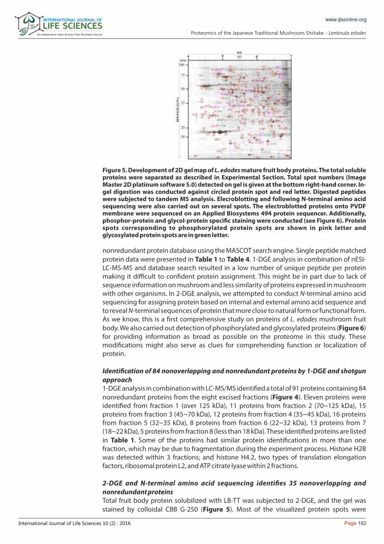
2-DGE and N-terminal amino acid sequencing identi�es 35 nonoverlapping and nonredundant proteinsTotal fruit body protein solubilized with LB-TT was subjected to 2-DGE, and the gel was stained by colloidal CBB G-250 (Figure 5). Most of the visualized protein spots were
Figure 5. Development of 2D gel map of L. edodes mature fruit body proteins. The total soluble proteins were separated as described in Experimental Section. Total spot numbers (Image Master 2D platinum software 5.0) detected on gel is given at the bottom right-hand corner. In-gel digestion was conducted against circled protein spot and red letter. Digested peptides were subjected to tandem MS analysis. Elecroblotting and following N-terminal amino acid sequencing were also carried out on several spots. The electroblotted proteins onto PVDF membrane were sequenced on an Applied Biosystems 494 protein sequencer. Additionally, phosphor-protein and glycol-protein speci�c staining were conducted (see Figure 6). Protein spots corresponding to phosphorylated protein spots are shown in pink letter and glycosylated protein spots are in green letter.
Proteomics of the Japanese Traditional Mushroom Shiitake - Lentinula edodes
Page 183International Journal of Life Sciences 10 (2) : 2016
www.ijlsonline.org
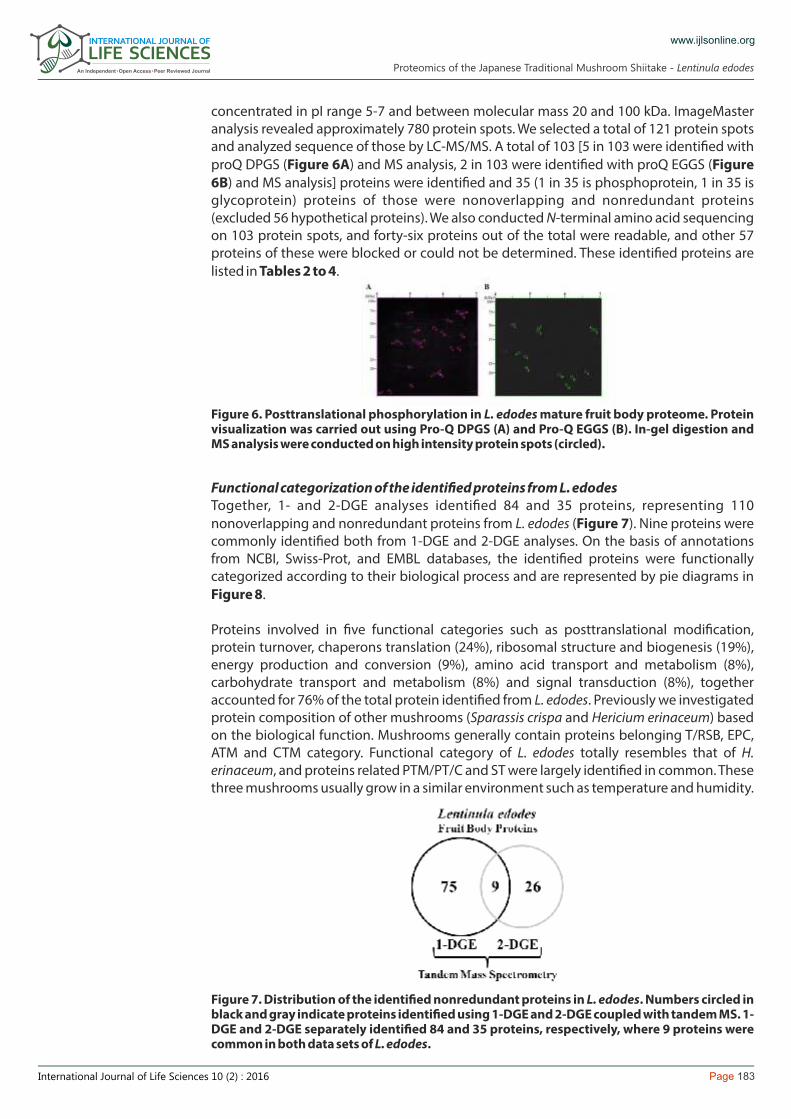
concentrated in pI range 5-7 and between molecular mass 20 and 100 kDa. ImageMaster analysis revealed approximately 780 protein spots. We selected a total of 121 protein spots and analyzed sequence of those by LC-MS/MS. A total of 103 [5 in 103 were identi�ed with proQ DPGS (Figure 6A) and MS analysis, 2 in 103 were identi�ed with proQ EGGS (Figure 6B) and MS analysis] proteins were identi�ed and 35 (1 in 35 is phosphoprotein, 1 in 35 is glycoprotein) proteins of those were nonoverlapping and nonredundant proteins (excluded 56 hypothetical proteins). We also conducted N-terminal amino acid sequencing on 103 protein spots, and forty-six proteins out of the total were readable, and other 57 proteins of these were blocked or could not be determined. These identi�ed proteins are listed in Tables 2 to 4.
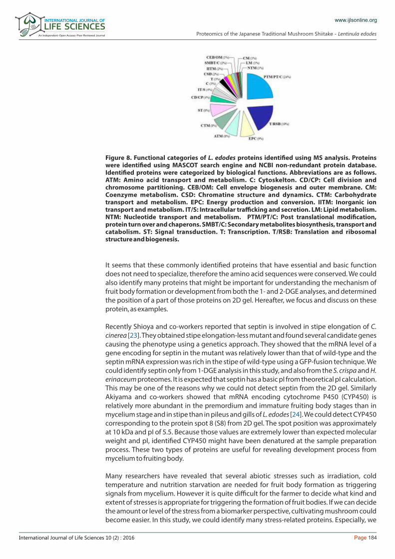
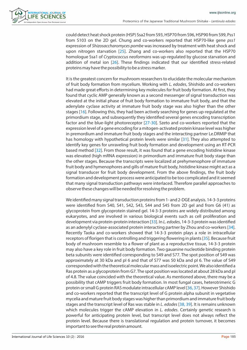
Functional categorization of the identi�ed proteins from L. edodesTogether, 1- and 2-DGE analyses identi�ed 84 and 35 proteins, representing 110 nonoverlapping and nonredundant proteins from L. edodes (Figure 7). Nine proteins were commonly identi�ed both from 1-DGE and 2-DGE analyses. On the basis of annotations from NCBI, Swiss-Prot, and EMBL databases, the identi�ed proteins were functionally categorized according to their biological process and are represented by pie diagrams in Figure 8.
Proteins involved in �ve functional categories such as posttranslational modi�cation, protein turnover, chaperons translation (24%), ribosomal structure and biogenesis (19%), energy production and conversion (9%), amino acid transport and metabolism (8%), carbohydrate transport and metabolism (8%) and signal transduction (8%), together accounted for 76% of the total protein identi�ed from L. edodes. Previously we investigated protein composition of other mushrooms (Sparassis crispa and Hericium erinaceum) based on the biological function. Mushrooms generally contain proteins belonging T/RSB, EPC, ATM and CTM category. Functional category of L. edodes totally resembles that of H. erinaceum, and proteins related PTM/PT/C and ST were largely identi�ed in common. These three mushrooms usually grow in a similar environment such as temperature and humidity.
Figure 6. Posttranslational phosphorylation in L. edodes mature fruit body proteome. Protein visualization was carried out using Pro-Q DPGS (A) and Pro-Q EGGS (B). In-gel digestion and MS analysis were conducted on high intensity protein spots (circled).
Figure 7. Distribution of the identi�ed nonredundant proteins in L. edodes. Numbers circled in black and gray indicate proteins identi�ed using 1-DGE and 2-DGE coupled with tandem MS. 1-DGE and 2-DGE separately identi�ed 84 and 35 proteins, respectively, where 9 proteins were common in both data sets of L. edodes.
Proteomics of the Japanese Traditional Mushroom Shiitake - Lentinula edodes
Page 184International Journal of Life Sciences 10 (2) : 2016
www.ijlsonline.org
It seems that these commonly identi�ed proteins that have essential and basic function does not need to specialize, therefore the amino acid sequences were conserved. We could also identify many proteins that might be important for understanding the mechanism of fruit body formation or development from both the 1- and 2-DGE analyses, and determined the position of a part of those proteins on 2D gel. Hereafter, we focus and discuss on these protein, as examples.
Recently Shioya and co-workers reported that septin is involved in stipe elongation of C. cinerea [ ]. They obtained stipe elongation-less mutant and found several candidate genes 23causing the phenotype using a genetics approach. They showed that the mRNA level of a gene encoding for septin in the mutant was relatively lower than that of wild-type and the septin mRNA expression was rich in the stipe of wild-type using a GFP-fusion technique. We could identify septin only from 1-DGE analysis in this study, and also from the S. crispa and H. erinaceum proteomes. It is expected that septin has a basic pI from theoretical pI calculation. This may be one of the reasons why we could not detect septin from the 2D gel. Similarly Akiyama and co-workers showed that mRNA encoding cytochrome P450 (CYP450) is relatively more abundant in the premordium and immature fruiting body stages than in mycelium stage and in stipe than in pileus and gills of L. edodes [ ]. We could detect CYP450 24corresponding to the protein spot 8 (S8) from 2D gel. The spot position was approximately at 10 kDa and pI of 5.5. Because those values are extremely lower than expected molecular weight and pI, identi�ed CYP450 might have been denatured at the sample preparation process. These two types of proteins are useful for revealing development process from mycelium to fruiting body.
Many researchers have revealed that several abiotic stresses such as irradiation, cold temperature and nutrition starvation are needed for fruit body formation as triggering signals from mycelium. However it is quite difficult for the farmer to decide what kind and extent of stresses is appropriate for triggering the formation of fruit bodies. If we can decide the amount or level of the stress from a biomarker perspective, cultivating mushroom could become easier. In this study, we could identify many stress-related proteins. Especially, we
Figure 8. Functional categories of L. edodes proteins identi�ed using MS analysis. Proteins were identi�ed using MASCOT search engine and NCBI non-redundant protein database. Identi�ed proteins were categorized by biological functions. Abbreviations are as follows. ATM: Amino acid transport and metabolism. C: Cytoskelton. CD/CP: Cell division and chromosome partitioning. CEB/OM: Cell envelope biogenesis and outer membrane. CM: Coenzyme metabolism. CSD: Chromatine structure and dynamics. CTM: Carbohydrate transport and metabolism. EPC: Energy production and conversion. IITM: Inorganic ion transport and metabolism. IT/S: Intracellular trafficking and secretion. LM: Lipid metabolism. NTM: Nucleotide transport and metabolism. PTM/PT/C: Post translational modi�cation, protein turn over and chaperons. SMBT/C: Secondary metabolites biosynthesis, transport and catabolism. ST: Signal transduction. T: Transcription. T/RSB: Translation and ribosomal structure and biogenesis.
Proteomics of the Japanese Traditional Mushroom Shiitake - Lentinula edodes
Page 185International Journal of Life Sciences 10 (2) : 2016
www.ijlsonline.org
could detect heat shock protein (HSP) Ssa2 from S93, HSP70 from S96, HSP90 from S99, Pss1 from S103 on the 2D gel. Chung and co-workers reported that HSP70-like gene pss1 expression of Shizosaccharomyces pombe was increased by treatment with heat shock and upon nitrogen starvation [ ]. Zhang and co-workers also reported that the HSP70 25homologue Ssa1 of Cryptococcus neoformans was up-regulated by glucose starvation and addition of metal ion [ ]. These �ndings indicated that our identi�ed stress-related 26proteins may have the possibility to be a stress marker.
It is the greatest concern for mushroom researchers to elucidate the molecular mechanism of fruit body formation from mycelium. Working with L. edodes, Shishido and co-workers had made great efforts in determining key molecules for fruit body formation. At �rst, they found that cyclic AMP generally known as a second messenger of signal transduction was elevated at the initial phase of fruit body formation to immature fruit body, and that the adenylate cyclase activity at immature fruit body stage was also higher than the other stages [ ]. Following this, they had been actively searching for genes up-regulated at the 16primordium stage, and subsequently they identi�ed several genes encoding transcription factor and the blue-light photoreceptor [ ]. Szeto and co-workers reported that the 27-30expression level of a gene encoding for a mitogen-activated protein kinase level was higher in premordium and immature fruit body stages and the interacting partner Le.DRMIP that has homology with hypothetical protein levels were similar [ ]. They also attempted to 31identify key genes for unraveling fruit body formation and development using an RT-PCR based method [ ]. From those result, it was found that a gene encoding histidine kinase 32was elevated (high mRNA expression) in primordium and immature fruit body stage than the other stages. Because the transcripts were localized at prehymenophore of immature fruit body and hymenophores and gills of mature fruit body, histidine kinase might act as a signal transducer for fruit body development. From the above �ndings, the fruit body formation and development process were anticipated to be too complicated and it seemed that many signal transduction pathways were interlaced. Therefore parallel approaches to observe these changes will be needed for resolving the problem.
We identi�ed many signal transduction proteins from 1- and 2-DGE analysis. 14-3-3 proteins were identi�ed from S40, S41, S42, S43, S44 and S45 from 2D gel and from G6 (41) as glycoprotein from glycoprotein stained gel. 14-3-3 proteins are widely distributed among eukaryotes, and are involved in various biological events such as cell proliferation and development via protein-protein interaction [ ]. In L. edodes, 14-3-3 protein was identi�ed 33as an adenylyl cyclase-associated protein interacting partner by Zhou and co-workers [ ]. 34Recently Taoka and co-workers showed that 14-3-3 protein plays a role in intracellular receptors of �origen that is controlling and triggering �owering in plants [ ]. Because fruit 35body of mushroom resemble to a �ower of plant as a reproductive tissue, 14-3-3 protein may also have a key role in fruit body formation. Two gauanine nucleotide binding protein beta subunits were identi�ed corresponding to S49 and S77. The spot position of S49 was approximately at 30 kDa and pI 6 and that of S77 was 50 kDa and pI 6. The value of S49 corresponded with the theoretical molecular mass and isoelectric point. We also identi�ed a Ras protein as a glycoprotein from G7. The spot position was located at about 28 kDa and pI of 4.8. The value coincided with the theoretical value. As mentioned above, there may be a possibility that cAMP triggers fruit body formation. In most fungal cases, heterotrimeric G protein or small G protein RAS modulate intracellular cAMP level [ ]. However Shishido 36, 37and co-workers reported that the transcript level of G protein alpha subunit in vegetative mycelia and mature fruit body stages was higher than primordium and immature fruit body stages and the transcript level of Ras was stable in L. edodes [ ]. It is remains unknown 38, 39which molecules trigger the cAMP elevation in L. edodes. Certainly genetic research is powerful for anticipating protein level, but transcript level does not always re�ect the protein level. Because there is translational regulation and protein turnover, it becomes important to see the real protein amount.
Proteomics of the Japanese Traditional Mushroom Shiitake - Lentinula edodes
Page 186International Journal of Life Sciences 10 (2) : 2016
www.ijlsonline.org
Mushroom contains a large amount of trehalose, which accounts for over 10% of its dry weight. It is well known that trehalose protects DNA and proteins from dryness and freezing conditions. A �y, Polypedium vanderplanki (P. vanderplanki) larva tolerates dehydration and cold stress by elevating trehalose level [ ]. Cornette and co-workers also showed that P. 40vanderplanki up-regulates not only trehalose biosynthesis enzyme but also antioxidative enzymes and HSPs [ ]. These elevations may contribute to protection from oxidative stress 41accompanied by desiccation and from denaturing. From a view point of food preservation, trehalose has important functions. Trehalose is widely used as a food additive for inhibiting change of color and maintaining food texture. In Japan, we produce dried shiitake, and then rehydrate them before use in various food preparations, and it is reported that the extent of reverting back to the original color depends on trehalose content [Hayashibara Co. Ltd., Japan; http://www.food.hayashibara.co.jp/index.html]. In this present study, we identi�ed trehalose synthase from 1-DGE, trehalose phosphorylase from S97, P20, P21, and P22 as phophoproteins, catalase from 1-DGE, two superoxide dismutases from S18 and S19 and many HSPs from both 1-DGE and 2-DGE. Therefore, by studying the change of these proteins amounts in post-harvest and during the drying process, we could have the possibility to contribute to the food preservation industry.
Previously we established and optimized methods for studying mushroom proteins using two edible medicinal mushrooms S. crispa and H. erinaceum. Continuing that research forward, in this study we have analyzed the proteome of the most traditional and popular Japanese mushroom, shiitake (L. edodes). Using 1-DGE and 2-DGE in conjunction with LC-MS/MS and N-terminal amino acid sequencing, we identi�ed 110 nonoverlaping and nonredundant proteins. Signi�cantly, we could detect and identify several signal transduction-related proteins such as 14-3-3 protein, guanine nucleotide binding protein beta subunit and small G protein Ras and protein putative involving in stipe formation like septin and CYP450. Because it has been anticipated that many signal transduction pathway such as trimeric G protein pathway, histidine kinase and MAP kinase cascade and small G protein pathway were mutually connected in fruit body formation and development process, our established technique and protein reference map will become a tool for speeding up the research further. Improvements to food processing and preservation techniques are highly desirable to the farmers (mushroom growers) for providing the �nal product (fresh or frozen or dried) to the consumers in a form they appreciate and want. By identifying several trehalose metabolic enzyme and stress-related protein, our research might also serve as a foundation for understanding the process in the post-harvest step. The protein inventory and 2D gel reference maps established for L. edodes will be helpful in pro�ling protein change during the cultivation and preservation of mushroom. This will involve comparative studies on deferent developmental stages among spore, mycelium, primordium, immature fruit body and mature fruit body and post-harvest fruit body or dried fruit body. In addition to this, a combination of genetic research and our established proteomic approach will help contribute more to fruit body formation and development. In mushroom research, C. cinerea genome has been completed by Pukkila et al. [ ] and 18Muraguchi and co-workers of Akita Prefectural University are now actively using this fundamental resource to gradual revealed key genes for fruit body development by employing mutants [ ]. In L. edodes, Shishido and co-workers have generated much 23information of candidate genes for fruit body formation and development. The next steps will involve functional genomics studies by generating over-expression or knockdown mutants in L. edodes. In this direction, our proteomics approach and data therein along the L. edodes genome after full annotation will be an important resource to speed up the research into the shiitake fruit body formation and mechanisms involved therein. We believe that these studies are connected to the expansion of food resource and improvement of mushroom cultivation and preservation.
Conclusion
Proteomics of the Japanese Traditional Mushroom Shiitake - Lentinula edodes
Page 187International Journal of Life Sciences 10 (2) : 2016
www.ijlsonline.org
This �rst report on Shiitake fruit body proteome, and both methods and results obtained herein could be applied for further understanding fruit body formation, development and preservation.
KH gratefully acknowledges members of Professor Masami Yonekura laboratory (Ibaraki University) and HTRC-AIST West team members under Prof. Yoshinori Masuo (currently Toho University) for their support and encouragement during this study. Authors appreciate the International Plant Proteomics Organization (INPPO) initiative (www.inppo.com) for connecting proteomics researchers among India, Nepal, South Korea and Japan.
1. Miles, P.G.; Chang, S.T. Mushroom Biology: Concise Basics and Current Development. River Edge, NJ: World Scienti�c Publishing Co. Inc., 1997.
2. Miles, P.G.; Chang, S.T. Mushrooms: Cultivation, Nutritional Value, Medicinal Effect, and Environmental Impact. Second Edition, Boca Raton, Florida: CRC Press, 2004.
3. Nakai, Y. Cytological studies on shiitake, Lentinus edodes (Berk.) Sing. Rept. Tottori Mycol. Inst. (Japan) 1986, 24, 1-202.4. Kues, U.; Liu, Y. Fruiting body production in basidomycetes. Appl. Microbial. Biotechnol. 2000, 54, 141-52.5. Kues, U. History and developmental processes in Basidiomycete Coprinus cinereus. Microbiol. Mol. Biol. Rev. 2000, 64, 316-353.6. Yamanaka, K. Mushroom cultivation in Japan. WSMBMP Bulletin 2011, 4, 1-10.7. Ito, T. Cultivation of Lentinus edodes. In: The Biology and Cultivation of Edible Mushrooms; Chang, S.T. and Hayes, W.A. Hayes.
Eds. Academic Press Inc.: New York, 1978; pp. 461-473.8. Chang, S.T. World production of cultivated edible and medicinal mushrooms in 1997 with emphasis on Lentinus edodes
(Berk.) Sing. China. Intl. J. Medicinal Mushrooms 1999, 1, 291-300.9. Chung, R. Functional properties of edible mushrooms. Nutr. Rev. 1996, 54, S91-93.10. Chihara, G.; Maeda, Y.; Hamuro, J.; Sasaki, T.; Fukuoka, F. Inhibition of mouse sarcoma 180 by polysaccharides from Lentinus
edodes (Berk.) Sing. Nature 1969, 222, 687-688.11. Chihara, G.; Hamuro, J.; Arai, Y.; Fukuoka, F. Fractionation and puri�cation of the polysaccharides with marked antitumor
activity, especially lentinan, from Lentinus edodes (Berk.) Sing. (an edible mushroom). Cancer Res. 1970, 30, 2776-2781.12. Chibata, I.; Okumura, K.; Takeyama, S.; Kotera, K. Lentinacin: A new hypocholesterolemic substance in Lentinus edodes.
Experienta 1969, 25, 1237-1238.13. Sugiyama, K.; Yamakawa, A.; Kawagishi, H.; Saeki, S. Hypocholesterolemic action of eritadenine is mediated by a
modi�cation of hepatic phospholipid metabolism in rats. J. Nutr. 1997, 127, 593-599.14. Viraj, J.J.; Conrad, O.P. Distribution of ergosterol in defferent tissues of mushrooms and its effect on the conversion of
ergosterol to vitamin D2 by UV irradiation. Food Chem. 2005, 92, 541-546.15. Brauer, D.; Kimmons, T.; Phillips, M. Effects of management on the yield and high-molecular-weight polysaccharide content
of shiitake (Lentinula edodes) mushrooms. J. Agric. Food. Chem. 2002, 50, 5333-5337.16. Takagi, Y.; Katayose, Y.; Shishid, K. Intracellular levels of cyclic AMP and adenylate cyclase activity during mycelia
development in fruiting body formation in Lentinus edodes. FEMS Microbiol. Let. 1988, 55, 275-278.17. Coprinus cinereus Sequencing Project. Broad Institute of MIT and Harvard
(http://www.broadinstitute.org/annotation/genome/coprinus_cinereus/Info.html).18. Burns, C.; Stajich, J.E.; Rechtsteiner, A.; Casselton, L.; Hanlon, S.E.; Wilke, S.K.; Savytskyy, O.P.; Gathman, A.C.; Lily, W.W.; Lieb,
J.D.; et al. Analysis of the basidiomycete Coprinopsis cinerea reveals conservation of the core meiotic expression program over half a billion years of evolution. PLoS Genet. 2010, 6, e1001135.
19. Agrawal, G.K.; Rakwal, R. Plant Proteomics: Technologies, Strategies, and Applications. Hoboken, NJ, USA: John Wiley Sons, 2008.
20. Horie, K.; Rakwal, R.; Hirano, M.; Shibato, J.; Nam, H.W.; Kim, Y.S.; Kouzuma, Y.; Agrawal, G.K.; Masuo, Y.; Yonekura, M. Proteomics of two cultivated mushrooms Sparassis crispa and Hericium erinaceum provides insight into their numerous functional protein components and diversity. J. Proteome Res. 2008, 7, 1819-1835.
21. Hurkman, W.J.; Tanaka, C.K. Solubilization of plant membrane proteins for analysis by two-dimensional electrophoresis. Plant Physiol. 1986, 81, 802-806.
22. Furusawa, T.; Rakwal, R.; Nam, H.W.; Shibato, J.; Agrawal, G.K.; Kim, Y.S.; Ogawa, Y.; Yoshida, Y.; Kouzuma, Y.; Masuo, Y.; Yonekura, M. Comprehensive royal jelly (RJ) proteomics using one- and two dimensional proteomics platforms reveals novel RJ proteins and potential phospho/glycoproteins. J. Proteome Res. 2008, 7, 3194-3129.
23. Shioya, T.; Nakamura, H.; Ishii, N.; Takahashi, N.; Sakamoto, Y.; Ozaki, N.; Kobayashi, M.; Okano, K.; Kamada, T.; Muraguchi, H. The Coprenopsis cinerea septin Cc. Cdc3 is involved in stipe elongation. Fungal Genet. Biol. 2013, 58-59, 80-90.
24. Akiyama, R.; Sato, Y.; Kajiwara, S.; Shishido, K. Cloning and expression of cytochrome P450 genes, belonging to a new P450 family, of the basidiomycete Lentinula edodes. Biosci. Biotechnol. Biochem. 2002, 10, 2183-2188.
25. Chung, K.S.; Hoe, K.L.; Kim, K.W.; Yoo, H.S. Isolation of a novel heat shock protein 70-like gene, pss1+ if Shizosaccharomyces pombe homologous to hsp110/SSE subfamily. Gene 1998, 210, 143-150.
26. Zhang, S.; Hacham, M.; Panepinto, J.; Guowu, H.; Shin, S.; Zhu, X.; Peter, R.W. The Hsp 70 member, Ssa1, acts as a DNA-binnding transctiptional co-activator of laccase in Cryptococcus neoformans. Mol. Microbiol. 2006, 62, 1090-1101.
27. Kajiwara, S.; Yamaoka, K.; Hori, K.; Miyazawa, H.; Saito, T. Isolation and sequence of a developmentally regulated putative novel gene, priA, from basidiomycete Lentinus edodes. Gene 1992, 114, 173-178.
28. Endo, H.; Kajiwara, S.; Tsunoka, O.; Shishido, K. A novel cDNA, priBc, encoding a protein with a Zn(II)2Cys6 Zinc cluster DNA-binding motif, derived from the basidiomycete Lentinus edodes. Gene 1994, 139, 117-121.
29. Miyazaki, Y.; Jojima, T.; Ono, T.; Yamazaki, T.; Shishido, K. A cDNA homologue Schizosaccharomyces pombe cdc5+ from the mushroom Lentinula edodes: characterization of the cDNA and its expressed product. Biochemica Biophysica Acta 2004, 1680, 93-102.
30. Sano, H.; Narikiyo, T.; Kaneko, S.; Yamazaki, T.; Shishido, K. Sequence analysis and expression of a blue-light photoreceptor
Acknowledgement
References and Notes
Proteomics of the Japanese Traditional Mushroom Shiitake - Lentinula edodes
gene, Le.phrA from the basidiomycetous mushroom Lentinula edodes. Biosci. Biotechnol. Biochem. 2007, 71, 2206-2213.31. Szeto, C.Y.Y.; Leung, G.S.; Kwan, H.S. Le.MAPK and its interacting partner, Le.DRMIP, in fruiting body development in
Lentinula edodes. Gene 2007, 393, 87-93.32. Szeto, C.Y.Y.; Wong, Q.W.L.; Leung, G.S.; Kwan, H.S. Isolation and transcript analysis of two-component histidine kinase gene
Le.nik1 in shiitake mushroom, Lentinula edodes. Mycol. Res. 2008, 112, 108-116.33. Kakiuchi, K.; Yamauchi, Y.; Taoka, T.; Iwago, M.; Fujita, T.; Ito, T.; Song, S.Y.; Sakai, A.; Isobe, T.; Ichimura, T. Proteomic analysis of
in vivo 14-3-3 interactions in the yeast Saccharomyces cerevisiae. Biochemistry 2007, 46, 7781-7792.34. Zhou, G.L.; Yamamoto, T.; Ozoe, F.; Yano, D.; Tanaka, K.; Matsuda, H.; Kawamukai, M. Identi�cation of a 14-3-3 protein from
Lentinus edodes that intercts with CAP (adenylyl cyclase-associated protein), and conservation of this interaction in �ssion yeast. Biosci. Biotechnol. Biochem. 2000, 64, 149-159.
35. Taoka, K.; Ohki, I.; Tsuji, H.; Furuita, K.; Hayashi, K.; Yanase, T.; Yamaguchi, M.; Nakashima, C.; Purwestri, Y.A.; Tamaki, S.; et al. 14-3-3 proteins act as intracellular receptors for rice Hd3a �origen. Nature 2011, 476, 332-335.
36. Toda, T.; Uno, I.; Ishikawa, T.; Powers, S.; Kataoka, T.; Broek, D.; Cameron, S.; Broach, J.; Matsumoto, K.; Wigler, M. In yeast, RAS proteins are controlling elements of adenylate cyclase. Cell 1985, 40, 27-36.
37. Alspaugh, J.A.; Pukkila-Worley, R.; Harashima, T.; Cavallo, L.M.; Funnell, D.; Cox, G.M.; Perfect, J.R.; Kronstad, J.W.; Heitman, J. Adenylyl cyclase functions downstream of Galpha protein Gpa1 and controls mating and pathogenicity of Cryptococcus neoformans. Eukaryot. Cell 2002, 1, 75-84.
38. Hori, K.; Kajiwara, S.; Saito, T.; Miyazawa, H.; Katayose, Y.; Shishido, K. Cloning, sequencing analysis and transctiptional expression of a ras gene of the edible basidiomycete Lentinus edodes. Gene 1991, 1, 91-96.
39. Tanaka, Y.; Kaneko, S.; Katsukawa, S.; Yamazaki, T.; Ohta, A.; Miyazaki, Y.; Shishido, K. Speci�c distribution in homobasidiomycete hymenophores of the transcripts of Ras protein and G-protein α-subunit genes. FEMS Microbiol. Letts. 2005, 242, 169-175.
40. Hinton, H.E. A �y larva that tolerates dehydration and temperature of -270ºC to +102ºC. Nature 1960, 188, 336-337.41. Cornette, R.; Kanamori, Y.; Watanabe, M.; Nakahara, Y.; Gusev, O.; Mitsumasu, K.; Kadono-Okuda, K.; Shimomura, M.; Mita, K.;
Kikawada, T.; Okuda, T. Identi�cation of anhydrobiosis-related genes from an expressed sequence tag database in the cryptobiotic midge Polypedilum vanderplanki (Diptera; Chironomidae). J. Biol. Chem. 2010, 285, 35889-35899.
International Journal of Life Sciences - Volume 10 Issue 2 - 2016
ISSN: 2091-0525 | www.ijlsonline.org
All rights are reserved © International Journal Life Sciences
Submit your next manuscript to International Journal of Life Sciences with a-
1. Convenient online submission, 2. Rapid editorial review followed by peer review,
3. Immediate publication on acceptance.
www.ijlsonline.org
Page 188International Journal of Life Sciences 10 (2) : 2016
Proteomics of the Japanese Traditional Mushroom Shiitake - Lentinula edodes