Embed Size (px)
Citation preview
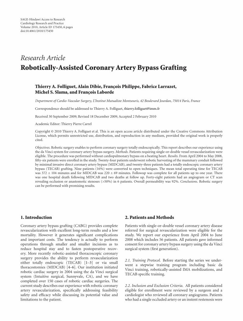
SAGE-Hindawi Access to ResearchCardiology Research and PracticeVolume 2010, Article ID 175450, 6 pagesdoi:10.4061/2010/175450
Research Article
Robotically-Assisted Coronary Artery Bypass Grafting
Thierry A. Folliguet, Alain Dibie, Francois Philippe, Fabrice Larrazet,Michel S. Slama, and Francois Laborde
Department of Cardio-Vascular Surgery, L’Institut Mutualiste Montsouris, 42 Boulevard Jourdan, 75014 Paris, France
Correspondence should be addressed to Thierry A. Folliguet, [email protected]
Received 30 September 2009; Revised 18 December 2009; Accepted 2 February 2010
Academic Editor: Thierry Pierre Carrel
Copyright © 2010 Thierry A. Folliguet et al. This is an open access article distributed under the Creative Commons AttributionLicense, which permits unrestricted use, distribution, and reproduction in any medium, provided the original work is properlycited.
Objectives. Robotic surgery enables to perform coronary surgery totally endoscopically. This report describes our experience usingthe da Vinci system for coronary artery bypass surgery. Methods. Patients requiring single-or-double vessel revascularization wereeligible. The procedure was performed without cardiopulmonary bypass on a beating heart. Results. From April 2004 to May 2008,fifty-six patients were enrolled in the study. Twenty-four patients underwent robotic harvesting of the mammary conduit followedby minimal invasive direct coronary artery bypass (MIDCAB), and twenty-three patients had a totally endoscopic coronary arterybypass (TECAB) grafting. Nine patients (16%) were converted to open techniques. The mean total operating time for TECABwas 372 ± 104 minutes and for MIDCAB was 220 ± 69 minutes. Followup was complete for all patients up to one year. Therewas one hospital death following MIDCAB and two deaths at follow up. Forty-eight patients had an angiogram or CT scanrevealing occlusion or anastomotic stenoses (>50%) in 6 patients. Overall permeability was 92%. Conclusions. Robotic surgerycan be performed with promising results.
1. Introduction
Coronary artery bypass grafting (CABG) provides completerevascularization with excellent long-term results and a lowmortality. However it generates significant complicationsand important costs. The tendency is actually to performoperations through smaller and smaller incisions as toreduce hospital stay and to fasten postoperative recov-ery. More recently robotic-assisted thoracoscopic coronarysurgery provides the ability to perform revascularizationeither totally endoscopic (TECAB) [1–3] or via smallthoracostomies (MIDCAB) [4–6]. Our institution initiatedrobotic cardiac surgery in 2004 using the da Vinci surgicalsystem (Intuitive surgical, Sunnyvale, CA), and we havecompleted over 150 cases of robotic cardiac surgeries. Thecurrent study describes our experience with robotic coronaryartery revascularization, specifically addressing feasibilitysafety and efficacy while discussing its potential value andlimitations to the patient.
2. Patients and Methods
Patients with single-or-double vessel coronary artery diseasereferred for surgical revascularization were eligible for thestudy. We report our experience from April 2004 to June2008 which includes 56 patients. All patients gave informedconsent for coronary artery bypass surgery using the da Vincisurgical system (first generation).
2.1. Training Protocol. Before starting the series we under-went a stepwise training program including basic daVinci training, robotically-assisted IMA mobilizations, andTECAB specific training.
2.2. Inclusion and Exclusion Criteria. All patients consideredeligible for enrollment were reviewed by a surgeon and acardiologist who reviewed all coronary angiograms. Patientswho had a single occluded artery or an instent restenosis were
2 Cardiology Research and Practice
considered, as well as patient with double-vessel lesion, orpatients who could benefit from a hybrid procedure. Hybridprocedures were defined as patients with two lesions: onewhich could easily be treated by angioplasty, whereas theother lesion was better suited for surgical revascularization(occluded artery, long calcified type C lesion).
All patients had a preoperative surgical and anesthesiol-ogist visit which included medical history, physical examina-tion, pulmonary function tests, computer tomography of thechest and/or gated coronary CT scan, and echocardiography.Based on the information and the established inclusion-exclusion criteria the patient was offered robotically-assistedcoronary revascularization. Exclusion criteria included leftventricular ejection fraction <0.30, emergency surgery, pul-monary edema, inability to ventilate on one lung (asthma,chronic obstructive pulmonary disease, pulmonary fibrosis),hemodynamic instability, anatomy unsuitable for endoscopicsurgery (scoliosis, kyphosis, morbid obesity), previous lungsurgery, pleural adhesions visible on CT scan, and patientunwilling to sign an informed consent.
Depending on the location of the coronary as well asthe caliber and the degree of calcifications, a TECAB or aMIDCAB was chosen. If the circumflex or the right coronaryarteries needed to be revascularized, a MIDCAB was chosen,as well as for patients needing a double revascularizationleft anterior descending (LAD) diagonal. A TECAB wasproposed only for patients in need of revascularization of anLAD or a large diagonal branch.
2.3. Operative Technique. Anesthesia was slightly modifiedcompared to patients undergoing CABG with ECC. Betablo-cants are continued in the hospital and given the day ofsurgery in order to maintain a cardiac rhythm between50 and 60 beats per minute. Anesthesia was maintainedwith reduced doses, Sulfentanyl (Sufenta) 2 mcg/kg, Fluri-trazepam (Narcozep) 0.03 mg/kg, and Bromure of Pancuro-nium (Pavulon) 0,1 mg/kg, associated with an inhalationagent (Enflurane, Ethrane). In case of tachycardia heartrate can be slowed with intrasvenous betablocking agents(Esmolol IV), or calcium channel blockers (Diltiazem). Allpatients were monitored with a radial continuous cardiacoutput monitor.
Heparin doses were reduced 50 UI/Kg IV, without anyprotamin reversal.
A double-lumen endotracheal tube is placed during thesurgery; it is subsequently changed to a single-lumen tube atthe end of the procedure.
A camera was inserted through a 5th intercostal spacemidaxillary line and two 8 mm robotic instruments trocarswere placed in the 2nd and 7th intercostal space midaxillaryline in order to obtain a triangulation with the camera. Theleft or right mammary artery was dissected entirely fromits insertion on the subclavian artery to its bifurcation. Thepericardium was then open over the target vessel and anendoscopic stabilizer was inserted through a 12 mm portplaced in the subxiphoid region and positioned over thetarget vessel. After heparinization, occlusion of the arterywas done with silastic loops; arteriotomy was performed as
an end to side anastomosis using running 8-0 monofilament(Goretex or Prolene) sutures. At completion of the anasto-mosis all trocars are removed and two chest tubes are placed.
During mammary harvesting both lungs were venti-lated while during coronary artery anastomosis single rightlung ventilation was maintained. The procedure has beendescribed in detail in other studies [7, 8].
All TECAB or MIDCAB was performed with the endo-scopic stabilizer which was either placed Trans xiphoid ordirectly through the mini thoracic incision. In fifteen patientswith incomplete coronary artery occlusion, we placed acoronary shunt, and the anastomosis was performed overa shunt. No patient needed pump assistance through groincannulation.
When a double bypass was performed, both mammaryarteries were harvested through the left chest. The anasto-mosis of the right mammary artery to the right coronarywas made through a small 4th right intercostals thoracotomy.The endostabilizer was placed directly through the incisionand the anastomosis was hand sawn. When the circumflexartery was chosen, the incision was slightly more posterior onthe left 4th intercostals space. The endo stabilizer was placedthrough the incision, and the anastomosis was hand made.
2.4. Exclusion versus Conversion. Once the patients wereenrolled, the surgical team decided to perform either aMIDCAB or a TECAB. Intraoperative conversions weredefined as cases in which a TECAB was scheduled but hadto be abandoned for various reasons and converted to eithera MIDCAB or a midline sternotomy.
2.5. Followup Postoperative Assessment. All immediate post-surgical clinical information was recorded per standardhospital practice and this included a postoperative 12-leadECG, chest X-ray, cardiac enzymes, and echocardiogram.
Patients had a followup visit between one to threemonths. Supplemental postoperative studies and physicalexam, resting 12-lead ECG, stress ECG or SPECT, andtransthoracic echo were obtained. All patients were offered acontrol angiogram after three months, and in case of refusal,a gated CT scan was performed. All angiograms or CT scanwas seen and reviewed by the team which included a surgeonand an angiographer.
All information was prospectively entered in the data-base.
All patients continued to be followed through regularinformation obtained by the referring cardiologist.
Comparisons between groups were performed by thestandard X2 test (or Fisher’s exact test when an expectedfrequency was less than 5) to compare categorical variablesand operative mortality, and the t test to compare continuousvariables. Statistical significance was accepted at P < .05.
3. Results
3.1. Patient Population. A total of 56 patients were poten-tial candidates for robotic-assisted surgery and agreed tosign an informed consent. Demographics and baseline
Cardiology Research and Practice 3
Table 1: Patient Demographics.
Variable Number (%)
Number of patients 56
male 46 (80)
female 10 (20)
Mean age (years) 66 ± 11 (38–82)
Hypertension 32 (57)
Diabetes 14 (25)
History of PTCA restenosis 26 (46 )
Myocardial infarction 37 (66)
Chronic renal failure 2 (3)
Angina 54 (96)
Average preoperative LVEF 49 ± 6 (30–65)
Redo surgery 2 (3)
EuroScore 3.9 ± 2 (0–9)
PTCA: percutaneous angioplasty.
characteristics are summarized in Table 1. The proceduresscheduled were a TECAB in 32 patients and a MIDCABin 24 patients. The TECAB procedure was proposed if thecoronary anatomy seemed favorable (large vessel > 3 mm,no major calcification, no intramyocardial course), and if theejection fraction was normal. In other cases a MIDCAB wasproposed with harvesting of the LIMA or RIMA roboticallyand the coronary anastomosis was done through a smallthoracotomy (left or right depending on the vessel).
Two patients were redo patients, both patients withpatent left-sided graft and occluded right venous grafts.Twelve patients underwent hybrid procedures with surgicalrevascularization on the LAD and angioplasty of anothervessel. All patients had a LIMA LAD anastomosis performedwhile the other vessels stented were the right coronary (8patients) and the circumflex (4 patients). The order of theprocedures varied as some patients had angioplasty priorto surgery (4 patients), or surgery first (8 patients). Noprocedures were done simultaneously. Patients were alwaysdischarged after the first procedure and readmitted for thesecond procedure.
The primary efficacy endpoint of the study was thecomposite endpoint of LIMA-LAD graft patency and free-dom from target vessel reintervention during the period ofobservation. The primary safety endpoints of the study werefreedom from major adverse cardiac events, including mor-tality, target vessel reintervention, and myocardial infarction.
For each case, intraoperative times were determinedbased on perfusion, anesthesia, and operative records or wereexplicitly recorded during the operation. The details of thesurgical procedure, including revascularization scheme, stepsof the surgical procedure that were completed robotically,conversion to alternate techniques, time to conversion, andreason for conversion, were documented in the operativereport forms.
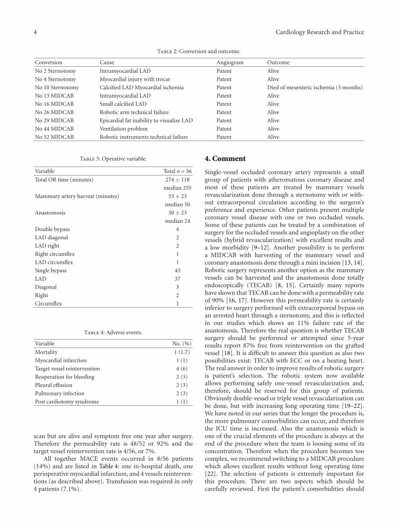
3.2. In-Hospital Morbidity and In-Hospital Outcome. Ninepatients in the TECAB group had to be converted, 3 tosternotomy and 6 to MIDCAB. Table 2 lists the cause of
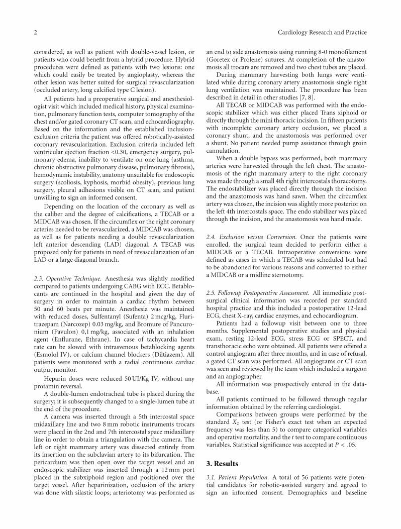
3 7 11 15 19 23 27 31 35 39 43 47 51 55
Case number
Mammary artery harvestingAnastomosisTotal operating room
0
100
200
300
400
500
600
700
800
(min
ute
s)
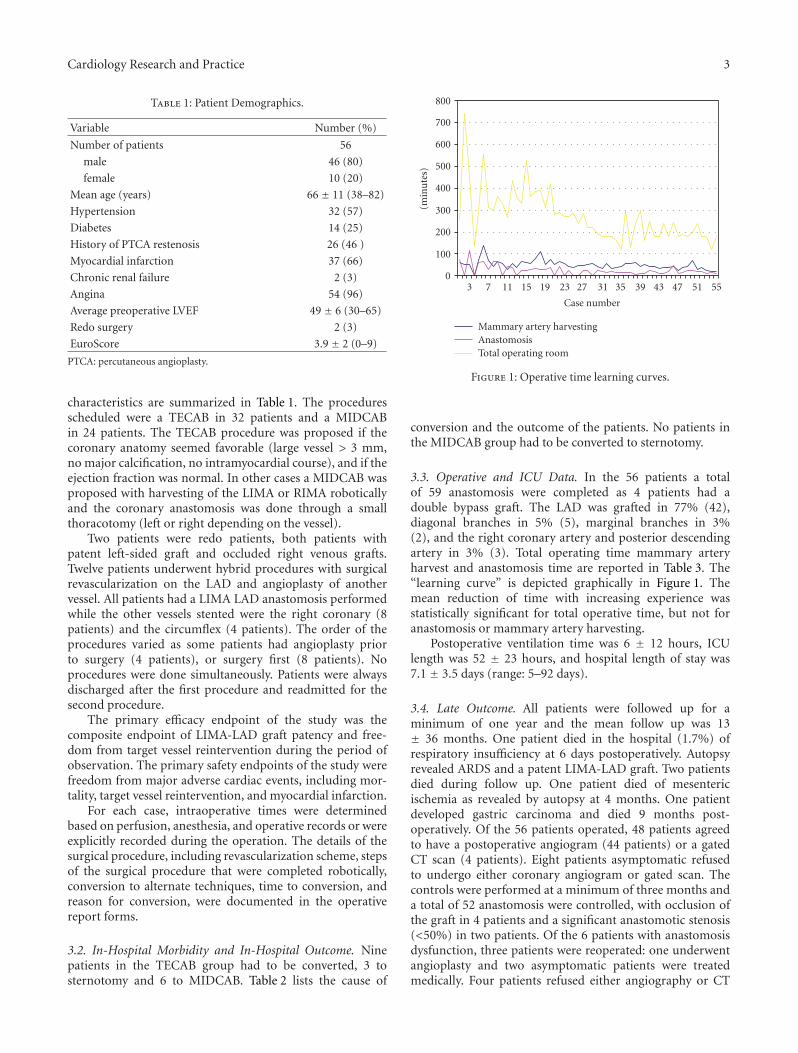
Figure 1: Operative time learning curves.
conversion and the outcome of the patients. No patients inthe MIDCAB group had to be converted to sternotomy.
3.3. Operative and ICU Data. In the 56 patients a totalof 59 anastomosis were completed as 4 patients had adouble bypass graft. The LAD was grafted in 77% (42),diagonal branches in 5% (5), marginal branches in 3%(2), and the right coronary artery and posterior descendingartery in 3% (3). Total operating time mammary arteryharvest and anastomosis time are reported in Table 3. The“learning curve” is depicted graphically in Figure 1. Themean reduction of time with increasing experience wasstatistically significant for total operative time, but not foranastomosis or mammary artery harvesting.
Postoperative ventilation time was 6 ± 12 hours, ICUlength was 52 ± 23 hours, and hospital length of stay was7.1 ± 3.5 days (range: 5–92 days).
3.4. Late Outcome. All patients were followed up for aminimum of one year and the mean follow up was 13± 36 months. One patient died in the hospital (1.7%) ofrespiratory insufficiency at 6 days postoperatively. Autopsyrevealed ARDS and a patent LIMA-LAD graft. Two patientsdied during follow up. One patient died of mesentericischemia as revealed by autopsy at 4 months. One patientdeveloped gastric carcinoma and died 9 months post-operatively. Of the 56 patients operated, 48 patients agreedto have a postoperative angiogram (44 patients) or a gatedCT scan (4 patients). Eight patients asymptomatic refusedto undergo either coronary angiogram or gated scan. Thecontrols were performed at a minimum of three months anda total of 52 anastomosis were controlled, with occlusion ofthe graft in 4 patients and a significant anastomotic stenosis(<50%) in two patients. Of the 6 patients with anastomosisdysfunction, three patients were reoperated: one underwentangioplasty and two asymptomatic patients were treatedmedically. Four patients refused either angiography or CT
4 Cardiology Research and Practice
Table 2: Conversion and outcome.
Conversion Cause Angiogram Outcome
No 2 Sternotomy Intramyocardial LAD Patent Alive
No 4 Sternotomy Myocardial injury with trocar Patent Alive
No 10 Sternotomy Calcified LAD Myocardial ischemia Patent Died of mesenteric ischemia (3 months)
No 13 MIDCAB Intramyocardial LAD Patent Alive
No 16 MIDCAB Small calcified LAD Patent Alive
No 26 MIDCAB Robotic arm technical failure Patent Alive
No 29 MIDCAB Epicardial fat inability to visualize LAD Patent Alive
No 44 MIDCAB Ventilation problem Patent Alive
No 52 MIDCAB Robotic instruments technical failure Patent Alive
Table 3: Operative variable.
Variable Total n = 56
Total OR time (minutes) 274 ± 118
median 255
Mammary artery harvest (minutes) 53 ± 23
median 50
Anastomosis 30 ± 23
median 24
Double bypass 4
LAD diagonal 2
LAD right 2
Right circumflex 1
LAD circumflex 1
Single bypass 43
LAD 37
Diagonal 3
Right 2
Circumflex 1
Table 4: Adverse events.
Variable No. (%)
Mortality 1 (1.7)
Myocardial infarction 1 (1)
Target vessel reintervention 4 (6)
Reoperation for bleeding 2 (3)
Pleural effusion 2 (3)
Pulmonary infection 2 (3)
Post cardiotomy syndrome 1 (1)
scan but are alive and symptom free one year after surgery.Therefore the permeability rate is 48/52 or 92% and thetarget vessel reintervention rate is 4/56, or 7%.
All together MACE events occurred in 8/56 patients(14%) and are listed in Table 4: one in-hospital death, oneperioperative myocardial infarction, and 4 vessels reinterven-tions (as described above). Transfusion was required in only4 patients (7.1%).
4. Comment
Single-vessel occluded coronary artery represents a smallgroup of patients with atheromatous coronary disease andmost of these patients are treated by mammary vesselsrevascularization done through a sternotomy with or with-out extracorporeal circulation according to the surgeon’spreference and experience. Other patients present multiplecoronary vessel disease with one or two occluded vessels.Some of these patients can be treated by a combination ofsurgery for the occluded vessels and angioplasty on the othervessels (hybrid revascularization) with excellent results anda low morbidity [9–12]. Another possibility is to performa MIDCAB with harvesting of the mammary vessel andcoronary anastomosis done through a mini incision [13, 14].Robotic surgery represents another option as the mammaryvessels can be harvested and the anastomosis done totallyendoscopically (TECAB) [8, 15]. Certainly many reportshave shown that TECAB can be done with a permeability rateof 90% [16, 17]. However this permeability rate is certainlyinferior to surgery performed with extracorporeal bypass onan arrested heart through a sternotomy, and this is reflectedin our studies which shows an 11% failure rate of theanastomosis. Therefore the real question is whether TECABsurgery should be performed or attempted since 5-yearresults report 87% free from reintervention on the graftedvessel [18]. It is difficult to answer this question as also twopossibilities exist: TECAB with ECC or on a beating heart.The real answer in order to improve results of robotic surgeryis patient’s selection. The robotic system now availableallows performing safely one-vessel revascularization and,therefore, should be reserved for this group of patients.Obviously double-vessel or triple vessel revascularization canbe done, but with increasing long operating time [19–22].We have noted in our series that the longer the procedure is,the more pulmonary comorbidities can occur, and thereforethe ICU time is increased. Also the anastomosis which isone of the crucial elements of the procedure is always at theend of the procedure when the team is loosing some of itsconcentration. Therefore when the procedure becomes toocomplex, we recommend switching to a MIDCAB procedurewhich allows excellent results without long operating time[22]. The selection of patients is extremely important forthis procedure. There are two aspects which should becarefully reviewed. First the patient’s comorbidities should
Cardiology Research and Practice 5
be reviewed. As we have already mentioned, all patients withpulmonary disease and/or poor ejection fraction should beexcluded since poor hemodynamic or low cardiac output candevelop during the procedure due to CO2 insufflations whichdecreases venous return, and also cardiac stabilization whichsometimes decreases cardiac output. For these reasons weroutinely monitor cardiac output throughout the procedurewith a radial monitor. When cardiac output decreases andis not improved despite simple measures (adding extravolume), we generally convert to a MIDCAB procedure[23]. The other aspects which should be carefully reviewedare the location, quality, and trajectory of the target vessel.Therefore, we have performed preoperative gated CT scanwhich allows us to appreciate the degree of calcifications andthe portion of the artery either intramyocardial or epicardial.This remains more important than in open surgery as theartery sometimes can be quite difficult to find if the vesselis in the fat or intramyocardial. Also if the vessel is calcified,it can be more difficult to perform the anastomosis with therobot. So we think that diffuse calcified and intramyocardialvessels should be exclusion criteria for the TECAB procedure.Finally the diameter of the vessel is important. It is difficultto select a number as to which to recommend not to attempta TECAB, but certainly the bigger the vessel, the easier theanastomosis and better the result will be. Occluded arteriesare an ideal situation since there is rarely any ischemia duringvessels occlusion, and therefore the time to perform thecoronary anastomosis is not an issue. Another interestinggroup of patients are redo coronary surgery. Sometimes onlyone vessel needs to be revascularised as the other graftsare still patent and certainly mini access or robotic surgerycan be a simple option [24]. It avoids redo sternotomy andits complications; the stabilization of the heart is usuallysimpler as the adhesions decrease the beating motion of themyocardium. However it can be sometimes difficult to findthe target vessel and a preoperative CT scan can help infinding the vessel.
The length of stay in this study was 7 days which is notmuch different then for sternotomy patients. This is certainlymuch higher than in various countries but is the norm inour country where patients are kept around a week in thehospital before being sent to a rehabilitation center. Howevermost of the patients (48/56) were discharged directly home,whereas the patient who underwent a sternotomy was sent toa rehabilitation center.
5. Conclusion
Robotic coronary surgery is basically feasible and can beperformed, but with a high conversion rate and a lowerpermeability rate than conventional surgery. Selection of thepatient and also of the target vessel is extremely importantto avoid conversion or poor vessel patency. In case ofhemodynamic instability or increased difficulty occurringduring the surgery, quick conversion should be performed.New techniques are needed in order to improve permeabilityrates.
References
[1] J. Bonatti, T. Schachner, O. Bernecker, et al., “Robotic totallyendoscopic coronary artery bypass: program development andlearning curve issues,” Journal of Thoracic and CardiovascularSurgery, vol. 127, no. 2, pp. 504–510, 2004.
[2] D. Loulmet, A. Carpentier, N. D’Attellis, et al., “Endoscopiccoronary artery bypass grafting with the aid of robotic assistedinstruments,” Journal of Thoracic and Cardiovascular Surgery,vol. 118, no. 1, pp. 4–10, 1999.
[3] V. Falk, A. Diegeler, T. Walther, S. Jacobs, J. Raumans, and F.W. Mohr, “Total endoscopic off-pump coronary artery bypassgrafting,” The Heart Surgery Forum, vol. 3, no. 1, pp. 29–31,2000.
[4] T. A. Vassiliades Jr., E. W. Rogers, J. L. Nielsen, andJ. L. Lonquist, “Minimally invasive direct coronary arterybypass grafting: intermediate-term results,” Annals of ThoracicSurgery, vol. 70, no. 3, pp. 1063–1065, 2000.
[5] V. A. Subramanian, N. U. Patel, N. C. Patel, and D. F. Loulmet,“Robotic assisted multivessel minimally invasive direct coro-nary artery bypass with port-access stabilization and cardiacpositioning: paving the way for outpatient coronary surgery?”Annals of Thoracic Surgery, vol. 79, no. 5, pp. 1590–1596, 2005.
[6] W. F. Turner Jr. and J. H. Sloan, “Robotic-assisted coronaryartery bypass on a beating heart: initial experience andimplications for the future,” Annals of Thoracic Surgery, vol.82, no. 3, pp. 790–794, 2006.
[7] U. Kappert, R. Cichon, J. Schneider, et al., “Closed-chestcoronary artery surgery on the beating heart with the useof a robotic system,” Journal of Thoracic and CardiovascularSurgery, vol. 120, no. 4, pp. 809–811, 2000.
[8] S. Dogan, T. Aybek, P. Risteski, et al., “Totally endoscopiccoronary artery bypass graft: initial experience with anadditional instrument arm and an advanced camera system,”Surgical Endoscopy and Other Interventional Techniques, vol.18, no. 11, pp. 1587–1591, 2004.
[9] J. Bonatti, T. Schachner, N. Bonaros, et al., “Simultaneoushybrid coronary revascularization using totally endoscopic leftinternal mammary artery bypass grafting and placement ofrapamycin eluting stents in the same interventional session:the combination pilot study,” Cardiology, vol. 110, no. 2, pp.92–95, 2008.
[10] C. Gao, M. Yang, Y. Wu, et al., “Hybrid coronary revasculariza-tion by endoscopic robotic coronary artery bypass grafting onbeating heart and stent placement,” Annals of Thoracic Surgery,vol. 87, no. 3, pp. 737–741, 2009.
[11] J. Bonatti, T. Schachner, N. Bonaros, et al., “Robotic totallyendoscopic coronary artery bypass and catheter based coro-nary intervention in one operative session,” Annals of ThoracicSurgery, vol. 79, no. 6, pp. 2138–2141, 2005.
[12] D. M. Holzhey, S. Jacobs, M. Mochalski, et al., “Minimallyinvasive hybrid coronary artery revascularization,” Annals ofThoracic Surgery, vol. 86, no. 6, pp. 1856–1860, 2008.
[13] W. D. Boyd, B. Kiaii, R. J. Novick, et al., “RAVECAB:improving outcome in off-pump minimal access surgery withrobotic assistance and video enhancement,” Canadian Journalof Surgery, vol. 44, no. 1, pp. 45–50, 2001.
[14] U. Kappert, R. Cichon, J. Schneider, et al., “Technique of closedchest coronary artery surgery on the beating heart,” EuropeanJournal of Cardio-Thoracic Surgery, vol. 20, no. 4, pp. 765–769,2001.
[15] F. W. Mohr, V. Falk, A. Diegeler, et al., “Computer-enhanced“robotic” cardiac surgery: experience in 148 patients,” Journal
6 Cardiology Research and Practice
of Thoracic and Cardiovascular Surgery, vol. 121, no. 5, pp.842–853, 2001.
[16] D. de Canniere, G. Wimmer-Greinecker, R. Cichon, et al.,“Feasibility, safety, and efficacy of totally endoscopic coronaryartery bypass grafting: multicenter European experience,”Journal of Thoracic and Cardiovascular Surgery, vol. 134, no.3, pp. 710–716, 2007.
[17] R. J. Damiano Jr., W. J. Ehrman, C. T. Ducko, et al., “InitialUnited States clinical trial of robotically assisted endoscopiccoronary artery bypass grafting,” Journal of Thoracic andCardiovascular Surgery, vol. 119, no. 1, pp. 77–82, 2000.
[18] U. Kappert, S.-M. Tugtekin, R. Cichon, M. Braun, and K.Matschke, “Robotic totally endoscopic coronary artery bypass:a word of caution implicated by a five-year follow-up,” Journalof Thoracic and Cardiovascular Surgery, vol. 135, no. 4, pp.857–862, 2008.
[19] J. Bonatti, T. Schachner, N. Bonaros, et al., “Robotic totallyendoscopic double-vessel bypass grafting: a further steptoward closed-chest surgical treatment of multivessel coronaryartery disease,” The Heart Surgery Forum, vol. 10, no. 3, pp.E239–E242, 2007.
[20] U. Kappert, R. Cichon, J. Schneider, I. Schramm, and S.Schuler, “Closed chest bilateral mammary artery grafting indouble-vessel coronary artery disease,” Annals of ThoracicSurgery, vol. 70, no. 5, pp. 1699–1701, 2000.
[21] F. Farhat, S. Aubert, P. Blanc, and O. Jegaden, “Totallyendoscopic off-pump bilateral internal thoracic artery bypassgrafting,” European Journal of Cardio-Thoracic Surgery, vol. 26,no. 4, pp. 845–847, 2004.
[22] S. Srivastava, S. Gadasalli, M. Agusala, et al., “Use of bilateralinternal thoracic arteries in CABG through lateral thora-cotomy with robotic assistance in 150 patients,” Annals ofThoracic Surgery, vol. 81, no. 3, pp. 800–806, 2006.
[23] S. Ganapathy, “Anaesthesia for minimally invasive cardiacsurgery,” Best Practice and Research: Clinical Anaesthesiology,vol. 16, no. 1, pp. 63–80, 2002.
[24] T. P. Martens, M. M. Hefti, R. Kalimi, C. R. Smith, andM. Argenziano, “Robot-assisted off-pump minimally invasivereoperative coronary artery bypass grafting: case report,” TheHeart Surgery Forum, vol. 7, no. 6, pp. E533–E534, 2004.

Submit your manuscripts athttp://www.hindawi.com
Stem CellsInternational
Hindawi Publishing Corporationhttp://www.hindawi.com Volume 2014
Hindawi Publishing Corporationhttp://www.hindawi.com Volume 2014
MEDIATORSINFLAMMATION
of
Hindawi Publishing Corporationhttp://www.hindawi.com Volume 2014
Behavioural Neurology
EndocrinologyInternational Journal of
Hindawi Publishing Corporationhttp://www.hindawi.com Volume 2014
Hindawi Publishing Corporationhttp://www.hindawi.com Volume 2014
Disease Markers
Hindawi Publishing Corporationhttp://www.hindawi.com Volume 2014
BioMed Research International
OncologyJournal of
Hindawi Publishing Corporationhttp://www.hindawi.com Volume 2014
Hindawi Publishing Corporationhttp://www.hindawi.com Volume 2014
Oxidative Medicine and Cellular Longevity
Hindawi Publishing Corporationhttp://www.hindawi.com Volume 2014
PPAR Research
The Scientific World JournalHindawi Publishing Corporation http://www.hindawi.com Volume 2014
Immunology ResearchHindawi Publishing Corporationhttp://www.hindawi.com Volume 2014
Journal of
ObesityJournal of
Hindawi Publishing Corporationhttp://www.hindawi.com Volume 2014
Hindawi Publishing Corporationhttp://www.hindawi.com Volume 2014
Computational and Mathematical Methods in Medicine
OphthalmologyJournal of
Hindawi Publishing Corporationhttp://www.hindawi.com Volume 2014
Diabetes ResearchJournal of
Hindawi Publishing Corporationhttp://www.hindawi.com Volume 2014
Hindawi Publishing Corporationhttp://www.hindawi.com Volume 2014
Research and TreatmentAIDS
Hindawi Publishing Corporationhttp://www.hindawi.com Volume 2014
Gastroenterology Research and Practice
Hindawi Publishing Corporationhttp://www.hindawi.com Volume 2014
Parkinson’s Disease
Evidence-Based Complementary and Alternative Medicine
Volume 2014Hindawi Publishing Corporationhttp://www.hindawi.com