Embed Size (px)
Citation preview

McGill Science Undergraduate Research Journal - msurj.mcgill.ca30
Selecting for multicellularity in the unicellular alga Chlamydomonas reinhardtiiTyler Moulton1*, Dr. Graham Bell1
1 Department of Biology, McGill University, Montreal, QC
Abstract Background: Researchers have recently begun experimentally exploring the origins of multicellularity (4-6). Their studies have found that the transition to a multicellular state may have been surprisingly simple, considering its profound implications for the history of life (3). This study experimentally selected for multicellularity in the unicellular biflagellated alga Chlamydomonas reinhardtii. This organism is especially interesting because it is basal to the Volvocaceae—a family of biflagellates whose evolutionary transition from unicellular organisms to complex forms have been meticulously characterised (2). The present study aimed to recreate the early stages of this transition, starting from incomplete cytokinesis after cell division.
Methods: The procedure was modeled loosely on the experiment performed by Ratcliff et al. (4) in which the authors successfully selected for multicellular Saccharomyces Cerevisiae—unicellular baker’s yeast. Three experimental replicates and one control for nine strains of C. reinhardtii were cultured in round-bottom vials in shaking incubators. Prior to each transfer (every 3-4 days), each culture was slowly mixed, and selection lines were then gently and briefly centrifuged. This applied a selection pressure which rendered heavier (clustered) cells more fit. The very bottom ~2% of the tubes’ contents was transferred, and cell cultures were examined for multicellularity.
Results: Six of nine lines of C. reinhardtii demonstrated an increased frequency of C. reinhardtii existing in a two- to four-celled state (the paired cell state accounted for 88% of these clusters)—consistent with the first step toward multicellularity as outlined by Kirk (2). A close study of cell division in the line which exhibited the strongest shift towards the multicellular phenotype suggests that true multicellularity began to evolve in this experiment. A multicellular phenotype did not become fixed in any population.
Conclusion: Our findings suggest that an artificial selection pressure is capable of inducing the evolution of multicellularity. Expanding upon this study could help us understand the mechanisms underlying the evolution of multicellularity.
Limitations: Sporadic data, possibly the result of difficulties in the procedure, prevented us from rigorously examining the effect of selection through time, limiting our ability to describe the evolutionary response. In addition, the study of individual cells, due to its time-consuming nature, was limited to one replicate of one line exhibiting a pronounced multicellular response. Thorough replication would be required before drawing a strong conclusion from this assay.
RESEARCH ARTICLE
Introduction
Multicellularity is one of the most crucial innovations in the history of life, allowing for drastic increases in organismal size and complex-ity. Multiple phyla and clades within these phyla have independently evolved this trait (1, 2) suggesting that this innovation may be fairly easy to achieve (3, 4). Boraas (5) found that in a predator-prey chemo-
stat, unicellular Chlorella vulgaris evolved a multicellular phenotype in response to predation from a flagellated protist. Koschwanez et al. (6) demonstrated that cell clumping greatly increases fitness of Sac-charomyces cerevisiae in dilute sucrose solution due to metabolite sharing from secreted hydrolytic enzymes. Ratcliff et al. (4)* have recently reported the successful evolution of multicellular yeast sim-ply by selecting for heavier cells via centrifugation. The yeast even
*Email Correspondence:[email protected]
Keywords:Multicellularity: The state defined by an organism comprised of more than one cell, often associated with division of labour among cells.Volvocaceae: Family of algae repre-senting a gradient of unicellular to multicellular species.Chlamydomonas reinhardtii: A unicellular, flagellated, volvocacean alga.Artificial Selection: Consistent, biased, guided selection for a pheno-typic trait in a population.
* The paper by Ratcliff et al. was published after the present study was carried out.Citation: W.C. Ratcliff F. Denison, M Borrello, M. Travisano. Proc. Natl. Acad. Sci U.S.A. 109,1595-1600 (2012)

Volume 8 - Issue 1 - March 2013 31
Selecting for multicellularity in the unicellular alga Chlamydomonas reinhardtii
exhibited division of labor, indicating increased complexity. Such results are promising, but skeptics point out that yeast may have a multicellular ancestor within the Ascomycota, which may trivialize these results (7, 8). It is critical, therefore, that these types of studies be expanded to include other model organisms.
The family Volvocaceae presents an ideal context to study the evo-lution of multicellularity because the transition to multicellularity from a unicellular volvocacean ancestor occurred recently on the evolutionary timescale (between 30 and 70 mya). As a result, the fam-ily contains a group of species which very clearly demonstrates the adaptive progression from small simple colonies to the larger and comparatively complex organisms (2, 9). Chlamydomonas reinhardtii, a unicellular biflagellated alga, is typically used as the outgroup in Vol-vocacean phylogenies, a practice supported by phylogenetic stud-ies of rRNA and ITS sequences of the Chlorphyceae (10-12). Kirk (2) outlined a putative twelve-step progression of adaptations from an ancestral volvocacean similar to C. reinhardtii all the way to the larg-est, most complex member of the family—Volvox carteri. Therefore, C. reinhardtii is an excellent starting point for experimentally examin-ing the transition to multicellularity.
Several studies have already shown that C. reinhardtii can exist in small, somewhat ordered clumps known as palmelloids (13). This phenotype is readily induced by certain organic acids (14) and by
small algal grazers (15). Both Iwasa and Murakami (14) and Lurling and Beekman (15) reported that palmelloids occur in 2n clumps more often than expected by chance, suggesting that members originate from a common mother cell. In addition, Lurling and Beekman (15) demonstrated that palmelloid formation requires light (and presum-ably growth and division). But the palmelloid phenotype is a plastic response (i.e. certain stimuli trigger this phenotypic change) which is too rapid to be evolutionary.
In this study we subjected several lines of C. reinhardtii to a simple treatment—brief, gentle centrifugation at each transfer (after Rat-cliff (4))—to select for increased individual size in an attempt to in-duce the evolution of multicellularity.
Materials and Methods
Nine C. reinhardtii strains were cultured in 5ml of sterile Bold’s mini-mal media (16) in 11ml Kimax® glass culture tubes with Bacti Capall® lids and held loosely in white Nalgene® tube racks in a New Brunswick Scientific® Innova 43® incubator-shaker set to 123 rpm and 25.1°C. Available nitrogen prevented sexual reproduction, thus all cultures reproduced vegetatively (13). There were three replicates of the ex-perimental treatment for each line, and one control (See Table 1).
Strains used in this experiment
Code Strain Description and HistoryA3 CC 2935 (mt-) Wild strains isolated from Farnham, Quebec: 1993. Cultured on Bold’s minimal media plates (agar 1.5%)
at McGill University since 1993 (27). The population is grown on Bold’s plates (1.5% agar) in the light, and is transferred every 15 days.A4 CC 2936 (mt+)
B2 A2S3 1/2 Base population for lines B2-C4. The ancestor was a cross of 3 lines performed by Clifford Zeyl in October 1992 as follows: (X•[Y•(Y•Z)]) where X is a mutant of lab strain CC-253 (mt-), Y is CC-1952 wild-type (mt-), and Z is CC-2343 wild-type (mt+) (17). The population is grown on Bold’s plates (1.5% agar) in the light, and is transferred every 15 days.
B3 ZIF Mass 2B Zygotes in Flasks (ZIF): descendants of strain B2, cultured in flasks of Bold’s minimal media (nitrogen-free to induce gametogenesis) with a suspended Nitex filter to serve as a substrate. Cultures are bubbled with a Pasteur pipet. After a cycle of 5 days of growth in the light followed by 5 days in the dark, the filter is removed and chloroformed, leaving only zygotes alive. A minimum of 200 zygotes is transferred to the next cycle.
B4 ZIF Mass 4
C1 MOP Mass 1A Mating on Plates (MOP): descendants of strain B2, grown on (1.5)% agar plates of Bold’s minimal media (nitrogen-free to induce gametogenesis) in light for 10 days followed by 5 days in the dark (extra time in light because growth is slower on plates than in liquid media (13)). The plates are then chloroformed, and a minimum of 200 zygotes are transferred to the next cycle.
C2 MOP Mass 1C
C3 Asex 1A Descendants of strain B2, grown on nitrogen-containing Bold’s minimal media plates (agar 1.5%) in light for 10 days followed by 5 days in the dark. Gametogenesis is not induced and the plates are never chloroformed, thus reproduction is strictly asexual.
C4 Unsel 2A Descendants of strain B2, grown on Bold’s minimal media plates (agar 1.5%) in light for 10 days followed by 5 days in the dark. A minimum of 200 individuals from each cycle for the subsequent inocculum. The medium is nitrogen deprived, thus allowing for gametogenesis, but the population is never chloroformed (i.e. sexual reproduction is permitted, but not selected for). This line has been exclusively asexually reproducing since the early 1990’s.
Table 1: Names and descriptions of the Chlamydamonas reinhardtii strains used in the present study.

McGill Science Undergraduate Research Journal - msurj.mcgill.ca32
Selecting for multicellularity in the unicellular Alga Chlamydomonas reinhardtii
Fig. 1

Volume 8 - Issue 1 - March 2013 33
Selecting for multicellularity in the unicellular alga Chlamydomonas reinhardtii
Six of the nine strains were derived from a seventh ancestral strain (B2). The ancestor was comprised of a cross of 3 wild-derived strains in 1992 (See Table 1) (17). These 7 strains were cultured under differ-ent conditions in the Bell laboratory at McGill University for 20 years prior to this study (see Table 1). All were grown in 500 μl liquid Bold’s cultures for one week prior to the experiment. During the study, the cultures were kept in areas which never housed any algae other than C. reinhardtii, and procedures were carried out in a laminar flow hood. Thus the risk of contamination, especially from algal species, was minimal. Nine 2.8 cmX45 cm 15W Sylvania®Gro-Lux®F15T8/GRO/AQ/RP fluorescent lights (rated at 325 lumens) provided continuous il-lumination from the lid of the incubator, and the door window was covered with white plastic boards to create a uniformly lit environ-ment.
Fig. 1 depicts step by step the transfer and selection procedures car-ried out every 3-4 days, corresponding to approximately 15 popu-lations doublings (i.e. 5-6 cell cycles) (13, 18). The procedures were performed in a laminar flow hood to prevent contamination. Once removed from the incubator, all cultures (experimental and control treatments) were re-suspended by gentle pumping with a pipettor (Fig.1.1, Fig 1.4). At this point, 100 μl samples from each tube were stored in a 96-well plate for subsequent analysis (see below). Ex-perimental treatment tubes were then centrifuged for 10 seconds at 500rpm (set to maximum acceleration and deceleration) using an IEC Centra GP8 ventilated centrifuge (cat. no. 216 4-place swinging buck-et rotor with cat. no. 316S buckets and cat.no. 5712 bucket adaptors). All but the bottom 100 μl (2%) ‘pellet’ of each culture (experimental and controls) was then removed with a pipet and discarded. Cen-trifugation resulted in a higher proportion of heavy (clumped) cells in the experimental tubes’ pellets compared to controls. This con-stituted a selection pressure in experimental lines which rendered the clumped phenotype more fit than a non-clumped or unicellular phenotype. The ‘pellets’ were re-suspended in fresh media (Fig. 1.6) and transferred to new culture tubes (~1.5% inoculum density). This procedure aimed to select solely on the basis of individual size by keeping environmental conditions the same between control and ex-perimental cultures. This ruled out the possibility of external stimuli inducing a plastic response (15, 19).
Data Collection
The 100 μl culture samples were used for counting cells and track-ing changes in individual morphology. Samples were loaded onto a haemocytometer, and viewed with a 10X objective (100X magnifi-cation). Pictures were taken with Q-Imaging© QICAM FAST-1394 in conjunction with the QCapture 2.68.6 software (©2001 Quantitative Imaging Corporation). The software settings were left at their default levels. Photographs showed the central portion of the grid, and cells were counted over an area of 0.8 mm2 from these pictures. The pho-tographs were taken of all populations on the day of each transfer. Counts were made for single cells, cell pairs, triplets, and quadru-
plets. Clumps of cells were photographed at higher magnifications to get more information about their structures.
Data Analysis
As Kirk (2) explains, the first step towards multicellularity should be the failure of daughter cells to separate following division. Therefore, we expected that putative multicellular individuals would contain 2n cells. As the experiment continued, the frequency of cells existing in pairs increased, aligning closely with our expectations. During some samplings, 3 and 4 cell clumps were also prevalent. In such instances, the microscope hampered our ability to differentiate to differentiate between 3 and 4-celled individuals, so they were lumped together in the analysis. Larger clumps were extremely rare, and thus not quan-tifiable (see Notable Observations section).The evolutionary response to selection was quantified as the frequency of 2-, 3-, and 4-celled in-dividuals relative to the total number of individuals as the response variable. In this analysis, each clump (1-cell, 2-celled, etc) was count-ed as an ‘individual’. This fraction (log transformed to normalize the data) is plotted against time (transfer number) in Fig. 2 (generated in R with ‘ggplot2’, Hadley Wickham, 2012). For each strain, odds ra-tios compared the multicellular response of each experimental repli-cate to its control treatment using a logistic regression model where transfer (time) is a covariate (12 transfers, therefore DF=47, α=0.05) (Using ‘glm’, The R Core Team, 2007). A likelihood ratio test was then used to compute the significance of the overall effect of experimental selection treatment for each strain compared to its control (χ2 test DF=3, α=0.05) (computed in Microsoft Excel© 2010).

McGill Science Undergraduate Research Journal - msurj.mcgill.ca34
Selecting for multicellularity in the unicellular Alga Chlamydomonas reinhardtii
Study of Paired-Cell Division
The object of this assay was to observe pairs of cells—putatively mul-ticellular individuals—undergoing division. Synchronized timing of division within a pair would suggest that the individuals are daugh-ter cells to a common mother, therefore representing a primitive multicellular state. A sterile screw-top vial containing 3.0ml of Bold’s minimal medium (agarose 0.9 g/l at 45 °C) was inoculated with 50 μl of culture from line C1 line, which had by this point demonstrated the clearest trend toward purported multicellularity. The mixture was plated over solidified Bold’s medium (agar 1.5 g/l), cooled, and placed open under the microscope. Fluorescent lighting in the room plus additional fluorescent and incandescent lamps around the mi-croscope provided ample light for growth. Photographs were taken hourly when possible. When cleavage initiation was observed, pic-tures were taken every 20-30 min. During the initial 7 hours of illumi-nation, photographs of 3 pairs and 2 single cells were taken from the 20X objective (200X magnification). The plate was left in dim light overnight with the lid on. Twelve hours later, illumination was re-stored, and a new group of cells was followed (again with the 20X objective). Once the plate was sufficiently dried out, it was possible to switch to the 40X objective (400X magnification). After another 10 hours of illumination, the room was left in absolute darkness for 24 hours, save for hourly 5 minute breaks for photography. Through-out this entire procedure, the stage of the microscope was left as
undisturbed as possible so the progress of individual cells could be tracked.
Results
Count Data
The likelihood ratio tests showed significantly higher frequencies of multicellular individuals in experimental treatments compared to their respective controls in the ancestral line B2 and all of its de-scendants (B3-C4) except for line B4, which showed a significant re-sult, but was skewed by a replicate which had significantly lower mul-ticellularity frequencies than the control (See Table 2). Of the total individuals classified as multicellular (2-3-4-cell clusters) 88% were in the paired cell form, deviating minimally across strains. Selection replicates in lines C1 and C2 had the highest odds ratios (OR ranges 5.570-17.190 and 3.211-3.881 respectively) indicating a particularly high frequency of multicellular individuals compared to controls. Likelihood ratio tests also implicate treatment as a highly significant factor for these two strains, especially C1. These two strains repre-sent historically sexually reproducing lines cultured on gel plates. Weaker responses (though still significant) were observed in C3 and C4 (historically asexual) and B3 and B4 (historically reproduced sexu-
Fig. 2: Plots showing the relative frequency of multicellular individuals through time. Refer to Table 1 for strain description.
Volume 8 - Issue 1 - March 2013 35
Selecting for multicellularity in the unicellular alga Chlamydomonas reinhardtii
ally in liquid media). While the selection pressure was a significant factor influencing multicellularity in the ancestor, strain B2, the re-sponse to selection was comparatively weak (OR range 0.999-2.208).The wild type isolates A3 and A4 (both single mating types) showed no significant response to selection.
Odds ratio and likelihood ratio tests for effect of selec-tion treatment on multicellular phenotypic response (±�=0.05 for all tests)
Strain Replicate Odds Ratio P|Z|(df=43)
Likelihood ratio test p|χ2|
(df=3)
CC 2935 (mt-)
1 1.001 1.00E+00
3.96E-012 0.989 9.60E-01
3 0.687 1.80E-01
CC 2936 (mt+)
1 1.477 3.50E-01
6.82E-012 1.573 3.10E-01
3 1.153 7.60E-01
A2S3 1/2
1 0.999 9.90E-01
1.37E-09*2 1.354 1.10E-01
3 2.208 7.00E-06
ZIF Mass 2B
1 3.391 5.10E-02
3.62E-02*2 1.762 3.91E-01
3 4.046 3.00E-02
ZIF Mass 4
1 1.316 3.40E-01
2.57E-02*2 1.379 2.40E-01
3 0.695 2.40E-01
MOP Mass 1A
1 5.570 1.80E-08
5.37E-52*2 6.863 1.60E-10
3 17.190 < 2.00E-16
MOP Mass 1C
1 3.881 4.30E-06
8.37E-07*2 3.211 8.70E-05
3 3.764 1.30E-05
Asex 1A
1 2.685 3.60E-05
3.32E-05*2 2.287 4.90E-04
3 1.674 3.60E-02
Unsel 2A1 2.041 1.10E-02
5.43E-05*2 0.764 4.41E-01
3 1.478 2.23E-01
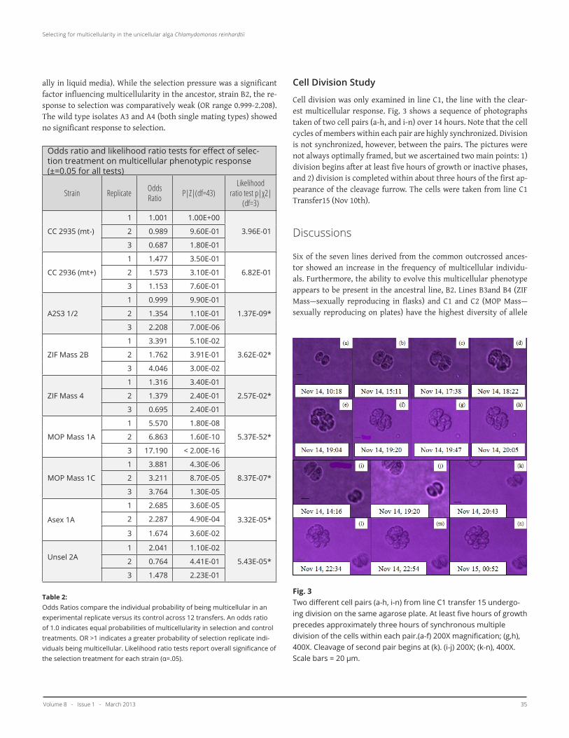
Table 2: Odds Ratios compare the individual probability of being multicellular in an experimental replicate versus its control across 12 transfers. An odds ratio of 1.0 indicates equal probabilities of multicellularity in selection and control treatments. OR >1 indicates a greater probability of selection replicate indi-viduals being multicellular. Likelihood ratio tests report overall significance of the selection treatment for each strain (α=.05).
Cell Division Study
Cell division was only examined in line C1, the line with the clear-est multicellular response. Fig. 3 shows a sequence of photographs taken of two cell pairs (a-h, and i-n) over 14 hours. Note that the cell cycles of members within each pair are highly synchronized. Division is not synchronized, however, between the pairs. The pictures were not always optimally framed, but we ascertained two main points: 1) division begins after at least five hours of growth or inactive phases, and 2) division is completed within about three hours of the first ap-pearance of the cleavage furrow. The cells were taken from line C1 Transfer15 (Nov 10th).
Discussions
Six of the seven lines derived from the common outcrossed ances-tor showed an increase in the frequency of multicellular individu-als. Furthermore, the ability to evolve this multicellular phenotype appears to be present in the ancestral line, B2. Lines B3and B4 (ZIF Mass—sexually reproducing in flasks) and C1 and C2 (MOP Mass—sexually reproducing on plates) have the highest diversity of allele
Fig. 3Two different cell pairs (a-h, i-n) from line C1 transfer 15 undergo-ing division on the same agarose plate. At least five hours of growth precedes approximately three hours of synchronous multiple division of the cells within each pair.(a-f) 200X magnification; (g,h), 400X. Cleavage of second pair begins at (k). (i-j) 200X; (k-n), 400X. Scale bars = 20 µm.

McGill Science Undergraduate Research Journal - msurj.mcgill.ca36
Selecting for multicellularity in the unicellular Alga Chlamydomonas reinhardtii
combinations present in the ancestral A2S3 population because these four had been sexually reproducing since 1992 (17). Errors in cross-ing over could also have led to gene duplication and subsequent di-vergence of function, or to gene modification. That the line emerg-ing with the strongest tendency towards multicellularity was one of these four previously sexually reproducing is consistent with the expectation that genetic diversity increases evolutionary potential (20). It is unclear, however, why line C1 exhibits such a remarkably strong and statistically significant response. Agar-base ancestral cul-ture conditions may have somehow predisposed lines C1 and C2 to evolving multicellularity.
The multicellular response could mean one of three things: 1) cells have increased the duration of cell division or the frequency of divi-sion, 2) cells are stickier, and tend to cohere, or 3) individuals actually exist in a paired or multicellular state. To evaluate these three hy-potheses, we monitored division of several cell pairs from one trans-fer of line C1—the line where selection appeared to be having the greatest effect (See Fig. 3). C. reinhardtii typically undergo a number of cleavages in rapid succession following 6-12 hours of growth (18, 21-23). The number of cleavages is determined in each cell during inter-phase (prior to division) and varies within a population (18, 21-23). The two pairs shown in Fig. 3 grow or retain a constant volume for at least five hours while in a paired state before rapid multiple cleavage occurs over about three hours. If the paired cells were actually just dividing cells, we would not have observed a gap between the first and subsequent cleavages. Furthermore, the cells within a pair are the same size as single cells (not shown). In addition, cultures were not synchronously dividing. (synchroniziationrequires prolonged exposure to alternating 12 hour light-dark periods (13)). There is evi-dence for asynchronicity in Fig. 3—one pair begins cleavage prior to 17:30, the other begins after 19:20. Within the pairs, however, cleav-age is highly synchronized between cells, and each cell gives rise to the same number of daughter cells. In Fig. 3, pair 1 (i-n) undergoes one more cleavage than pair 2 (a-h). If the pairs were simply com-prised of unrelated cells which had stuck together, then one would not expect this intra-pair synchronization. Therefore, these pho-tographs suggest that both cells within a pair are derived from the same mother cell. Cytoplasmic junctions may be maintained between members of a pair of daughter cells—a crucial step in the evolution of volvocacean multicellularity (2). Or, if the cells are siblings, their cell cycles could be inherently synchronized with no need for retaining cytoplasmic junctions. An alternative possibility is that cells within a pair are unrelated, but cohered following a collision, and formed cytoplasmic junctions which allowed for cell cycle synchronization.
That a multicellular phenotype never became fixed in any line is noteworthy. It has dramatically increased in frequency, but by no means dominates the cultures. Perhaps this is due to a trade-off ef-fect; cell clumping reduces the nutrients available for cells to con-sume (15). Centrifugation may have selected for heavier cells at each transfer, and thus multicellularity, but any single-celled individuals
which managed to be transferred would have proliferated faster. This would keep the frequency of multicellular individuals relatively low. Another possibility is that the experiment was too short to allow for fixation of the multicellular phenotype.
Notable Observations
Fig. 4Two different cell pairs (a-h, i-n) from line C1 transfer 15 undergo-ing division on the same agarose plate. At least five hours of growth
Flat Green Aggregates: On three separate occasions, we observed flat clumps of green cells arranged neatly in an alternating hexa-gon/pentagon pattern (particularly Fig. 4.1 a,c,f). These aggregates were generally circular, although several were irregular. When we at-tempted to wash these clumps from the haemocytometer and into a 12 well plate, they proved to be very sticky, often disintegrating rather than washing off. Given this observation and that several of

Volume 8 - Issue 1 - March 2013 37
Selecting for multicellularity in the unicellular alga Chlamydomonas reinhardtii
these aggregates appeared in a sample taken from a control line, we hypothesize that these clumps were a result of wall growth. Though the phenomenon was rare, such aggregates tended to co-occur, with several being found on the same slide. Perhaps this is an evolved phe-notype selected by the transfer procedure. But if this is the case, it is obviously not a tremendously advantageous phenotype, because it never achieved a frequency of greater than 1% in any given culture tube. Like colonies of the genus Gonium, these aggregates were flat, and the interlocking cells resembled those of Pandorina colonies (24, 25). Unlike either of these genera—the two next highest major clades in the Volvocacea (2)—these aggregates were non-motile.
Spherical Aggregates: During the sixth transfer, we discovered an apparent sphere of 13 or more cells (Fig. 4.1 g) in line C2. It was im-motile, but clearly three dimensional, and approximately 50um in diameter. Unfortunately, we failed to preserve the specimen. During the next transfer we noticed a smaller spherical group of cells (about 8) in line C1 (Fig. 4.1 h). We were unable to preserve this specimen. Both aggregates (Fig. 4.1 g,h) resembled Pandorina colonies, exhib-iting the “keystone” morphology described by Angeler (25) though again, both were immotile.On several other slides, we observed the occasional structure similar to that of palmelloids (Fig. 4.1 i) shown in Lurling and Beekman (15). Again, these occurred at very low frequencies (less than five instanc-es observed during the whole experiment). The significance of the above formations is unknown.
Motile Aggregates: C. reinhardtii absorb their flagella during divi-sion, as their anchoring basal bodies are required for mitosis (26). Therefore, moving aggregates would be convincing evidence of mul-ticellularity. Once in a control of line C4 and once in an experimen-tal replicate of line C1, we observed loosely associated aggregates of around seven cells cartwheeling about the slide. The C1 aggregate was fairly symmetrical, but it lost a few cells over the course of 30 minutes and was determined to be nothing more than temporarily cohering cells. During transfer seven, however, we observed that nearly 25% of individuals in one replicate of line C1 (Fig. 4.2) were 3-4 cell clumps—and most were motile! They slowly rolled in apparently random paths across the surface of the slide, in a manner similar to that of Pandorina. We never witnessed such an event again, despite efforts to preserve this unique population.
Limitations
While this study yielded promising results, it has several limitations. One concern with the design is the imprecision of the transfer. Un-equal evaporation between tubes prevented implementation of a constant and precise selection pressure. In addition, removing the supernatant proved to be a very difficult and imprecise procedure. Because there was no true ‘pellet’, there was inevitable mixing be-tween the ‘supernatant’ and the presumptive inoculum. Undoubted-ly, this lessened the selection pressure. In addition, large mats often
formed at the water line on the walls of many of the tubes. These occasionally were dislodged, and ended up in the pellet. The effect of this contamination is unknown.This imprecision may have led to the erratic data seen across trans-fers. In analyzing the data, we were unable to find any significant effect of time on the frequency of multicellular individuals. This is troubling, because if the response is evolutionary, we would expect to see an increase in the multicellular phenotype through time. While Fig. 2 suggests that lines B2, C1 and C2 do in fact have an increased frequency of multicellular individuals over time, we were not able to find statistical significance in this trend. This prevented us from completely ruling out the possibility that the selection procedure somehow induced a plastic palmelloid response.In addition, low-resolution microscopy precluding detailed observa-tions of cell morphology. As mentioned earlier, the ability to see the flagella of cells would have been very informative. Furthermore, cells in clumps may have obscured other cells within the clumps (i.e. a clump counted as 3 cells may actually have contained 4). Electron mi-croscopy could resolve the ambiguities left by the current pictures.Finally, this investigation lacks rigorous replication of controls. Only one control line was used per strain to compare against three experi-mental replicates. Given the sporadic nature of the data, a balanced design with more controls would have improved statistical power. Similarly, the study of cell division was not sufficiently replicated to extrapolate quantitative conclusions to all paired cells in this experi-ment. Future studies could incorporate this into their design using automated photography.
Conclusions
Selection of heavier individuals through centrifugation prior to each transfer appears sufficient to induce the beginning of a transition to true multicellularity in genetically diverse strains of C. reinhardtii. The increase in small clusters, particularly in the form of pairs, is consistent with the putative first steps in achieving multicellular-ity—incomplete daughter cell separation (2). We have gathered pho-tographic evidence that suggests that the multicellular phenotype in this line is not merely an artifact of division or cell collision and cohesion, and that the cells within a pair are likely genetically identi-cal. The response occurred over a fairly short period of time (around 50 generations) but the multicellular phenotype was never fixed in the population. Expanding upon this study could be invaluable in elucidating the mechanisms underlying one of the most important innovations in the history of life.
Acknowledgements
Many thanks to Etienne Low-Decarie for providing the bulk of the R scripts, and to Josianne Lachapelle for her help with various R-related issues. Both also gave plenty of much-needed advice in set-

McGill Science Undergraduate Research Journal - msurj.mcgill.ca38
Selecting for multicellularity in the unicellular Alga Chlamydomonas reinhardtii
ting up the experiment. Thanks to Larry for consultation regarding statistics. Many thanks to Dr. Bell for giving me the opportunity to undertake this experiment, and for sharing his wealth of knowledge along the way. This study was conducted September to December of 2011. Funding was provided by the National Sciences and Engineer-ing Research Council of Canada.
References
[1] J.T. Bonner. Integr. Biol.: Issues, News, and Reviews 1, 27-36 (1998).[2] D.L. Kirk. BioEssays 27, 299-310 (2005).[3] R.K. Grosberg, R.R. Strathmann. Annu. Rev. Ecol. Evol. Syst. 38, 621-654 (2007).[4] W.C. Ratcliff, paper presented to the Society for the Study of Evolution, Norman, Okl, USA. (18 June 2011).[5] M. Boraas, D. Seale, J. Boxhorn. Evol. Ecol. 12, 153-164 (1998).[6] J.H. Koschwanez, K.R. Foster, A.W. Murray. PLoS Biol. 9, e1001122 (2011).[7] B. Holmes. New Sci. (2011).[8] Y.J. Liu, B. D. Hall. Proc. Natl. Acad. Sci. U.S.A. 101, 4507-4512 (2004).[9.] M.D. Herron, R.E. Michod. Evolution. 62, 436-451 (2008).[10] T. Pröschold, B. Marin, U.G. Schlösser, M. Melkonian. Protist 152, 265-300 (2001).[11] T. Muller, S. Rahmann, T. Dandekar, M. Wolf. BMC Evol. Biol. 4, 20 (2004).[12] A. Larson, M. Kirk, D.L. Kirk. Mol. Biol. Evol. 9, 85-105 (1992).[13] E.H. Harris, D.B. Stern, G. Witman. The Chlamydomonas Sourcebook: Introduction to Chlamydomonas and its laboratory use. (Academic Press, 2008).[14] K. Iwasa, S. Murakami. Physiol. Plant. 21, 1224-& (1968).[15] M. Lurling, W. Beekman. Ann. Limnol. - Int. J. Lim. 42, 65-72 (2006).[16] H.C. Bold. Bull. Torr. Bot. Cl. 76, 101-108 (1949).[17] G. Bell. J. evol. biol. 18, 722-34 (2005)[18] M. Vítová, et al. Planta. 234, 599-608 (2011).[19] D.O. Hessen, E. Vandonk. Arch. Hydrobiol. 127, 129-140 (1993).[20] R.D.H. Barrett, D. Schluter. Trends Ecol. Evol. 23, 38-44 (2008).[21] K. Bisova, D.M. Krylov, J.G. Umen. Plant Physiol. 137, 475-491 (2005).[22] L. Donnan, P.C.L. John. Nat. 304, 630-633 (1983).[23] H. Oldenhof, V. Zachleder, H. Van den Ende. Folia Microbiol. 52, 53-60 (2007).[24] M. Hayama, T. Nakada, T. Hamaji, H. Nozaki. Phycol. 49, 221-234 (2010).24.[25] D.G. Angeler. Hydrobiol. 369-370, 269-275 (1998).[26] U.G.Johnson, K.R. Porter. J. Cell Biol. 38, 403-425 (1968). 27. L. Sack et al. Note. J. Phycol.. 30, 770–773(1994)


![[BIO] 02 - Origin of Multicellularity (Calsado)](https://img.pdfslide.net/doc/110x75/54686e2bb4af9f3a3f8b5cfa/bio-02-origin-of-multicellularity-calsado.jpg)