Embed Size (px)
Citation preview

Research ArticleThe Production of Nitric Oxide, IL-6, andTNF-Alpha in Palmitate-Stimulated PBMNCs Is Enhancedthrough Hyperglycemia in Diabetes
Caroline Maria Oliveira Volpe,1 Luana Farnese Machado Abreu,1
Pollyanna Stephanie Gomes,1 Raquel Miranda Gonzaga,1
Clara Araújo Veloso,2 and José Augusto Nogueira-Machado1
1 Nucleo de Pos-Graduacao e Pesquisa (NPGP), Hospital Santa Casa de Belo Horizonte, Domingos Vieira 590,Santa Efigenia, 30150-240 Belo Horizonte, MG, Brazil
2 Centro Universitario de Belo Horizonte (UniBH), Professor Mario Werneck, 1685 Estoril, 30455-610 Belo Horizonte, MG, Brazil
Correspondence should be addressed to Jose Augusto Nogueira-Machado; [email protected]
Received 23 January 2014; Accepted 1 March 2014; Published 6 April 2014
Academic Editor: Daniela Giustarini
Copyright © 2014 Caroline Maria Oliveira Volpe et al. This is an open access article distributed under the Creative CommonsAttribution License, which permits unrestricted use, distribution, and reproduction in any medium, provided the original work isproperly cited.
We examinednitric oxide (NO), IL-6, andTNF-𝛼 secretion fromcultured palmitate-stimulatedPBMNCsor in the plasma from type2 diabetesmellitus (T2MD)patients or nondiabetic (ND) controls. Free fatty acids (FFA) have been suggested to induce chronic low-grade inflammation, activate the innate immune system, and cause deleterious effects on vascular cells and other tissues throughinflammatory processes. The levels of NO, IL-6, TNF-𝛼, and MDA were higher in supernatant of palmitate stimulated blood cells(PBMNC) or from plasma from patients. The results obtained in the present study demonstrated that hyperglycemia in diabetesexacerbates in vitro inflammatory responses in PBMNCs stimulated with high levels of SFA (palmitate). These results suggest thathyperglycemia primes PBMNCs for NO, IL-6, and TNF-alpha secretion under in vitro FFA stimulation are associated with thesecretion of inflammatory biomarkers in diabetes. A combined therapy targeting signaling pathways activated by hyperglycemia inconjunction with simultaneous control of hyperglycemia and hypertriglyceridemia would be suggested for controlling the progressof diabetic complications.
1. Introduction
Circulating free fatty acids (FFAs) are elevated in patientswith type 2 diabetes mellitus (T2DM), obesity, metabolicsyndrome, and dyslipidemia [1–4]. FFAs represent a complexgroup of structurally variable molecules stored in the bodyas triglycerides and released through lipolysis [3, 5]. FFAsare classified according to the carbon chain length in short-, medium-, and long-chain fatty acids, the presence orabsence of double bonds as saturated (SFA) and unsaturatedfatty acids, respectively, and the number of double bondsas mono- or polyunsaturated (PUFA) [6, 7]. The effect ofFFA on cellular signaling pathways depends on the chemicalstructure. It has been reported that chronic exposure to SFA
increases oxidative stress and inflammation, leading to thedevelopment of cardiovascular diseases and insulin resistance[8–12].
Oxidative stress, reflecting an imbalance between proox-idant and antioxidant effectors, plays an important role indiabetic vascular complications [13]. Superoxide, nitric oxide,and lipid peroxidation are indicators of oxidative stress in thebody. Despite the number of studies concerning FFA-inducedsuperoxide overproduction [14–22], there are few reportsconcerning FFA-induced nitric oxide (NO) production. NOis a highly diffusible and unstable gas that acts as a modulatorof vascular tone, glucose transport in skeletalmuscle cells andadipocytes, blood flow, force generation in skeletal muscle,cytotoxicity, and inflammation [23–26].
Hindawi Publishing CorporationOxidative Medicine and Cellular LongevityVolume 2014, Article ID 479587, 12 pageshttp://dx.doi.org/10.1155/2014/479587

2 Oxidative Medicine and Cellular Longevity
FFA also regulates the immune system through interac-tions with specific cell surface receptors, such as Toll-likereceptors (TLR) and G-protein-coupled receptors (GPCR),thereby activating NF-kappaB and c-Jun amino-terminalkinase (JNK) pathways, which stimulate the secretion ofproinflammatory cytokines (IL-1beta, IL-6 and TNF-alpha)and chemokines [27–30].
It is well known the effects of hyperglycemia and hyper-lipidemia on peripheral blood mononuclear cells (PBMNCs)by activation of NADPH oxidase system leading to reactiveoxygen species production, TLR expression, enhancing NF-kappaB activity, and inducing proinflammatory cytokines,chemokines, and circulating adhesion molecules secretion[8, 21, 31–41].
Thus, elevated plasma FFA levels act as inflammatoryinducers, which potentially contribute to vascular disorders[27–30, 42, 43]. Thus, the aim of the present study was toinvestigate the in vitro effects of palmitate (C16:0), the majorSFA in plasma [44, 45], on the modulation of oxidative stressand inflammation in T2DM patients. Nitric oxide, with orwithout palmitate induction, was quantified and correlatedwith proinflammatory cytokines secreted in the culturedsupernatant of PBMNCs from type 2 diabetes patients. Theassociation among plasmatic triglycerides, NO, proinflam-matory cytokines (IL-6 and TNF-alpha), and oxidative stress(malondialdehyde) is discussed.
2. Material and Methods
This study was approved through the Ethical Committee ofSanta CasaHospital (BeloHorizonte-MG, Brazil) andwritteninformed consent was obtained from all participants prior tothe study.
2.1. Subjects. T2DM patients (𝑛 = 29), diagnosed accordingto the criteria of the AmericanDiabetes Association [46], andnondiabetic controls (𝑛 = 16), ranging from 45 to 70 years ofage, were recruited from the Endocrinology Department ofSanta Casa Hospital. Type 2 DM patients were treated withstatins and beta-blockers in addition to hypoglycemic drugs.Prior to the study, all volunteers received complete physicalexaminations, and detailed evaluations of medical historiesand laboratory analyses were performed (Table 1). Pregnantwomen and individuals suffering from alcoholism, infection,inflammation, dementia, or malignant diseases and smokingaddictions were excluded from this study.
2.2. Preparation of Fatty Acids. Palmitate and low-endotoxinbovine serum albumin (BSA, FFA-free) were purchased fromSigma-Aldrich Co. FFA was dissolved in 0.1MNaOH at 70∘Cand subsequently complexedwith 10%BSA at 55∘C for 10minto obtain a final FFA concentration of 500𝜇M (molar ratio2.4 : 1) [42, 47]. A 10mM fatty acid-albumin complex stocksolution and a 0.5 𝜇M BSA control solution were freshlyprepared, filtrated, and diluted prior to each experiment.
2.3. Preparation of Peripheral Blood Mononuclear Cells.PBMNCs were purified from 10.0mL of heparinized venous
Table 1: Clinical and biochemical characteristics of the studiedpopulation.
Parameters T2DM (𝑛 = 29) ND (𝑛 = 16) 𝑃
Female/Male ratio 19/10 11/5 NA
Age (years) 58.3 ± 9.0 57.1 ± 10.0 ns
Body mass index (kg/m2) 30.8 ± 9.8 24.6 ± 4.1 <0.05
Disease duration (years) 6.7 ± 6.4 NA NA
Systolic pressure (mmHg) 127.9 ± 14.5 122.3 ± 15.9 ns
Diastolic pressure (mmHg) 86.6 ± 8.6 88.9 ± 7.9 ns
Fasting glucose (mg/dL) 147.0 ± 40.7 89.0 ± 9.0 <0.05
Glycated hemoglobin (%) 8.1 ± 1.1 5.3 ± 0.2 <0.05
Total cholesterol (mg/dL) 191.6 ± 65.7 160.7 ± 20.0 ns
Low density lipoprotein(mg/dL)
115.3 ± 39.7 104.5 ± 32.6 ns
High density lipoprotein(mg/dL)
45.6 ± 10.6 50.2 ± 14.0 ns
Triglycerides (mg/dL) 142.0 ± 51.0 108.6 ± 37.7 <0.05Data as means ± SD.NA: not applicable; ns: not significant.Significant differences between the groups were determined using Student’s𝑡-test (𝑃 < 0.05).
blood, using a Ficoll-Hypaque gradient as previouslydescribed [48], with slight modifications. The trypan blueexclusion test showed that the cell viability in all samples was>95%.
2.4. Preparation of Plasma. EDTA venous blood sampleswere collected using a standard venipuncture technique.The plasma was obtained through centrifugation (200 g for15min, at room temperature), and the samples were storedat −80∘C until further analysis. Subsequent analyses wereperformed within 3 months from the day of storage.
2.5. Quantification of Proinflammatory Cytokines and NO inSupernatant of PBMNCs. Aliquots (100 𝜇L) of a PBMNCsuspension (1 × 10⁶/mL) from T2DM patients and NDcontrols in Dulbecco’s modified Eagle’s medium (DMEM)supplemented with 10% fetal bovine serum (FBS) wereincubated in the presence or absence of BSA (0.5 𝜇M) orpalmitate (500𝜇M) for 72 hours at 37∘C under 5% CO
2. The
final volume was adjusted to 300 𝜇L in DMEM supplementedwith 10% FBS. After incubation, the cells were centrifugedand the supernatant was collected. The interleukin-6 (IL-6human EIA Kit—Enzo Life Sciences, Inc., New York, USA)and tumor necrosis factor-alpha (TNF-𝛼 human EIA Kit—Enzo Life Sciences, Inc., New York, USA) concentrationswere determined through enzyme-linked immunosorbentassay (ELISA). Because NO is unstable, the quantitative ofNO was indirectly determined based on the detection ofthe blood nitrite and nitrate levels. The NO concentrationwas measured using the Total Nitric Oxide Assay Kit (AssayDesigns, Enzo Life Sciences, Inc., New York, USA).

Oxidative Medicine and Cellular Longevity 3
2
4
6
8
10
12
14
16
18
a a
bb
c
a
Nitr
ite (𝜇
M)
0Unstimulated BSA control
T2DMND
500𝜇M palmitate
(a)
0
100
200
300
400
500
600
700
a
b
a
b
c
b
Unstimulated BSA control 500𝜇M palmitate
IL-6
conc
entr
atio
n (p
g/m
L)
T2DMND
(b)
0
20
40
60
80
100
120
140
160
b ba
b
a
c
Unstimulated BSA control
TNF-𝛼
conc
entr
atio
n (p
g/m
L)
500𝜇M palmitate
T2DMND
(c)
Figure 1: Palmitate induces NO, IL-6, and TNF-alpha secretion in peripheral blood mononuclear cells (PBMNC) from patients with type2 diabetes. (a) Nitrite production; (b) IL-6 production; (c) TNF-alpha production. Different letters denote significance at 𝑃 < 0.05 usingStudent’s t-test. 𝑛 = 10 for each group.
2.6. Quantification of NO, MDA, and ProinflammatoryCytokines in Plasma. The plasma levels of NO, IL-6, andTNF-alpha were determined as described above. The plasmaMDA concentration was measured using the TBARS AssayKit (ZeptoMetrix Corp., New York, USA) according to themanufacturer’s instructions.
2.7. Statistical Analyses. The values are presented as themeans ± standard deviation (SD). The nonparametricKolmogorov-Smirnov test was used to assess the normaldistribution of the continuous variables. Comparisonsbetween groups were performed using unpaired Student’st-tests. Within-group correlations were performed usingPearson’s 𝑟 correlation. All analyses were consideredsignificant at 𝑃 values < 0.05 using Origin 6.0 software(Microcal Software Inc., Northampton, MA, USA).
3. Result
3.1. PBMNCs from T2DM Patients Are More Sensitive toPalmitate Stimulation Than the Cells from ND Controls. Asdepicted in Figure 1, palmitate activated the secretion ofNO, IL-6, and TNF-alpha in PBMNCs from T2DM patientscompared with those from ND controls (𝑃 < 0.05). Theresults of the induced effect of palmitate on PBMNCs fromT2DM patients and ND controls, expressed as the means ±SD, were NO, 11.5 ± 1.3 and 13.6 ± 2.2; IL-6, 86.1 ± 14.1 and126.0 ± 29.0; and TNF-alpha, 140.0 ± 28.1 and 535.8 ± 115,respectively. The results shown in Figure 1 also demonstratedthat PBMNCs from T2DM patients secreted significantly(𝑃 < 0.05) higher amounts of IL-6 (256.7 ± 81.1) and TNF-alpha (96.1±17.5) compared with the cells fromND controls(IL-6: 128.3 ± 32.3, TNF-alpha: 78.0 ± 13.6). No difference(𝑃 > 0.05) was observed in NO production in PBMNCs
4 Oxidative Medicine and Cellular Longevity
from T2DMpatients (10.9±1.7) and ND controls (10.9±1.2)without stimulation.
The production of NO and proinflammatory cytokineswas not altered in the presence of BSA (𝑃 > 0.05) in T2DMpatients and ND controls: NO, 11.5 ± 1.3 and 13.6 ± 2.2; IL-6,86.1±14.1 and 126.0±29.0; and TNF-alpha, 140.0±28.1 and535.8 ± 115, respectively.
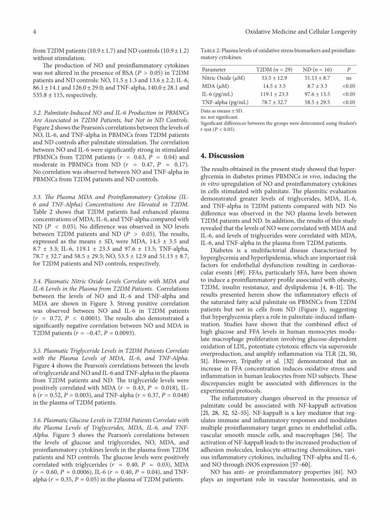
3.2. Palmitate-Induced NO and IL-6 Production in PBMNCsAre Associated in T2DM Patients, but Not in ND Controls.Figure 2 shows the Pearson’s correlations between the levels ofNO, IL-6, and TNF-alpha in PBMNCs from T2DM patientsand ND controls after palmitate stimulation. The correlationbetween NO and IL-6 were significantly strong in stimulatedPBMNCs from T2DM patients (𝑟 = 0.63, 𝑃 = 0.04) andmoderate in PBMNCs from ND (𝑟 = 0.47, 𝑃 = 0.17).No correlation was observed between NO and TNF-alpha inPBMNCs from T2DM patients and ND controls.
3.3. The Plasma MDA and Proinflammatory Cytokine (IL-6 and TNF-Alpha) Concentrations Are Elevated in T2DM.Table 2 shows that T2DM patients had enhanced plasmaconcentrations ofMDA, IL-6, and TNF-alpha comparedwithND (𝑃 < 0.05). No difference was observed in NO levelsbetween T2DM patients and ND (𝑃 > 0.05). The results,expressed as the means ± SD, were MDA, 14.5 ± 3.5 and8.7 ± 3.3; IL-6, 119.1 ± 23.3 and 97.6 ± 13.5; TNF-alpha,78.7 ± 32.7 and 58.5 ± 29.5; NO, 53.5 ± 12.9 and 51.13 ± 8.7,for T2DM patients and ND controls, respectively.
3.4. Plasmatic Nitric Oxide Levels Correlate with MDA andIL-6 Levels in the Plasma from T2DM Patients. Correlationsbetween the levels of NO and IL-6 and TNF-alpha andMDA are shown in Figure 3. Strong positive correlationwas observed between NO and IL-6 in T2DM patients(𝑟 = 0.72, 𝑃 < 0.0001). The results also demonstrated asignificantly negative correlation between NO and MDA inT2DM patients (𝑟 = −0.47, 𝑃 = 0.0093).
3.5. Plasmatic Triglyceride Levels in T2DM Patients Correlatewith the Plasma Levels of MDA, IL-6, and TNF-Alpha.Figure 4 shows the Pearson’s correlations between the levelsof triglyceride andNO and IL-6 and TNF-alpha in the plasmafrom T2DM patients and ND. The triglyceride levels werepositively correlated with MDA (𝑟 = 0.43, 𝑃 = 0.018), IL-6 (𝑟 = 0.52, 𝑃 = 0.003), and TNF-alpha (𝑟 = 0.37, 𝑃 = 0.048)in the plasma of T2DM patients.
3.6. Plasmatic Glucose Levels in T2DM Patients Correlate withthe Plasma Levels of Triglycerides, MDA, IL-6, and TNF-Alpha. Figure 5 shows the Pearson’s correlations betweenthe levels of glucose and triglycerides, NO, MDA, andproinflammatory cytokines levels in the plasma from T2DMpatients and ND controls. The glucose levels were positivelycorrelated with triglycerides (𝑟 = 0.40, 𝑃 = 0.03), MDA(𝑟 = 0.60, 𝑃 = 0.0006), IL-6 (𝑟 = 0.40, 𝑃 = 0.04), and TNF-alpha (𝑟 = 0.35, 𝑃 = 0.05) in the plasma of T2DM patients.
Table 2: Plasma levels of oxidative stress biomarkers and proinflam-matory cytokines.
Parameter T2DM (𝑛 = 29) ND (𝑛 = 16) 𝑃
Nitric Oxide (𝜇M) 53.5 ± 12.9 51.13 ± 8.7 nsMDA (𝜇M) 14.5 ± 3.5 8.7 ± 3.3 <0.05IL-6 (pg/mL) 119.1 ± 23.3 97.6 ± 13.5 <0.05TNF-alpha (pg/mL) 78.7 ± 32.7 58.5 ± 29.5 <0.05Data as means ± SD.ns: not significant.Significant differences between the groups were determined using Student’s𝑡-test (𝑃 < 0.05).
4. Discussion
The results obtained in the present study showed that hyper-glycemia in diabetes primes PBMNCs in vivo, inducing thein vitro upregulation of NO and proinflammatory cytokinesin cells stimulated with palmitate. The plasmitic evaluationdemonstrated greater levels of triglycerides, MDA, IL-6,and TNF-alpha in T2DM patients compared with ND. Nodifference was observed in the NO plasma levels betweenT2DM patients and ND. In addition, the results of this studyrevealed that the levels of NO were correlated withMDA andIL-6, and levels of triglycerides were correlated with MDA,IL-6, and TNF-alpha in the plasma from T2DM patients.
Diabetes is a multifactorial disease characterized byhyperglycemia and hyperlipidemia, which are important riskfactors for endothelial dysfunction resulting in cardiovas-cular events [49]. FFAs, particularly SFA, have been shownto induce a proinflammatory profile associated with obesity,T2DM, insulin resistance, and dyslipidemia [4, 8–11]. Theresults presented herein show the inflammatory effects ofthe saturated fatty acid palmitate on PBMNCs from T2DMpatients but not in cells from ND (Figure 1), suggestingthat hyperglycemia plays a role in palmitate-induced inflam-mation. Studies have shown that the combined effect ofhigh glucose and FFA levels in human monocytes modu-late macrophage proliferation involving glucose-dependentoxidation of LDL, potentiate cytotoxic effects via superoxideoverproduction, and amplify inflammation via TLR [21, 50,51]. However, Tripathy et al. [32] demonstrated that anincrease in FFA concentration induces oxidative stress andinflammation in human leukocytes from ND subjects. Thesediscrepancies might be associated with differences in theexperimental protocols.
The inflammatory changes observed in the presence ofpalmitate could be associated with NF-kappaB activation[21, 28, 32, 52–55]. NF-kappaB is a key mediator that reg-ulates immune and inflammatory responses and modulatesmultiple proinflammatory target genes in endothelial cells,vascular smooth muscle cells, and macrophages [56]. Theactivation of NF-kappaB leads to the increased production ofadhesion molecules, leukocyte-attracting chemokines, vari-ous inflammatory cytokines, including TNF-alpha and IL-6,and NO through iNOS expression [57–60].
NO has anti- or proinflammatory properties [61]. NOplays an important role in vascular homeostasis, and in

Oxidative Medicine and Cellular Longevity 5
1000
800
600
400
200
0
IL-6
(pg/
mL)
TNF-𝛼
(pg/
mL)
Nitrite (𝜇M)0 5 10 15
200
150
100
50
20
Nitrite (𝜇M)0 5 10 15 20
0
r = 0.63
P = 0.04
r = 0.02P = 0.94
(a)
200
150
100
50
00 5 10 15
IL-6
(pg/
mL)
Nitrite (𝜇M)
r = 0.47
P = 0.17
150
100
50
00 5 10 15
Nitrite (𝜇M)
r = 0.08
P = 0.83
TNF-𝛼
(pg/
mL)
(b)
Figure 2: Pearson’s correlation coefficients betweenNOandproinflammatory cytokines in PMBNCs fromT2DMpatients (a) andnondiabeticcontrols (b) after stimulation with palmitate. 𝑛 = 10 for each group.
immune cells, NO regulates antimicrobial and antitumoractivities, although excess NO production might cause tissuedamage and is associated with acute and chronic inflamma-tion [56, 62]. Nitric oxide synthase (NOS) synthesizes NOfrom L-arginine using NADPH and oxygen as cosubstrates[63]. Three isoforms of NO synthase have been described:neuronal (nNOS or NOS 1), inducible (iNOS or NOS 2), andendothelial (eNOS or NOS 3) [64]. Activated macrophagesand neutrophils produce large amounts of NO through iNOSactivity [65, 66]. The results of this study demonstratedincreased NO production and a positive correlation betweenNO and IL-6 levels in palmitate-stimulated PBMNCs fromT2DM patients, suggesting that iNOS expression can be ele-vated through palmitate-induced proinflammatory cytokinesecretion. No differences were observed in the cells from NDcontrols (Figures 1 and 2). Unbound palmitic acid treatmentincreased NO production in skeletal muscle [67]. However,
in endothelial cells, FFA induced the inhibition of eNOS,thereby attenuating NO production [68–71].
To evaluate in vivo inflammation, we quantified theplasma levels of NO, the oxidative stress biomarker (MDA),and proinflammatory cytokines (IL-6 and TNF-alpha) inT2DM patients and ND controls. Consistent with otherstudies [72–93], the results of the present study demon-strated elevated levels of IL-6 and TNF-alpha, reflectingthe activation of innate immune cells, and high levels ofMDA, indicating the presence of oxidative stress in T2DMpatients compared with ND controls. Diabetic conditions(hyperglycemia and hyperlipidemia) increase proinflamma-tory and oxidative stress levels, culminating in endotheliumdysfunction [1, 27, 42, 56, 90, 94, 95]. Oxidative stress reducedNO production through eNOS [56], and the increased levelsof superoxide could react with NO to produce peroxyni-trite, a highly toxic product [23, 96]. Peroxynitrite nitrates

6 Oxidative Medicine and Cellular Longevity
IL-6
(pg/
mL)
TNF-𝛼
(pg/
mL)
Nitrite (𝜇M)
Nitrite (𝜇M)
Nitrite (𝜇M)
200
150
100
50
0
150
200
100
50
0
100806040200
100
80
60
40
20
00 5 10 15 20 25
MD
A (𝜇
M)
30 50 70 90 110
P = 0.40
r = −0.47
r = −0.16
P = 0.0093
r = 0.72
P < 0.0001
(a)
IL-6
(pg/
mL)
TNF-𝛼
(pg/
mL)
Nitrite (𝜇M)
150
100
50
0
100
806040200
80
60
40
20
0
0
5
10
15
20
MD
A (𝜇
M)
r = 0.12P = 0.63
r = 0.09P = 0.74
r = −0.16P = 0.57
Nitrite (𝜇M)
Nitrite (𝜇M)
806040200
806040200
(b)
Figure 3: Pearson’s correlation coefficients between nitric oxide and proinflammatory cytokines andMDA in the plasma fromT2DMpatients(a) and nondiabetic controls (b). 𝑛 = 29 for T2DM patients and 16 for nondiabetic controls.
the tyrosine residues in a number of proteins and modulatestheir functions [97, 98]. The results in the present study didnot show any differences in the plasmaNO levels between thestudied groups (Table 2). However, we observed a negativeassociation between NO and MDA levels in the plasmafrom T2DM patients, suggesting that increased oxidative
stress could affect NO biodisponibility, leading to endothelialdysfunction in diabetes (Figure 3).
The results obtained in the present study also demon-strated high levels of triglycerides in the plasma from T2DMpatients compared with ND controls (Table 1). FFAs arestored in the body in the formof triglycerides and are released

Oxidative Medicine and Cellular Longevity 7
IL-6
(pg/
mL)
TNF-𝛼
(pg/
mL)
Nitr
ite (𝜇
M)
MD
A (𝜇
M)
200
150
100
50
0
200
150
100
50
0
100
80
60
40
20
0
0
5
10
15
20
25
0 50 100 150 200 250
Triglycerides (mg/dL)
r = −0.011P = 0.95
r = 0.43P = 0.018
r = 0.52
P = 0.003
r = 0.37
P = 0.048
0 50 100 150 200 250
Triglycerides (mg/dL)
0 50 100 150 200 250
Triglycerides (mg/dL)
0 50 100 150 200 250
Triglycerides (mg/dL)
(a)
IL-6
(pg/
mL)
TNF-𝛼
(pg/
mL)
Nitr
ite (𝜇
M)
MD
A (𝜇
M)
150
100
50
0
80
60
40
20
0
100
80
60
40
20
0
0
5
10
15
20
0 50 100 150 200
Triglycerides (mg/dL)
r = 0.059
P = 0.826
r = 0.20
P = 0.46
r = 0.29
P = 0.27
r = 0.04P = 0.88
0 50 100 150 200
Triglycerides (mg/dL)
0 50 100 150 200
Triglycerides (mg/dL)
0 50 100 150 200
Triglycerides (mg/dL)
(b)
Figure 4: Pearson’s correlation coefficients between triglycerides and proinflammatory cytokines and MDA in the plasma of T2DM patients(a) and nondiabetic controls (b). 𝑛 = 29 for T2DM patients and 16 for nondiabetic controls.

8 Oxidative Medicine and Cellular Longevity
IL-6
(pg/
mL)
TNF-𝛼
(pg/
mL)
Nitr
ite (𝜇
M)
MD
A (𝜇
M)
200
150
100
50
0
200
150
100
50
0
100
80
60
40
20
0
0
5
10
15
20
25
0
50
100
150
200
250
0 50 100 150 200 250
Trig
lyce
rides
(mg/
dL)
Glucose (mg/dL)
r = 0.40P = 0.03
r = 0.60
P = 0.0006
r = −0.08P = 0.67
r = 0.40P = 0.04
r = 0.35P = 0.05
0 50 100 150 200 250
Glucose (mg/dL)
0 50 100 150 200 250
Glucose (mg/dL)
0 50 100 150 200 250
Glucose (mg/dL)
0 50 100 150 200 250
Glucose (mg/dL)
(a)
IL-6
(pg/
mL)
TNF-𝛼
(pg/
mL)
Nitr
ite (𝜇
M)
MD
A (𝜇
M)
200
150
100
50
0
150
100
50
0
60
40
20
0
100
80
60
40
20
0100806040200
0
5
10
15
20
Trig
lyce
rides
(mg/
dL)
Glucose (mg/dL)
r = 0.31P = 0.25
r = 0.13P = 0.61
r = −0.12P = 0.65
r = 0.09P = 0.72
r = 0.18P = 0.52
100806040200
Glucose (mg/dL)
10080
80
6040200
Glucose (mg/dL)
100806040200
Glucose (mg/dL)
100806040200
Glucose (mg/dL)
(b)
Figure 5: Pearson’s correlation coefficients between glucose and triglycerides, MDA, nitric oxide, and proinflammatory cytokines in plasmaof T2DM (a) patients and nondiabetic controls (b). 𝑛 = 29 for T2DM and 16 for nondiabetic controls.

Oxidative Medicine and Cellular Longevity 9
into tissues through lipolysis, a process regulated throughinsulin [99]. Impaired insulin signaling increases lipolysis,resulting in increased FFA levels [100, 101]. The results of thepresent study showed that triglycerides levels are positivelyassociated with the MDA, IL-6, and TNF-alpha levels in theplasma from T2DM patients, but this correlation was notobserved in the plasma fromND controls. No correlationwasobserved between triglycerides and NO in the plasma fromthe studied groups (Figure 4). Glucose levels are positivelycorrelated with the triglycerides, MDA, IL-6, and TNF-alphalevels in the plasma from T2DM patients, but not in theplasma from ND controls (Figure 5).
Accumulating evidence has shown that the regulationof dyslipidemia is of equal importance for the regulation ofhyperglycemia and hypertension in the care of patients withT2DM. Hyperlipidemia represents a major risk factor forthe development of vascular dysfunction and atherosclerosis[27–30, 42, 43]. Most T2DM patients are obese and haveelevated plasma FFA levels [102, 103]. Moreover, high-fatdiets might induce metabolic dysfunction and inflammationthrough the release of FFA through lipolysis and proinflam-matory cytokines through downstream signaling [104, 105].
FFAs have been suggested to induce chronic low-gradeinflammation, activate the innate immune system, and causedeleterious effects on vascular cells and other tissues throughinflammatory processes. The results obtained in the presentstudy demonstrated that hyperglycemia in diabetes exacer-bates in vitro inflammatory responses in PBMNCs stimu-lated with high levels of SFA (palmitate). Furthermore, theresults suggest that the endothelium levels of NO could beregulated through oxidative stress and high levels of triglyc-erides are correlated with oxidative stress and proinflam-matory cytokine secretion in T2DM patients. Endothelialdysfunction is associated with several pathophysiologicalconditions in diabetes [56]. Combined therapy targeting theintracellular mechanisms underlying metabolic alterationsleading to endothelial dysfunction is an important issue inthe prevention of vascular complications associated withdiabetes. The simultaneous control of hyperglycemia andhypertriglyceridemia is necessary to ameliorate the progres-sion to diabetic vasculopathy.
Conflict of Interests
The authors confirm that there is no conflict of interests.
Acknowledgments
The authors would like to thank FAPEMIG, CNPq, CAPES,Rede Mineira de Toxina Terapeutica 26/12, and Toxinologiaproject for financial support.
References
[1] R. N. Bergman and M. Ader, “Free fatty acids and pathogenesisof type 2 diabetes mellitus,” Trends in Endocrinology andMetabolism, vol. 11, no. 9, pp. 351–356, 2000.
[2] G. Boden, “Role of fatty acids in the pathogenesis of insulinresistance and NIDDM,” Diabetes, vol. 46, no. 1, pp. 3–10, 1997.
[3] V. Stich andM. Berlan, “Physiological regulation ofNEFA avail-ability: lipolysis pathway,” Proceedings of the Nutrition Society,vol. 63, no. 2, pp. 369–374, 2004.
[4] S. Tsimikas and P. D. Reaven, “The role of dietary fatty acids inlipoprotein oxidation and atherosclerosis,” Current Opinion inLipidology, vol. 9, no. 4, pp. 301–307, 1998.
[5] G. M. Reaven, “Role of insulin resistance in human disease,”Diabetes, vol. 37, no. 12, pp. 1595–1607, 1988.
[6] T. Ulven, “Short-chain free fatty acid receptors FFA2/GPR43and FFA3/GPR41 as newpotential therapeutic targets,” Frontiersin Endocrinology, vol. 3, p. 111, 2012.
[7] M. A. R. Vinolo, S. M. Hirabara, and R. Curi, “G-protein-coupled receptors as fat sensors,” Current Opinion in ClinicalNutrition and Metabolic Care, vol. 15, no. 2, pp. 112–116, 2012.
[8] R. C. Bunn, G. E. Cockrell, Y. Ou, K. M.Thrailkill, C. K. Lump-kin Jr., and J. L. Fowlkes, “Palmitate and insulin synergisticallyinduce IL-6 expression in human monocytes,” CardiovascularDiabetology, vol. 9, article no. 73, 2010.
[9] B. M. Egan, G. Lu, and E. L. Greene, “Vascular effects of non-esterified fatty acids: implications for the cardiovascular riskfactor cluster,” Prostaglandins Leukotrienes and Essential FattyAcids, vol. 60, no. 5-6, pp. 411–420, 1999.
[10] B. Hennig, D. M. Shasby, and A. A. Spector, “Exposure to fattyacid increases human low density lipoprotein transfer acrosscultured endothelial monolayers,” Circulation Research, vol. 57,no. 5, pp. 776–780, 1985.
[11] D. F. Horrobin, “Abnormal membrane concentrations of 20and 22-carbon essential fatty acids: a common link betweenrisk factors and coronary and peripheral vascular disease?”Prostaglandins Leukotrienes and Essential Fatty Acids, vol. 53,no. 6, pp. 385–396, 1995.
[12] G. I. Shulman, “Cellular mechanisms of insulin resistance,”Journal of Clinical Investigation, vol. 106, no. 2, pp. 171–176, 2000.
[13] D. Giugliano, A. Ceriello, and G. Paolisso, “Oxidative stress anddiabetic vascular complications,” Diabetes Care, vol. 19, no. 3,pp. 257–267, 1996.
[14] E. Hatanaka, A. C. Levada-Pires, T. C. Pithon-Curi, and R.Curi, “Systematic study on ROS production induced by oleic,linoleic, and 𝛾-linolenic acids in human and rat neutrophils,”Free Radical Biology and Medicine, vol. 41, no. 7, pp. 1124–1132,2006.
[15] U. Wenzel, A. Nickel, and H. Daniel, “Increased mitochon-drial palmitoylcarnitine/carnitine countertransport by flavonecauses oxidative stress and apoptosis in colon cancer cells,”Cellular and Molecular Life Sciences, vol. 62, no. 24, pp. 3100–3105, 2005.
[16] G. S. Dhaunsi, J. Kaur, K. Alsaeid, R. B. Turner, and M. S. Bitar,“Very long chain fatty acids activate NADPH oxidase in humandermal fibroblasts,” Cell Biochemistry and Function, vol. 23, no.1, pp. 65–68, 2005.
[17] M. H. Horani, M. J. Haas, and A. D. Mooradian, “Saturated,unsaturated, and trans-fatty acids modulate oxidative burstinduced by high dextrose in human umbilical vein endothelialcells,” Nutrition, vol. 22, no. 2, pp. 123–127, 2006.
[18] C. Duval, Y. Camara, E. Hondares, B. Sibille, and F. Villarroya,“Overexpression of mitochondrial uncoupling protein-3 doesnot decrease production of the reactive oxygen species, elevatedby palmitate in skeletal muscle cells,” FEBS Letters, vol. 581, no.5, pp. 955–961, 2007.
[19] S. Srivastava and C. Chan, “Hydrogen peroxide and hydroxylradicals mediate palmitate-induced cytotoxicity to hepatoma

10 Oxidative Medicine and Cellular Longevity
cells: relation to mitochondrial permeability transition,” FreeRadical Research, vol. 41, no. 1, pp. 38–49, 2007.
[20] L. I. Rachek, S. I. Musiyenko, S. P. LeDoux, and G. L. Wilson,“Palmitate induced mitochondrial deoxyribonucleic acid dam-age and apoptosis in L6 rat skeletal muscle cells,” Endocrinology,vol. 148, no. 1, pp. 293–299, 2007.
[21] M. R. Dasu and I. Jialal, “Free fatty acids in the presenceof high glucose amplify monocyte inflammation via Toll-likereceptors,” American Journal of Physiology, vol. 300, no. 1, pp.E145–E154, 2011.
[22] R. H. Lambertucci, S. M. Hirabara, L. D. R. Silveira, A.C. Levada-Pires, R. Curi, and T. C. Pithon-Curi, “Palmitateincreases superoxide production through mitochondrial elec-tron transport chain and NADPH oxidase activity in skeletalmuscle cells,” Journal of Cellular Physiology, vol. 216, no. 3, pp.796–804, 2008.
[23] V. Darley-Usmar, H. Wiseman, and B. Halliwell, “Nitric oxideand oxygen radicals: a question of balance,” FEBS Letters, vol.369, no. 2-3, pp. 131–135, 1995.
[24] D. McGrowder, D. Ragoobirsingh, and P. Brown, “Acute effectsof exogenous nitric oxide on glucose uptake in skeletal muscleof normoglycaemic and diabetic rats,”Medical Science Monitor,vol. 12, no. 1, pp. BR28–BR35, 2006.
[25] M. J. Jackson, D. Pye, and J. Palomero, “The productionof reactive oxygen and nitrogen species by skeletal muscle,”Journal of Applied Physiology, vol. 102, no. 4, pp. 1664–1670,2007.
[26] D. M. Pattwell, A. McArdle, J. E. Morgan, T. A. Patridge, andM. J. Jackson, “Release of reactive oxygen and nitrogen speciesfrom contracting skeletal muscle cells,” Free Radical Biology andMedicine, vol. 37, pp. 1064–1072, 2004.
[27] J. K. Kim, “Fat uses a TOLL-road to connect inflammation anddiabetes,” Cell Metabolism, vol. 4, no. 6, pp. 417–419, 2006.
[28] T. Martins de Lima, R. Gorjao, E. Hatanaka et al., “Mechanismsby which fatty acids regulate leucocyte function,” ClinicalScience, vol. 113, no. 1-2, pp. 65–77, 2007.
[29] H. Shi, M. V. Kokoeva, K. Inouye, I. Tzameli, H. Yin, and J.S. Flier, “TLR4 links innate immunity and fatty acid-inducedinsulin resistance,” Journal of Clinical Investigation, vol. 116, no.11, pp. 3015–3025, 2006.
[30] L. A. Stoddart, N. J. Smith, and G. Milligan, “Internationalunion of pharmacology. LXXI. Free fatty acid receptors FFA1,-2, and -3: pharmacology and pathophysiological functions,”Pharmacological Reviews, vol. 60, no. 4, pp. 405–417, 2008.
[31] H. Kolb and T. Mandrup-Poulsen, “An immune origin of type 2diabetes?” Diabetologia, vol. 48, no. 6, pp. 1038–1050, 2005.
[32] D. Tripathy, P.Mohanty, S. Dhindsa et al., “Elevation of free fattyacids induces inflammation and impairs vascular reactivity inhealthy subjects,” Diabetes, vol. 52, no. 12, pp. 2882–2887, 2003.
[33] Y. Azekoshi, T. Yasu, S. Watanabe et al., “Free fatty acid causesleukocyte activation and resultant endothelial dysfunctionthrough enhanced angiotensin II production in mononuclearand polymorphonuclear cells,” Hypertension, vol. 56, no. 1, pp.136–142, 2010.
[34] M. Morohoshi, K. Fujisawa, I. Uchimura, and F. Numano,“Glucose-dependent interleukin 6 and tumor necrosis factorproduction by human peripheral blood monocytes in vitro,”Diabetes, vol. 45, no. 3, pp. 954–959, 1996.
[35] M. R. Dasu, S. Devaraj, L. Zhao, D. H. Hwang, and I. Jialal,“High glucose induces toll-like receptor expression in humanmonocytes mechanism of activation,” Diabetes, vol. 57, no. 11,pp. 3090–3098, 2008.
[36] T. Inoguchi, P. Li, F. Umeda et al., “High glucose level and freefatty acid stimulate reactive oxygen species production throughprotein kinase C-dependent activation of NAD(P)H oxidase incultured vascular cells,” Diabetes, vol. 49, no. 11, pp. 1939–1945,2000.
[37] L. Haversen, K. N. Danielsson, L. Fogelstrand, and O.Wiklund,“Induction of proinflammatory cytokines by long-chain satu-rated fatty acids in human macrophages,” Atherosclerosis, vol.202, no. 2, pp. 382–393, 2009.
[38] M. Morigi, S. Angioletti, B. Imberti et al., “Leukocyte-endothelial interaction is augmented by high glucose concen-trations and hyperglycemia in a NF-kB-dependent fashion,”Journal of Clinical Investigation, vol. 101, no. 9, pp. 1905–1915,1998.
[39] M. Guha, W. Bai, J. L. Nadler, and R. Natarajan, “Molecularmechanisms of tumor necrosis factor 𝛼 gene expression inmonocytic cells via hyperglycemia-induced oxidant stress-dependent and -independent pathways,” Journal of BiologicalChemistry, vol. 275, no. 23, pp. 17728–17739, 2000.
[40] P. S. Laine, E. A. Schwartz, Y.Wang et al., “Palmitic acid inducesIP-10 expression in human macrophages via NF-𝜅B activation,”Biochemical and Biophysical Research Communications, vol. 358,no. 1, pp. 150–155, 2007.
[41] J. Y. Lee, J. Ye, Z. Gao et al., “Reciprocal modulation oftoll-like receptor-4 signaling pathways involving MyD88 andphosphatidylinositol 3-kinase/AKT by saturated and polyun-saturated fatty acids,” Journal of Biological Chemistry, vol. 278,no. 39, pp. 37041–37051, 2003.
[42] E. Maloney, I. R. Sweet, D. M. Hockenbery et al., “Activation ofNF-𝜅B by palmitate in endothelial cells: a key role for NADPHoxidase-derived superoxide in response to TLR4 activation,”Arteriosclerosis, Thrombosis, and Vascular Biology, vol. 29, no.9, pp. 1370–1375, 2009.
[43] D. K. Covington, C. A. Briscoe, A. J. Brown, and C. K. Jayaw-ickreme, “The G-protein-coupled receptor 40 family (GPR40-GPR43) and its role in nutrient sensing,” Biochemical SocietyTransactions, vol. 34, no. 5, pp. 770–773, 2006.
[44] S. M. Grundy and M. A. Denke, “Dietary influences on serumlipids and lipoproteins,” Journal of Lipid Research, vol. 31, no. 7,pp. 1149–1172, 1990.
[45] G. V. Richieri and A. M. Kleinfeld, “Unbound free fatty acidlevels in human serum,” Journal of Lipid Research, vol. 36, no.2, pp. 229–240, 1995.
[46] “Diagnosis and classification of diabetes mellitus,” DiabetesCare, vol. 33, supplement 1, pp. S62–S69, 2010.
[47] S. P. Cousin, S. R. Hugl, C. E. Wrede, H. Kajio, M. G. Myers Jr.,and C. J. Rhodes, “Free fatty acid-induced inhibition of glucoseand insulin-like growth factor I-induced deoxyribonucleic acidsynthesis in the pancreatic𝛽-cell line INS-1,”Endocrinology, vol.142, no. 1, pp. 229–240, 2001.
[48] H. M. S. Bicalho, C. M. Gontijo, and J. A. Nogueira-Machado,“A simple technique for simultaneous human leukocytes sep-aration,” Journal of Immunological Methods, vol. 40, no. 1, pp.115–116, 1981.
[49] M. A. Creager, T. F. Luscher, F. Cosentino, and J. A. Beckman,“Diabetes and vascular disease. Pathophysiology, clinical conse-quences, and medical therapy: part I,” Circulation, vol. 108, no.12, pp. 1527–1532, 2003.
[50] N. Lamharzi, C. B. Renard, F. Kramer et al., “Hyperlipidemiain concert with hyperglycemia stimulates the proliferationof macrophages in atherosclerotic lesions: potential role of

Oxidative Medicine and Cellular Longevity 11
glucose-oxidized LDL,” Diabetes, vol. 53, no. 12, pp. 3217–3225,2004.
[51] R.Okuyama, T. Fujiwara, and J. Ohsumi, “High glucose potenti-ates palmitate-inducedNO-mediated cytotoxicity through gen-eration of superoxide in clonal 𝛽-cell HIT-T15,” FEBS Letters,vol. 545, no. 2-3, pp. 219–223, 2003.
[52] G. Boden, P. She, M. Mozzoli et al., “Free fatty acids produceinsulin resistance and activate the proinflammatory nuclearfactor-𝜅b pathway in rat liver,”Diabetes, vol. 54, no. 12, pp. 3458–3465, 2005.
[53] Z. Gao, X. Zhang, A. Zuberi et al., “Inhibition of insulin sen-sitivity by free fatty acids requires activation of multiple serinekinases in 3T3-L1 adipocytes,”Molecular Endocrinology, vol. 18,no. 8, pp. 2024–2034, 2004.
[54] M. Jove, A. Planavila, R. M. Sanchez, M. Merlos, J. C. Laguna,and M. Vazquez-Carrera, “Palmitate induces tumor necro-sis factor-𝛼 expression in C2C12 skeletal muscle cells by amechanism involving protein kinase C and nuclear factor-𝜅Bactivation,” Endocrinology, vol. 147, no. 1, pp. 552–561, 2006.
[55] H. K. Takahashi, T. D. Cambiaghi, A. D. Luchessi et al.,“Activation of survival and apoptotic signaling pathways inlymphocytes exposed to palmitic acid,” Journal of CellularPhysiology, vol. 227, no. 1, pp. 339–350, 2012.
[56] C. M. Sena, A. M. Pereira, and R. Seica, “Endothelialdysfunction—a major mediator of diabetic vascular disease,”Biochimica et Biophysica Acta, vol. 1832, pp. 2216–2231, 2013.
[57] T. Suganami, K. Tanimoto-Koyama, J. Nishida et al., “Roleof the Toll-like receptor 4/NF-𝜅B pathway in saturated fattyacid-induced inflammatory changes in the interaction betweenadipocytes and macrophages,” Arteriosclerosis, Thrombosis, andVascular Biology, vol. 27, no. 1, pp. 84–91, 2007.
[58] A. S. Baldwin Jr., “The NF-kappa B and I kappa B proteins: newdiscoveries and insights,”Annual Review of Immunology, vol. 14,pp. 649–683, 1996.
[59] K. E. Wellen and G. S. Hotamisligil, “Inflammation, stress, anddiabetes,” Journal of Clinical Investigation, vol. 115, no. 5, pp. 1111–1119, 2005.
[60] H. L. Pahl, “Activators and target genes of Rel/NF-𝜅B transcrip-tion factors,” Oncogene, vol. 18, no. 49, pp. 6853–6866, 1999.
[61] P. Kubes, “Polymorphonuclear leukocyte-endothelium inter-actions: a role for pro-inflammatory and anti-inflammatorymolecules,” Canadian Journal of Physiology and Pharmacology,vol. 71, no. 1, pp. 88–97, 1993.
[62] J. MacMicking, Q.-W. Xie, and C. Nathan, “Nitric oxide andmacrophage function,” Annual Review of Immunology, vol. 15,pp. 323–350, 1997.
[63] M. A. Marletta, A. R. Hurshman, and K. M. Rusche, “Catalysisby nitric oxide synthase,” Current Opinion in Chemical Biology,vol. 2, no. 5, pp. 656–663, 1998.
[64] Z. Grozdanovic, “NO message from muscle,” MicroscopyResearch and Technique, vol. 55, no. 3, pp. 148–153, 2001.
[65] S. Moncada, R. M. J. Palmer, and E. A. Higgs, “Nitric oxide:physiology, pathophysiology, and pharmacology,” Pharmaco-logical Reviews, vol. 43, no. 2, pp. 109–142, 1991.
[66] C. Nathan and Q.-W. Xie, “Nitric oxide synthases: roles, tolls,and controls,” Cell, vol. 78, no. 6, pp. 915–918, 1994.
[67] R. H. Lambertucci, C. G. Leandro, M. A. Vinolo et al., “Theeffects of palmitic acid on nitric oxide production by rat skeletalmuscle: mechanism via superoxide and iNOS activation,”Cellu-lar Physiology and Biochemistry, vol. 30, pp. 1169–1180, 2012.
[68] V. E. Esenabhalu, G. Schaeffer, and W. F. Graier, “Free fattyacid overload attenuates Ca2+ signaling and NO production inendothelial cells,” Antioxidants and Redox Signaling, vol. 5, no.2, pp. 147–153, 2003.
[69] H. Li, H. Li, Y. Bao, X. Zhang, and Y. Yu, “Free fatty acids induceendothelial dysfunction and activate protein kinase C andnuclear factor-𝜅B pathway in rat aorta,” International Journal ofCardiology, vol. 152, no. 2, pp. 218–224, 2011.
[70] Y. Tang and G. Li, “Chronic exposure to high fatty acidsimpedes receptor agonist-induced nitric oxide production andincrements of cytosolic Ca2+ levels in endothelial cells,” Journalof Molecular Endocrinology, vol. 47, no. 3, pp. 315–326, 2011.
[71] F. Kim, K. A. Tysseling, J. Rice et al., “Free fatty acid impairmentof nitric oxide production in endothelial cells is mediated byIKK𝛽,” Arteriosclerosis, Thrombosis, and Vascular Biology, vol.25, no. 5, pp. 989–994, 2005.
[72] A. D.Meleth, E. Agron, C.-C. Chan et al., “Serum inflammatorymarkers in diabetic retinopathy,” Investigative Ophthalmologyand Visual Science, vol. 46, no. 11, pp. 4295–4301, 2005.
[73] A. Celebiler Cavusoglu, S. Bilgili, A. Alaluf et al., “Vascularendothelial growth factor level in the serum of diabetic patientswith retinopathy,” Annals of Ophthalmology, vol. 39, no. 3, pp.205–208, 2007.
[74] B. T. Ozturk, B. Bozkurt, H. Kerimoglu, M. Okka, U. Kamis,and K. Gunduz, “Effect of serum cytokines and VEGF levels ondiabetic retinopathy and macular thickness,” Molecular Vision,vol. 15, pp. 1906–1914, 2009.
[75] M. Kalousova, T. Zima, V. Tesar, S. Dusilova-Sulkova, andJ. Skrha, “Advanced glycoxidation end products in chronicdiseases—clinical chemistry and genetic background,” Muta-tion Research, vol. 579, no. 1-2, pp. 37–46, 2005.
[76] K. Alexandraki, C. Piperi, C. Kalofoutis, J. Singh, A. Alaveras,andA. Kalofoutis, “Inflammatory process in type 2 diabetes: therole of cytokines,” Annals of the New York Academy of Sciences,vol. 1084, pp. 89–117, 2006.
[77] K. I. Alexandraki, C. Piperi, P. D. Ziakas et al., “Cytokinesecretion in long-standing diabetes mellitus type 1 and 2:associations with low-grade systemic inflammation,” Journal ofClinical Immunology, vol. 28, no. 4, pp. 314–321, 2008.
[78] J. C. Pickup, “Inflammation and activated innate immunity inthe pathogenesis of type 2 diabletes,” Diabetes Care, vol. 27, no.3, pp. 813–823, 2004.
[79] J. C. Pickup, G. D. Chusney, S.M.Thomas, andD. Burt, “Plasmainterleukin-6, tumour necrosis factor 𝛼 and blood cytokineproduction in type 2 diabetes,” Life Sciences, vol. 67, no. 3, pp.291–300, 2000.
[80] J. C. Pickup and M. A. Crook, “Is type II diabetes mellitus adisease of the innate immune system?”Diabetologia, vol. 41, no.10, pp. 1241–1248, 1998.
[81] A. Ceriello, “New insights on oxidative stress and diabeticcomplications may lead to a “causal” antioxidant therapy,”Diabetes Care, vol. 26, no. 5, pp. 1589–1596, 2003.
[82] P. Dandona, A. Aljada, and A. Bandyopadhyay, “Inflammation:the link between insulin resistance, obesity and diabetes,”Trends in Immunology, vol. 25, no. 1, pp. 4–7, 2004.
[83] P. Dandona, A. Aljada, A. Chaudhuri, and A. Bandyopadhyay,“The potential influence of inflammation and insulin resistanceon the pathogenesis and treatment of atherosclerosis-relatedcomplications in type 2 diabetes,” Journal of Clinical Endocrinol-ogy and Metabolism, vol. 88, no. 6, pp. 2422–2429, 2003.

12 Oxidative Medicine and Cellular Longevity
[84] M. Y. Donath and S. E. Shoelson, “Type 2 diabetes as an inflam-matory disease,” Nature Reviews Immunology, vol. 11, no. 2, pp.98–107, 2011.
[85] R. Esper, J. Vilarino, R.Machado, andA. Paragano, “Endothelialdysfunction in normal and abnormal glucose metabolism,”Advances in Cardiology, vol. 45, pp. 17–43, 2008.
[86] E. Hatanaka, P. T. Monteagudo, M. S. M. Marrocos, and A.Campa, “Neutrophils and monocytes as potentially importantsources of proinflammatory cytokines in diabetes,” Clinical andExperimental Immunology, vol. 146, no. 3, pp. 443–447, 2006.
[87] C. Herder, E. J. Brunner, W. Rathmann et al., “Elevated levelsof the anti-inflammatory interleukin-1 receptor antagonist pre-cede the onset of type 2 diabetes: thewhitehall II study,”DiabetesCare, vol. 32, no. 3, pp. 421–423, 2009.
[88] C.Herder, T. Illig,W.Rathmann et al., “Inflammation and type 2diabetes: results fromKORAAugsburg,”Gesundheitswesen, vol.67, no. 1, pp. S115–S121, 2005.
[89] S. Mirza, M. Hossain, C. Mathews et al., “Type 2-diabetesis associated with elevated levels of TNF-alpha, IL-6 andadiponectin and low levels of leptin in a population of MexicanAmericans: a cross-sectional study,” Cytokine, vol. 57, no. 1, pp.136–142, 2012.
[90] P. Rosen, P. P. Nawroth, G. King, W. Moller, H.-J. Tritschler,and L. Packer, “The role of oxidative stress in the onset andprogression of diabetes and its complications: a summary of acongress series sponsored by UNESCO-MCBN, the Americandiabetes association and the German diabetes society,” Dia-betes/Metabolism Research and Reviews, vol. 17, no. 3, pp. 189–212, 2001.
[91] S. E. Shoelson, J. Lee, and A. B. Goldfine, “Inflammation andinsulin resistance,” Journal of Clinical Investigation, vol. 116, no.7, pp. 1793–1801, 2006.
[92] J. Spranger, A. Kroke,M.Mohlig et al., “Inflammatory cytokinesand the risk to develop type 2 diabetes: results of the prospec-tive population-based European Prospective Investigation intoCancer and Nutrition (EPIC)-Potsdam study,”Diabetes, vol. 52,no. 3, pp. 812–817, 2003.
[93] M. A. Syed, E. Barinas-Mitchell, S. L. Pietropaolo et al., “Istype 2 diabetes a chronic inflammatory/autoimmune disease?”Diabetes, Nutrition and Metabolism, vol. 15, no. 2, pp. 68–83,2002.
[94] J. A. Beckman, M. A. Creager, and P. Libby, “Diabetes andatherosclerosis epidemiology, pathophysiology, and manage-ment,” Journal of the AmericanMedical Association, vol. 287, no.19, pp. 2570–2581, 2002.
[95] R. W. Nesto, “Correlation between cardiovascular diseaseand diabetes mellitus: current concepts,” American Journal ofMedicine, vol. 116, supplement 5A, pp. 11S–22S, 2004.
[96] J. McAndrew, R. P. Patel, H. Jo et al., “The interplay of nitricoxide and peroxynitrite with signal transduction pathways:implications for disease,” Seminars in Perinatology, vol. 21, no.5, pp. 351–366, 1997.
[97] S. Bartesaghi, G. Ferrer-Sueta, G. Peluffo et al., “Protein tyro-sine nitration in hydrophilic and hydrophobic environments,”Amino Acids, vol. 32, no. 4, pp. 501–515, 2007.
[98] R. Radi, “Nitric oxide, oxidants, and protein tyrosine nitration,”Proceedings of the National Academy of Sciences of the UnitedStates of America, vol. 101, no. 12, pp. 4003–4008, 2004.
[99] V. Large andP.Arner, “Regulation of lipolysis in humans. Patho-physiological modulation in obesity diabetes, and hyperlipi-daemia,” Diabetes and Metabolism, vol. 24, no. 5, pp. 409–418,1998.
[100] Y.-D. I. Chen, A. Golay, A. L. M. Swislocki, and G. M. Reaven,“Resistance to insulin suppression of plasma free fatty acidconcentrations and insulin stimulation of glucose uptake innoninsulin-dependent diabetes mellitus,” Journal of ClinicalEndocrinology and Metabolism, vol. 64, no. 1, pp. 17–21, 1987.
[101] R. H. Unger, “Lipotoxicity in the pathogenesis of obesity-dependent NIDDM: genetic and clinical implications,” Dia-betes, vol. 44, no. 8, pp. 863–870, 1995.
[102] G. M. Reaven, C. Hollenbeck, C.-Y. Jeng, M. S. Wu, and Y.-D. I.Chen, “Measurement of plasma glucose, free fatty acid, lactate,and insulin for 24 h in patients with NIDDM,” Diabetes, vol. 37,no. 8, pp. 1020–1024, 1988.
[103] G. Boden, “Fatty acids and insulin resistance,” Diabetes Care,vol. 19, no. 4, pp. 394–395, 1996.
[104] A. Guilherme, J. V. Virbasius, V. Puri, and M. P. Czech, “Adipo-cyte dysfunctions linking obesity to insulin resistance and type2 diabetes,” Nature Reviews Molecular Cell Biology, vol. 9, no. 5,pp. 367–377, 2008.
[105] H. Bays, L. Mandarino, and R. A. DeFronzo, “Role of the adipo-cyte, free fatty acids, and ectopic fat in pathogenesis of type2 diabetes mellitus: peroxisomal proliferator-activated receptoragonists provide a rational therapeutic approach,” Journal ofClinical Endocrinology and Metabolism, vol. 89, no. 2, pp. 463–478, 2004.

Submit your manuscripts athttp://www.hindawi.com
Stem CellsInternational
Hindawi Publishing Corporationhttp://www.hindawi.com Volume 2014
Hindawi Publishing Corporationhttp://www.hindawi.com Volume 2014
MEDIATORSINFLAMMATION
of
Hindawi Publishing Corporationhttp://www.hindawi.com Volume 2014
Behavioural Neurology
EndocrinologyInternational Journal of
Hindawi Publishing Corporationhttp://www.hindawi.com Volume 2014
Hindawi Publishing Corporationhttp://www.hindawi.com Volume 2014
Disease Markers
Hindawi Publishing Corporationhttp://www.hindawi.com Volume 2014
BioMed Research International
OncologyJournal of
Hindawi Publishing Corporationhttp://www.hindawi.com Volume 2014
Hindawi Publishing Corporationhttp://www.hindawi.com Volume 2014
Oxidative Medicine and Cellular Longevity
Hindawi Publishing Corporationhttp://www.hindawi.com Volume 2014
PPAR Research
The Scientific World JournalHindawi Publishing Corporation http://www.hindawi.com Volume 2014
Immunology ResearchHindawi Publishing Corporationhttp://www.hindawi.com Volume 2014
Journal of
ObesityJournal of
Hindawi Publishing Corporationhttp://www.hindawi.com Volume 2014
Hindawi Publishing Corporationhttp://www.hindawi.com Volume 2014
Computational and Mathematical Methods in Medicine
OphthalmologyJournal of
Hindawi Publishing Corporationhttp://www.hindawi.com Volume 2014
Diabetes ResearchJournal of
Hindawi Publishing Corporationhttp://www.hindawi.com Volume 2014
Hindawi Publishing Corporationhttp://www.hindawi.com Volume 2014
Research and TreatmentAIDS
Hindawi Publishing Corporationhttp://www.hindawi.com Volume 2014
Gastroenterology Research and Practice
Hindawi Publishing Corporationhttp://www.hindawi.com Volume 2014
Parkinson’s Disease
Evidence-Based Complementary and Alternative Medicine
Volume 2014Hindawi Publishing Corporationhttp://www.hindawi.com