Embed Size (px)
Citation preview

November, 2005
Final Report on Research into the Toxicological Effects of Chemicals used in the F-111 Deseal/Reseal Programs
C ont r ibut or s : Dr Diana OakesDr Helen RitchieDr Patricia WoodmanProfessor Bill Webster
CCHHAALLUUSS
Chemical Hazard Assessment LaboratoryUniversity of Sydney

2CHALUS
CONTENTS
1. Executive Summary
Proposed Future Research
2. Abbreviations
3. Background
4. Does exposure to SR51 induce memory loss?
5. How chemically stable is SR51?The effect of temperature changes on the toxicity profile of SR51.
6. Does SR51 cause DNA damage in cells?The development of an in vitro genotoxicity assay (the Comet assay) using mouse lymphoma cells.
7. Establishing a Drososphila Testing Laboratory.Drosophila as a test model for investigating the effect of chemicals on mitochondrial function and aging.
8. References
9. Acknowledgements

3CHALUS
1. Executive Summary
1. Due to concerns about memory loss in the F-111 cohort, we undertook a study in mice to
examine working memory after exposure to SR51. Due to methodological and
paradigmatic deficiencies, the results neither proved nor disproved SR51 exposure in
mice affects memory.
2. Post-mortem histological examination of mice used in the memory test showed in some
of the high dose SR51 exposed animals the presence of enlarged spleens with evidence of
haemolysis. This is consistent with thiophenol (a component of SR51) and its oxidation
products undergoing redox activity and causing subsequent damage to red blood cells.
3. SR51 was shown to be affected by increasing temperature. This is relevant to the F-111
cohort since SR51 was often used at relatively high temperatures (>40ºC). With increase
in temperature it was shown thiopehenol is oxidsed to diphenyl disulfide. This could
potentially alter the toxicity profile of SR51 if exposed via inhalation.
4. Previous reports have described experiments testing SR51 in the Ames test, mouse
lymphoma assay and the mouse micronucleus test. All of these tests were negative for
mutagenesis. A further investigation of genotoxicity was performed using the Comet
assay which detects DNA damage in single cells. There was no evidence exposure to
SR51 damages DNA. This confirms our previous studies that SR51 is unlikely to be
carcinogenic via a direct genotoxic mechanism.
5. An hypothesis has been developed that SR51 or other chemicals used in the F-111 DSRS
maintenance programs may increase oxidative stress on mitochondria and may hasten the
ageing process. The detection of mitochondrial disorders in several of the DSRS
personnel and the apparent increase in disease progression was the origin of this
hypothesis. Drosophila was examined as a possible model for investing mitochondrial
function and ageing. The use of Drosophila offers a viable and convenient alternative to
animals as a test model to examine the effects of chemical exposures on mitochondrial
function and ageing.
6. Proposed Future Research - to conduct experiments examining the effects of a range of
chemicals (of particular concern to the military) on mitochondrial function and ageing
using Drosophila as the test model.

4CHALUS
2. Abbreviations
Abbreviations
Deseal/ Reseal DSRS Dimethylacetamide - DMA Thiophenol - TP Triethyl phosphate - TEP Diphenyl disulfide - DPDS Aromatic petroleum solvent – ARO 150Retention time - RT Dimethylsulfoxide DMSO Low dose SR51 LD SR51Mid-dose SR51 MD SR51High dose SR51 HD SR51 days dper week pw

5CHALUS
3. Background
F-111 aircraft used by the Royal Australian Airforce had a design fault that resulted in
fuel tanks that leaked. From 1977-2000 a fuel tank DSRS maintenance programme was
established to regularly remove old sealant and to apply new sealant. A solvent mixture,
SR51, was initially used as the chemical desealant. SR51 is a volatile mixture of four
solvents: high flash aromatic petroleum solvent (75%), thiophenol (10%), dimethyl
acetamide (10%) and triethyl phosphate (5%).
The maintenance workers who used SR51 complained about headaches, skin rashes,
memory loss and other neurological symptoms, and later also expressed fears that the
chemicals may cause cancer (Report of the Board of Inquiry into F-111, 2001). Many of
these adverse effects were supported by subsequent epidemiological studies (SHOAMP
2003, SHOAMP 2004).
Two of the studies described in this report are attempts to investigate these claims using
animal models.

6CHALUS
4. Does exposure to SR51 induce memory loss?
There were a number of complaints from the men who worked on the DeSeal ReSeal
programme of poor memory (Report of the Board of Inquiry into F-111, 2001). A Study
of Health Outcomes in Aircraft Maintenance Personnel (SHOAMP) reported 74% of the
exposed cohort reported forgetfulness, as compared with 44% of the Richmond cohort
and 41% of the Amberley cohort (SHOAMP, 2004).
This led us to investigate whether SR51 exposure might have a direct effect on the brain
leading to decreased memory in exposed animals. We selected a test called "object
recognition task" since expertise in the use of this test was available at University of
Sydney.
Object recognition tasks (ORTs) are widely used in humans to test some aspects of
working memory. This type of testing has been developed in rodents (rats and to a lesser
extent mice) and is based on the spontaneous tendency of rodents to explore a novel
object more than a familiar one (Ennaceur and Delacour, 1988; Dodart et al. 1997;
Messier C, 1997; Naveen and Kohli, 1999; Morley et al, 2001; Ryabinin et al. 2002;
Sik et al 2003).
In an initial study, male mice were exposed to varying doses of SR51 for two weeks and
then their memory was tested using ORT. The results of this study were inconclusive but
the results were of sufficient quality to warrant further investigation. The experimental
protocol was refined to increase outcome sensitivity. A major refinement was to include
a 3 month mouse handling (daily) regime in an effort to increase the time the mice spent
exploring the test objects (ie exploration time). A second improvement was the inclusion
of a group of mice treated with scopolamine which has been reported to cause memory
loss in mice and was included as a positive control (Dodart et al. 1997).
Aim:
The Object Recognition Task was designed to investigate if exposure to SR51 induced
memory loss in a mouse model.

7CHALUS
MethodologyAnimals Housing: Sixty male mice (C57Bl/J strain) were isolated in separate cages to prevent fighting and handled daily for 3 months before testing. They were housed in a room at 20ºC with a l2 hour light/dark cycle. They were provided with food and water ad libertumAnimal Dosing:The mice were divided into 5 treatment groups of 12 mice per group (n=12).
G ro u p 1 : Po s itive c on t r o l. Mice were treated intraperitonally with a 1mg/kg of scopolamine 30 min before the start of Testing (Trial 1 – see below under Testing Procedure).G ro u p 2 : N e ga tive C on t r o l: Mice were treated daily (5 days per week for 2 week) by oral gavage with light peanut oil.G ro u p 3 : L o w D o s e SR51 : Mice were treated daily (5 days per week for 2 week) by oral gavage with 90mg/kg SR51 prepared in light peanut oilG ro u p 4 :Mid D o s e SR51 : Mice were treated daily (5 days per week for 2 week) by oral gavage with 180mg/kg SR51 prepared in light peanut oilG ro u p 5 : Hi g h D o s e SR51 : Mice were treated daily (5 days per week for 2 week) by oral gavage with 360mg/kg SR51 prepared in light peanut oil.
To reduce the stress of gavaging, all mice from all groups were lightly anaethetised with ether at time oftreatment.
Object Recognition Task:The apparatus used for testing was a rounded arena (48x48x40cm) with black walls and a white floor. The testing apparatus was located in a quiet dimly lit room. A series of objects were used: a small glass bottle (1.5 cm x 4 cm), a larger amber bottle (3 cm x 6 cm), a circular foil covered objected filled with lead beads (5 cm diameter x 1.5 cm), and a circular cast iron object (7cm diameter x 1cm).
HabituationIn the week before testing began, each mouse was placed in the empty testing arena for 3-5min, 15 min later the mouse was allowed to explore the arena in the presence of 2 objects for 3 min. This was performed daily in order to habituate the mouse to the testing environment.
Testing ProcedureThe experiment consisted of two testing periods, Trial 1 and Trial 2.
T r i a l 1 : Each mouse was allowed to explore the testing arena in the presence of two identical objects(object A) for 3 min.
Figure 4.1 Object Recognition Testing arena (48x48x40cm) showing a mouse exploring two identical objects (circular cast iron object)
T r i a l 2 : Fifteen (15) minutes after Trial 1, the same mouse was placed in the testing arena for 3 min in the presence of two objects, the familiar object A and a novel object B.
Figure 4.2 Object Recognition Testing arena (48x48x40cm) showing a mouse exploring a familiar object (circular cast iron object) and a novel object(a circular foil object).
8CHALUS
The above testing was performed 2h, 24h and 48h after the final dose of SR51. The time exploring each object A and Object B (exploration times: tA and tB, respectively) were recorded. A recognition index was calculated for each mouse by the ratio (tB x 100)/ (tA + tB).
Mouse EuthanasiaAt the conclusion of the Object Recognition Task the mice were killed and kidney, liver and spleens weights were recorded. Liver and kidneys were chosen because these tissues are directly involved in metabolism and excretion of xenobiotic chemicals. Spleen was of interest because it is the organ which is directly involved in blood storage and breaking down of old and damaged red blood cells. Thiophenol (one of the components of SR51) has been reported to cause red blood cell lysis (Munday and Manns, 1985; Amrolia et al., 1989; Munday et al., 1990).
Kidney, liver, spleen and brain tissue was collected and fixed in 10% buffered formalin. The collected organs from untreated and high dose mice were later examined histologically by staining with haematoxylin and eosin (H&E) and viewed under light microscopy.
Immunohistochemistry (TUNEL assay) was also performed on brain sections to determine if there were apoptotic cells present (ie programmed cell death).
TUNEL Assay of mouse brain tissueApoptosis was measured in paraffin-embedded sections (6µ m) of mouse brain sections using the terminal deoxynucleotidyl transferase (TDT)-mediated dUTP-X nick end labeling NEURO TAC® kit purchased from Trevigen Inc. A common biochemical property of apoptosis is the endonucleolytic cleavage of chromatin, initially to large fragments of 50-300 kilobase pairs and subsequently to monomers and multimers of 180-200 base pairs. The NeuroTAC TUNEL apoptosis kit used on the mouse brain sections (see p 18) detects these nuclear fragments. No apoptosis was detected brain sections analysed form HD SR51 treated mice (Figure 4.9). The assay was performed according to manufacturer's instructions.
Statistical Analysis
Object recognition TestTo compare the recognition index between each treatment groups, a non-parametric Kruskall-Wallis test and a Mann-Whitney U-test were performed. In addition, the time spent exploring object A was compared with that spent exploring object B using a within-group analysis, the Wilcoxon signed ranks test. Exploration times of <10s were excluded from analysis. A minimum of 10 seconds of total exploration time was recommended as the criteria for acceptance of data to ensure a measurable difference between time spent with novel object compared to time spent with the familiar object (Sik et al., 2003).
Organ weightsAbsolute weight and organ body weight ratios for kidney, liver and spleen were compared between treated and untreated mice using a one-way ANOVA.

Re
cognitio
n I
nd
ex
9CHALUS
Results
Object Recognition Test:
The rec o g nition ind e x is basically the time spent with the novel object over the time spent
with the familiar object. The more time spent with the novel object, the higher the ratio
and the better is the memory.
Exploration Times
Exploration times by mice from all groups during the trial time of 3 minutes averaged
15s. In all data, mice failed to achieve an exploration time of >10s in 26% of cases.
Object recognition 2 hours after last SR51 dose
There was no significant difference in the recognition index between all groups 2 hours
after the final dose of SR51 (p> 0.05) [Figure 4.3]. A within-group analysis showed that
A within-group analysis showed that both the MD and HD treated mice explored the
novel object more than the familiar object but the untreated, scopolamine-treated or LD-
treated mice did not explore the novel object more than the familiar object.
Object Recognition test 2h post final dose of SR51
50
0
Figure 4.3. Object recognition test 2 hours after final SR51 dose. Each testing group consisted of 12 male mice.

Re
cogn
itio
n I
nd
ex
10CHALUS
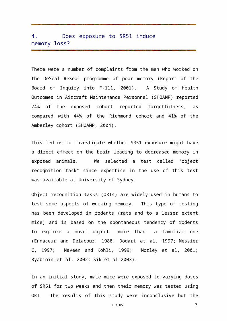
Object recognition 24 hours after last SR51 dose
There was no significant difference in the recognition index between all groups 24 hours
after the final dose of SR51 (p> 0.05) [Figure 4.4]. A within-group analysis showed the
negative control mice explored the novel object more than the familiar object. The
scopolamine-treated mice did not explore the novel object significantly more than the
familiar object. The LD, MD and HD-treated mice explored the novel object less than the
familiar object.
Object Recognition test 24h post final dose of SR51
50
0
Figure 4.4. Object recognition test 2 hours after final SR51 dose. Each testing group consisted of 12 male mice.

Re
cogn
itio
n I
nd
ex
11CHALUS
Object recognition 48 hours after last SR51 dose
There was no significant difference in the recognition index between all groups 48 hours
after the final dose of SR51 (p> 0.05) [Figure 4.5]. A within-group analysis revealed that
none of the groups explored the novel object more than the familiar object.
Object recognition test 48h post final dose of SR51
50
0
Figure 4.5. Object recognition test 48 hours after final SR51 dose. Each testing group consisted of 12 male mice.

12CHALUS
Necropsy Data.
Organ weights (absolute weights and organ- to- body weight ratios)
Livers, kidneys and spleens were collected from treated mice 3 days after the final dose of
SR51. Table 1 summarises all the organ weight data. There were no significant changes
in the absolute or relative kidney or liver weights between the SR51-treated and control
groups (Table 4.1 and Figure 4.6b and 4.6c).
Spleens collected from the HD SR51-treated mice showed a significant increase
(P<0.001) in both absolute weight and spleen to body weight ratios compared to control
and LD and MD treatment groups (Table 1 and Figure 4.6a).
Mouse Treatment Group (n=12)
ORGAN Control(0 mg/kg) Group 1
LD SR51(90 mg/kg)
Group 2
MD SR51(180 mg/kg)
Group 3
HD SR51(360 mg/kg)
Group 4
Absolute weightsBody (g) 29.39 ± 1.65 28.91 ± 1.41 28.80 ± 1.87 28.15 ± 2.84
Liver (g) 1.488 ± 0.161 1.494 ± 0.118 1.450 ± 0.137 1.499 ± 0.210
Kidney (g) 0.226 ± 0.020 0.217 ± 0.015 0.226 ± 0.030 0.214 ± 0.039
Spleen 0.072 ± 0.007 0.076 ± 0.009 0.075 ± 0.009 0.097 ± 0.028***
Organ-to-body weight ratiosLiver 5.057 ± 0.393 5.164 ± 0.254 5.042 ± 0.432 5.310 ± 0.394
Kidney 0.771 ± 0.059 0.752 ± 0.033 0.782 ± 0.076 0.760 ± 0.064
Spleen 0.246 ± 0.023 0.262 ± 0.029 0.259 ± 0.031 0.348 ± 0.113***
*** significantly different at p<0.001
Table 4.1. Summary of absolute organ weights and organ-to-body weight ratios of kidneys, spleens and livers collected from mice dosed for the Object Recognition Test. Organs were collected 3 days after the final dose of SR51. Mice were orally dosed via gavage for 5 days pw for 2w.

liver
to b
ody
wt r
atio
kidn
ey t
o bo
dy w
t rat
iosp
leen
to
body
wt r
atio
13CHALUS
a)
Effect of SR-51 oral dosing of mice on spleen weights
0.40
0.35
0.30
0.25
0.20
0.15
0.10
0.05
0.00
control 90mg/kg 180mg/kg 360mg/kg
SR-51 (oral dose 5 days pw for 2w)
b)
Effect of SR-51 treatment in mice on kidney weights
1.81.61.41.21.00.80.60.40.20.0
control 90mg/kg 180mg/kg 360mg/kg
SR-51 (oral dose 5 days pw for 2w)
c)
Effect of SR-51 treatment in mice on liver weights
6
5
4
3
2
1
0control 90mg/kg 180mg/kg 360mg/kg
SR-51 (oral dose 5 days pw for 2w)
Figure 4.6. Effect of SR51 treatment on organ-to-body weight ratios in mice. Organ weights were collected for a) spleen b) kidney and c) liver (n=12). *** indicates a significant difference to the control group at P<0.001.

14CHALUSs
Histology of Mouse Spleen
Typically the spleen consists of a dense connective tissue capsule protecting the inner
pulp. The white pulp (mainly lymphocytes) is organized into nodules while the re d pulp
(blood-filled sinusoids and macrophages, lymphocytes and plasma cells) arranged into
cords. The red pulp is concerned with the phagocytosis and destruction of old red blood
cells. In mice the red pulp normally contains nests of extramedullary hemopoiesis. The
mice exposed to HD SR51 exhibited enlarged spleens typified by the presence of
extensive areas of extramedullary hemopoiesis.
Splenic nodule
White pulp
Red pulplymphocyte
a) Spleen taken from control mouse (untreated). b) Area within square enlarged showing the perifollicular zone with scattered lymphocytes.
Splenic nodule
c) Spleen taken from mouse treated with HD SR51 d) Area within square enlarged.
Figure 4.7. Effect of oral dosing of mice with HD SR51 (360mg/kg 5d pw for 2w) on the spleen histology. Paraffin-embedded spleen sections -6µ m thick and stained with H&E stain.Frames a) and c) are at x22.5 magnification. Frames b) and d) are at x100 magnification.

15CHALUS
Histology of Mouse Brain
The hippocampus is involved in spatial learning memory (Morris et al., 2003;
Eichenbaum and Fortin, 2003). The hippocampus has three cell layers – mol ec ul a r l a y e r
consisting of interacting axons and dendrites; p y ra mid a l ce ll l a y e r comprising large
pyramidal cells with dendrites in the molecular layer and axons making connections
outside the hippocampus and a pol y mo r phic l a y e r containing axons, dendrites and inter-
neurons.
There was no evidence that in vivo exposure to HD SR51 (daily oral dosing with
360mg/kg 5 days pw for 2 w) in mice caused any gross morphological changes in brain
tissue collected 3 days after the final dose.
Polymorphic cellular layer
Pyramidal cell layer
Molecular layer
a) H&E stain of brain section collected from untreated control mouse (paraffin-embedded 6µ m sections at x10 magnification)
b) H&E stain of brain section collected from mouse treated with HD SR51 (paraffin-embedded6µ m sections at x10 magnification)
Figure 4.8. Histology of brain collected from mouse orally exposed to HD SR51 (daily oral dosing with360mg/kg 5 days pw for 2 w). Paraffin-embedded 6µ m sections stained with H&E stain. a) shows brain tissue from and untreated control mouse (x10 magnification).b) shows brain tissue from and HD SR51 treated mouse (x10 magnification).
e) Untreated control (x10 mag.) f) Brain collected from mouse treated withHD SR51 (x10 mag.)
Figure 4.9.
16CHALUS
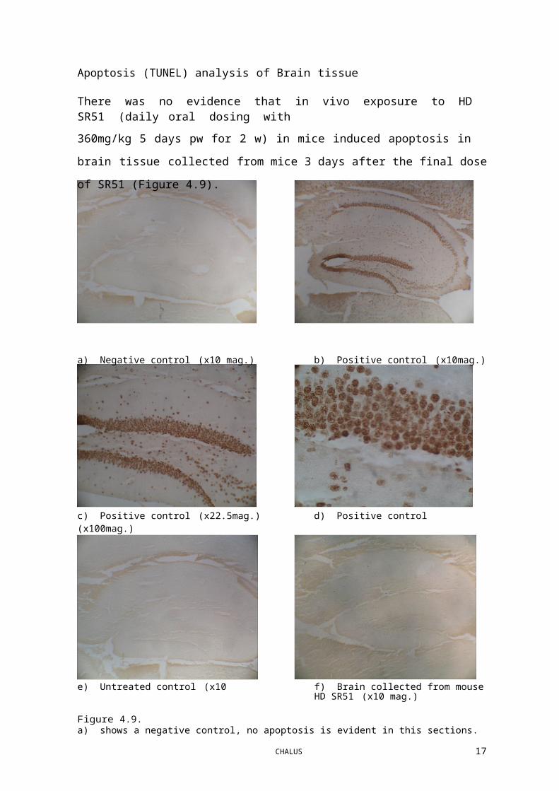
Apoptosis (TUNEL) analysis of Brain tissue
There was no evidence that in vivo exposure to HD SR51 (daily oral dosing with
360mg/kg 5 days pw for 2 w) in mice induced apoptosis in brain tissue collected from
mice 3 days after the final dose of SR51 (Figure 4.9).
a) Negative control (x10 mag.) b) Positive control (x10mag.)
c) Positive control (x22.5mag.) d) Positive control (x100mag.)
a) shows a negative control, no apoptosis is evident in this sections.b)-d) shows a positive control. Sections were artificially treated with enzymes to force a positive result. Apoptotic cell nuclei are stained a brown colour.e) shows an untreated control mouse hippocampus. The stain for apoptosis is no different than the negative control ie. there is no cell death.f) shows the hippocampus of mouse treated with the highest dose of SR51. The stain for apoptosis is no different than the negative control ie. there is no cell death.

17CHALUS
Discussion
The present study was undertaken to determine if SR51 interfered with the acquisition of
new memories in mice. The apparently negative results have been seriously
compromised by unexpected findings in the negative controls and in the positive controls.
For the negative controls, the mice did not consistently spend any more time examining a
novel object than they did a familiar object. It is a basic tenet of the test that they will
spend more time examining a novel object. To indicate a memory deficit in positive
control, the mice should spend equal times with novel and familiar object since they have
lost the ability to discriminate. However, for the positive controls (scopolamine treated
mice), the mice did not consistently spend equal time with novel and familiar object.
Overall, these results suggest serious methodological flaws.
A possible explanation could have been that exploration times were too short (ie less than
10 sec) (Sik et al., 2003) but this was not the situation since the average exploration time
was in excess of 10 seconds for all groups. Another possible explanation is that the ether
that was used to lightly anaesthetise the mice before each SR51 dose may have interfered
with memory.
The use of scopolamine as a positive control is based on numerous studies (Giovannini et
al., 1999; Naveen and Kohli, 2003; Dodart et al, 2003) indicating that this drug causes
temporary memory loss in rodents. The absence of this effect in the present study is a
second fundamental flaw and throws into doubt all of the results obtained with SR51.
There is no recommendation to repeat these experiments. Great care and preparation
went into the conduct of the experiments and it is our conclusion that the experimental
paradigm underlying the object recognition test is either flawed or too susceptible to
small variations in the experimental protocol.
The experiments have neither proved nor disproved that SR51 can interfere with memory.
The absence of morphological changes in the brains of treated mice is also inconclusive.
The basic morphological structure of the hippocampus is established during embryonic
and early postnatal life. Loss of cells in the hippocampus is associated with Alzheimer's

18CHALUS
disease and it is possible that a toxic mixture such as SR51 could lead to hippocampal cell
loss but it was not detected in this study.
The changes seen in the spleen may reflect a haemolytic effect of SR51 specifically the
thiophenol component. Thiophenol, is readily autooxidised at neutral pH in a reaction
which generates superoxide radical and hydrogen peroxide. The oxidation product,
diphenyl disulphide, may be reduced back to thiophenol by glutathione and a
reduction/autoxidation cycle for generation of 'active oxygen' species is established. The
autoxidation reaction is strongly catalysed by haematin; haemoglobin is also an effective
mediator of 'active oxygen' generation from the diphenyl disulphide/glutathione couple,
being oxidized to methaemoglobin in the process. In vivo haemolysis may be anticipated
from any disulphide or thiol which undergoes appreciable autoxidation at neutral pH
(Munday and Manns, 1985).
Thiophenol has been reported to cause red blood cell lysis in vitro and in vivo (Munday
and Manns 1985; Munday et al, 1990; Amrolia et al, 1989; Fairchild and Stockinger,
1958). This type of damage frequently results in formation of haemosiderin in spleen
(Haschek and Rousseaux, 1998). Perl's prussion blue stain can be used to demonstrate
haemosiderin pigment deposition. In the present study, haemosiderin analysis was not
performed. Enlarged spleens in the high dose groups suggest increased blood lysis may
have occurred in these animals. We have not undertaken blood tests of the SR51-treated
mice to see if they are anemic.

19CHALUS
5. How chemically stable is SR51?The effect of temperature changes on the toxicity profile of SR51.
Background:
Tanks containing SR51 were left on the runway at 45-60oC during the DSRS programs
performed on F111 aircraft fuel tanks. Personnel were exposed to the vapour coming
from the heated SR51. Preliminary experiments by CHALUS found that when SR51 was
raised to these temperatures, one of the components, thiophenol converted to diphenyl
disulfide, a reaction which occurs in the presence of oxygen. This reaction could
effectively change the toxicity profile of the SR51.
Aim:
To use gas chromatography to firstly determine the components of SR51 and the
components of the vapour phase above the liquid. Secondly, to monitor changes in both
the liquid SR51 and in the vapour phase above the liquid after heating at temperatures
from 20-60oC. The latter study was undertaken in order to ascertain whether the chemical
composition of SR51 and the vapour produced above the liquid changes at increasing
temperatures.

20CHALUSs
Methodology:
Two series of experiments were undertaken:
1. Analysis of SR51 liquid
Vials containing 200µ l SR51 were heated at for 30 min at : 20oC, 30oC, 40oC, 50oC, or 60oC. A0.04µ l aliquot of each heated liquid was injected in to the GC. Experiments were performed in duplicate.
2. Analysis of the SR51 vapour above the heated liquid
Three ml of SR51 were placed in a series of headspace analysis vials. The vials were heated at20oC, 40oC 50oC, or 60oC. for 30 min. During the heating the StableFlex fibre was inserted through the vial septum into the SR51 vapour and left in place for the 30 min for absorption ofvapour components onto the fibre to occur.
The fibre was placed in the GC injection port for 1.5 min for desorption to occur.
Experiments were performed in duplicate.
A separation method, using gas chromatography, was developed to separate and identify the major components of SR51.
Gas Chromatography (GC) parameters used for analysis
GC: GC was carried out on a Hewlett Packard 5890 Series II gas chromatograph fitted with a split/splitless capillary injector (split ratio approximately 50:1) and flame ionisation detector (FID) with helium as carrier gas. Column head pressure 110 kpa.
Column: Phenomenex ZB-5 length 30 m x 0.25 mm internal diameter, film 0.25 µ m (non-polar column).
Detector temperature: 270oC.
Injector temperature: 270oC for analysis of liquid SR51, 250oC for analysis of headspace vapour.
Oven temperature program:
1. Analysis of liquid SR51: 40oC for 1 min, to 320oC at 20oC per min, remain at 320oC for 5 min.
2. Headspace analysis of SR51 vapour: 40oC for 3 min, to 320oC at 20oC per min, remain at320oC for 5 min.
Fibre used for analysis of vapour in headspace above SR51
Supelco solid phase microextraction holder equipped with a StableFlex fibre coated with polydimethylsiloxane/divinylbenzene, film thickness 65µ m.

21CHALUS
Results:
Analysis of SR51
The chromatogram of the components of liquid SR51 at room temperature, 20oC (Figure
5.1) shows that SR51 is composed of a large number of individual components (in excess
of 40 chemicals). Of particular interest is the thiophenol (TP) peak (peak area % =
6.96689), thiophenol being one of the most volatile components of SR51. The presence
of a small diphenyl disulphide (DPDS) peak (peak area % = 1.02029) demonstrates that
even at room temperature there is some oxidation of thiophenol to diphenyl disulphide.
Figure 5.1: Chromatogram of components of SR51. The following retention times (min) for each of the major components were recorded: 4.560 - DMA; 5.602 – TP; 7.108 – TEP; 12.368 – DPDS; the remaining peaks represent the components of ARO 150.
Analysis of heated SR51
Comparison of the chromatogram of SR51 obtained at room temperature (Figure 5.1)
with that obtained after heating for 30 min at 60oC indicates there was little change in the
conversion of TP (peak area % = 7.93330) to DPDS in the liquid SR51 (peak area % =
0.62091).

22CHALUS
Figure 5.2. Chromatogram of components of liquid SR51 heated to 60oC for 30 min. The following RTs were recorded: 5.604 – TP; 12.350 - DPDS.
Analysis of headspace vapour above heated SR51
The chromatogram obtained following absorption of the vapour contents above SR51
onto the fibre for 30 min at room temperature is shown in Figure 5.3. It should be noted
that the retention time for each component is approximately 2 minutes longer than those
recorded in Figure 5.1, since the initial time spent at 40oC in the temperature program was
increased from 1 min to 3 min to allow time for desorption from the fibre to occur in the
injection port of the GC before the temperature increments commenced. TP is found in
considerable amounts in the headspace (peak area % = 7.37873). The oxidation product
of TP, DPDS is also found in the headspace (peak area % = 3.22851)
Figure 5.3. Chromatogram of components of vapour phase above liquid SR51 at room temperature (20oC).
The following RTs were recorded: 7.280 – TP; 14.264 - DPDS.

23CHALUS
The chromatogram obtained following absorption of the vapour contents above SR51
onto the fibre for 30 min at a temperature of 60oC is shown in Figure 5.4. TP is found in a
slightly higher concentration in the headspace (peak area % = 8.97424) than at room
temperature. However, the oxidation product of TP, DPDS is found in the headspace in a
considerably higher concentration (peak area % = 12.51849).
Figure 5.4. Chromatogram of components of vapour phase above liquid SR51 heated for 30 min at a temperature of 60oC. The following RTs were recorded: 7.268 – TP; 14.283 - DPDS.
A final experiment was performed in which a sample of SR51 was heated at 60oC for 30
min. A 100 µ l aliquot of the headspace vapour was drawn into a warmed gas tight
syringe. This aliquot was injected into the GC (Figure 5.5). The TP peak is large (peak
area % 10.46052), as TP is volatile. An appreciable amount of the DPDS is also found
(peak area % = 2.33942). These concentrations provide a good estimate of the levels in
the vapour the personnel were inhaling at elevated ambient temperatures. However, this
technique may underestimate the levels of the oxidation product since it could be
deposited on the walls of the vial as it forms. The analysis using the fibre overcomes this
possible inaccuracy.

24CHALUS
Figure 5.5. Chromatogram of components of vapour phase above liquid SR51 60oC, assayed by direct injection of the headspace vapour. The following RTs were recorded: 7.241 – TP; 14.237 – DPDS.
25CHALUS
Conclusions
As SR51 is heated, an increasing amount of thiophenol is oxidised to the diphenyl
disulphide as the temperature increases. The higher the temperature on the tarmac, the
greater the concentration of thiophenol in the inhaled vapour and the greater the amount
of the oxidation product diphenyl disulphide that would be present.
The implications for personnel exposed to the SR51 vapour is that the inhaled vapour
would contain significant amounts of both thiophenol and the oxidation product diphenyl
disulphide. As the ambient temperature increased, increasing concentrations of diphenyl
disulphide that would be present in the inhaled vapour. There is relatively little
toxicological data available for diphenyl disulphide compared to thiophenol.
In general diphenyl disulphide is thought to be less toxic than thiophenol but the
combination of the chemicals in the blood is associated with a reduction/autoxidation
cycle for generation of 'active oxygen' species and subsequent damage to red blood cells.

26CHALUS
6. Does SR51 damage DNA in cells?The development of an in v i t ro genotoxicity assay (the Comet assay)
using mouse lymphoma cells.
As mentioned in the introduction the men who worked with SR51 have expressed
concern that it might cause cancer. Much of the concern with SR51 probably arises from
the fact that the thiophenol component has such an unpleasant odour which is detectable
at incredibly low concentrations. Hence working with the chemical is always unpleasant
since you can almost always smell it whatever precautions are taken.
We undertook a wide range of studies to examine whether SR51 could damage DNA as a
conventional mutagen. Previous reports have described experiments testing SR51 in the
Ames test, mouse lymphoma assay and the mouse micronucleus test. All of these tests
were negative for mutagenesis.
This report describes an additional test - the COMET assay.
The assay is based on the principle that the negatively charged DNA within a cell will
migrate in a gel (agarose) matrix when in an electric field. When cells are exposed to a
test agent/chemical, any damaged DNA (DNA with strand breaks) will migrate faster in
the gel resulting in a characteristic comet shape. The larger the comet, the more damage
to the cellular DNA.
Aim:
The Comet assay was used as an in vitro test to test the genotoxic potential of SR51. The
test was optimised using cultured mammalian cells (mouse lymphoma cells) in the
presence and absence of endogenous metabolic enzymes. The contribution of apoptosis
(programmed cell death) in any chemically induced cell damage was also investigated.

27CHALUS
Methodology:DNA damage was evaluated using a single cell gel electrophoresis (Comet assay). Mouse lymphoma cells were exposed in vitro for 3 h to SR51 (solubilised in DMSO at 1% (v/v)) at concentrations ranging from 5.5 to 22.3µ g/ml in the presence and absence of S9 metabolic activation enzymes.
Cell cultureMouse lymphoma cells (were obtained from the American Tissue Culture Collection (Cryosite, Australia) and maintained in RPMI-1640 media (Sigma, Australia) containing 10% heat-inactivated horse serum (Sigma, Australia) at 37oC, 5% CO2 humidified incubator. Cytotoxicity was assessed using the Trypan blue live cell exclusion dye.
Treated cells
SR51 treatmentFor the comet assay, cells were seeded in 25 cm2 flasks and exposed to SR51 in RPMI-1640 containing10% horse serum for 3 h. SR51 was diluted in DMSO such that the final DMSO concentration was1%(v/v), a concentration previously determined to be non-toxic to the cells. The highest concentration tested resulted caused <15% cytotoxicity. The experiment was performed using three concentrations ofSR51 (5.625, 11.25 and 22.5 µg/ml). Sealed tissue culture flasks were used for the incubation with the chemical due to the volatility of SR51. The cultures were gassed with CO2 prior to incubation in order to saturate the cultures with CO2. The chemicals were added last to minimize loss of SR51 due to vaporization. The cultures were incubated for 3 hours at 37° on a rocking incubator.
ControlsNegative control cells were exposed to 1% (v/v) DMSO only. Positive control cells were exposed to100µ M (30min 4°C,) hydrogen peroxide, a known in vitro inducer of DNA damage. For cells incubated in the presence of metabolic activation liver enzymes (S9 enzymes), cells exposed to 250µ M cyclophosphamide (3h, 37°C) were used as the positive control.
Single cell gel electrophoresis assay (Comet Assay)Comet assays were performed using alkaline unwinding of DNA electrophoresis following an adapted protocol of Donnelly et al., (2000). A mixture of the exposed cell suspension (1 x 10⁵ cells / ml) was mixed with low melting agarose and applied directly to a fully-frosted slide pre-coated with a layer normal melting agarose. The slides were then placed at 4°C for 10 min to solidify, followed by careful immersion in freshly prepared ice-cold lysing solution (2.5 M NaCl, 100 mM EDTA, 10mM Tris base, 1% Triton-X, pH 7.4) for60 min at 4°C in the dark. Following lysis, the slides were placed in an alkali buffer (300mM NaOH, 1 mM EDTA pH > 13) for 20 min to unwind the DNA. Electrophoresis was conducted at room temperature in the same buffer for 10 min at 25 V (300 mA). Slides were then neutralized and washed twice in Neutralisation buffer (Tris 100 mM, Borate 90 mM, EDTA 1.0 mM pH 7.5) buffer. Slides were immediately stained with Ethidium Bromide (20 µ M) and stored at RT in the dark until photographed the next day. The DNA damage was visualized with a Zeiss Axioplan 2 upright fluorescence microscope equipped with an excitation filter (515-560 nm) and barrier filter (590 nm) at 100x magnification,. a Zeiss AxioCam HR digital monochrome CCD camera using Zeiss AxioVision 4.0 image acquisition software to store comet images. All images were obtained using the same exposure time (763 ms). The comet images were subsequently analysed using the CASP online software and Komet 5.1® software to obtain a range of measurements including percent DNA in tail, olive tail moment and tail length. All calculations were based on the intensities after correction for background illumination. For each concentration, 100 non-overlapping comets per two slides were randomly captured at a constant depth of the gel, avoiding edges and damaged gel regions. The parameters of olive tail moment (tail length integrated over intensity of the tail) and percent DNA (percent migrated DNA) in the tail were used as indicators of the severity of DNA damage. Each experiment was performed in duplicate.
SR51 exposed cells were incubated both in the absence and presence of liver enzymes (S9 metabolic activation enzymes). Experiments performed with cells exposed to SR51 in the presence of S9 metabolic activation enzymes were not successful due to inactivity of the S9 enzymes.
Apoptosis AssayThe induction of apoptosis was determined in SR51 exposed cells (3h) using the Annexin V apoptosis kit purchased from Sigma, Australia. The Annexin V kit detects a phospholipid protein that typically localizes on the outer surface of the cell membrane in apoptotic cells. Cells were processed according to the manufacturers instructions.

28CHALUS
Statistical analysisData was analysed using CASP on-line program and Komet 5.1® analyses software. The Kruskall-Wallis one-way analysis of variance by ranks was used as a non-parametric test to determine whether the distributions of the various tail parameters differed in exposure groups within a SR51 treatment. Statistically significant results were subjected to the Dunn's post-test to compare the differences in the groups with the expected average difference.

No.
of c
ells
Incr
easi
ng D
NA
dam
age
====
> (%
DN
A in
tail)
29CHALUS
Results
Hydrogen peroxide exposed cells
Hydrogen peroxide caused significant DNA damage (increased % tail DNA, olive tail
moment) in mouse lymphoma cells after 30min exposure (at 4°C). There was a dose
response (see Figure 6.1).
80
70
60
50
40
30
20
10
0
control(saline)
50uM H202 100uM H202 200uM H202
Figure 6.1. Dose Response Curve showing the effect of in vitro exposure to Hydrogen Peroxide in Mouse
Lymphoma cells using the COMET assay. The COMET assay detects DNA damage in single cells. A total
of 100 cells were examined per concentration.
80
70
60
50
40
30
20
10
0 200 uM H202
< 1.65
26.2375
50.825
75.4125
100 uM H202
50 uM H202
control (saline)
Increasing damage ====> (%DNA in tail)
100
Figure 6.2. Distribution of cell DNA damage within each treatment group. In vitro exposure to SR51 of
mouse lymphoma cells and detection of DNA damage using the COMET assay

30CHALUS
No.
of c
ells
Incr
easi
ng D
NA
dam
age
===>
(%
DN
A in
tail)
SR51 exposed cells
DNA damage
SR51 did not cause significant DNA damage (increased % tail DNA, olive tail moment)
in mouse lymphoma cells after 3h exposure (at 37oC). The concentration of SR51 was
tested up to the concentration where >85% cells remained viable. There was no dose
response (see Figure 6.3).
70
60
50
40
30
20
10
0
control (1%
DMSO)
5.625 uM SR51
11.25 uM SR51
22.5 uM SR51
200uM H202
Figure 6.3. Dose Response Curve showing the effect of in vitro exposure to SR51 in Mouse Lymphoma
cells using the COMET assay. The COMET assay detects DNA damage in single cells. Approximately
100 cells were examined per concentration.
Figure 6.4 shows the distribution of damage of cells within each treatment group.
120
100
80
60
40
20
0
< 0.31
25.2325
50.155
75.0775
200uM 'H202HD SR51
MD SR51
LD SR51
controlIncreasing damage ===>
(% DNA in tail)100
Figure 6.4. Distribution of cell DNA damage within each treatment group. In vitro exposure to SR51 of
mouse lymphoma cells and detection of DNA damage using the COMET assay. Approximately 100
cells were examined per concentration.

31CHALUS
Cytotoxicity and Induction of Apoptosis
SR51, in the concentrations used in this study, did not result in cytotoxicity (<15% cell
death) or a significant induction of apoptosis (see Figure 6.5).
a)
b)
Figure 6.5. Induction of Apoptosis in Mouse lymphoma cells. Mouse lymphoma cells were incubated for3h (at 37oC) in:a) 10% DMSO - remained viable (cells fluoresced green) but were apoptotic (cells fluoresce red)b) 22ug SR51 /ml – cells remained viable (cells fluoresce green) but not apoptotic (cells did not fluoresce
red).

32CHALUS
Discussion
Comet Assay
There are many mechanisms to induce cancer including induction of DNA damage and
impairment of DNA repair processes. This study was designed to determine whether
SR51 induced DNA damage. The comet assay quantifies the extent of breaks in the DNA
of single cells. In the present study using the COMET assay the positive control
(hydrogen peroxide) gave concentration-dependent positive results confirming the
validity of the test. We found that SR51 did not induce DNA damage in the mouse
lymphoma cells even at doses that were marginally cytotoxic. The results suggest that if
SR51 was carcinogenic then its mechanism of action does not involve overt DNA
damage.
These results are further confirmation that SR51 is not a mutagen It has now been tested
in the AMES test, mouse lymphoma assay, mouse micronucleus test and in the COMET
assay and in each case it was negative. In the AMES test and the mouse lymphoma
assay it has also been shown to be negative with metabolic activation.
Apoptosis
It is important when assaying cells for genotoxicity that the test cells are exposed to the
highest concentration of the cells but not to the point of causing extensive cell death. The
highest concentration of SR51 used in the Comet assay was determined to be marginally
cytotoxic as determined by the trypan blue viability stain (<15% dead cells). The cell
death at this concentration appeared to be necrotic rather than apoptotic. Apoptosis is the
term for programmed cell death characterized by specific morphologic and biochemical
properties.

33CHALUSs
7. Drosophila as a test model for investigating the effect of chemicals on mitochondrial function and aging.
Establishing a Drosophila Testing Laboratory.
Background
A hypothesis has been developed that SR51 or other chemicals used in the F-111 DSRS
maintenance programs may increase oxidative stress on mitochondria and may hasten the
ageing process. The detection of mitochondrial disorders in several of the DSRS
personnel and the apparent increase in disease progression was the origin of this
hypothesis. Furthermore, our own studies showing adverse effects of SR51 on
mitochondrial function support this hypothesis (Oakes et al, 2004). A possible way of
investigating this hypothesis is to examine the life expectancy in animals exposed long-
term to the test chemicals. Since cost would be prohibitive with mice and rats, the
possibility of using Drosophila has been investigated. The structure of mitochondria is
highly conserved across species and the use of Drosophila offers a viable and convenient
alternative to examine mitochondrial function and ageing.
Aim: To determine the feasibility of using Drosophila to test a range of chemicals that
may be of concern to the military.
Establishing a Drosophila Testing Laboratory
Bill Webster and Diana Oakes visited two major Drosophila Laboratories at the
University of Melbourne in the Department of Genetics (Jill Williamson) and Department
of Anatomy and Cell Biology (Dr Gary Hime) in August, 2005. Both contacts have
agreed to supply our Laboratory with a Drosophila strain (Drosophila melanogaster) and
provided practical advice that will enable us to establish a breeding colony at the
University of Sydney.
The required equipment and consumables have been are currently being ordered to enable
the set-up of a basic Drosophila Breeding and Testing Facility.
The following pictures were taken at the University of Melbourne and show the basic set
up for breeding and handling Drosophila.

34CHALUS
Figure 7.1. Drosophila colonies bred in small plastic container within and incubator maintained at 18oC
(stock maintenance) or 25oC (running experiments).
Figure 7.2. Drosophila can be ‘immobilised’ on a specialised viewing platform that enables a continuous
flow of carbon dioxide across the platform surface.
Figure 7.3. Whilst immobilised, Drosophila characteristics can be identified clearly under a microscope
eg sorting of male and female flies.

35CHALUS
Figure 7.4. There are a range of ‘population vessels’ that can be utilised for running toxicity testing
experiments on the effect of chemicals on the lifespan of Drosophila (see below). Exposed Flies can also
be examined for effects on function such as effects on mitochondrial function.
Life Span Determination
Survival as an end-point of toxicity
Percent survival is a convenient measure in the fly model since the mean life-span of a
long-lived strain of D. melanogaster is approximately 70 days. This compares to 3
5years if using rodent models.
Newly emerged flies are collected and raised in standard corn meal agar medium which
has been inoculated with the test substance. Control bottles are included. For each
experiment 10 vials, each containing 20 flies, are maintained at 29oC or 25oC and
transferred to fresh vials every 3 days. The number of dead flies are counted everyday.

36CHALUS
Literature Review highlighting the Potential Use of Drosophila for studying the effects
of chemical exposure on mitochondrial function and aging.
The focus of the review is to highlight the effect of chemical exposure on lifespan,
induction of reactive oxygen species and potential adverse effects on mitochondrial
function.
The fruit fly Drosophila melanogaster has been studied for use as a cheap and rapid assay
to assess toxicity. Mutagenic studies have been its main use with assays to detect the
genotoxic effects of reactive oxygen inducing compounds (Gaivao and Comendador,
1996; Gaivao et al, 1999). Drosophila has also been used in ageing studies. It has a mean
life span of 40-60 days, hence studies are rapid and manageable. Drosophila may be
useful to study the effect of chemicals on mitochondrial function with Kaplan-Meier
survival curves the endpoint for determining toxicity.
Mitochondrial function in Drosophila
Mitochondria are the predominant intracellular generators of reactive oxygen species
(ROS), specifically superoxide anion radical (O2-) and H2O2. As in humans, the cellular
oxidative defence system in flies consists predominantly of the enzymes superoxide
dismutase (SOD) and catalase. Superoxide, the initial ROS derived from the electron
transport chain (ETC) is converted to H2O2 by SOD, and catalase reduces H2O2 to water
and molecular oxygen. If H2O2 cannot be eliminated, hydroxyl free radicals, thought to
be the main species inflicting oxidative damage, are formed.
Interference with the ROS defense system can affect lifespan in Drosphila. Drosophila
with low catalase expression have a mean life span similar to controls, however, when
exposed to H2O2, mortality in the mutant line is greater. Catalase null mutations have a
greatly reduced life span and hypersensitivity to H2O2. A cSOD null mutation also has a
shorter life span and hypersensitivity to the superoxide generator paraquat.
Mitochondria are the prime targets of oxidative damage due to close proximity to the
ETC and the absence of protective histone proteins and DNA repair enzymes in
mitochondria.

37CHALUS
The D. melanogaster mitochondrion contains a 19517 bp genome encoding 22 tRNAs, 2
rRNAs and 13 proteins of the ETC and oxidative phosphorylation. The ETC consists of 4
enzyme complexes assembled with complexes I, II and IV being derived from both
mitochondrial and nuclear genes, while complex II is entirely nuclear encoded. Mt DNA
is a target of ROS, mutations have been shown to occur at a 5-10 fold higher rate that in
nuclear DNA. One of the consequences of cells bearing a large mtDNA deletion load
include accumulation of morphologically abnormal mitochondria and loss of cytochrome
oxidase activity. Thoraces of D. melanogaster, consisting primarily of flight muscle, may
be used as a source of mitochondria to measure changes in respiration rates and activities
of oxidoreductases within the ETC as a function of age.
Reported ageing effects in Drosophila mitochondria
Both state 3 mitochondrial respiration and cytochrome c oxidase (complex IV) activity
decreases with increasing age (the latter is associated with increased H2O2 production).
Both of these effects may result from and contribute to an age-related increase in
oxidative stress (Ferguson et al 2005). Thus mitochondrial respiratory capacity decreases
with age. Cytochrome c oxidase activity (COX) declined progressively from 2 days post
eclosion (Schwarze et al 1998). The abundance of 4 mitochondrial encoded ETC
transcripts declined 5-10 fold with advancing age (Schwarze et al 1998).
D. melanogaster displays an age-related increase in oxidative damage and a decrease in
mitochondrial transcripts. From days 2 – 45 post-eclosion declines were found in
complex IV cytochrome c oxidase activity. Oxidative stress of chemical or genetic origin
leads to reduction in levels of the mitochondrial transcript coxI, which is associated with
declines in COX activity and ATP levels. Age-related reductions in COX activity may
lead to impaired generation of ATP (Schwarze et al 1998). Since deficiencies in ATP
production may not be apparent in resting flies, the environmental temperature was raised
(36oC) as a means of elevating metabolic activity. ATP levels decreased with age; heat
stress caused a greater decline with age.
As flies age, the accrued oxidative damage may result in a loss of COX activity. As an
indicator of oxidative damage, levels of oxidised lipid by-products may be measured
using the TBARS assay. There is an increase in oxidised lipid by-products with ageing
(Schwarze et al 1998).

38CHALUS
Levels of several mitochondrial transcripts decrease with age. Flies with genetic
impairments in either catalase or cSOD antioxidant defence systems were used to study
the effects of oxidative stress on mtRNA levels. Anti-sense RNA probes complementary
to cytochrome oxidase I (coxI, ETS complex IV) and ribosomal protein 49 (rp49) were
used to detect mitochondrial and nuclear-encoded transcripts, respectively. Levels of
coxI mtRNA declined by 68% from 2 to 30 days of age. Levels decreased to a greater
extent in the catalase deficient mutant compared to flies that expressed catalase. A
similar result was obtained in the cSOD null flies (Schwarze et al 1998).
Toxicology studies using Drosophila
The genetically modified Drosophila Strain ORR-flr3/TM3 has a high cytochrome P-450
dependent metabolism of xenobiotics. It has been used to screen for the toxic effects of
volatile organic compounds. Since respiratory activity is indicative of overall metabolic
conditions of an organism, it was used as an indicator of intoxication from various
substances, including volatile solvents with CO2 production being the end-product
measured (Wasserkort & Koller 1997).
In another test, newly enclosed (<24 h) adults were transferred to vials containing various
percentages of H2O2 in fly medium. After 3 days, flies were transferred to fresh H2O2
containing medium. Percent survival was calculated at the end of the 6-day experiment.

39CHALUS
8. References
Amrolia, P., Sullivan, S. G., Stern, A., Munday, R. (1989). "Toxicity of aromatic thiols in the human red blood cell." Journal of Applied Toxicology 9(2): 113-8.
Dodart J.C., Mathis C. and Ungerer A. Scopolamine-induced deficits in a two-trial object recognition task in mice. Neuroreport, 1997, 8, 1173-1178.
Donnelly E.T., O'Connell M., McClure N., Lewis S.E.M. (2000). Differences in nuclear DNA fragmentation and mitochondrial integrity of semen and prepared human spermatozoa. Human Reproduction, 15(7):1552-1561
Eichenbaum H, Fortin N (2003) Episodic memory and the hippocampus: it's about time. Curr Dir Psychol Sci 12: 53-57.
Ennaceur A., Delacour J. A new one-trial test for neurobiological studies of memory in rats. 1: behavioral data. Behav. Brain Res., 1988, 31,47-59
Fairchild, E. J. and Stockinger, H. E. (1958). “Toxicologic studies on organic sulfur compounds. 1. Acute toxicity.” American Industrial Hygiene Association Journal 19 :171-189.
Ferguson M., Mockett R.J., Shen Y., Orr W.C., Sohal R.S. (2005). Age-associated decline in mitochondrial respiration and electron transport in Drosophila melanogaster. Biochemical Journal (in press).
Gaivao I, Comendador MA. 1996. The w/w+ somatic mutation and recombination test (SMART) of Drosophila melanogaster for detecting reactive oxygen species: characterization of 6 strains. Mutation Research 360(2):145-151.
Gaivao I, Sierra LM, Comendador MA. 1999. The w/w+ SMART assay of Drosophila melanogaster detects the genotoxic effects of reactive oxygen species inducing compounds. Mutation Research 440(2):139-145.
Giovannini MG, Bartolini L, Bacciottini L, Greco L, Blandina P. (1999). Effect of histamin H3 receptor agonists and antagonists on cognitive perormance and scopolamine- induced amnesia. Behavioural Brain Research. 104:147-155
Haschek, W. M. and Rousseaux, C. G.(1998). Fundamentals of tocicologic pathology. Academic Press, UK. pp.57-232.
Messier C. Object Recognition in Mice: Improvement of Memory by Glucose. Neurobiol. Learn. Memory, 1997, 67, 172-175.
Morley K.C., Gallate J.E., Hunt G.E., Mallet P.E. and McGregor I. Increased anxiety and impaired memory in rats 3 months after administration of 3,4methylenedioxymethamphetamine (“Ecstasy”). Europ. J. Pharm., 2001, 433, 91-99.

40CHALUS
Morris RGM, Moser EI, Riedel G, Martin SJ, Sandin J, Day M, O'Carroll C (2003) Elements of a neurobiological theory of the hippocampus: the role of activity-dependent synaptic plasticity in memory. Philos Trans R Soc Lond B Biol Sci 358: 773-786.
Munday, R., Manns, E., Fowke, E. A. (1990). "Steric effects on the haemolytic activity of aromatic disulphides in rats." Food & Chemical Toxicology 28(8): 561-6.
Munday R. and Manns E. Toxicity of aromatic disulphides III. In vivo haemolytic activity of aromatic disulphides. J Appl. Tox., 1985, 5, 414-417.
Miwa S. St-Pierre J., Partridge L, Brand M.D. (2003). Superoxide and hydrogen peroxide production by Drosophila mitochondria. Free Radical Biology & Medicine 35:938-948.
Morel F., Debise R. Renoux M., Touraille S., Ragno M., Alziari S. (1999). Biochemical and molecular consequences of ethidium bromide treatment on Drosophila cells. Insect. Biochemistry and Molecular Biology 29: 835-843.
Naveen K and Kohli K. (2003). Effect of Metoclopramide on scolpolamine-induced working memory impairment in rats. Indian Journal of Pharmacology. 35:104-108
Royal Australian Airforce (2001). Chemical exposure of Air Force maintenance workers. Report of the Board of Inquiry into F-111 (Fuel Tank) Deseal/Reseal and Spray Seal Programs”, Airforce Headquarters, Canberra.
The Study of Health Outcomes in Aircraft Maintenance Personnel (SHOAMP). Phase II– Mortality and Cancer Incidence (2003). The University of Newcastle ResearchAssociates (TUNRA).
The Study of Health Outcomes in Aircraft Maintenance Personnel (SHOAMP). Phase III– Report of the General Health and Medical Study (2004). The University of NewcastleResearch Associates (TUNRA).
Schwarze S.R., Weindruch, J.M. Aiken (1998). Decreased mitochondrial RNA levels without accumulation of mitochondrial deletions in ageing Drosophila melanogaster. Mutation research Genomics 382: 99-107.
Ryabinin A. E., Miller M.N. and Durrant S. (2002) Effects of acute alcohol administration on object recognition learning in C57BL/6J mice. Pharmacol. Biochem. Behav. 2002, 71, 315-320.
Sik A., van Nieuwehuyzen P., Prickaerts J. and Blokland A. Performance of different fdmouse strains in an object recognition task. Behav. Brain Res., 2003, 147, 49-54.
Wasserkort R., Koller T. (1997). Screening toxic effects of volatile organic compounds using Drosophila melanogaster. J. Appl. Toxicol. 17 (119-125).

41CHALUS
9. Acknowledgements
Invaluable technical assistance was kindly provided by Belinda Hughes (Comet assay),
Hege Jeffring (Comet assay and data collection for Object Recognition Task), Aparna
Rajagopalan (Animal care and handling for the Object Recognition Task) and Mohsen
Pourghasem (mouse organ histology).
Expert advice and assistance with the design, set-up and running of the Object
Recognition Task was given by Petra van Nieuwehuysen and Assoc. Professor Iain
McGregor from the School of Psychology, University of Sydney.
Dr Kelvin Picker from the School of Chemistry, University of Sydney provided expert
advice and assisted with the Gas Chromatographic analysis of SR51.
This research was supported by a grant from the Australian Department of Veterans’
Affairs.


![SUMMARY OF SAFETY AND EFFECTIVENESS DATA ...Mouse Peripheral Blood Micronucleus Study [According to ISO 10993-3 (2003) / GLP (21 CFR 58 - 2002)] Hemostatic powder in a bellows PASS](https://img.pdfslide.net/doc/110x75/5f45201fb8de3636e51339bd/summary-of-safety-and-effectiveness-data-mouse-peripheral-blood-micronucleus.jpg)