Embed Size (px)
Citation preview

rsos.royalsocietypublishing.org
ResearchCite this article: Takaku Y et al. 2017 Amodified ‘NanoSuit®’ preserves wet samples inhigh vacuum: direct observations on cells andtissues in field-emission scanning electronmicroscopy. R. Soc. open sci. 4: 160887.http://dx.doi.org/10.1098/rsos.160887
Received: 8 November 2016Accepted: 2 February 2017
Subject Category:Biochemistry and biophysics
Subject Areas:structural biology
Keywords:field-emission scanning electron microscope(FE-SEM), high vacuo, living specimen, surfaceshield effect, NanoSuit®
Author for correspondence:Takahiko Hariyamae-mail: [email protected]
†These authors contributed equally to thiswork.
Electronic supplementary material is availableonline at https://doi.org/10.6084/m9.figshare.c.3699076
A modified ‘NanoSuit®’preserves wet samples inhigh vacuum: directobservations on cells andtissues in field-emissionscanning electronmicroscopyYasuharu Takaku1,†, Hiroshi Suzuki2,†, Hideya
Kawasaki3, Isao Ohta4, Daisuke Ishii7, Satoshi
Hirakawa5, Takami Tsutsui1, Haruko Matsumoto1,
Sayuri Takehara1, Chinatsu Nakane1, Kana Sakaida1,
Chiaki Suzuki1, Yoshinori Muranaka1, Hirotoshi
Kikuchi6, Hiroyuki Konno6, Masatsugu Shimomura8
and Takahiko Hariyama1,†1Department of Biology, 2Department of Chemistry, 3Department of Regenerative andInfectious Pathology, 4Laboratory for Ultrastructure Research and Research EquipmentCenter, 5Department of Dermatology and 6Second Department of Surgery,Hamamatsu University School of Medicine, Higashi-ku, Hamamatsu 431-3192, Japan7Life Science and Applied Chemistry, Graduate School of Engineering, Nagoya Instituteof Technology, Gokiso-cho, Showa-ku, Nagoya 466-8555, Japan8Departments of Bio- and Material Photonics, Chitose Institute of Science andTechnology, Hokkaido 066-8655, Japan
TH, 0000-0001-9623-1011
Although field-emission scanning electron microscopy (FE-SEM) has proven very useful in biomedical research, thehigh vacuum required (10−3 to 10−7 Pa) precludes directobservations of living cells and tissues at high resolutionand often produces unwanted structural changes. We havepreviously described a method that allows the investigatorto keep a variety of insect larvae alive in the high vacuumenvironment of the electron microscope by encasing the
2017 The Authors. Published by the Royal Society under the terms of the Creative CommonsAttribution License http://creativecommons.org/licenses/by/4.0/, which permits unrestricteduse, provided the original author and source are credited.
on July 18, 2018http://rsos.royalsocietypublishing.org/Downloaded from

2
rsos.royalsocietypublishing.orgR.Soc.opensci.4:160887
................................................organisms in a thin, vacuum-proof suit, the ‘NanoSuit®’. However, it was impossible to protectwet tissues freshly excised from intact organisms or cultured cells. Here we describe an improved‘NanoSuit’ technique to overcome this limitation. We protected the specimens with a surface shieldenhancer (SSE) solution that consists of glycerine and electrolytes and found that the fine structure ofthe SSE-treated specimens is superior to that of conventionally prepared specimens. The SSE-basedNanoSuit affords a much stronger barrier to gas and/or liquid loss than the previous NanoSuit didand, since it allows more detailed images, it could significantly help to elucidate the ‘real’ organizationof cells and their functions.
1. IntroductionField-emission scanning electron microscopes (FE-SEMs) can generate images of nanosized objects byshooting electrons at them and detecting how the electrons react. To use a beam of electrons effectively,it is necessary to evacuate the specimen chamber to a high vacuum level (10−3 to 10−7 Pa) to preventscattering by molecules in the air. In this vacuum living organisms die quickly of dehydration. To avoidtissue damage and to stabilize structures for SEM, biological specimens need to be chemically fixed priorto undergoing dehydration, freeze drying and coating with a thin layer of metal [1]. These complexprocedures preclude the direct observation of living tissue and often produce unwanted structuralchanges even in fixed tissue. In these procedures, the drying process causes the most serious damage tothe specimen, because approximately 70–80% of living tissue is water. Therefore, researchers have tried tomodify SEM procedures to allow lower vacuum levels, as in low-vacuum scanning electron microscopyor by use of an environmental scanning electron microscope [2–5]. All such methods, however, resultedin inferior resolution and, thus, less information.
We have now found a way to keep multicellular organisms alive in the high vacuum of an electronmicroscope by encasing them in a thin, vacuum-proof suit, the ‘NanoSuit®’ [6]. The basic technique is touse the natural extracellular substance (ECS) covering some organisms or to add a substance mimickingthe ECS (e.g. 1% aqueous solution of the surfactant polyoxyethylene sorbitan monolaurate; TW 20) andto polymerize these substances by plasma or electron beam irradiation. Since the NanoSuit was not onlyflexible but also dense enough to keep the living organisms’ bodily gases and liquids from evaporating(a property that we call the ‘surface shield effect’), it works like a miniature space suit.
Although the TW 20-based NanoSuit protected some organisms in the SEM, it was unable to protectisolated tissues excised from intact organisms and cultured cells. To overcome this limitation, we havemodified the technique and developed a new solution, which enables FE-SEM observations on wettissues and/or cultured cells. The new NanoSuit allows high-resolution images to be taken of wetspecimens, both living and fixed, and could significantly help to elucidate the ‘real’ organization of cellsand their functions.
2. Material and methods2.1. Experimental organismsMale mice (BALB/c background, 20–25 g) were housed at the animal facility of Hamamatsu UniversitySchool of Medicine. Mice were kept in plastic cages with free access to food and fresh water in a roomwith controlled temperature (22–24°C) and light (12 h/12 h light/dark cycle) at the experimental animalfacility until use. Mice, 8 weeks of age, were subjected to a chronic aseptic peritonitis model as previouslydescribed [7]. Briefly, peritoneal inflammation was induced by intra-peritoneal injection of thioglycollatemedium (BD Biosciences, Franklin Lakes, NJ) (5 mg per cavity in 500 µl of sterile saline) once every 3 daysup to 3 weeks. All experimental procedures were performed according to The Committee on Ethical Useof Laboratory Animals of Hamamatsu University School of Medicine.
Mouse embryonic fibroblast (MEF) cells were prepared from 12.5-day-old embryos of BALB/cmice (SLC Japan, Hamamatsu, Japan). Neonatal human dermal fibroblast was purchased fromLonza (Walkersville, MD, USA). Both of the cell lines were grown in DMEM containing penicillin(100 units ml−1), streptomycin (50 µg ml−1) and 10% fetal bovine serum on the glass.
The Smith strain of MCMV was passaged in MEFs. Infectious supernatants from infected MEFcultures were made cell-free by centrifugation at 3000g for 20 min at 16°C. The supernatants were then
on July 18, 2018http://rsos.royalsocietypublishing.org/Downloaded from

3
rsos.royalsocietypublishing.orgR.Soc.opensci.4:160887
................................................ultracentrifuged for 40 min at 70 000g. Pellets containing virions and other particles were resuspendedin 1 ml of Tris-buffered saline and transferred onto a preformed linear sorbitol gradient (25–70%), whichwas ultracentrifuged at 70 000g for 60 min. The virion-containing band was harvested with a syringe andthe virions were washed and pelleted by additional ultracentrifugation at 70 000g for 40 min. The pelletwas resuspended in phosphate-buffered saline and stored at −80°C until infection experiments.
Resected stomach tissue containing a malignant tumour was fixed in 10% neutral-buffered formalin.For SEM observation 3–5 mm thick tissue slices were cut with a scalpel. The samples included the borderbetween malignant and normal tissue.
2.2. MicroscopyField-emission scanning electron microscopy was carried out with a JEM-7100F (JEOL) and a HitachiS-4800 instrument operated at an acceleration voltage of 1.0 kV. The vacuum level of the observationchamber was 10−3 to 10−7 Pa. The detector for secondary electrons was a mixture of signals from upperand lower detectors. Other details are as follows; working distance: 8 mm, aperture size: ϕ 100 µm,scan speed: each beam is 10–15 frames s−1. Transmission electron microscopy (TEM) observations werecarried out using a JEM-1220 (JEOL) at an acceleration voltage of 120 kV.
2.3. Preparation of surface shield enhancer solutions and sample preparation for the FE-SEMobservations
The newly developed surface shield enhancer (SSE) was used in all experiments and consisted of sucrose(5 g), fructose (5 g) and sodium chloride (5 g) dissolved in distilled water (500 ml), to which were thenadded under further stirring citric acid (1.25 g) and sodium glutamate (0.05 g) (pH 7.4). This aqueoussolution and glycerine were mixed in a ratio of 1 : 2.
To form the NanoSuit, the specimens were dipped for 1 min into the SSE solution and blotted brieflythereafter on dry filter paper to remove excess solution. Specimens were then directly introduced intothe SEM where a NanoSuit formed following irradiation by the electron beam. Alternatively, a NanoSuitwas formed by pre-irradiating specimens with plasma as follows: the metal-emitter from a standard ion-sputtering device (JFC-1100, JEOL) was removed, so that the plasma ions produced within the chamberwere derived from the remaining gas molecules in the chamber. Specimens were irradiated with plasmainside this device for 3 min at a vacuum level of ca 1.0 Pa and 1.0 kV DC (8.0 mA) at room temperature.
2.4. Weight loss experiment for tissues excised from intact organismsTo remove excess water remaining on the surface of the specimens, tissues, untreated and treated withSSE or Tween 20 solution [6], were exposed to low vacuum (ca 10 Pa) for 1 min 30 s, and then weighed forthe first time. During this period, treated specimens were irradiated by plasma to construct the NanoSuit.After the SEM observations, specimens were weighed a second time. The difference before and afterobservations is indicated as a percentage.
2.5. Preparation for standard scanning and transmission electron microscopyFor standard SEM observation, samples were prefixed with 4% glutaraldehyde in 0.1 M cacodylate buffer(pH 7.4) and postfixed in 1% OsO4 in the same buffer. The specimens were then dehydrated, freeze dried(JFD300, JEOL) and ultra-thin coated with OsO4 (PMC-5000, Meiwa). For TEM to observe the surfacefine structure of the samples, specimens were prefixed in 2% glutaraldehyde and 2% paraformaldehydein 0.1 M cacodylate buffer (pH 7.4), and then postfixed in 1% OsO4 in the same buffer. The dehydratedspecimens were embedded in an Epon–Araldite mixture. Ultra-thin sections (approximately 70 nm) werecut vertical to the surface of the sample. Sections were stained with 2% uranyl acetate followed by 0.4%lead citrate for 5 min each. To make the SSE-based NanoSuit visible, 10% platinum blue (Nisshin EM)was added to the SSE solution used to treat samples (figure 3).
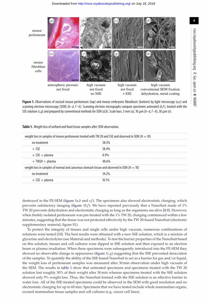
3. ResultsWe introduced numerous living tissues and cells into the SEM to see how they changed under highvacuum (10−3 to 10−7 Pa). Figure 1 shows typical results for the peritoneum of mouse and for mousefibroblast cells: all the untreated specimens dried up rapidly and their structural integrity was completely
on July 18, 2018http://rsos.royalsocietypublishing.org/Downloaded from
4
rsos.royalsocietypublishing.orgR.Soc.opensci.4:160887
................................................
mouseperitoneum
mousefibroblast
cells
high vacuumnot fixedno SSE
atmospheric pressurenot fixed
high vacuumnot fixed
+ SSE
high vacuumconventional SEM fixation,dehydration, metal coating
(b)(a) (c) (d )
(e) (g) (h)( f )
Figure 1. Observations of excised mouse peritoneum (top) and mouse embryonic fibroblasts (bottom) by light microscopy (a,e) andscanning electron microscopy (SEM) (b–d, f–h). Scanning electron micrographs compare specimens untreated (b,f ), treated with theSSE solution (c,g) and prepared by conventional methods for SEM (d,h). Scale bars, 5 mm (a), 10 µm (b–d, f–h), 30 µm (e).
Table 1. Weight loss of unfixed and fixed tissue samples after SEM observation.. . . . . . . . . . . . . . . . . . . . . . . . . . . . . . . . . . . . . . . . . . . . . . . . . . . . . . . . . . . . . . . . . . . . . . . . . . . . . . . . . . . . . . . . . . . . . . . . . . . . . . . . . . . . . . . . . . . . . . . . . . . . . . . . . . . . . . . . . . . . . . . . . . . . . . . . . . . . . . . . . . . . . . . . . . . . . . . . . . . . . . . . . . . . . . . . . . . . . . . . . . . . . . . . . . . . . . . . .
weight loss in samples of mouse peritoneum treated with TW 20 and SSE and observed in SEM (N= 10). . . . . . . . . . . . . . . . . . . . . . . . . . . . . . . . . . . . . . . . . . . . . . . . . . . . . . . . . . . . . . . . . . . . . . . . . . . . . . . . . . . . . . . . . . . . . . . . . . . . . . . . . . . . . . . . . . . . . . . . . . . . . . . . . . . . . . . . . . . . . . . . . . . . . . . . . . . . . . . . . . . . . . . . . . . . . . . . . . . . . . . . . . . . . . . . . . . . . . . . . . . . . . . . . . . . . . . . .
no treatment 58.3%. . . . . . . . . . . . . . . . . . . . . . . . . . . . . . . . . . . . . . . . . . . . . . . . . . . . . . . . . . . . . . . . . . . . . . . . . . . . . . . . . . . . . . . . . . . . . . . . . . . . . . . . . . . . . . . . . . . . . . . . . . . . . . . . . . . . . . . . . . . . . . . . . . . . . . . . . . . . . . . . . . . . . . . . . . . . . . . . . . . . . . . . . . . . . . . . . . . . . . . . . . . . . . . . . . . . . . . . .
+ SSE 38.4%. . . . . . . . . . . . . . . . . . . . . . . . . . . . . . . . . . . . . . . . . . . . . . . . . . . . . . . . . . . . . . . . . . . . . . . . . . . . . . . . . . . . . . . . . . . . . . . . . . . . . . . . . . . . . . . . . . . . . . . . . . . . . . . . . . . . . . . . . . . . . . . . . . . . . . . . . . . . . . . . . . . . . . . . . . . . . . . . . . . . . . . . . . . . . . . . . . . . . . . . . . . . . . . . . . . . . . . . .
+ SSE+ plasma 8.9%. . . . . . . . . . . . . . . . . . . . . . . . . . . . . . . . . . . . . . . . . . . . . . . . . . . . . . . . . . . . . . . . . . . . . . . . . . . . . . . . . . . . . . . . . . . . . . . . . . . . . . . . . . . . . . . . . . . . . . . . . . . . . . . . . . . . . . . . . . . . . . . . . . . . . . . . . . . . . . . . . . . . . . . . . . . . . . . . . . . . . . . . . . . . . . . . . . . . . . . . . . . . . . . . . . . . . . . . .
+ TW20+ plasma 48.6%. . . . . . . . . . . . . . . . . . . . . . . . . . . . . . . . . . . . . . . . . . . . . . . . . . . . . . . . . . . . . . . . . . . . . . . . . . . . . . . . . . . . . . . . . . . . . . . . . . . . . . . . . . . . . . . . . . . . . . . . . . . . . . . . . . . . . . . . . . . . . . . . . . . . . . . . . . . . . . . . . . . . . . . . . . . . . . . . . . . . . . . . . . . . . . . . . . . . . . . . . . . . . . . . . . . . . . . . .
weight loss in samples of normal and cancerous stomach tissue and observed in SEM (N= 10). . . . . . . . . . . . . . . . . . . . . . . . . . . . . . . . . . . . . . . . . . . . . . . . . . . . . . . . . . . . . . . . . . . . . . . . . . . . . . . . . . . . . . . . . . . . . . . . . . . . . . . . . . . . . . . . . . . . . . . . . . . . . . . . . . . . . . . . . . . . . . . . . . . . . . . . . . . . . . . . . . . . . . . . . . . . . . . . . . . . . . . . . . . . . . . . . . . . . . . . . . . . . . . . . . . . . . . . .
no treatment 54.2%. . . . . . . . . . . . . . . . . . . . . . . . . . . . . . . . . . . . . . . . . . . . . . . . . . . . . . . . . . . . . . . . . . . . . . . . . . . . . . . . . . . . . . . . . . . . . . . . . . . . . . . . . . . . . . . . . . . . . . . . . . . . . . . . . . . . . . . . . . . . . . . . . . . . . . . . . . . . . . . . . . . . . . . . . . . . . . . . . . . . . . . . . . . . . . . . . . . . . . . . . . . . . . . . . . . . . . . . .
+ SSE+ plasma 10.1%. . . . . . . . . . . . . . . . . . . . . . . . . . . . . . . . . . . . . . . . . . . . . . . . . . . . . . . . . . . . . . . . . . . . . . . . . . . . . . . . . . . . . . . . . . . . . . . . . . . . . . . . . . . . . . . . . . . . . . . . . . . . . . . . . . . . . . . . . . . . . . . . . . . . . . . . . . . . . . . . . . . . . . . . . . . . . . . . . . . . . . . . . . . . . . . . . . . . . . . . . . . . . . . . . . . . . . . . .
destroyed in the FE-SEM (figure 1a,b and e,f ). The specimens also showed electrostatic charging, whichprevents satisfactory imaging (figure 1b,f ). We have reported previously that a NanoSuit made of 1%TW 20 prevents dehydration and electrostatic charging as long as the organisms are alive [8,9]. However,when freshly isolated peritoneum was pre-treated with the 1% TW 20, charging commenced within a fewminutes, suggesting that the tissue was not protected effectively by the TW 20-based NanoSuit (electronicsupplementary material, figure S1).
To protect the integrity of tissues and single cells under high vacuum, numerous combinations ofsolutions were tested [10]. The best results were obtained with a new SSE solution, which is a mixture ofglycerine and electrolytes (see Material and methods). To test the barrier properties of the NanoSuit basedon this solution, tissues and cell cultures were dipped in SSE solution and then exposed to an electronbeam or plasma irradiation. When these specimens were subsequently introduced into the FE-SEM theyshowed no observable change in appearance (figure 1c,g) suggesting that the SSE prevented desiccationof the samples. To quantify the ability of the SSE-based NanoSuit to act as a barrier for gas and/or liquid,the weight loss of peritoneum samples was measured after 30 min observation under high vacuum ofthe SEM. The results in table 1 show that untreated specimens and specimens treated with the TW 20solution lost roughly 50% of their weight after 30 min whereas specimens treated with the SSE solutionshowed only 9% weight loss. Thus, the NanoSuit formed with the SSE solution is an effective barrier towater loss. All of the SSE-treated specimens could be observed in the SEM with good resolution and noelectrostatic charging for up to 60 min. Specimens that we have tested include whole mammalian organs,excised mammalian tissue samples and cell cultures (e.g. cancer cell lines).
on July 18, 2018http://rsos.royalsocietypublishing.org/Downloaded from

5
rsos.royalsocietypublishing.orgR.Soc.opensci.4:160887
................................................(b)(a) (c) (d )
(e) (g) (h)( f )
+SSE,not fixedspecimen
conventionalSEM fixation(dehydration
and metal coating)
Figure 2. Comparison of images of human fibroblast treated with the SSE solution (a–d) and prepared by traditional methods (e–h).Fibres of the cells (b,f ), nucleus (c,g), high magnifications of the cell surface (d,h). In traditional methods, some cells show protrusionson the surface (arrow in h). Scale bars, 20µm (a,e), 10µm (b,c,f,g), 5 µm (d,h).
(b)(a) (c)
(d ) (e)
Figure 3. Imaging the SSE NanoSuit. (a,b) TEM images of cells treated with SSE and then fixed and sectioned for TEM. The SSE solutioncontained platinum blue to increase contrast of the NanoSuit. The SSE NanoSuit is about 10 nm thick. (c) TEM image of control cells nottreated with SSE but fixed and sectioned as in a and b. The TEM section in a is similar to b but was stained with 2% uranyl acetate and0.4% lead citrate to reveal subcellular structures. The red and blue squares in b, c are shown at high magnifications in d, e, respectively.Scale bars, 300 nm (a–c), 100 nm (d,e).
In addition to the apparent barrier effect, we found that the fine structure of the specimens treatedwith SSE solution was completely different from that of conventionally prepared, i.e. fixed and metalcoated, specimens. Following conventional preparation, shrinkage of tissues and cells was inevitableowing to dehydration (figures 1d,h, 2e–h). By comparison, the overall morphology of SSE-treatedspecimens seemed well preserved and intact (figures 1c,g, 2a–d).
To investigate the structure of the SSE-based NanoSuit we fixed and stained cultured cells treatedwith SSE and plasma irradiation for TEM (figure 3). Cross-sections showed that specimens produced anextra layer (less than 10 nm thickness) over their surface (figure 3a,b,d). Since no such layer was detectedin specimens, which were not treated with the SSE (figure 3c,e), we conclude that this ‘surface shield’ iswhat protects specimens from desiccation in the SEM. The SSE-based surface shield is much thinner thanthe TW 20-based NanoSuit (50–200 nm) that we previously developed [9]. As a consequence it is possibleto image surface structures at much higher resolution.
To determine the effectiveness of the new SSE-based NanoSuit to identify morphological features athigh magnification, we investigated the difference between normal and inflamed mouse peritoneum
on July 18, 2018http://rsos.royalsocietypublishing.org/Downloaded from

6
rsos.royalsocietypublishing.orgR.Soc.opensci.4:160887
................................................(b)(a) (c)
(d ) (e) ( f )
control
inflammation(3 weeks)
Figure 4. Control living peritoneum in mouse (a–c) and images of inflamed specimen with adjuvand for 3 weeks (d–f ). All specimenswere treated with the SSE solution, without chemical fixation and dehydration. Scale bars, 200µm (a,d), 20 µm (b,e), 2 µm (c,f ).
(b) non-infection 5 min 10 min 30 min
3 min(a)
(c) (d ) (e)
Figure 5. SEM images of the cell surface of mouse fibroblasts infected with cytomegalovirus. Cells were treated with SSE and imageddirectly in SEM. (a) Infected cell after 3 min. (b) Uninfected cell. (c–e) Infected cells after 5, 10 and 30 min, respectively. Scale bars, 2µm(a), 500 nm (b–e).
(figure 4a–c). Using conventional electron microscopic methods, it was difficult to distinguish if theobservable damage to tissue was caused by inflammation or by the fixation and dehydration processes(data not shown). However, specimens treated with SSE appeared normal and showed smooth surfaceswhen compared with tissues inflamed with adjuvant for 3 weeks (figure 4d–f ). This suggests that themorphological differences are the result of the inflammation.
To further investigate the ability of the SSE NanoSuit to improve high-resolution imaging of livingcells, we made direct observations of an animal virus infecting cells. Mouse fibroblasts infected withcytomegalovirus were treated with the SSE solution and introduced into the SEM. Images taken
on July 18, 2018http://rsos.royalsocietypublishing.org/Downloaded from

7
rsos.royalsocietypublishing.orgR.Soc.opensci.4:160887
................................................
(c)
(d )
border
+SSE,fixed ‘wet’specimen
(a) (b)
(e)
Figure 6. Comparison of normal and cancerous stomach tissue protected with SSE-based NanoSuit and observed directly in SEM (a) orstereo dissecting images of haematoxylin and eosin (H&E); cancerous (b) and normal tissue (c) in the same specimen. Highmagnificationimages of cancerous (d) and normal (e) regions protected with SSE-based NanoSuit in SEM. Scale bars, 20 µm (a), 0.2 mm (b,c),10 µm (d,e).
3–5 min after infection showed large numbers of virus particles attached to the cell surface (figure 5a,c).Uninfected cell had no such particles on the cell surface (figure 5b). Interestingly, we observed thatthe appearance of the cell surface in the SEM changed with time. After 3–5 min virus particles wereclearly visible on the cell surface but after 10 and 30 min the virus particles appeared to be taken upby endocytosis while being observed in the SEM (N.B. the temperature in the SEM chamber is 20°C)(figure 5d,e). A similar but more rapid change in cell surface morphology was observed in infected cells,which were fixed at various times after infection and then treated with SSE and observed in the SEM(electronic supplementary material, figure S2b–d). Although the specimens in electronic supplementarymaterial, figure S2b–d were fixed with 4% glutaraldehyde, they remained ‘wet’ even in high vacuum ofthe SEM owing to the presence of the SSE NanoSuit.
To explore the effect of the SSE-based NanoSuit for ‘wet fixed specimens’, observations were carriedout on chemically fixed pathology samples. Figure 6 shows images of a surgical explant of humanstomach wall including areas of cancerous and normal tissue. Morphological features were comparedusing two different preparations: specimens treated with SSE solution and imaged directly in the SEM(figure 6) and specimens prepared by conventional methods for SEM observation (fixation, freeze dryingand metal coating) (electronic supplementary material, figure S3). Specimens treated with SSE solution(figure 6a,d,e) appeared intact. By comparison, fixed specimens prepared with conventional metal coatingfor SEM showed obvious structural damage (electronic supplementary material, figure S3). Consistentwith these results, specimens treated with SSE showed little weight loss following 30 min exposure tothe SEM whereas untreated samples showed major weight loss in the SEM (table 1).
To identify cancerous and normal tissue in the explants, we compared the results of haematoxylin andeosin (H&E) staining (figure 6b,c) with the electron microscopic imaging (figure 6a,d,e). The SSE-treatedspecimens preserved fine structures and remained ‘wet’ during the SEM observation and normal andcancerous tissue showed clear differences in morphology. Cancerous cells had a flat surface (figure 6d)compared with clearly rounded surface profiles of normal cells (figure 6e). These results indicate that
on July 18, 2018http://rsos.royalsocietypublishing.org/Downloaded from

8
rsos.royalsocietypublishing.orgR.Soc.opensci.4:160887
................................................the SSE NanoSuit played a significant role as a barrier preventing desiccation in the high vacuum. Insummary, covering the surface of fixed tissue explants with an SSE-based NanoSuit permits rapid andeasy identification of abnormal and normal regions in tissue.
4. DiscussionTo maintain life under natural conditions, water, which possesses many unique properties and plays anirreplaceable role in organisms, is an essential chemical. Thus, water limitation is one of the harsheststressors in an extreme environment, viz. a high vacuum. We previously found that after modification ofmaterial on the surface of organisms through exposure to an electron beam [11] or plasma ionization [12],i.e. conditions known to enhance polymer formation, some treated animals survived under the highvacuum condition of the FE-SEM owing to the protective properties of what we termed the NanoSuit.The NanoSuit allows more sophisticated observation methods for studying living organisms in aFE-SEM.
In our earlier report, we used TW 20 to form the NanoSuit on the living organisms [6,8–10]. Inwhole animals, which were successfully protected with a TW 20-based NanoSuit, the outer surfacewas covered with epithelium or cuticle. By contrast, excised tissues or single cells do not have sucha protective cover, so that an alternative barrier was needed. In the present experiment, we used anSSE solution with glycerine as a main component. Since glycerine is strongly hygroscopic, it has beenused as a humectant in cosmetics [13]. To increase the barrier effect, we have combined glycerine withelectrolytes and polymerized a thin liquid film over the sample in order to prepare a NanoSuit. In somecases this treatment yielded an effective NanoSuit and permitted imaging in the SEM (figures 1c,g,2a–d, 4, 5 and 6a,d,e, and the electronic supplementary material, figure S2). The SSE constitutes avery effective diffusion barrier (table 1) and it is very thin (less than 10 nm) (figure 3a,b,d). The SSE-based surface shield is much thinner than the TW 20-based NanoSuit (50–200 nm) that we previouslydeveloped [9]. As a consequence it is possible to image surface structures at much higher resolution.Since glycerine alone is unlikely to polymerize to form a stable NanoSuit, it seems likely that it interactswith protein/proteoglycan on the surface of cells and tissues to form a stabilizing polymer coat afterplasma irradiation or exposure to the electron beam in the SEM.
There have been several previous attempts to adapt SEM for the observation of wet samples.Thiberge et al. used polyimide or silicon nitride membranes to protect the sample from the vacuum [14].However, this method required the use of high acceleration voltages (15–30 kV) to penetrate the relativelythick membranes. The intense radiation of the electron beam during high magnification imaging wassufficient to cause damage to the specimens. A related technique for imaging wet tissue in SEM hasrecently been described by Wojcik et al. [15]. This method covers tissue specimens with a monolayer ofgraphene. In addition to being technically challenging, these methods appear only to work successfullywith fixed tissue samples. By contrast, our SSE-based NanoSuit method can be applied to both livingspecimens (figures 1c,g, 2a–d, 4 and 5) and to fixed tissue (figure 6a,d,e; electronic supplementary material,figure S2b–d) and requires only use of low voltage electrons (1 kV). Imaging occurs in a hydrous/wetstate closely approximating the natural condition. Moreover, even after SSE treatment and exposure tothe high vacuum in the SEM, some cells survived and could be re-cultured when they were returnedto atmospheric pressure and placed in culture medium (Takaku Y et al., in preparation). Our resultssuggest that, to protect cells for long periods under high vacuo conditions, very effective barriers againstthe extreme environment need to be devised, and at the same time improvements in the SEM instrumentto accommodate living organisms will be necessary. This ongoing progress in electron microscopy isstarting to have a significant impact on our understanding of the subcellular world.
Ethics. This study was approved by the institutional review board of the Hamamatsu University School of Medicine.Animal ethics. Handling of animals was performed in accordance with the Guide for the Care and Use of LaboratoryAnimals, Hamamatsu University School of Medicine.Data accessibility. Our data can be found in the electronic supplementary material.Authors’ contributions. Y.T., H.S. and T.H. conceived and designed the experiments, performed research, analysed the dataand made contribution in manuscript preparation and final editing; H.S. also contributed new reagents/analytic tools;H.K., I.O., D.I., S.H., T.T., H.M., S.T., C.N., K.S., C.S. and Y.M. performed research; H.K., H.K. and M.S. helped in dataanalysis and contributed in manuscript preparation. All authors gave final approval for publication.Competing interests. We have no competing interests.Funding. This work was supported by Grants-in-Aid for Scientific Research for Y.T. (JP25292198) and I.O. (JP26506008),and by Grant-in-Aid for Challenging Exploratory Research for Y.T. (15K14558) and S.H. (15K15413), and by Grant-in-Aid for Scientific Research on Innovative Areas in ‘Innovative Materials Engineering Based on Biological Diversity’
on July 18, 2018http://rsos.royalsocietypublishing.org/Downloaded from

9
rsos.royalsocietypublishing.orgR.Soc.opensci.4:160887
................................................for T.H., D.I. M.S. (JP 24120001 and JP24120004) and for Y.T. (15H01598) and by grants from the Takeda ScienceFoundation.Acknowledgement. We wish to express our thanks to Prof. V. B. Meyer-Rochow and to two anonymous referees for helpfulsuggestions and comments on the manuscript. We are also grateful to JEOL for the technical assistance.
References1. Suzuki E. 2002 High-resolution scanning electron
microscopy of immunogold-labelled cells by the useof thin plasma coating of osmium. J. Microsc. 208,153–157. (doi:10.1046/j.1365-2818.2002.01082.x)
2. Danilatos GD. 1991 Review and outline ofenvironmental SEM at present. J. Microsc. 162,391–402. (doi:10.1111/j.1365-2818.1991.tb03149.x)
3. Mohan A, Khanna N, Hwu J, Joy DC. 1998 Secondaryelectron imaging in the variable pressure scanningelectron microscope. J. Scan Microsc. 20, 436–441.(doi:10.1002/sca.1998.4950200603)
4. Symondson WOC, Williams IB. 2003 Low-vacuumelectron microscopy of carabid chemoreceptors: anew tool for the identification of live and valuablemuseum specimens. Entomol. Exp. Appl. 85, 75–82.(doi:10.1046/j.1570-7458.1997.00235.x)
5. Stokes DJ. 2003 Investigating biologicalultrastructure using environmental scanningelectron microscopy (ESEM). Sci. Technol. Ed.Microsc. Overview 564–570
6. Takaku Y, Suzuki H, Ohta I, Ishii D, Muranaka Y,Shimomura M, Hariyama T. 2013 A thin polymer
membrane, nano-suit, enhancing survival acrossthe continuum between air and high vacuum. Proc.Natl. Acad. Sci. USA 110, 7631–7635. (doi:10.1073/pnas.1221341110)
7. Iwata C et al. 2007 Inhibition of cyclooxygenase-2suppresses lymph node metastasis via reduction oflymphangiogenesis. Cancer Res. 67, 10181–10189.(doi:10.1158/0008-5472.CAN-07-2366)
8. Ohta I, Takaku Y, Suzuki H, Ishii D, Muranaka Y,Shimomura M, Hariyama T. 2014 Dressing livingorganisms in a thin polymer membrane, NanoSuit,for high vacuum FE-SEM observation.Microscopy63, 295–300. (doi:10.1093/jmicro/dfu015)
9. Takaku Y, Suzuki H, Ohta I, Tsutsui T, Matsumoto H,Shimomura M, Hariyama T. 2015 A ‘NanoSuit’surface shield successfully protects organisms inhigh vacuum: observations on living organisms in aFE-SEM. Proc. R. Soc. B 282, 2014.2857. (doi:10.1098/rspb.2014.2857)
10. Suzuki H, Takaku Y, Ohta I, Ishii D, Muranaka Y,Shimomura M, Hariyama T. 2013 In-situ preparationof biomimetic thin films and their surface shield
effect for organisms in high vacuum. PLoS ONE 8,e78563. (doi:10.1371/journal.pone.0078563)
11. Sun KH. 1954 Effects of atomic radiation on highpolymers. Plastics 141.
12. Friedrich J. 2011 Mechanisms of plasmapolymerization – reviewed from a chemical point ofview. Plasma Processes Polym 8, 783–802.(doi:10.1002/ppap.201100038)
13. Kellett EG. 1949 The principles and practice ofmodern cosmetics. Nature 163, 588. (doi:10.1038/163588a0)
14. Thiberge S et al. 2004 Scanning electron microscopyof cells and tissues under fully hydrated conditions.Proc. Natl. Acad. Sci. USA 101, 3346–3351.(doi:10.1073/pnas.0400088101)
15. Wojcik M, Hauser M, Li W, Moon S, Xu K. 2015Graphene-enabled electron microscopy andcorrelated super-resolution microscopy of wet cells.Nat. Commun. 6, 7384. (doi:10.1038/ncomms8384)
on July 18, 2018http://rsos.royalsocietypublishing.org/Downloaded from


![rsos.royalsocietypublishing.org Canisaureus …rsos.royalsocietypublishing.org/content/royopensci/2/12/150450...cannot rule out the hypothesis of ancient hybridization events [10,11]](https://img.pdfslide.net/doc/110x75/5b3499c67f8b9ae1108e650d/rsosro-canisaureus-rsosro-rule-out-the-hypothesis-of-ancient-hybridization-events.jpg)


![endangeredCrossRiver Research gorilla( …rsos.royalsocietypublishing.org/content/royopensci/2/2/140423.full.pdfand group composition [1,2,5]. However, ... of passage), teams would](https://img.pdfslide.net/doc/110x75/5acd8f2f7f8b9a27628dc742/endangeredcrossriver-research-gorilla-rsosro-group-composition-125-however.jpg)


![humpbackwhales Research - Portal · 3 rsos.royalsocietypublishing.org R.Soc.opensci. 3:160616..... also found in deep waters [26,27] and in shallow waters extended offshore during](https://img.pdfslide.net/doc/110x75/5ea1d4344125f0619e1831bd/humpbackwhales-research-portal-3-rsosroyalsocietypublishingorg-rsocopensci.jpg)
![Theperilousstateof seagrassintheBritishIslesorca.cf.ac.uk/85062/1/150596.full.pdf · 2016. 2. 4. · 2 rsos.royalsocietypublishing.org R.Soc.opensci. 3:150596..... [2,3] with anthropogenic](https://img.pdfslide.net/doc/110x75/600a90f34966be29c122c676/theperilousstateof-seagrassi-2016-2-4-2-rsosroyalsocietypublishingorg-rsocopensci.jpg)