Embed Size (px)
Citation preview
RESEARCH Open Access
Detection of hepatitis B virus DNA amongaccepted blood donors in Nanjing, ChinaYong Liu1,4†, Ping Li2†, Cuiping Li2, Jinyong Zhou1,4, Chao Wu3, Yi-Hua Zhou1,3,4*
Abstract
Background: Posttransfusion hepatitis B virus (HBV) infection still occurs although its incidence has beensubstantially reduced since the introduction of screening of hepatitis B surface antigen (HBsAg) in blood donors.This study aimed to investigate the occult HBV infection in accepted blood donors in Nanjing, China.
Results: The lower detection limit of the nested PCR in this study was estimated to be 20 copies/ml HBV DNA.The positive rate of occult HBV infection was 0.13% (5 of 2972) in the accepted blood donors. Sequencing datashowed that the amplified HBV sequences were not identical each other and to the known sequences cloned inour laboratory, excluding the false-positive caused by cross-contamination. Phylogenetic analysis showed that theHBV in all five donors was genotype B; a single base deletion was detected in the S region of HBV DNA from onedonor, and no mutation was observed in the “a” determinant of HBsAg from four other donors. All five donorswere negative for anti-HBs and one was positive for anti-HBc.
Conclusions: The prevalence of occult HBV infection in the accepted blood donors in Nanjing, China is relativelyhigh. The data would be meaningful in adapting strategy to eliminate posttransfusion HBV infection in China.
BackgroundHepatitis B virus (HBV) infection is one of the majorhealth problems worldwide. The infection is usuallydefined by the presence of hepatitis B surface antigen(HBsAg) in serum or plasma. However, HBV may existin humans without detectable HBsAg but with presenceof HBV DNA in the serum and/or in the liver, i.e. theoccult HBV infection [1]. The occult infection mayresult from the low viral load in circulation (usually<200 IU/ml) [2] or a mutant HBsAg which is not recog-nized by the monoclonal antibodies against HBsAg(anti-HBs) used in some commercial detection kits [1,3].Because of routine screening of blood donors for
HBsAg, the incidence of transfusion-transmitted hepati-tis B has been steadily reduced over the last four dec-ades; however HBV transmission remains the mostfrequent transfusion-transmitted viral infection [4-6].The residual risk of HBV transmission by transfusion ismainly associated with occult HBV infection in blood
donors. Additionally, occult HBV infection also hassignificance in bone marrow and organ transplantations[2,7-9].Attributed to widespread use of hepatitis B vaccine
and other strategies for control of HBV infection, theprevalence of HBsAg carrier rate in general populationin China decreased from some 10.0% in 1980 s to 7.2%in 2006 [10-12]. However, HBV infection is still endemicin China. Studies have shown that the prevalence ofoccult HBV infection is closely related to the endemicityof HBV infection [13,14], however, the occult HBVinfection in China has been less studied [15-17]. Thus,we performed this study to investigate the prevalence ofoccult HBV infection in blood donors in Nanjing, aneastern region of China.
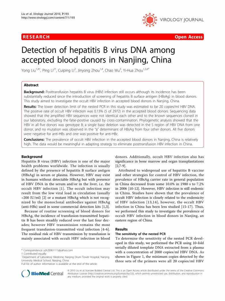
ResultsThe sensitivity of the nested PCRTo determine the sensitivity of the nested PCR devel-oped in this study, we performed the PCR using 10-foldserially diluted template DNA extracted from a plasmawith a concentration of 2000 copies/ml HBV DNA. Asshown in Figure 1, the minimum copies detected by thethree sets of the primers were all 20 copies/ml HBV
* Correspondence: [email protected]† Contributed equally1Department of Laboratory Medicine, Nanjing Drum Tower Hospital, NanjingUniversity Medical School, Nanjing, ChinaFull list of author information is available at the end of the article
Liu et al. Virology Journal 2010, 7:193http://www.virologyj.com/content/7/1/193
© 2010 Liu et al; licensee BioMed Central Ltd. This is an Open Access article distributed under the terms of the Creative CommonsAttribution License (http://creativecommons.org/licenses/by/2.0), which permits unrestricted use, distribution, and reproduction inany medium, provided the original work is properly cited.
DNA, equivalent to 1 copy per reaction. Thus, the lowerdetection limit of the nested PCR using the differentprimers was comparable.
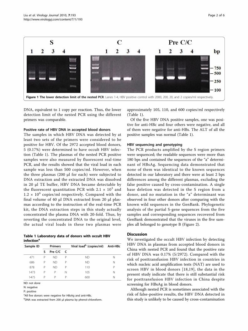
Positive rate of HBV DNA in accepted blood donorsThe samples in which HBV DNA was detected by atleast two sets of the primers were considered to bepositive for HBV. Of the 2972 accepted blood donors,5 (0.17%) were determined to have occult HBV infec-tion (Table 1). The plasmas of the nested PCR positivesamples were also measured by fluorescent real-timePCR, and the results showed that the viral load in eachsample was less than 500 copies/ml. However, whenthe three plasmas (200 μl for each) were subjected toDNA extraction and the extracted DNA was dissolvedin 20 μl TE buffer, HBV DNA became detectable bythe fluorescent quantitation PCR with 2.1 × 10³ and1.2 × 104 copies/ml respectively. Compared with thefinal volume of 40 μl DNA extracted from 20 μl plas-mas according to the instruction of the real-time PCRkit, the DNA extraction steps in this study actuallyconcentrated the plasma DNA with 20-fold. Thus, byreverting the concentrated DNA to the original level,the actual viral loads in these two plasmas were
approximately 105, 110, and 600 copies/ml respectively(Table 1).Of the five HBV DNA positive samples, one was posi-
tive for anti-HBc and four others were negative, and allof them were negative for anti-HBs. The ALT of all thepositive samples was normal (Table 1).
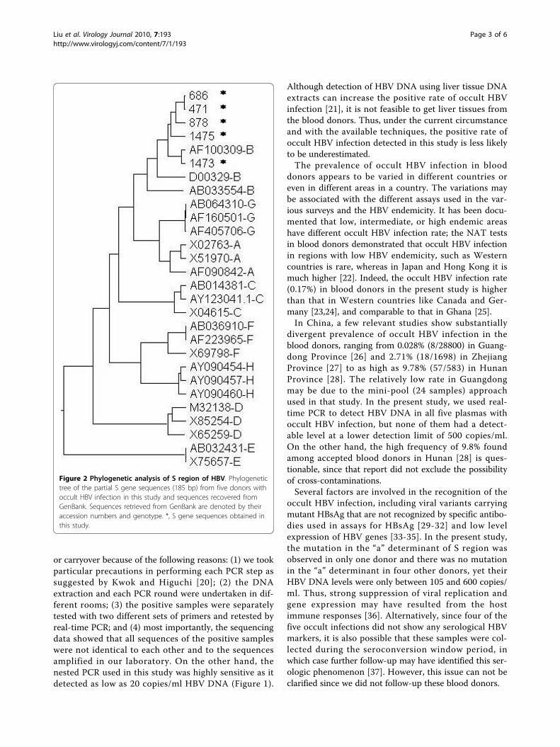
HBV sequencing and genotypingThe PCR products amplified by the S region primerswere sequenced; the readable sequences were more than180 bps and contained the sequences of the “a” determi-nant of HBsAg. Sequencing data demonstrated thatnone of them was identical to the known sequencesdetected in our laboratory and there were at least 2 bpsdifferences among the different plasmas, excluding thefalse positive caused by cross-contamination. A singlebase deletion was detected in the S region from adonor, and no mutation in the “a” determinant wasobserved in four other donors after comparing with theknown wild sequences in the GenBank. Phylogeneticanalysis of the partial S-gene sequences from the fivesamples and corresponding sequences recovered fromGenBank demonstrated that the viruses in the five sam-ples all belonged to genotype B (Figure 2).
DiscussionWe investigated the occult HBV infection by detectingHBV DNA in plasmas from accepted blood donors inChina with nested PCR and found that the positive rateof HBV DNA was 0.17% (5/2972). Compared with therisk of posttransfusion HBV infection in countries inwhich nucleic acid amplification tests (NAT) are used toscreen HBV in blood donors [18,19], the data in thepresent study indicate that there is still substantial riskfor posttransfusion HBV infection in China despitescreening for HBsAg in blood donors.Although nested PCR is sometimes associated with the
risk of false-positive results, the HBV DNA detected inthis study is unlikely to be caused by cross-contamination
Figure 1 The lower detection limit of the nested PCR. Lanes 1-4, HBV positive control with 2000, 200, 20, and 2 copies/ml respectively.
Table 1 Laboratory data of donors with occult HBVinfectiona
Sample ID Primers Viral loadb (copies/ml) Anti-HBc
S Pre C/C C
471 P ND P ND N
686 P ND P ND N
878 P ND P 110 P
1473 P P N 105 N
1475 P P P 600 N
ND: not done
N: negative
P: positiveaAll five donors were negative for HBsAg and anti-HBs.bDNA was extracted from 200 μl plasma by phenol-chloroform.
Liu et al. Virology Journal 2010, 7:193http://www.virologyj.com/content/7/1/193
Page 2 of 6
or carryover because of the following reasons: (1) we tookparticular precautions in performing each PCR step assuggested by Kwok and Higuchi [20]; (2) the DNAextraction and each PCR round were undertaken in dif-ferent rooms; (3) the positive samples were separatelytested with two different sets of primers and retested byreal-time PCR; and (4) most importantly, the sequencingdata showed that all sequences of the positive sampleswere not identical to each other and to the sequencesamplified in our laboratory. On the other hand, thenested PCR used in this study was highly sensitive as itdetected as low as 20 copies/ml HBV DNA (Figure 1).
Although detection of HBV DNA using liver tissue DNAextracts can increase the positive rate of occult HBVinfection [21], it is not feasible to get liver tissues fromthe blood donors. Thus, under the current circumstanceand with the available techniques, the positive rate ofoccult HBV infection detected in this study is less likelyto be underestimated.The prevalence of occult HBV infection in blood
donors appears to be varied in different countries oreven in different areas in a country. The variations maybe associated with the different assays used in the var-ious surveys and the HBV endemicity. It has been docu-mented that low, intermediate, or high endemic areashave different occult HBV infection rate; the NAT testsin blood donors demonstrated that occult HBV infectionin regions with low HBV endemicity, such as Westerncountries is rare, whereas in Japan and Hong Kong it ismuch higher [22]. Indeed, the occult HBV infection rate(0.17%) in blood donors in the present study is higherthan that in Western countries like Canada and Ger-many [23,24], and comparable to that in Ghana [25].In China, a few relevant studies show substantially
divergent prevalence of occult HBV infection in theblood donors, ranging from 0.028% (8/28800) in Guang-dong Province [26] and 2.71% (18/1698) in ZhejiangProvince [27] to as high as 9.78% (57/583) in HunanProvince [28]. The relatively low rate in Guangdongmay be due to the mini-pool (24 samples) approachused in that study. In the present study, we used real-time PCR to detect HBV DNA in all five plasmas withoccult HBV infection, but none of them had a detect-able level at a lower detection limit of 500 copies/ml.On the other hand, the high frequency of 9.8% foundamong accepted blood donors in Hunan [28] is ques-tionable, since that report did not exclude the possibilityof cross-contaminations.Several factors are involved in the recognition of the
occult HBV infection, including viral variants carryingmutant HBsAg that are not recognized by specific antibo-dies used in assays for HBsAg [29-32] and low levelexpression of HBV genes [33-35]. In the present study,the mutation in the “a” determinant of S region wasobserved in only one donor and there was no mutationin the “a” determinant in four other donors, yet theirHBV DNA levels were only between 105 and 600 copies/ml. Thus, strong suppression of viral replication andgene expression may have resulted from the hostimmune responses [36]. Alternatively, since four of thefive occult infections did not show any serological HBVmarkers, it is also possible that these samples were col-lected during the seroconversion window period, inwhich case further follow-up may have identified this ser-ologic phenomenon [37]. However, this issue can not beclarified since we did not follow-up these blood donors.
Figure 2 Phylogenetic analysis of S region of HBV. Phylogenetictree of the partial S gene sequences (185 bp) from five donors withoccult HBV infection in this study and sequences recovered fromGenBank. Sequences retrieved from GenBank are denoted by theiraccession numbers and genotype. *, S gene sequences obtained inthis study.
Liu et al. Virology Journal 2010, 7:193http://www.virologyj.com/content/7/1/193
Page 3 of 6
The occurrence of posttransfusion hepatitis B in Chinahas been substantially reduced since the introduction ofHBsAg screening of blood donors. However, the trueincidence remains unknown because of the obstacles inconducting such studies. In the present survey, wefound that 0.17% of the accepted blood donors hadoccult HBV infection, which is much higher than thatobserved in Western counties as well as in Japan andHong Kong. Although the conversion of residual risk ofoccult HBV infection into the true rate of infection islargely unknown, the results in the chimpanzee modeldemonstrate that as low as 10 copies of HBV may resultin half of the animals being infected, irrespective ofHBV genotype [38,39]. Considering that our blooddonors had HBV DNA exceed 100 copies/ml, the recipi-ents of these five occult HBV carrier donors were highlypossible to be infected with HBV although we did notfollow-up the recipients. Indeed, numerous reports havedocumented that occult HBV infection in blood donorsmay cause posttransfusion hepatitis B [40-42].Currently, only a few financially prosperous cities in
China detect HBV DNA in blood donors by minipoolNAT. We consider that NAT for HBV DNA as a rou-tine screening in blood donors in China will be cost-effectiveness, since the risk of HBV transmission bytransfusion in China is relatively high based on our data.Even in countries with low HBsAg prevalence such asUSA and some European countries, minipool NAT hasbeen used as a routine test in blood donors [43].
ConclusionsThe data in the present study will be meaningful in set-ting up a strategy to prevent posttransfusion hepatitis Bin China. Because of relatively high rate of occult HBVinfection in blood donors in China, tests for HBV DNAin blood donors would substantially reduce the inci-dence of posttransfusion hepatitis B.
MethodsBlood donorsWe randomly collected plasma samples from 2972accepted volunteer donors at the Nanjing Red CrossBlood Center between February 2007 and April 2008.Each sample was not directly collected from the donor,but collected from one tubing segment connected to thebag of the donor’s blood; each tubing segment was cutwith a fresh blade to avoid cross-contamination amongsamples. According to the policy set up by the ChinaHealth Ministry, an accepted donor is defined as a gen-erally healthy person with normal alanine aminotrans-ferase (ALT), negative for HBsAg and antibodies againsthepatitis C virus (HCV), HIV, and treponema pallidum.Thus, the laboratory tests in all the 2972 donors showedto be normal ALT and negative for HBsAg, anti-HCV,
anti-HIV, and antibody against treponema pallidum; theHBsAg in each donor was tested in parallel by two dif-ferent commercial enzyme-linked immunosorbent assay(ELISA) kits. The donors’ average age was 24.9 years(range, 18-53). They were 1475 males and 1497 females.The plasmas were stored at -20°C. All the experimentswere approved by the Ethics Committee of NanjingDrum Tower Hospital, Nanjing University MedicalSchool, in accordance with guidelines of the NationHealth and Medical Research Council of China.
Plasma DNA isolationDNA was extracted from plasma using phenol-chloro-form extraction by the standard method. Briefly, 200 μlplasma was treated with proteinase K at 50°C for 3 h,and then DNA was extracted twice by phenol-chloro-form and rinsed once by 70% ethanol. Finally, 20 μlTris-EDTA (TE) buffer was added to each sample todissolve DNA.
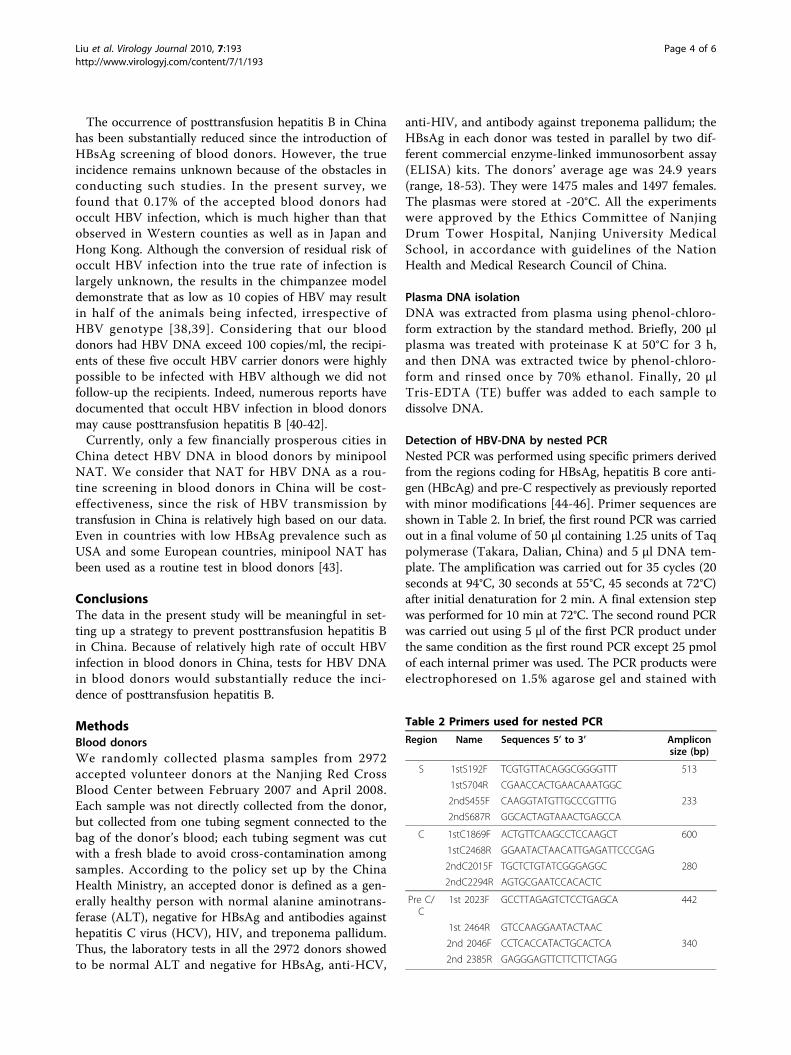
Detection of HBV-DNA by nested PCRNested PCR was performed using specific primers derivedfrom the regions coding for HBsAg, hepatitis B core anti-gen (HBcAg) and pre-C respectively as previously reportedwith minor modifications [44-46]. Primer sequences areshown in Table 2. In brief, the first round PCR was carriedout in a final volume of 50 μl containing 1.25 units of Taqpolymerase (Takara, Dalian, China) and 5 μl DNA tem-plate. The amplification was carried out for 35 cycles (20seconds at 94°C, 30 seconds at 55°C, 45 seconds at 72°C)after initial denaturation for 2 min. A final extension stepwas performed for 10 min at 72°C. The second round PCRwas carried out using 5 μl of the first PCR product underthe same condition as the first round PCR except 25 pmolof each internal primer was used. The PCR products wereelectrophoresed on 1.5% agarose gel and stained with
Table 2 Primers used for nested PCR
Region Name Sequences 5’ to 3’ Ampliconsize (bp)
S 1stS192F TCGTGTTACAGGCGGGGTTT 513
1stS704R CGAACCACTGAACAAATGGC
2ndS455F CAAGGTATGTTGCCCGTTTG 233
2ndS687R GGCACTAGTAAACTGAGCCA
C 1stC1869F ACTGTTCAAGCCTCCAAGCT 600
1stC2468R GGAATACTAACATTGAGATTCCCGAG
2ndC2015F TGCTCTGTATCGGGAGGC 280
2ndC2294R AGTGCGAATCCACACTC
Pre C/C
1st 2023F GCCTTAGAGTCTCCTGAGCA 442
1st 2464R GTCCAAGGAATACTAAC
2nd 2046F CCTCACCATACTGCACTCA 340
2nd 2385R GAGGGAGTTCTTCTTCTAGG
Liu et al. Virology Journal 2010, 7:193http://www.virologyj.com/content/7/1/193
Page 4 of 6
ethidium bromide, and then photographed by imageanalysis system (UVP, Upland, CA).To determine the lower detection limit of the nested
PCR in this study, we carried out the PCR using 10-foldserially diluted template DNA, which was extractedfrom a sample with known concentration of HBV DNAwith 2000 copies/ml. The DNA was extracted from200 μl serum and dissolved in 200 μl TE.To prevent carryover or cross contamination during
the extraction of DNA from plasmas and PCR, eachstep of the procedure was performed in separate areaswith dedicated equipment. Negative controls, includingplasmas DNA from normal subjects without HBV infec-tion and distilled water, and a positive control (an HBVDNA positive plasma diluted to 20 copies/ml) werealways included in every nested PCR test.
Quantitative assay of HBV DNAPlasma HBV DNA in occult HBV infections was quanti-fied by real-time PCR with a commercially availablefluorescent real-time PCR assay (Shenyou Biotechnol-ogy, Shanghai, China) which had a strict internal qualitycontrol and passed the Clinical Laboratory Center ofChinese Ministry of Health external quality assessmenton the DNA Engine Opticon 2 System (MJ Research,Waltham, MA). Plasma DNA was extracted using acommercial kit (Shenyou Biotechnology) from 20 μlplasma by boiling. The PCR was run for 40 cycles andthe fluorescence signal of the amplicons was detected bythe DNA Engine Opticon 2 System. The lower detectionlimit of this assay was 500 copies/ml with a linear rangeof up to 108 copies/ml.
Detection of serological markers for HBV infectionAnti-HBc of the HBV DNA positive samples was mea-sured by ELISA using the Diagnostic kit (Huakang Bio-technology, Shenzhen, China) for anti-HBc.
HBV DNA sequencing and HBV genotypingThe PCR products amplified by S region primers werepurified and directly sequenced on an ABI Prism 3130sequencer (Applied Biosystems, Hitachi, Tokyo, Japan)after reaction with BigDye Terminator v3.1 (AppliedBiosystems, Foster, CA). Sequence analysis and compari-son were conducted by using molecular programsdeposited in the web site of the National Centre for Bio-technology Information http://www.ncbi.nlm.nih.gov/.The sequences were compared with the same region ofHBV sequences from different genotypes found in thegenotyping reference set available on the NCBI website(URL: http://www.ncbi.nih.gov/projects/genotyping/view.cgi?db=2). The phylogenetic tree was constructedaccording to previous methods [47].
AcknowledgementsThis study was supported by a Special Research Grant (RC2007005) for thePrincipal Fellow from the Department of Health, Jiangsu Province, and by agrant (No. 200801088) from the Department of Science and Technology ofNanjing City, China.
Author details1Department of Laboratory Medicine, Nanjing Drum Tower Hospital, NanjingUniversity Medical School, Nanjing, China. 2Department of TransfusionMedicine, Nanjing Drum Tower Hospital, Nanjing University Medical School,Nanjing, China. 3Department of Infectious Diseases, Nanjing Drum TowerHospital, Nanjing University Medical School, Nanjing, China. 4Jiangsu KeyLaboratory for Molecular Medicine, Nanjing University Medical School,Nanjing, China.
Authors’ contributionsYL and PL performed the experiments, analyzed the data, and drafted themanuscript and contributed equally to this work. CL collected the samplesand assisted in the performance of the experiments. JZ carried out the DNAsequencing. CW interpreted the data and revised the manuscript. YHZdesigned the study, interpreted the data and critically revised themanuscript. All authors read and approved the final manuscript.
Competing interestsThe authors declare that they have no competing interests.
Received: 2 July 2010 Accepted: 19 August 2010Published: 19 August 2010
References1. Gerlich WH, Bremer C, Saniewski M, Schuttler CG, Wend UC, Willems WR,
Glebe D: Occult hepatitis B virus infection: detection and significance.Digest Dis 2010, 28:116-125.
2. Raimondo G, Allain JP, Brunetto MR, Buendia MA, Chen DS, Colombo M,Craxi A, Donato F, Ferrari C, Gaeta GB, Gerlich WH, Levrero M, Locarnini S,Michalak T, Mondelli MU, Pawlotsky JM, Pollicino T, Prati D, Puoti M,Samuel D, Shouval D, Smedile A, Squadrito G, Trepo C, Villa E, Will H,Zanetti AR, Zoulim F: Statements from the Taormina expert meeting onoccult hepatitis B virus infection. J Hepatol 2008, 49:652-657.
3. Zhang R, Wang L, Li J: Hepatitis B virus transfusion risk in China:proficiency testing for the detection of hepatitis B surface antigen.Transfusion Med 2010.
4. Niederhauser C, Mansouri Taleghani B, Graziani M, Stolz M, Tinguely C,Schneider P: Blood donor screening: how to decrease the risk oftransfusion-transmitted hepatitis B virus? Swiss Med Wkly 2008,138:134-141.
5. Calderon GM, Gonzalez-Velazquez F, Gonzalez-Bonilla CR, Novelo-Garza B,Terrazas JJ, Martinez-Rodriguez ML, Cortes-Marquez SR, Blanco-Flores JP,Rodriguez-Rodriguez A, Del Campo MA, Cortes-Gomez R, Mejia-Bocanegra MG: Prevalence and risk factors of hepatitis C virus, hepatitisB virus, and human immunodeficiency virus in multiply transfusedrecipients in Mexico. Transfusion 2009, 49:2200-2207.
6. Kafi-abad SA, Rezvan H, Abolghasemi H, Talebian A: Prevalence and trendsof human immunodeficiency virus, hepatitis B virus, and hepatitis Cvirus among blood donors in Iran, 2004 through 2007. Transfusion 2009,49:2214-2220.
7. Hollinger FB: Hepatitis B virus infection and transfusion medicine:science and the occult. Transfusion 2008, 48:1001-1026.
8. Giudice CL, Martinengo M, Pietrasanta P, Bocciardo L, Malavasi C, Rastelli S,Faraci M, Tripodi G: Occult hepatitis B virus infection: a case ofreactivation in a patient receiving immunosuppressive treatment forallogeneic bone marrow transplantation. Blood transfus 2008, 6:46-50.
9. Raimondo G, Pollicino T, Romano L, Zanetti AR: A 2010 update on occulthepatitis B infection. Pathol Biol 2010, 58:254-257.
10. Liang X, Bi S, Yang W, Wang L, Cui G, Cui F, Zhang Y, Liu J, Gong X,Chen Y, Wang F, Zheng H, Wang F, Guo J, Jia Z, Ma J, Wang H, Luo H, Li L,Jin S, Hadler SC, Wang Y: Epidemiological serosurvey of hepatitis B inChina–declining HBV prevalence due to hepatitis B vaccination. Vaccine2009, 27:6550-6557.
11. Zhou YH, Wu C, Zhuang H: Vaccination against hepatitis B: the Chineseexperience. Chin Med J (Engl) 2009, 122:98-102.
Liu et al. Virology Journal 2010, 7:193http://www.virologyj.com/content/7/1/193
Page 5 of 6
12. Zhang S, Li RT, Wang Y, Liu Q, Zhou YH, Hu Y: Seroprevalence of hepatitisB surface antigen among pregnant women in Jiangsu, China, 17 yearsafter introduction of hepatitis B vaccine. Int J Gynecol Obstet 2010,109:194-197.
13. Brechot C, Thiers V, Kremsdorf D, Nalpas B, Pol S, Paterlini-Brechot P:Persistent hepatitis B virus infection in subjects without hepatitis Bsurface antigen: clinically significant or purely “occult"? Hepatology 2001,34:194-203.
14. Zervou EK, Dalekos GN, Boumba DS, Tsianos EV: Value of anti-HBcscreening of blood donors for prevention of HBV infection: results of a3-year prospective study in Northwestern Greece. Transfusion 2001,41:652-658.
15. Shang G, Seed CR, Wang F, Nie D, Farrugia A: Residual risk of transfusion-transmitted viral infections in Shenzhen, China, 2001 through 2004.Transfusion 2007, 47:529-539.
16. Fang Y, Shang QL, Liu JY, Li D, Xu WZ, Teng X, Zhao HW, Fu LJ, Zhang FM,Gu HX: Prevalence of occult hepatitis B virus infection amonghepatopathy patients and healthy people in China. J Infect 2009,58:383-388.
17. Shang G, Yan Y, Yang B, Shao C, Wang F, Li Q, Seed CR: Two HBV DNA+/HBsAg-blood donors identified by HBV NAT in Shenzhen, China.Transfus Apher Sci 2009, 41:3-7.
18. Bamaga MS, Azahar EI, Al-Ghamdi AK, Al-Enzi FQ, Farahat FM: Nucleic acidamplification technology for hepatitis B virus, and its role in blooddonation screening in blood banks. Saudi Med J 2009, 30:1416-1421.
19. Gonzalez R, Torres P, Castro E, Barbolla L, Candotti D, Koppelman M,Zaaijer HL, Lelie N, Allain JP, Echevarria JM: Efficacy of hepatitis B virus(HBV) DNA screening and characterization of acute and occult HBVinfections among blood donors from Madrid, Spain. Transfusion 2009,50:221-230.
20. Kwok S, Higuchi R: Avoiding false positives with PCR. Nature 1989,339:237-238.
21. Raimondo G, Pollicino T, Cacciola I, Squadrito G: Occult hepatitis B virusinfection. J Hepatol 2007, 46:160-170.
22. Lelie N, Heaton A: Hepatitis B - a review of the role of NAT in enhancingblood safety. J Clin Virol 2006, 36(Suppl 1):S1-2.
23. Offergeld R, Faensen D, Ritter S, Hamouda O: Human immunodeficiencyvirus, hepatitis C and hepatitis B infections among blood donors inGermany 2000-2002: risk of virus transmission and the impact of nucleicacid amplification testing. Euro Surveill 2005, 10:8-11.
24. Chevrier MC, St-Louis M, Perreault J, Caron B, Castilloux C, Laroche J,Delage G: Detection and characterization of hepatitis B virus of anti-hepatitis B core antigen-reactive blood donors in Quebec with an in-house nucleic acid testing assay. Transfusion 2007, 47:1794-1802.
25. Owusu-Ofori S, Temple J, Sarkodie F, Anokwa M, Candotti D, Allain JP:Predonation screening of blood donors with rapid tests: implementationand efficacy of a novel approach to blood safety in resource-poorsettings. Transfusion 2005, 45:133-140.
26. Wang DW, Wang TB, Liu FP, Shi LL: Study about seroconversion of HBVNAT screening-positive crowd from blood donors. Chinese Journal ofExperimental and Clinical Virology 2008, 22:127-129.
27. Chen BY, Shen J: HBV-DNA detection in HBsAg negative blood donorsand its clinical significance. Chinese Journal of Nosocomiology 2007,10:1240-1241.
28. Wang D, Tan D, Cao X: A prospective study of posttransfusion hepatitis Bvirus infection. Chinese Journal of Experimental and Clinical Virology 2000,14:77-79.
29. Kreutz C: Molecular, immunological and clinical properties of mutatedhepatitis B viruses. J Cell Mol Med 2002, 6:113-143.
30. Laulu SL, Roberts WL: The analytic sensitivity and mutant detectioncapability of six hepatitis B surface antigen assays. Am J Clin Pathol 2006,125:748-751.
31. Weber B: Diagnostic impact of the genetic variability of the hepatitis Bvirus surface antigen gene. J Med Virol 2006, 78(Suppl 1):S59-65.
32. Hollinger FB: Hepatitis B virus genetic diversity and its impact ondiagnostic assays. J Viral Hepat 2007, 14(Suppl 1):S11-15.
33. Chemin I, Trepo C: Clinical impact of occult HBV infections. J Clin Virol2005, 34(Suppl 1):S15-21.
34. Gerlich WH, Glebe D, Schuttler CG: Deficiencies in the standardization andsensitivity of diagnostic tests for hepatitis B virus. J Viral Hepat 2007,14(Suppl 1):16-21.
35. Hollinger FB, Sood G: Occult hepatitis B virus infection: a covertoperation. J Viral Hepat 2010, 17:1-15.
36. Zerbini A, Pilli M, Boni C, Fisicaro P, Penna A, Di Vincenzo P, Giuberti T,Orlandini A, Raffa G, Pollicino T, Raimondo G, Ferrari C, Missale G: Thecharacteristics of the cell-mediated immune response identify differentprofiles of occult hepatitis B virus infection. Gastroenterology 2008,134:1470-1481.
37. Torbenson M, Thomas DL: Occult hepatitis B. Lancet Infect Dis 2002,2:479-486.
38. Komiya Y, Katayama K, Yugi H, Mizui M, Matsukura H, Tomoguri T,Miyakawa Y, Tabuchi A, Tanaka J, Yoshizawa H: Minimum infectious doseof hepatitis B virus in chimpanzees and difference in the dynamics ofviremia between genotype A and genotype C. Transfusion 2008,48:286-294.
39. Candotti D, Allain JP: Transfusion-transmitted hepatitis B virus infection. JHepatol 2009, 51:798-809.
40. Hoofnagle JH, Waggoner JG: Hepatitis A and B virus markers in immuneserum globulin. Gastroenterology 1980, 78:259-263.
41. Levicnik-Stezinar S, Rahne-Potokar U, Candotti D, Lelie N, Allain JP: Anti-HBspositive occult hepatitis B virus carrier blood infectious in twotransfusion recipients. J Hepatol 2008, 48:1022-1025.
42. Wendel S, Levi JE, Biagini S, Candotti D, Allain JP: A probable case ofhepatitis B virus transfusion transmission revealed after a 13-month-longwindow period. Transfusion 2008, 48:1602-1608.
43. Jackson BR, Busch MP, Stramer SL, AuBuchon JP: The cost-effectiveness ofNAT for HIV, HCV, and HBV in whole-blood donations. Transfusion 2003,43:721-729.
44. Toyoda H, Hayashi K, Murakami Y, Honda T, Katano Y, Nakano I, Goto H,Kumada T, Takamatsu J: Prevalence and clinical implications of occulthepatitis B viral infection in hemophilia patients in Japan. J Med Virol2004, 73:195-199.
45. Hui CK, Sun J, Au WY, Lie AK, Yueng YH, Zhang HY, Lee NP, Hou JL,Liang R, Lau GK: Occult hepatitis B virus infection in hematopoietic stemcell donors in a hepatitis B virus endemic area. J Hepatol 2005,42:813-819.
46. Kim SM, Lee KS, Park CJ, Lee JY, Kim KH, Park JY, Lee JH, Kim HY, Yoo JY,Jang MK: Prevalence of occult HBV infection among subjects withnormal serum ALT levels in Korea. J Infect 2007, 54:185-191.
47. Utama A, Octavia TI, Dhenni R, Miskad UA, Yusuf I, Tai S: Hepatitis B virusgenotypes/subgenotypes in voluntary blood donors in Makassar, SouthSulawesi, Indonesia. Virol J 2009, 6:128.
doi:10.1186/1743-422X-7-193Cite this article as: Liu et al.: Detection of hepatitis B virus DNA amongaccepted blood donors in Nanjing, China. Virology Journal 2010 7:193.
Submit your next manuscript to BioMed Centraland take full advantage of:
• Convenient online submission
• Thorough peer review
• No space constraints or color figure charges
• Immediate publication on acceptance
• Inclusion in PubMed, CAS, Scopus and Google Scholar
• Research which is freely available for redistribution
Submit your manuscript at www.biomedcentral.com/submit
Liu et al. Virology Journal 2010, 7:193http://www.virologyj.com/content/7/1/193
Page 6 of 6