Embed Size (px)
Citation preview
RESEARCH Open Access
Effect of chronic treatment with Rosiglitazone onLeydig cell steroidogenesis in rats: In vivo and exvivo studiesJanaína A Couto1, Karina LA Saraiva2, Cleiton D Barros3, Daniel P Udrisar4, Christina A Peixoto2,Juliany SB César Vieira4, Maria C Lima3, Suely L Galdino3, Ivan R Pitta3, Maria I Wanderley4*
Abstract
Background: The present study was designed to examine the effect of chronic treatment with rosiglitazone -thiazolidinedione used in the treatment of type 2 diabetes mellitus for its insulin sensitizing effects - on the Leydigcell steroidogenic capacity and expression of the steroidogenic acute regulatory protein (StAR) and cholesterolside-chain cleavage enzyme (P450scc) in normal adult rats.
Methods: Twelve adult male Wistar rats were treated with rosiglitazone (5 mg/kg) administered by gavage for 15days. Twelve control animals were treated with the vehicle. The ability of rosiglitazone to directly affect theproduction of testosterone by Leydig cells ex vivo was evaluated using isolated Leydig cells from rosiglitazone-treated rats. Testosterone production was induced either by activators of the cAMP/PKA pathway (hCG anddbcAMP) or substrates of steroidogenesis [22(R)-hydroxy-cholesterol (22(R)-OH-C), which is a substrate for theP450scc enzyme, and pregnenolone, which is the product of the P450scc-catalyzed step]. Testosterone in plasmaand in incubation medium was measured by radioimmunoassay. The StAR and P450scc expression was detectedby immunocytochemistry.
Results: The levels of total circulating testosterone were not altered by rosiglitazone treatment. A decrease in basalor induced testosterone production occurred in the Leydig cells of rosiglitazone-treated rats. The ultrastructural andimmunocytochemical analysis of Leydig cells from rosiglitazone-treated rats revealed cells with characteristics ofincreased activity as well as increased StAR and P450scc expression, which are key proteins in androgenbiosynthesis. However, a number of rosiglitazone-treated cells exhibited significant mitochondrial damage.
Conclusion: The results revealed that the Leydig cells from rosiglitazone-treated rats showed significant reductionin testosterone production under basal, hCG/dbcAMP- or 22 (R)-OH-C/pregnenolone-induced conditions, althoughincreased labeling of StAR and P450scc was detected in these cells by immunocytochemistry. The ultrastructuralstudy suggested that the lower levels of testosterone produced by these cells could be due to mitochondrialdamage induced by rosiglitazone.
BackgroundRosiglitazone is a PPARg synthetic activator from thegroup of thiazolidinediones (TZDs) often used in thetreatment of chronic diseases such as type 2 diabetesand other forms of insulin resistance, as seen in polycys-tic ovary syndrome (PCOS). Activation of PPARg byTZDs improves insulin sensitivity and, consequently,
bodily glycemia and lipid control and reduces the con-centration of plasma androgen in patients with PCOS[1-5]. Besides their well-known effects on insulin sensi-tivity and energy metabolism, TZDs have also beenreported to modulate steroid production in gonad tis-sues. For example, TZDs stimulate progesterone secre-tion in MA-10 Leydig tumor cells [6,7] and ovarian cells[8]. Opposing effects of TZDs on androgen levels and/or production in male humans [9-11] and animal mod-els have been described [7,12-20]. An inhibitory effect of
* Correspondence: [email protected] of Physiology and Pharmacology, Universidade Federal dePernambuco, Recife, 50.670-901, Brazil
Couto et al. Reproductive Biology and Endocrinology 2010, 8:13http://www.rbej.com/content/8/1/13
© 2010 Couto et al; licensee BioMed Central Ltd. This is an Open Access article distributed under the terms of the Creative CommonsAttribution License (http://creativecommons.org/licenses/by/2.0), which permits unrestricted use, distribution, and reproduction inany medium, provided the original work is properly cited.
rosiglitazone on the production of testosterone has beendemonstrated in healthy men [11], while in obese maleZucker rats [19] plasma testosterone was not affected bythis TZD. TZDs have also been reported to directlyinhibit steroidogenic enzymes and steroid secretion invitro, as evidenced by a decrease in the activity of 17a-hydroxylase, 17,20-lyase, aromatase and 3b-hydroxyster-oid dehydrogenase [7,12-18]. Although there is evidenceof the modulation of testosterone action and/or produc-tion by TZDs, the effects of oral rosiglitazone treatmenton circulating plasma testosterone and/or productionhave not been reported in experimental animal models.The aim of the present study was to determine
whether oral rosiglitazone treatment influences testicularproduction of testosterone using an ex-vivo model ofLeydig cells isolated from rosiglitazone-treated adultmale rats. Ultrastructural and immunocytochemical ana-lysis of Leydig cell was performed to assess the cellularintegrity and the expression of StAR and P450scc, whichare key proteins in androgen biosynthesis.
MethodsAnimals and experimental proceduresTwelve male Wistar-Albino rats (aged 8-9 weeks andweighing 200-250 g) were used and obtained from theBioterium of the Department of Antibiotics, Universi-dade Federal de Pernambuco, Brazil. The rats were keptin a small colony in the animal house at a temperatureof 22 ± 3°C, with a 12:12 hour light/dark cycle, receivingstandard feed (Purina®) and water as required. Theexperimental procedure was approved by the localEthics Committee for Animal Experimentation. The ratswere randomly divided into two groups of six animalseach. The test group received rosiglitazon (GlaxoS-mithKline, Aranda de Duero, Spain) prepared using 1%(v/v) Tween-80 (Sigma Chemical Co., St. Louis, MO,USA) and administered daily via oral gavage at 5 mg/kg/d. The control group received 1% vehicle. Bothgroups were treated for 15 consecutive days. After treat-ment, the rats were sacrificed with carbon dioxide. Fol-lowing decapitation, truncal blood was collected inheparinized tubes and kept on ice until the plasma wasobtained by centrifugation (600 × g, 15 min. 6°C). Theplasma was aliquoted and stored at -20°C until analysisfor testosterone concentration. The testes and seminalvesicles were removed and weighed. The testes wereused for ex vivo assays and morphological analysis. Thedry weight of the seminal vesicles was determined bydrying these tissues in an oven overnight at 110°C.Ex vivo studyFor the ex vivo study, the testes from rosiglitazone-treatedor control rats were decapsulated and the Leydig cellswere isolated and purified as described in Wanderley andNegro-Vilar [21], with slight modifications. Briefly, the
decapsulated testes were incubated in an enzyme solutionof 0.5 mg/ml collagenase (Sigma), 0.2 mg/ml soybean tryp-sin inhibitor (Sigma) and 5 μg/ml leupeptin (Sigma) inPBS (136.9 mM NaCl, 2.68 mM KCl, 8.1 mM Na2H-PO4.7H2O, 1.47 mM KH2PO4) containing 0.1% bovineserum albumin (PBS/BSA) (BSA, fraction V, ICN Biome-dical, California, CA, USA), pH 7.4, in a shaking waterbath (20 min, 90 Hz, 34°C). The dispersed testes were sus-pended in 50 ml (final volume) PBS/BSA and the disso-ciated tubules were allowed to settle (5 min). Thesupernatant was filtered and washed with 5 ml PBS/BSA.The filtered cell suspension was centrifuged (150 × g, 15min, 20°C). The pellet was re-suspended in 5 ml PBS/BSA,loaded onto the top of a discontinuous Percoll (HEHealthcare Bio-Sciences AB, Uppsala, Sweden) densitygradient (20%, 35%, 43%, 68% and 90%) and centrifuged at800 × g for 30 min at 20°C. Cells in the 43-68% interface(specific gravity: 1.0640-1.0960 g/ml) were collected,washed twice with medium 199 (M199) (Gibco, GrandIsland, NY, USA) containing 0.1% BSA, re-suspended inM199/0.1% BSA and used immediately for the experi-ments. Cells (0.3 × 10⁶ cells/0.5 ml) were treated (incu-bated) for 2 h with M199 (basal testosterone), hCG(Sigma) (1 mIU/ml), dbcAMP (Sigma) (1 mM), 22-hydro-xycholesterol (Sigma) (10 μM) or pregnenolone (Sigma) (1μM) (stimulated/induced testosterone) in a shaking waterbath (60 Hz, 34°C) in an atmosphere of 95% O2 and 5%CO2. At the end of incubation, the cells were centrifuged.The supernatant was collected and stored at -20°C untiltestosterone measurement by radioimmunoassay (RIA).To assess the effects of chronic treatment with rosiglita-zone on cell viability the trypan blue exclusion was used.The trypan blue exclusion is widely used screeningmethod to measure plasma membrane integrity. The try-pan blue assay was performed after the period of 2 h incu-bation of cells. Cells were incubated with trypan blue(0,5%) for 20 min and the percentage of blue cells, indicat-ing a capture of the colorant due to plasma membranerupture, were counted. 90-95% of no colored cells wasconsidered normal cell viability.Light microscopyThe testes were fixed in Bouin’s solution for eight hours,then dehydrated in an alcohol series and embedded inparaffin wax. Serial sections of 4 μm were cut with amicrotome (Leica RM 3125RT) and stained with hema-toxylin-eosin for histological analysis [22].Electron transmission microscopyFor routines procedures, the fragments of testes werefixed overnight in a solution containing 2.5% glutaralde-hyde (Sigma) and 4% paraformaldehyde (Sigma) in 0.1M cacodylate (Sigma) buffer. After fixation, the sampleswere washed twice in the same buffer and post-fixed ina solution containing 1% osmium tetroxide (Sigma), 2mM calcium chloride and 0.8% potassium ferricyanide
Couto et al. Reproductive Biology and Endocrinology 2010, 8:13http://www.rbej.com/content/8/1/13
Page 2 of 9
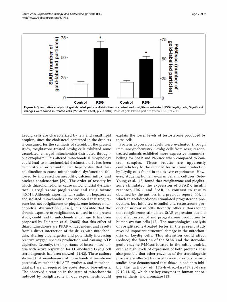
in 0.1 cacodylate buffer, pH 7.2, dehydrated in acetoneand embedded in SPIN-PON resin (Embed 812). Poly-merization was performed at 60°C for three days [23].Ultrathin sections were collected on 300-mesh nickelgrids, counterstained with 5% uranyl acetate and leadcitrate and examined with a FEI Morgani 268D trans-mission electron microscope. For the immunocytochem-ical study, the isolated and hCG-stimulated Leydig cellswere fixed overnight in a solution containing 0.5% glu-taraldehyde and 4% paraformaldehyde in 0.1 M phos-phate buffer. After fixation, the samples were washedthree times in the same buffer, incubated with 50 mMammonium chloride for 40 min, dehydrated in alcoholand embedded in LR-White resin (Electron MicroscopyScience, Washington, PA, USA). Polymerization wasperformed at 30°C for five days. This procedure was car-ried out as described by Peixoto et al. [24].ImmunocytochemistryUltrathin sections of isolated and hCG-stimulated Leydigcells were cut with a diamond knife, collected on nickelgrids and incubated for 30 min. at room temperature in0.02 M PBS, pH 7.2, containing 1% BSA and 0.1% Tween20 (PBS-BT). The sections were then incubated for onehour with primary antibodies against StAR and P450scc atdilutions of 1:25 and 1:200, respectively, in PBS-BT. Thesections were then washed in PBS-BT and incubated witha secondary antibody, 10 nm colloidal gold-labeled goatanti-rabbit IgG. As an antibody control, sections wereincubated only in the presence of the gold-labeled marker.Following the immunostaining procedures, the sectionswere counterstained with 5% uranyl acetate and leadcitrate [24]. Quantitative analysis was performed onphotomicrographs at a final magnification of 28’000× of10 different Leydig cells, showing the entire profile andrandomly chosen, in order to compare the number ofgold-labeled particles in the control and rosiglitazone-trea-ted cells using the Student’s t test. Since the experimen-tally treated cells and control samples were processed inan identical method, no correction for tissue shrinkagewas performed.AntibodiesThe polyclonal antibodies StAR (sc-25806, Santa CruzBiotechnology, INC., Santa Cruz, CA, USA) and cyto-chrome P450scc enzyme (AB1244, Chemicon Interna-tional, Inc., Canada) were raised in rabbits againstdifferent peptides corresponding to amino acids 1-285representing full-length StAR of human origin andamino acids 421-441 of the rat cytochrome P450sccenzyme, respectively. The 10-nm colloidal gold-labeledgoat anti-rabbit IgG was purchased from Sigma Chemi-cal Co. (St. Louis, MO, USA).Radioimmunoassay and statistical analysisTestosterone was measured in plasma (with extraction)and directly (without extraction) in the incubation
medium by a charcoal-dextran RIA [25] that employs[3H]-testosterone as a tracer and primary antiserumraised in rabbits in our laboratory against testosterone-3-(0-carboxymethyl)oxime:BSA. Intra-assay and inter-assay coefficients of variation were 8.1% and 15.1%,respectively. The testosterone antibody demonstrated <0.1% cross-reactivity with androstenedione, dehydroe-piandrosterone, androsterone, 17a-hydroxyprogesterone,b-estradiol and estrone. None of the substances testedinterfered with the assays. The data from the differentanalyses are expressed as the mean ± SEM of six repli-cates determinations for plasma and triplicate determi-nations for incubation medium and were representativeof results obtained in at least two similar experiments.The Student’s t test, one-way analysis of variance(ANOVA) and Dunnett’s test were used to determinedifferences between control and rosiglitazone-treatedplasma and cells, respectively. P-values less than 0.05were considered statistically significant.
ResultsBody weight, testis weight, seminal vesicle weight andplasma testosteroneThe treatment of rats with rosiglitazone did not inducechanges in body weight or the relative weight of thetestes or dried seminal vesicles (data not shown). Totalplasma testosterone was not significantly modified bythe treatment with rosiglitazone (control: 25.01 ± 3.5;rosiglitazone: 38.4 ± 7.0 ng/ml), which may explain theunaltered weight of the seminal vesicles, since it isknown that testosterone supports the trophism of thisorgan.Ex vivo testosterone secretionThese experiments examined the impact of chronictreatment with rosiglitazone on the steroidogenicresponse of Leydig cells to direct induction by activatorsof the cAMP/PKA pathway (hCG and dbcAMP), whichis the major signaling pathway regulating steroidogenesis[26], and by substrates of steroidogenesis (22-OH-C andpregnenolone). The objective of the use of these sub-strates was to determine whether the limiting-steps ofsteroidogenesis - the transportation of cholesterol fromouter to inner mitochondrial membrane by the StARprotein and the cleavage of the cholesterol side chain bymitochondrial P450scc to yield pregnenolone - wereaffected by rosiglitazone treatment. Rosiglitazone treat-ment modified the steroidogenic response, resulting in adecrease in testosterone production under basal, hCG/dbcAMP- or 22 (R)-OH-C/pregnenolone-induced condi-tions (Figure 1). This reduced steroidogenic response tostimulators/inducers of testosterone production can beseen by the reduction in the magnitude of the stimula-tion/induction obtained in both the control and rosigli-tazone-treated groups. The increase in testosterone
Couto et al. Reproductive Biology and Endocrinology 2010, 8:13http://www.rbej.com/content/8/1/13
Page 3 of 9
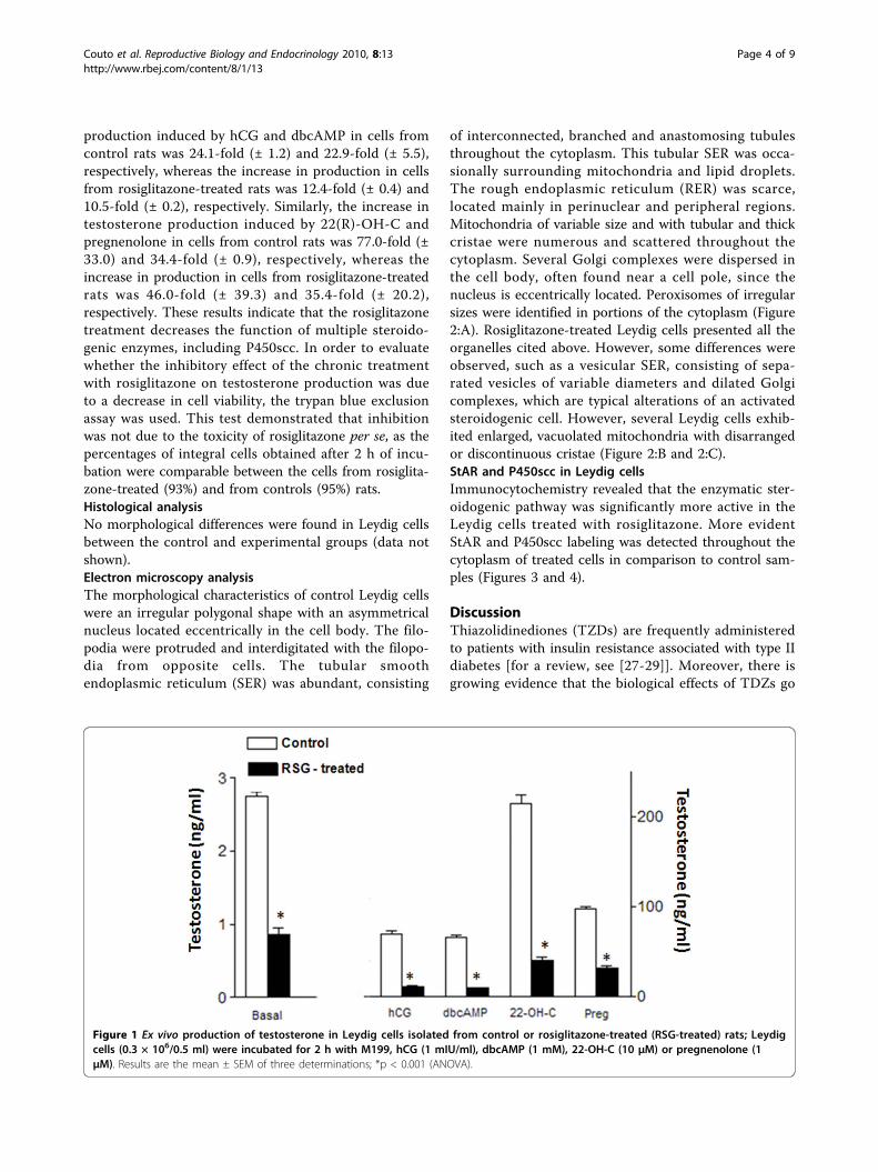
production induced by hCG and dbcAMP in cells fromcontrol rats was 24.1-fold (± 1.2) and 22.9-fold (± 5.5),respectively, whereas the increase in production in cellsfrom rosiglitazone-treated rats was 12.4-fold (± 0.4) and10.5-fold (± 0.2), respectively. Similarly, the increase intestosterone production induced by 22(R)-OH-C andpregnenolone in cells from control rats was 77.0-fold (±33.0) and 34.4-fold (± 0.9), respectively, whereas theincrease in production in cells from rosiglitazone-treatedrats was 46.0-fold (± 39.3) and 35.4-fold (± 20.2),respectively. These results indicate that the rosiglitazonetreatment decreases the function of multiple steroido-genic enzymes, including P450scc. In order to evaluatewhether the inhibitory effect of the chronic treatmentwith rosiglitazone on testosterone production was dueto a decrease in cell viability, the trypan blue exclusionassay was used. This test demonstrated that inhibitionwas not due to the toxicity of rosiglitazone per se, as thepercentages of integral cells obtained after 2 h of incu-bation were comparable between the cells from rosiglita-zone-treated (93%) and from controls (95%) rats.Histological analysisNo morphological differences were found in Leydig cellsbetween the control and experimental groups (data notshown).Electron microscopy analysisThe morphological characteristics of control Leydig cellswere an irregular polygonal shape with an asymmetricalnucleus located eccentrically in the cell body. The filo-podia were protruded and interdigitated with the filopo-dia from opposite cells. The tubular smoothendoplasmic reticulum (SER) was abundant, consisting
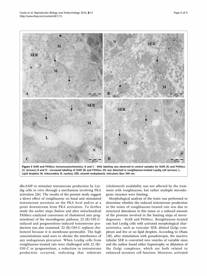
of interconnected, branched and anastomosing tubulesthroughout the cytoplasm. This tubular SER was occa-sionally surrounding mitochondria and lipid droplets.The rough endoplasmic reticulum (RER) was scarce,located mainly in perinuclear and peripheral regions.Mitochondria of variable size and with tubular and thickcristae were numerous and scattered throughout thecytoplasm. Several Golgi complexes were dispersed inthe cell body, often found near a cell pole, since thenucleus is eccentrically located. Peroxisomes of irregularsizes were identified in portions of the cytoplasm (Figure2:A). Rosiglitazone-treated Leydig cells presented all theorganelles cited above. However, some differences wereobserved, such as a vesicular SER, consisting of sepa-rated vesicles of variable diameters and dilated Golgicomplexes, which are typical alterations of an activatedsteroidogenic cell. However, several Leydig cells exhib-ited enlarged, vacuolated mitochondria with disarrangedor discontinuous cristae (Figure 2:B and 2:C).StAR and P450scc in Leydig cellsImmunocytochemistry revealed that the enzymatic ster-oidogenic pathway was significantly more active in theLeydig cells treated with rosiglitazone. More evidentStAR and P450scc labeling was detected throughout thecytoplasm of treated cells in comparison to control sam-ples (Figures 3 and 4).
DiscussionThiazolidinediones (TZDs) are frequently administeredto patients with insulin resistance associated with type IIdiabetes [for a review, see [27-29]]. Moreover, there isgrowing evidence that the biological effects of TDZs go
Figure 1 Ex vivo production of testosterone in Leydig cells isolated from control or rosiglitazone-treated (RSG-treated) rats; Leydigcells (0.3 × 10⁶/0.5 ml) were incubated for 2 h with M199, hCG (1 mIU/ml), dbcAMP (1 mM), 22-OH-C (10 μM) or pregnenolone (1μM). Results are the mean ± SEM of three determinations; *p < 0.001 (ANOVA).
Couto et al. Reproductive Biology and Endocrinology 2010, 8:13http://www.rbej.com/content/8/1/13
Page 4 of 9

beyond insulin-sensitizing [30]. It has been demon-strated that TDZs have a considerable impact on theproduction and metabolism of gonad hormones[6,8,14,17-20]. Rosiglitazone and other thiazolidine-diones are known to affect testosterone production inhumans [10,11,31]. However, effects of rosiglitazone ontestosterone level and synthesis have not been reportedin experimental animal models. Therefore, the presentstudy investigated the effect of chronic treatment withrosiglitazone on plasma testosterone levels, steroidogenicresponse and morphology of Leydig cells in normal rats.Steroid hormones are synthesized from cholesterol in
the gonads in response to pituitary hormones, such asLH/hCG via the classic cAMP/PKA pathway. The mainrate-limiting step in the steroidogenenic pathway is thetransportation of cholesterol from the outer to innermitochondrial membrane by a transmembrane protein,StAR protein, the expression and activation of which ismaintained by cAMP modulated PKA under maximalstimulation of LH [32].In the present study, normal rats exposed to rosiglita-
zone underwent no changes in the level of total plasmatestosterone when compared with the controls. A num-ber of authors have reported contrasting results regard-ing the impact of TZDs on plasma testosterone levels indifferent clinical and experimental models. In maleZucker diabetic fatty (ZDP) rats, rosiglitazone treatmentdid not alter plasma testosterone levels [19,33]. Inhealthy men, rosiglitazone decreased testosterone anddihydrotestosterone (DHT) production rates [11]. Inwomen with PCOS and hyperandrogenism, TZD treat-ment increases sex hormone binding globulin (SHBG)levels in plasma, leading to a decrease in free-circulatingtestosterone levels [34]. Circulating testosterone isknown to be present in three major fractions: free, albu-min-bound and sex hormone binding globulin (SHBG)[35]. In contrast to women, men with type 2 diabeteshave low testosterone levels and treatment with rosigli-tazone induces an increase in the three fractions of cir-culating testosterone and SHBG levels [36]. As thetestosterone measured in the present study representsthe total circulating fraction, the measurement of theother two fractions (free and albumin-bound) would benecessary for a better assessment of the effect of rosigli-tazone treatment in the present study. However, unlikethe human, adult rats do not express SHBG [37]. Addi-tionally, the possibility of decreased clearance rate oftestosterone can not be excluded.In the ex vivo experiments, basal testosterone pro-
duction was reduced by chronic treatment with rosigli-tazone and Leydig cells from rosiglitazone-treated ratswere less responsive to cAMP/PKA pathway activationof testosterone production than those from controlrats. The ex vivo model is based on the ability of hCG/
Figure 2 Testis Leydig Cells; A - Untreated Leydig cells withnucleus, Golgi complex (arrows), SER, RER and mitochondria;Bar 1 μm; B and C - Leydig cells treated with rosiglitazoneshowing dilated and vacuolated mitochondria (star), vesicularsmooth endoplasmic reticulum and dilated Golgi complex(arrows); Bars 1 μm and 0.5 μm, respectively; M, mitochondria;N, nucleus; RER, rough endoplasmic reticulum; SER, smoothendoplasmic reticulum.
Couto et al. Reproductive Biology and Endocrinology 2010, 8:13http://www.rbej.com/content/8/1/13
Page 5 of 9
dbcAMP to stimulate testosterone production by Ley-dig cells in vitro through a mechanism involving PKAactivation [26]. The results of the present study suggesta direct effect of rosiglitazone on basal and stimulatedtestosterone secretion on the PKA level and/or at apoint downstream from PKA activation. To furtherstudy the earlier steps (before and after mitochondrialP450scc-catalyzed conversion of cholesterol into preg-nenolone) of the steroidogenic pathway, 22 (R)-OH-C-induced and pregnenolone-induced testosterone pro-duction was also examined. 22 (R)-OH-C replaces cho-lesterol because it is membrane-permeable. The highconcentrations used were to obviate the interference ofany endogenous precursor. When Leydig cells fromrosiglitazone-treated rats were challenged with 22 (R)-OH-C or pregnenolone, a reduction in testosteroneproduction occurred, indicating that substrate
(cholesterol) availability was not affected by the treat-ment with rosiglitazone, but rather multiple steroido-genic enzymes were limiting.Morphological analysis of the testis was performed to
determine whether the reduced testosterone productionin the testes of rosiglitazone-treated rats was due tostructural alterations in this tissue or a reduced amountof the proteins involved in the limiting steps of steroi-dogenesis - StAR and P450scc. Rosiglitazone-treatedrats had Leydig cells with activated morphological char-acteristics, such as vesicular SER, dilated Golgi com-plexes and few or no lipid droplets. According to Ohata[38], after stimulation with gonadotropin, the inactivetubular SER is converted into vesicles of variable sizesand the author found either hypertrophy or dilatation ofthe Golgi complexes, which are both related toenhanced secretory cell function. Moreover, activated
Figure 3 StAR and P450scc immunocytochemistry; A and C - little labeling was observed in control samples for StAR (A) and P450scc(C) (arrows); B and D - increased labeling of StAR (B) and P450scc (D) was detected in rosiglitazone-treated Leydig cell (arrows); L,Lipid droplets; M, mitocondria; N, nucleus; SER, smooth endoplasmic reticulum; Bars 500 nm.
Couto et al. Reproductive Biology and Endocrinology 2010, 8:13http://www.rbej.com/content/8/1/13
Page 6 of 9
Leydig cells are characterized by few and small lipiddroplets, since the cholesterol contained in the dropletsis consumed for the synthesis of steroid. In the presentstudy, rosiglitazone-treated Leydig cells exhibited somevacuolated, enlarged mitochondria distributed through-out cytoplasm. This altered mitochondrial morphologycould lead to mitochondrial dysfunction. It has beendemonstrated in rat and human hepatocytes, that thia-zolidinediones cause mitochondrial dysfunction, fol-lowed by increased permeability, calcium influx, andnuclear condensation [39]. The order of toxicity bywhich thiazolidinediones cause mitochondrial dysfunc-tion is troglitazone pioglitazone and rosiglitazone[40,41]. Although experimental studies on hepatocytesand isolated mitochondria have indicated that troglita-zone but not rosiglitazone or pioglitazone induces mito-chondrial dysfunction [39,40], it is possible that thechronic exposure to rosiglitazone, as used in the presentstudy, could lead to mitochondrial damage. It has beenproposed by Feinstein et al. (2005) that this effect ofthiazolidinediones are PPARg-independent and resultsfrom a direct interaction of the drugs with mitochon-dria, altering bioenergetics and potentially increasingreactive oxygen species production and causing ATPdepletion. Recently, the importance of intact mitochon-dria with active respiration for LH-mediated Leydig cellsteroidogenesis has been showed [41,42]. These authorsshowed that maintenance of mitochondrial membranepotencial, mitochondrial ATP synthesis, and mitochon-drial pH are all required for acute steroid biosynthesis.The observed alteration in the state of mitochondriainduced by rosiglitazone in our experiments could
explain the lower levels of testosterone produced bythese cells.Protein expression levels were evaluated through
immunocytochemistry. Leydig cells from rosiglitazone-treated animals exhibited more expressive immunola-belling for StAR and P450scc when compared to con-trol samples. These results are apparentlycontradictory to the reduced testosterone productionby Leydig cells found in the ex vivo experiments. How-ever, studying human ovarian cells in cultures, Seto-Young et al. [43] found that rosiglitazone and pioglita-zone stimulated the expression of PPARg, insulinreceptor, IRS-1 and StAR, in contrast to resultsobtained by the authors in a previous report [44], inwhich thiazolidinediones stimulated progesterone pro-duction, but inhibited estradiol and testosterone pro-duction in ovarian cells. Recently, other authors foundthat rosiglitazone stimulated StAR expression but didnot affect estradiol and progesterone production byhuman ovarian cells [45]. The morphological analysisof rosiglitazone-treated testes in the present studyrevealed important structural damage in the mitochon-dria of Leydig cells. This alteration could affect(reduce) the function of the StAR and the steroido-genic enzyme P450scc located in the mitochondria,even at high levels of expression of both proteins. It isalso possible that other enzymes of the steroidogenicprocess are affected by rosiglitazone. Previous in vitrostudies have demonstrated that thiazolidinediones inhi-bit the activity of 17a-hydroxylase/17,20-lyase[7,12,14,15], which are key enzymes in human andro-gen synthesis, and aromatase [13].
Figure 4 Quantitative analysis of gold-labeled particle distribution in control and rosiglitazone-treated (RSG) Leydig cells; Significantchanges were found in treated cells (*Student’s t test, p < 0.0002). Mean of gold-labeled particles (mean ± S.D); N = 10.
Couto et al. Reproductive Biology and Endocrinology 2010, 8:13http://www.rbej.com/content/8/1/13
Page 7 of 9
Rosiglitazone is a ligand for PPARg. Recently, theexpression of PPARg has been demonstrated in Leydigcells and an increase in PPARg mRNA and proteinlevels has been observed in these cells after chronictreatment with glitazones [33,46]. Therefore, it seemsreasonable that the expression of StAR and P450sccobserved in the present study may be mediated by thenuclear receptor PPARg. However, the mitochondrialdamage could indicate that the influence of rosiglitazoneon testosterone production could also be PPARg-independent.
ConclusionIn summary, the present study was designed to examinethe effect of chronic treatment with rosiglitazone on thesteroidogenic capacity of Leydig cells in normal adultrats. The results revealed that the Leydig cells from rosi-glitazone-treated rats showed significant reduction intestosterone production under basal, hCG/dbcAMP- or22 (R)-OH-C/pregnenolone-induced conditions,although increased labeling of StAR and P450scc wasdetected in these cells by immunocytochemistry. Theultrastructural study suggested that the lower levels oftestosterone produced by these cells could be due tomitochondrial damage induced by rosiglitazone. Furtherstudies are necessary to evaluate the impact of chronictreatment with rosiglitazone on the activity of the StARand steroidogenic enzymes involved in testosterone pro-duction. Although the present study does not permitdistinguishing a pituitary gland from the direct effect ofrosiglitazone, the observation that this TDZ reduces tes-tosterone production in an ex vivo model indicates thatrosiglitazone has direct effects on Leydig cells that areindependent of the effects of the drug on the secretionof gonadotropin. Finally, rosiglitazone action in testicu-lar steroidogenesis is potentially of physiological andpathophysiological significance.
AcknowledgementsThis study was supported by the Universidade Federal de Pernambuco(UFPE), Fundação Oswaldo Cruz (FIOCRUZ), Centro de TecnologiasEstratégicas do Nordeste (CETENE) and Conselho Nacional deDesenvolvimento Científico e Tecnológico (CNPq).
Author details1Department of Morphology and Physiology, Universidade Federal Rural dePernambuco, Recife, 52.171-900, Brazil. 2Ultrastructure Laboratory, AggeuMagalhães Research Center (FIOCRUZ) and Center for Strategic Technologiesof the Northeast (CETENE), Recife, 50.670-901, Brazil. 3Department ofAntibiotics, Universidade Federal de Pernambuco, Recife, 50.670-901, Brazil.4Department of Physiology and Pharmacology, Universidade Federal dePernambuco, Recife, 50.670-901, Brazil.
Authors’ contributionsJAC, CDB, DPU, JSBCV, MIW carried out the treatment of animals, Leydigcells isolation, radioimmunoassay and performed the statistical analysis. KLAS,CAP carried out the ultrastructural analysis and immunocytochemistry. MIWand CAP conceived of the study, and participated in its design and
coordination. MIW, DPU, CAP, KLAS, JAC, CDB, JSBCV, MCL, SLG and IRPparticipated in the interpretation and analysis of the data and helped todraft the manuscript. All authors read and approved the final manuscript.
Competing interestsThe authors declare that they have no competing interests.
Received: 29 December 2009Accepted: 9 February 2010 Published: 9 February 2010
References1. Inzucchi SE, Maggs DG, Spollett GR, Page SL, Rife FS, Walton V, Shulman GI:
Efficacy and metabolic effects of metformin and troglitazone in type IIdiabetes mellitus. N Engl J Med 1998, 338:867-872.
2. De Leo V, La Marca A, Ditto A, Morgante G, Cianci A: Effects of metforminon gonadotropin-induced ovulation in women with polycystic ovarysyndrome. Fertil Steril 1999, 72:282-285.
3. Miyazaki Y, Mahankali A, Matsuda M, Glass L, Mahankali S, Ferrannini E,Cusi K, Mandarino LJ, DeFronzo RA: Improved glycemic control andenhanced insulin sensitivity in type 2 diabetic subjects treated withpioglitazone. Diabetes Care 2001, 24:710-719.
4. Lord JM, Flight IH, Norman RJ: Insulin-sensitising drugs (metformin,troglitazone, rosiglitazone, pioglitazone, D-chiro-inositol for polycysticovary syndrome. Cochrane Database Syst Ver 2003, 3:CD003053.
5. Sepilian V, Nagamani M: Effects of rosiglitazone in obese women withpolycystic ovary syndrome and severe insulin resistance. J Clin EndocrinolMetab 2005, 90:60-65.
6. Freeman DA, Romero A: Effects of troglitazone on cells n intracellularcholesterol distribution and cholesterol-dependent cell functions in MA-10 Leydig tumour cells. Biochem Pharmacol 2003, 66:307-313.
7. Kempná P, Hofer G, Mullis PE, Flück CE: Pioglitazone inhibits androgenproduction in NCI-H295R cells by regulating gene expression of CYP17and HSD3B2. Mol Pharmacol 2007, 71:787-798.
8. Froment P, Gizard F, Defever D, Staels B, Dupont J, Monget P: Peroxisomeproliferator-activated receptors in reproductive tissues: fromgametogenesis to parturition. J Endocrinol 2006, 189:199-209.
9. Dunaif A, Scott D, Finegood D, Quintana B, Whitcomb R: The insulin-sensitizing agent troglitazone improves metabolic and reproductiveabnormalities in the polycystic ovary syndrome. J Clin Endocrinol Metab1996, 81:3299-3306.
10. Bloomgarden ZT, Futterweit W, Poretsky L: Use of insulin-sensitizingagents in patients with polycystic ovary syndrome. Endocr Pract 2001,7:279-286.
11. Vierhapper H, Nowontny P, Waldhäusl W: Reduced production rates oftestosterone and dihydrotestosterone in healthy men treated withrosiglitasone. Metabolism 2003, 52:230-232.
12. Gasic S, Bodenburg Y, Nagamani M, Green A, Urban R: Troglitazoneinhibits progesterone production in porcine granulosa cells.Endocrinology 1998, 139:4962-4966.
13. Mu YM, Yanase T, Nishi Y, Waseda N, Oda T, Tanaka A, Takayanagi R,Nawata H: Isulin sensitizer, troglitazone, directly inhibits aromataseactivity in human ovarian granulosa cells. Biochem Biophys Res Commun2000, 271:710-713.
14. Arlt W, Auchus RJ, Miller WL: Thiazolidinediones but not metformindirectly inhibit the steroidogenic enzymes P450c and 3b-hydroxysteroiddehydrogenase. J Biol Chem 2001, 276:16767-16771.
15. Gasic S, Nagamani M, Green A, Urban RJ: Troglitazone is a competitiveinhibitor of 3b-hydroxysteroid dehydrogenase enzyme in the ovary. AmJ Obst Ginecol 2001, 184:575-579.
16. Schoppee PD, Garmey JC, Valdhuis J: Putative activaction of theperoxixome proliferator-activated receptor g impairs androgen andenhances progesterone biosynthesis in primary cultures of porcinetheca cells. Biol Reprod 2002, 66:190-198.
17. Veldhuis JD, Zhang G, Garmey JC: Troglitazone, an insulin-sensitizingthiazolidinedione, represses combined stimulation by LH and insulin ofde novo androgen biosynthesis by thecal cells in vitro. J Clin EndocrinolMetab 2002, 87:1129-1133.
18. Rubin GL, Duong JH, Clyne CD, Speed CJ, Murata Y, Gong C, Simpson ER:Ligands for the peroxisomal proliferator-activated receptor g and theretinoid × receptor inhibit aromatase cytochrome P450 (CYP19)
Couto et al. Reproductive Biology and Endocrinology 2010, 8:13http://www.rbej.com/content/8/1/13
Page 8 of 9
expression mediated by promoter II in human breast adipose.Endocrinology 2002, 143:2863-2871.
19. Fürnsinn C, Nowotny B, Brunmair B, Gras F, Roden M, Waldhäusl W,Vierhapper H: Thiazolidinediones influence plasma steroides of maleobese Zucker rats. Endocrinology 2002, 143:327-330.
20. Minge CE, Robker RL, Norman RJ: PPAR gamma coordinating metabolicand immune contributions to female fertility. PPAR Res 2008, Article ID243791.
21. Wanderley MI, Negro-Vilar A: Pretreatment with phorbol ester and LHRHagonist reduces testosterone production and protein kinase C activity inrat Leydig cells challenged with PDBu and LHRH. Braz J Med Biol Res1996, 29:1557-65.
22. Weng Q, Medan MS, Watanabe G, Tsubota T, Tanioka Y, Taya K:Immunolocalization of steroidogenic enzymes P450scc, 3bHSD, P450c17,and P450arom in Göttingen miniature pig testes. J Reprod Dev 2005,51:299-304.
23. Saraiva KLA, Silva Junior VA, Dias ESF, Peixoto CA: Morphological changesin the testis induced by diethylcarbamazine. Reprod Toxicol 2006,22:754-759.
24. Peixoto CA, Norões J, Rocha A, Dreyer G: Immunocytochemicallocalization and distribution of human albumin in Wuchereria bancroftiadult worms. Arch Pathol Lab Med 1999, 123:173-177.
25. Niswender GD, Akbar AM, Nett TM: Use of specific antibodies forquantification of steroid hormones. Methods in Enzymology. Vol Part AXXXVI New York: Academic PressO’Malley BW, Hardman JG 1975, 16.
26. Dehejia A, Nozu K, Catt KJ, Dufau ML: Luteinizing hormone receptors andgonadotropic activation of purified rat Leydig cells. J Biol Chem 1982,257:13781-86.
27. Houseknecht KL, Cole BM, Steele PJ: Peroxisome proliferator-activatedreceptor gamma (PPAR gamma) and its ligands: a review. Domest AnimEndocrinol 2002, 22:1-23.
28. Gurnell M, Savage DB, Chatterjee VK, O’Rahilly S: The metabolic syndrome:peroxisome proliferator-activated receptor gamma and its therapeuticmodulation. J Clin Encocrinol Metab 2003, 88:2412-2421.
29. Staels B, Fruchart JC: Therapeutic roles of peroxisome proliferator-activated receptor agonists. Diabetes 2005, 54:2460-2470.
30. Kalaitzidis RG, Sarafidis PA, Bakris GL: Effects of thiazolidinediones beyondglycaemic control. Curr Pharm Des 2009, 15:529-536.
31. Azziz R, Ehrmann D, Legro RS, Whitcomb RW, Hanley R, Fereshetian AG,O’Keefe M, Ghazzi MN: Troglitazone improves ovulation and hirsutism inthe polycystic ovary syndrome: a multicenter, double blind, placebo-controlled trial. J Clin Endocrinol Metab 2001, 86:1626-1632.
32. Stocco DM, Clark BJ: The role of the steroidogenic acute regulatoryprotein in steroidogenesis. Steroids 1997, 62:29-36.
33. Mansour M, Coleman E, Dennis J, Akingbemi B, Ashwartz D, Braden T,Judd R, Plaisance E, Ken Stuart L, Morrison E: Activation of PPARg byrosiglitazone does not negatively impact male sex steroid hormones indiabetic rats. PPAR Res 2009, 101857, doi: 10.1155/2009/101857.
34. Brettenthaler N, De Geyter C, Huber PR, Keller U: Effect of the insulinsensitizer pioglitazone on insulin resistance, hyperandrogenism, andovulatory dysfunction in women with polycystic ovary syndrome. J ClinEndocrinol Metab 2004, 89:3835-3840.
35. Kapoor D, Malkin CJ, Channer KS, Jones TH: Androgens, insulin resistanceand vascular disease in men. Clin Endocrinol (Oxf) 2005, 63:239-250.
36. Kapoor D, Channer KS, Jones TH: Rosiglitazone increases bioactivetestosterone and reduces waist circumference in hypogonadal men withtype 2 diabetes. Diabetes Vasc Dis Res 2008, 5:135-137.
37. Reventos J, Sullivan PM, Joseph DR, Gordon JW: Tissue-specific expressionof the rat androgen-binding protein/sex hormone-binding globulin genein transgenic mice. Mol Cell Endocrinol 1993, 96:69-73.
38. Ohata M: Electron microscopy study on the testicular interstitial cells inthe mouse. Arch Histol Jpn 1979, 42:51-79.
39. Feinstein DL, Spagnolo A, Akar C, Weinberg G, Murphy P, Gavrilyuk V, DelloRusso C: Receptor-independent actions of PPAR thiazolidinedioneagonists: is mitochondrial function the key?. Biochem Pharmacol 2005,70:177-188.
40. Masubuchi Y: Metabolic and non-metabolic factors determiningtroglitazone hepatotoxicity: a review. Drug Metab Pharmacokinet 2006,21:347-356.
41. Allen JA, Shankara T, Janus P, Buck S, Diemer T, Hales KH, Hales DB:Energized, polarized, and actively respiring mitochondria are requiredfor acute Leydig cell steroidogenesis. Endocrinology 2006, 147:3924-3935.
42. Midzak AS, Liu J, Zirkin BR, Chen H: Effects of myxothiazol on Leydig cellsteroidogenesis: inhibition of luteinizing hormone-mediatedtestosterone synyhesis but stimulation of basal steroidogenesis.Endocrinology 2007, 148:2583-2590.
43. Seto-Young D, Avtanski D, Strizhevsky M, Parikh G, Patel P, Kaplun J,Holcomb K, Rosenwaks Z, Poretsky L: Interactions among peroxisomeproliferator activated receptor-g, insulin signaling pathways, andsteroidogenic acute regulatory protein in human ovarian cells. J ClinEndocrinol Metab 2007, 92:2232-2239.
44. Seto-Young D, Paliou M, Schlosser J, Avtanski D, Park A, Patel P, Holcomb K,Chang P, Poretsky L: Direct thiazolidinedione action in the human ovary:insulin-independent and insulin-sensitizing effects on steroidigenesisand insulin-like growth factor binding protein-1 production. J ClinEndocrinol Metab 2005, 90:6099-6105.
45. Chen Q, Sun X, Chen J, Cheng L, Wang J, Wang Y, Sun Z: Directrosiglitazone action on steroidogenesis and proinflammatory factorproduction in human granulosa-lutein cells. Reprod Biol Endocrinol 2009,7:147.
46. Kowalewski MP, Dyson MT, Manna PR, Stocco DM: Involvement ofperoxisome proliferator-activated receptor g in gonadal steroidogenesisand steroidogenic acute regulatory protein expression. Reprod Fertil Dev2009, 21:909-922.
doi:10.1186/1477-7827-8-13Cite this article as: Couto et al.: Effect of chronic treatment withRosiglitazone on Leydig cell steroidogenesis in rats: In vivo and ex vivostudies. Reproductive Biology and Endocrinology 2010 8:13.
Submit your next manuscript to BioMed Centraland take full advantage of:
• Convenient online submission
• Thorough peer review
• No space constraints or color figure charges
• Immediate publication on acceptance
• Inclusion in PubMed, CAS, Scopus and Google Scholar
• Research which is freely available for redistribution
Submit your manuscript at www.biomedcentral.com/submit
Couto et al. Reproductive Biology and Endocrinology 2010, 8:13http://www.rbej.com/content/8/1/13
Page 9 of 9