Embed Size (px)
Citation preview
RESEARCH Open Access
Identification of harmful cyanobacteria in theSacramento-San Joaquin Delta and Clear Lake,California by DNA barcodingTomofumi Kurobe1, Dolores V Baxa1*, Cécile E Mioni2, Raphael M Kudela2, Thomas R Smythe3, Scott Waller4,Andrew D Chapman5 and Swee J Teh1
Abstract
Accurate identification of cyanobacteria using traditional morphological taxonomy is challenging due to themagnitude of phenotypic plasticity among natural algal assemblages. In this study, molecular approach was utilizedto facilitate the accurate identification of cyanobacteria in the Sacramento-San Joaquin Delta and in Clear Lake inNorthern California where recurring blooms have been observed over the past decades. Algal samples werecollected from both water bodies in 2011 and the samples containing diverse cyanobacteria as identified bymorphological taxonomy were chosen for the molecular analysis. The 16S ribosomal RNA genes (16S rDNA) andthe adjacent internal transcribed spacer (ITS) regions were amplified by PCR from the mixed algal samples usingcyanobacteria generic primers. The obtained sequences were analyzed by similarity search (BLASTN) andphylogenetic analysis (16S rDNA) to differentiate species sharing significantly similar sequences. A total of 185plasmid clones were obtained of which 77 were successfully identified to the species level: Aphanizomenon flos-aquae, Dolichospermum lemmermannii (taxonomic synonym: Anabaena lemmermannii), Limnoraphis robusta(taxonomic synonym: Lyngbya hieronymusii f. robusta) and Microcystis aeruginosa. To date, Dolichospermum andLimnoraphis found in Clear Lake have only been identified to the genus lavel by microscopy. During the course ofthis study, morphological identification and DNA barcoding confirmed A. flos-aquae as the predominantcyanobacterium in the Sacramento-San Joaquin Delta indicating a shift from M. aeruginosa that have dominatedthe blooms in the past decade. Lastly, the species-specific identification of Limnoraphis robusta in Clear Lake isanother significant finding as this cyanobacterium has, thus far, only been reported in Lake Atitlan blooms inGuatemala.
Keywords: Harmful cyanobacteria; DNA barcoding; 16S ribosomal DNA; Internal transcribed spacer region
BackgroundHarmful cyanobacterial blooms (CyanoHABs) are aserious global concern and are often associated withodorous metabolites in drinking water and toxins inaquaculture facilities and in the environment (Mankiewiczet al. 2003; Smith et al. 2008). Different types of toxins areproduced from several cyanobacterial species includinghepatotoxins (microcystins), cytotoxins (cylindrospermop-sin), neurotoxins (anatoxin-a, antillatoxin, saxitoxins), anddermatoxins (lyngbyatoxins). These potent toxins render
serious consequences to the health of ecosystems,aquatic organisms, domestic animals, and humans upondirect contact or consumption of CyanoHAB impactedwater (Mankiewicz et al. 2003; Osswald et al. 2007;Puschner et al. 2010; Acuña et al. 2012).The current study focused on molecular analysis of
cyanobacterial species from two ecosystems that areecologically and economically important in California.The Sacramento-San Joaquin Delta is a critical watersupply system in Northern California, which providesdrinking water to two-thirds of the California popu-lation (more than 20 million people) and irrigates4.5 million acres of farmlands (Jassby 2008). Theestuary also provides essential habitats for many
* Correspondence: [email protected] of Anatomy, Physiology, and Cell Biology, School of VeterinaryMedicine, University of California, Davis, CA 95616, USAFull list of author information is available at the end of the article
a SpringerOpen Journal
© 2013 Kurobe et al.; licensee Springer. This is an Open Access article distributed under the terms of the Creative CommonsAttribution License (http://creativecommons.org/licenses/by/2.0), which permits unrestricted use, distribution, and reproductionin any medium, provided the original work is properly cited.
Kurobe et al. SpringerPlus 2013, 2:491http://www.springerplus.com/content/2/1/491
SDWA 221
anadromous, commercial, and recreational fish such asstriped bass (Morone saxatilis), Chinook salmon (Onco-rhynchus tshawytscha), and several endangered fish spe-cies such as the delta smelt (Hypomesus transpacificus)(Sommer et al. 2007). Blooms of the hepatotoxin-producing cyanobacterium Microcystis aeruginosa werefirst recorded in the Sacramento-San Joaquin Delta in1999, and since then, cyanobacterial blooms have re-occurred and have been monitored for biomass and tox-icity (Lehman et al. 2005, 2008, 2010; Spier et al. 2010).Colonial forms of M. aeruginosa are widely distributedalong the 180 km of freshwater and brackish waterwaysof the delta that may affect indigenous invertebrates andfishes (Lehman et al. 2005, 2008, 2010; Ger et al. 2010).Other harmful cyanobacteria such as Aphanizomenon,Dolichospermum (formerly recognized as plankticAnabaena) (Wacklin et al. 2009), and Oscillatoria havebeen observed in the Sacramento-San Joaquin Delta,although to a lesser extent than Microcystis (Cloern andDufford 2005; Lehman et al. 2010; Spier et al. 2010).Because algal bloom studies in the Delta have mainlyfocused on M. aeruginosa (Lehman et al. 2005, 2008,2010), the occurrence, abundance, and potential role ofother toxin-producing cyanobacteria to indigenousfisheries resources are largely unknown.Clear Lake is the largest natural lake in California and
provides drinking water to local communities. The lakesupports recreational activities and tourism for sportfishing and water contact sports, forming an industrygreater than 50 million dollars in the local county (Gold-stein and Tolsdorf 1994). Although considered “impaired”in terms of hyper-eutrophication from phosphorus andsulfate overload from anthropogenic activities, the lake isused for storage of irrigation water for downstreamagricultural lands. Land use such as construction of farm-lands, road building, livestock grazing, logging, and fire-wood cutting have accelerated erosion resulting in largephosphorus inputs mostly from basins around the lake(Richerson et al. 1994). Cyanobacterial assemblages in thelake reached the highest densities from the mid 1970’s to1990 (Horne 1975; Richerson et al. 1994; Winder et al.2010) and were dominated by diazotrophic cyanobacteriasuch as Aphanizomenon, Dolichospermum, and the non-nitrogen fixing cyanobacterium Microcystis (Horne1975; Richerson et al. 1994). Lyngbya (now known asLimnoraphis) blooms have also been recorded since2009 (Mioni and Kudela 2011, Mioni et al. 2012).These cyanobacteria form scum on the water surfaceand deteriorate water quality (Smith et al. 2008; Mioniand Kudela 2011, Mioni et al. 2012).Cyanobacteria are traditionally classified and identified
by microscopic analysis of morphological characterssuch as shape and size of vegetative cells, heterocytes,akinetes, presence/absence of sheath, and morphology of
terminal cell. This task is challenging even for a well-experienced taxonomist due to significant phenotypicchanges that may occur in natural assemblages andmorphological transformation upon cultivation in thelaboratory environment (Palinska et al. 1996). Compre-hensive morphological identification combined withmolecular characteristics have been reported forcyanobacteria found in Nordic countries belonging tothe order Nostocales such as Anabaena, Aphanizomenon,Dolichospermum,Trichormus, and Nostoc (Rajaniemi et al.2005; Wacklin et al. 2009). Genetic relationships havebeen characterized among Chroococcales (Cyanobium,Synechocystis, and Synechococcus), Oscillatoriales (Lep-tolyngbya, Microcoleus, Phormidium, and Romeria), andNostocales (Nostoc and Nodularia) in Portuguese estua-ries (Lopes et al. 2012). These studies have greatlyenriched the cyanobacterial database by linking genetic in-formation and morphological features to facilitate speciesidentification.DNA barcoding is a taxonomic identification method
that relies on the use of standardized species-specificDNA regions known as “barcodes” (Hebert et al. 2003).Species identification by DNA barcodes provides a rapidand specific detection tool for various organisms suchas mammals (Murphy et al. 2001), birds (Khan et al.2010), amphibians (SanMauro et al. 2005), and fish(Kochzius et al. 2010). Because each organism possessesunique gene sequences, DNA barcoding offers an accu-rate identification of known species and leads to thediscovery of unique organisms with discrete geneticprofiles. DNA barcoding has been employed for assess-ment of cyanobacterial assemblages (Betournay et al.2007; Lopez-Legentil et al. 2011) and genetic diversityof diatoms and dinoflagellates (Litaker et al. 2007; Linet al. 2009; Moniz and Kaczmarska 2010). DNA bar-coding has also been used to analyze changes in bacte-rial community composition potentially affecting bioticinteractions due to Microcystis blooms (Cheng et al.2011).Over the last decades, the species composition of re-
curring blooms in the Sacramento-San Joaquin Deltaand Clear Lake has been assessed by traditional morpho-logical taxonomy. As morphological identification is notalways conclusive, molecular analysis such as sequencingof species−specific regions followed by phylogenetic ana-lysis is a widely applied technique for obtaining precisetaxonomic classification of biological specimens (Robert-son et al. 2001; Casamatta et al. 2005; Rajaniemi et al.2005; Ezhilarasi and Anand 2009; Lopes et al. 2012).Our goal in the current study is to facilitate the accurateidentification of dominant cyanobacterial species fromtwo water bodies in California impacted by seasonalCyanoHABs by traditional taxonomic identificationcombined with molecular techniques.
Kurobe et al. SpringerPlus 2013, 2:491 Page 2 of 12http://www.springerplus.com/content/2/1/491
SDWA 221
ResultsMicroscopyMicroscopic observation of samples collected in ClearLake showed four filamentous (Aphanizomenon spp.,Dolichospermum (formerly Anabaena) spp., Limnoraphis(formerly Lyngbya) spp., and Gloeotrichia echinulata)and two colonial (M. aeruginosa and Woronichinianaegeliana) cyanobacteria (Table 1). Although the sam-ples from the Sacramento-San Joaquin Delta showedthat Aphanizomenon spp., Dolichospermum spp., and M.aeruginosa were dominant as observed by microscopy,other cyanobacterial species such as Limnoraphis, Gloeo-trichia, and Woronichinia that were found in Clear Lakewere not observed in the Delta by traditional microscopy(Table 1). As briefly mentioned above, all plankticmorphospecies in the genus Anabaena have been trans-ferred into the new genus Dolichospermum (Wacklinet al. 2009). Likewise, a tropical planktic filamentouscyanobacteria found only in Lake Atitlan, Guatemala,formerly identified as Lyngbya, has been classified into anew genus, Limnoraphis (Komárek et al. 2013).
Molecular analysesWe obtained a total of 185 clones showing similarity to se-quences of potentially toxin-producing cyanobacteria in-cluding Aphanizomenon, Dolichospermum, Limnoraphis,Microcystis as well as various types of bacteria such asSynechococcus, Bacillus, Paenibacillus, Fluviicola, alpha-
proteobacteria, and Rhodobacter (Table 2). Among theclones, 77 sequences showing similarity to Aphanizo-menon, Dolichospermum, Limnoraphis, and Microcystiswere classified into 14 genotypes (Table 3) based on thedegree of the similarities of their 16S ribosomal RNA gene(rDNA) and internal transcribed spacer (ITS) sequencesusing a 98.5% cutoff value as stringent criteria for speciesidentification (Janda and Abbott 2007). The sequences ofthe type clone for each group were deposited in NCBIGenBank (accession numbers JX006082 to JX006095).Although BLASTN search is a commonly used and
powerful tool for similarity analysis, it is incapable ofdistinguishing species that share very similar genesequences. For example, Aphanizomenon, Anabaena,and Dolichospermum share significantly high similarityscores (>98%) in the 16S rDNA sequence rendering aninconclusive molecular identification. This difficulty wasaddressed by constructing phylogenetic trees as depictedin this study, providing an accurate identification of thecyanobacterial species. We obtained over 40 clonesshowing sequence similarity to either Aphanizomenon orDolichospermum by BLASTN search from both theSacramento-San Joaquin Delta and Clear Lake. Phylo-genetic analysis successfully classified the majority of thesequences as Aphanizomenon (A.) flos-aquae (Figure 1).The genotypes, SWMP11-01, -02, -04, -07, and −08formed a clade with morphologically identified A. flos-aquae strain 1tu29s19 as described in Rajaniemi et al.
Table 1 Algal samples used for molecular analysis
Location Sample APHN DLCH GLTR LMNR MCRC-AE WRNC
ID (filmt/mL) (cells/mL) (filmt/mL) (filmt/mL) (cell/mL) (cell/mL)
Clear Lake CL3(6)a 54,950 312,430 136,982 2,355 819,540 ND
(4.1%) (23.6%) (10.3%) (0.2%) (61.8%)
CL4(7) ND ND 648,606 15,700 2,219,587 ND
(22.5%) (0.5%) (77.0%)
S2(7) ND 516,137 1,018,537 406,237 ND ND
(26.6%) (52.5%) (20.9%)
S1(8) 245 179,814 25,267 10,058 ND ND
(0.1%) (83.5%) (11.7%) (4.7%)
CL1(9) ND 132,273 ND 5,888 153,467 547,538
(15.8%) (0.7%) (18.3%) 65.2%
Sacramento- D16(7) ND 41,998 ND ND ND ND
San Joaquin (100%)
Delta D16(9) 41,409 ND ND ND 38,858 ND
(51.6%) (48.4%)
D19(9) 70,846 2,551 ND ND ND ND
(96.5%) (3.5%)
The species composition of cyanobacterial species, indicated as percentage, was determined by morphological identification (Mioni et al. 2012).Abbreviations: APHN Aphanizomenon spp.: DLCH Dolichospermum spp.: GLTR Gloeotrichia spp.: LMNR Limnoraphis spp.: MCRC-AE Microcystis aeruginosa:WRNC Woronichinia spp: ND not detected.aThe sampling month is shown in parenthesis in Sample ID.
Kurobe et al. SpringerPlus 2013, 2:491 Page 3 of 12http://www.springerplus.com/content/2/1/491
SDWA 221
Table 2 Cyanobacteria and other bacteria in the Sacramento-San Joaquin Delta and Clear Lake identified by molecular analyses
Location SampleID APHN-FL
DLCH-LE
DLCH-sp
LMNC-LI
LMNR-RO
LMNR-sp
MCRC-AE
ALGR-sp
aPRTB BCLL-PU
FLVC-TA
PNBC-AL
PNBC-sp
RHDB-SP
RHDB-sp
SYNC-sp
UNKWNa
Clear Lake CL3(6)b 21 2 1 2 6 9 6
CL4(7) 14 6
S2 1 1 1 12 1 1 2
S1(8) 4 2 1 12
CL1(9) 20
Sacramento-San JoaquinDelta
D16(7)c 23 2
D19(9) 20
D19(9) 13 7
Numbers indicate the number of clones classified into each category.Twenty clones were analyzed for each sample, except for CL3(6) that used 50 clones for sequencing. The sequencing reaction did not work for some of the clones, affecting the total number of clones available in thisTable. The obtained 16S rDNA sequences were subjected to clustering into Operational Taxonomic Units and similarity search by BLASTN program. Phylogenetic analysis was conducted to identify closely related taxa.Abbreviations: [cyanobacteria] APHN-FL: Aphanizomenon flos-aquae, DLCH-LE: Dolichospermum lemmermannii, DLCH-sp: Dolichospermum sp., LMNC-LI: Limnococcus limneticus, LMNR-RO: Limnoraphis robusta, LMNR-sp:Limnoraphis-sp., MCRC-AE: Microcystis aeruginosa, [other bacteria] ALGR-sp: Algoriphagus sp., aPRTB: alpha proteobacterium, BCLL-PU: Bacillus pumilus, FLVC-TA: Fluviicola taffensis, PNBC-AL: Paenibacillus alvei, PNBC-sp:Paenibacillus sp., RHDB-SP: Rhodobacter sphaeroides, RHDB-sp: Rhodobacter sp., SYNC-sp: Synechococcus sp., UNKWN: unknown bacteriaaThese two sequences were designated as unidentified bacteria (maximum identity by BLASTN search <95%).bThe sampling month is shown in parenthesis in Sample ID.cDifferent primer set (CYA108F and CYA16S SCYR) was used for the sample D16(7).
Kurobeet
al.SpringerPlus2013,2:491
Page4of
12http://w
ww.springerplus.com
/content/2/1/491
SDWA 221
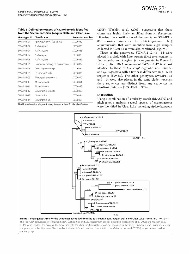
(2005); Wacklin et al. (2009), suggesting that theseclones are highly likely amplified from A. flos-aquae.Likewise, the classification of the genotypes SWMP11-05 showing similarity to Dolichospermum (D.)lemmermannii that were amplified from algal samplescollected in Clear Lake were also confirmed (Figure 1).Three of the genotypes, SWMP11-12 to −14 were
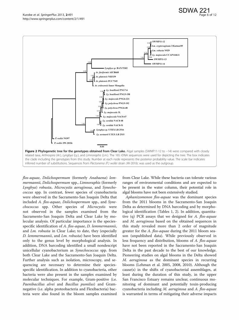
placed in a clade with Limnoraphis (Lm.) cryptovaginata,Lm. robusta, and Lyngbya (Ly.) majuscula in Figure 2.Notably, 16S rDNA sequence of SWMP11-12 is almostidentical to those of Lm. cryptovaginata, Lm. robusta,and Ly. majuscula with a few base differences in a 1.5 kbsequence (>99.8%). The other genotypes, SWMP11-13and −14 were also placed in the same clade, however,these sequences are distinct from any sequences inGenBank Database (16S rDNA, <95%).
DiscussionUsing a combination of similarity search (BLASTN) andphylogenetic analysis, several species of cyanobacteriawere identified in Clear Lake including Aphanizomenon
Table 3 Defined genotypes of cyanobacteria identifiedfrom the Sacramento-San Joaquin Delta and Clear Lake
Genotype ID Classification Accession number
SWMP11-01 Aphanizomenon flos-aquae JX006082
SWMP11-02 A. flos-aquae JX006083
SWMP11-04 A. flos-aquae JX006085
SWMP11-07 A. flos-aquae JX006088
SWMP11-08 A. flos-aquae JX006089
SWMP11-06 Unknown, belong to Nostocaceae JX006087
SWMP11-03 Dolichospermum sp. JX006084
SWMP11-05 D. lemmermannii JX006086
SWMP11-09 Microcystis aeruginosa JX006090
SWMP11-10 M. aeruginosa JX006091
SWMP11-11 M. aeruginosa JX006092
SWMP11-12 Limnoraphis robusta JX006093
SWMP11-13 Limnoraphis sp. JX006094
SWMP11-14 Limnoraphis sp. JX006095
BLAST search and phylogenetic analysis were utilized for the classification.
Figure 1 Phylogenetic tree for the genotypes identified from the Sacramento-San Joaquin Delta and Clear Lake (SWMP11-01 to −08).The 16S rDNA sequences for Aphanizomenon, Cuspidothrix, and Dolichospermum species described in Rajaniemi et al. (2005) and Wacklin et al.(2009) were used for the analysis. The boxes indicate the clades including the genotypes obtained in this study. Number at each node representsthe posterior probability value. The scale bar indicates inferred number of substitutions. Nodularia sp. (strain PCC7804) sequence was used asthe outgroup.
Kurobe et al. SpringerPlus 2013, 2:491 Page 5 of 12http://www.springerplus.com/content/2/1/491
SDWA 221
flos-aquae, Dolichospermum (formerly Anabaena) lem-mermannii, Dolichospermum spp., Limnoraphis (formerlyLyngbya) robusta, Microcystis aeruginosa, and Synecho-coccus spp. In contrast, fewer species of cyanobacteriawere observed in the Sacramento-San Joaquin Delta thatincluded A. flos-aquae, Dolichospermum spp., and Syne-chococcus spp. Other species of Microcystis werenot observed in the samples examined from theSacramento-San Joaquin Delta and Clear Lake by mo-lecular analysis. Of particular importance is the species-specific identification of A. flos-aquae, D. lemmermannii,and Lm. robusta in Clear Lake; to date, they (especiallyD. lemmermannii, and Lm. robusta) have been identifiedonly to the genus level by morphological analysis. Inaddition, DNA barcoding identified a small nondescriptunicellular cyanobacterium as Synechococcus spp. fromboth Clear Lake and the Sacramento-San Joaquin Delta.Further analysis such as isolation, microscopy, and se-quencing are necessary to determine their species-specific identification. In addition to cyanobacteria, otherbacteria were also present in the samples examined bymolecular techniques. For example, Gram-positive (i.e.Paenibacillus alvei and Bacillus pumilus) and Gram-negative (i.e. alpha proteobacteria and Flexibacteria) bac-teria were also found in the bloom samples examined
from Clear Lake. While these bacteria can tolerate variousranges of environmental conditions and are expected tobe present in the water column, their potential role inalgal blooms have not been extensively studied.Aphanizomenon flos-aquae was the dominant species
from the 2011 blooms in the Sacramento-San JoaquinDelta as determined by DNA barcoding and by morpho-logical identification (Tables 1, 2). In addition, quantita-tive (q) PCR assays that we designed for A. flos-aquaeand M. aeruginosa based on the obtained sequences inthis study revealed more than 2 order of magnitudegreater for the A. flos-aquae during the 2011 bloom sea-son (unpublished data). While previously observed inless frequency and distribution, blooms of A. flos-aquaehave not been reported in the Sacramento-San JoaquinDelta in the past decade to the best of our knowledge.Pioneering studies on algal blooms in the Delta showedM. aeruginosa as the dominant species in recurringblooms (Lehman et al. 2005, 2008, 2010). Although thecause(s) in the shifts of cyanobacterial assemblages, atleast during the duration of this study, in the upperSan Francisco Estuary remains unclear, continuous mo-nitoring of dominant and potentially toxin-producingcyanobacteria including M. aeruginosa and A. flos-aquaeis warranted in terms of mitigating their adverse impacts
Figure 2 Phylogenetic tree for the genotypes obtained from Clear Lake. Algal samples (SWMP11-12 to −14) were compared with closelyrelated taxa, Arthrospira (Ar.), Lyngbya (Ly.), and Limnoraphis (Lm.). The 16S rDNA sequences were used for depicting the tree. The box indicatesthe clade including the genotypes from this study. Number at each node represents the posterior probability value. The scale bar indicatesinferred number of substitutions. Sequences from Plectonema (P.) wollei strain JW-2010c was used as the outgroup.
Kurobe et al. SpringerPlus 2013, 2:491 Page 6 of 12http://www.springerplus.com/content/2/1/491
SDWA 221
to aquatic organisms and conservation of water quality.Water temperature and other physicochemical factorshave been associated with the emergence of A. flos-aquae(Cloern and Dufford 2005) and other cyanobacterial spe-cies during the 2011 blooms in the Sacramento-SanJoaquin Delta (Mioni et al. 2012). A. flos-aquae is adiazotrophic cyanobacterium which produce endotoxinssuch as anatoxin-a, saxitoxins, and cylindrospermopsin(Sivonen and Jones 1999; Castle and Rogers 2009).A species identification system using a combination of
BLASTN search and phylogenetic analysis for 16S rDNAis a powerful method; however this approach has limita-tions when used to analyze taxa with nearly identical se-quences or to classify unknown sequences. For example,Lm. robusta, Lm. cryptovaginata, and Ly. majuscula shareidentical 16S rDNA sequences (1.5 kb) with a few basepairs differences as observed from 6 clones (SWMP11-12)amplified from Clear Lake samples (Jüttner and Watson2007; Guiry and Guiry 2012). Although the phylogenetictree (Figure 2) implies that these clones are most closelyrelated to Lm. cryptovaginata, SWMP11-12 is most likelyLm. robusta by virtue of their characteristic morphologicalfeatures (Komárek 2003, Komárek et al. 2013; Rejmánkováet al. 2011). While Ly. majuscula is also placed in the sameclade, SWMP11-12 is unlikely Ly. majuscula as it is amarine species (Jones et al. 2011). Lm. robusta blooms inLake Atitlan, Guatemala was first reported in 2008 by(Rejmánková et al. 2011). This cyanobacterium formeddense patches covering approximately 40% of Lake Atitlansurface during the peak of the blooms although cya-notoxin production from this species remains unclear(Rejmánková et al. 2011). Cylindrospermopsin and saxi-toxins were detected during the Atitlan bloom in 2009,but the concentrations remained low (12 and 58 ng g-1
from the freeze dried specimen) (Rejmánková et al. 2011;Komárek et al. 2013). Lm. robusta found in Clear Lake,California is unlikely introduced from Lake Atitlan asthere are no reports documenting the introduction of thisspecies across the two water bodies. Further analysis suchas comparison of variable gene region will provide a betterunderstanding of the relationship between the same spe-cies found in two distant locations. Initial findings showedthat environmental factors such as water temperature andnutrient concentrations may affect the growth and abun-dance of Lm. robusta and other emerging cyanobacteria inClear Lake during the 2011 blooms (Mioni et al. 2012).The classification of clone SWMP11-06 in the phylo-
genetic tree is debatable (Figure 1). The genotype isplaced in a clade of Dolichospermum, however, the refer-ence strain A. flos-aquae strain 1tu37s13, which wasmorphologically identified by Rajaniemi et al. (2005), isalso placed in the same group. The values of posteriorprobability are relatively low for the branching (< 0.70),precluding a conclusive identification of the genotype.
SWMP11-13 and −14 from Clear Lake showed simi-larity to Lm. robusta by BLASTN search but interes-tingly these sequences are distinct from Lm. robusta,Lm. cryptovaginata, or any other sequences in theNCBI-GenBank database (16S rDNA Pairwise% Identity:96.8%). Stackebrandt and Goebel (1994) suggested a cut-off value of 97.5% (or higher) for acceptable similarityvalues for species identification using the 16S rDNA se-quence. These two genotypes are tentatively designatedas Limnoraphis sp. as placed in the phylogenetic tree;further analysis is necessary for species-specific identifi-cation (Figure 2).Although M. aeruginosa was detected at different sam-
pling sites and times in the Sacramento-San JoaquinDelta by microscopic observation, samples that wereexamined by molecular analysis did not detect this spe-cies. This result may be due to the following reasons:1) M. aeruginosa was lacking in the samples examinedfor molecular analysis due to its colonial nature and het-erogeneity across subsamples, and 2) inhibition by otherabundant cyanobacteria precluding the amplification ofM. aeruginosa in the samples that were PCR tested. It isimportant to note that A. flos-aquae, instead of the his-torically recurring M. aeruginosa, dominated the bloomsin the Sacramento-San Joaquin Delta during the dur-ation of these studies. Although the field samples thatwe chose for molecular analyses were based on morpho-logic microscopic analysis (qualitative and quantitative),the DNA fragments of the expected algal species werenot obtained from the subsamples. Another potential ex-planation may be due to the small number of clones thatwere analyzed that may not represent the wide variety ofcyanobacterial species present in the blooms. Analyzingmore clones from appropriate field samples using emer-ging sequencing technologies would probably yield agreater number of sequences from potentially toxin-producing cyanobacteria with a sample such as CL3(6)from Clear Lake that showed more diverse bacterial spe-cies (Table 2). Another ideal approach is to use parallelalgal samples for morphologic taxonomic identificationand molecular analyses to better understand the cyano-bacterial composition in the Sacramento-San JoaquinDelta and Clear Lake.Most of the cyanobacterial sequences deposited in
NCBI-GenBank database originate from geographicallydistant locations such as Portugal, Japan, India, andNordic countries (Robertson et al. 2001; Rajaniemi et al.2005; Ezhilarasi and Anand 2009; Lopes et al. 2012).Despite the distant origins of species-specific sequences,we were able to successfully identify the taxonomic clas-sification of the clones based on the 16S rDNA. Usingthe 16S rDNA for classification and identification ofcyanobacteria is widely accepted because 1) the gene ispresent in all bacterial genomes, and 2) the frequency of
Kurobe et al. SpringerPlus 2013, 2:491 Page 7 of 12http://www.springerplus.com/content/2/1/491
SDWA 221
sequence variations and insertions in this gene serves as amolecular clock and reflects evolutional history, allowingthe distinction of a broad range of taxonomic groups andidentification of individual species (Casamatta et al. 2005;Janda and Abbott 2007). Although the resolution of the16S rDNA for specific identification remains debatabledue to the high degree of their sequence conservation(Janda and Abbott 2007), the gene has been a reliablebarcode providing identification to the genus level, insome cases to specific species level as we have demon-strated in this study. We attempted to use the ITS regionin our analysis, however, the sequences were not suitablefor alignments due to their high variability when com-pared with the reference strains from other geographiclocations (unpublished observation). Another gene in-volved in nitrogen fixation, nifH, has been used as analternate barcode for classification and identification ofcyanobacteria by providing better resolution for speciesidentification (Zehr et al. 1997). However, the nifH gene isnot appropriate for analyzing complex algal assemblagesby DNA barcoding as non-diazotrophic cyanobacteriasuch as M. aeruginosa do not possess this gene in theirgenome.
ConclusionMicroscopic observation coupled with DNA barcoding ef-fectively identified cyanobacterial species in theSacramento-San Joaquin Delta and in Clear Lake. For thefirst time in Northern California, this tiered approachprovided species-specific identification of dominant spe-cies in the blooms including Microcystis aeruginosa,Aphanizomenon flos-aquae, Dolichospermum (formerlyAnabaena) lemmermannii, Dolichospermum spp., Lim-noraphis (formerly Lyngbya) robusta, Limnoraphis spp.,and Synechococcus spp. The precise identification usingDNA barcoding provides two important ecological impli-cations in these water bodies. First, we have identifiedA. flos-aquae as the new dominant species in theSacramento-San Joaquin Delta during the course of thisstudy, an apparent shift from M. aeruginosa that havedominated the recurring blooms at the delta in the pastdecade. Second, DNA barcoding documented the first oc-currence of Lm. robusta in North America. To date, thisharmful cyanobacterium has only been reported fromLake Atitlan in Guatemala where the climate is differentfrom that in California (Komárek et al. 2013). It is impor-tant to understand the factors affecting the emergence ofLm. robusta in California and the potential link promotingthe growth of the cyanobacterium between the two geo-graphically distant water bodies. Lastly, the identificationof prokaryote assemblages by DNA barcoding will en-hance the current cyanobacterial monitoring efforts byallowing us to develop specific quantitative PCR (qPCR)assays using the sequences obtained in this study. We are
currently validating the reliability and reproducibility of theqPCR tests for estimating the abundance of key cyanobac-terial species with potential toxin production. Assessmentof cyanobacterial assemblages using the interdisciplinaryapproach (i.e. DNA barcoding and qPCR supported bymorphological identification) will aid in formulating effec-tive mitigation measures by addressing the specific identityof cyanobacteria, their corresponding physiological fea-tures, and determining the effects of fundamental environ-mental factors on species-specific toxicity.
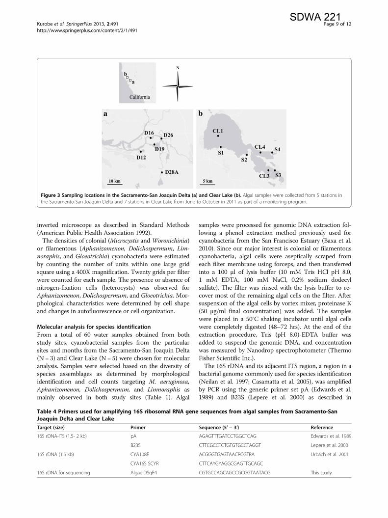
Materials and methodsStudy sites and collection of algal samplesAlgal samples were collected for the period of June toOctober in 2011 from five and seven stations in theSacramento-San Joaquin Delta and Clear Lake, respec-tively (Figure 3). These sampling stations have beenpreviously established by the Department of WaterResources as standard monitoring sites with correspond-ing environmental data such as water quality, nutrientloading, and phytoplankton records (Richerson et al.1994; Winder et al. 2010). Algal samples were collectedaccording to standard protocol (Fetscher et al. 2009) andestablished procedures (Mioni and Kudela 2011). Briefly,samples for microscopy were fixed with 2.5% (v/v) glu-taraldehyde in the field and were filtered through a1-μm pore size, 25-mm diameter, black polycarbonatefilters (GE Osmonics, Monroe, NC). Algal samples formolecular analysis were collected as follows: appro-ximately 600 mL of surface water (grab) samples werefiltered with a 0.45-μm membrane using a clean filtra-tion device (hand pump) on site. Each filter was placedin a sterile microcentrifuge tube and stored on dry iceand in the dark upon collection and transported to thelab for analysis. The samples were stored in a freezer(−80°C) until processing.
Microscopic analysisAlgal samples were sent to two independent laboratories:University of California, Santa Cruz, and GreenwaterLaboratories (http://greenwaterlab.com/) for morphologictaxonomic identification of cyanobacteria (Karlson et al.2010; Mioni and Kudela 2011). In UC Santa Cruz,epifluorescence microscopy was used to identify andenumerate cyanobacteria present in environmental sam-ples following established procedures (Mioni and Kudela2011). The abundance of autofluorescing phycoerythrincontaining cells (i.e. cyanobacteria) was determined on aZeiss Axioplan epifluorescence microscope at 400X mag-nification using green excitation (Zeiss Filter Set 20, exci-tation 546 nm bandpass, and emission 575–640 nmbandpass filters). At Greenwater Laboratories, sampleswere preserved in Lugol’s Iodine solution, and cyanobac-terial cells were enumerated on a Nikon Eclipse TE200
Kurobe et al. SpringerPlus 2013, 2:491 Page 8 of 12http://www.springerplus.com/content/2/1/491
SDWA 221
inverted microscope as described in Standard Methods(American Public Health Association 1992).The densities of colonial (Microcystis and Woronichinia)
or filamentous (Aphanizomenon, Dolichospermum, Lim-noraphis, and Gloeotrichia) cyanobacteria were estimatedby counting the number of units within one large gridsquare using a 400X magnification. Twenty grids per filterwere counted for each sample. The presence or absence ofnitrogen-fixation cells (heterocysts) was observed forAphanizomenon, Dolichospermum, and Gloeotrichia. Mor-phological characteristics were determined by cell shapeand changes in autofluorescence or cell organization.
Molecular analysis for species identificationFrom a total of 60 water samples obtained from bothstudy sites, cyanobacterial samples from the particularsites and months from the Sacramento-San Joaquin Delta(N = 3) and Clear Lake (N = 5) were chosen for molecularanalysis. Samples were selected based on the diversity ofspecies assemblages as determined by morphologicalidentification and cell counts targeting M. aeruginosa,Aphanizomenon, Dolichospermum, and Limnoraphis asmainly observed in both study sites (Table 1). Algal
samples were processed for genomic DNA extraction fol-lowing a phenol extraction method previously used forcyanobacteria from the San Francisco Estuary (Baxa et al.2010). Since our major interest is colonial or filamentouscyanobacteria, algal cells were aseptically scraped fromeach filter membrane using forceps, and then transferredinto a 100 μl of lysis buffer (10 mM Tris HCl pH 8.0,1 mM EDTA, 100 mM NaCl, 0.2% sodium dodecylsulfate). The filter was rinsed with the lysis buffer to re-cover most of the remaining algal cells on the filter. Aftersuspension of the algal cells by vortex mixer, proteinase K(50 μg/ml final concentration) was added. The sampleswere placed in a 50°C shaking incubator until algal cellswere completely digested (48–72 hrs). At the end of theextraction procedure, Tris (pH 8.0)-EDTA buffer wasadded to suspend the genomic DNA, and concentrationwas measured by Nanodrop spectrophotometer (ThermoFisher Scientific Inc.).The 16S rDNA and its adjacent ITS region, a region in a
bacterial genome commonly used for species identification(Neilan et al. 1997; Casamatta et al. 2005), was amplifiedby PCR using the generic primer set pA (Edwards et al.1989) and B23S (Lepere et al. 2000) as described in
Figure 3 Sampling locations in the Sacramento-San Joaquin Delta (a) and Clear Lake (b). Algal samples were collected from 5 stations inthe Sacramento-San Joaquin Delta and 7 stations in Clear Lake from June to October in 2011 as part of a monitoring program.
Table 4 Primers used for amplifying 16S ribosomal RNA gene sequences from algal samples from Sacramento-SanJoaquin Delta and Clear Lake
Target (size) Primer Sequence (5′ − 3′) Reference
16S rDNA-ITS (1.5- 2 kb) pA AGAGTTTGATCCTGGCTCAG Edwards et al. 1989
B23S CTTCGCCTCTGTGTGCCTAGGT Lepere et al. 2000
16S rDNA (1.5 kb) CYA108F ACGGGTGAGTAACRCGTRA Urbach et al. 2001
CYA16S SCYR CTTCAYGYAGGCGAGTTGCAGC
16S rDNA for sequencing AlgaeIDSqF4 CGTGCCAGCAGCCGCGGTAATACG This study
Kurobe et al. SpringerPlus 2013, 2:491 Page 9 of 12http://www.springerplus.com/content/2/1/491
SDWA 221
Rajaniemi et al. (2005) (Table 4). In addition, another setof primers (CYA 108 F and CYA16S SCYR, Table 4) amp-lifying a partial fragment of the 16S rDNA, but not theITS, was used for one of the algal samples, D16(7), fromthe Sacramento-San Joaquin Delta as we observed inhi-bition of PCR amplification with the primer set describedabove. The volume of the PCR cocktail was 50 μlcontaining 200 μM each of dNTP, 1.5 mM of MgCl2,40 pmol of each primer, 2 units of Taq DNA polymerase(High Fidelity Platinum Taq polymerase, Invitrogen Corp)and 10X buffer at 1/10 the volume of the reaction. Bovineserum albumin (0.1 mg/ml final concentration) was addedto the reaction cocktail for the algal samples from theSacramento-San Joaquin Delta to resolve the inhibition ofthe PCR. The PCR cycling condition was performed asfollows: initial denaturation step of 95°C for 5 min,40 cycles of 95°C for 30 s, 50°C for 30 s, and 72°C for2 min 30 s, followed by a final extension step at 72°C for10 min and then held at 4°C. The PCR product wasseparated on 1% agarose gel and observed by a transillu-minator after staining with 0.5 μg/ml ethidium bromidefor 20 min.The DNA bands at the expected size (1.5-2 kb) were
excised from the gel and extracted using QIAquick II ex-traction kit (Qiagen). The eluted DNA was ligated intopGEM-T Easy vector (Promega BioSciences) that wasused to transform Escherichia coli DH5α competent cells(Invitrogen). The length of the inserted DNA fragmentwas verified by running a PCR on colonies carrying theplasmid. The PCR cocktail (50 μl) contained 200 μMeach of dNTP, 1.5 mM of MgCl2, 40 pmol of M13 for-ward and reverse primers, 0.5 unit Platinum Taq DNApolymerase (Invitrogen) and 10X buffer at 1/10 thevolume of the reaction. The PCR cycling condition wasthe same as above except for the annealing temperatureat 55°C. Clones carrying the inserted fragment size of1.5 to 2 kb with variable length were chosen for plasmidextraction and sequencing. Twenty clones were analyzedfor each of the algal samples except for the sample CL3(6) from which additional 30 clones were submitted forsequencing because various types of cyanobacterialsequences were observed from the first 20 clones. Theplasmid was extracted using QIAprep Spin Mini Kit(Qiagen) according to the manufacturer’s instruction.The sequence of the inserted DNA fragment was deter-mined from both ends using M13 forward and reverseprimers in addition to the primer that we designed(AlgaeIDSqF4) for sequencing the middle fragments(Table 4). The samples were submitted to Davis Sequen-cing (http://www.davissequencing.com/) for sequencingreactions using an ABI 377 automated DNA sequencer(Applied Biosciences). The obtained sequences fromeach clone were processed to correct ambiguous basesand to remove vector and primer sequences; a consensus
sequence was generated using Geneious software ver.5.0.3 (Drummond et al. 2011).The entire sequence of the 16S rDNA-ITS region was
used for defining Operational Taxonomic Units (OTUs)using UCLUST ver. 1.2.22 with a threshold of 98.5% (Ed-gar 2010). A representative sequence for each of theOTU cluster was selected as a genotype and was usedfor similarity search using BLASTN (Altschul et al.1990). The sequences showing similarity to cyanobac-teria with potential ability to produce toxins were se-lected for further analysis.The phylogenetic trees were constructed to distinguish
species that share nearly identical 16S rDNA sequenceswith a few base pair differences such as those observedbetween Aphanizomenon and Dolichospermum, and bet-ween Limnoraphis and closely related species such asArthrospira and Lyngbya. The 16S rDNA sequences, ap-proximately 1.4 kb covering almost the entire sequencebut not including the ITS regions, were used for phylo-genetic analysis. The sequences used for phylogenetictree analysis were taken from other studies as listed inAdditional File 1 (Lehtimäki et al. 2000; Lyra et al. 2001;Gugger et al. 2002; Rajaniemi et al. 2005; Engene et al.2011). Multiple alignments were generated by MUSCLEver. 3.8.31 (Edgar 2004). The phylogenetic trees weregenerated by MrBayes program ver. 3.2 for 16S rDNAsequences using Markov chain Monte Carlo methodwith the following settings: Ngen = 10000000, Nchain =4, Temp = 0.5, Stopval = 0.01, Samplefreq = 50, Printfreq =1000 (Ronquist et al. 2012). The General Time Reversiblemodel with a proportion of invariable sites and a gamma-shaped distribution of rates was selected by jModeltestver. 2.1.4 (Darriba et al. 2012) as the best model for thedatasets for the family Nostocaceae (Aphanizomenon,Cuspidothrix, and Dolichospermum) and Oscillatoriaceae(Anthrospira, Limnoraphis, and Lyngbya). Nodularia sp.(strain PCC7804) or Plectonema wollei (strain JW-2010c)was used as the outgroup for the phylogenetic tree ofNostocaceae (Figure 1) or Oscillatoriaceae (Figure 2),respectively. FigTree ver. 1.4 was used for depicting thephylogenetic trees (http://tree.bio.ed.ac.uk/software/figtree/).
AbbreviationsCyanoHABs: Cyanobacterial blooms; ITS: Internal transcribed spacer;OTU: Operational taxonomic unit; rDNA: Ribosomal RNA gene.
Competing interestsThe authors declare that they have no competing interests.
Authors’ contributionsCEM directed field sampling with the support of TS and SW and supervisionof RMK. SW also provided ancillary data in this study. CEM and ADCconducted taxonomic identification by microscopy. TK and DVB carried outmolecular analysis. TK analyzed the molecular data, generated thephylogenetic trees, and wrote the paper. DVB supervised the research andprovided major suggestions and revisions of the manuscript. SJT providedlogistics support for the molecular analyses. All the co-authors contributed to
Kurobe et al. SpringerPlus 2013, 2:491 Page 10 of 12http://www.springerplus.com/content/2/1/491
SDWA 221
several revisions of the manuscript. All authors read and approve the finalmanuscript.
Authors’ informationTK, DVB, SJT are members of the Aquatic Health Program at UC Davis,School of Veterinary Medicine: http://www.vetmed.ucdavis.edu/aquatic_health/index.cfm.
AcknowledgmentsFunding support for this study was provided by the Surface Water AmbientMonitoring Program (Grant No. 10−058−150) to CEM with a subcontract(Grant No. 201119053) to DB and TK at the UC Davis Aquatic HealthProgram, and by the California Department of Water Resources(Grant No. 4600008137) to ST. We extend our appreciation to the LakeCounty Water Resources Department for providing field assistance for ClearLake sampling, and the California Department of Water Resources(Environmental Monitoring Program) for the Sacramento-San Joaquin Deltasampling (R/V San Carlos). Dr. Thomas B. Waltzek from the University ofFlorida is acknowledged for his insights on phylogenetic analysis and Mr.Michael O. Park and Ms. Samah Abdelrazek at the Aquatic Health Program,UC Davis for assistance on the cloning work. From UC Santa Cruz, we thankKendra Hayashi, Jonathan Zehr, and Robert Franks for their technicalassistance and use of instrumentation and other facilities. We acknowledgeCharles Ingwell (EcoAnalysts, Inc.) for his assistance with taxonomy andhelpful discussion.
Author details1Department of Anatomy, Physiology, and Cell Biology, School of VeterinaryMedicine, University of California, Davis, CA 95616, USA. 2Institute of MarineSciences, University of California, Santa Cruz, CA 95064, USA. 3Lake CountyWater Resources Department, Lakeport, CA 95453, USA. 4CaliforniaDepartment of Water Resources, Environmental Monitoring Program, WestSacramento, CA 95691, USA. 5Greenwater Laboratories, Palatka, FL 32177,USA.
Received: 6 September 2013 Accepted: 24 September 2013Published: 30 September 2013
ReferencesAcuña S, Deng DF, Lehman P, Teh SJ (2012) Sublethal dietary effects of
Microcystis on Sacramento splittail, Pogonichthys macrolepidotus. AquatToxicol 110–111:1–8
Altschul SF, Gish W, Miller W, Myers EW, Lipman DJ (1990) Basic local alignmentsearch tool. J Mol Evol 215:403–410
American Public Health Association (1992) Standard Methods of Water andWastewater. American Public Health Association, American Water WorksAssociation, Water Environment Federation publication, 18th edition.APHA, Washington D.C
Baxa DV, Kurobe T, Ger KA, Lehman PW, Teh SJ (2010) Estimating the abundanceof toxic Microcystis in the San Francisco Estuary using quantitative real-timePCR. Harmful Algae 9:342–349
Betournay S, Marsh AC, Donello N, Stiller JW (2007) Selective recovery ofmicroalgae from diverse habitats using “Phyto-specific” 16S rDNA primers.J Phycol 43:609–613
Casamatta DA, Johansen JR, Vis ML, Broadwater ST (2005) Molecular andmorphological characterization of ten polar and near-polar strains within theOscillatoriales (cyanobacteria). J Phycol 41:421–435
Castle JW, Rogers JH (2009) Hypothesis for the role of toxin-producing algae inPhanerozoic mass extinctions based on evidence from the geologic recordand modern environments. Environ Geosci 16:1–23
Cheng C, Zaichao Z, Aizhong D, Jiayan W, Jingfa X, Yujiao S (2011) Bar-codedpyrosequencing reveals the bacterial community during Microcystis waterBloom in Guanting Reservoir, Beijing. Procedia Eng 18:341–346
Cloern JE, Dufford R (2005) Phytoplankton community ecology: principles appliedin San Francisco Bay. Mar Ecol-Prog Ser 285:11–28
Darriba D, Taboada GL, Doallo R, Posada D (2012) jModelTest 2: more models,new heuristics and parallel computing. Nat Methods 30:772
Drummond AJ, Ashton B, Buxton S, Cheung M, Cooper A, Duran C, Field M,Heled J, Kearse M, Markowitz S, Moir R, Stones-Havas S, Sturrock S, Thierer T,Wilson A (2011) Geneious v5.0.3. Available from http://www.geneious.com
Edgar RC (2004) MUSCLE: multiple sequence alignment with high accuracy andhigh throughput. Nucleic Acids Res 32:1792–1797
Edgar RC (2010) Search and clustering orders of magnitude faster than BLAST.Bioinformatics 26:2460–2461
Edwards U, Rogall T, Blocker H, Emde M, Bottger EC (1989) Isolation and directcomplete nucleotide determination of entire genes. Characterization of agene coding for 16S ribosomal RNA. Nuc Aci Res 17:7843–7853
Engene N, Choi H, Esquenazi E, Rottacker EC, Ellisman MH, Dorrestein PC,Gerwick WH (2011) Underestimated biodiversity as a major explanation forthe perceived rich secondary metabolite capacity of the cyanobacterialgenus Lyngbya. Env Microbiol 13:1601–1610
Ezhilarasi A, Anand N (2009) Phylogenetic analysis of Anabaena spp.(Cyanobacteria) using sequences of 16S rRNA Gene. Aust J Basic Appl Sci3:4026–4031
Fetscher AE, Busse L, Ode PR (2009) Standard operating procedures for collectingstream algae samples and associated physical habitat and chemical data forambient bioassessments in California. California State Water ResourcesControl Board Surface Water Ambient Monitoring Program (SWAMP)Bioassessment SOP 002. http://swamp.mpsl.mlml.calstate.edu/wp-content/uploads/2010/06/SWAMP_SOP_Algae_Field_Collection_050110.pdf. updatedMay 2010
Ger KA, Teh SJ, Baxa DV, Lesmeister SA, Goldman CR (2010) The effects of dietaryMicrocystis aeruginosa and microcystin on the copepods of the upperSan Francisco Estuary. Freshwater Biol 55:1548–1559
Goldstein JJ, Tolsdorf TN (1994) An economic analysis of potential water qualityimprovement in Clear Lake. Department of Agriculture Soil ConservationService, U.S, pp 1–42
Gugger M, Lyra C, Henriksen P, Coute A, Humbert JF, Sivonen K (2002)Phylogenetic comparison of the cyanobacterial genera Anabaena andAphanizomenon. Int J Syst Evol Microbiol 52:1867–1880
Guiry MD, Guiry GM (2012) AlgaeBase. World-wide electronic publication,National University of Ireland, Galway. http://www.algaebase.org(searched on 29 October 2012)
Hebert PD, Ratnasingham S, deWaard JR (2003) Barcoding animal life:cytochrome c oxidase subunit 1 divergences among closely related species.Proc Bio Soc 7:S96–99
Horne AJ (1975) The ecology of Clear Lake phytoplankton. Lakeport Clear LakeAlgal Research Unit, pp 1–116
Janda JM, Abbott SL (2007) 16S rRNA gene sequencing for bacterial identificationin the diagnostic laboratory: pluses, perils, and pitfalls. J Clin Microbiol45:2761–2764
Jassby AD (2008) Phytoplankton in the upper San Francisco Estuary: recentbiomass trends, their causes and their trophic significance. San Fran EstWatershed Sci 6:1–24
Jones AC, Monroe EA, Podell S, Hess WR, Klages S, Esquenazi E, Niessen S,Hoover H, Rothmann M, Lasken RS, Yates JR, Reinhardt R, Kube M, BurkartMD, Allen EE, Dorrestein PC, Gerwick WH, Gerwick L (2011) Genomic insightsinto the physiology and ecology of the marine filamentous cyanobacteriumLyngbya majuscula. Proc Natl Acad Sci U S A 108:8815–8820
Jüttner F, Watson SB (2007) Biochemical and ecological control of geosmin and2-methylisoborneol in source waters. Appl Env Microbiol73:4395–4406
Karlson B, Cusack C, Bresnan E (2010) Microscopic and molecular methodsfor quantitative phytoplankton analysis. Intergov OceanographCommission of UNESCO. (IOC Manuals and Guides, no. 55.). http://hab.ioc-unesco.org/index.php?option=com_oe&task=viewDocumentRecord&docID=5440
Khan HA, Arif IA, Shobrak M (2010) DNA Barcodes of Arabian partridge andPhilby’s Rock partridge: Implications for phylogeny and species identification.Evol Bioinform 5:151–158
Kochzius M, Seidel C, Antoniou A, Botla SK, Campo D, Cariani A, Vazquez EG,Hauschild J, Hervet C, Hjörleifsdottir S, Hreggvidsson G, Kappel K, Landi M,Magoulas A, Marteinsson V, Nölte M, Planes S, Tinti F, Turan C, VenugopalMN, Weber H, Blohm D (2010) Identifying fishes through DNA barcodes andmicroarrays. PLoS ONE 5:e12620
Komárek J (2003) Planktic oscillatorialean cyanoprokaryotes. Hydrobiologia502:367–382
Komárek J, Zapomělová E, Šmarda J, Kopecký J, Rejmánková E, Woodhouse J,Neilan BA, Komárková J (2013) Polyphasic evaluation of Limnoraphis robusta,a water-bloom forming cyanobacterium from Lake Atitlán, Guatemala, with adescription of Limnoraphis gen. nov. Fottea 13:39–52
Kurobe et al. SpringerPlus 2013, 2:491 Page 11 of 12http://www.springerplus.com/content/2/1/491
SDWA 221
Lehman PW, Boyer G, Hall C, Waller S, Gehrts K (2005) Distribution and toxicity ofa new colonial Microcystis aeruginosa bloom in the San Francisco Bay Estuary,California. Hydrobiologia 541:87–99
Lehman PW, Boyer G, Satchwell M, Waller S (2008) The influence ofenvironmental conditions on the seasonal variation of Microcystis cell densityand microcystins concentration in San Francisco Estuary. Hydrobiologia600:187–204
Lehman PW, Teh SJ, Boyer GL, Nobriga ML, Bass E, Hogle C (2010) Initial Impactsof Microcystis aeruginosa blooms on the aquatic food web in the SanFrancisco Estuary. Hydrobiologia 637:229–248
Lehtimäki J, Lyra C, Suomalainen S, Sundman P, Rouhiainen L, Paulin L,Salkinoja-Salonen M, Sivonen K (2000) Characterization of Nodularia strains,cyanobacteria from brackish waters, by genotypic and phenotypic methods.Int J Syst Evol Microbiol 50:1043–1053
Lepere C, Wilmotte A, Meyer B (2000) Molecular diversity of Microcystis strains(Cyanophyceae, Chroococcales) based on 16S rRNA sequences. Syst GeogrPlants 70:275–283
Lin S, Zhang H, Hou Y, Zhuang Y, Miranda L (2009) High-level diversity ofdinoflagellates in the natural environment, revealed by assessment ofmitochondrial cox1 and cob genes for dinoflagellate DNA barcoding.Appl Env Microbiol 75:1279–1290
Litaker WR, Vandersea WM, Kibler RS, Reece SK, Stokes AN, Lutzoni MF, Yonish AB,West AM, Black NDM, Tester AP (2007) Recognizing dinoflagellate speciesusing ITS rDNA sequences. J Phycol 43:344–355
Lopes VR, Ramos V, Martins A, Sousa M, Welker M, Antunes A, Vasconcelos VM(2012) Phylogenetic, chemical and morphological diversity of cyanobacteriafrom Portuguese temperate estuaries. Mar Env Res 73:7–16
Lopez-Legentil S, Song B, Bosch M, Pawlik JR, Turon X (2011) Cyanobacterialdiversity and a new acaryochloris-like symbiont from Bahamian sea-squirts.PLoS One 6:e23938
Lyra C, Suomalainen S, Gugger M, Vezie C, Sundman P, Paulin L, Sivonen K (2001)Molecular characterization of planktic cyanobacteria of Anabaena,Aphanizomenon, Microcystis and Planktothrix genera. Int J Syst Evol Microbiol51:513–526
Mankiewicz J, Tarczynska M, Walter Z, Zalewski M (2003) Natural toxins fromcyanobacteria. Acta Biol Cracov Bot 45:9–20
Mioni CE, Kudela RM (2011) Algal toxins bioassessment - Clear Lake, July/August2010. Final report for Lake County, April 2011. The University of California,Santa Cruz: Institute of Marine Sciences. http://www.swrcb.ca.gov/rwqcb5/water_issues/basin_plans/tr_living_rivers_council.pdf (accessed May 2, 2012)
Mioni CE, Kudela RM, Baxa DV (2012) Harmful cyanobacteria blooms and theirtoxins in Clear Lake and the Delta (California). Final Report, Project #: 10-058-150. Surface Water Ambient Monitoring Program (SWAMP), prepared for theCentral Valley Quality Control Board. http://www.co.lake.ca.us/Assets/WaterResources/Algae/2011+Cyanobacteria+Report.pdf (accessed May 2,2012)
Moniz MB, Kaczmarska I (2010) Barcoding of diatoms: nuclear encoded ITSrevisited. Protist 161:7–34
Murphy WJ, Eizirik E, Johnson WE, Zhang YP, Ryder OA, O’Brien SJ (2001)Molecular phylogenetics and the origins of placental mammals.Nature 409:614–618
Neilan BA, Jacobs J, Del Dot T, Blackall LL, Hawkins PR, Cox PT, Goodman AE(1997) rDNA sequences and evolutionary relationships among toxic andnontoxic cyanobacteria of the genus Microcystis. Int J Syst Bacteriol47:693–697
Osswald J, Rellan S, Carvalho AP, Gago A, Vasconcelos V (2007) Acute effects ofan anatoxin-a producing cyanobacterium on juvenile fish Cyprinus carpio L.Toxicon 49:693–698
Palinska KA, Liesack W, Rhiel E, Krumbein WE (1996) Phenotype variability ofidentical genotypes: the need for a combined approach in cyanobacterialtaxonomy demonstrated on Merismopedia-like isolates. Arch Microbiol166:224–233
Puschner B, Pratt C, Tor ER (2010) Treatment and diagnosis of a dog withfulminant neurological deterioration due to anatoxin-a intoxication.J Vet Emergency and Crit Care 20:518–522
Rajaniemi P, Hrouzek P, Kastovská K, Willame R, Rantala A, Hoffmann L,Komárek J, Sivonen K (2005) Phylogenetic and morphological evaluation ofthe genera Anabaena, Aphanizomenon, Trichormus and Nostoc(Nostocales, Cyanobacteria). Int J Syst Evol Microbiol 55:11–26
Rejmánková E, Komárek J, Dix M, Komárková J, Girón N (2011) Cyanobacterialblooms in Lake Atitlan, Guatemala. Limnologica 41:296–302
Richerson PJ, Suchanek TH, Why SJ (1994) The causes and control of algalblooms in Clear Lake. Final report, July 28, 1994. Clean Lake Diagnostic/Feasibility Study for Clear Lake, California, pp 1–182
Robertson BR, Tezuka N, Watanabe MM (2001) Phylogenetic analyses ofSynechococcus strains (cyanobacteria) using sequences of 16S rDNA and partof the phycocyanin operon reveal multiple evolutionary lines and reflectphycobilin content. Int J Syst Evol Microbiol 51:861–871
Ronquist F, Teslenko M, Van der Mark P, Ayres DL, Darling A, Höhna S, Larget B,Liu L, Suchard MA, Huelsenbeck JP (2012) MrBayes 3.2: efficient Bayesianphylogenetic inference and model choice across a large model space.Syst Biol 61:539–542
SanMauro D, Vences M, Alcobendas M, Zardoya R, Meyer A (2005) Initialdiversification of living amphibians predated the breakup of Pangaea.Am Nat 165:590–599
Sivonen K, Jones G (1999) Cyanobacterial toxins. In: Chorus I, Bartram J (ed) Toxiccyanobacteria in water: a guide to their public health consequences,monitoring and management. Spon Press, London, pp 41–111
Smith JL, Boyer GL, Zimba PV (2008) A review of cyanobacterial odorous andbioactive metabolites: Impacts and management alternatives in aquaculture.Aquaculture 280:5–20
Sommer T, Armor C, Baxter R, Breuer R, Brown L, Chotkowski M, Culberson S,Feyrer F, Gingras M, Herbold B, Kimmerer W, Mueller-Solger A, Nobriga M,Souza K (2007) The collapse of pelagic fishes in the upper San FranciscoEstuary. Fisheries 32:270–277
Spier C, Koski T, Graham J, Hanlon J, Brunell M, Borglin S, Stringfellow W (2010)Distribution of Microcystis and microcystin in the southern reach of theSacramento San Joaquin Delta. Final report, January 2010. University of thePacific Ecological Engineering Research, Program, pp 1–38
Stackebrandt E, Goebel BM (1994) Taxonomic note: a place for DNA-DNAreassociation and 16S rRNA sequence analysis in the present speciesdefinition in Bacteriology. Int J Syst Evol Microbiol 44:846–849
Urbach E, Vergin KL, Young L, Morse A, Larson GL, Giovannoni SJ (2001) Unusualbacterioplankton community structure in ultra-oligotrophic Crater Lake.Limnol Oceanogr 46:557–572
Wacklin P, Hoffmann L, Komárek J (2009) Nomenclatural validation of thegenetically revised cyanobacterial genus Dolichospermum (RALFS ex BORNETet FLAHAULT) comb. nova. Fottea 9:59–64
Winder M, Reuter J, Schladow G (2010) Clear Lake historical data analysis. ClearLake final report for Lake County. University of California, Davis, pp pp 1–51
Zehr JP, Mellon MT, Hiorns WD (1997) Phylogeny of cyanobacterial nifH genes:evolutionary implications and potential applications to natural assemblages.Microbiology 143:1443–1450
doi:10.1186/2193-1801-2-491Cite this article as: Kurobe et al.: Identification of harmful cyanobacteriain the Sacramento-San Joaquin Delta and Clear Lake, California by DNAbarcoding. SpringerPlus 2013 2:491.
Submit your manuscript to a journal and benefi t from:
7 Convenient online submission
7 Rigorous peer review
7 Immediate publication on acceptance
7 Open access: articles freely available online
7 High visibility within the fi eld
7 Retaining the copyright to your article
Submit your next manuscript at 7 springeropen.com
Kurobe et al. SpringerPlus 2013, 2:491 Page 12 of 12http://www.springerplus.com/content/2/1/491
SDWA 221