Embed Size (px)
Citation preview

RESEARCH Open Access
RNAi reveals proteins for metabolism andprotein processing associated with Langatvirus infection in Ixodes scapularis (black-legged tick) ISE6 cellsJeffrey M. Grabowski1,2,4, Monika Gulia-Nuss1,5, Richard J. Kuhn2,3 and Catherine A. Hill1,3*
Abstract
Background: Tick-borne flaviviruses (TBFs) cause thousands of human cases of encephalitis worldwide each year,with some TBF infections progressing to hemorrhagic fever. TBFs are of medical and veterinary importance andstrategies to reduce flavivirus transmission by the tick vector may have significant application. Analyses of theproteome of ISE6 cells derived from the black legged tick, Ixodes scapularis infected with the TBF, Langat virus(LGTV), have provided insights into proteins and cellular processes involved with LGTV infection.
Methods: RNA interference (RNAi)-induced knockdown of transcripts was used to investigate the role of ten tickproteins in the LGTV infection cycle in ISE6 cells. LGTV-infected cells were separately transfected with dsRNAcorresponding to each gene of interest and the effect on LGTV genome replication and release of infectious viruswas assessed by RT-qPCR and plaque assays, respectively.
Results: RNAi-induced knockdown of transcripts for two enzymes that likely function in amino acid, carbohydrate,lipid, terpenoid/polykeytide and vitamin metabolism, and a transcript for one protein of unknown function wereassociated with decreased replication of the LGTV genome and release of infectious virus from cells. Theknockdown of transcripts for five enzymes predicted to function in metabolism, a protein likely associated withfolding, sorting and degradation, and a protein of unknown function was associated with a decrease only in theamount of infectious LGTV released from cells.
Conclusions: These data suggest tick proteins potentially associated with metabolism and protein processing maybe involved in LGTV infection of ISE6 cells. Our study provides information to begin to elucidate the function ofthese proteins and identify targets for the development of new interventions aimed at controlling the transmissionof TBFs.
Keywords: Ixodes scapularis, Tick-borne disease, Flavivirus, Langat, ISE6, RNAi, Functional studies, Metabolism,Protein-processing
* Correspondence: [email protected] of Entomology, College of Agriculture, Purdue University, 901W State Street, West Lafayette, IN 47907, USA3Purdue Institute for Inflammation, Immunology and Infectious Disease,Purdue University, West Lafayette, IN 47907, USAFull list of author information is available at the end of the article
© The Author(s). 2017 Open Access This article is distributed under the terms of the Creative Commons Attribution 4.0International License (http://creativecommons.org/licenses/by/4.0/), which permits unrestricted use, distribution, andreproduction in any medium, provided you give appropriate credit to the original author(s) and the source, provide a link tothe Creative Commons license, and indicate if changes were made. The Creative Commons Public Domain Dedication waiver(http://creativecommons.org/publicdomain/zero/1.0/) applies to the data made available in this article, unless otherwise stated.
Grabowski et al. Parasites & Vectors (2017) 10:24 DOI 10.1186/s13071-016-1944-0
brought to you by COREView metadata, citation and similar papers at core.ac.uk
provided by Springer - Publisher Connector
BackgroundTick-borne flaviviruses (TBFs) impact human and animalhealth worldwide. These positive, single-stranded RNAviruses are transmitted by an infected tick (subphlymChelicerata, subclass Acari, family Ixodidae) during blood-feeding. Tick-borne encephalitis virus (TBEV), Powassanvirus (POWV), Kyasanur Forest Disease virus (KFDV)and Omsk hemorrhagic fever virus (OHFV) are membersof the TBF complex and can cause encephalitis (TBEVand POWV) and hemorrhagic fever (KFDV and OHFV).Many TBFs are considered a biosecurity threat and areclassified as biosafety containment level (BSL) three orfour. Research is ongoing to develop vaccines and ther-apeutics to prevent or treat TBF infections. The lessvirulent Langat virus (LGTV), classified BSL-2, has beenwidely used as a model for more virulent TBFs. Manystudies have employed LGTV and cell lines derived fromthe black-legged tick, Ixodes scapularis to investigate viralpathogenesis in the tick host cell, although I. scapularis isnot a natural vector of LGTV.Studies to understand the pathogenesis of tick-virus
interactions will benefit from the recently published as-sembly of the I. scapularis genome. As the first such re-source for a tick vector of disease [1–3], the assembly isexpected to help advance investigations of tick-virus in-teractions at the molecular level. Proteomic studies haveidentified hundreds of I. scapularis proteins [4–7] andhelped to define the proteome of this vector. Using massspectrometry, Grabowski et al. identified 486 proteins inthe I. scapularis ISE6 cell line, 266 of which were differ-entially regulated in cells infected with LGTV [4]. Pro-teins likely associated with metabolic processes exhibitedincreased or decreased expression following LGTV in-fection. These and other studies [7–9] provide a logicalstarting point for detailed molecular research to determinethe role of tick proteins during the TBF life-cycle in thevector.RNA interference (RNAi) is a tool widely used for
functional studies of arthropod proteins, including pro-teins produced by the I. scapularis IDE8 cell line duringinfection with flavivirus [7–9]. At least one of these studiessuggests induction of the RNAi-based antiviral pathwayidentified in other organisms and a role for Argonauteand Dicer in suppression of LGTV genome replication [9],although a role for the RNAi-pathway protein, Tudor-SNin LGTV replication or release of infectious virus is ques-tioned [8]. Other IDE8 gene products implicated in theantiviral response of the tick cell against LGTV includeFactor H, trypsin, HSP90 and HSP70 [7], with the lattertwo proteins predicted to function in protein foldingand/or processing. Previous studies shed light on lipidsand metabolic processes potentially manipulated by den-gue virus (DENV) to facilitate infection and replication inhuman and mosquito systems [10–12]. Equivalent studies
are required to better understand metabolic processesaffected during tick-flavivirus interaction.Efforts are underway to develop new transmission-
blocking technologies that target proteins produced bythe host cell (i.e. host factors) that are essential to virusinfection and replication [13–16]. Here, we investigatedthe hypothesis that proteins which exhibited increasedexpression in LGTV-infected I. sapularis ISE6 cells andare predicted to function in (i) the metabolism of aminoacids, vitamins/cofactors, carbohydrates, nucleotides andlipids, (ii) DNA replication/repair or (iii) protein folding/sorting/degradation [4] are involved in flaviviral infection.The functional roles performed by these proteins duringLGTV infection was pursued using loss-of-function, RNAiknockdown assays. Ten genes of interest were selected foranalyses: fumarylacetoacetase (FAH; ISCW020196),endoplasmic reticulum protein 29 (ERP29; ISCW018425),aldehyde dehydrogenase (ALDH; ISCW015982), carbon-nitrogen hydrolase/pantetheine hydrolase/vanin-like (VNN;ISCW004822), malate dehydrogenase (MDH2; ISCW003528),poly [ADP-ribose] polymerase (PARP; ISCW019519),cytidine/uridine monophosphate kinase (CMPK; ISCW012446),acetyl-CoA acetyltransferase (ACAT1; ISCW016117)and two hypothetical proteins (Hypo195; ISCW011195and Hypo576; ISCW020576). The process used to se-lect these ten genes is summarized in Additional file 1:Figure S1. Transcripts corresponding to the above geneswere confirmed in ISE6 cells and adult I. scapularis. Subse-quently, ISE6 cells were separately transfected with dsRNAcorresponding to each gene of interest, the knockdown oftranscripts was confirmed by reverse transcriptase quanti-tative PCR (RT-qPCR) and the effect on LGTV genomereplication and release of infectious virus was assessed byRT-qPCR and plaque assay, respectively. Knockdown oftranscripts for VNN, ACAT1, and Hypo576 was associatedwith decreased LGTV genome replication and LGTV re-lease, while knockdown of transcripts for FAH, ERP29,ALDH, MDH2, CMPK, and Hypo195 was associated withdecreased LGTV release only. These proteins are candi-dates for further functional analyses and studies aimed atdevelopment of new technologies to prevent transmissionof TBF infection.
MethodsCell and LGTV cultureThe ISE6 cell line derived from I. scapularis embryoniccells (obtained from T. Kurtti, University of Minnesota,Minneapolis, MN) was cultured at 34 °C in L15B-300medium in the absence of CO2 [17, 18]. Baby hamsterkidney 15 cells (BHK15) and Green African monkeykidney cells (Vero) originally obtained from AmericanTissue Culture Collection (ATCC), were cultured at 37 °Cin Minimum Essential Medium (MEM) supplementedwith L-glutamine, non-essential amino acids, and 10%
Grabowski et al. Parasites & Vectors (2017) 10:24 Page 2 of 14
heat-inactivated fetal calf serum (FCS) with 5% CO2.LGTV TP21 wildtype strain (passage 2) was obtainedfrom A. Pletnev (NIH-NAID, Bethesda, MD) and amp-lified using a multiplicity of infection (MOI) of 0.01[19, 20] in Vero cells (cells were grown as describedabove, with 2.5% heat-inactivated FCS) to produce aworking stock (passage 4). The titer of the LGTV p4stock was determined via serial immunofluorescenceassays (IFAs) in Vero cells using expression of the LGTVnonstructural protein 3 (NS3) as described by Junjhon etal. [21] and Grabowski et al. [4]. LGTV infection of ISE6cells was performed using an MOI of 10 to achieve max-imal infection of the cell population [4] and capture thesynchronized release of the first population of infectiousvirus. Manual cell counts (cells/ml) were conducted usinga hemocytometer [22] to quantify cell numbers before(a) seeding and (b) infection with LGTV.
Preparation of RNA from adult I. scapularis and ISE6 cellsand cDNA synthesisA single I. scapularis female collected by flagging fromTippecanoe State Park, Winamac, IN (October 29, 2013)was flash frozen in liquid N2 and ground in TRIzol re-agent (Invitrogen, Carlsbad, USA) using mortar and pestle,and RNA was extracted as per manufacturer instructions.RNA was isolated from ISE6 cells (passage 96–100) grownin 96 well plates using the RNeasy mini kit (Qiagen,Hilden, Germany) and processed according to kit instruc-tions. cDNA was synthesized from RNA samples usingthe iScript cDNA synthesis kit (BioRad, Hercules, USA).Thermocycler conditions used for cDNA synthesis wereas follows: 25 °C for 5 min, 42 °C for 50 min, and 85 °Cfor 5 min.
Confirmation of transcripts for genes of interest in adult I.scapularis and ISE6 cellsPrimers were designed using Primer3 software [23, 24]and NCBI Primer-BLAST [25] (http://www.ncbi.nlm.nih.gov/tools/primer-blast/). GenBank accession numbers forthe genes of interest are as follows: fumarylacetoacetase(FAH; XP_002407463), endoplasmic reticulum protein 29(ERP29; XP_002435676), aldehyde dehydrogenase (ALDH;XP_002399265), carbon-nitrogen hydrolase/pantetheinehydrolase/vanin-like (VNN; XP_002402506), malate de-hydrogenase (MDH2; XP_002402153), poly [ADP-ribose]polymerase (PARP; XP_002409668), cytidine/uridinemonohydrate kinase (CMPK; XP_002413690), acetyl-CoAacetyltransferase (ACAT1; XP_002402965), hypotheticalprotein (Hypo195; XP_002411582), and hypothetical pro-tein (Hypo576; XP_002408828). Primers were designed toamplify products ranging from 300 to 607 bp and span-ning at least one intron (Additional file 1: Table S1).PCR was conducted using Phusion high-fidelity PCR
master mix with HF buffer (NE Biolabs, Ipswich, USA),
cDNA template prepared from female I. scapularis orISE6 cell RNA, and the following thermocycler condi-tions: 94 °C for 5 min; 32 cycles of 94 °C for 30 s, 58 °Cfor 30 s and 72 °C for 2 min, and 72 °C for 7 min. Fol-lowing gel electrophoresis on 1.5% TBE agarose gel,amplicons of expected size were excised and purifiedusing the Qiagen gel extraction kit (Qiagen) and the se-quence was confirmed via direct sequencing at the PurdueGenomics Core Facility (PGCF), Purdue University.
Synthesis of dsRNA corresponding to I. scapularis genesof interestT7-tagged cDNA template was generated using cDNAprepared from ISE6 cell RNA and T7-tagged primers(Additional file 1: Table S1). Two-step PCR was performedusing the following thermocycler conditions: 94 °C for 5min; 5 cycles at 94 °C for 30 s, 58 °C for 30 s, 72 °C for 2min; 27 cycles at 94 °C for 30 s, 68 °C for 30 s, 72 °C for 2min, and final extension at 72 °C for 7 min. dsRNA wassynthesized from T7-tagged cDNA template using theMEGAscript RNAi synthesis kit according to manufac-turer’s instructions and Barry et al. [26]. ConceptualcDNA sequences were searched by BLASTn against theI. scapularis IscaW1.4 transcript dataset at VectorBase(https://www.vectorbase.org/) to confirm the specificityof expected siRNA products for the target gene (i.e. tolimit off-target effects).
Transfection of ISE6 cells with dsRNA and transcriptknockdownISE6 cells were seeded at ~ 1 × 10⁵ cells per well in 96-well flat-bottom cell culture plates pretreated with poly-L-lysine (Sigma Aldrich, St. Louis, USA). For dsRNAtransfections, ISE6 cells were cultured for 48 h, follow-ing which half of the media was removed from each welland replaced with an equal volume of transfection mix(OptiMEM Reduced Serum Medium, Glutamax supple-ment [Invitrogen] and X-tremeGENE siRNA transfectionreagent [Roche, Basel, Switzerland] prepared according to[26]) and 10 ng dsRNA per well. Cells were incubated for60 h, following which media/transfection mix was re-moved and cells were incubated with resazurin salt (SigmaAldrich) complete tick media (0.275 mM final concentra-tion) for 12 h and cell viability was assessed via fluores-cent readout on a Molecular Devices SpectraMax M5plate reader coupled with SoftMax Pro v4.8 software (ex-citation at 560nm, emission at 590 nm) as described inGrabowski et al. [4]. In parallel, RNA was extracted fromcells collected at 60 h post-transfection using RLT lysisbuffer (from Qiagen RNeasy kit) to confirm knockdown oftranscripts.To assess the effect of dsRNA-mediated knockdown
on replication of the LGTV genome, media/transfectionmix was removed at 60 h post-transfection, cells were
Grabowski et al. Parasites & Vectors (2017) 10:24 Page 3 of 14
infected with LGTV (1 h rocking adsorption at roomtemperature), rinsed 3 times with 1× PBS, and incubatedwith fresh media for 12 h, following which RNA was ex-tracted using RLT buffer for RT-qPCR analyses.To assess the effect of dsRNA-mediated knockdown
on the release of infectious LGTV, cells were eithertransfected with dsRNA (i) prior to (pre-treatment withdsRNA) or (ii) immediately following (post-treatment withdsRNA) LGTV infection. For pretreated cells, media/transfection mix was removed at 60 h post-transfection,cells were infected with LGTV (as above), rinsed 3 timeswith 1× PBS and incubated with fresh media for 16 hpost-infection (hpi), following which media was collectedfor plaque assays. Post-treated cells were first infectedwith LGTV as above, rinsed three times with 1× PBS, in-cubated with media/transfection mix for 60 h and mediawas collected for plaque assays. Unpaired, two-tailed t-teststatistical analyses were performed with GraphPad Prism(v4.03) software.The amount of RNA extracted per 96-well following
dsRNA transfections or LGTV infection ranged from 5.8to 29.9 ng/μl. To confirm dsRNA-mediated knockdownof mRNA, the relative levels of ISE6 cell transcriptswere determined using the Quantifast SYBR GreenPCR kit (Qiagen) and RT-qPCR primers (Additional file 1:Table S2) relative to the I. scapularis β-actin gene. Primerstargeting the negative strand of the replicative LGTV gen-ome intermediate were used to quantify LGTV transcriptsas described by Mitzel et al. [27] as a measure of LGTVgenome replication relative to the β-tubulin gene. Reac-tions were performed on the Applied Biosystems 7300PCR system (Life Technologies, Carlsbad, USA) in Micro-Amp Optical 96-well reaction plates with labeled barcode(Life Technologies). The SDS RQ study software (v1.4.1)was used to collect raw Ct cycle values and the Compara-tive CT Method (ΔΔCt Method) [28, 29] was used to de-termine relative transcript expression and an unpaired,two-tailed t-test was performed with GraphPad Prism(v4.03) software.
Analysis of proteins using the DAVID functional clusteringsoftwareThe annotation tool DAVID (http://david.abcc.ncifcrf.gov/) [30], was used to assign putative biological function toclusters of ISE6 proteins that exhibited increased expres-sion following incubation with LGTV (LGTV), UVinactivated LGTV (UV-LGTV) or both (LGTV & UV-LGTV) identified in Grabowski et al. [4]. The GenBankaccessions of orthologous proteins that mapped to I.scapularis KEGG pathways were used as input. GeneOntology (GO) options were selected as output for DA-VID clustering and an enrichment score of “≥1.3 is equalto a P-value ≤ 0.05” was used as cut-off. For each cluster, a
modified Fisher’s exact test P-value ≤ 0.05 was used as anadditional cut-off.
Prediction of protein-protein interactions using STRINGSTRING (v9.1; string91.embl.de; [31, 32]) was used topredict protein-protein interactions for the conceptualproducts of the 10 genes analyzed in this study. Bindingpartners were predicted for each gene product usingVectorBase accession ID as input and a cut-off score of ≥0.70 (high confidence score).
ResultsSelection and characterization of I. scapularis genes ofinterest for dsRNA-mediated knockdown of ISE6transcriptsTen genes of interest were selected based on (i) evidenceof increased protein expression following LGTV infec-tion, (ii) quality of the protein identification data fromLC-MS/MS (proteins supported by ≥ 2 peptides), and(iii) orthology to vertebrate/invertebrate proteins (basedon KEGG) [4] (Additional file 1: Figure S1). The geneswere fumarylacetoacetase (FAH; VectorBase accession IDISCW020196), secreted protein (ERP29; ISCW018425),aldehyde dehydrogenase (ALDH; ISCW015982), carbon-nitrogen hydrolase (VNN; ISCW004822), malate de-hydrogenase (MDH2; ISCW003528), poly [ADP-ribose]polymerase (PARP; ISCW019519), cytidine/uridine mono-phosphate kinase (CMPK; ISCW012446), acetyl-CoAacetyltransferase (ACAT1; ISCW016117), hypotheticalprotein (Hypo195; ISCW011195), and hypothetical pro-tein (Hypo576; ISCW020576) (Table 1).The DAVID cluster analyses of biological function
provided insight into the response of ISE6 cells to in-fection with LGTV and UV-LGTV. ISE6 cell proteinsreported in Grabowski et al. [4] that exhibited increasedexpression following exposure to LGTV or UV-inactivated LGTV (i.e. assigned to one or more of thefollowing four datasets: LGTV, UV-LGTV, LGTV/UV-LGTV or LGTV & UV-LGTV) and mapped to KEGGpathways, were used to identify “clusters of biologicalprocesses” (Additional file 1: Table S3). Of the 10 ISE6proteins selected for knockdown studies, MDH2 andPARP were identified in the cluster “translation, riboso-mal function, and protein metabolic processing”, FAH,ALDH, and CMPK in both “nitrogen metabolic processing”and “nitrogen/amine/amino acid metabolic processing” clus-ters, and ACAT1 in the cluster “nitrogen metabolic process-ing function”. FAH, ALDH, MDH2, PARP, and CMPK werealso identified in the cluster “ribonucleoprotein/ribosomal/translation/protein metabolic function.”Sequence similarity analyses revealed that four of the
10 proteins (ERP29, VNN, PARP, and CMPK) had < 50%amino acid identity to the human ortholog (Table 1).Searches of the I. scapularis genome revealed no evidence
Grabowski et al. Parasites & Vectors (2017) 10:24 Page 4 of 14
Table
1Summaryof
Ixodes
scapularisge
nesselected
forRN
Aianalyses
Tick
Protein;KEGGaEntry
I.scapularisVectorBase
accessionID
Gen
Bank
accession
numbe
r
Pred
ictedfunctio
n%
aminoacid
iden
tityto
H.
sapiensortholog
Locatio
nof
gene
onIscaW1(scaffo
ld:b
prang
e)
Num
berof
paralogs
iden
tifiedin
IscaW1
assembly
Pred
ictednu
mbe
rof
proteinbind
ing
partne
rsb
Fumarylacetoacetase
(FAH)
ISCW020196
XP_002407463
Aminoacid
metabolism
65.9
DS831757:33,311–44,738
–2
Endo
plasmicreticulum
protein
29(ERP29)
ISCW018425
XP_002435676
Proteinfolding,
sorting,
&de
gradation
17DS758338:84,585–88,966
–0
Aldeh
ydede
hydrog
enase
(ALD
H)
ISCW015982
XP_002399265
Aminoacid
metabolism
56.7
DS612682:208,981–
240,639
166
Carbo
n-nitrog
enhydrolase/
pantethe
inehydrolase/vanin-
like(VNN)
ISCW004822
XP_002402506
Metabolism
ofcofactors&
vitamins
33.9
DS712062:22,027–47,303
–0
Malatede
hydrog
enase(M
DH2)
ISCW003528
XP_002402153
Carbo
hydratemetabolism
68.9
DS711115:47,872–64,175
–21
Poly[ADP-rib
ose]
polymerase
(PARP)
ISCW019519
XP_002409668
DNAreplication&repair
48.6
DS807313:23,492–74,749
230
Cytidine/uridinemon
opho
sphate
kinase
(CMPK)
ISCW012446
XP_002413690
Nucleotidemetabolism
40.2
DS915558:7422–14,364
32
Acetyl-C
oAC-acetyltransferase
(ACAT1)
ISCW016117
XP_002402965
Carbo
hydrate,lipid,amino
acid,terpe
noid/polykeytid
emetabolism
60.2
DS624476:15,968–41,821
224
Hypothe
ticalprotein(Hypo1
95)
ISCW011195
XP_002411582
Unkno
wn
–DS857119:268,751–
271,378
––
Hypothe
ticalprotein(Hypo5
76)
ISCW020576
XP_002408828
Unkno
wn
–DS835548:71,893–118,756
––
a Kyo
toEn
cyclop
edia
ofGen
esan
dGen
omes;h
ttp://www.gen
ome.jp/keg
g/bNum
berof
potentialp
rotein
bind
ingpa
rtne
rsas
pred
ictedviaSTRING
Grabowski et al. Parasites & Vectors (2017) 10:24 Page 5 of 14
of paralogs for the genes FAH, ERP29, VNN, MDH2,Hypo195, and Hypo576. Analyses suggest that ALDH,PARP, CMPK, and ACAT1 are members of multi-genefamilies comprising 16, 2, 3, and 2 genes respectively(Table 1). STRING predicted multiple binding partnersfor PARP (30), ACAT1 (24), MDH2 (21), ALDH (6), FAH(2), and CMPK (2) in I. scapularis, suggesting these pro-teins may be associated with multiple protein-protein in-teractions and cellular processes. Binding partners werenot predicted for ERP29 and VNN in I. scapularis.Transcripts for the 10 genes of interest amplified from
a single female I. scapularis or the ISE6 cell line had aminimum of 96% nucleotide identity to the correspond-ing IscaW1 gene model (Additional file 1: Table S4), sug-gesting significant conservation between the genome ofthe ISE6 cell line, field collected material, and the Wikelreference strain used to produce the IscaW1 genome as-sembly and annotation.
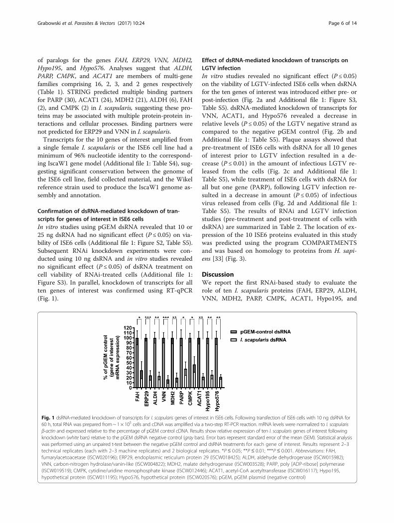
Confirmation of dsRNA-mediated knockdown of tran-scripts for genes of interest in ISE6 cellsIn vitro studies using pGEM dsRNA revealed that 10 or25 ng dsRNA had no significant effect (P ≤ 0.05) on via-bility of ISE6 cells (Additional file 1: Figure S2, Table S5).Subsequent RNAi knockdown experiments were con-ducted using 10 ng dsRNA and in vitro studies revealedno significant effect (P ≤ 0.05) of dsRNA treatment oncell viability of RNAi-treated cells (Additional file 1:Figure S3). In parallel, knockdown of transcripts for allten genes of interest was confirmed using RT-qPCR(Fig. 1).
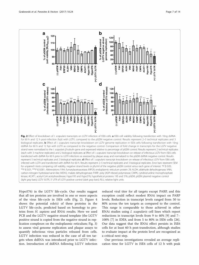
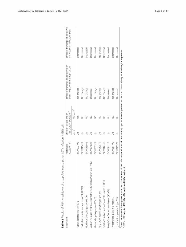
Effect of dsRNA-mediated knockdown of transcripts onLGTV infectionIn vitro studies revealed no significant effect (P ≤ 0.05)on the viability of LGTV-infected ISE6 cells when dsRNAfor the ten genes of interest was introduced either pre- orpost-infection (Fig. 2a and Additional file 1: Figure S3,Table S5). dsRNA-mediated knockdown of transcripts forVNN, ACAT1, and Hypo576 revealed a decrease inrelative levels (P ≤ 0.05) of the LGTV negative strand ascompared to the negative pGEM control (Fig. 2b andAdditional file 1: Table S5). Plaque assays showed thatpre-treatment of ISE6 cells with dsRNA for all 10 genesof interest prior to LGTV infection resulted in a de-crease (P ≤ 0.01) in the amount of infectious LGTV re-leased from the cells (Fig. 2c and Additional file 1:Table S5), while treatment of ISE6 cells with dsRNA forall but one gene (PARP), following LGTV infection re-sulted in a decrease in amount (P ≤ 0.05) of infectiousvirus released from cells (Fig. 2d and Additional file 1:Table S5). The results of RNAi and LGTV infectionstudies (pre-treatment and post-treatment of cells withdsRNA) are summarized in Table 2. The location of ex-pression of the 10 ISE6 proteins evaluated in this studywas predicted using the program COMPARTMENTSand was based on homology to proteins from H. sapi-ens [33] (Fig. 3).
DiscussionWe report the first RNAi-based study to evaluate therole of ten I. scapularis proteins (FAH, ERP29, ALDH,VNN, MDH2, PARP, CMPK, ACAT1, Hypo195, and
Fig. 1 dsRNA-mediated knockdown of transcripts for I. scapularis genes of interest in ISE6 cells. Following transfection of ISE6 cells with 10 ng dsRNA for60 h, total RNA was prepared from~ 1 × 10⁵ cells and cDNA was amplified via a two-step RT-PCR reaction. mRNA levels were normalized to I. scapularisβ-actin and expressed relative to the percentage of pGEM control cDNA. Results show relative expression of ten I. scapularis genes of interest followingknockdown (white bars) relative to the pGEM dsRNA negative control (gray bars). Error bars represent standard error of the mean (SEM). Statistical analysiswas performed using an unpaired t-test between the negative pGEM control and dsRNA treatments for each gene of interest. Results represent 2–3technical replicates (each with 2–3 machine replicates) and 2 biological replicates. *P ≤ 0.05; **P ≤ 0.01; ***P ≤ 0.001. Abbreviations: FAH,fumarylacetoacetase (ISCW020196); ERP29, endoplasmic reticulum protein 29 (ISCW018425); ALDH, aldehyde dehydrogenase (ISCW015982);VNN, carbon-nitrogen hydrolase/vanin-like (ISCW004822); MDH2, malate dehydrogenase (ISCW003528); PARP, poly [ADP-ribose] polymerase(ISCW019519); CMPK, cytidine/uridine monophosphate kinase (ISCW012446); ACAT1, acetyl-CoA acetyltransferase (ISCW016117); Hypo195,hypothetical protein (ISCW011195); Hypo576, hypothetical protein (ISCW020576); pGEM, pGEM plasmid (negative control)
Grabowski et al. Parasites & Vectors (2017) 10:24 Page 6 of 14
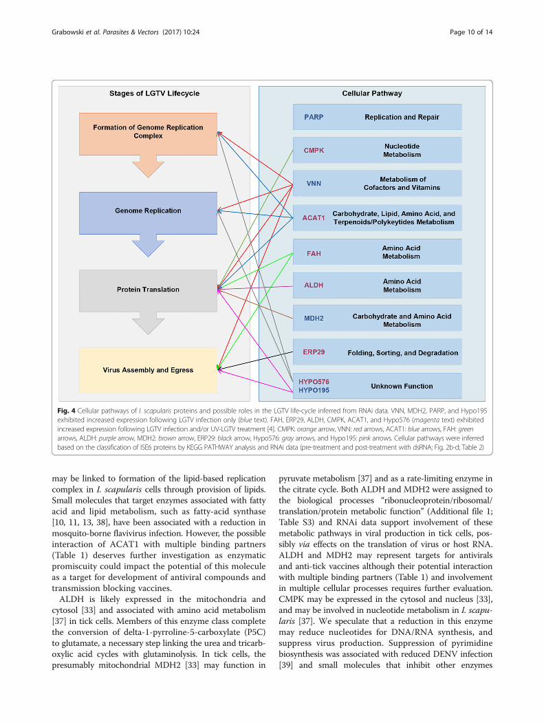
Hypo576) in the LGTV life-cycle. Our results suggestthat all ten proteins are involved in one or more aspectsof the virus life-cycle in ISE6 cells (Fig. 2). Figure 4shows the potential role(s) of these proteins in theLGTV life-cycle, predicted based on homology to pro-teins from H. sapiens and RNAi results. Here we usedPCR and the LGTV negative strand template (the LGTVpositive strand is copied from the negative strand in rep-lication complexes on the endoplasmic reticulum; Fig. 3)to assess viral genome replication and plaque assays toquantify infectious virus particles released from cells.LGTV infection was reduced in the case of all ten tar-gets when dsRNA was introduced prior to LGTV infec-tion. Introduction of dsRNA following LGTV infection
reduced viral titer for all targets except PARP, and thisexception could reflect modest RNAi impact on PARPlevels. Reduction in transcript levels ranged from 50 to80% across the ten targets as compared to the control.This range is comparable to those achieved in otherRNAi studies using I. scapularis cell lines which reportreductions in transcript levels from 9 to 40% [9] and 7–100% [7] in IDE8, and from 5 to 80% in ISE6 cells [26].Our data suggest that the RNAi effect persists in ISE6cells for at least 60 h post-transfection, although studiesto evaluate impact at the protein level are recognized asa critical next step.Our previous investigations revealed an average repli-
cation time for LGTV in ISE6 cells of 12 h with peak
Fig. 2 Effect of knockdown of I. scapularis transcripts on LGTV infection of ISE6 cells. a ISE6 cell viability following transfection with 10ng dsRNAfor 60 h and 12 h post-infection (hpi) with LGTV, compared to the pGEM negative control. Results represent 2–5 technical replicates and 3biological replicates. b Effect of I. scapularis transcript knockdown on LGTV genome replication in ISE6 cells following transfection with 10ngdsRNA for 60 h and 12 hpi with LGTV as compared to the negative control. Comparison of fold change in transcripts for the LGTV negativestrand were normalized to the I. scapularis β-tubulin gene and expressed relative to percentage of pGEM control. Results represent 2 technical replicates(each with 3 machine replicates) and 2 biological replicates. c Effect of I. scapularis transcript knockdown on release of infectious LGTV from ISE6 cellstransfected with dsRNA for 60 h prior to LGTV infections as assessed by plaque assay and normalized to the pGEM dsRNA negative control. Resultsrepresent 5 technical replicates and 2 biological replicates. d Effect of I. scapularis transcript knockdown on release of infectious LGTV from ISE6 cellsinfected with LGTV and transfected with dsRNA for 60 h. Results represent 2–5 technical replicates and 3 biological replicates. Error bars represent SEMfor unpaired t-tests comparing cell viability, negative strand levels or pfu/ml of the negative pGEM control versus each gene of interest. *P≤ 0.05;**P≤ 0.01; ***P≤ 0.001. Abbreviations: FAH, fumarylacetoacetase; ERP29, endoplasmic reticulum protein 29; ALDH, aldehyde dehydrogenase; VNN,carbon-nitrogen hydrolase/vanin-like; MDH2, malate dehydrogenase; PARP, poly [ADP-ribose] polymerase; CMPK, cytidine/urdine monophosphatekinase; ACAT1, acetyl-CoA acetyltransferase; Hypo195 and Hypo576, hypothetical proteins 195 and 576; pGEM, pGEM plasmid negative control(light gray bars); LGTV 3UTR, 3’ UTR of LGTV positive control (dark gray bars); RLU, relative light units
Grabowski et al. Parasites & Vectors (2017) 10:24 Page 7 of 14
Table
2Results
ofRN
Aikno
ckdo
wnof
I.scapularistranscrip
tson
LGTV
infectionin
ISE6
cells
Tick
Protein
VectorBase
accessionID
Effect
oftreatm
enton
ISE6
proteinexpression
aEffect
oftranscrip
tknockdow
non
LGTV
negativestrand
replication
Effect
oftranscrip
tknockdow
non
releaseof
infectious
LGTV
LGTV
bUV-LG
TVb
Fumarylacetoacetase
(FAH)
ISCW020196
Up
Up
Nochange
Decreased
Endo
plasmicreticulum
protein29
(ERP29)
ISCW018425
Up
Up
Nochange
Decreased
Aldeh
ydede
hydrog
enase(ALD
H)
ISCW015982
Up
Up
Nochange
Decreased
Carbo
n-nitrog
enhydrolase/pantethe
inehydrolase/vanin-like(VNN)
ISCW004822
Up
NC
Decreased
Decreased
Malatede
hydrog
enase(M
DH2)
ISCW003528
Up
NC
Nochange
Decreased
Poly[ADP-rib
ose]
polymerase(PARP)
ISCW019519
Up
NC
Nochange
Nochange
Cytidine/uridinemon
opho
sphate
kinase
(CMPK)
ISCW012446
Up
Up
Nochange
Decreased
Acetyl-C
oAC-acetyltransferase(ACAT1)
ISCW016117
Up
Up
Decreased
Decreased
Hypothe
ticalprotein(Hypo1
95)
ISCW011195
Up
NC
Nochange
Decreased
Hypothe
ticalprotein(Hypo5
76)
ISCW020576
Up
Up
Decreased
Decreased
a Protein
expression
followingLG
TVan
d/or
UV-LG
TVtreatm
entof
ISE6
cells
compa
redto
mocktreatm
ent[4].Up=increasedexpression
andNC=no
statistically-significan
tchan
gein
expression
bLG
TV=LG
TVinfection;
UV-LG
TV=UVinactiv
ated
LGTV
treatm
ent
Grabowski et al. Parasites & Vectors (2017) 10:24 Page 8 of 14

release of infectious LGTV at 36 hpi [4]. Here, we pre-treated cells with dsRNA for 60 h (hereafter referred toas “pre-treatment with dsRNA”) and subsequently mea-sured infectious LGTV particles at 16 hpi (Fig. 2c). Thistime-point was selected in an attempt to capture the firstpopulation of infectious virus particles released fromcells. For comparative purposes, we also assessed infec-tious LGTV release from ISE6 cells exposed to LGTVand subsequently transfected with dsRNA for 60 h(post-treatment with dsRNA; Fig. 2d). The latter meas-urement may reflect several rounds of virus infectionand in the case of FAH, ERP29, ALDH, MDH2, PARP,and Hypo576, may explain our observation of higherviral titers in comparison to the results for pre-treatedcells.The proposed cellular location and function of the ten
I. scapularis proteins investigated in this study, and thepredicted role(s) of these proteins in the LGTV life-cycleis depicted in Figs. 3 and 4. The knockdown of tran-scripts for VNN, ACAT1, and Hypo576 was associatedwith a decrease in both LGTV genome replication andrelease of infectious virus from cells (Fig. 2b-d). Furtherstudies are required to dissect the contributions of these
proteins to the LGTV infection process and determinewhether the observed reduction in release of LGTV re-flects the disruption of processes pre- and/or post-replication of the viral genome. The knockdown oftranscripts for FAH, ERP29, ALDH, MDH2, PARP,CMPK, and Hypo195 was associated with a decrease ininfectious LGTV only, suggesting that these proteinsmay be involved in processes independent of viral gen-ome replication (i.e. viral protein synthesis, packaging,egress, and release).VNN is a glycosylphosphatidylinositol (GPI)-anchored
protein [34] found at the surface of many different verte-brate cells [35] and may also be secreted [33]. GPI pro-teins perform diverse cellular functions, including lipidremodeling, which occurs during the replication of flavi-viruses. KEGG pathway analyses suggest that VNN mayfunction in pantothenate/coenzyme-A (CoA) biosynthesis[36], which is critical for lipid metabolism. This proteinfamily is worthy of further investigation in I. scapularisand other TBF vectors as antiviral targets. ACAT1 may beexpressed in the mitochondria and potentially secreted[33]. This enzyme likely utilizes CoA metabolites duringmetabolism of fatty acids (lipids) and amino acids [37] and
Fig. 3 Proposed cellular location of I. scapularis proteins investigated in the present study. Expression of FAH, ERP29, ALDH, VNN, MDH2, PARP,CMPK, ACAT1, Hypo195 and Hypo576 (light blue) is shown based on studies of orthologous proteins in H. sapiens (excluding Hypo195 and 576).Black circles, ribosomes; dark blue lines, LGTV positive strand; orange lines, LGTV negative strand; red circles, viral proteins associated with replicationcomplexes on the endoplasmic reticulum; purple particles, immature LGTV prior to cleavage of the pr peptide; magenta circles, furin protease producedby the host cell and responsible for cleavage of the pr peptide from the Membrane protein, producing mature LGTV; extracellular purple circles, cleavedpr peptide; extracellular black smooth particles, mature LGTV. Protein localization was based on COMPARTMENTS [33] predictions using data fromH. sapiens
Grabowski et al. Parasites & Vectors (2017) 10:24 Page 9 of 14
may be linked to formation of the lipid-based replicationcomplex in I. scapularis cells through provision of lipids.Small molecules that target enzymes associated with fattyacid and lipid metabolism, such as fatty-acid synthase[10, 11, 13, 38], have been associated with a reduction inmosquito-borne flavivirus infection. However, the possibleinteraction of ACAT1 with multiple binding partners(Table 1) deserves further investigation as enzymaticpromiscuity could impact the potential of this moleculeas a target for development of antiviral compounds andtransmission blocking vaccines.ALDH is likely expressed in the mitochondria and
cytosol [33] and associated with amino acid metabolism[37] in tick cells. Members of this enzyme class completethe conversion of delta-1-pyrroline-5-carboxylate (P5C)to glutamate, a necessary step linking the urea and tricarb-oxylic acid cycles with glutaminolysis. In tick cells, thepresumably mitochondrial MDH2 [33] may function in
pyruvate metabolism [37] and as a rate-limiting enzyme inthe citrate cycle. Both ALDH and MDH2 were assigned tothe biological processes “ribonucleoprotein/ribosomal/translation/protein metabolic function” (Additional file 1;Table S3) and RNAi data support involvement of thesemetabolic pathways in viral production in tick cells, pos-sibly via effects on the translation of virus or host RNA.ALDH and MDH2 may represent targets for antiviralsand anti-tick vaccines although their potential interactionwith multiple binding partners (Table 1) and involvementin multiple cellular processes requires further evaluation.CMPK may be expressed in the cytosol and nucleus [33],and may be involved in nucleotide metabolism in I. scapu-laris [37]. We speculate that a reduction in this enzymemay reduce nucleotides for DNA/RNA synthesis, andsuppress virus production. Suppression of pyrimidinebiosynthesis was associated with reduced DENV infection[39] and small molecules that inhibit other enzymes
Fig. 4 Cellular pathways of I. scapularis proteins and possible roles in the LGTV life-cycle inferred from RNAi data. VNN, MDH2, PARP, and Hypo195exhibited increased expression following LGTV infection only (blue text). FAH, ERP29, ALDH, CMPK, ACAT1, and Hypo576 (magenta text) exhibitedincreased expression following LGTV infection and/or UV-LGTV treatment [4]. CMPK: orange arrow, VNN: red arrows, ACAT1: blue arrows, FAH: greenarrows, ALDH: purple arrow, MDH2: brown arrow, ERP29: black arrow, Hypo576: gray arrows, and Hypo195: pink arrows. Cellular pathways were inferredbased on the classification of ISE6 proteins by KEGG PATHWAY analysis and RNAi data (pre-treatment and post-treatment with dsRNA; Fig. 2b-d; Table 2)
Grabowski et al. Parasites & Vectors (2017) 10:24 Page 10 of 14
involved in nucleotide biosynthesis reduced arboviruses incell culture [13, 40, 41]. Inhibition of CMPK may reduceTBF infection but would likely have broad impacts oncellular processes. ALDH, MDH2 and CMPK exhibitedincreased expression in human hepatoma 7.5 cells(HUH7.5) infected with Hepatitis C virus (HCV) [42]and FAH was decreased in HCV-associated carcinomatissue [43]. Products that disrupt host proteins involvedin enzymatic pathways commonly manipulated by flavi-viruses, may provide broad anti-viral effect.Analyses suggest FAH is a secreted and cytosolic pro-
tein that may perform amino acid metabolism [33, 37]in I. scapularis. Disruption of amino acid synthesis couldaffect the production of virus or host proteins at one ormore points in the virus life-cycle. ERP29 is likely associ-ated with the endoplasmic reticulum (ER) and also se-creted [33]), and may be involved in protein processingand ER associated degradation [37] in the tick. ERP29showed increased expression in mouse brain tissue fol-lowing infection with the Japanese Encephalitis virus(JEV) [44]. Studies of polyomavirus-infected cells alsosuggest a role for ERP29 in viral binding and releasefrom the ER lumen [45–47]. Additional studies are re-quired to investigate the role of ERP in LGTV infectionand evaluate the potential of this target.PARP is likely expressed in the cytosol, mitochondria,
and nucleus [33] and may be involved in DNA replica-tion and repair [37] in the tick. This protein has a rolein pro-apoptotic signaling and is activated by oxidativestress [48]. The HCV non-structural protein 5A (NS5A)can create oxidative stress, leading to activation of PARP[49]. JEV and DENV also induced cleavage of PARP1,resulting in a variety of pro-apoptotic responses [50]. Theincreased expression of the ISE6 proteins observed on in-fection with LGTV may reflect a generalized cellular re-sponse or metabolic processes and products used by thevirus [4]. Flaviviruses are thought to exploit the cellularstress response to aid replication [51] and oxidative stressis thought to aid the replication of positive-strand RNAviruses [52]. Further, it has been proposed that the bal-ance between antioxidant responses maintains an anti-apoptotic environment during flavivirus infection of thecell [53, 54], presumably facilitating virus replicationand transmission.LGTV concentration has been linked to efficiency of
establishment of infection in I. scapularis larvae [27]. Inthe present study, transcript knockdown produced a mod-est reduction in LGTV replication and genome replica-tion. It is recognized that a small reduction in amount ofinfectious virus can profoundly affect transmission; furtherfunctional analyses might investigate transcript knock-down over time and it will be necessary to establish that areduction in protein level or impairment of enzymatic ac-tivity in vivo translates to a meaningful reduction in virus
transmission. Recapitulation of such studies in the naturalvectors of LGTV will also be important as flavivirus repli-cation may differ among natural vector and non-vectorcell types [55]. Our predictions regarding cellular com-partments associated with protein expression were madebased on homology to proteins from H. sapiens andstudies are needed to determine the spatio-temporal ex-pression of tick proteins. Transcripts for MDH2 wereidentified in the salivary glands of blood-fed I. scapu-laris nymphs [56] and both MDH2 and Hypo195 wereidentified in the synganglia of I. scapularis [6] suggest-ing potential roles in neurological processes. Functionalstudies would be of particular value in the case ofHypo195 and Hypo576. Orthologs of Hypo195 andHypo576 have not been identified in H. sapiens (Table 1)and these proteins may represent targets for development ofvector-specific products to control flavivirus transmission.
ConclusionsOur work provides for investigations of orthologousprotein targets in transmission of more virulent TBFs,including POWV and TBEV. Theoretically, small mole-cules that disrupt one or more tick proteins could beused to limit virus transmission from the tick to mamma-lian reservoirs and intermediate hosts. Small molecule in-hibitors of FAH [57, 58] and PARP [59, 60] are known buttheir potential to regulate flavivirus infection in arthropodshas not been investigated. In addition, there is precedentfor development of transmission blocking vaccines againstTBFs. The outer surface protein A (OspA) of the Borreliaburgdorferi bacterium is the basis for a Lyme disease (LD)vaccine and has been deployed in the Peromyscus leucopus(white-footed mouse) reservoir [61, 62]. This vaccine, de-livered via oral bait, offers one strategy to reduce circula-tion of B. burgdorferi in the reservoir, and subsequenttransmission to the tick vector. Functional studies de-scribed here highlight proteins associated with pathogen-esis of TBFs and are a necessary precursor to anti-tickvaccine development [63]. Further functional studies willreveal the potential of these proteins as targets for develop-ment of new strategies to prevent TBF infections.
Additional file
Additional file 1: Figure S1. Summary of the process employed toselect I. scapularis genes for RNAi knockdown experiments. Δ ISE6 proteinsfrom the differential proteomic analysis at 36 hpi were analyzed. Proteinswere selected based on (1) increased expression level, (2) strength ofproteomic support (minimum 2 peptides identified from LC-MS-MS perprotein) from proteins identified in Grabowski et al. [4], and (3) orthologyto vertebrate/invertebrate proteins; * orthologous proteins identified inpublished proteomic studies [4–6, 8]. LGTV denotes proteins that exhibitedincreased expression following LGTV infection and LGTV & UV-LGTV denotesproteins that exhibited increased expression following both LGTV infectionand UV-LGTV treatment. + proteins that exhibited increased expressionfollowing LGTV infection as compared to UV-LGTV treatment. FAH,
Grabowski et al. Parasites & Vectors (2017) 10:24 Page 11 of 14
fumarylacetoacetase; ERP29, endoplasmic reticulum protein 29; ALDH,1-pyrroline-5-carboxylate dehydrogenase; VNN, pantetheine hydrolase;MDH2, malate dehydrogenase; PARP, poly [ADP-ribose] polymerase;CMPK, UMP-CMP kinase; ACAT1, acetyl-CoA acetyltransferase; Hypo195,hypothetical protein; Hypo576. The prefix “ISCW” denotes VectorBaseaccession IDs. Figure S2 Effect of pGEM dsRNA concentrations on ISE6cell viability following transfection for 60 h. X-tremeGENE (Xtr) transfectionreagent was used to optimize pGEM dsRNA (RNAi negative control)concentrations in ISE6 cells at 60 h post transfection. Cell viability readingswere compared to the Xtr + OptiMEM (Opti) control (gray bar). Red boxesindicate increased or no significant decrease in ISE6 cell viability. RLU560,590,relative light units 560 nm excitation and 590 nm emission. Error bars repre-sent SEM. Statistical analysis was performed using an unpaired t-test be-tween Xtr + Opti control and each pGEM dsRNA concentration. *pvalue ≤ 0.05 and **p value ≤ 0.01. Results represent 3 technical replicatesand 1 biological replicate (multiple biological replicates completed with 10ng concentration). Figure S3 Effect of transfection with dsRNA on ISE6 cellviability. FAH, fumarylacetoacetase; ERP29, endoplasmic reticulum protein29; ALDH, 1-pyrroline-5-carboxylate dehydrogenase; VNN, pantetheinehydrolase; MDH2, malate dehydrogenase; PARP, poly [ADP-ribose]polymerase; CMPK, UMP-CMP kinase; ACAT1, acetyl-CoA acetyltransfer-ase; Hypo195, hypothetical protein; Hypo576, hypotheticalprotein; pGEM, pGEM plasmid (negative control; light gray bars); LGTV 3UTR,3’ UTR of LGTV TP21 strain (positive control; dark gray bars), RLU560,590,relative light units 560 nm excitation and 590 nm emission. ISE6 cellviability following transfection with 10ng dsRNA for 60 h normalized tothe negative control pGEM dsRNA. Results represent 2–5 technical replicatesand 3 biological replicates. Error bars represent SEM and unpaired t-tests forcomparison of cell viability of the negative pGEM control versus each geneof interest. Table S1 T7-tagged primers used to amplify cDNA andsynthesize dsRNA. Table S2 Primers used to amplify cDNA for I. scapularisgenes of interest by RT-qPCR. Table S3 Enrichment/cluster analysis of ISE6proteins that exhibited increased expression following LGTV and UV-LGTVtreatment. ISE6 proteins with increased expression following LGTV infectionand/or UV-LGTV treatment from [4] were searched via DAVID enrichmentanalysis. For each cluster, the P value represents a modified FisherExact P value, and EASE score implemented in DAVID gene enrichment andfunctional annotation analysis. Enrichment (E) score of ≥ 1.3 is equal toP value of ≤ 0.05. Table S4 Nucleotide similarity of RT-PCR productsamplified from I. scapularis and ISE6 cells and IscaW1 gene models.Table S5 Summary of statistically significant values corresponding tofigures. (DOCX 329 kb)
AbbreviationsACAT1: Acetyl-CoA acetyltransferase; ALDH: Aldehyde dehydrogenase;ATCC: American Tissue Culture Collection; cDNA: Complementary DNA;CMPK: Cytidine/uridine monophosphate kinase; DENV: Dengue virus;dsRNA: Double stranded RNA; ER: Endoplasmic reticulum;ERP29: Endoplasmic reticulum protein 29; FAH: Fumarylacetoacetase;GO: Gene ontology; HCV: Hepatitis C virus; HUH: Human hepatoma cell line;Hypo195/576: Hypothetical proteins 195/576; IDE8/ISE6: Ixodes scapularis cellline 8/6; IFA: Immunofluorescence assay; JEV: Japanese encephalitis;KEGG: Kyoto Encyclopedia of Genes and Genomes; LD: Lyme disease;LGTV: Langat virus; MDH2: Malate dehydrogenase 2; MOI: Multiplicity ofinfection; Opti: OptiMEM; PARP: Poly [ADP-ribose] polymerase; RNAi: RNAinterference; RT-qPCR: reverse transcriptase-quantitative polymerase chainreaction; SEM: Standard error of the mean; TBF: Tick-borne flavivirus;VNN: Carbon nitrogen hydrolase/vanin-like; Xtr: X-tremeGENE transfectionreagent
AcknowledgementsThe authors are grateful to Alexander Pletnev (NIH/NIAID, Bethesda, MD) andTimothy J. Kurtti (University of Minnesota) for providing the LGTV TP21 strainand ISE6 cell line, respectively. We thank Esther Schnettler (MRC-University ofGlasgow Centre for Virus Research; Roslin Institute and Royal (Dick) School ofVeterinary Studies, University of Edinburgh), Sonja M. Best (NIH/NIAID,Hamilton, MT) and Andres F. Sandoval-Mojica (Department of Entomology,Purdue University) for advice on RNAi studies. pGEM plasmid and pGEMprimers with T7 promoter were kindly supplied by Michael E. Scharf(Department of Entomology, Purdue University).
FundingJMG was supported by an NSF Graduate Research Fellowship (ProgramDGE-1333468) and the Indiana Clinical and Translational Science InstitutePre-doctoral Training Program (NIH/NCATS-ICTSI TL1 TR000162). Research fundsprovided by the Entomological Society of America, Monsanto Research GrantAward to JMG are gratefully acknowledged. The funders had no role in studydesign, data collection and analysis, decision to publish, or preparation of themanuscript.
Availability of data and materialAll data are presented in the main paper and Additional file 1.
Authors’ contributionsConceived and designed the experiments: JMG, MGN, RJK, and CAH.Performed the experiments: JMG. Analyzed the data: JMG, MGN, RJK, andCAH. Contributed reagents/materials/analysis tools: JMG, RJK, and CAH.Wrote the paper: JMG and CAH. All authors read and approved the finalmanuscript.
Competing interestsThe authors declare that they have no competing interests.
Consent for publicationNot applicable.
Ethics approval and consent to participateNot applicable.
Author details1Department of Entomology, College of Agriculture, Purdue University, 901W State Street, West Lafayette, IN 47907, USA. 2Markey Center for StructuralBiology, Department of Biological Sciences, College of Science, PurdueUniversity, 915 W State Street, West Lafayette, IN 47907, USA. 3PurdueInstitute for Inflammation, Immunology and Infectious Disease, PurdueUniversity, West Lafayette, IN 47907, USA. 4Current Address: NIH/NIAID, RockyMountain Laboratories, Laboratory of Virology, Biology of Vector-BorneViruses Section, 903 S 4th St, Hamilton, MT 59840, USA. 5Current Address:Department of Biochemistry and Molecular Biology, College of Agriculture,Biotechnology, and Natural Resources, University of Nevada-Reno, 1664 NVirginia Street, Reno, NV 89503, USA.
Received: 29 May 2016 Accepted: 16 December 2016
References1. Pagel Van Zee J, Geraci NS, Guerrero FD, Wikel SK, Stuart JJ, Nene VM, et al.
Tick genomics: the Ixodes genome project and beyond. Int J Parasitol.2007;37(12):1297–305.
2. Hill CA, Wikel SK. The Ixodes scapularis Genome Project: an opportunity foradvancing tick research. Trends Parasitol. 2005;21(4):151–3.
3. Gulia-Nuss M, Nuss AB, Meyer JM, Sonenshine DE, Roe RM, Waterhouse RM,et al. Genomic insights into the Ixodes scapularis tick vector of Lyme disease.Nat Commun. 2016;7:10507.
4. Grabowski JM, Perera R, Roumani AM, Hedrick VE, Inerowicz HD, Hill CA,et al. Changes in the proteome of Langat-infected Ixodes scapularis ISE6cells: Metabolic pathways associated with flavivirus infection. PLoS NeglTrop Dis. 2016;10(2):e0004180.
5. Ayllon N, Villar M, Galindo RC, Kocan KM, Sima R, Lopez JA, et al. Systemsbiology of tissue-specific response to Anaplasma phagocytophilum revealsdifferentiated apoptosis in the tick vector Ixodes scapularis. PLoS Genet.2015;11(3):e1005120.
6. Oliver JD, Chavez AS, Felsheim RF, Kurtti TJ, Munderloh UG. An Ixodesscapularis cell line with a predominantly neuron-like phenotype. Exp ApplAcarol. 2015;66(3):427–42.
7. Weisheit S, Villar M, Tykalova H, Popara M, Loecherbach J, Watson M, et al.Ixodes scapularis and Ixodes ricinus tick cell lines respond to infection withtick-borne encephalitis virus: transcriptomic and proteomic analysis. ParasitVectors. 2015;8:599.
8. Ayllon N, Naranjo V, Hajdusek O, Villar M, Galindo RC, Kocan KM, et al.Nuclease tudor-SN is involved in Tick dsRNA-mediated RNA interference
Grabowski et al. Parasites & Vectors (2017) 10:24 Page 12 of 14
and feeding but not in defense against Flaviviral or Anaplasmaphagocytophilum Rickettsial Infection. PLoS One. 2015;10(7):e0133038.
9. Schnettler E, Tykalova H, Watson M, Sharma M, Sterken MG, Obbard DJ,et al. Induction and suppression of tick cell antiviral RNAi responses bytick-borne flaviviruses. Nucleic Acids Res. 2014;42(14):9436–46.
10. Perera R, Riley C, Isaac G, Hopf-Jannasch AS, Moore RJ, Weitz KW, et al.Dengue virus infection perturbs lipid homeostasis in infected mosquitocells. PLoS Pathog. 2012;8(3):e1002584.
11. Heaton NS, Perera R, Berger KL, Khadka S, Lacount DJ, Kuhn RJ, Randall G.Dengue virus nonstructural protein 3 redistributes fatty acid synthase tosites of viral replication and increases cellular fatty acid synthesis. Proc NatlAcad Sci USA. 2010;107(40):17345–50.
12. Heaton NS, Randall G. Dengue virus-induced autophagy regulates lipidmetabolism. Cell Host Microbe. 2010;8(5):422–32.
13. Krishnan MN, Garcia-Blanco MA. Targeting host factors to treat West Nileand dengue viral infections. Viruses. 2014;6(2):683–708.
14. Pastorino B, Nougairede A, Wurtz N, Gould E, de Lamballerie X. Role of hostcell factors in flavivirus infection: implications for pathogenesis anddevelopment of antiviral drugs. Antiviral Res. 2010;87(3):281–94.
15. Leyssen P, Balzarini J, De Clercq E, Neyts J. The predominant mechanism bywhich ribavirin exerts its antiviral activity in vitro against flaviviruses andparamyxoviruses is mediated by inhibition of IMP dehydrogenase. J Virol.2005;79(3):1943–7.
16. Chang J, Schul W, Yip A, Xu X, Guo JT, Block TM. Competitive inhibitor ofcellular alpha-glucosidases protects mice from lethal dengue virus infection.Antiviral Res. 2011;92(2):369–71.
17. Munderloh UG, Kurtti TJ. Formulation of medium for tick cell culture. ExpAppl Acarol. 1989;7(3):219–29.
18. Munderloh UG, Liu Y, Wang M, Chen C, Kurtti TJ. Establishment, maintenanceand description of cell lines from the tick Ixodes scapularis. J Parasitol.1994;80(4):533–43.
19. Pletnev AG, Men R. Attenuation of the Langat tick-borne flavivirus bychimerization with mosquito-borne flavivirus dengue type 4. Proc Natl AcadSci USA. 1998;95(4):1746–51.
20. Campbell MS, Pletnev AG. Infectious cDNA clones of Langat tick-borneflavivirus that differ from their parent in peripheral neurovirulence. Virology.2000;269(1):225–37.
21. Junjhon J, Lausumpao M, Supasa S, Noisakran S, Songjaeng A, Saraithong P,et al. Differential modulation of prM cleavage, extracellular particle distribution,and virus infectivity by conserved residues at nonfurin consensus positions ofthe dengue virus pr-M junction. J Virol. 2008;82(21):10776–91.
22. Strober W. Monitoring cell growth. Curr Protoc Immunol. 2001;Appendix 3:Appendix 3A.
23. Untergasser A, Cutcutache I, Koressaar T, Ye J, Faircloth BC, Remm M, et al.Primer3 - new capabilities and interfaces. Nucleic Acids Res. 2012;40(15):e115.
24. Koressaar T, Remm M. Enhancements and modifications of primer designprogram Primer3. Bioinformatics. 2007;23(10):1289–91.
25. Ye J, Coulouris G, Zaretskaya I, Cutcutache I, Rozen S, Madden TL. Primer-BLAST: a tool to design target-specific primers for polymerase chain reaction.BMC Bioinformatics. 2012;13:134.
26. Barry G, Alberdi P, Schnettler E, Weisheit S, Kohl A, Fazakerley JK, et al. Genesilencing in tick cell lines using small interfering or long double-strandedRNA. Exp Appl Acarol. 2013;59(3):319–38.
27. Mitzel DN, Wolfinbarger JB, Long RD, Masnick M, Best SM, Bloom ME. Tick-borne flavivirus infection in Ixodes scapularis larvae: development of a novelmethod for synchronous viral infection of ticks. Virology. 2007;365(2):410–8.
28. Livak KJ, Schmittgen TD. Analysis of relative gene expression data usingreal-time quantitative PCR and the 2(-Delta Delta C(T)) Method. Methods.2001;25(4):402–8.
29. Rao X, Huang X, Zhou Z, Lin X. An improvement of the 2^(-delta delta CT)method for quantitative real-time polymerase chain reaction data analysis.Biostat Bioinforma Biomath. 2013;3(3):71–85.
30. Dennis Jr G, Sherman BT, Hosack DA, Yang J, Gao W, Lane HC, et al. DAVID:Database for Annotation, Visualization, and Integrated Discovery. GenomeBiol. 2003;4(5):3.
31. von Mering C, Jensen LJ, Snel B, Hooper SD, Krupp M, Foglierini M, et al. STRING:known and predicted protein-protein associations, integrated and transferredacross organisms. Nucleic Acids Res. 2005;33(Database issue):D433–437.
32. Szklarczyk D, Franceschini A, Wyder S, Forslund K, Heller D, Huerta-Cepas J,et al. STRING v10: protein-protein interaction networks, integrated over thetree of life. Nucleic Acids Res. 2015;43(Database issue):D447–452.
33. Binder JX, Pletscher-Frankild S, Tsafou K, Stolte C, O’Donoghue SI, Schneider R,et al. COMPARTMENTS: unification and visualization of protein subcellularlocalization evidence. Database (Oxford). 2014;2014:bau012.
34. Aurrand-Lions M, Galland F, Bazin H, Zakharyev VM, Imhof BA, Naquet P.Vanin-1, a novel GPI-linked perivascular molecule involved in thymushoming. Immunity. 1996;5(5):391–405.
35. Boersma YL, Newman J, Adams TE, Cowieson N, Krippner G, Bozaoglu K,et al. The structure of vanin 1: a key enzyme linking metabolic disease andinflammation. Acta Crystallogr D Biol Crystallogr. 2014;70(Pt 12):3320–9.
36. Kinoshita T, Fujita M. Biosynthesis of GPI-anchored proteins: specialemphasis on GPI lipid remodeling. J Lipid Res. 2016;57(1):6–24.
37. Kanehisa M, Sato Y, Kawashima M, Furumichi M, Tanabe M. KEGG as areference resource for gene and protein annotation. Nucleic Acids Res.2016;44(D1):D457–462.
38. Martin-Acebes MA, Blazquez AB, Jimenez de Oya N, Escribano-Romero E,Saiz JC. West Nile virus replication requires fatty acid synthesis but isindependent on phosphatidylinositol-4-phosphate lipids. PLoS One.2011;6(9):e24970.
39. Wang QY, Bushell S, Qing M, Xu HY, Bonavia A, Nunes S, et al. Inhibition ofdengue virus through suppression of host pyrimidine biosynthesis. J Virol.2011;85(13):6548–56.
40. Morrey JD, Smee DF, Sidwell RW, Tseng C. Identification of active antiviralcompounds against a New York isolate of West Nile virus. Antiviral Res.2002;55(1):107–16.
41. Qing M, Zou G, Wang QY, Xu HY, Dong H, Yuan Z, et al. Characterization ofdengue virus resistance to brequinar in cell culture. Antimicrob AgentsChemother. 2010;54(9):3686–95.
42. Diamond DL, Syder AJ, Jacobs JM, Sorensen CM, Walters KA, Proll SC, et al.Temporal proteome and lipidome profiles reveal hepatitis C virus-associatedreprogramming of hepatocellular metabolism and bioenergetics. PLoS Pathog.2010;6(1):e1000719.
43. Kim W, Oe Lim S, Kim JS, Ryu YH, Byeon JY, Kim HJ, et al. Comparison ofproteome between hepatitis B virus- and hepatitis C virus-associatedhepatocellular carcinoma. Clin Cancer Res. 2003;9(15):5493–500.
44. Sengupta N, Ghosh S, Vasaikar SV, Gomes J, Basu A. Modulation of neuronalproteome profile in response to Japanese encephalitis virus infection. PLoSOne. 2014;9(3):e90211.
45. Magnuson B, Rainey EK, Benjamin T, Baryshev M, Mkrtchian S, Tsai B. ERp29triggers a conformational change in polyomavirus to stimulate membranebinding. Mol Cell. 2005;20(2):289–300.
46. Walczak CP, Tsai B. A PDI family network acts distinctly and coordinatelywith ERp29 to facilitate polyomavirus infection. J Virol. 2011;85(5):2386–96.
47. Inoue T, Tsai B. How viruses use the endoplasmic reticulum for entry,replication, and assembly. Cold Spring Harb Perspect Biol. 2013;5(1):a013250.
48. Mao Z, Hine C, Tian X, Van Meter M, Au M, Vaidya A, et al. SIRT6promotes DNA repair under stress by activating PARP1. Science. 2011;332(6036):1443–6.
49. Paracha UZ, Fatima K, Alqahtani M, Chaudhary A, Abuzenadah A,Damanhouri G, et al. Oxidative stress and hepatitis C virus. Virol J. 2013;10:251.
50. Lee CJ, Liao CL, Lin YL. Flavivirus activates phosphatidylinositol 3-kinasesignaling to block caspase-dependent apoptotic cell death at the earlystage of virus infection. J Virol. 2005;79(13):8388–99.
51. Blazquez AB, Escribano-Romero E, Merino-Ramos T, Saiz JC, Martin-Acebes MA.Stress responses in flavivirus-infected cells: activation of unfolded proteinresponse and autophagy. Front Microbiol. 2014;5:266.
52. Gullberg RC, Jordan Steel J, Moon SL, Soltani E, Geiss BJ. Oxidative stressinfluences positive strand RNA virus genome synthesis and capping.Virology. 2015;475:219–29.
53. Chen TH, Lo YP, Yang CF, Chen WJ. Additive protection by antioxidant andapoptosis-inhibiting effects on mosquito cells with dengue 2 virus infection.PLoS Negl Trop Dis. 2012;6(4):e1613.
54. Chen TH, Tang P, Yang CF, Kao LH, Lo YP, Chuang CK, et al. Antioxidantdefense is one of the mechanisms by which mosquito cells survive dengue2 viral infection. Virology. 2011;410(2):410–7.
55. Ruzek D, Bell-Sakyi L, Kopecky J, Grubhoffer L. Growth of tick-borneencephalitis virus (European subtype) in cell lines from vector and non-vectorticks. Virus Res. 2008;137(1):142–6.
56. McNally KL, Mitzel DN, Anderson JM, Ribeiro JM, Valenzuela JG, Myers TG,et al. Differential salivary gland transcript expression profile in Ixodesscapularis nymphs upon feeding or flavivirus infection. Ticks Tick Borne Dis.2012;3(1):18–26.
Grabowski et al. Parasites & Vectors (2017) 10:24 Page 13 of 14

57. Bateman RL, Bhanumoorthy P, Witte JF, McClard RW, Grompe M, Timm DE.Mechanistic inferences from the crystal structure of fumarylacetoacetatehydrolase with a bound phosphorus-based inhibitor. J Biol Chem.2001;276(18):15284–91.
58. Bateman RL, Ashworth J, Witte JF, Baker LJ, Bhanumoorthy P, Timm DE,et al. Slow-onset inhibition of fumarylacetoacetate hydrolase by phosphinatemimics of the tetrahedral intermediate: kinetics, crystal structure andpharmacokinetics. Biochem J. 2007;402(2):251–60.
59. Tempera I, Deng Z, Atanasiu C, Chen CJ, D’Erme M, Lieberman PM.Regulation of Epstein-Barr virus OriP replication by poly(ADP-ribose)polymerase 1. J Virol. 2010;84(10):4988–97.
60. Gibson BA, Kraus WL. New insights into the molecular and cellular functionsof poly(ADP-ribose) and PARPs. Nat Rev Mol Cell Biol. 2012;13(7):411–24.
61. Bhattacharya D, Bensaci M, Luker KE, Luker G, Wisdom S, Telford SR, et al.Development of a baited oral vaccine for use in reservoir-targeted strategiesagainst Lyme disease. Vaccine. 2011;29(44):7818–25.
62. Richer LM, Brisson D, Melo R, Ostfeld RS, Zeidner N, Gomes-Solecki M.Reservoir targeted vaccine against Borrelia burgdorferi: a new strategy toprevent Lyme disease transmission. J Infect Dis. 2014;209(12):1972–80.
63. de la Fuente J, Merino O. Vaccinomics, the new road to tick vaccines. Vaccine.2013;31(50):5923–9.
• We accept pre-submission inquiries
• Our selector tool helps you to find the most relevant journal
• We provide round the clock customer support
• Convenient online submission
• Thorough peer review
• Inclusion in PubMed and all major indexing services
• Maximum visibility for your research
Submit your manuscript atwww.biomedcentral.com/submit
Submit your next manuscript to BioMed Central and we will help you at every step:
Grabowski et al. Parasites & Vectors (2017) 10:24 Page 14 of 14


![In Vivo NADH/NAD+ Biosensing Reveals the Dynamics of ...In Vivo NADH/NAD1 Biosensing Reveals the Dynamics of Cytosolic Redox Metabolism in Plants[OPEN] Janina Steinbeck,a Philippe](https://img.pdfslide.net/doc/110x75/6089901585c6453b995ebe0f/in-vivo-nadhnad-biosensing-reveals-the-dynamics-of-in-vivo-nadhnad1-biosensing.jpg)