Embed Size (px)
Citation preview

Rahn et al. Biology of Sex Differences 2014, 5:4http://www.bsd-journal.com/content/5/1/4
RESEARCH Open Access
Sex differences in a mouse model of multiplesclerosis: neuropathic pain behavior in femalesbut not males and protection from neurologicaldeficits during proestrusElizabeth J Rahn1,2, Tommaso Iannitti1,3, Renee R Donahue1 and Bradley K Taylor1*
Abstract
Background: Multiple sclerosis (MS), a demyelinating disease of the central nervous system, is one of the mostprevalent neurological disorders in the industrialized world. This disease afflicts more than two million peopleworldwide, over two thirds of which are women. MS is typically diagnosed between the ages of 20–40 and canproduce debilitating neurological impairments including muscle spasticity, muscle paralysis, and chronic pain.Despite the large sex disparity in MS prevalence, clinical and basic research investigations of how sex and estrouscycle impact development, duration, and severity of neurological impairments and pain symptoms are limited. Tohelp address these questions, we evaluated behavioral signs of sensory and motor functions in one of the mostwidely characterized animal models of MS, the experimental autoimmune encephalomyelitis (EAE) model.
Methods: C57BL/6 male and female mice received flank injection of complete Freund’s adjuvant (CFA) or CFA plusmyelin oligodendrocyte glycoprotein 35-55 (MOG35-55) to induce EAE. Experiment 1 evaluated sex differences ofEAE-induced neurological motor deficits and neuropathic pain-like behavior over 3 weeks, while experiment 2evaluated the effect of estrous phase in female mice on the same behavioral measures for 3 months. EAE-inducedneurological motor deficits including gait analysis and forelimb grip strength were assessed. Neuropathic pain-likebehaviors evaluated included sensitivity to mechanical, cold, and heat stimulations. Estrous cycle was determineddaily via vaginal lavage.
Results: MOG35-55-induced EAE produced neurological impairments (i.e., motor dysfunction) including mild paralysisand decreases in grip strength in both females and males. MOG35-55 produced behavioral signs of neuropathicpain—mechanical and cold hypersensitivity—in females, but not males. MOG35-55 did not change cutaneous heatsensitivity in either sex. Administration of CFA or CFA + MOG35-55 prolonged the time spent in diestrus for 2 weeks,after which normal cycling returned. MOG35-55 produced fewer neurological motor deficits when mice were inproestrus relative to non-proestrus phases.
Conclusions: We conclude that female mice are superior to males for the study of neuropathic pain-like behaviorsassociated with MOG35-55-induced EAE. Further, proestrus may be protective against EAE-induced neurologicaldeficits, thus necessitating further investigation into the impact that estrous cycle exerts on MS symptoms.
Keywords: Experimental autoimmune encephalomyelitis, Estrous, MOG35-55, Proestrus
* Correspondence: [email protected] of Physiology, University of Kentucky, 800 Rose Street,Lexington, KY 40536, USAFull list of author information is available at the end of the article
© 2014 Rahn et al.; licensee BioMed Central Ltd. This is an Open Access article distributed under the terms of the CreativeCommons Attribution License (http://creativecommons.org/licenses/by/2.0), which permits unrestricted use, distribution, andreproduction in any medium, provided the original work is properly credited.

Rahn et al. Biology of Sex Differences 2014, 5:4 Page 2 of 18http://www.bsd-journal.com/content/5/1/4
BackgroundMultiple sclerosis (MS) is an autoimmune demyelinatingdisease of the central nervous system (CNS) that afflictstwice the number of women as compared to men [1].Sex has been shown to impact both the pathology andseverity of MS. While the immune response associatedwith MS is more robust in women, the pathology andprognosis for men are generally associated with moreprogressive neurodegeneration [2]. Despite this large sexdisparity in disease prevalence and severity, the impactof estrous cycle and correlated alterations in circulatingovarian hormones (estrogens/progestogens) on MS hasyet to be fully characterized. Limited reports indicatethat clinical manifestations of MS (e.g., weakness, numb-ness, tingling, and tiredness) tend to increase when ovar-ian hormones are low and decrease when they are high[3-5]. Additionally, pregnancy-associated remission ofsymptoms is believed to be associated with increased cir-culating ovarian hormones [6,7].Although neurological deficits such as muscle weak-
ness, spasticity, and paralysis are considered canonicalsymptoms of MS, chronic pain is a symptom frequentlyexperienced by patients which can antedate other neuro-logical impairments. Pain is considered a primary factorin the suffering and poor quality of life experienced byMS patients, with an overall prevalence of approximately63% [8-11]. Cardinal features of neuropathic pain inmultiple sclerosis include hypersensitivity to cutaneousmechanical and thermal stimuli in the distal extremities[10,12-14]. Sex differences in pain symptoms of MS isan understudied area of research, and the literatureyields limited and conflicting results. Some clinical stud-ies suggest that pain is worse in females [15,16], whileothers report no differences [17-19].The serendipitous advent of animal models mimicking
the pathology and symptoms of MS has allowed basicresearchers to investigate disease progression, sympto-mology, and therapeutic interventions. These widely uti-lized models include the experimental autoimmuneencephalomyelitis (EAE) and Theiler’s murine enceph-alomyelitis virus (TMEV) models. Research evaluatingsex differences in both models have yielded variable re-sults, undoubtedly influenced by a number of factors in-cluding autosomal genotype of murine strains and theimmunization agent/protocol used [20-23]. Although anumber of studies characterize manipulation of ovarianhormones in these models, we were unable to find a singlestudy evaluating natural estrous cycle and correspondingalterations in motor impairments, disease progression, orany other behavioral measure. Preclinical animal modelsof MS virtually ignore the question of neuropathic pain-like behaviors in males versus females with the exceptionof two reports indicating that allodynia and/or hyperalge-sia is more pronounced in females [22,24].
The present study was designed to investigate the effectsof sex and estrous state on the development, duration,and severity of neurological deficits and neuropathic pain-like behavior in a mouse EAE model induced by myelinoligodendrocyte glycoprotein 35–55 (MOG35-55) [25]. TheMOG35-55 EAE model has previously been shown to pro-duce robust and reproducible motor dysfunction, demye-lination, and neuropathic pain-like behaviors [26-28]. Wehypothesized that while both males and females woulddemonstrate development of neurological impairments(i.e., motor dysfunction) typical of EAE, females wouldshow more severe neuropathic pain-like behaviors.Based on the clinical findings of reduced MS symptomsduring the luteal phase and remission of MS symptomsduring pregnancy, we further hypothesized that thesesymptoms would be attenuated during the proestrusphase, when circulating ovarian hormones peak.
MethodsAnimalsThis study used 80 C57BL/6 mice purchased from CharlesRivers (Indianapolis, IN, USA), aged 12–14 weeks whenthe studies began. Mice were housed four to a cage, main-tained in a temperature- and humidity-controlled environ-ment on a 14/10 h light/dark cycle (lights on 4:00 a.m.,lights off 6:00 p.m.). Since the estrous phase is stimulatedby the presence of male pheromones [29,30], male and fe-male cages were interspersed with each other. We did notcontrol for pheromone exposure as the primary purposewas to keep female mice cycling; however, care was takento spatially intersperse cages of females with equal num-bers of cages housing male mice. Food and water wereavailable ad libitum. Animals were allowed a minimum of1 week to habituate to the facility prior to their entranceinto the study. All animal procedures were approved bythe Institutional Animal Care and Use Committee of theUniversity of Kentucky, followed the guidelines for thetreatment of animals of the International Association forthe Study of Pain, and conducted in full compliance withthe Association for Assessment and Accreditation ofLaboratory Animal Care (AAALAC).
General experimental methodsTwo experimental studies were completed. Experiment 1was designed to characterize sex differences observed inthe EAE model induced by immunization with MOG35-55
(detailed protocol below). In this study, female and maleC57BL/6 mice were immunized with complete Freund’sadjuvant (CFA) (females, n = 13; males, n = 10) or CFA +MOG35-55 (females, n = 11; males, n = 11) and evaluatedbehaviorally for neurological motor deficits and alterationsto tactile and thermal stimulations for 21 days post-immunization. Experiment 2 evaluated the impact ofestrous cycle on neurological motor impairments and
Rahn et al. Biology of Sex Differences 2014, 5:4 Page 3 of 18http://www.bsd-journal.com/content/5/1/4
responses to tactile and thermal stimulation in the EAEmodel. In experiment 2, female C57BL/6 mice were im-munized with either CFA (n = 17) or CFA +MOG35-55
(n = 18) and evaluated for 43 days post-immunization.Estrous cycle was determined daily via vaginal lavagefrom day −7 through day 43 (detailed protocol below).To evaluate the duration of neuropathic pain-like be-havior in the EAE model, a randomly chosen subset ofanimals from experiment 2 (CFA, n = 8; CFA +MOG35-55,n = 9) were tested for an additional period, from days 60–90 post-immunization.All animals were weighed and evaluated daily for al-
terations in neurological motor function using a clinicalassessment scoring system as previously described [31](experiment 1: day −2 through day 21; experiment 2:day −7 through day 43, days 60, 75, and 90). Baselinegrip strength, as well as mechanical and cold stimula-tions, was evaluated on day −2 for both experiments 1and 2. Baseline thermal responses were measured onday −1 for both experiments. Immunization with eitherCFA or CFA +MOG35-55 occurred on days 0 and 6. Al-terations in grip strength and responsivity to mechanicaland cold stimulation were evaluated on days −2, 1, 3, 5,7, 9, 11, 13, 17, and 21 in both experiments and contin-ued on days 25, 29, 35, 42, 60, 75, and 90 for animals inexperiment 2. Heat sensitivity was assessed on days −1,4, 10, and 16 in both experiments and continued withdays 28, 34, and 43 in experiment 2. Following the in-duction of EAE, which produces mild to moderatemotor dysfunction, mice were given access to DietGel®76A (Clear H2O®, Portland, OR, USA) in the bottom ofcages to ensure that they maintained body weight. In ex-periment 1, males and females were tested concurrentlybut on different behavioral testing platforms that hadbeen thoroughly cleaned with MB-10 (Quip Laboratories,Inc., Wilmington, DE, USA).
Induction of EAEEAE was induced with an immunization protocol utilizingMOG33-55 (AnaSpec Inc., Fremont, CA, USA), whichleads to T cell infiltration in the central nervous system,in combination with pertussis toxin (List Biological La-boratories, Campbell, CA, USA), an agent which en-hances EAE severity and disease onset (for review, see[32]). MOG33-55 was emulsified in a 1:1 solution of 1×phosphate-buffered saline (Fisher Scientific, Pittsburgh,PA, USA) and CFA. CFA was prepared at a concentra-tion of 5 mg/ml of mycobacterium tuberculosis (VoigtGlobal Distribution, Lawrence, KS, USA) in incompleteFreund’s adjuvant (IFA, Sigma-Aldrich, St. Louis, MO,USA). On the afternoon of day 0 following behavioralassessment, MOG35-55 (150 μg, s.c. per flank) was bilat-erally injected (100 μl) at the flank of each hindlimbunder light isoflurane (1.5%–3% in oxygen; Butler Schein,
Dublin, OH, USA) anesthesia (experiment 1) or gentlemanual restraint (experiment 2). A booster injection ofMOG35-55 (150 μg, s.c. per flank) was administered on day6. Pertussis toxin was injected (200 ng/200 μl, i.p.) on days0 and 2. Age- and sex-matched controls received identicaltreatment but were immunized with CFA only. Emulsifi-cation of MOG35-55 in CFA is necessary for the immuno-logical response observed in canonical EAE models [33].Therefore, although CFA is known to produce an inflam-matory and pain response, this control group was criticalas it allowed for differential pain behavior to be attributedto MOG35-55 rather than CFA immunization, therebyincreasing our understanding of behaviors associatedwith the pathophysiology of experimental autoimmuneencephalomyelitis.
Behavioral assessment of sensory and motor functionsFluctuations in room noise, vibrations, and temperaturewere minimized so as to facilitate acclimation and re-sponse reliability. Prior to sensory testing, the mice wereacclimated for 30 min/day for 3 days to individual Plexi-glas (10.16 × 10.16 × 25.4 cm) chambers. These boxes wereplaced on either an elevated stainless steel wire mesh (fortests of cold and mechanical sensitivity) or a Plexiglasfloor (for tests of heat sensitivity). An additional habitu-ation period of at least 30 min was provided before datacollection on each testing day. Mechanical testing wasperformed prior to cold testing, and a minimum of 1 hwas allowed between tests. Heat testing and cold/mechan-ical testing were conducted on alternating days to avoidsensitization. All behavioral measurements and injectionswere performed by a single experimenter (EJR). Animalswere assigned numbers which did not indicate group con-dition. Coded testing sheets were used throughout behav-ioral testing to keep the experimenter blind to condition.
Mechanical sensitivityMechanical sensitivity was assessed using a digital elec-tronic von Frey Anesthesiometer (model Alemo 2450;IITC Life Science, Woodland Hills, CA, USA), connectedto a 90-g probe equipped with a flexible tip. The tip wasapplied to the plantar surface of the paw until paw with-drawal. Duplicate determinations were measured for eachpaw and averaged. A minimum inter-trial interval of2 min was allowed to elapse between evaluations of paws.The testing took place in the following order: right, right,left, and left.
Cold sensitivityCold allodynia was assessed following the application ofan acetone drop to the plantar surface of the hind paw aspreviously described [34]. Acetone was loaded into a syr-inge barrel, and air bubbles were cleared from the syringeprior to acetone application. One drop of acetone

Rahn et al. Biology of Sex Differences 2014, 5:4 Page 4 of 18http://www.bsd-journal.com/content/5/1/4
(approximately 10–12 μl) was applied through the meshplatform onto the plantar surface of the hind paw. Carewas taken to gently apply the bubble of acetone (and notthe tip of the applicator) to the plantar skin. The durationof time the animal shook, licked, or completely lifted itspaw off the floor was recorded. The duration of paw with-drawal was recorded with a 60-s cutoff. Three observa-tions were taken for each paw and averaged. A minimuminter-trial interval of 5 min was allowed to pass betweenobservations for each pair of paws (i.e., right and left). Thetesting took place in the following order: right, left, right,left, right, and left.
Heat sensitivityMice were tested for sensitivity to heat using a radiantheat paw-withdrawal (Hargreaves) device [35]. The ther-mal stimulus consisted of a radiant heat source (8 V, 50-W lamp, Ugo Basile, Comerio, Italy) positioned under theglass floor directly beneath the hind paw. When triggered,a timer was activated, and light passed through a smallaperture at the top of a movable case. One day before test-ing, voltage intensity was adjusted to standardize the aver-age paw withdrawal latency at 10 ± 2 s. At specified timepoints, paw withdrawal latencies were measured in dupli-cate for each paw. A minimum inter-trial interval of 5 minwas allowed between evaluations of the paws. Testing tookplace with the following order: right, right, left, and left. Ifthe mouse did not respond within 30 s, the heat was dis-continued to prevent tissue damage.
Neurological motor functionWe monitored animals as they walked across a flat planeand checked their righting reflex after turning themover. Responses were scored according to the followingclinical assessment scale [31]: grade 0, absence of clinicalsigns; grade 1, hanging tail or impaired righting; grade 2,mild paresis of one hind limb; grade 3, paresis of twohind limbs; and grade 4, full paralysis of one or two hindlimbs/moribund.
Neuromuscular functionNeuromuscular function of the forelimbs was tested witha grip strength meter (Columbus Instruments, Columbus,OH, USA). The meter was positioned horizontally, andthe mice were held by the tail and lowered toward the ap-paratus. The mice were allowed to grasp the smooth metalgrid (forelimbs only) and were then pulled backward inthe horizontal plane. The force applied to the grid at themoment the grasp was released was recorded as the peaktension (Newtons). Grip strength was measured in tripli-cate and averaged.
Estrous cycle and vaginal lavageEstrous cycle was monitored daily for female mice in ex-periment 2, from 7 days before through 43 days afterMOG33-55 and/or CFA. Vaginal lavage was performedbetween 6 and 8 a.m. using gentle manual restraint. Ani-mals were returned to their home cages following lavageand allowed approximately 2 h prior to behavioral evalu-ations, sufficient time to allow any stress-induced anal-gesia to subside. To collect vaginal cells, a glass Pasteurpipette (14.6 cm, Fischer Scientific, Pittsburg, PA, USA)that had been pulled over a flame to create an angled tipwith a narrow opening was used. The tip of the pipettewas fire-polished and examined under a microscope toconfirm it was free of jagged edges. The pipette was at-tached to a bulb and filled with 100–200 ul of 0.9% sa-line (Sigma-Aldrich). The pipette tip was gently pressedagainst the vaginal opening, and the saline was slowlyforced into the vagina (approximately 5–7 s) and with-drawn over several repetitions to obtain a representativesample of vaginal cells. Samples were placed in a 96-wellplate, and classification was made using a Nikon Diaphot300 microscope (Melville, NY, USA). On rare occasions,lavage did not yield enough cells for classification, andthis is reflected in varying degrees of freedom in the sta-tistics for experiment 2 comparing behavioral measuresacross estrous phases. The cycle stage was classified asestrus (cornified), proestrus (nucleated cells), diestrus(leukocyte cells), or metestrus (mixture of cells fromvarious stages) as previously described [36,37]. Earlytime points in the study (prior to day 14) were associ-ated with prolonged diestrus which resulted in decreasednumbers of animals within estrus, proestrus, and metes-trus as would have been present otherwise with normalcycling.
Statistical analysesData were analyzed using analysis of variance (ANOVA)for repeated measures, two-way or one-way ANOVA asappropriate.
Experiment 1Three-way repeated measures (RM) ANOVAs were per-formed to determine the effect of sex (females vs. males)and treatment (MOG35-55 vs. CFA) over time. Followingthese analyses, two-way RM ANOVAs were performedto examine the effects of treatment (MOG35-55 vs. CFA)and time (Table 1). In cases where a main effect of treat-ment or interaction of treatment by time was observedwith the two-way RM ANOVA, one-way ANOVAs weresubsequently performed, comparing the treatment con-ditions at each time point. Area under the curve (AUC)transformations were performed on days 7–21 (mechan-ical/cold) and days 10–16 (heat) to correspond with thefirst behavioral assessment following the final MOG35-55

Table 1 Statistics for experiment 1
Measure Two-way RM ANOVA (treatment × time) Three-way RM ANOVA(treatment × sex × time)Females Males
Figure Figure
Clinicalscores
Figure 1A Day: F23,506 = 25.1, P < 0.001 Figure 1B Day: F23,437 = 14.3, P < 0.001 Day: F23,943 = 37.1, P < 0.001
Treatment: F1,22 = 9.0, P < 0.01 Treatment: F1,19 = 8.6, P < 0.01 Treatment: F1,41 = 16.9, P < 0.001
Day × treatment: F23,506 = 3.2,P < 0.05
Day × treatment: F23,437 = 2.2,P < 0.01
Sex: F1,41 = 0.5, P = 0.4
Treatment × sex: F1,41 = 0.2,P = 0.6
Day × treatment: F23,943 = 2.8,P < 0.01
Day × sex: F23,943 = 1.3, P = 0.2
Day × sex × treatment: F23,943 = 0.4,P = 0.8
Gripstrength
Figure 1C Day: F9,198 = 3.3, P < 0.001 Figure 1D Day: F9,171 = 3.7, P < 0.001 Day: F9,369 = 5.8, P < 0.001
Treatment: F1,22 = 2.0, P = 0.17 Treatment: F1,19 = 7.3, P < 0.05 Treatment: F1,41 = 8.2, P < 0.01
Day × treatment: F9,198 = 1.2, P = 0.3 Day × treatment: F9,171 = 1.3,P = 0.2
Sex: F1,41 = 32.7, P < 0.001
Treatment × sex: F1,41 = 0.7,P = 0.4
Day × treatment: F9,369 = 1.9,P < 0.05
Day × sex: F9,369 = 1.2, P = 0.2
Day × sex × treatment: F9,369 = 0.6,P = 0.7
Weight Figure 1E Day: F22,484 = 16.0, P < 0.001 Figure 1 F Day: F22,418 = 49.7, P < 0.001 Day: F22,902 = 54.4, P < 0.001
Treatment: F1,22 = 0.2, P = 0.6 Treatment: F1,19 = 0.06, P = 0.8 Treatment: F1,41 = 0.2, P = 0.6
Day × treatment: F22,484 = 0.8,P = 0.5
Day × treatment: F22,418 = 0.6,P = 0.9
Sex: F1,41 = 1.4, P = 0.2
Treatment × sex: F1,41 = 0.005,P = 0.9
Day × treatment: F22,902 = 0.6,P = 0.7
Day × sex: F22,902 = 3.2, P < 0.01
Day × sex × treatment: F22,902 = 0.7,P = 0.6
Mechanicalthresholds
Figure 2A Day: F9,198 = 4.2, P < 0.001 Figure 2B Day: F9,171 = 2.0, P < 0.05 Day: F9,369 = 4.8, P < 0.001
Treatment: F1,22 = 13.8, P < 0.01 Treatment: F1,19 = 1.3, P = 0.2 Treatment: F1,41 = 10.2, P < 0.01
Day × treatment: F9,198 = 4.5,P < 0.001
Day × treatment: F9,171 = 0.5,P = 0.8
Sex: F1,41 = 7.3, P < 0.05
Treatment × sex: F1,41 = 1.6,P = 0.2
Day × treatment: F9,369 = 2.8,P < 0.01
Day × sex: F9,369 = 0.8, P = 0.5
Day × sex × treatment: F9,369 = 1.1,P = 0.3
Acetonewithdrawal
Figure 2C Day: F9,198 = 14.5, P < 0.001 Figure 2D Day: F9,171 = 7.0, P < 0.001 Day: F9,369 = 20.2, P < 0.001
Treatment: F1,22 = 6.5, P < 0.05 Treatment: F1,19 = 0.02, P = 0.8 Treatment: F1,41 = 2.7, P = 0.16
Day × treatment: F9,198 = 8.2,P < 0.001
Day × treatment: F9,171 = 0.6,P = 0.6
Sex: F1,41 = 22.5, P < 0.001
Rahn et al. Biology of Sex Differences 2014, 5:4 Page 5 of 18http://www.bsd-journal.com/content/5/1/4

Table 1 Statistics for experiment 1 (Continued)
Treatment × sex: F1,41 = 3.4,P = 0.07
Day × treatment: F9,369 = 4.3,P < 0.001
Day × sex: F9,369 = 1.6, P = 0.13
Day × sex × treatment: F9,369 = 5.2,P < 0.001
Heat latency Figure 2E Day: F3,66 = 0.06, P = 0.9 Figure 2F Day: F3,57 = 1.8, P = 0.14 Day: F3,123 = 1.052, P = 0.3
Treatment: F1,22 = 0.7, P = 0.3 Treatment: F1,19 = 0.2, P = 0.5 Treatment: F1,41 = 0.01, P = 0.8
Day × treatment: F3,66 = 1.8,P = 0.15
Day × treatment: F3,57 = 0.2,P = 0.8
Sex: F1,41 = 2.1, P = 0.14
Treatment × sex: F1,41 = 0.9,P = 0.3
Day × treatment: F3,123 = 1.5,P = 0.19
Day × sex: F3,123 = 1.1, P = 0.3
Day × sex × treatment: F3,123 = 0.3,P = 0.8
AUC Figure 2G Mechanical thresholds: one-wayANOVA: F3,41 = 11.1, P < 0.001
Figure 2H Acetone withdrawal: one-wayANOVA: F3,41 = 13.2, P < 0.001
Heat latency: one-way ANOVA:F3,41 = 0.8, P = 0.4 (Figure 2I)
Treatment conditions: CFA (females n = 13, males n = 10), CFA +MOG35-55 (females n = 11, males n = 11). AUC area under the curve.
Rahn et al. Biology of Sex Differences 2014, 5:4 Page 6 of 18http://www.bsd-journal.com/content/5/1/4
immunization through the final testing day. AUC trans-formations were performed to differentiate the effects ofMOG35-55 from those of the CFA control condition andto graphically illustrate changes observed in femaleMOG35-55 mice relative to same-sex controls and malemice from both treatment conditions (Figures 1 and 2).
Experiment 2Data were analyzed with a two-way RM ANOVA com-paring treatment (MOG35-55 vs. CFA) and time. For thedata of Figure 3, due to the difference in animal num-bers (BL–43 days: n = 17–18/group; 60–90 days: n = 8–9/group, see ‘Methods’ section), separate analyses wereconducted for each of these two time intervals (Table 2).If the two-way RM ANOVA revealed a main effect oftreatment or treatment-by-time interaction, then one-way ANOVAs were performed to compare treatmentconditions at each time point. Estrous data (classified viavaginal lavage, Figure 4) was analyzed with two-wayANOVAs comparing treatment condition (MOG35-55 vs.CFA) and estrous phase (proestrus vs. non-proestrus)(Figure 5; Table 3). When the interactions of treatmentby estrous were present, a one-way ANOVA was per-formed followed by a Bonferroni correction. In the smallnumber of instances where estrous phase could not bedetermined due to technical problems (9 cases out of245), data could not be considered for the analysis sum-marized in Figure 5 (this accounts for differences in ani-mal numbers in the statistics shown in Table 3). AUCtransformations examining the effects of estrous phaseon neurological motor deficits and pain-like behaviors at
representative time points, randomly selected at eachweek of estrous monitoring, were analyzed with a one-way ANOVA.The Greenhouse-Geisser correction was applied to all
RM ANOVAs, where the epsilon value from Mauchly’stest of sphericity was <0.75, and the significance levelwas P < 0.05 (the assumption of sphericity was violated).In these cases where the Greenhouse-Geisser correctionfactor was applied, degrees of freedom reported reflectthe uncorrected values. AUC data were analyzed withone-way ANOVAs followed by Bonferroni correction.SPSS 19.0 (SPSS Inc., Chicago, IL, USA) statistical soft-ware was employed. P < 0.05 was considered statisticallysignificant.
ResultsExperiment 1: sex differences in the EAE modelMOG35-55 produces neurological motor dysfunction in bothfemales and males
Clinical scores As illustrated in Table 1, three-way RMANOVA comparing sex (female vs. male) by treatment(MOG35-55 vs. CFA) by time revealed that MOG35-55 pro-duced greater neurological motor impairments (muscleweakness/paralysis) relative to CFA (P < 0.001; main effectof treatment) over the 3-week time course (P < 0.01;treatment-by-time interaction). Although female EAEmice began to show neurological motor deficits relative tosame-sex CFA controls on day 10, whereas male EAEmice did not begin to show these same deficits relative tosame-sex CFA controls until day 14, this conclusion is

0
1
2
3
BL 7 14 210
Female MOG35-55Female CFA
Time (days post-immunization)
Clin
ical
Sco
re
0
1
2
3
BL 7 14 210
Male MOG35-55
Male CFA
Time (days post-immunization)
0
1
2
BL 7 14 210
Time (days post-immunization)
Fo
rce
(N)
0
1
2
BL 7 14 210
Time (days post-immunization)
0
1
2
3
BL 7 14 210
Time (days post-immunization)
Ch
ang
e in
Wei
gh
t (g
)
0
1
2
3
BL 7 14 210
Time (days post-immunization)
BA
DC
FE
Figure 1 EAE-induced neurological motor deficits are similar between 3–4-month-old female and male C57BL/6 mice. Administration ofthe EAE-inducing agent myelin oligodendrocyte glycoprotein 35–55 (MOG35-55) was associated with neurological motor deficits as assessed witha clinical scoring system in female (A) and male (B) mice. Weak effects were also observed on grip strength (C and D, but see Figure 3B for significanteffects at later time points). No differences in body weight were observed between the animals that received MOG35-55 versus CFA in eitherfemale (E) or male (F) mice. MOG35-55 injections began on day 0. BL baseline. Values represent mean ± SEM. ★P < 0.05 compared to sex- andage-matched CFA control (one-way ANOVA). N = 10–13/group.
Rahn et al. Biology of Sex Differences 2014, 5:4 Page 7 of 18http://www.bsd-journal.com/content/5/1/4

A B G
C D H
E F I
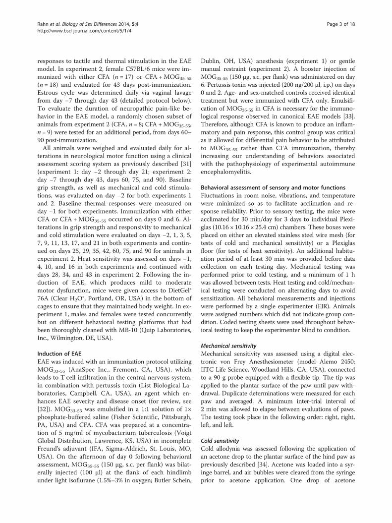
Figure 2 EAE induces mechanical and cold hypersensitivity in female, but not male C57BL/6 mice. Administration of MOG35-55 increasedsensitivity to mechanical and cold stimulation in females (A and C, respectively), but not their male counterparts (B and D, respectively) relativeto CFA sex- and age-matched controls. MOG35-55 did not alter paw withdrawal latencies in response to radiant heat in either female (E) or male(F) mice as compared to CFA age- and sex-matched controls. Area under the curve (AUC) analyses on days 7–21 examining sensitivity tomechanical (G) and cold (H) and days 10–16 examining thermal stimulation (I) in females (F) and males (M). MOG35-55 injections began on day 0.BL baseline. Values represent mean ± SEM. ★★★P < 0.001, ★★P < 0.01, and ★P < 0.05 compared to sex- and age-matched CFA control (one-wayANOVA) or comparison as indicated in AUC figures (one-way ANOVA and Bonferroni Correction). N = 10–13/group.
Rahn et al. Biology of Sex Differences 2014, 5:4 Page 8 of 18http://www.bsd-journal.com/content/5/1/4
tentative because we found no main effect of sex and nointeraction of sex by treatment by time (both P > 0.05;Table 1). As illustrated in Figure 1A,B, subsequent two-way RM ANOVAs revealed that bilateral flank injectionsof CFA produced small neurological motor function defi-cits in either females or males, such as hanging tail or im-paired righting reflex. MOG35-55 treatment producedgreater impairments relative to CFA controls, includingmild paralysis of one or both limbs, in female (P < 0.01;Table 1; Figure 1A) and male (P < 0.01; Table 1; Figure 1B)mice over the 3-week evaluation period (females, P < 0.05;males, P < 0.01; treatment-by-time interaction; Table 1).
Grip strength As illustrated in Table 1, three-way RMANOVA comparing sex (female vs. male) by treatment(MOG35-55 vs. CFA) by time revealed that both female
and male EAE mice presented with decreased forelimbgrip strength when compared to the CFA controls (P <0.01) over the 3-week time course (P < 0.05; treatment-by-time interaction). Male mice demonstrated greaterforelimb grip strength values relative to females (P <0.001; main effect of sex), consistent with the previousfinding that male mice have greater muscular strengththan females [38]. However, MOG35-55 did not interactwith sex: there was neither a sex-by-time interaction,treatment-by-sex interaction, nor a sex-by-treatment-by-time interaction (all P > 0.05; Table 1). As illustrated inFigure 1C,D, subsequent two-way RM ANOVAs indi-cated that MOG35-55 treatment, relative to sex-matchedCFA controls, did not significantly impact responses infemales (P = 0.1; Figure 1C), whereas an effect of treat-ment was evident in males (P < 0.05; Table 1; Figure 1D).

0
1
2
3
BL 7 14 21 28 24530
Time (days post-immunization)
Clin
ical
Sco
re
Female MOG35-55Female CFA
60 75 90
0
1
2
BL 7 14 21 28 35 42
Time (days post-immunization)
Fo
rce
(N)
60 75 900
2
4
6
8
BL 7 14 21 35 4228
Time (days post-immunization)
Co
ld R
esp
on
se (s
)
60 75 90
0
5
10
BL 7 14 21 28 35 42
Time (days post-immunization)
Ch
ang
e in
Wei
gh
t (g
)
60 75 900
5
10
15
BL 7 14 21 2453820
Time (days post-immunization)
Hea
t Lat
ency
(s)
0
2
4
6
BL 7 14 21 28 35 42
Time (days post-immunization)
Mec
han
ical
Th
resh
old
(g)
60 75 90
DA
EB
FC
Figure 3 EAE-induced neuropathic pain-like behaviors persist for 6 weeks in female C57BL/6 mice. Administration of MOG35-55 increasedneurological motor deficits (A) and decreased forelimb grip strength (B), but did not change weight gain (C) relative to CFA controls. Femalemice that received MOG35-55 displayed decreased mechanical withdrawal thresholds (D) and increased duration of response to acetonestimulation (E) when compared to CFA-treated controls. Response to heat stimulation did not differ between mice that received MOG35-55 or CFAalone (F). MOG35-55 injections began on day 0. BL baseline. Values represent mean ± SEM. ★P < 0.05 compared to sex- and age-matched CFAcontrol (one-way ANOVA). N = 8–18/group.
Rahn et al. Biology of Sex Differences 2014, 5:4 Page 9 of 18http://www.bsd-journal.com/content/5/1/4
The trend of MOG35-55-induced decreases on gripstrength for females at later time points is confirmed inFigure 3A.
Body weight Three-way RM ANOVA comparing sex(female vs. male) by treatment (MOG35-55 vs. CFA) bytime revealed no differences in weight between MOG35-55

Table 2 Statistics for experiment 2
Measure Two-way RM ANOVA (treatment × time (BL–day 43)) Two-way RM ANOVA (treatment × time (days 60–90))
Clinical scores Figure 3A Day: F37,1221 = 21.3, P < 0.001 Day: F2,30 = 0.8, P = 0.4
Treatment: F1,33 = 21.7, P < 0.001 Treatment: F1,15 = 0.003, P = 0.9
Day × treatment: F37,1221 = 4.1, P < 0.001 Day × treatment: F2,30 = 1.7, P = 0.19
Grip strength Figure 3B Day: F13,429 = 17.9, P < 0.001 Day: F2,30 = 4.7, P < 0.05
Treatment: F1,33 = 22.6, P < 0.001 Treatment: F1,15 = 0.16, P = 0.6
Day × treatment: F13,429 = 0.9, P = 0.5 Day × treatment: F2,30 = 1.4, P = 0.2
Weight Figure 3C Day: F36,1188 = 76.3, P < 0.001 Day: F2,30 = 17.1, P < 0.001
Treatment: F1,33 = 1.0, P = 0.3 Treatment: F1,15 = 0.5, P = 0.4
Day × treatment: F36,1188 = 0.5, P = 0.9 Day × treatment: F2,30 = 0.6, P = 0.4
Mechanical thresholds Figure 3D Day: F13,429 = 5.5, P < 0.001 Day: F2,30 = 30.4, P = 0.06
Treatment: F1,33 = 20.9, P < 0.001 Treatment: F1,15 = 0.5, P = 0.4
Day × treatment: F13,429 = 2.8, P < 0.01 Day × treatment: F2,30 = 1.0, P = 0.3
Acetone withdrawal Figure 3E Day: F13,429 = 11.1, P < 0.001 Day: F2,30 = 0.5, P = 0.5
Treatment: F1,33 = 8.9, P < 0.01 Treatment: F1,15 = 0.2, P = 0.6
Day × treatment: F13,429 = 2.0, P = 0.06 Day × treatment: F2,30 = 1.6, P = 0.2
Heat latency Figure 3F Day: F7,231 = 1.8, P = 0.07 -
Treatment: F1,33 = 1.9, P = 0.17
Day × treatment: F7,231 = 0.3, P = 0.9
Treatment conditions: CFA, n = 17 (BL–day 43) or n = 8 (days 60–90). CFA +MOG35-55, n = 18 (BL–day 43) or n = 9 (days 60–90).
Rahn et al. Biology of Sex Differences 2014, 5:4 Page 10 of 18http://www.bsd-journal.com/content/5/1/4
and CFA controls (P = 0.6; Table 1); as expected, males didgain weight throughout the course of the study at a morerapid rate than their female counterparts (P < 0.01; sex-by-time interaction; Table 1). Subsequent two-way RM ANO-VAs comparing treatment by time also found no differ-ences in body weight between animals treated with
A
B
Figure 4 CFA transiently increased the time spent in diestrus.Representative traces of female mice that received either CFA (A) orCFA + MOG35-55 (B). Note that estrous cycling resumed 2 weeks afterCFA or CFA + MOG35-55.
MOG35-55 vs. CFA (females: P = 0.6, Table 1, Figure 1E;males: P = 0.8, Table 1, Figure 1F) over the 3-week timecourse for either sex.
EAE produces mechanical and cold hypersensitivity infemale, but not male miceNo study to date has evaluated sex differences in re-sponse to mechanical, cold, and thermal stimulation inmale and female mice using the MOG35-55-induced EAEmodel.
Mechanical hyperalgesia As illustrated in Table 1,three-way RM ANOVA comparing sex by treatment bytime revealed higher mechanical withdrawal thresholdsin males than females (P < 0.05; main effect of sex) andin MOG35-55-treated animals compared to CFA controls(main effect of treatment: P < 0.01; interaction of treat-ment by time: P < 0.01). Two-way RM ANOVAs revealedthat MOG35-55 treatment decreased mechanical with-drawal thresholds in females (P < 0.01 vs. CFA controls;Table 1; Figure 2A) from days 11–21 post-immunization(P < 0.001; treatment-by-time interaction; Table 1) relativeto sex-matched CFA controls; by contrast, MOG35-55 didnot decrease mechanical withdrawal thresholds in males(P = 0.2; Table 1; Figure 2B). One-way ANOVA of AUCfor mechanical withdrawal thresholds on testing days 7–21 visually confirms that female MOG35-55 mice presentedwith lower thresholds than either males or sex-matchedfemale CFA controls (P < 0.001; Figure 2G).

A B
C D
E F
G H
Figure 5 Proestrus produces protective effects in the EAE model at late time points. Neurological motor deficits (A), grip strength (C),mechanical withdrawal thresholds (E), and responses to acetone application (G) in female mice analyzed across phases of the estrous cycle. Areaunder the curve (AUC) analyses examining neurological motor deficits (B), grip strength (D), and sensitivity to mechanical (F) and cold (H)stimulation in females analyzed across the phases of the estrous cycle. Non-proestrus includes the following phases: diestrus, metestrus, andestrus. BL baseline. Values represent mean ± SEM. ***P < 0.001, **P < 0.01, *P < 0.05 main effect of treatment as indicated (two-way ANOVAcomparing treatment by estrous). ★★★P < 0.001, ★★P < 0.01, ★P < 0.05 comparison as indicated (one-way ANOVA and Bonferroni correction).N = 3–16/group, except n = 2 in groups CFA proestrus and MOG35-55 proestrus on day 13.
Rahn et al. Biology of Sex Differences 2014, 5:4 Page 11 of 18http://www.bsd-journal.com/content/5/1/4
Cold hyperalgesia Three-way RM ANOVA comparingsex by treatment by time revealed that cold hyperalgesiawas significantly impacted by sex, with females demon-strating greater response withdrawal durations to acetoneapplication relative to their male counterparts (P < 0.001;
Table 1). A treatment-by-sex-by-time interaction suggeststhat female, but not male, mice receiving MOG35-55 dem-onstrated increased responsivity to acetone followingimmunization (P < 0.001; Table 1). Two-way RM ANO-VAs revealed that MOG35-55 increased the duration of

Table 3 Statistics for experiment 2 to determine the effects of estrous phase (proestrus vs. non-proestrus phases)
Two-way RM ANOVA (treatment × estrous)
BL Day 7 Day 13 Day 21 Day 29 Day 35 Day 42
Clinical scores, Figure 5A
Treatment F1,28 = 0.2, P = 0.6 F1, 27 = 2.0, P = 0.16 F1,30 = 1.09, P = 0.3 F1,30 = 10.3, P < 0.05 F1,31 = 8.5, P < 0.01 F1,31 = 0.5, P = 0.4 F1,31 = 2.2, P = 0.1
Estrous F1,28 = 0.2, P = 0.6 F1, 27 = 0.5, P = 0.4 F1,30 = 0.2, P = 0.5 F1,30 = 0.6, P = 0.4 F1,31 = 1.1, P = 0.2 F1,31 = 3.0, P = 0.09 F1,31 = 2.8, P = 0.053
Treatment × estrous F1,28 = 0.2, P = 0.6 F1, 27 = 0.001, P = 0.9 F1,30 = 1.09, P = 0.3 F1,30 = 0.6, P = 0.4 F1,31 = 0.2, P = 0.9 F1,31 = 3.7, P = 0.6 F1,31 = 3.7, P < 0.05
Grip strength, Figure 5C
Treatment F1,28 = 0.3, P = 0.5 F1. 27 = 2.7, P = 0.1 F1,30 = 0.09, P = 0.7 F1,30 = 11.0, P < 0.05 F1,31 = 19.8, P < 0.001 F1,31 = 5.8, P < 0.05 F1,31 = 6.4, P < 0.05
Estrous F1,28 = 0.04, P = 0.8 F1, 27 = 0.7, P = 0.3 F1,30 = 0.6, P = 0.4 F1,30 = 0.002, P = 0.9 F1,31 = 0.05, P = 0.8 F1,31 = 0.1, P = 0.7 F1,31 = 0.1, P = 0.7
Treatment × estrous F1,28 = 0.3, P = 0.5 F1, 27 = 0.06, P = 0.8 F1,30 = 2.1, P = 0.15 F1,30 = 0.5, P = 0.4 F1,31 = 0.02, P = 0.8 F1,31 = 0.009, P = 0.9 F1,31 = 0.02, P = 0.8
Mechanical thresholds, Figure 5E
Treatment F1,28 = 0.08, P = 0.7 F1, 27 = 4.2, P < 0.05 F1,30 = 0.7, P = 0.3 F1,30 = 7.4, P < 0.05 F1,31 = 25.9, P < 0.001 F1,31 = 2.0, P = 0.16 F1,31 = 3.5, P = 0.07
Estrous F1,28 = 0.9, P = 0.3 F1, 27 = 0.4, P = 0.4 F1,30 = 2.3, P = 0.13 F1,30 = 0.09, P = 0.7 F1,31 = 5.2, P < 0.05 F1,31 = 3.1, P = 0.08 F1,31 = 0.2, P = 0.6
Treatment × estrous F1,28 = 0.001, P = 0.9 F1, 27 = 0.09, P = 0.7 F1,30 = 0.1, P = 0.7 F1,30 = 1.5, P = 0.2 F1,31 = 8.7, P < 0.01 F1,31 = 2.1, P = 0.14 F1,31 = 2.6, P = 0.11
Acetone withdrawal, Figure 5G
Treatment F1,28 = 0.1, P = 0.7 F1,27 = 1.9, P =0.17 F1,30 = 4.2, P < 0.05 F1,30 = 4.2, P < 0.05 F1,31 = 5.2, P < 0.05 F1,31 = 1.0, P = 0.3 F1,31 = 7.2, P < 0.05
Estrous F1,28 = 0.01, P = 0.9 F1,27 = 0.000, P = 0.9 F1,30 = 0.01, P = 0.9 F1,30 = 0.02, P = 0.6 F1,31 = 0.001, P = 0.9 F1,31 = 0.8, P = 0.7 F1,31 = 0.09, P = 0.7
Treatment × estrous F1,28 = 0.9, P = 0.3 F1,27 = 0.06, P = 0.7 F1,30 = 0.001, P = 0.9 F1,30 = 1.2, P = 0.2 F1,31 = 0.2, P = 0.6 F1,31 = 3.7, P = 0.06 F1,31 = 6.3, P < 0.05
AUC Clinical scores: Grip strength: Mechanical thresholds: Acetone withdrawal:
one-way ANOVA: one-way ANOVA: one-way ANOVA: one-way ANOVA:
F3,44 = 8.8, P < 0.001 F3,44 = 5.3, P < 0.01 F3,44 = 5.8, P < 0.01 F3,44 = 2.8, P < 0.05
(Figure 5B) (Figure 5D) (Figure 5F) (Figure 5H)
Treatment conditions: CFA, n = 17; CFA +MOG35-55, n = 18. Estrous phase: proestrus (n = 4–18/day) or non-proestrus phases (n = 17–30/day). AUC area under the curve.
Rahnet
al.Biologyof
SexDifferences
2014,5:4Page
12of
18http://w
ww.bsd-journal.com
/content/5/1/4

Rahn et al. Biology of Sex Differences 2014, 5:4 Page 13 of 18http://www.bsd-journal.com/content/5/1/4
response following acetone application to the hindpaws in female mice relative to sex-matched CFA con-trols (P < 0.05; Table 1; Figure 2C) observed on days 13–21 (P < 0.001; treatment-by-time interaction; Table 1); noalterations in response duration were observed in malemice (P = 0.8; Table 1; Figure 2D). One-way ANOVA ofAUC over testing days 7–21 confirmed that cold responseduration was greater in female MOG35-55 mice relative tosex-matched CFA controls and males (P < 0.001; Table 1;Figure 2H).
Heat responses Three-way RM ANOVA of heat stimu-lation responses yielded no main effects of either sex(P = 0.14; females, Figure 2E; males, Figure 2F; Table 1)or treatment (P = 0.8; Table 1). AUC analysis of with-drawal latency to thermal stimulation also failed to yielddifferences (P = 0.4; one-way ANOVA; Table 1; Figure 2I).
Experiment 2: time course of EAE and effects of estrousstateExtended time course of EAE-induced neuropathic pain-likebehavior in female miceThe time course of previous studies of neuropathic painin EAE models are generally limited to 1 month or less,allowing a description of the onset and peak of hyper-algesia, but not remission [24,28]. The one exception is aMOG35-55 study that followed mice over a 50-day timecourse; although mechanical allodynia decreased overthis time period, full remission did not occur [23]. Todetermine the duration of neuropathic pain-like behav-iors and their correlation with neurological motor im-pairment, we repeated our measurements of sensory andmotor functions in females—this time with a timecourse of 90 days—and analyzed the data with two-wayRM ANOVA.
Clinical scores As illustrated in Figure 3, MOG35-55
treatment induced neurological motor deficits (P < 0.001;Table 2; Figure 3A). Motor impairments varied with time(P < 0.001; treatment-by-time interaction; Table 2); be-ginning on day 10 and lasting through day 35, deficitsre-appeared for one additional day (day 43). We note atrend of increased neurological motor impairments inmice receiving MOG35-55 on days 35–38 (P < 0.10 foreach comparison). Subsequent evaluations between days60 and 90 failed to yield differences between theMOG35-55 and CFA mice (P = 0.19; Table 2).
Grip strength MOG35-55 consistently decreased fore-limb grip strength (P < 0.001; Table 2; Figure 3B) fromdays 17–42 (P < 0.05 for each comparison), with twoearlier time points (days 3 and 11) also showing a differ-ence. Later time points failed to yield differences in grip
strength between female MOG35-55 and CFA mice (P = 0.2;Table 2).
Body weight MOG35-55 did not alter body weight whencompared to CFA controls (P = 0.3; Table 2; Figure 3C).
Mechanical hyperalgesia MOG35-55 decreased mech-anical withdrawal thresholds relative to CFA controls(P < 0.001; Table 2; Figure 3D) beginning on day 5 andcontinuing through day 42 (P < 0.01; treatment-by-timeinteraction; Table 2), with no differences over days 60–90(P = 0.3; Table 2).
Cold hyperalgesia MOG35-55 increased the responsive-ness to topical acetone application (P < 0.01; Table 2;Figure 3E) beginning on day 7 and lasting through day42 (P < 0.05 for each comparison), with no differencesover days 60–90 (P = 0.2; Table 2).
Heat responses Compared to the CFA controls,MOG35-55 did not produce heat hypersensitivity (Table 2;Figure 3F) through day 43 (P = 0.9); therefore, heat testingwas discontinued.
EAE is associated with fewer neurological motor deficitsduring proestrusLittle is known about the relationship between estrouscycle and neuropathic pain-like behaviors in animalmodels of MS. To address this question, we evaluatedmechanical and cold allodynia while monitoring the es-trous cycle. As illustrated in Figure 4, female mice thatreceived either CFA (Figure 4A) or CFA +MOG35-55
(Figure 4B) demonstrated prolonged periods of timespent in the diestrus phase as classified by vaginal lavage.This is consistent with previous reports indicating thatintraplantar CFA prolonged the leukocytic phase of theestrous cycle [39]. Within approximately 14 days afterinitial CFA or CFA +MOG35-55 administration, estrouscycling returned to normal. The CFA-induced prolonga-tion of diestrus resulted in unequal representation of theestrous phases within our study. Therefore, in order toinvestigate effects of the phase with the greatest hormo-nal fluctuations, we chose to bin our analyses into proes-trus (progesterone, estradiol, and luteinizing hormonesurge) and ‘non-proestrus’ phases (diestrus, metestrus,and estrus). The effect of estrous phase on neurologicalmotor deficits and pain-like behaviors were further ana-lyzed using two-way ANOVA. Though the treatmentcondition (MOG35-55 vs. CFA) was consistent at eachtime point, the animals classified as being within proes-trus or non-proestrus was not constant. Thus, the num-ber of animals in each of the different phases of estrousvaried at each post-immunization time point. Therefore,

Rahn et al. Biology of Sex Differences 2014, 5:4 Page 14 of 18http://www.bsd-journal.com/content/5/1/4
it was not possible to analyze our data by RM ANOVAwith time as the repeated measure.
Clinical scores As illustrated in Figure 5A,B, two-wayANOVAs at each time point confirmed that MOG35-55,compared to CFA controls, produced neurological motordeficits, significantly on days 21 and 29 (P < 0.05;Table 3). We found an estrous-by-treatment interactionon day 42 (P < 0.05; Table 3), but when the same datawere subjected to a one-way ANOVA, no significancewas noted (F3,34 = 1.0, P = 0.3). One-way ANOVA of alltime points, transformed as AUC, suggests that proes-trus was protective against neurological motor deficits inMOG35-55 animals (P < 0.001; bottom row of Table 3);however, this conclusion is presented with caution as wedid not find a significant main effect of estrous at anyparticular time point (P > 0.05).
Grip strength MOG35-55 reduced forelimb grip strengthat several time points (day 21: P < 0.05; day 29: P < 0.001;day 35: P < 0.05; day 42: P < 0.05; Table 3). However, gripstrength did not change with estrous cycle at any par-ticular day (effect of estrous: P > 0.3 for all time points,Table 3; estrous-by-treatment interaction P > 0.15 for alltime points, Table 3) (Figure 5C). Analysis of gripstrength data using AUC revealed that proestrus was asso-ciated with lower grip strength values as compared to non-proestrus in CFA controls (P < 0.01; one-way ANOVA;Figure 5D).
Mechanical hyperalgesia MOG35-55 reduced mechan-ical thresholds on days 7, 21, and 29 (P < 0.05; Table 3).When the data was transformed as AUC, we found amain effect of MOG35-55 (P < 0.01; Table 3; Figure 5E,F).On day 29, two-way ANOVA revealed an effect of es-trous (P < 0.05; Table 3) and an estrous-by-treatmentinteraction (P < 0.01; Table 3; Figure 5E); however, wecaution against over-interpretation of this result sincethis occurred at just one time point, and AUC trans-formation yielded no effect of estrous over multiple test-ing days.
Cold hyperalgesia MOG35-55 increased the response tocold stimulation on days 13, 21, 29, and 42 (P < 0.05;Table 3; Figure 5G). Although we found an interactionof treatment by estrous on day 42 (P < 0.05; Table 3), theconclusions are tentative as there was no main effect ofestrous on cold stimulation at any particular time point(P > 0.6 for each time point; Table 3). A subsequent one-way ANOVA revealed that cold hyperalgesia was greaterin the MOG35-55-proestrus animals as compared toCFA-proestrus animals on day 42 (F3,34 = 3.7, P < 0.05).When all time points were transformed as AUC, we founda main effect of group (P < 0.05; Table 3; Figure 5H);
however, subsequent post hoc analysis did not reveal sig-nificant differences between groups (P > 0.05 for eachcomparison).
DiscussionTo date, no study has rigorously characterized the ef-fects of sex and estrous state on the development, dur-ation, and severity of pain symptoms associated withMOG35-55-induced EAE. Our present study addressedthis gap with behavioral assessment of sensory andmotor functions in C57BL/6 female mice for 3 monthsafter induction of EAE with MOG35-55 immunizationand compared select time points with males. We reportfour general findings. First, in experiment 1, MOG35-55
produced neurological motor impairments in both fe-males and males, including mild paralysis of hind limbsand decreases in forelimb grip strength (particularly inexperiment 2). Second, MOG35-55 produced mechanicaland cold hypersensitivity only in females. Third, the dur-ation of pain-like behavior in 3–4-month-old (at studyonset) C57BL/6 female mice in experiment 2 was42 days. Subsequent studies using younger female micehave yielded much longer durations of pain-like behavior(unpublished observations from our laboratory). Fourth,experiment 2 revealed that MOG35-55 produced lessneurological motor dysfunction when female mice werein the proestrus phase.
Sex differences in the clinical manifestations of EAEMultiple sclerosis is a disease dominated by female pa-tients with a 2:1 prevalence in females relative to males.Our study tested the hypothesis that neurological motorimpairment would be greater in females in the mostcommonly utilized animal model of MS, the mouseMOG35-55 EAE model [21,28,40]. As described previ-ously, we found that MOG35-55 produced motor dys-function, characterized by mild paralysis of one or bothhind limbs [28], for review see [41]. And, in agreementwith one previous study [33], we found that MOG35-55
decreased grip strength, an effect most evident 2–3 weeksafter initial immunization. This robust behavior per-sisted in females for up to 42 days post-immunization(Figure 3B).Similar to the results of Okuda and colleagues, we
found that MOG35-55 produced motor impairments anddecreases in grip strength in both male and femaleC57BL/6 mice, indicating an absence of sex differencesin EAE severity; however, in our study, females devel-oped neurological deficits as reported with a clinicalscoring assessment 4 days prior to the development ofsuch deficits in males when the mice are compared tosame-sex CFA controls, whereas they reported no differ-ence in onset of clinical deficits [20]. By contrast, femaleSJL and ASW mice develop more severe clinical

Rahn et al. Biology of Sex Differences 2014, 5:4 Page 15 of 18http://www.bsd-journal.com/content/5/1/4
symptoms than males when treated with the EAE-inducing encephalitogenic peptides myelin basic protein(MBP) and proteolipid protein (PLP), respectively [21].Conversely, males develop more severe neurological defi-cits (bilateral hind limb paralysis) in the TMEV model, amouse model of progressive MS [22]. Further studies areneeded to determine the importance of immunizationprotocol, dose of adjuvant, mouse strain, age, and otherfactors on sex differences in murine models of MS.
Sex differences in EAE-induced mechanical and coldhypersensitivityMS is one of many disease states which show a particu-larly high prevalence of pain in women [42], necessitatinginvestigation of sex-associated hypersensitivity in preclin-ical MS models. Our study is the first to compare EAE-associated nociception between male and female miceusing a MOG35-55 immunization protocol. For two rea-sons, we feel it unlikely that these pain-like behaviors wereindirectly inhibited by concomitant neurological motordeficits. First, we found that pain-like behaviors occurredprior to the onset of clinical signs, in accordance with pre-vious observations. Second, we observed robust nocicep-tive responses despite motor deficits.As observed previously by Olechowski and colleagues
[28], we found that MOG35-55, as compared to sex- andage-matched CFA controls, produced hypersensitivity tomechanical and cold stimulations in 3–4-month-oldC57BL/6 female mice. In females, hyperalgesia waspresent up to 42 days following initial immunization,and subsided by 60 days, with no relapse noted by day90. Remarkably, we did not observe hyperalgesia in theirmale MOG35-55 counterparts. Our results are consistentwith the finding that female SLJ/J mice in the TMEVmodel of multiple sclerosis exhibited a faster onset andgreater peak of mechanical allodynia as compared tomales [22,43]. Similarly, heat hyperalgesia was more pro-nounced in females in the PLP model of EAE in SJLmice [24]. Our study did not reveal heat hypersensitivity,perhaps due to different localization of the heat stimulusin our study (hindpaw) vs. the previous study (tail andforepaws) [24]. Olechowski and colleagues also reporteda lack of thermal hyperalgesia in MOG35-55 females thatnevertheless demonstrate robust mechanical and coldallodynia [28].
Duration of pain-like behaviors in MOG35-55-induced EAETo date, no study has examined neuropathic pain-like be-haviors beyond 50 days, when minor mechanical allodyniawas noted in female subjects [23,24,28]. Here, we reportthat mechanical and cold hyperalgesia may antedate, butdo not outlast MOG35-55-induced neurological motor im-pairments. Both neuropathic pain-like behaviors andneurological motor impairments were present up to 42 days
post-immunization, and we failed to observe a relapse;however, further assessment beyond 90 days is needed todetermine whether our model reflects the relapsing-remitting form of EAE.
Neurological motor impairment is dampened duringproestrusThe lack of information regarding the relationship be-tween neurological deficits, pain behaviors, and estrouscycle prompted our investigation of cycle-related alter-ations in the sensory and motor disturbances associatedwith EAE. In experiment 2, we observed that CFA ad-ministration alone, or in combination with MOG35-55,prolonged the time spent in diestrus, thereby haltingnormal estrous cycling for 2 weeks—consistent with aprevious report [39]. Strikingly, neurological motor defi-cits measured via daily clinical assessments were attenu-ated during proestrus as compared to the other phases;this protection was only evident with AUC transforma-tions that allowed us to examine the more subtle effectsof estrous phase over multiple testing days. Because pro-estrus is characterized by relatively high plasma levels ofestrogen and progesterone [44], our results are consist-ent with the hypothesis that circulating ovarian hor-mones decrease neurological motor deficits in femaleMOG35-55 mice. Progesterone administered prior to EAEimmunization delayed onset and attenuates progression ofneurological deficits [45]. Work in the EAE models hasalso demonstrated the possible role of estrogens/proges-terone in promoting remyelination and reducing the pres-ence of pro-inflammatory mediators (e.g., TNF-α) andmicroglial activation [46-50], providing potential mecha-nisms through which ovarian hormones may produce pro-tection against neurological motor deficits.We were unable to conclusively demonstrate estrous-
related alterations in pain behaviors. A veritable host ofstudies have investigated sex- and hormone-related alter-ations of somatosensation in other preclinical models ofchronic pain, with conflicting findings. Some studies re-port greater hyperalgesic responses in females duringthe proestrus phase [51,52], while others report antinoci-ception associated with the proestrus phase [53] or withthe exogenous administration of estrogens [54-56]. Moreinvestigations are necessary to determine how naturaland directed alterations in hormone levels may impactEAE pain-like behaviors.While our study is the first to report on estrous cycle
alterations in EAE-induced neurological motor dysfunc-tion, our findings do have important commonalities withclinical MS populations. A recent report examined men-strual cycle effects on MS symptoms and found thatwomen taking oral contraceptives experienced increasedweakness, numbness, and tiredness during their menstrualcycle when not taking oral steroids [3]. A similar study

Rahn et al. Biology of Sex Differences 2014, 5:4 Page 16 of 18http://www.bsd-journal.com/content/5/1/4
reported menstruation-related increases in MS symptoms,including pain, in normal cycling women [5]. The onset ofmenses is associated with low levels of estrogen and pro-gesterone [57]; therefore, these studies collectively suggesta worsening of symptoms when ovarian hormones are lowand, conversely, a potentially protective effect when levelsare high. These clinical studies parallel our findings in theEAE model and point to the clinical therapeutic potentialoffered from further investigations into mechanisms medi-ating the protective and antinociceptive effects of ovarianhormones in the EAE model.It is well established that clinical symptoms of MS are at-
tenuated during pregnancy, likely due to elevations in estro-gens and progesterone. This is followed by an increasedrisk of postpartum relapse when levels of estrogens andprogesterone are low [6,58]. Similarly, studies in pregnantSJL and C57BL/6 mice reported pregnancy-associated im-provement in EAE symptoms and a subsequent increasein postpartum relapse, concomitant with decreased estro-gen levels [59,60]. Further, administration of estriol, an es-trogen produced only during pregnancy, amelioratedsymptoms and lesions as assessed by magnetic resonanceimaging in non-pregnant female MS patients [61] andclinical impairments in EAE mice [62,63]. It remains to beinvestigated if pregnancy-induced remission in EAE symp-toms would include reduction of hypersensitivity to cuta-neous and cold stimulation, in addition to the possibleanalgesic role that exogenously applied estrogens/proges-terone may play.
ConclusionsIn agreement with previous reports [24,28], for reviewsee [64], we conclude that the MOG35-55 EAE model in-duces neurological motor dysfunction and neuropathicpain-like behavior, similar to symptom profiles observedin clinical MS populations. While male C57BL/6 micedevelop neurological motor impairments typical of theEAE model, they fail to develop the neuropathic pain-like behaviors observed in their female counterparts.EAE-induced neurological motor impairments were re-duced during the proestrus phase, a finding that war-rants further investigations into the contributing role ofcirculating ovarian hormones to EAE pathology. Wesuggest that female C57BL/6 mice receive preference infuture studies of neuropathic pain-like behaviors associ-ated with the MOG35-55 model of EAE. Care must betaken to consider the effects that estrous cycling mayhave on neurological motor deficits.
AbbreviationsAUC: area under the curve; CFA: complete Freund’s adjuvant;EAE: experimental autoimmune encephalomyelitis; MOG35-55: myelinoligodendrocyte glycoprotein 35–55; MS: multiple sclerosis; PLP: proteolipid
protein; TMEV: Theiler’s murine encephalomyelitis virus.
Competing interestsThe authors declare that they have no competing interests.
Authors’ contributionsEJR helped conceive the study; carried out vaginal lavages, all behavioralevaluations, and immunizations; and helped draft the manuscript. TI helpeddraft the manuscript. RRD assisted in vaginal lavage and immunizations. BKThelped conceive the study, participated in its design and coordination, andhelped draft the manuscript. All authors read and approved the finalmanuscript.
Authors’ informationFor the research description and interests of BKT, please refer to https://physiology.med.uky.edu/users/bkta222.
AcknowledgementsThis work was funded by the University of Kentucky start-up funds, NIHNS62306, and the Canadian Institute for Health Research grant #MOP119338to BKT (consortium PI).
Author details1Department of Physiology, University of Kentucky, 800 Rose Street,Lexington, KY 40536, USA. 2Present Address: Department of Neurobiology,University of Alabama at Birmingham, 1825 University Blvd. SHEL 1070C,Birmingham, AL 35294, USA. 3Present Address: School of BiomedicalSciences, University of Leeds, Leeds LS2 9JT, UK.
Received: 20 August 2013 Accepted: 31 January 2014Published: 28 February 2014
References1. El-Etr M, Vukusic S, Gignoux L, Durand-Dubief F, Achiti I, Baulieu EE,
Confavreux C: Steroid hormones in multiple sclerosis. J Neurol Sci 2005,233:49–54.
2. Voskuhl RR, Gold SM: Sex-related factors in multiple sclerosissusceptibility and progression. Nat Rev Neurol 2012, 8:255–263.
3. Holmqvist P, Hammar M, Landtblom AM, Brynhildsen J: Symptoms ofmultiple sclerosis in women in relation to cyclical hormone changes.Eur J Contracept Reprod Health Care 2009, 14:365–370.
4. Holmqvist P, Wallberg M, Hammar M, Landtblom AM, Brynhildsen J:Symptoms of multiple sclerosis in women in relation to sex steroidexposure. Maturitas 2006, 54:149–153.
5. Zorgdrager A, De Keyser J: Menstrually related worsening of symptoms inmultiple sclerosis. J Neurol Sci 1997, 149:95–97.
6. Confavreux C, Hutchinson M, Hours MM, Cortinovis-Tourniaire P,Moreau T: Rate of pregnancy-related relapse in multiple sclerosis.Pregnancy in Multiple Sclerosis Group. N Engl J Med 1998,339:285–291.
7. Zhu WH, Lu CZ, Huang YM, Link H, Xiao BG: A putative mechanism onremission of multiple sclerosis during pregnancy: estrogen-inducedindoleamine 2,3-dioxygenase by dendritic cells. Mult Scler 2007,13:33–40.
8. Nurmikko TJ, Gupta S, Maclver K: Multiple sclerosis-related central paindisorders. Curr Pain Headache Rep 2010, 14:189–195.
9. O’Connor AB, Schwid SR, Herrmann DN, Markman JD, Dworkin RH: Painassociated with multiple sclerosis: systematic review and proposedclassification. Pain 2008, 137:96–111.
10. Truini A, Barbanti P, Pozzilli C, Cruccu G: A mechanism-based classificationof pain in multiple sclerosis. J Neurol 2012, 260(2):351–367.
11. Foley PL, Vesterinen HM, Laird BJ, Sena ES, Colvin LA, Chandran S, MacleodMR, Fallon MT: Prevalence and natural history of pain in adults withmultiple sclerosis: systematic review and meta-analysis. Pain 2013,154:632–642.
12. Osterberg A, Boivie J, Thuomas KA: Central pain in multiple sclerosis—prevalence and clinical characteristics. Eur J Pain 2005, 9:531–542.

Rahn et al. Biology of Sex Differences 2014, 5:4 Page 17 of 18http://www.bsd-journal.com/content/5/1/4
13. Truini A, Galeotti F, Cruccu G: Treating pain in multiple sclerosis. ExpertOpin Pharmacother 2011, 12:2355–2368.
14. Svendsen KB, Jensen TS, Overvad K, Hansen HJ, Koch-Henriksen N, Bach FW:Pain in patients with multiple sclerosis: a population-based study. ArchNeur 2003, 60:1089–1094.
15. Buchanan RJ, Wang S, Ju H: Gender analyses of nursing home residentswith multiple sclerosis. J Gend Spec Med 2003, 6:35–46.
16. Warnell P: The pain experience of a multiple sclerosis population: adescriptive study. Axone 1991, 13:26–28.
17. Beiske AG, Pedersen ED, Czujko B, Myhr KM: Pain and sensory complaintsin multiple sclerosis. Eur J Neurol 2004, 11:479–482.
18. Casetta I, Riise T, Wamme Nortvedt M, Economou NT, De Gennaro R, Fazio P,Cesnik E, Govoni V, Granieri E: Gender differences in health-related quality oflife in multiple sclerosis. Mult Scler 2009, 15:1339–1346.
19. Archibald CJ, McGrath PJ, Ritvo PG, Fisk JD, Bhan V, Maxner CE, Murray TJ:Pain prevalence, severity and impact in a clinic sample of multiplesclerosis patients. Pain 1994, 58:89–93.
20. Okuda Y, Okuda M, Bernard CC: Gender does not influence thesusceptibility of C57BL/6 mice to develop chronic experimentalautoimmune encephalomyelitis induced by myelin oligodendrocyteglycoprotein. Immunol Lett 2002, 81:25–29.
21. Papenfuss TL, Rogers CJ, Gienapp I, Yurrita M, McClain M, Damico N, Valo J,Song F, Whitacre CC: Sex differences in experimental autoimmuneencephalomyelitis in multiple murine strains. J Neuroimmun 2004, 150:59–69.
22. Lynch JL, Gallus NJ, Ericson ME, Beitz AJ: Analysis of nociception, sex andperipheral nerve innervation in the TMEV animal model of multiplesclerosis. Pain 2008, 136:293–304.
23. Lu J, Kurejova M, Wirotanseng LN, Linker RA, Kuner R, Tappe-TheodorA: Pain in experimental autoimmune encephalitis: a comparativestudy between different mouse models. J Neuroinflammation 2012,9:233.
24. Aicher SA, Silverman MB, Winkler CW, Bebo BF Jr: Hyperalgesia in ananimal model of multiple sclerosis. Pain 2004, 110:560–570.
25. Constantinescu CS, Farooqi N, O’Brien K, Gran B: Experimental autoimmuneencephalomyelitis (EAE) as a model for multiple sclerosis (MS). Brit JPharmacol 2011, 164:1079–1106.
26. Berard JL, Wolak K, Fournier S, David S: Characterization of relapsing-remittingand chronic forms of experimental autoimmune encephalomyelitis inC57BL/6 mice. Glia 2010, 58:434–445.
27. Kuerten S, Gruppe TL, Laurentius LM, Kirch C, Tary-Lehmann M, Lehmann PV,Addicks K: Differential patterns of spinal cord pathology induced by MP4,MOG peptide 35–55, and PLP peptide 178–191 in C57BL/6 mice. APMIS2011, 119:336–346.
28. Olechowski CJ, Truong JJ, Kerr BJ: Neuropathic pain behaviours in achronic-relapsing model of experimental autoimmune encephalomyelitis(EAE). Pain 2009, 141:156–164.
29. Whitten WK: Modification of the oestrous cycle of the mouse by externalstimuli associated with the male. J Endocrinol 1956, 13:399–404.
30. Whitten WK: Occurrence of anoestrus in mice caged in groups. JEndocrinol 1959, 18:102–107.
31. Lisi L, Navarra P, Cirocchi R, Sharp A, Stigliano E, Feinstein DL, Dello Russo C:Rapamycin reduces clinical signs and neuropathic pain in a chronicmodel of experimental autoimmune encephalomyelitis. J Neuroimmunol2012, 243:43–51.
32. Rangachari M, Kuchroo VK: Using EAE to better understand principles ofimmune function and autoimmune pathology. J Autoimmun 2013, 45:31–39.
33. Jones MV, Nguyen TT, Deboy CA, Griffin JW, Whartenby KA, Kerr DA, CalabresiPA: Behavioral and pathological outcomes in MOG 35–55 experimentalautoimmune encephalomyelitis. J Neuroimmunol 2008, 199:83–93.
34. Choi Y, Yoon YW, Na HS, Kim SH, Chung JM: Behavioral signs of ongoing painand cold allodynia in a rat model of neuropathic pain. Pain 1994, 59:369–376.
35. Hargreaves K, Dubner R, Brown F, Flores C, Joris J: A new and sensitivemethod for measuring thermal nociception in cutaneous hyperalgesia.Pain 1988, 32:77–88.
36. Goldman JM, Murr AS, Cooper RL: The rodent estrous cycle:characterization of vaginal cytology and its utility in toxicologicalstudies. Birth Defects Res B Dev Reprod Toxicol 2007, 80:84–97.
37. Byers SL, Wiles MV, Dunn SL, Taft RA: Mouse estrous cycle identificationtool and images. PloS One 2012, 7:e35538.
38. Glenmark B, Nilsson M, Gao H, Gustafsson JA, Dahlman-Wright K,Westerblad H: Difference in skeletal muscle function in males vs.
females: role of estrogen receptor-beta. Am J Physiol Endocrinol Metab2004, 287:E1125–E1131.
39. Battisto JR, Pinto M, Joseph S: Disturbance of oestrous cycles in ratsfollowing administration of Freund’s complete adjuvant. J Reprod Fertil1967, 14:147–150.
40. Lin F, Kaminski HJ, Conti-Fine BM, Wang W, Richmonds C, Medof ME:Markedly enhanced susceptibility to experimental autoimmunemyasthenia gravis in the absence of decay-accelerating factorprotection. J Clin Inves 2002, 110:1269–1274.
41. Truini A, Barbanti P, Pozzilli C, Cruccu G: A mechanism-based classificationof pain in multiple sclerosis. J Neurol 2013, 260:351–367.
42. Mogil JS, Bailey AL: Sex and gender differences in pain and analgesia.Prog Brain Res 2010, 186:141–157.
43. Lynch JL, Alley JF, Wellman L, Beitz AJ: Decreased spinal cord opioidreceptor mRNA expression and antinociception in a Theiler’s murineencephalomyelitis virus model of multiple sclerosis. Brain Res 2008,1191:180–191.
44. Smith MS, Freeman ME, Neill JD: The control of progesterone secretionduring the estrous cycle and early pseudopregnancy in the rat:prolactin, gonadotropin and steroid levels associated with rescue ofthe corpus luteum of pseudopregnancy. Endocrinology 1975,96:219–226.
45. de Graaf KL, Wallstrom E, Muhallab S, Wiesmuller KH, Olsson T, Weissert R:MHC and non-MHC gene regulation of disease susceptibility and diseasecourse in experimental inflammatory peripheral neuropathy.J Neuroimmunol 2004, 155:73–84.
46. Garay LI, Gonzalez Deniselle MC, Brocca ME, Lima A, Roig P, De Nicola AF:Progesterone down-regulates spinal cord inflammatory mediators andincreases myelination in experimental autoimmune encephalomyelitis.Neuroscience 2012, 226:40–50.
47. Yu HJ, Fei J, Chen XS, Cai QY, Liu HL, Liu GD, Yao ZX: Progesteroneattenuates neurological behavioral deficits of experimental autoimmuneencephalomyelitis through remyelination with nucleus-sublocalizedOlig1 protein. Neurosci Lett 2010, 476:42–45.
48. Kumar S, Patel R, Moore S, Crawford DK, Suwanna N, Mangiardi M,Tiwari-Woodruff SK: Estrogen receptor beta ligand therapy activatesPI3K/Akt/mTOR signaling in oligodendrocytes and promotesremyelination in a mouse model of multiple sclerosis. Neurobiol Dis2013, 56C:131–144.
49. Ito A, Buenafe AC, Matejuk A, Zamora A, Silverman M, Dwyer J, Vandenbark AA,Offner H: Estrogen inhibits systemic T cell expression of TNF-alpha andrecruitment of TNF-alpha(+) T cells and macrophages into the CNS ofmice developing experimental encephalomyelitis. Clin Immunol 2002,102:275–282.
50. De Nicola AF, Gonzalez-Deniselle MC, Garay L, Meyer M, Gargiulo-Monachelli G,Guennoun R, Schumacher M, Carreras MC, Poderoso JJ: Progesteroneprotective effects in neurodegeneration and neuroinflammation.J Neuroendocrinol 2013. doi: 10.1111/jne.12043.
51. Bradshaw H, Miller J, Ling Q, Malsnee K, Ruda MA: Sex differences andphases of the estrous cycle alter the response of spinal cord dynorphinneurons to peripheral inflammation and hyperalgesia. Pain 2000,85:93–99.
52. Cook CD, Nickerson MD: Nociceptive sensitivity and opioidantinociception and antihyperalgesia in Freund’s adjuvant-inducedarthritic male and female rats. J Pharmacol Exp Ther 2005,313:449–459.
53. Carmichael NM, Charlton MP, Dostrovsky JO: Sex differences ininflammation evoked by noxious chemical, heat and electricalstimulation. Brain Res 2009, 1276:103–111.
54. Gaumond I, Arsenault P, Marchand S: Specificity of female and male sexhormones on excitatory and inhibitory phases of formalin-inducednociceptive responses. Brain Res 2005, 1052:105–111.
55. Kuba T, Wu HB, Nazarian A, Festa ED, Barr GA, Jenab S, Inturrisi CE,Quinones-Jenab V: Estradiol and progesterone differentially regulateformalin-induced nociception in ovariectomized female rats. Horm Behav2006, 49:441–449.
56. Mannino CA, South SM, Quinones-Jenab V, Inturrisi CE: Estradiolreplacement in ovariectomized rats is antihyperalgesic in the formalintest. J Pain 2007, 8:334–342.
57. Farage MA, Neill S, MacLean AB: Physiological changes associated withthe menstrual cycle: a review. Obstet Gynecol Surv 2009, 64:58–72.

Rahn et al. Biology of Sex Differences 2014, 5:4 Page 18 of 18http://www.bsd-journal.com/content/5/1/4
58. Vukusic S, Hutchinson M, Hours M, Moreau T, Cortinovis-Tourniaire P,Adeleine P, Confavreux C, The Pregnancy In Multiple Sclerosis Group:Pregnancy and multiple sclerosis (the PRIMS study): clinical predictors ofpost-partum relapse. Brain 2004, 127:1353–1360.
59. Vukusic S, Ionescu I, El-Etr M, Schumacher M, Baulieu EE, Cornu C,Confavreux C: The Prevention of Post-Partum Relapses withProgestin and Estradiol in Multiple Sclerosis (POPART’MUS) trial:rationale, objectives and state of advancement. J Neurol Sci 2009,286:114–118.
60. Spence RD, Voskuhl RR: Neuroprotective effects of estrogens andandrogens in CNS inflammation and neurodegeneration. FrontNeuroendocrinol 2012, 33:105–115.
61. Sicotte NL, Liva SM, Klutch R, Pfeiffer P, Bouvier S, Odesa S, Wu TC, Voskuhl RR:Treatment of multiple sclerosis with the pregnancy hormone estriol. AnnNeurol 2002, 52:421–428.
62. Kim S, Liva SM, Dalal MA, Verity MA, Voskuhl RR: Estriol amelioratesautoimmune demyelinating disease: implications for multiple sclerosis.Neurology 1999, 52:1230–1238.
63. Palaszynski KM, Liu H, Loo KK, Voskuhl RR: Estriol treatmentameliorates disease in males with experimental autoimmuneencephalomyelitis: implications for multiple sclerosis. J Neuroimmunol2004, 149:84–89.
64. Tian DH, Perera CJ, Moalem-Taylor G: Neuropathic pain in animal modelsof nervous system autoimmune diseases. Mediators Inflamm 2013,2013:298326.
doi:10.1186/2042-6410-5-4Cite this article as: Rahn et al.: Sex differences in a mouse model ofmultiple sclerosis: neuropathic pain behavior in females but not malesand protection from neurological deficits during proestrus. Biology of SexDifferences 2014 5:4.
Submit your next manuscript to BioMed Centraland take full advantage of:
• Convenient online submission
• Thorough peer review
• No space constraints or color figure charges
• Immediate publication on acceptance
• Inclusion in PubMed, CAS, Scopus and Google Scholar
• Research which is freely available for redistribution
Submit your manuscript at www.biomedcentral.com/submit


![3 sex differences-in_bronchiolar_epithelial_injury.5[1]](https://img.pdfslide.net/doc/110x75/55ab96151a28abb2588b4632/3-sex-differences-inbronchiolarepithelialinjury51.jpg)