Embed Size (px)
Citation preview
www.aging-us.com 5764 AGING
INTRODUCTION
Bladder cancer is the second most common
genitourinary malignancy in the world, with an
estimated 549,393 new cases and 199,922 deaths in
2018 [1]. In the United States, an estimated 80,470
people will be diagnosed with this disease and 17,670
people are expected to have died from it in 2019 [2].
Statistically, 70% of patients are newly diagnosed with
non-muscle invasive bladder cancers (NMIBC), which
have a five-year survival rate of ~90%. However,
NMIBCs have a high recurrence rate and a high
probability of progressing toward muscle invasive
bladder cancers (MIBCs) [3], with a dramatically re-
duced five-year survival rate once the disease becomes
metastatic [4], since treatment for metastatic bladder
cancer has seen little progress in decades [3]. For
MIBC, the standard of care is radical cystectomy with
platinum-based chemotherapy. The most active
regimens are methotrexate, vinblastine, doxorubicin,
and cisplatin (MVAC), dose-dense MVAC, and gem-
citabine plus cisplatin [5, 6]. Patients who received
platinum-based chemotherapy have an overall survival
rate of 9-15 months. Still, the median survival is
reduced to 5 to 7 months in patients with resistance to
platinum-based chemotherapy [7]. Immunotherapy by
checkpoint inhibitors is the second-line of therapy for
patients who fail to respond to first-line chemotherapy
[8]. While erdafitinib, a pan-FGFR inhibitor, has been
recently approved as a monotherapy option for patients
www.aging-us.com AGING 2020, Vol. 12, No. 7
Research Paper
CK1δ as a potential therapeutic target to treat bladder cancer
Yu-Chen Lin1, Mei-Chuan Chen2,3, Tsung-Han Hsieh4, Jing-Ping Liou5, Chun-Han Chen1,6,7 1Department of Pharmacology, School of Medicine, College of Medicine, Taipei Medical University, Taipei, Taiwan 2Ph.D. Program in Clinical Drug Development of Herbal Medicine, College of Pharmacy, Taipei Medical University, Taipei, Taiwan 3Traditional Herbal Medicine Research Center of Taipei Medical University Hospital, Taipei, Taiwan 4Joint Biobank, Office of Human Research, Taipei Medical University, Taipei, Taiwan 5School of Pharmacy, College of Pharmacy, Taipei Medical University, Taipei, Taiwan 6Cell Physiology and Molecular Image Research Center, Wan Fang Hospital, Taipei Medical University, Taipei, Taiwan 7TMU Research Center of Cancer Translational Medicine, Taipei Medical University, Taipei, Taiwan
Correspondence to: Chun-Han Chen; email: [email protected] Keywords: bladder cancer, CK1δ, apoptosis, necroptosis, migration Received: November 16, 2019 Accepted: January 27, 2020 Published: April 13, 2020
Copyright: Lin et al. This is an open-access article distributed under the terms of the Creative Commons Attribution License (CC BY 3.0), which permits unrestricted use, distribution, and reproduction in any medium, provided the original author and source are credited.
ABSTRACT
Bladder cancer is the second most common genitourinary malignancy in the world. However, only immune-checkpoint inhibitors and erdafitinib are available to treat advanced bladder cancer. Our previous study reported that 4-((4-(4-ethylpiperazin-1-yl) phenyl)amino)-N-(3,4,5-trichlorophenyl)-7H-pyrrolo-[2, 3-d]pyrimidine- 7-carboxamide hydrochloride (13i HCl) is a potent CK1δ inhibitor showing significant anti-bladder cancer activity. In this study, we elucidated the pharmacological mechanisms underlying 13i HCl’s inhibition of human bladder cancer. Our results demonstrate that expression of the CSNK1D gene, which codes for CK1δ, is upregulated in superficial and infiltrating bladder cancer patients in two independent datasets. CK1δ knockdown decreased β-catenin expression in bladder cancer cells and inhibited their growth. Additionally, 13i HCl suppressed bladder cancer cell proliferation and increased apoptosis. We also observed that inhibition of CK1δ using 13i HCl or PF-670462 triggers necroptosis in bladder cancer cells. Finally, 13i HCl inhibited bladder cancer cell migration and reversed their mesenchymal characteristics. These findings suggest further development of 13i HCl as a potential therapeutic agent to treat bladder cancer is warranted.
www.aging-us.com 5765 AGING
with locally-advanced or metastatic urothelial cancer
[9], caring for bladder cancer patients remains a huge
social problem due to the high economic burden from
end-of-life care, high recurrence rate of NMIBCs, and
lack of effective treatments [10]. Accordingly, there is
an unmet need to develop novel therapeutic agents for
advanced bladder cancer patients.
CK1δ and CK1ε are two structure-related
serine/threonine kinases with high homology in their
kinase (98%) and C-terminal regulatory domains (53%)
[11]. Several of their common substrates are involved in
oncogenic signaling, such as Wnt (APC, β-catenin,
NFATC3), p53 (TP53, MDM2), and death-receptor
signaling (FADD, BID) [12], triggering gene
transcription [13]. Due to the structural similarity and
functional overlap, the contributions of CK1δ and
CK1ε to the progression of human cancers remain
elusive. Rosenberg et al. reported that CK1δ is widely
overexpressed within a subset of breast tumors across
all major classes, while CK1ε overexpression is
restricted to the basal-like subclass by analyzing the
transcription level of CK1 isoforms in datasets from the
Cancer Genome Atlas (TCGA). Meanwhile, copy
number gains of the CSNK1D locus were found in 36%
of breast tumors, with higher frequencies in the basal-
like and luminal B subtypes. The authors also revealed
that CK1δ is a driver of Wnt/β-catenin activation, a
molecular phenotype known to associate with poor
prognosis in breast cancer patients [14, 15].
Importantly, either APC mutations or nuclear β-catenin
accumulation are associated with poor outcome in
patients with invasive bladder cancer [16]. Evidence
from the microarray database of tumor cell lines and
tissue samples indicated that CK1δ is overexpressed in
many types of malignancy, including bladder cancer
[12]. A TCGA dataset also showed that the copy
number of CSNK1D, the gene that codes for CK1δ, is
amplified in ~50% of bladder tumors, which correlated
with enhanced CK1δ expression [14]. In addition, there
was a large overlap between the CK1δ gene signature
and Wnt signaling genes in bladder cancer [14, 15].
Together, the evidence suggests that CK1δ inhibition
may be a promising strategy to treat human bladder
cancer.
We previously identified 7H-pyrrolo-[2,3-d]pyrimidine
derivatives as novel anticancer agents with potent anti-
CK1δ activity [17]. Importantly, 4-((4-(4-ethylpiperazin-
1-yl)phenyl)amino)-N-(3,4,5-trichlorophenyl)-7H-
pyrrolo-[2, 3-d]pyrimidine-7-carboxamide hydrochloride
(13i HCl) exhibits stronger anticancer activities than
known CK1δ/ε inhibitors (PF-4800567, D4476, PF-
670462) in human bladder and ovarian cancer cells. The
inhibition of the CK1 δ/β-catenin pathway partly
contributes to 13i HCl-mediated cell death. In the
present study, we further elucidated the action
mechanisms of 13i HCl in human bladder cancers. Our
results here demonstrated that CSNK1D was
upregulated in superficial and infiltrating bladder cancer
patients from two independent datasets. Furthermore,
compound 13i HCl suppresses proliferation and
increases apoptosis in bladder cancer cells. For the first
time, our data suggested that inhibition of CK1δ
activates necroptosis in bladder cancer cells. Finally,
13i HCl inhibits migration of bladder cancer cells and
reverses their mesenchymal characteristics. In
conclusion, our findings describe the pharmacological
mechanisms of compound 13i HCl in a preclinical
setting, highlighting it as a potential therapeutic agent to
treat bladder cancer.
RESULTS
CK1δ is crucial to the growth of bladder cancer cells
To explore the relationship between CK1δ levels and
bladder cancer progression in a clinical setting, we
analyzed two independent microarray datasets of
mRNA levels in normal tissues and patient samples.
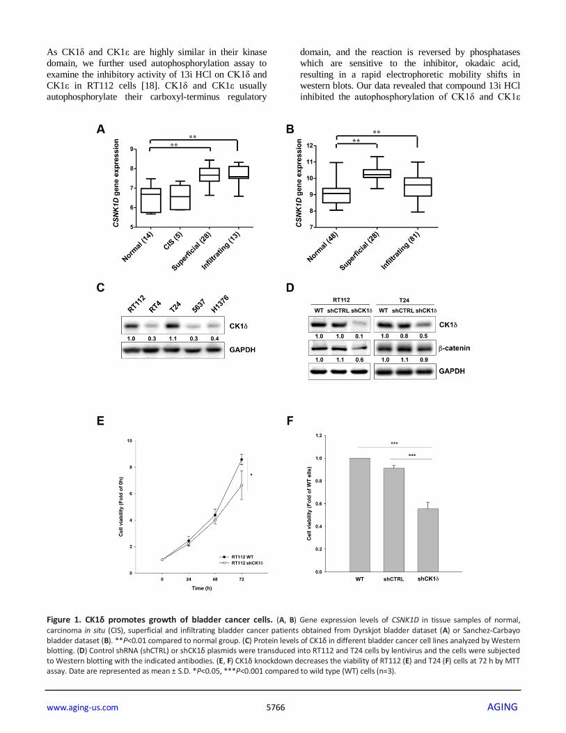
The results demonstrated that the gene expression of
CSNK1D was upregulated in superficial and infiltrating
bladder cancer patients (Figure 1A, 1B). We also
examined CK1δ protein levels in different bladder
cancer cell lines, and found that RT112 and T24 express
the highest levels of CK1δ (Figure 1C). We therefore
chose these two cell lines for subsequent experiments.
To evaluate the contribution of CK1δ to cell growth, we
stably knocked down CSNK1D by lentiviral
transduction. The data suggested that CK1δ levels and
those of its downstream target, β-catenin, were
decreased in RT112 and T24 cells (Figure 1D).
Meanwhile, viability decreased for RT112 and T24 cells
at 72 h (Figure 1E, 1F). Together, the data suggest that
CK1δ contributes to cell growth in bladder cancer cells.
Compound 13i HCl exhibits anti-proliferative
activity in bladder cancer cells
We previously reported 7H-pyrrolo-[2,3-d]pyrimidine
derivatives as novel anticancer agents with potent anti-
CK1δ activity [17]. Among them, 13i HCl is the most
potent against human RT-112 bladder cancer cells. In the
current study, we evaluated the anti-proliferative activity
of 13i HCl using MTT and SRB assay in two bladder
cancer cell lines which express the highest levels of
CK1δ. The data revealed that 13i HCl decreased the
viability of RT112 and T24 cells in a concentration-
dependent manner (Figure 2A) and inhibited their
proliferation (Figure 2B). Notably, 13i HCl displayed
weaker effects on the viability and proliferation of
normal uroepithelial SV-HUC-1 cells (Figure 2A, 2B).
www.aging-us.com 5766 AGING
As CK1δ and CK1ε are highly similar in their kinase
domain, we further used autophosphorylation assay to
examine the inhibitory activity of 13i HCl on CK1δ and
CK1ε in RT112 cells [18]. CK1δ and CK1ε usually
autophosphorylate their carboxyl-terminus regulatory
domain, and the reaction is reversed by phosphatases
which are sensitive to the inhibitor, okadaic acid,
resulting in a rapid electrophoretic mobility shifts in
western blots. Our data revealed that compound 13i HCl
inhibited the autophosphorylation of CK1δ and CK1ε
Figure 1. CK1δ promotes growth of bladder cancer cells. (A, B) Gene expression levels of CSNK1D in tissue samples of normal, carcinoma in situ (CIS), superficial and infiltrating bladder cancer patients obtained from Dyrskjot bladder dataset (A) or Sanchez-Carbayo bladder dataset (B). **P<0.01 compared to normal group. (C) Protein levels of CK1δ in different bladder cancer cell lines analyzed by Western blotting. (D) Control shRNA (shCTRL) or shCK1δ plasmids were transduced into RT112 and T24 cells by lentivirus and the cells were subjected to Western blotting with the indicated antibodies. (E, F) CK1δ knockdown decreases the viability of RT112 (E) and T24 (F) cells at 72 h by MTT assay. Date are represented as mean ± S.D. *P<0.05, ***P<0.001 compared to wild type (WT) cells (n=3).

www.aging-us.com 5767 AGING
in a concentration-dependent manner, resembling the
known dual CK1δ/ε inhibitor PF670462 (Figure 2C,
2D). In summary, 13i HCl exhibits potent anti-cancer
activity in bladder cancer cell lines.
Effects of compound 13i HCl on cell cycle progression
and apoptotic pathways in bladder cancer cells
To elucidate the mechanism underlying 13i HCl-
induced cell death, we first examined 13i HCl’s effect
on cell cycle progression by propidium iodide (PI)
staining and flow cytometry. The data revealed that
13i HCl increased the population of sub-G1 cells in
RT112 cells in a time- and concentration-dependent
manner (Figure 3A, 3B). Accordingly, we further
examined the regulatory proteins of apoptosis by
western blotting. Compound 13i HCl activated the
cleavage of caspase-3, -8, -9 as well as PARP in a
time- and concentration-dependent manner in RT112
cells (Figure 3C, 3D). We also confirmed that 13i HCl
increased the number of cells at the sub-G1 phase and
apoptosis in T24 cells in a concentration-dependent
manner (Supplementary Figure 1). Collectively, these
data suggest that 13i HCl increases apoptosis in
bladder cancer cells.
Inhibition of CK1δ activates necroptosis in bladder
cancer cells
From the above findings, we observed that the protein
levels of GAPDH were decreased under high
concentrations of 13i HCl (Figure 3C, 3D). We
hypothesized that compound 13i HCl increased
membrane permeability in bladder cancer cells. Both
apoptosis and necroptosis are classified as programmed
cell death under drug-induced stress [19, 20]. We
therefore examined the effect of apoptosis- and
necroptosis-inhibitors on 13i HCl-induced cell death.
The results showed that a pan-caspase inhibitor, Z-
VAD-FMK, rescued cell death in the presence of 5 μM
13i HCl. Necrosulfonamide (NSA), a necroptosis
inhibitor, reversed the cell death induced by 2.5 and 5
μM 13i HCl (Figure 4A). However, neither of these
drugs reversed cell death at 10 μM. Because inhibition of
Figure 2. Compound 13i HCl exhibits anti-proliferative activity in bladder cancer cells. (A, B) RT112, T24 and SV-HUC-1 cells were exposed to the indicated concentrations of 13i HCl for 48 h and subjected to MTT assay (A) or SRB assay (B) to analyze cell viability and proliferation, respectively. Data are represented as mean ± S.D. (n=3) *P<0.05, **P<0.01, ***P<0.001 compared to control cells. (C, D) RT112 cells were treated with the indicated concentrations of 13i HCl and PF670462 in the presence of okadaic acid (2 µM) for 1 h and subjected to Western blotting by using CK1δ (C) and CK1ε (D) antibodies.

www.aging-us.com 5768 AGING
caspases by Z-VAD-FMK might further increase the
number of cells undergoing necroptosis, we therefore
combined Z-VAD-FMK and NSA to examine their
compounded effect on 13i HCl-induced cell death. The
data showed that Z-VAD-FMK together with NSA
reversed cell death induced by 2.5 to 10 μM of 13i HCl
(Figure 4B). To further confirm the contribution of
necroptosis in 13i HCl-induced cell death, we stably
knocked down mixed lineage kinase domain-like
protein (MLKL), the key signaling molecule in
necroptosis. Surprisingly, MLKL-knockdown rescued
13i HCl-induced cell death (Figure 4C). Meanwhile,
cell death induced by PF-670462, a specific CK1δ/ε
inhibitor, was also rescued by MLKL-knockdown
(Figure 4D). The knockdown efficiency was confirmed
by western blotting (Figure 4E). Together, the data
suggest that inhibition of CK1δ triggers not only
apoptosis, but also necroptosis in bladder cancer cells.
Compound 13i HCl triggers the phosphorylation of
MLKL and ROS production in bladder cancer cells
To further test whether compound 13i HCl increases
necroptosis in bladder cancer cells, we evaluated the
marker of necroptosis, phosphorylated MLKL
(pMLKL) by western blotting. The data showed that 13i
HCl increased the phosphorylation of MLKL in a
concentration-dependent manner at 72 h (Figure 5A).
The same phenomenon was observed in RT112 cells
treated with a known CK1δ/ε inhibitor, PF-670462
Figure 3. Compound 13i HCl induces apoptosis in RT112 cells. (A) RT112 cells were exposed to the indicated concentrations of 13i HCl for 72 h and subjected to cell cycle analysis. Data are represented as mean ± S.D. *P<0.05, **P<0.01, ***P<0.001 compared to control cells (n=3). (B) RT112 cells were treated with 13i HCl (10 µM) for the indicated times and cell cycle distribution was measured by PI staining and flow cytometry (n=2). (C, D) RT112 cells were exposed to the indicated concentrations of 13i HCl for 72 h (C) or 13i HCl (10 µM) for different times (D) and subjected to Western blotting with the indicated antibodies.

www.aging-us.com 5769 AGING
(Figure 5B). Meanwhile, 13i HCl increased extra-
cellular LDH levels in a concentration-dependent
manner (Figure 5C). It is known that reactive oxygen
species (ROS) contribute to triggering necroptosis and
apoptosis [21, 22]. Mitochondrial ROS reportedly
promote RIP1 autophosphorylation, which is crucial
for the recruitment of RIP3 in the necroptosome [23].
We therefore examined intracellular and mitochondrial
ROS levels by using H2DCFDA and MitoSOX
staining, respectively. Here we show that 13i HCl
increased intracellular and mitochondrial ROS levels
in RT112 cells (Figure 5D, 5E). Together, the data
suggested that 13i HCl triggers necroptosis in bladder
cancer cells.
Figure 4. Inhibition of CK1δ activates necroptosis in bladder cancer cells. (A) RT112 cells were treated with the indicated concentrations of 13i HCl in the presence or absence of zVAD (20 µM) or NSA (10 µM) for 48 h and subjected to MTT assay. Data are represented as mean ± S.D. **P<0.01 compared to the group of 13i HCl alone (n=3). (B) RT112 cells were treated with the indicated concentrations of 13i HCl in the presence or absence of zVAD (20 µM) plus NSA (10 µM) for 48 h and subjected to MTT assay. Data are represented as mean ± S.D. *P<0.05, **P<0.01 compared to the group of 13i HCl alone (n=3). (C, D) RT112 and MLKL stable knocked-down clone (shMLKL-1, shMLKL-2) cells were treated with the indicated concentrations of 13i HCl (C) or PF-670462 (D) for 72 h and subjected to MTT assay. Data are represented as mean ± S.D. *P<0.05, **P<0.01, ***P<0.001compared to the group of 13i HCl or PF-670462 alone (n=3). (E) The knockdown efficiency of MLKL in RT112 cells was examined by Western blotting.

www.aging-us.com 5770 AGING
Effects of 13i HCl on migratory activity in bladder
cancer cells
Our previous study demonstrated that compound 13i HCl
displays inhibitory activity against CK1δ [17]. As CK1δ
regulates several mediators of cancer metastasis, such as
wnt/β-catenin and metastasis suppressor 1 (MTSS1) [12,
24], we examined the effects of 13i HCl on migration by
wound-healing assay. Our data showed that 13i HCl
inhibited the migratory activity of RT112 and T24 cells in
a concentration-dependent manner (Figure 6A, 6B;
Supplementary Figure 2A, 2B). Additionally, knocking-
down CK1δ also decreased wound-healing in RT112 and
T24 cells (Supplementary Figure 3A, 3B). Meanwhile,
13i HCl reversed the expression of epithelial-
mesenchymal transition (EMT) markers, such as the
increase in E-cadherin, and decrease in snail expression in
RT112 cells (Figure 6C). Therefore, the data suggested
that 13i HCl decreases migration and reversed EMT
marker levels in bladder cancer cells.
Figure 5. Compound 13i HCl increases MLKL phosphorylation and ROS production in bladder cancer cells. (A, B) RT112 cells were treated with the indicated concentrations of 13i HCl (A) or PF-670462 (B) for 24 h, 48 h and 72 h. The cells were subjected to Western blotting with the indicated antibodies. (C) RT112 cells were exposed to 13i HCl for 72 h and LDH levels in the supernatants were determined. (D, E) RT112 cells were exposed to the indicated concentrations of 13i HCl for 72 h or H2O2 (10 µM) for 1 h. The cells were stained with H2DCFDA (D) or MitoSOX (E) and subjected to flow cytometry. Data are represented as mean ± S.D. *P<0.05, **P<0.01, ***P<0.001 compared to control cells (n=3).

www.aging-us.com 5771 AGING
DISCUSSION
Bladder cancer is the second most common
genitourinary malignancy worldwide [1]. Appro-
ximately 30% of patients are first diagnosed as MIBC
with a five-year survival rate of 50%, and that drops to
only 6% once it progresses to the metastatic stage [4].
For decades, platinum-based chemotherapy has been the
standard-of-care for advanced bladder cancer, but
patients are often non-responsive or develop of
resistance [3]. Hence, there is an unmet need to develop
novel therapeutic agents to treat bladder cancer. CK1δ
is a serine/threonine-protein kinase that regulates many
cellular processes implicated in cancer, including
Wnt/β-catenin signaling, apoptosis, DNA damage
response and circadian rhythms [12]. Copy number
amplification was observed in 50% of bladder tumors,
which correlated with CK1δ overexpression [12, 14].
Figure 6. Effects of 13i HCl on migratory activity of bladder cancer cells. (A) RT112 cells were seeded into a 2-well insert on 6-well plate and allowed to attach overnight. The wound was created by removing the insert, and the cells were treated with or without 13i HCl and the images were captured at indicated times by EVOS XL Core Cell Imaging System (Thermo Scientific). Scale bar = 100 µm. (B) Quantification of wound healing assay. Data are represented as mean ± S.D. (n=2) *P<0.05 compared to control cells. (C) RT112 cells were exposed to indicated concentrations of 13i HCl for 72 h and subjected to Western blotting for the detection of EMT markers.

www.aging-us.com 5772 AGING
However, the anticancer activity and pharmacological
mechanisms of CK1δ inhibitors on bladder cancer cells
remain elusive. In the current study, we reported that
mRNA levels of CSNK1D, the coding gene of CK1δ,
are upregulated in both of superficial and invasive
bladder cancers by analyzing two independent datasets
(Figure 1A, 1B). Meanwhile, CK1δ is crucial to the
growth and migration of bladder cancer cells (Figure
1D, 1F; Figure 6B). We also demonstrated that
compound 13i HCl, a derivative of 7H-pyrrolo-[2,3-
d]pyrimidines with CK1δ-inhibitory activity, inhibited
proliferation and migration, and induced apoptosis in
bladder cancer cells. Importantly, we describe for the
first time that inhibition of CK1δ by 13i HCl or PF-
670462 triggers necroptosis in bladder cancer cells.
These results suggest that CK1δ could be a valuable
therapeutic target to treat advanced bladder cancer, and
that further development of compound 13i HCl is
warranted to improve patient outcomes.
Several small molecules have shown Ck1δ-inhibitory
activity, such as CKI-7, D4476, IC261, (R)-DRF053,
Bischof-524, and PF-670462 [25], but all present various
problems that limit their application to bladder cancer
therapy [26–31]. Recently, a series of purine scaffold
inhibitors were discovered with IC50 values in the low
nanomolar range, such as SR-1277, SR-2890, and SR-
3029 [31]. Rosenberg et al. successfully demonstrated that
the CK1δ/ε dual inhibitor SR-3029 inhibits growth in
CK1δ-high breast cancer cells and several tumor
xenografts in mice [14]. We also observed that compound
13i HCl inhibits both CK1δ and CK1ε isoforms as
evidenced by autophosphorylation assays (Figure 2C,
2D). Currently, the challenge of using CK1 inhibitors to
treat bladder cancer is the lack of selectivity between
CK1δ and CK1ε, which may result from the high
similarity in their kinase domain. Hence, drug design
should focus on their highly variable C-terminal domains
or the inhibition of auto-phosphorylation. The discovery
of selective CK1δ- or CK1ε-specific inhibitors will
provide new therapeutic possibilities for personalized
medicine.
Although CK1δ inhibitors have shown promising
anticancer activity in several studies, the molecular
mechanisms underlying drug-mediated cell death are
still unclear. In the current study, we observed that
inhibition of CK1δ activity by compound 13i HCl
triggers apoptotic and necroptotic cell death in bladder
cancer cells. We also provided evidence that 13i HCl
treatment or CK1δ knockdown inhibits RT112 cell
migration (Figure 6A, 6B). CK1δ reportedly activates
Wnt/β-catenin in breast cancer [14, 15] and CK1δ
silencing reduces the migration and invasion of triple-
negative breast cancer cells [32]. Importantly, either
APC mutations or nuclear β-catenin accumulation are
associated with poor outcome in patients with invasive
bladder cancer [16]. In a previous study, we found that
forced expression of β-catenin rescued cytotoxicity
induced by 13i HCl in RT112 cells [17]. Therefore,
targeting the CK1δ/β-catenin pathway is an attractive
strategy to treat Wnt-driven bladder cancer. On the
other hand, CK1δ has emerged as a potential target for
other diseases. CK1δ inhibitors have been proposed to
inhibit the phosphorylation of TDP-43, a pathological
hallmark of central nervous system (CNS) diseases,
such as amyotrophic lateral sclerosis (ALS) and
frontotemporal dementia [33, 34]. CK1δ is also a
potential target for Parkinson’s disease and pulmonary
fibrosis [35, 36]. These observations further support the
development of compound 13i HCl as a potential
therapeutic target for other diseases.
Apoptosis, necroptosis and autophagic cell death are
three main types of programmed cell death (PCD)
induced by anticancer agents [19]. Apoptosis is the
most studied PCD, characterized by cytoplasmic
shrinkage, chromatin condensation, nuclear con-
densation, and membrane blebbing. The activation of
caspases is a feature of apoptosis, accompanied by
DNA/protein breakdown, and mitochondrial outer
membrane permeabilization [37]. Necroptosis is a
caspase-independent PCD, a form of necrotic death
executed by receptor-interacting protein 1 (RIP1), RIP3,
and Mixed Lineage Kinase Domain-Like (MLKL)
protein [38]. Since resistance to apoptosis is one of the
hallmarks of cancer, necroptosis-based cancer therapy
has been proposed as a novel strategy for antitumor
treatment [39]. Necroptosis is a unique cell-killing
mechanism in response to severe stress or impaired
apoptosis, which can be triggered by inflammatory
cytokines, chemotherapeutic agents, and natural
compounds [40]. Meanwhile, necroptotic tumor cells
initiate adaptive immune responses by releasing
damage-associated molecular patterns (DAMPs) into
the microenvironment, activating dendritic and
cytotoxic T cells that may suppress tumor progression
[41]. For the first time, we observed that inhibition of
CK1δ activity by compound 13i HCl or PF-670462
triggers necroptosis in bladder cancer cells. Knocking
down of MLKL rescued cytotoxicity induced by 13i
HCl or PF-670462 in RT112 cells (Figure 4C, 4D).
However, the mechanisms by which CK1δ regulates
necroptosis remain elusive. From the literature, CK1δ is
able to phosphorylate T362 in the catalytic domain of
protein phosphatase 5 (PP5), enhancing its phosphatase
activity [42]. A crucial target of PP5 is Cdc37, a
cochaperone of the Hsp90 complex, regulating the
activation of protein kinase clients by Hsp90-Cdc37
[43, 44]. Interestingly, RIPK3 and MLKL, two
important regulators in necrosomes, are clients of
Hsp90-Cdc37 [45, 46]. Therefore, it would be useful to

www.aging-us.com 5773 AGING
elucidate any potential linkages between CK1δ
inhibition and of necroptosis induction in future studies.
MATERIALS AND METHODS
Cell culture, antibodies, and reagents
RT112, RT4, 5637 cells were cultured in RPMI 1640;
T24 cells were cultured in McCoy's 5a, H1376 cells
were cultured in EMEM, and SV-HUC-1 were cultured
in F-12K, supplemented with 10% (v/v) FBS and 1%
(v/v) antibiotic-antimycotic solution (Thermo Fisher
Scientific, Waltham, MA, USA) at 37 °C in a
humidified incubator containing 5% CO2. 4-((4-(4-
ethylpiperazin-1-yl)phenyl)amino)-N-(3,4,5-trichloro
phenyl)-7H-pyrrolo-[2, 3-d]pyrimidine-7-carboxamide
hydrochloride (13i HCl) was synthesized by Dr. Jing-
Ping Liou, as previously described [17]. PF-670462,
okadaic acid, necrosulfonamide (NSA), and z-VAD-
FMK were purchased from Cayman Chemical (Ann
Arbor, MI, USA). 3-(4,5-Dimethylthiazol-2-yl)-2,5-
diphenyltetrazolium bromide (MTT) and
Sulforhodamine B (SRB) were obtained from Sigma
Chemical Corp (St. Louis, MO, USA). Antibodies
against various proteins were obtained from the
following sources: caspase-3 from Novus biologicals
(Littleton, CO, USA); CK1δ, pMLKL from Abcam
(Cambridge, MA, USA); β-catenin from Santa Cruz
Biotechnology (Santa Cruz, CA, USA); PARP, caspase-
8, caspase-9 from Cell Signaling Technology (Danvers,
MA, USA), and β-actin, MLKL, GAPDH from Genetex
(Irvine, CA, USA).
Cell viability, proliferation and lactate
dehydrogenase (LDH) assays
Cells were seeded in 96-well plates and exposed to
indicated compounds for 48 or 72 h. Cell viability was
examined by MTT assay as described previously [47].
Cell proliferation was measured with the SRB assay as
described previously [48]. For LDH assay, cells were
seeded in 96-well plates and treated with drugs at the
indicated concentrations for 72 h. LDH levels in culture
supernatants were analyzed by using the CytoTox 96
Non-Radioactive Cytotoxicity Assay Kit (Promega;
Madison, WI, USA) according to the manufacturer's
protocol.
Cell cycle analysis
Cells were seeded in 6-well plates, and exposed to
indicated compounds for 24 to 72 h. The cells were
collected by trypsinization, washed one time by PBS,
and fixed with ethanol (70%) at -20 °C overnight. The
cells were pelleted by centrifugation, and incubated in
0.1 mL of phosphate-citric acid buffer (0.2 M NaHPO4,
0.1 M citric acid, and pH 7.8) for 15 min at room
temperature, and then resuspended in propidium iodide
staining buffer containing Triton X-100 (0.1%, v/v),
RNase A (100 μg/mL), and propidium iodide (80
μg/mL) for 30 min in the dark. Cell cycle distribution
was analyzed by flow cytometry with CellQuest
software (Becton Dickinson, Mountain View, CA,
USA) following previously published methods [17].
Intracellular and mitochondrial ROS analysis
Cells were seeded in 6-well plates, exposed to DMSO or
indicated compounds for 72 h, and harvested by
trypsinization. For total cell ROS analysis, cells were
stained with 0.1 µM H2DCFDA (Biotium, Fremont, CA,
USA) at 37 °C for 20 min. For mitochondrial ROS
analysis, cells were stained with 5 µM MitoSOX Red
(Thermo Fisher Scientific, Waltham, MA, USA) at 37 °C
for 10 min. After washing with PBS three times, cells
were subjected to ROS detection via flow cytometry with
CellQuest software according to the manufacturer's
instructions (Becton Dickinson, Mountain View, CA,
USA), as previously described [47].
Western blot analysis and lentiviral expression
system
The cells were seeded in 6-well plates or 60 mm dishes
and exposed to different compounds for the indicated
times. After treatment, equal amounts of protein were
separated via SDS–PAGE, transferred to PVDF
membrane and immunoblotted with specific antibodies,
as described previously [47]. Lentiviral particles
containing shRNA plasmids of shCSNK1D
(TRCN0000001552) and shMLKL (TRCN0000194846
and TRCN0000196741) were purchased from the
National RNAi Core Facility (Academia Sinica,
Taiwan). Stable cell lines were selected by the treatment
of puromycin (InvivoGen; San Diego, CA, USA).
Cell migration assay
Cells were seeded into a 2-well insert (Ibidi, Munich,
Germany) on a 6-well plate and allowed to attach
overnight. A wound was created by removing the insert,
and the cell-free gap was around 500 μm. Cells were
allowed to migrate in the presence or absence of drugs,
and images were captured at the indicated times by EVOS
XL Core Cell Imaging System (Thermo Scientific).
Microarray datasets analysis
Dyrskjot et al. published a dataset in which 14 normal
bladder, 5 carcinoma in situ (CIS), 28 superficial
bladder cancer, and 13 invasive bladder cancer samples
were analyzed using Affymetrix U133A microarrays

www.aging-us.com 5774 AGING
[49]. Array data were obtained from the NCBI
Gene expression omnibus (GEO; http://www.ncbi.nlm.
nih.gov/geo/) database with the accession number
GSE3167. RMA log expression units were calculated
using ‘affy’ package for the R statistical programming
language. The default RMA settings were used to
background correct, normalize and summarize all
expression values. Second dataset was published by
Sanchez-Carbayo et al., in which 81 infiltrating bladder
urothelial carcinoma, 28 superficial bladder cancer, and
48 normal bladder samples were analyzed on
Affymetrix U133A microarrays [50]. The gene
expression level of CSNK1D was obtained from this
study, and log2 expression level was used for statistical
analysis. A 2-tailed Student’s t-test was then applied for
the calculation of the p value between two different
groups.
Statistical analysis
Each experiment was performed independently with at
least two biological replicates. Data in the bar graphs are
presented as means ± S.D and analyzed by using the
Student’s t-test with p values < 0.05 considered significant.
Abbreviations
BC: bladder cancer; EMT: epithelial-mesenchymal
transition; MIBC: muscle invasive bladder cancer;
MTSS1: metastasis suppressor 1; NMIBC: non-muscle
invasive bladder cancer; PCD: programmed cell death;
ROS: reactive oxygen species.
AUTHOR CONTRIBUTIONS
Conceptualization, CHC.; investigation, YCL and MCC;
data curation, YC. and CHC; Analysis, MCC and THH;
resources, JPL; writing original draft, YCL and CHC;
review and editing of manuscript, MCC and CHC;
funding acquisition: CHC; supervision, CHC.
CONFLICTS OF INTEREST
The authors declare that they have no competing
interests.
FUNDING
This research was funded by the Ministry of Science
and Technology of the Republic of China, grant No.
MOST-107-2320-B-038-039. This work was also
financially supported by the “TMU Research Center of
Cancer Translational Medicine” from The Featured
Areas Research Center Program within the framework
of the Higher Education Sprout Project by the Ministry
of Education (MOE) in Taiwan.
REFERENCES
1. Bray F, Ferlay J, Soerjomataram I, Siegel RL, Torre LA, Jemal A. Global cancer statistics 2018: GLOBOCAN estimates of incidence and mortality worldwide for 36 cancers in 185 countries. CA Cancer J Clin. 2018; 68:394–424.
https://doi.org/10.3322/caac.21492 PMID:30207593
2. Siegel RL, Miller KD, Jemal A. Cancer statistics, 2019. CA Cancer J Clin. 2019; 69:7–34.
https://doi.org/10.3322/caac.21551 PMID:30620402
3. Berdik C. Unlocking bladder cancer. Nature. 2017; 551:S34–35.
https://doi.org/10.1038/551S34a PMID:29117159
4. Funt SA, Rosenberg JE. Systemic, perioperative management of muscle-invasive bladder cancer and future horizons. Nat Rev Clin Oncol. 2017; 14:221–34.
https://doi.org/10.1038/nrclinonc.2016.188 PMID:27874062
5. Ghosh M, Brancato SJ, Agarwal PK, Apolo AB. Targeted therapies in urothelial carcinoma. Curr Opin Oncol. 2014; 26:305–20.
https://doi.org/10.1097/CCO.0000000000000064 PMID:24685646
6. Bukhari N, Al-Shamsi HO, Azam F. Update on the Treatment of Metastatic Urothelial Carcinoma. ScientificWorldJournal. 2018; 2018:5682078.
https://doi.org/10.1155/2018/5682078 PMID:29977169
7. Flaig TW, Spiess PE, Agarwal N, Bangs R, Boorjian SA, Buyyounouski MK, Downs TM, Efstathiou JA, Friedlander T, Greenberg RE, Guru KA, Hahn N, Herr HW, et al. NCCN Guidelines Insights: Bladder Cancer, Version 5.2018. J Natl Compr Canc Netw. 2018; 16:1041–53.
https://doi.org/10.6004/jnccn.2018.0072 PMID:30181416
8. Ghasemzadeh A, Bivalacqua TJ, Hahn NM, Drake CG. New Strategies in Bladder Cancer: A Second Coming for Immunotherapy. Clin Cancer Res. 2016; 22:793–801.
https://doi.org/10.1158/1078-0432.CCR-15-1135 PMID:26683632
9. Nadal R, Bellmunt J. Management of metastatic bladder cancer. Cancer Treat Rev. 2019; 76:10–21.
https://doi.org/10.1016/j.ctrv.2019.04.002 PMID:31030123
10. Svatek RS, Hollenbeck BK, Holmäng S, Lee R, Kim SP, Stenzl A, Lotan Y. The economics of bladder cancer:

www.aging-us.com 5775 AGING
costs and considerations of caring for this disease. Eur Urol. 2014; 66:253–62.
https://doi.org/10.1016/j.eururo.2014.01.006 PMID:24472711
11. Cheong JK, Virshup DM. Casein kinase 1: complexity in the family. Int J Biochem Cell Biol. 2011; 43:465–69.
https://doi.org/10.1016/j.biocel.2010.12.004 PMID:21145983
12. Schittek B, Sinnberg T. Biological functions of casein kinase 1 isoforms and putative roles in tumorigenesis. Mol Cancer. 2014; 13:231.
https://doi.org/10.1186/1476-4598-13-231 PMID:25306547
13. Logan CY, Nusse R. The Wnt signaling pathway in development and disease. Annu Rev Cell Dev Biol. 2004; 20:781–810.
https://doi.org/10.1146/annurev.cellbio.20.010403.113126 PMID:15473860
14. Rosenberg LH, Lafitte M, Quereda V, Grant W, Chen W, Bibian M, Noguchi Y, Fallahi M, Yang C, Chang JC, Roush WR, Cleveland JL, Duckett DR. Therapeutic targeting of casein kinase 1δ in breast cancer. Sci Transl Med. 2015; 7:318ra202.
https://doi.org/10.1126/scitranslmed.aac8773 PMID:26676609
15. Cheong JK, Virshup DM. CK1δ: a pharmacologically tractable Achilles’ heel of Wnt-driven cancers? Ann Transl Med. 2016; 4:433.
https://doi.org/10.21037/atm.2016.11.07 PMID:27942524
16. Kastritis E, Murray S, Kyriakou F, Horti M, Tamvakis N, Kavantzas N, Patsouris ES, Noni A, Legaki S, Dimopoulos MA, Bamias A. Somatic mutations of adenomatous polyposis coli gene and nuclear b-catenin accumulation have prognostic significance in invasive urothelial carcinomas: evidence for Wnt pathway implication. Int J Cancer. 2009; 124:103–08.
https://doi.org/10.1002/ijc.23917 PMID:18844223
17. Liu YM, Chen CH, Yeh TK, Liou JP. Synthesis and evaluation of novel 7H-pyrrolo-[2,3-d]pyrimidine derivatives as potential anticancer agents. Future Med Chem. 2019; 11:959–74.
https://doi.org/10.4155/fmc-2018-0564 PMID:30789758
18. Rivers A, Gietzen KF, Vielhaber E, Virshup DM. Regulation of casein kinase I epsilon and casein kinase I delta by an in vivo futile phosphorylation cycle. J Biol Chem. 1998; 273:15980–84.
https://doi.org/10.1074/jbc.273.26.15980 PMID:9632646
19. Mishra AP, Salehi B, Sharifi-Rad M, Pezzani R, Kobarfard F, Sharifi-Rad J, Nigam M. Programmed Cell
Death, from a Cancer Perspective: an Overview. Mol Diagn Ther. 2018; 22:281–95.
https://doi.org/10.1007/s40291-018-0329-9 PMID:29560608
20. Zhou W, Yuan J. Necroptosis in health and diseases. Semin Cell Dev Biol. 2014; 35:14–23.
https://doi.org/10.1016/j.semcdb.2014.07.013 PMID:25087983
21. Schulze-Osthoff K, Bakker AC, Vanhaesebroeck B, Beyaert R, Jacob WA, Fiers W. Cytotoxic activity of tumor necrosis factor is mediated by early damage of mitochondrial functions. Evidence for the involvement of mitochondrial radical generation. J Biol Chem. 1992; 267:5317–23.
PMID:1312087
22. Redza-Dutordoir M, Averill-Bates DA. Activation of apoptosis signalling pathways by reactive oxygen species. Biochim Biophys Acta. 2016; 1863:2977–92.
https://doi.org/10.1016/j.bbamcr.2016.09.012 PMID:27646922
23. Zhang Y, Su SS, Zhao S, Yang Z, Zhong CQ, Chen X, Cai Q, Yang ZH, Huang D, Wu R, Han J. RIP1 autophosphorylation is promoted by mitochondrial ROS and is essential for RIP3 recruitment into necrosome. Nat Commun. 2017; 8:14329.
https://doi.org/10.1038/ncomms14329 PMID:28176780
24. Zhong J, Shaik S, Wan L, Tron AE, Wang Z, Sun L, Inuzuka H, Wei W. SCF β-TRCP targets MTSS1 for ubiquitination-mediated destruction to regulate cancer cell proliferation and migration. Oncotarget. 2013; 4:2339–53.
https://doi.org/10.18632/oncotarget.1446 PMID:24318128
25. Knippschild U, Krüger M, Richter J, Xu P, García-Reyes B, Peifer C, Halekotte J, Bakulev V, Bischof J. The CK1 Family: Contribution to Cellular Stress Response and Its Role in Carcinogenesis. Front Oncol. 2014; 4:96.
https://doi.org/10.3389/fonc.2014.00096 PMID:24904820
26. Chijiwa T, Hagiwara M, Hidaka H. A newly synthesized selective casein kinase I inhibitor, N-(2-aminoethyl)-5-chloroisoquinoline-8-sulfonamide, and affinity purification of casein kinase I from bovine testis. J Biol Chem. 1989; 264:4924–27.
PMID:2925675
27. Badura L, Swanson T, Adamowicz W, Adams J, Cianfrogna J, Fisher K, Holland J, Kleiman R, Nelson F, Reynolds L, St Germain K, Schaeffer E, Tate B, Sprouse J. An inhibitor of casein kinase I epsilon induces phase delays in circadian rhythms under free-running and entrained conditions. J Pharmacol Exp Ther. 2007; 322:730–38.

www.aging-us.com 5776 AGING
https://doi.org/10.1124/jpet.107.122846 PMID:17502429
28. Cheong JK, Nguyen TH, Wang H, Tan P, Voorhoeve PM, Lee SH, Virshup DM. IC261 induces cell cycle arrest and apoptosis of human cancer cells via CK1δ/ɛ and Wnt/β-catenin independent inhibition of mitotic spindle formation. Oncogene. 2011; 30:2558–69.
https://doi.org/10.1038/onc.2010.627 PMID:21258417
29. Rena G, Bain J, Elliott M, Cohen P. D4476, a cell-permeant inhibitor of CK1, suppresses the site-specific phosphorylation and nuclear exclusion of FOXO1a. EMBO Rep. 2004; 5:60–65.
https://doi.org/10.1038/sj.embor.7400048 PMID:14710188
30. Oumata N, Bettayeb K, Ferandin Y, Demange L, Lopez-Giral A, Goddard ML, Myrianthopoulos V, Mikros E, Flajolet M, Greengard P, Meijer L, Galons H. Roscovitine-derived, dual-specificity inhibitors of cyclin-dependent kinases and casein kinases 1. J Med Chem. 2008; 51:5229–42.
https://doi.org/10.1021/jm800109e PMID:18698753
31. Bibian M, Rahaim RJ, Choi JY, Noguchi Y, Schürer S, Chen W, Nakanishi S, Licht K, Rosenberg LH, Li L, Feng Y, Cameron MD, Duckett DR, et al. Development of highly selective casein kinase 1δ/1ε (CK1δ/ε) inhibitors with potent antiproliferative properties. Bioorg Med Chem Lett. 2013; 23:4374–80.
https://doi.org/10.1016/j.bmcl.2013.05.075 PMID:23787102
32. Bar I, Merhi A, Larbanoix L, Constant M, Haussy S, Laurent S, Canon JL, Delrée P. Silencing of casein kinase 1 delta reduces migration and metastasis of triple negative breast cancer cells. Oncotarget. 2018; 9:30821–36.
https://doi.org/10.18632/oncotarget.25738 PMID:30112110
33. Bissaro M, Federico S, Salmaso V, Sturlese M, Spalluto G, Moro S. Targeting Protein Kinase CK1δ with Riluzole: Could It Be One of the Possible Missing Bricks to Interpret Its Effect in the Treatment of ALS from a Molecular Point of View? ChemMedChem. 2018; 13:2601–05.
https://doi.org/10.1002/cmdc.201800632 PMID:30359484
34. Alquezar C, Salado IG, de la Encarnación A, Pérez DI, Moreno F, Gil C, de Munain AL, Martínez A, Martín-Requero Á. Targeting TDP-43 phosphorylation by Casein Kinase-1δ inhibitors: a novel strategy for the treatment of frontotemporal dementia. Mol Neurodegener. 2016; 11:36.
https://doi.org/10.1186/s13024-016-0102-7 PMID:27138926
35. Morales-Garcia JA, Salado IG, Sanz-San Cristobal M, Gil C, Pérez-Castillo A, Martínez A, Pérez DI. Biological and Pharmacological Characterization of Benzothiazole-Based CK-1δ Inhibitors in Models of Parkinson’s Disease. ACS Omega. 2017; 2:5215–20.
https://doi.org/10.1021/acsomega.7b00869 PMID:30023743
36. Keenan CR, Langenbach SY, Jativa F, Harris T, Li M, Chen Q, Xia Y, Gao B, Schuliga MJ, Jaffar J, Prodanovic D, Tu Y, Berhan A, et al. Casein Kinase 1δ/ε Inhibitor, PF670462 Attenuates the Fibrogenic Effects of Transforming Growth Factor-β in Pulmonary Fibrosis. Front Pharmacol. 2018; 9:738.
https://doi.org/10.3389/fphar.2018.00738 PMID:30042678
37. Mariño G, Niso-Santano M, Baehrecke EH, Kroemer G. Self-consumption: the interplay of autophagy and apoptosis. Nat Rev Mol Cell Biol. 2014; 15:81–94.
https://doi.org/10.1038/nrm3735 PMID:24401948
38. Su Z, Yang Z, Xie L, DeWitt JP, Chen Y. Cancer therapy in the necroptosis era. Cell Death Differ. 2016; 23:748–56.
https://doi.org/10.1038/cdd.2016.8 PMID:26915291
39. Grootjans S, Vanden Berghe T, Vandenabeele P. Initiation and execution mechanisms of necroptosis: an overview. Cell Death Differ. 2017; 24:1184–95.
https://doi.org/10.1038/cdd.2017.65 PMID:28498367
40. Chen D, Yu J, Zhang L. Necroptosis: an alternative cell death program defending against cancer. Biochim Biophys Acta. 2016; 1865:228–36.
https://doi.org/10.1016/j.bbcan.2016.03.003 PMID:26968619
41. Gong Y, Fan Z, Luo G, Yang C, Huang Q, Fan K, Cheng H, Jin K, Ni Q, Yu X, Liu C. The role of necroptosis in cancer biology and therapy. Mol Cancer. 2019; 18:100.
https://doi.org/10.1186/s12943-019-1029-8 PMID:31122251
42. Dushukyan N, Dunn DM, Sager RA, Woodford MR, Loiselle DR, Daneshvar M, Baker-Williams AJ, Chisholm JD, Truman AW, Vaughan CK, Haystead TA, Bratslavsky G, Bourboulia D, Mollapour M. Phosphorylation and Ubiquitination Regulate Protein Phosphatase 5 Activity and Its Prosurvival Role in Kidney Cancer. Cell Rep. 2017; 21:1883–95.
https://doi.org/10.1016/j.celrep.2017.10.074 PMID:29141220
43. Vaughan CK, Mollapour M, Smith JR, Truman A, Hu B, Good VM, Panaretou B, Neckers L, Clarke PA, Workman P, Piper PW, Prodromou C, Pearl LH. Hsp90-

www.aging-us.com 5777 AGING
dependent activation of protein kinases is regulated by chaperone-targeted dephosphorylation of Cdc37. Mol Cell. 2008; 31:886–95.
https://doi.org/10.1016/j.molcel.2008.07.021 PMID:18922470
44. Li T, Jiang HL, Tong YG, Lu JJ. Targeting the Hsp90-Cdc37-client protein interaction to disrupt Hsp90 chaperone machinery. J Hematol Oncol. 2018; 11:59.
https://doi.org/10.1186/s13045-018-0602-8 PMID:29699578
45. Li D, Xu T, Cao Y, Wang H, Li L, Chen S, Wang X, Shen Z. A cytosolic heat shock protein 90 and cochaperone CDC37 complex is required for RIP3 activation during necroptosis. Proc Natl Acad Sci USA. 2015; 112:5017–22.
https://doi.org/10.1073/pnas.1505244112 PMID:25852146
46. Zhao XM, Chen Z, Zhao JB, Zhang PP, Pu YF, Jiang SH, Hou JJ, Cui YM, Jia XL, Zhang SQ. Hsp90 modulates the stability of MLKL and is required for TNF-induced necroptosis. Cell Death Dis. 2016; 7:e2089.
https://doi.org/10.1038/cddis.2015.390 PMID:26866270
47. Chen CH, Changou CA, Hsieh TH, Lee YC, Chu CY, Hsu KC, Wang HC, Lin YC, Lo YN, Liu YR, Liou JP, Yen Y. Dual Inhibition of PIK3C3 and FGFR as a New Therapeutic Approach to Treat Bladder Cancer. Clin Cancer Res. 2018; 24:1176–89.
https://doi.org/10.1158/1078-0432.CCR-17-2066 PMID:29222162
48. Liu YM, Lee HY, Chen CH, Lee CH, Wang LT, Pan SL, Lai MJ, Yeh TK, Liou JP. 1-Arylsulfonyl-5-(N-hydroxyacrylamide)tetrahydroquinolines as potent histone deacetylase inhibitors suppressing the growth of prostate cancer cells. Eur J Med Chem. 2015; 89:320–30.
https://doi.org/10.1016/j.ejmech.2014.10.052 PMID:25462248
49. Dyrskjøt L, Kruhøffer M, Thykjaer T, Marcussen N, Jensen JL, Møller K, Ørntoft TF. Gene expression in the urinary bladder: a common carcinoma in situ gene expression signature exists disregarding histopathological classification. Cancer Res. 2004; 64:4040–48.
https://doi.org/10.1158/0008-5472.CAN-03-3620 PMID:15173019
50. Sanchez-Carbayo M, Socci ND, Lozano J, Saint F, Cordon-Cardo C. Defining molecular profiles of poor outcome in patients with invasive bladder cancer using oligonucleotide microarrays. J Clin Oncol. 2006; 24:778–89.
https://doi.org/10.1200/JCO.2005.03.2375 PMID:16432078

www.aging-us.com 5778 AGING
SUPPLEMENTARY MATERIALS
Supplementary Figures
Supplementary Figure 1. Compound 13i HCl induces apoptosis in T24 cells. (A) T24 cells were exposed to the indicated concentrations of 13i HCl for 72 h and subjected to cell cycle analysis. Data are represented as mean ± S.D. *P<0.05, **P<0.01, ***P<0.001 compared to control cells (n=2). (B) T24 cells were exposed to the indicated concentrations of 13i HCl for 72 h and subjected to Western blotting with the indicated antibodies.

www.aging-us.com 5779 AGING
Supplementary Figure 2. Effects of 13i HCl on migratory activity in bladder cancer cells. (A) T24 cells were seeded into a 2-well insert on 6-well plate and allowed to attach overnight. The wound was created by removing the insert, and the cells were treated with or without 13i HCl. Images were captured at the indicated time points using an EVOS XL Core Cell Imaging System (Thermo Scientific). Scale bar = 100 mm. (B) Quantification of wound healing assay. Data are represented as mean ± S.D. (n=2) *P<0.05, **P<0.01 compared to control cells.

www.aging-us.com 5780 AGING
Supplementary Figure 3. Effects of CK1δ knockdown on migratory activity in bladder cancer cells. RT112 (A) and T24 (B) cells stably expressing control shRNA (shCTRL) or shCK1δ were seeded into a 2-well insert on 6-well plate and allowed to attach overnight. The wound was created by removing the insert, and the images were captured at the indicated times using an EVOS XL Core Cell Imaging System (Thermo Scientific). Scale bar = 100 mm. (B) Quantification of wound healing assay. Data are represented as mean ± S.D. (n=2) *P<0.05 compared to control cells.