Embed Size (px)
Citation preview
Brain Research 933 (2002) 50–59www.elsevier.com/ locate /bres
Research report
Phosphorylation of P42/P44 MAP kinase and DNA fragmentation inthe rat perforant pathway stimulation model of limbic epilepsy
a,b,d b,c,d a,c,d ,*Jonathan L. Brisman , G. Rees Cosgrove , Andrew J. ColeaEpilepsy Research Laboratory, Neurosurgical Service, Massachusetts General Hospital, Boston, MA, USA
bNeurosurgical Service, Massachusetts General Hospital, Boston, MA, USAcEpilepsy Service, Massachusetts General Hospital, Boston, MA, USA
dHarvard Medical School, Boston, MA 02114, USA
Accepted 20 December 2001
Abstract
The intracellular signaling pathways associated with neuronal injury after perforant pathway stimulation of the rodent hippocampushave not been examined. To determine whether activation of the p42/p44 (Erk1/2) MAP kinase (MAPK) phosphorylation cascade islinked to neuronal injury after perforant pathway stimulation (PPS), we stained for phosphorylated Erk1/2 (P-Erk1/2) and for DNAfragmentation, a marker of cell death after PPS. Eighteen Sprague–Dawley rats underwent PPS for 6 (n56), 12 (n56), or 24 (n56) h andwere sacrificed either immediately (n59) or 48 h (n59) after stimulation. Sham-operated non-stimulated control animals (n52) andcontrol animals receiving low frequency stimulation only (n54) were also examined. Brain sections were stained for DNA fragmentationand P-Erk1/2. DNA fragmentation was evident only in granule cells and CA3 pyramidal cells of the stimulated side 48 h after 24 h ofPPS. PPS resulted in robust phosphorylation of Erk1/2 that displayed a stereotyped timecourse, appearing first in hilar neurons on theipsilateral side and later in hilar neurons, granule cells, hippocampal pyramidal and non-neuronal cell populations on both the stimulatedand contralateral sides. Both Erk1/2 phosphorylation and DNA fragmentation show definite and reproducible staining patterns after PPSthat vary based on duration of stimulation. Populations displaying Erk1/2 activation appeared to differ from those showing DNAfragmentation and neuronal injury. 2002 Elsevier Science B.V. All rights reserved.
Theme: Disorders of the nervous system
Topic: Epilepsy: human studies and animal models
Keywords: MAP kinase; Perforant pathway stimulation; Epilepsy; Rat; DNA fragmentation
1. Introduction gyrus die via the apoptotic route while pyramidal cells ofCA1, CA2 and CA3 as well as hilar neurons die via
While selective neuronal injury has been described in a necrosis [2,17,35,36]. Both apoptosis and necrosis havevariety of animal models, recent studies have increasingly been linked to glutamate excitotoxicity, NMDA receptorfocused on the mechanisms and pathways leading to cell activation [30,31], and ischemia [25], but why certain celldeath. In particular, there has been ongoing debate as to populations die in one manner as opposed to another is notwhether seizure-induced cell death is necrotic, apoptotic, known. Granule cells represent a unique population ofor both. excitatory neurons within the hippocampus that are ideally
Accumulating evidence suggests that multiple mecha- positioned and connected to function as seizure-generatingnisms lead to cell injury after seizure activity in the rodent. cells [26,27]. If these cells die by apoptosis, while otherSome studies suggest that granule cells of the dentate cells in the hippocampus die by necrosis, multiple neuro-
protection strategies might be required to blockstimulation-induced injury. We have used DNA fragmenta-
*Corresponding author. Epilepsy Service, Massachusetts Generaltion as a readily identifiable marker of cell injury, whoseHospital VBK 830, 55 Fruit Street, Boston, MA 02114, USA. Tel.:correlation with apoptotic cell death remains controversial11-617-7263-311; fax: 11-617-7269-250.
E-mail address: [email protected] (A.J. Cole). [38].
0006-8993/02/$ – see front matter 2002 Elsevier Science B.V. All rights reserved.PI I : S0006-8993( 02 )02304-1
J.L. Brisman et al. / Brain Research 933 (2002) 50 –59 51
The mitogen-activated protein kinases (MAPKs) are a electroconvulsive shock treatment [1,37] and generalizedfamily of serine / threonine kinases that regulates intracellu- seizures [15]. Numerous targets for Erk1/2 have beenlar activity in response to extracellular signals. Two described, including cytoskeletal proteins and synapsin,neuronal isoforms of MAPK, p42 (ERK2) and p44 and activated MAPKs have been shown to translocate from(ERK1) have been immunocytochemically mapped and the cytoplasm into the nucleus and act to regulate tran-found to have high concentrations within the cortex, scription of early genes [41]. The exact function ofcerebellum and hippocampus of both developing and adult activation in vivo, however, is unknown. Moreover, al-brain [13]. Activation of these proteins requires dual though attempts have been made to correlate the activationphosphorylation on both serine and tyrosine residues. The of MAPKs with apoptotic cell death [6,20], whether thepresence of activated Erk1/2 in cell bodies and dendrites activation of MAPKs is beneficial or detrimental to a cellhas implicated this pathway in postsynaptic signal trans- undergoing stress is still not known. Interestingly, we haveduction. Activation of Erk1/2 and other MAPKs have previously shown that inhibition of Erk1/2 activationbeen described in response to ischemia [5], growth factors blocks cell death in a primary neuronal cell culture model[4,8,16,21], glutamate receptor activity [19,30,31,41], of seizure-like activity [23].
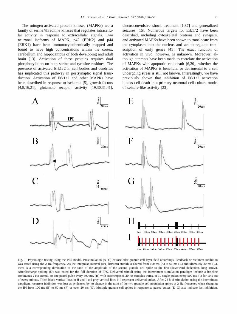
Fig. 1. Physiologic testing using the PPS model. Prestimulation (A–C) extracellular granule cell layer field recordings. Feedback or recurrent inhibitionwas tested using the 2 Hz frequency. As the interpulse interval (IPI) between stimuli is altered from 100 ms (A) to 60 ms (B) and ultimately 20 ms (C),there is a corresponding diminution of the ratio of the amplitude of the second granule cell spike to the first (downward deflection, long arrow).Afterdischarge spiking (D) was noted for the full duration of PPS. Delivered stimuli using the intermittent stimulation paradigm include a baselinecontinuous 2 Hz stimuli, or one paired pulse every 500 ms, (H) with superimposed 20 Hz stimulus trains, or 10 single pulses every 500 ms, (I) for 10 s outof every minute. Thick black vertical lines in H and I and grey vertical lines in I represent delivered pulses. After 24 h of stimulation using the intermittentparadigm, recurrent inhibition was lost as evidenced by no change in the ratio of the two granule cell population spikes at 2 Hz frequency when changingthe IPI from 100 ms (E) to 60 ms (F) or even 20 ms (G). Multiple granule cell spikes in response to paired pulses (E–G) also indicate lost inhibition.
52 J.L. Brisman et al. / Brain Research 933 (2002) 50 –59
To study cell death after abnormal neuronal activity, we electrophysiological homeostasis in response to granulehave utilized a rodent chronic perforant pathway stimula- cell firing within the hippocampus, was tested by examin-tion (PPS) model, in which prolonged repeated stimulation ing paired-pulse inhibition as a function of interpulseof the angular bundle results in a reproducible pattern of interval (IPI). The IPI was reduced in steps from 100 to 20unilateral hippocampal damage. We investigated whether ms (Fig. 1A–C). In normal animals this results in en-Erk1/2 is phosphorylated in this model and whether hanced paired pulse inhibition manifested by a decreasedphosphorylation occurs in neurons that subsequently mani- ratio of the amplitude of the second evoked potential to thefest injury as demonstrated by DNA fragmentation. first evoked potential, whereas when feedback inhibition is
reduced, the amplitude ratio between the first and secondevoked potentials remains close to unity regardless of the
2. Materials and methods IPI. At the beginning of each experiment animals wereexamined electrophysiologically to document the baseline
2.1. Perforant pathway stimulation integrity of inhibitory pathways (Fig. 1A–C). Evokedepileptiform activity after a 10 s 20 Hz train was also
Male Sprague–Dawley descendant rats (250–350 g, documented in each animal (Fig. 1D). For chronic PPS,Charles River Labs) were used in all experiments. All paired pulses 40 ms apart were delivered at 2 Hz continu-experiments were conducted in accordance with the guide- ously throughout the 6, 12, or 24 h stimulation period,lines for animal care set forth by the Subcommittee for along with a superimposed 20 Hz/10 s stimulus trainResearch and Animal Care of Massachusetts General delivered once per minute (Fig. 1H and I). The testingHospital. Rats were housed on a 12 h light /dark cycle and protocol described above utilizing varied IPIs was repeatedgiven ad libitum access to food and water. Rats were at the conclusion of chronic PPS to assess changes inanesthetized with urethane (1.25 g/kg s.c.) and placed in a recurrent inhibition (Fig. 1E–G). Subsequently, animalsKopf stereotaxic device. Supplemental doses of urethane were perfused immediately (n59) if they were to bewere given subcutaneously as needed to maintain surgical analyzed for P-Erk1/2 or after 48 h (n59) if they were toanesthesia throughout the experimental period. Rectal be analyzed for DNA fragmentation. Control animals intemperature was monitored continuously and maintained at which electrodes were implanted for 24 h but stimulation37 8C (61 8C) with a heating pad (Harvard Apparatus). was not given (n52) or continuous 24 h stimulation was
Two holes were drilled in the skull of the animal to given at 2 Hz without superimposed 20 Hz trains (n54)accommodate a recording and stimulating electrode. The were sacrificed at appropriate time points for P-Erk1/2stimulating electrode (bipolar stainless steel electrode, (n53) or DNA fragmentation (n53) staining.Rhodes Medical Instruments; NE-200; 0.5 mm tip sepa-ration) was placed 4.5 mm lateral to the midline suture and 2.2. DNA fragmentationimmediately rostral to the lambdoid suture and loweredinto the entorhinal cortex. The recording electrode (4 M We adopted the protocol developed by Wijsman et al.NaCl-filled glass microelectrode, 0.4–1.0 MV resistance, [39]. At appropriate time points, animals were anesthetized1.5 mm outer diameter) was placed 3.5 mm posterior and 2 with urethane and perfusion-fixed with 4% paraformal-mm lateral to bregma and lowered into the dorsal blade of dehyde. After storage in situ overnight at 4 8C, brains werethe granule cell layer (approximately 3.5 mm below the harvested and cut on a sliding vibratome in 0.1 Mbrain surface). Exact placement of the recording electrode phosphate buffer (50 mm thick sections).was determined by optimizing the characteristic shape of Free-floating sections were immersed in 23 salinethe evoked potential. Biphasic current pulses (0.1 ms sodium citrate (13 saline sodium citrate contains 150 mMduration) were generated using a Grass S88 stimulator with NaCl, 15 mM sodium citrate, pH 7.0) at 80 8C anda Grass stimulus isolation unit. A supramaximal stimulus incubated for 20 min. Sections were treated for 10 minvoltage was used and was usually 20–30 V. Potentials were with pronase (1 mg/ml; Boehringer Mannheim) and theamplified by a Grass preamplifier and displayed on a digestion process was stopped by immersing the sectionsdigital oscilloscope (Nicolet). in 2% glycine in phosphate-buffered saline (PBS), pH 7.4,
Inhibition was assessed as previously described by for 30 s. Sections were quickly rinsed with double-distilledSloviter [33]. The presence of feedback or recurrent water and then incubated in a buffer containing 50 mMinhibition, a measure of the animal’s ability to maintain Tris–HCl, pH 7.5, 5 mM MgCl , 10 mM b-mercap-2
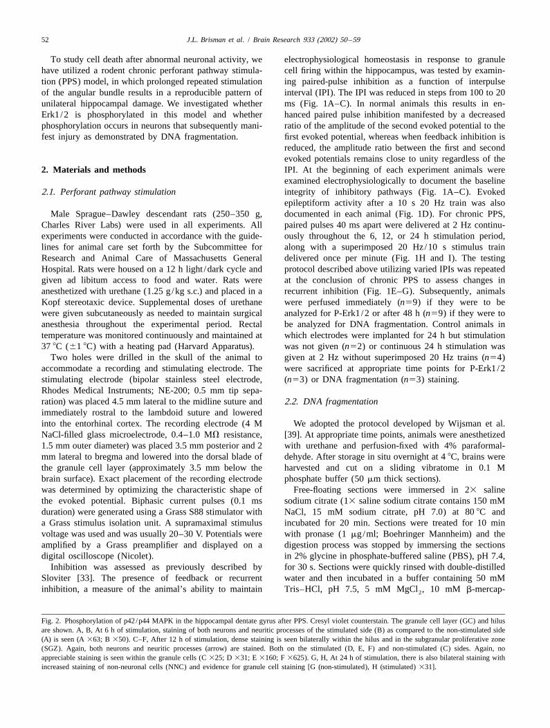
Fig. 2. Phosphorylation of p42/p44 MAPK in the hippocampal dentate gyrus after PPS. Cresyl violet counterstain. The granule cell layer (GC) and hilusare shown. A, B, At 6 h of stimulation, staining of both neurons and neuritic processes of the stimulated side (B) as compared to the non-stimulated side(A) is seen (A 363; B 350). C–F, After 12 h of stimulation, dense staining is seen bilaterally within the hilus and in the subgranular proliferative zone(SGZ). Again, both neurons and neuritic processes (arrow) are stained. Both on the stimulated (D, E, F) and non-stimulated (C) sides. Again, noappreciable staining is seen within the granule cells (C 325; D 331; E 3160; F 3625). G, H, At 24 h of stimulation, there is also bilateral staining withincreased staining of non-neuronal cells (NNC) and evidence for granule cell staining [G (non-stimulated), H (stimulated) 331].
J.L. Brisman et al. / Brain Research 933 (2002) 50 –59 53
54 J.L. Brisman et al. / Brain Research 933 (2002) 50 –59
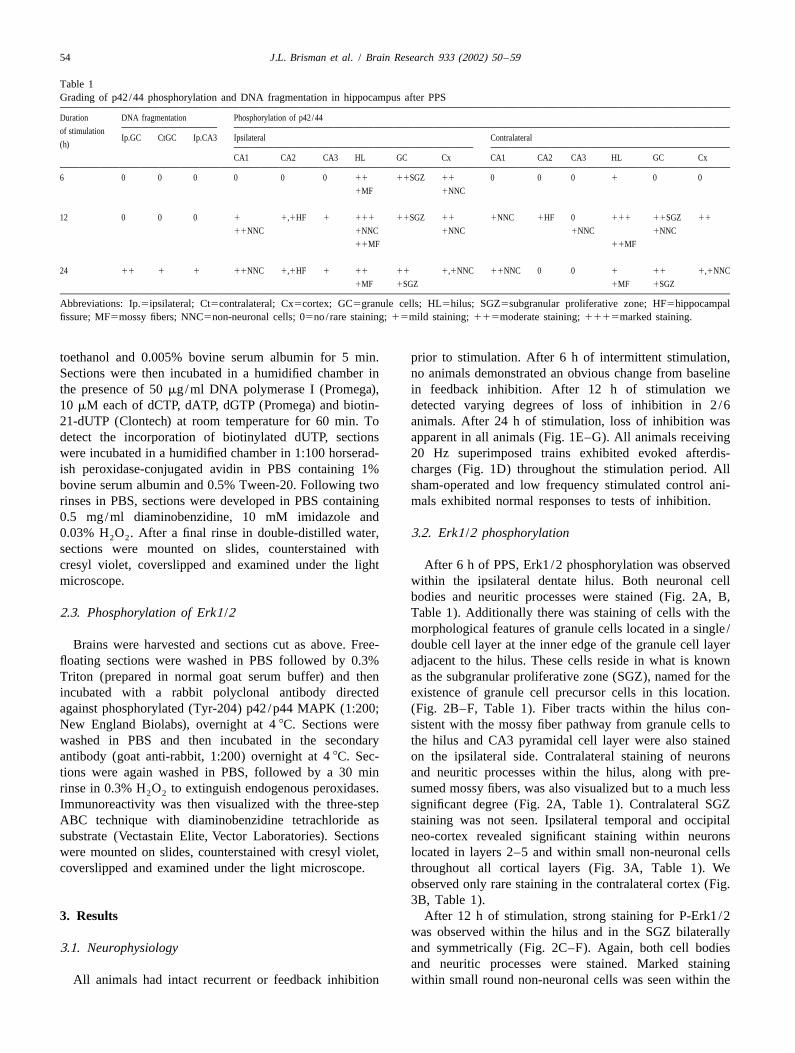
Table 1Grading of p42/44 phosphorylation and DNA fragmentation in hippocampus after PPS
Duration DNA fragmentation Phosphorylation of p42/44
of stimulationIp.GC CtGC Ip.CA3 Ipsilateral Contralateral
(h)
CA1 CA2 CA3 HL GC Cx CA1 CA2 CA3 HL GC Cx
6 0 0 0 0 0 0 11 11SGZ 11 0 0 0 1 0 0
1MF 1NNC
12 0 0 0 1 1,1HF 1 111 11SGZ 11 1NNC 1HF 0 111 11SGZ 11
11NNC 1NNC 1NNC 1NNC 1NNC
11MF 11MF
24 11 1 1 11NNC 1,1HF 1 11 11 1,1NNC 11NNC 0 0 1 11 1,1NNC
1MF 1SGZ 1MF 1SGZ
Abbreviations: Ip.5ipsilateral; Ct5contralateral; Cx5cortex; GC5granule cells; HL5hilus; SGZ5subgranular proliferative zone; HF5hippocampalfissure; MF5mossy fibers; NNC5non-neuronal cells; 05no/ rare staining; 15mild staining; 115moderate staining; 1115marked staining.
toethanol and 0.005% bovine serum albumin for 5 min. prior to stimulation. After 6 h of intermittent stimulation,Sections were then incubated in a humidified chamber in no animals demonstrated an obvious change from baselinethe presence of 50 mg/ml DNA polymerase I (Promega), in feedback inhibition. After 12 h of stimulation we10 mM each of dCTP, dATP, dGTP (Promega) and biotin- detected varying degrees of loss of inhibition in 2/621-dUTP (Clontech) at room temperature for 60 min. To animals. After 24 h of stimulation, loss of inhibition wasdetect the incorporation of biotinylated dUTP, sections apparent in all animals (Fig. 1E–G). All animals receivingwere incubated in a humidified chamber in 1:100 horserad- 20 Hz superimposed trains exhibited evoked afterdis-ish peroxidase-conjugated avidin in PBS containing 1% charges (Fig. 1D) throughout the stimulation period. Allbovine serum albumin and 0.5% Tween-20. Following two sham-operated and low frequency stimulated control ani-rinses in PBS, sections were developed in PBS containing mals exhibited normal responses to tests of inhibition.0.5 mg/ml diaminobenzidine, 10 mM imidazole and0.03% H O . After a final rinse in double-distilled water, 3.2. Erk1/2 phosphorylation2 2
sections were mounted on slides, counterstained withcresyl violet, coverslipped and examined under the light After 6 h of PPS, Erk1/2 phosphorylation was observedmicroscope. within the ipsilateral dentate hilus. Both neuronal cell
bodies and neuritic processes were stained (Fig. 2A, B,2.3. Phosphorylation of Erk1/2 Table 1). Additionally there was staining of cells with the
morphological features of granule cells located in a single /Brains were harvested and sections cut as above. Free- double cell layer at the inner edge of the granule cell layer
floating sections were washed in PBS followed by 0.3% adjacent to the hilus. These cells reside in what is knownTriton (prepared in normal goat serum buffer) and then as the subgranular proliferative zone (SGZ), named for theincubated with a rabbit polyclonal antibody directed existence of granule cell precursor cells in this location.against phosphorylated (Tyr-204) p42/p44 MAPK (1:200; (Fig. 2B–F, Table 1). Fiber tracts within the hilus con-New England Biolabs), overnight at 4 8C. Sections were sistent with the mossy fiber pathway from granule cells towashed in PBS and then incubated in the secondary the hilus and CA3 pyramidal cell layer were also stainedantibody (goat anti-rabbit, 1:200) overnight at 4 8C. Sec- on the ipsilateral side. Contralateral staining of neuronstions were again washed in PBS, followed by a 30 min and neuritic processes within the hilus, along with pre-rinse in 0.3% H O to extinguish endogenous peroxidases. sumed mossy fibers, was also visualized but to a much less2 2
Immunoreactivity was then visualized with the three-step significant degree (Fig. 2A, Table 1). Contralateral SGZABC technique with diaminobenzidine tetrachloride as staining was not seen. Ipsilateral temporal and occipitalsubstrate (Vectastain Elite, Vector Laboratories). Sections neo-cortex revealed significant staining within neuronswere mounted on slides, counterstained with cresyl violet, located in layers 2–5 and within small non-neuronal cellscoverslipped and examined under the light microscope. throughout all cortical layers (Fig. 3A, Table 1). We
observed only rare staining in the contralateral cortex (Fig.3B, Table 1).
3. Results After 12 h of stimulation, strong staining for P-Erk1/2was observed within the hilus and in the SGZ bilaterally
3.1. Neurophysiology and symmetrically (Fig. 2C–F). Again, both cell bodiesand neuritic processes were stained. Marked staining
All animals had intact recurrent or feedback inhibition within small round non-neuronal cells was seen within the
J.L. Brisman et al. / Brain Research 933 (2002) 50 –59 55
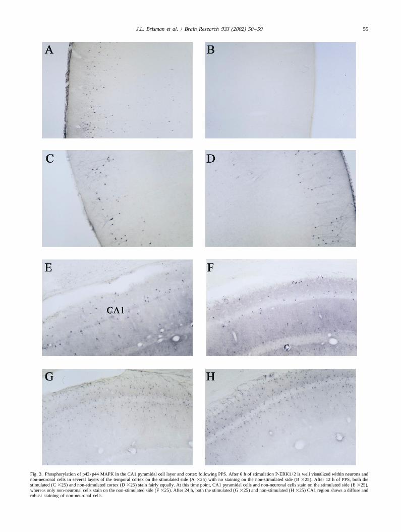
Fig. 3. Phosphorylation of p42/p44 MAPK in the CA1 pyramidal cell layer and cortex following PPS. After 6 h of stimulation P-ERK1/2 is well visualized within neurons andnon-neuronal cells in several layers of the temporal cortex on the stimulated side (A 325) with no staining on the non-stimulated side (B 325). After 12 h of PPS, both thestimulated (C 325) and non-stimulated cortex (D 325) stain fairly equally. At this time point, CA1 pyramidal cells and non-neuronal cells stain on the stimulated side (E 325),whereas only non-neuronal cells stain on the non-stimulated side (F 325). After 24 h, both the stimulated (G 325) and non-stimulated (H 325) CA1 region shows a diffuse androbust staining of non-neuronal cells.
56 J.L. Brisman et al. / Brain Research 933 (2002) 50 –59
hippocampal fissure, granule cell molecular layer and CA1 the extent of DNA fragmentation across multiple coronalmolecular layer (stratum lacunosum). These cells, which sections was noted. Staining on the contralateral side alsomay represent glia, were seen bilaterally (Fig. 3E–F, Table varied considerably from one animal to the next, but was1). Additionally there was faint staining of pyramidal cells always present to some degree and always clearly lessin the CA1, CA2 and CA3 pyramidal cell layer on the marked than on the stimulated side. Staining was uniform-stimulated side only (Fig. 3E–F, Table 1). Mossy fibers, ly nuclear. CA3 cells also revealed staining on thefibers of the CA1 molecular layer (stratum lacunosum) and stimulated side, mostly in the region of CA3 closest to thegranule cell molecular layer as well as temporal and hilus. Staining on the contralateral side in the CA3 regionoccipital neurons was also stained bilaterally (Fig. 3C–F, was not seen (Fig. 4B, Table 1). Staining was not evidentTable 1). in the cortex at any time point. Controls showed no DNA
After 24 h of PPS, P-Erk1/2 was visualized most fragmentation in the hippocampus.strongly in the hilus, granule cell layer, SGZ and innon-neuronal cell populations of the CA1 region on thestimulated side (Fig. 2G, H). To a lesser degree, pyramidal 4. Discussioncells of CA2 and CA3 region were also stained. Stainingwas more marked in the ventral blade of the dentate gyrus The main finding of this study is that after PPS, Erk1/2than in the dorsal blade. Again, both cell bodies and phosphorylation and DNA fragmentation appear in theneuronal processes were stained. Additional staining was hippocampus in incompletely overlapping neuronal popu-also seen in non-neuronal cells of the hippocampal fissure, lations. Within 6 h of beginning repetitive PPS, P-Erk1/2molecular layer (Fig. 3G, H) and occipito–temporal cortex is noted within mossy fibers, hilar neurons, corticalon the stimulated side. Occasional stained neurons in neurons, and neurons in the SGZ. By 12 h, pyramidallayers 3–5 of the occipito–temporal cortex were also seen staining is noted as well and cortical staining peaks. After(Table 1). 24 h, both ipsilateral and to a lesser extent, contralateral
At the 24-h time point, contralateral P-Erk1/2 staining staining is seen in broad regions of the pyramidal layer aswas seen in homologous regions with the exception of the well as in what appear to be glial cells. By contrast, DNApyramidal cell layer, which was stained only ipsilaterally fragmentation, which appears after 24–48 h is seen mainly(Table 1). in ipsilateral granule cells and occasional pyramidal cells,
Control animals, stimulated for 24 h at 2 Hz without the mostly in CA3, and in contralateral granule cells to a lessersuperimposed 20 Hz trains or sham-operated with elec- extent. Because DNA fragmentation is more restricted thantrodes placed and no stimulation given, showed only rare P-Erk1/2 expression, we conclude that activation of Erk1/stained cells along the electrode tract. 2 is not sufficient to commit neurons to early cell death
associated with DNA fragmentation.3.3. DNA fragmentation It is possible that additional P-Erk1/2 expression and/or
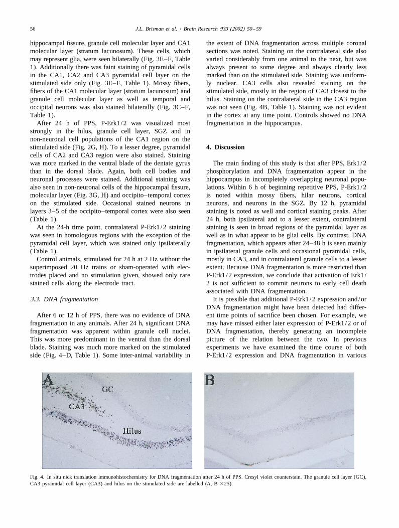
DNA fragmentation might have been detected had differ-After 6 or 12 h of PPS, there was no evidence of DNA ent time points of sacrifice been chosen. For example, we
fragmentation in any animals. After 24 h, significant DNA may have missed either later expression of P-Erk1/2 or offragmentation was apparent within granule cell nuclei. DNA fragmentation, thereby generating an incompleteThis was more predominant in the ventral than the dorsal picture of the relation between the two. In previousblade. Staining was much more marked on the stimulated experiments we have examined the time course of bothside (Fig. 4–D, Table 1). Some inter-animal variability in P-Erk1/2 expression and DNA fragmentation in various
Fig. 4. In situ nick translation immunohistochemistry for DNA fragmentation after 24 h of PPS. Cresyl violet counterstain. The granule cell layer (GC),CA3 pyramidal cell layer (CA3) and hilus on the stimulated side are labelled (A, B 325).
J.L. Brisman et al. / Brain Research 933 (2002) 50 –59 57
seizure models, including PPS. Based on these studies we In human surgical or autopsy specimens from patientsselected time points for analysis that we had previously with temporal lobe epilepsy and/or mesial temporalfound to yield maximal signals and that we thought most sclerosis, severe damage to CA1 and CA3 pyramidal cells,likely to reveal a relationship. It is also possible that our and dentate hilar interneurons with relative sparing of theassays were not sensitive enough to detect low but granule cell layer and the CA2 pyramidal cells is con-biologically important levels of either DNA fragmentation sistently appreciated. Chronic (24 h) PPS results in aor phosphorylation of Erk1/2. similar pattern of injury initially described by Olney and
Anatomic studies have shown that Erk1/2, in its inac- co-workers in 1983 using cresyl violet staining andtive form, is located throughout the adult rat brain, electron microscopy [24,33]. Cell loss is found mainly inparticularly in the superficial layers of the neocortex, the hilar and CA3 regions and to a less severe degree instriatum, cerebellum, and hippocampus [3,11]. In the the CA1 region. We used DNA fragmentation as an assayhippocampus, the inactive form of Erk1/2 exists in cell for neuronal injury. Our finding that DNA fragmentation isbodies and dendrites of dentate granule cells, CA3 pyrami- apparent in only a subset of neurons that have beendal cells, and to a lesser extent CA1 pyramidal cells, and in classically described as suffering injury after PPS supportsthe stratum lucidum (or the combination of mossy fibers the notion that in this system DNA fragmentation stainsand CA3 dendrites) [3,11]. Ours is the first report of an only for certain types of cell death, however, whetherincrease in the signal of Erk1/2 phosphorylation after DNA fragmentation is specific for apoptotic cell death isdirect brain electrical stimulation, i.e., PPS. Moreover, we controversial [30,31]. Our finding of marked granule cellhave documented the spatial and temporal distribution of DNA fragmentation on the stimulated side correspondsP-Erk1/2 after synaptic stimulation. Surprisingly, we well with electron microscopic evidence for apoptosisfound strong bilateral staining for P-Erk1/2 in the hilus restricted to the granule cells in the PPS model [36], andand SGZ, regions not traditionally thought to have an supports the notion that DNA fragmentation is a usefulabundance of endogenous Erk1/2. Interestingly, Parent et marker of apoptosis in this system. Granule cells may beal. recently showed that seizure activity secondary to predisposed to apoptotic cell death as demonstrated inpilocarpine or PPS led to new cell proliferation in the SGZ. previous studies after adrenalectomy or traumatic brainThese cells mature into granule cells in both the normal injury in rodents [7,17,35].location in the dentate gyrus as well as in ectopic locations DNA fragmentation has been examined in rat brain afterwith aberrant axonal projections [27]. It is possible that seizure activity generated by intra-amygdala or systemicErk1/2 activation is involved in this proliferative response. administration of kainic acid, systemic pilocarpine, andWe also found bilateral staining for P-Erk1/2 in granule maximal electroconvulsive treatment (MECS)cells. It seems likely that contralateral up-regulation of [10,12,29,38]. In contrast to our finding after PPS, kainateP-Erk1/2 is simply a response to comissural projections and MECS fail to induce DNA fragmentation in granulefrom the stimulated side. The relationship of this finding to cells [12,29,38]. It seems likely that after PPS, granule cellthe clinical phenomenon of secondary epileptogenesis injury is the result of excessive synaptic input. As such, the(development of a mirror focus) highlights the potential PPS model may parallel the clinical phenomenon ofimportance of this observation. neocortical epilepsy in which the hippocampus is only
Erk1/2 appears to transduce extracellular events into secondarily involved. This situation, where repeatedintracellular signals by triggering a cascade of phosphoryl- neocortical seizures are associated with distant hippocam-ation. MAPKs in general can be activated or up-regulated pal injury, has been described as dual pathology in humanby neurotrophic factors [8,16,21,28,32], neurotransmitters epilepsy [34].[19,41] ischemia [5] and seizures [14,15]. Previous studies The role of Erk1/2 activation in cell death is uncertain.showed a transient and rapid rise in phosphorylation of Some studies suggest that the phosphorylation of Erk1/2Erk1/2 after ECT [1,37], bicucilline-induced generalized actually plays a role in inhibiting apoptosis and neuronalseizures [15], pilocarpine [13] and kainic acid [18,22]. injury [40,42,43]. Other studies, however, suggest thatErk1/2 is also rapidly phosphorylated in a cell-culture phosphorylation of Erk1/2 is part of a biochemical cascademodel of seizure-like activity [23]. At the cellular level, resulting in neuronal injury [6,22,23]. Because we sawimmunostaining has been found in cytoplasm, nuclei and Erk1/2 phosphorylation in many cell groups after PPS thatneuritic processes [3,11]. Because Erk 1/2 is activated did not reveal DNA fragmentation, we conclude that Erk1/after numerous excitatory stimuli, is found in dendrites as 2 phosphorylation is not a sufficient intracellular signal towell as cell bodies, and has been linked to transcription of lead to DNA fragmentation at the times that we surveyed.early genes [41], it appears likely that this kinase cascade It remains possible, however, that Erk1/2 phosphorylationis important in mediating long-term effects of brief stimuli, is a necessary step in the pathway to injury in a subset ofor neuronal plasticity [9]. Moreover, the localization of cell death pathways. In the PPS model, 6 h of stimulationactivated MAPK to dendritic microtubules, which harbor results in neither identifiable hippocampal injury norMAP-2, responsible for microtubule assembly in vitro, detectable loss of physiological inhibition. Because thesuggests a role in dendritic remodeling [11,37]. Erk1/2 cascade here identified is turned on after only 6 h
58 J.L. Brisman et al. / Brain Research 933 (2002) 50 –59
[10] R.K. Filipkowski, M. Hetman, B. Kaminska, L. Kaczmarek, DNAof PPS, prior to irrevocable damage, it is possible thatfragmentation in rat brain after intraperitoneal administration ofinhibitors of Erk1/2 activation could be neuroprotective inkainate, Neuroreport 5 (1994) 1538–1540.
some neuronal populations. In fact we have previously [11] R.S. Fiore, V.E. Bayer, S.L. Pelech, J. Posada, J.A. Cooper, J.M.shown that the selective MEK (the enzyme directly Baraban, p42 mitogen-activated protein kinase in brain: prominentupstream of Erk1/2 and responsible for its phosphoryla- localization in neuronal cell bodies and dendrites, Neuroscience 55
(1993) 463–472.tion) inhibitor PD098059 prevents Erk1/2 phosphorylation[12] D.G. Fujikawa, S.S. Shinmei, B. Cai, Lithium-pilocarpine-inducedand cell death in a cell culture model of activity dependent
status epilepticus produces necrotic neurons with internucleosomalinjury [22]. The availability of additional potent MEKDNA fragmentation in adult rats, Eur. J. Neurosci. 11 (1999)
inhibitors such as U0126 and SL327 will permit detailed 1605–1614.examination of the role of Erk1/2 activation in the [13] Y.C. Garrido, E.R. Sanabria, M.G. Funke, E.A. Cavalheiro, M.G.
Naffah-Mazzacoratti, Mitogen-activated protein kinase is increaseddevelopment of injury and loss of inhibition.in the limbic structures of the rat brain during the early stages ofstatus epilepticus, Brain Res. Bull. 47 (1998) 223–229.
[14] P. Gass, A. Eckhardt, H. Schroder, R. Bravo, T. Herdegen, TransientAcknowledgements expression of the mitogen-activated protein kinase phosphatase
MKP-1 (3CH134/ERP1) in the rat brain after limbic epilepsy, BrainRes. Mol. Brain Res. 41 (1996) 74–80.The authors wish to acknowledge the technical expert
[15] P. Gass, M. Kiessling, H. Bading, Regionally selective stimulationassistance of Robert S. Sloviter, Ph.D. and Steven F.of mitogen activated protein (MAP) kinase tyrosine phosphorylation
Ronner, Ph.D. in the perforant pathway stimulation model, after generalized seizures in the rat brain, Neurosci. Lett. 162 (1993)Tessa Hedley-Whyte, M.D. for assistance with pathologic 39–42.
[16] N. Gomez, P. Cohen, Dissection of the protein kinase cascade byanalysis, Tom Beer for histological technical assistance,which nerve growth factor activates MAP kinases, Nature 353and David Feliciano and Ali Mian for their help with(1991) 170–173.figure preparation. This work was supported by a grant
[17] Z. Hu, K. Yuri, H. Ozawa, H. Lu, M. Kawata, The in vivo timefrom the Whitehall Foundation (A.J.C.) and the NINDS course for elimination of adrenalectomy-induced apoptotic profiles(NS036224) (A.J.C.) from the granule cell layer of the rat hippocampus, J. Neurosci. 17
(1997) 3981–3989.[18] Y.S. Kim, K.S. Hong, Y.S. Seong, J.B. Park, S. Kuroda, K. Kishi, K.
Kaibuchi, Y. Takai, Phosphorylation and activation of mitogen-References activated protein kinase by kainic acid-induced seizure in rat
hippocampus, Biochem. Biophys. Res. Commun. 202 (1994) 1163–1168.[1] J.M. Baraban, R.S. Fiore, J.S. Sanghera, H.B. Paddon, S.L. Pelech,
[19] M. Kurino, K. Fukunaga, Y. Ushio, E. Miyamoto, Activation ofIdentification of p42 mitogen-activated protein kinase as a tyrosinemitogen-activated protein kinase in cultured rat hippocampal neu-kinase substrate activated by maximal electroconvulsive shock inrons by stimulation of glutamate receptors, J. Neurochem. 65 (1995)hippocampus, J. Neurochem. 60 (1993) 330–336.1282–1289.[2] J. Bengzon, Z. Kokaia, E. Elmer, A. Nanobashvili, M. Kokaia, O.
[20] J.M. Lindquist, S. Rehnmark, Ambient temperature regulation ofLindvall, Apoptosis and proliferation of dentate gyrus neurons afterapoptosis in brown adipose tissue. Erk1/2 promotes norepinephrine-single and intermittent limbic seizures, Proc. Natl. Acad. Sci. USAdependent cell survival, J. Biol. Chem. 273 (1998) 30147–30156.94 (1997) 10432–10437.
[21] T. Moriguchi, Y. Gotoh, E. Nishida, Activation of two isoforms of[3] R.V. Bhat, T.M. Engber, J.P. Finn, E.J. Koury, P.C. Contreras, M.S.mitogen-activated protein kinase kinase in response to epidermalMiller, C.A. Dionne, K.M. Walton, Region-specific targets of p42/growth factor and nerve growth factor, Eur. J. Biochem. 234 (1995)p44MAPK signaling in rat brain, J. Neurochem. 70 (1998) 558–571.32–38.[4] T.G. Boulton, S.H. Nye, D.J. Robbins, N.Y. Ip, E. Radziejewska,
[22] B. Murray, T. Beer, A. Alessandrini, A.J. Cole, Pre- and post-S.D. Morgenbesser, R.A. DePinho, N. Panayotatos, M.H. Cobb,synaptic activation of p44/42 MAP kinase hippocampus afterG.D. Yancopoulos, ERKs: a family of protein-serine / threoninekainate-induced seizures, (Abstract) Epilepsia 40 (Suppl. 7) (1999)kinases that are activated and tyrosine phosphorylated in response to21.insulin and NGF, Cell 65 (1991) 663–675.
[23] B. Murray, A. Alessandrini, A.J. Cole, A.G. Yee, E.J. Furshpan,[5] R. Campos-Gonzalez, M.S. Kindy, Tyrosine phosphorylation ofInhibition of the p44/42 MAP kinase pathway protects hippocampalmicrotubule-associated protein kinase after transient ischemia in theneurons in a cell-culture model of seizure activity, Proc. Natl. Acad.gerbil brain, J. Neurochem. 59 (1992) 1955–1958.Sci. USA 95 (1998) 11975–11980.[6] B.Y. Chin, M.E. Choi, M.D. Burdick, R.M. Strieter, T.H. Risby,
[24] J.W. Olney, T. deGubareff, R.S. Sloviter, ‘Epileptic’ brain damage inA.M. Choi, Induction of apoptosis by particulate matter: role ofrats induced by sustained electrical stimulation of the perforant path.TNF-alpha and MAPK, Am. J. Physiol. 275 (1998) L942–L949.II. Ultrastructural analysis of acute hippocampal pathology, Brain[7] M.A. Colicos, P.K. Dash, Apoptotic morphology of dentate gyrusRes. Bull. 10 (1983) 699–712.granule cells following experimental cortical impact injury in rats:
[25] H. Ozawa, S. Shioda, K. Dohi, H. Matsumoto, H. Mizushima, C.J.possible role in spatial memory deficits, Brain Res. 739 (1996)Zhou, H. Funahashi, Y. Nakai, S. Nakajo, K. Matsumoto, Delayed120–131.neuronal cell death in the rat hippocampus is mediated by the[8] C. Creuzet, J. Loeb, G. Barbin, Fibroblast growth factors stimulatemitogen-activated protein kinase signal transduction pathway,protein tyrosine phosphorylation and mitogen-activated proteinNeurosci. Lett. 262 (1999) 57–60.kinase activity in primary cultures of hippocampal neurons, J.
Neurochem. 64 (1995) 1541–1547. [26] J.M. Parent, E. Tada, J.R. Fike, D.H. Lowenstein, Inhibition of[9] J.D. English, J.D. Sweatt, A requirement for the mitogen-activated dentate granule cell neurogenesis with brain irradiation does not
protein kinase cascade in hippocampal long term potentiation, J. prevent seizure-induced mossy fiber synaptic reorganization in theBiol. Chem. 272 (1997) 19103–19106. rat, J. Neurosci. 19 (1999) 4508–4519.
J.L. Brisman et al. / Brain Research 933 (2002) 50 –59 59
[27] J.M. Parent, T.W. Yu, R.T. Leibowitz, D.H. Geschwind, R.S. adrenalectomy-induced hippocampal granule cell degeneration in theSloviter, D.H. Lowenstein, Dentate granule cell neurogenesis is rat: apoptosis in the adult central nervous system, J. Comp Neurol.increased by seizures and contributes to aberrant network reorgani- 330 (1993) 337–351.zation in the adult rat hippocampus, J. Neurosci. 17 (1997) 3727– [36] R.S. Sloviter, E. Dean, A.L. Sollas, J.H. Goodman, Apoptosis and3738. necrosis induced in different hippocampal neuron populations by
[28] P. Peinado-Ramon, A. Wallen, F. Hallbook, MAP kinase repetitive perforant path stimulation in the rat, J. Comp Neurol. 366phosphatase-1 mRNA is expressed in embryonic sympathetic neu- (1996) 516–533.rons and is upregulated after NGF stimulation, Brain Res. Mol. [37] K.R. Stratton, P.F. Worley, J.S. Litz, S.J. Parsons, R.L. Huganir,Brain Res. 56 (1998) 256–267. J.M. Baraban, Electroconvulsive treatment induces a rapid and
[29] H. Pollard, M. Khrestchatisky, J. Moreau, Y. Ben Ari, A. Represa, transient increase in tyrosine phosphorylation of a 40-kilodaltonCorrelation between reactive sprouting and microtubule protein protein associated with microtubule-associated protein 2 kinaseexpression in epileptic hippocampus [published erratum appears in activity, J. Neurochem. 56 (1991) 147–152.Neuroscience 1994 Nov;63(2):627], Neuroscience 61 (1994) 773– [38] S. Weiss, O. Cataltepe, A.J. Cole, Anatomical studies of DNA787. fragmentation in rat brain after systemic kainate administration,
[30] C. Portera-Cailliau, D.L. Price, L.J. Martin, Excitotoxic neuronal Neuroscience 74 (1996) 541–551.death in the immature brain is an apoptosis-necrosis morphological [39] J.H. Wijsman, R.R. Jonker, R. Keijzer, C.J. van de Velde, C.J.continuum, J. Comp. Neurol. 378 (1997) 70–87. Cornelisse, J.H. van Dierendonck, A new method to detect apoptosis
[31] C. Portera-Cailliau, D.L. Price, L.J. Martin, Non-NMDA and in paraffin sections: in situ end-labeling of fragmented DNA, J.NMDA receptor-mediated excitotoxic neuronal deaths in adult brain Histochem. Cytochem. 41 (1993) 7–12.are morphologically distinct: further evidence for an apoptosis- [40] Z. Xia, M. Dickens, J. Raingeaud, R.J. Davis, M.E. Greenberg,necrosis continuum, J. Comp Neurol. 378 (1997) 88–104. Opposing effects of ERK and JNK-p38 MAP kinases on apoptosis,
[32] R.A. Segal, M.E. Greenberg, Intracellular signaling pathways acti- Science 270 (1995) 1326–1331.vated by neurotrophic factors, Annu. Rev. Neurosci. 19 (1996) [41] Z. Xia, H. Dudek, C.K. Miranti, M.E. Greenberg, Calcium influx via463–489. the NMDA receptor induces immediate early gene transcription by a
[33] R.S. Sloviter, ‘Epileptic’ brain damage in rats induced by sustained MAP kinase /ERK-dependent mechanism, J. Neurosci. 16 (1996)electrical stimulation of the perforant path. I. Acute electrophysio- 5425–5436.logical and light microscopic studies, Brain Res. Bull. 10 (1983) [42] C.Y.I. Yan, L.A. Greene, Prevention of PC12 cell death by N-675–697. acetylcysteine requires activation of the Ras pathway, J. Neurosci.
[34] R.S. Sloviter, The functional organization of the hippocampal 18 (1998) 4042–4049.dentate gyrus and its relevance to the pathogenesis of temporal lobe [43] S.O. Yoon, P. Casaccia-Bonnefil, B. Carter, M.V. Chao, Competitiveepilepsy, Ann. Neurol. 35 (1994) 640–654. signaling between TrkA and p75 nerve growth factor receptors
[35] R.S. Sloviter, E. Dean, S. Neubort, Electron microscopic analysis of determines cell survival, J. Neurosci. 18 (1998) 3273–3281.