Embed Size (px)
Citation preview
Research with Transcranial Magnetic Stimulation
to Improve Naming in Nonfluent Aphasia
Margaret Naeser, Ph.D.
Neuroimaging in Aphasia and TMS to Treat Aphasia Harold Goodglass Boston University Aphasia Research
Center VA Boston Healthcare System
Department of Neurology Boston University School of Medicine
Department of Veterans AffairsVA Boston Healthcare System
www.bu.edu/naeser/aphasia [email protected] www.tmslab.org
Co-Investigator: Alvaro Pascual-Leone, MD, PhD Director, Berenson-Allen Center for Noninvasive Brain Stimulation Beth Israel Deaconess Medical Center, Harvard Medical School Second Site: H. Branch Coslett, MD and Roy Hamilton, MD Hospital University of Penn., Philadelphia
Other Collaborators: Paula Martin Michael Ho, PhD Ethan Treglia, MA, CCC-SLP Elina Kaplan Karl Doron Jerome Kaplan, MA, CCC-SLP Kristine Lundgren, ScD, CCC-SLP Marjorie Nicholas, PhD, CCC-SLP Jacquie Kurland, PhD, CCC-SLP YunYan Wang
Felipe Fregni, PhD, MD Miguel Alonso, MD Masahito Kobayashi, MD Hugo Theoret, PhD Shahid Bashir, PhD
Antoni Valero, MD Daniel Press, MD Dae-Shik Kim, PhD Errol Baker, PhD
National Institutes of Health NIDCD, and Dept. Veterans Affairs
Four Topics for this Presentation
1. Review current TMS treatment protocol with Nonfluent aphasia patients to improve naming
3. Overt Naming fMRI studies, Pre- and Post- a series of TMS treatments
5. TMS plus Speech Therapy studies
7. DTI studies of two parts of Broca’s area: Pars Triangularis (PTr) vs. Pars Opercularis (POp) connections to Arcuate Fasciculus and posterior language zones.
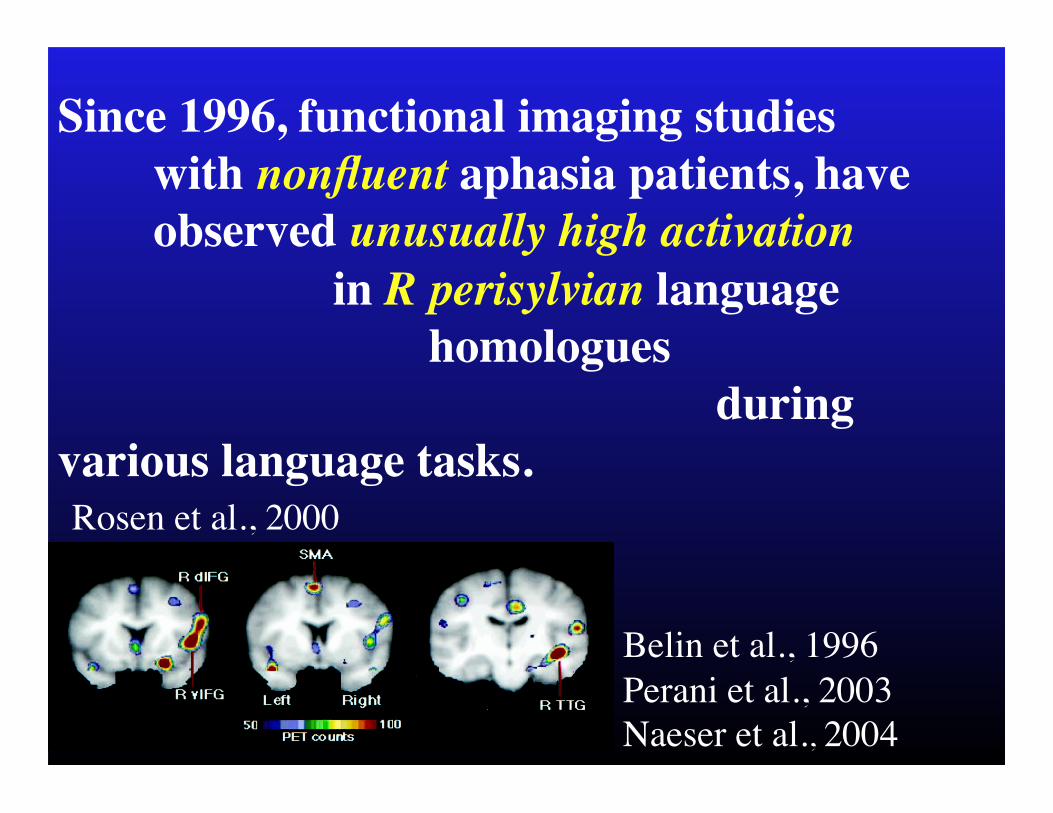
Since 1996, functional imaging studies with nonfluent aphasia patients, have observed unusually high activation in R perisylvian language homologues during
various language tasks.
Belin et al., 1996 Perani et al., 2003 Naeser et al., 2004 Martin et al., 2005
Rosen et al., 2000
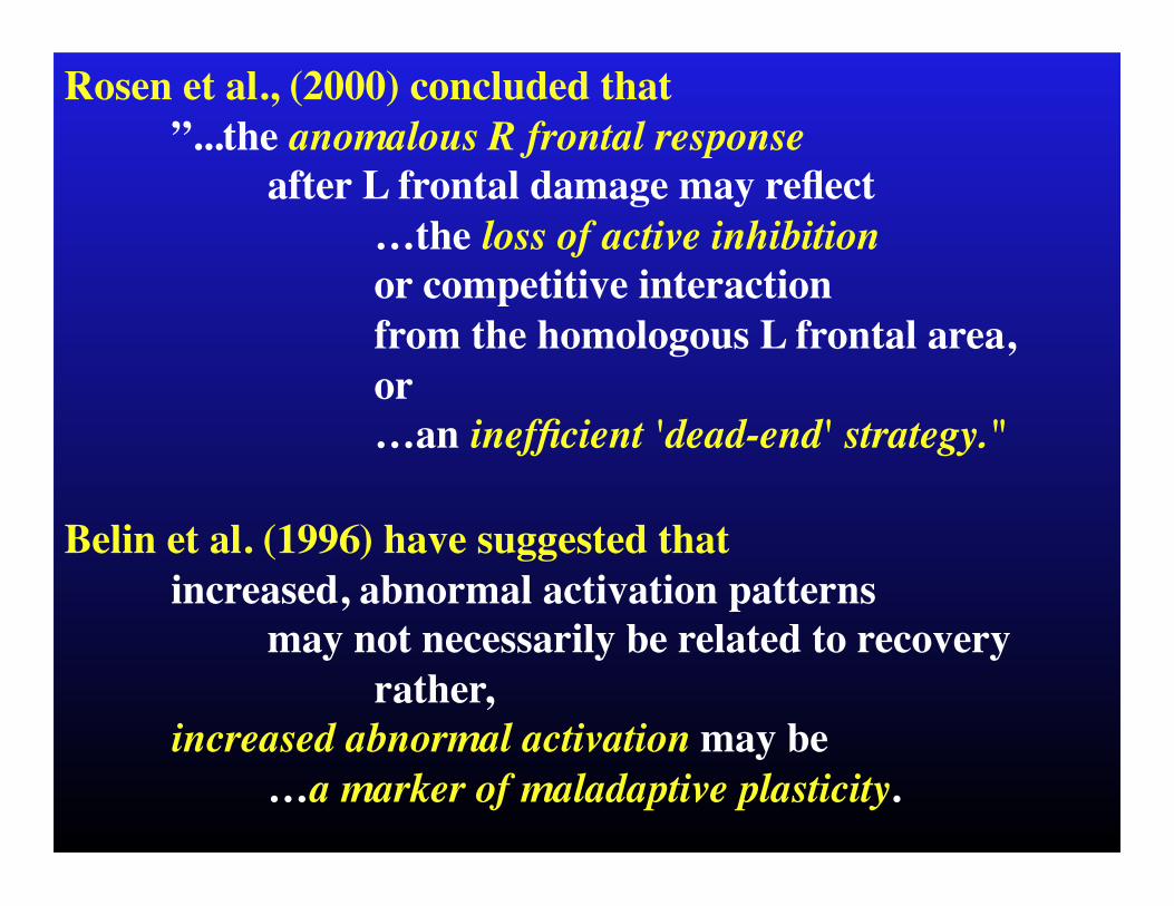
Belin et al. (1996) have suggested that increased, abnormal activation patterns may not necessarily be related to recovery rather, increased abnormal activation may be …a marker of maladaptive plasticity.
Rosen et al., (2000) concluded that ”...the anomalous R frontal response after L frontal damage may reflect …the loss of active inhibition or competitive interaction from the homologous L frontal area, or …an inefficient 'dead-end' strategy."
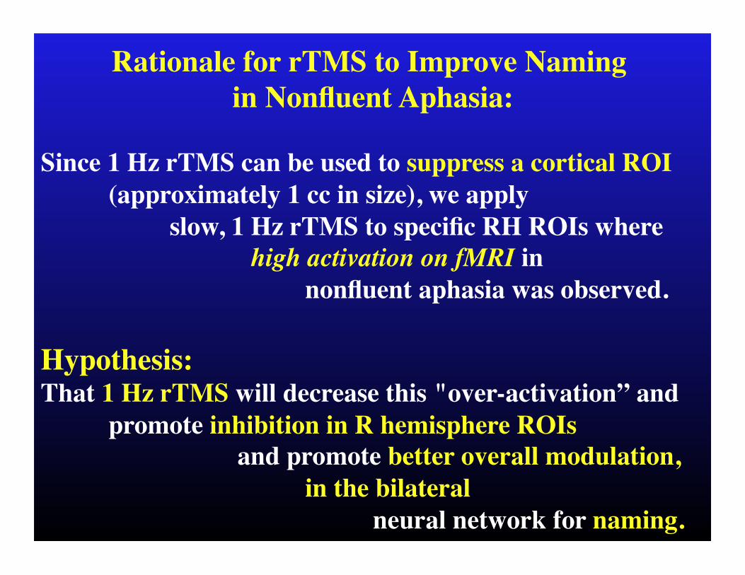
Rationale for rTMS to Improve Naming in Nonfluent Aphasia:
Since 1 Hz rTMS can be used to suppress a cortical ROI (approximately 1 cc in size), we apply slow, 1 Hz rTMS to specific RH ROIs where
high activation on fMRI in nonfluent aphasia was observed.
Hypothesis: That 1 Hz rTMS will decrease this "over-activation” and
promote inhibition in R hemisphere ROIs and promote better overall modulation, in the bilateral neural network for naming.
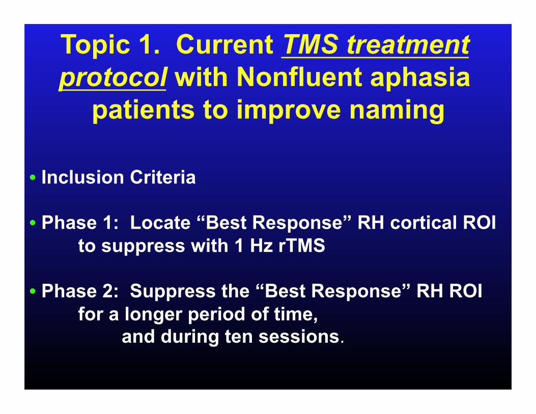
Topic 1. Current TMS treatment protocol with Nonfluent aphasia
patients to improve naming
• Inclusion Criteria
• Phase 1: Locate “Best Response” RH cortical ROI to suppress with 1 Hz rTMS
• Phase 2: Suppress the “Best Response” RH ROI for a longer period of time, and during ten sessions.
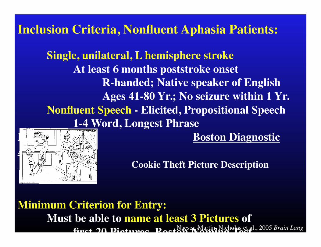
Inclusion Criteria, Nonfluent Aphasia Patients:
Single, unilateral, L hemisphere stroke At least 6 months poststroke onset R-handed; Native speaker of English Ages 41-80 Yr.; No seizure within 1 Yr. Nonfluent Speech - Elicited, Propositional Speech 1-4 Word, Longest Phrase
Length Boston Diagnostic Aphasia Exam,
Cookie Theft Picture Description
Minimum Criterion for Entry: Must be able to name at least 3 Pictures of first 20 Pictures, Boston Naming Test. Naeser, Martin, Nicholas et al., 2005 Brain Lang
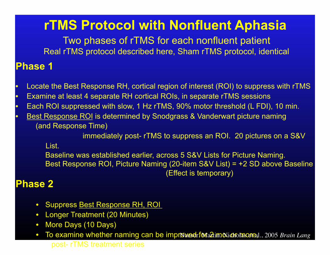
Phase 1
• Locate the Best Response RH, cortical region of interest (ROI) to suppress with rTMS • Examine at least 4 separate RH cortical ROIs, in separate rTMS sessions • Each ROI suppressed with slow, 1 Hz rTMS, 90% motor threshold (L FDI), 10 min. • Best Response ROI is determined by Snodgrass & Vanderwart picture naming
(and Response Time) immediately post- rTMS to suppress an ROI. 20 pictures on a S&V List.
Baseline was established earlier, across 5 S&V Lists for Picture Naming. Best Response ROI, Picture Naming (20-item S&V List) = +2 SD above Baseline
(Effect is temporary) Phase 2
• Suppress Best Response RH, ROI • Longer Treatment (20 Minutes) • More Days (10 Days) • To examine whether naming can be improved for 2 mo. or more,
post- rTMS treatment series
rTMS Protocol with Nonfluent Aphasia Two phases of rTMS for each nonfluent patient
Real rTMS protocol described here, Sham rTMS protocol, identical
Naeser, Martin, Nicholas et al., 2005 Brain Lang
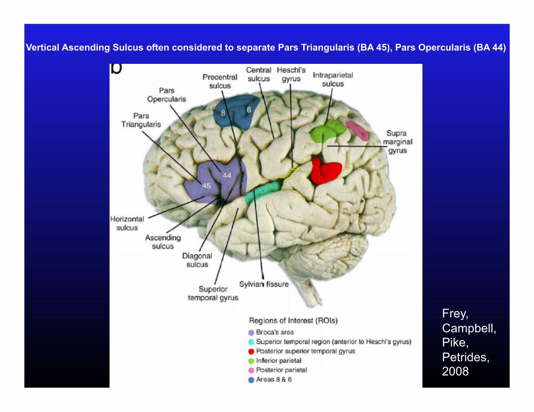
Vertical Ascending Sulcus often considered to separate Pars Triangularis (BA 45), Pars Opercularis (BA 44)
Frey, Campbell, Pike, Petrides, 2008
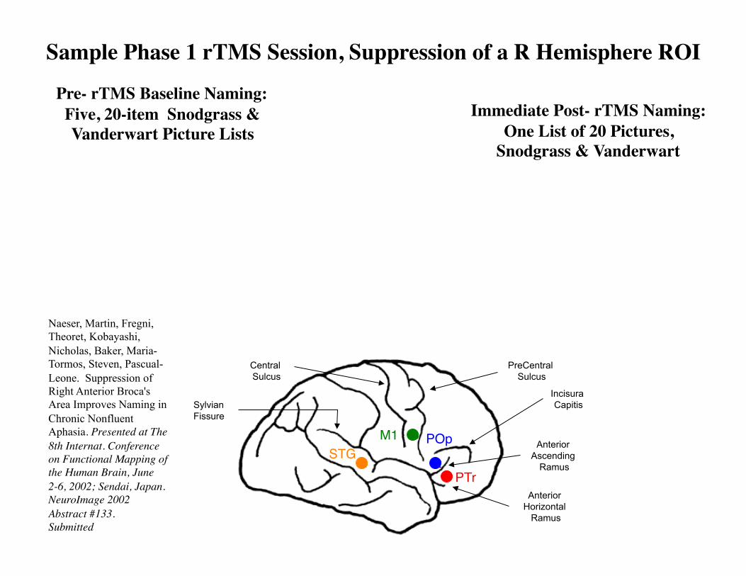
Sample Phase 1 rTMS Session, Suppression of a R Hemisphere ROI
Pre- rTMS Baseline Naming: Five, 20-item Snodgrass & Vanderwart Picture Lists
Immediate Post- rTMS Naming: One List of 20 Pictures,
Snodgrass & Vanderwart
Central Sulcus
Sylvian Fissure
Anterior Ascending
Ramus
Incisura Capitis
Anterior Horizontal
Ramus
PreCentral Sulcus
1 2
3
4 STG
M1 POp
PTr
Naeser, Martin, Fregni, Theoret, Kobayashi, Nicholas, Baker, Maria-Tormos, Steven, Pascual-Leone. Suppression of Right Anterior Broca's Area Improves Naming in Chronic Nonfluent Aphasia. Presented at The 8th Internat. Conference on Functional Mapping of the Human Brain, June 2-6, 2002; Sendai, Japan. NeuroImage 2002 Abstract #133. Submitted
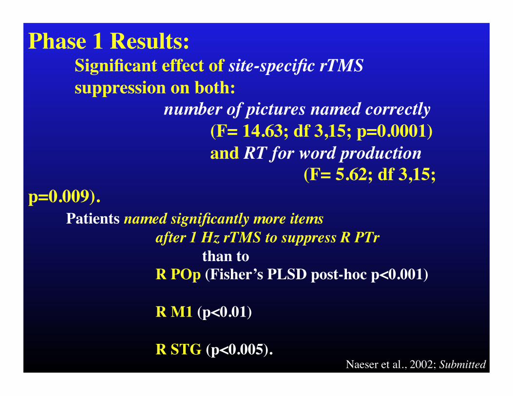
Phase 1 Results: Significant effect of site-specific rTMS suppression on both: number of pictures named correctly (F= 14.63; df 3,15; p=0.0001) and RT for word production (F= 5.62; df 3,15;
p=0.009). Patients named significantly more items
after 1 Hz rTMS to suppress R PTr than to R POp (Fisher’s PLSD post-hoc p<0.001) R M1 (p<0.01) R STG (p<0.005).
Naeser et al., 2002; Submitted
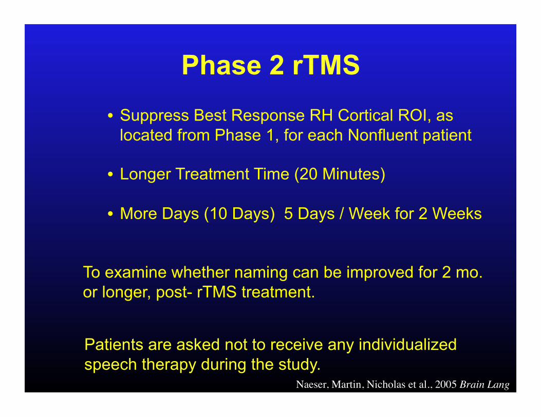
Phase 2 rTMS • Suppress Best Response RH Cortical ROI, as
located from Phase 1, for each Nonfluent patient
• Longer Treatment Time (20 Minutes)
• More Days (10 Days) 5 Days / Week for 2 Weeks
To examine whether naming can be improved for 2 mo. or longer, post- rTMS treatment.
Patients are asked not to receive any individualized speech therapy during the study.
Naeser, Martin, Nicholas et al., 2005 Brain Lang
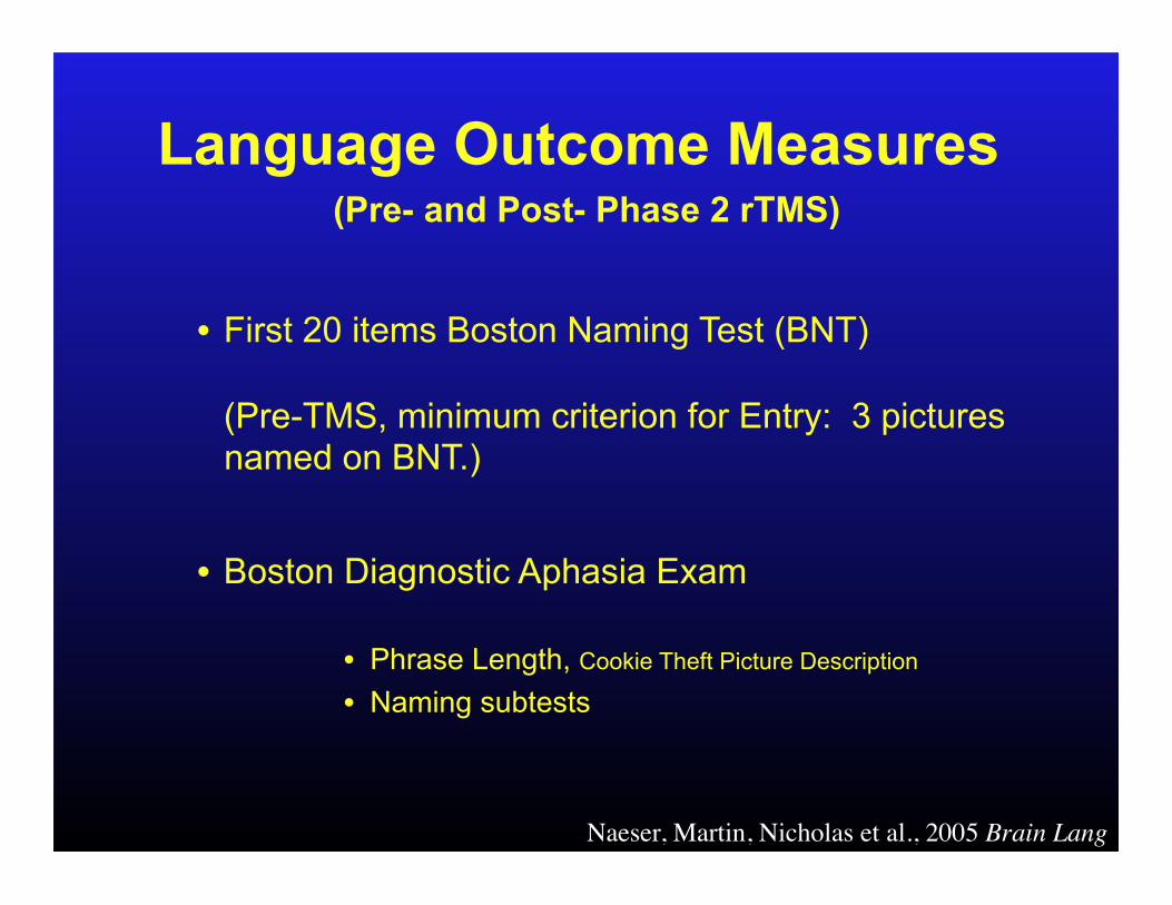
• First 20 items Boston Naming Test (BNT)
(Pre-TMS, minimum criterion for Entry: 3 pictures named on BNT.)
• Boston Diagnostic Aphasia Exam
• Phrase Length, Cookie Theft Picture Description • Naming subtests
Language Outcome Measures (Pre- and Post- Phase 2 rTMS)
Naeser, Martin, Nicholas et al., 2005 Brain Lang
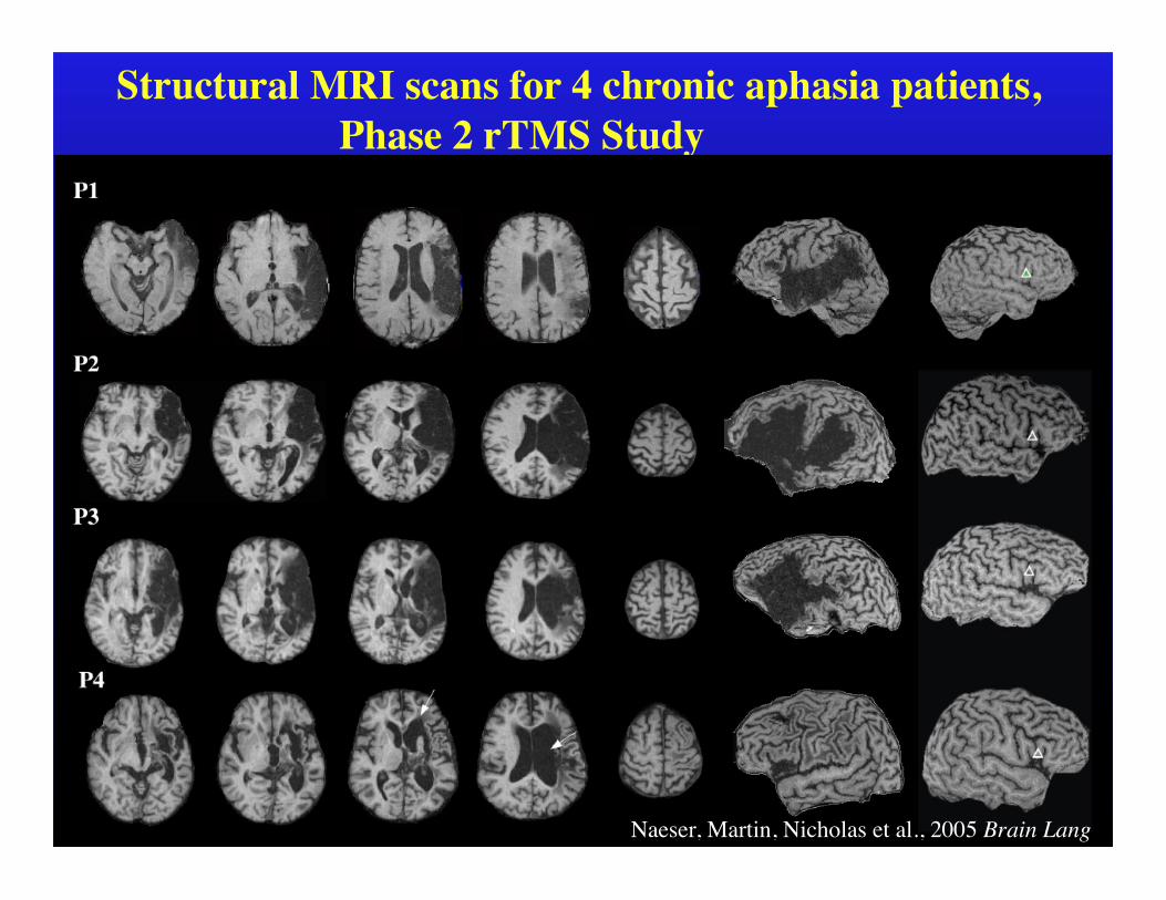
Structural MRI scans for 4 chronic aphasia patients, Phase 2 rTMS Study
Naeser, Martin, Nicholas et al., 2005 Brain Lang
Boston Naming Test(First 20 Pictures)
02468
101214161820
Pre-rTMS
2Wk Post- rTMS
2Mo Post- rTMS
8Mo Post- rTMS
Time of Testing
Num
ber
of P
ictu
res
Cor
rect
ly N
amed
P1P2P3P4
BDAE Naming SubtestAnimals
(Max=12)
02468
1012
Pre-rTMS
2Wk Post-rTMS
2Mo Post-rTMS
8Mo Post-
rTMSTime of Testing
Num
ber
of P
ictu
res
Cor
rect
ly N
amed
BDAE Naming SubtestTools/Implements
(Max=12)
02468
1012
Pre-rTMS
2Wk Post-rTMS
2Mo Post-rTMS
8Mo Post-rTMS
Time of Testing
Num
ber
of P
ictu
res
Cor
rect
ly N
amed
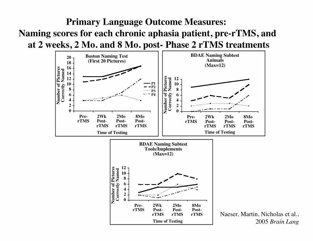
Primary Language Outcome Measures: Naming scores for each chronic aphasia patient, pre-rTMS, and
at 2 weeks, 2 Mo. and 8 Mo. post- Phase 2 rTMS treatments
Naeser, Martin, Nicholas et al., 2005 Brain Lang
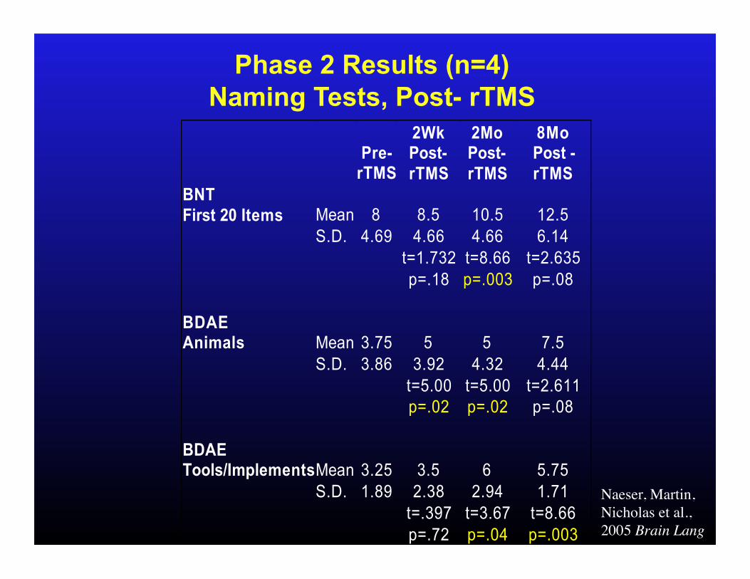
Phase 2 Results (n=4) Naming Tests, Post- rTMS
Pre-
rTMS
2Wk Post- rTMS
2Mo Post- rTMS
8Mo Post -rTMS
BNT First 20 Items Mean 8 8.5 10.5 12.5 S.D. 4.69 4.66 4.66 6.14 t=1.732 t=8.66 t=2.635 p=.18 p=.003 p=.08 BDAE Animals Mean 3.75 5 5 7.5 S.D. 3.86 3.92 4.32 4.44 t=5.00 t=5.00 t=2.611 p=.02 p=.02 p=.08 BDAE Tools/Implements Mean 3.25 3.5 6 5.75 S.D. 1.89 2.38 2.94 1.71 t=.397 t=3.67 t=8.66 p=.72 p=.04 p=.003
Naeser, Martin, Nicholas et al., 2005 Brain Lang
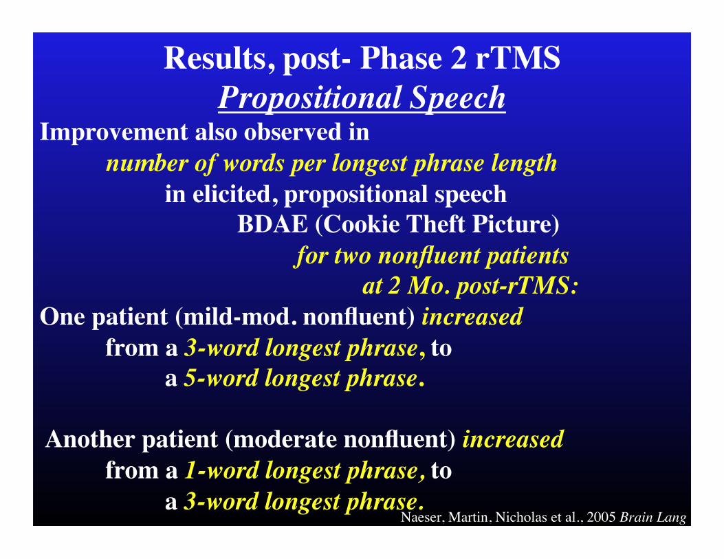
Results, post- Phase 2 rTMS Propositional Speech
Improvement also observed in number of words per longest phrase length in elicited, propositional speech BDAE (Cookie Theft Picture) for two nonfluent patients at 2 Mo. post-rTMS:
One patient (mild-mod. nonfluent) increased from a 3-word longest phrase, to a 5-word longest phrase.
Another patient (moderate nonfluent) increased from a 1-word longest phrase, to a 3-word longest phrase.
Naeser, Martin, Nicholas et al., 2005 Brain Lang
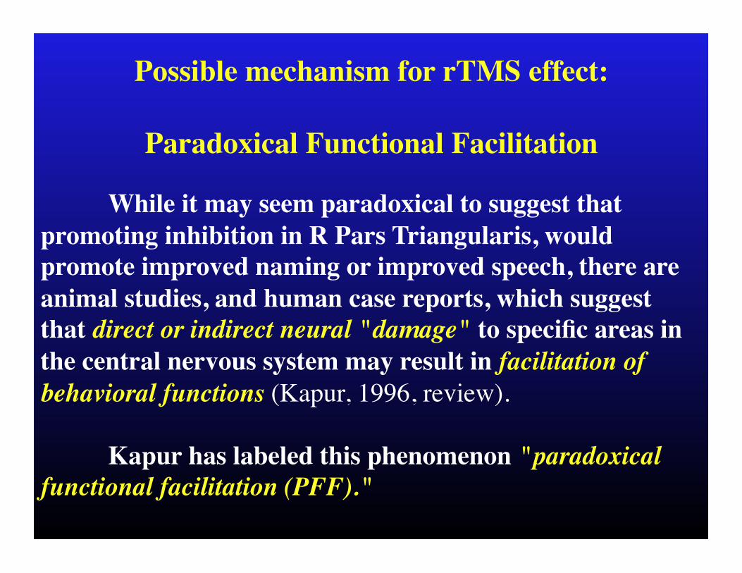
Possible mechanism for rTMS effect:
Paradoxical Functional Facilitation
While it may seem paradoxical to suggest that promoting inhibition in R Pars Triangularis, would promote improved naming or improved speech, there are animal studies, and human case reports, which suggest that direct or indirect neural "damage" to specific areas in the central nervous system may result in facilitation of behavioral functions (Kapur, 1996, review).
Kapur has labeled this phenomenon "paradoxical functional facilitation (PFF)."
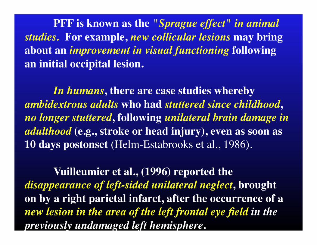
PFF is known as the "Sprague effect" in animal studies. For example, new collicular lesions may bring about an improvement in visual functioning following an initial occipital lesion.
In humans, there are case studies whereby ambidextrous adults who had stuttered since childhood, no longer stuttered, following unilateral brain damage in adulthood (e.g., stroke or head injury), even as soon as 10 days postonset (Helm-Estabrooks et al., 1986).
Vuilleumier et al., (1996) reported the disappearance of left-sided unilateral neglect, brought on by a right parietal infarct, after the occurrence of a new lesion in the area of the left frontal eye field in the previously undamaged left hemisphere.
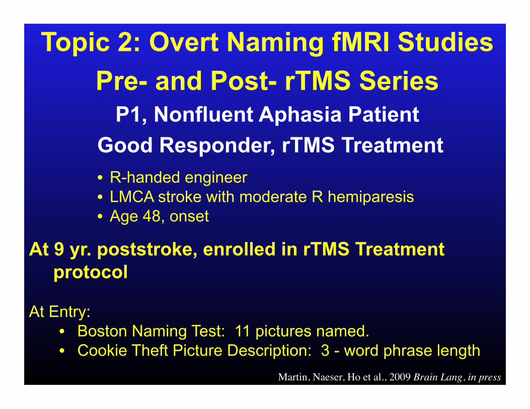
• R-handed engineer • LMCA stroke with moderate R hemiparesis • Age 48, onset
At 9 yr. poststroke, enrolled in rTMS Treatment protocol
At Entry: • Boston Naming Test: 11 pictures named. • Cookie Theft Picture Description: 3 - word phrase length
Topic 2: Overt Naming fMRI Studies Pre- and Post- rTMS Series
P1, Nonfluent Aphasia Patient Good Responder, rTMS Treatment
Martin, Naeser, Ho et al., 2009 Brain Lang, in press
Boston Naming Test
First 20 Items
1112
14
1817
15 15
0
5
10
15
20
Pre-
rTMS
2Wk.
Post-
rTMS
2Mo.
Post-
rTMS
6Mo.
Post-
rTMS
8Mo.
Post-
rTMS
1Yr. 4Mo.
Post-
rTMS
3Yr.
7Mo.
Post-
rTMSTime of Testing
Nu
mb
er
of
Pic
ture
s
Co
rre
ctl
y N
am
ed
Ma
x =
20
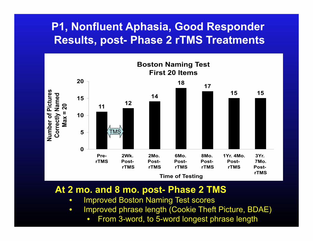
At 2 mo. and 8 mo. post- Phase 2 TMS • Improved Boston Naming Test scores • Improved phrase length (Cookie Theft Picture, BDAE)
• From 3-word, to 5-word longest phrase length
TMS
P1, Nonfluent Aphasia, Good Responder Results, post- Phase 2 rTMS Treatments
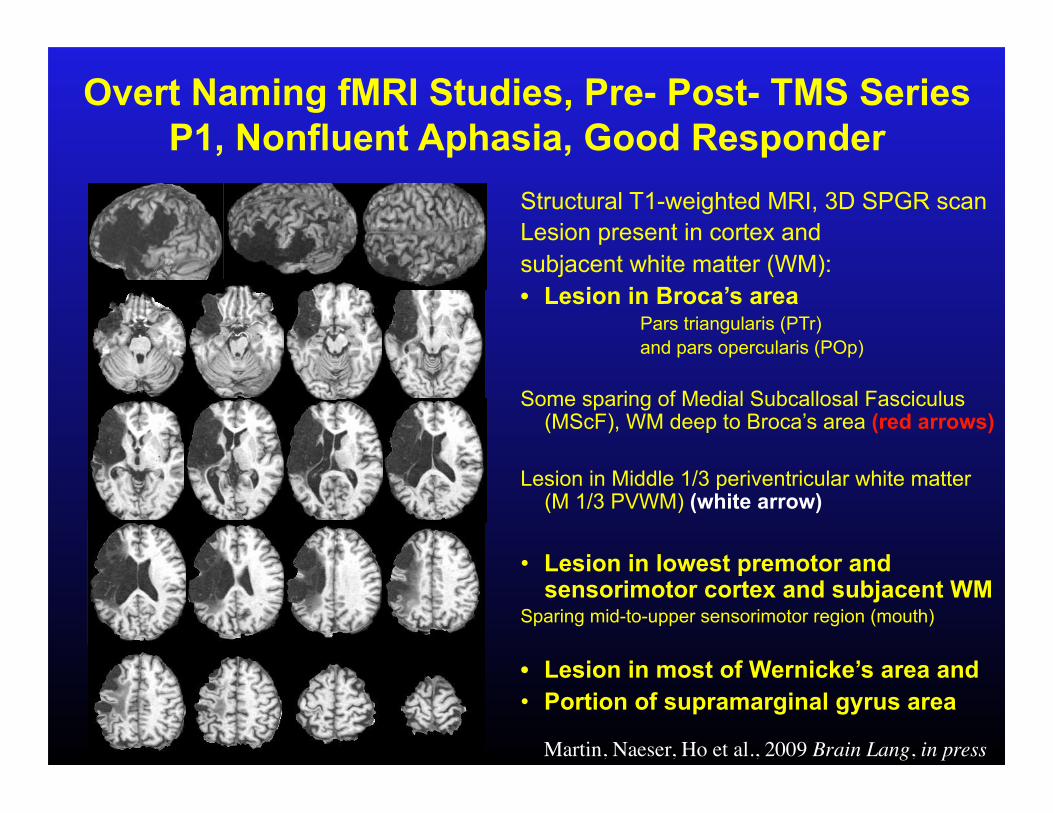
Structural T1-weighted MRI, 3D SPGR scan Lesion present in cortex and subjacent white matter (WM): • Lesion in Broca’s area
Pars triangularis (PTr) and pars opercularis (POp)
Some sparing of Medial Subcallosal Fasciculus (MScF), WM deep to Broca’s area (red arrows)
Lesion in Middle 1/3 periventricular white matter (M 1/3 PVWM) (white arrow)
• Lesion in lowest premotor and sensorimotor cortex and subjacent WM
Sparing mid-to-upper sensorimotor region (mouth)
• Lesion in most of Wernicke’s area and • Portion of supramarginal gyrus area
Overt Naming fMRI Studies, Pre- Post- TMS Series P1, Nonfluent Aphasia, Good Responder
Martin, Naeser, Ho et al., 2009 Brain Lang, in press
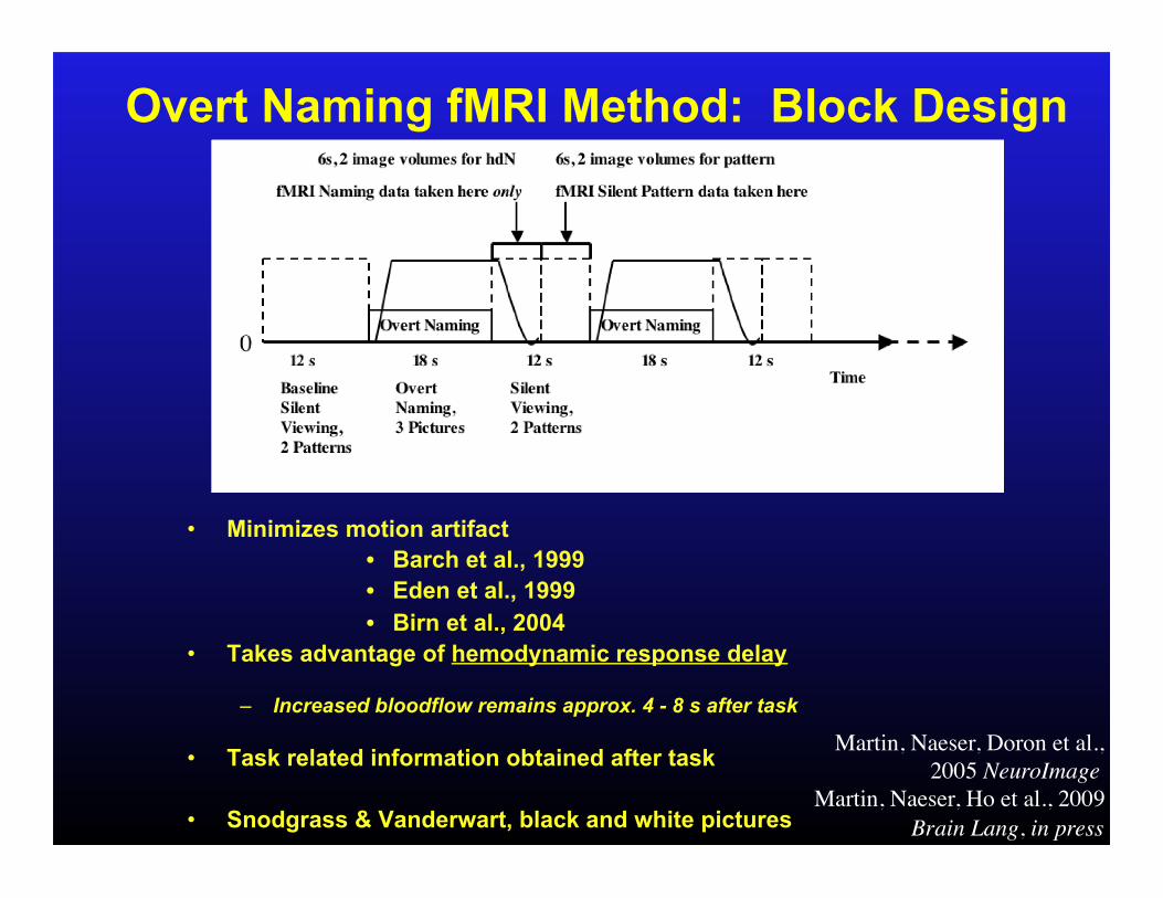
• Minimizes motion artifact • Barch et al., 1999 • Eden et al., 1999 • Birn et al., 2004
• Takes advantage of hemodynamic response delay
– Increased bloodflow remains approx. 4 - 8 s after task
• Task related information obtained after task
• Snodgrass & Vanderwart, black and white pictures
Overt Naming fMRI Method: Block Design
Martin, Naeser, Doron et al., 2005 NeuroImage
Martin, Naeser, Ho et al., 2009 Brain Lang, in press
0
0.5
1
1.5
2
2.5
3
Pre- TMS 3 Mo.
Post- TMS
16 Mo.
Post- TMS
46 Mo.
Post- TMS
Mean
Effect S
ize
L SensMot
R SensMot
P1, Nonfluent Aphasia, Good Responder Overt Naming fMRIs Pre- and Post-TMS Series
L
R
L
R L
R
L
Pre-TMS 17/60 28%
Pictures Named 9 Yr. Poststroke
3 Mo. Post-TMS 25/60 42%
Pictures Named 10;4 Yr. Poststroke
16 Mo. Post-TMS 35/60 58%
Pictures Named 11;5 Yr. Poststroke
46 Mo. Post-TMS 25/60 42%
Pictures Named 13;11 Yr. Poststroke
0
0.5
1
1.5
2
2.5
3
Pre- TMS 3 Mo.
Post- TMS
16 Mo.
Post- TMS
46 Mo.
Post- TMS
Mean
Effect S
ize
L SMA
R SMA
TMS
* *
* = p <.05
TMS
Martin, Naeser, Ho et al., 2009 Brain Lang, in press
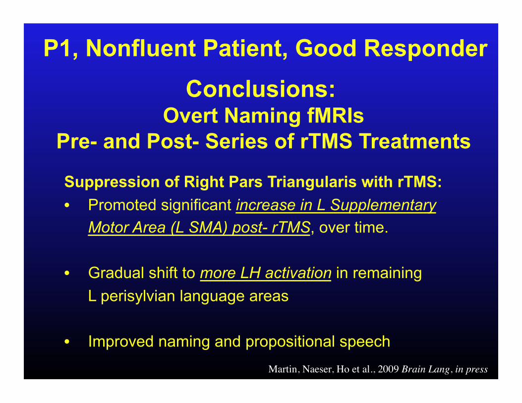
Conclusions: Overt Naming fMRIs
Pre- and Post- Series of rTMS Treatments
Suppression of Right Pars Triangularis with rTMS: • Promoted significant increase in L Supplementary
Motor Area (L SMA) post- rTMS, over time.
• Gradual shift to more LH activation in remaining L perisylvian language areas
• Improved naming and propositional speech
P1, Nonfluent Patient, Good Responder
Martin, Naeser, Ho et al., 2009 Brain Lang, in press
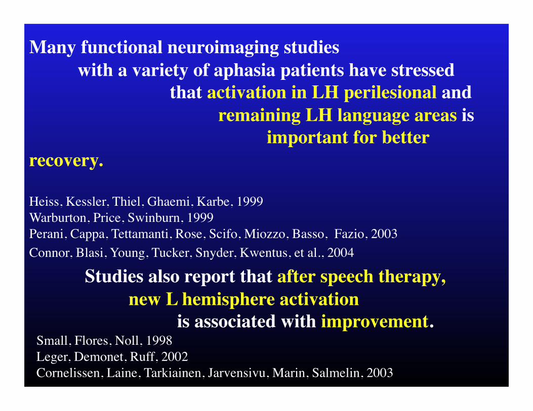
Many functional neuroimaging studies with a variety of aphasia patients have stressed that activation in LH perilesional and remaining LH language areas is important for better
recovery.
Heiss, Kessler, Thiel, Ghaemi, Karbe, 1999 Warburton, Price, Swinburn, 1999 Perani, Cappa, Tettamanti, Rose, Scifo, Miozzo, Basso, Fazio, 2003 Connor, Blasi, Young, Tucker, Snyder, Kwentus, et al., 2004
Studies also report that after speech therapy, new L hemisphere activation is associated with improvement.
Small, Flores, Noll, 1998 Leger, Demonet, Ruff, 2002 Cornelissen, Laine, Tarkiainen, Jarvensivu, Marin, Salmelin, 2003
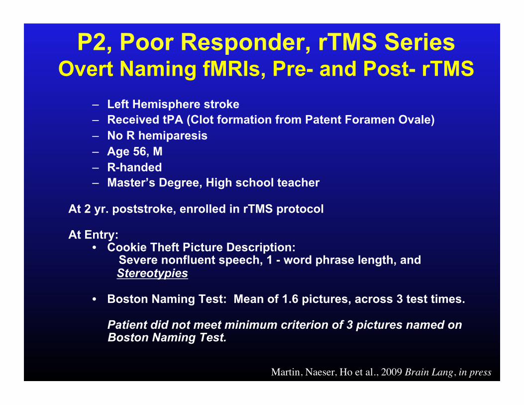
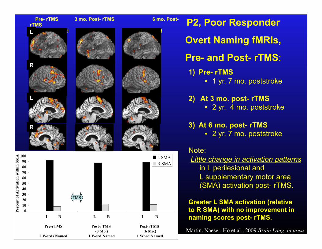
P2, Poor Responder, rTMS Series Overt Naming fMRIs, Pre- and Post- rTMS
– Left Hemisphere stroke – Received tPA (Clot formation from Patent Foramen Ovale) – No R hemiparesis – Age 56, M – R-handed – Master’s Degree, High school teacher
At 2 yr. poststroke, enrolled in rTMS protocol
At Entry: • Cookie Theft Picture Description:
Severe nonfluent speech, 1 - word phrase length, and Stereotypies
• Boston Naming Test: Mean of 1.6 pictures, across 3 test times.
Patient did not meet minimum criterion of 3 pictures named on Boston Naming Test.
Martin, Naeser, Ho et al., 2009 Brain Lang, in press
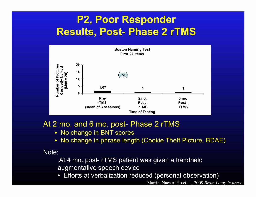
Boston Naming Test
First 20 Items
1.67 1 1
0
5
10
15
20
Pre-
rTMS
(Mean of 3 sessions)
2mo.
Post-
rTMS
6mo.
Post-
rTMS
Time of Testing
Nu
mb
er
of
Pic
ture
s
Co
rre
ctl
y N
am
ed
(Ma
x =
20
)
TMS
At 2 mo. and 6 mo. post- Phase 2 rTMS • No change in BNT scores • No change in phrase length (Cookie Theft Picture, BDAE)
Note: At 4 mo. post- rTMS patient was given a handheld augmentative speech device • Efforts at verbalization reduced (personal observation)
P2, Poor Responder Results, Post- Phase 2 rTMS
Martin, Naeser, Ho et al., 2009 Brain Lang, in press
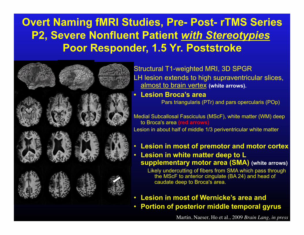
Overt Naming fMRI Studies, Pre- Post- rTMS Series P2, Severe Nonfluent Patient with Stereotypies
Poor Responder, 1.5 Yr. Poststroke Structural T1-weighted MRI, 3D SPGR LH lesion extends to high supraventricular slices,
almost to brain vertex (white arrows). • Lesion Broca’s area
Pars triangularis (PTr) and pars opercularis (POp)
Medial Subcallosal Fasciculus (MScF), white matter (WM) deep to Broca's area (red arrows)
Lesion in about half of middle 1/3 periventricular white matter
• Lesion in most of premotor and motor cortex • Lesion in white matter deep to L
supplementary motor area (SMA) (white arrows) Likely undercutting of fibers from SMA which pass through
the MScF to anterior cingulate (BA 24) and head of caudate deep to Broca's area.
• Lesion in most of Wernicke’s area and • Portion of posterior middle temporal gyrus
Martin, Naeser, Ho et al., 2009 Brain Lang, in press
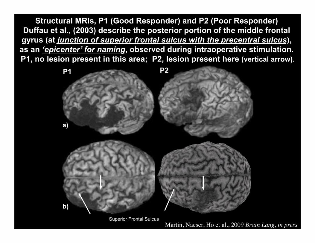
Structural MRIs, P1 (Good Responder) and P2 (Poor Responder) Duffau et al., (2003) describe the posterior portion of the middle frontal gyrus (at junction of superior frontal sulcus with the precentral sulcus), as an ‘epicenter’ for naming, observed during intraoperative stimulation. P1, no lesion present in this area; P2, lesion present here (vertical arrow).
P1 P2
a)
b)
Superior Frontal Sulcus Martin, Naeser, Ho et al., 2009 Brain Lang, in press
0
10
20
30
40
50
60
70
80
90
100
L R
Pre-rTMS
2 Words Named
L R
Post-rTMS
(3 Mo.)
1 Word Named
L R
Post-rTMS
(6 Mo.)
1 Word Named
Perc
en
t o
f A
cti
va
tio
n w
ith
in S
MA
L SMA
R SMA
Pre- rTMS 3 mo. Post- rTMS 6 mo. Post- rTMS 2 Pictures Named 1 Picture Named 1 Picture Named
P2, Poor Responder
Overt Naming fMRIs,
Pre- and Post- rTMS:
TMS
1) Pre- rTMS • 1 yr. 7 mo. poststroke
2) At 3 mo. post- rTMS • 2 yr. 4 mo. poststroke
3) At 6 mo. post- rTMS • 2 yr. 7 mo. poststroke
Note: Little change in activation patterns
in L perilesional and L supplementary motor area (SMA) activation post- rTMS.
Greater L SMA activation (relative to R SMA) with no improvement in naming scores post- rTMS.
R
L
L
R
Martin, Naeser, Ho et al., 2009 Brain Lang, in press
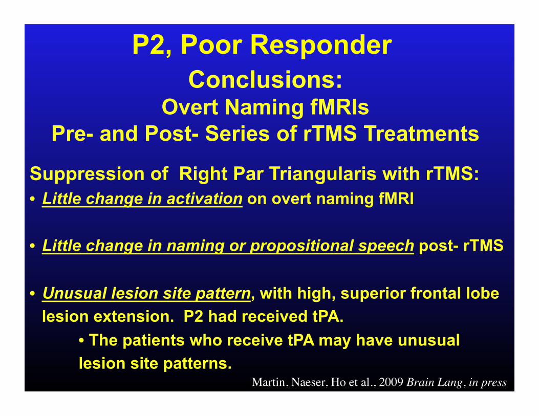
Suppression of Right Par Triangularis with rTMS: • Little change in activation on overt naming fMRI
• Little change in naming or propositional speech post- rTMS
• Unusual lesion site pattern, with high, superior frontal lobe lesion extension. P2 had received tPA.
• The patients who receive tPA may have unusual lesion site patterns.
P2, Poor Responder Conclusions:
Overt Naming fMRIs Pre- and Post- Series of rTMS Treatments
Martin, Naeser, Ho et al., 2009 Brain Lang, in press
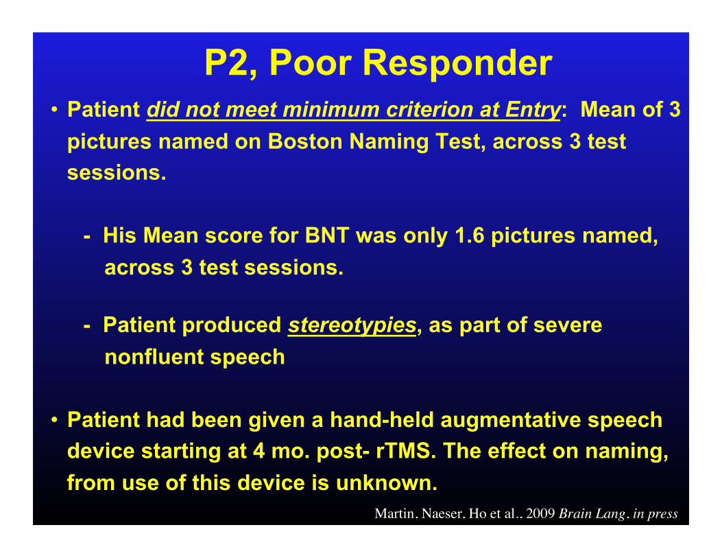
• Patient did not meet minimum criterion at Entry: Mean of 3 pictures named on Boston Naming Test, across 3 test sessions.
- His Mean score for BNT was only 1.6 pictures named, across 3 test sessions.
- Patient produced stereotypies, as part of severe nonfluent speech
• Patient had been given a hand-held augmentative speech device starting at 4 mo. post- rTMS. The effect on naming, from use of this device is unknown.
P2, Poor Responder
Martin, Naeser, Ho et al., 2009 Brain Lang, in press
Topic 3: Studies with TMS followed by Speech/Language Therapy
• Post-Phase 2, Begin Speech Therapy months later. OR
• New Pilot Study: Begin Speech Therapy immediately after, each 20 minute- rTMS treatment:
10 rTMS treatments, each followed by 3 hours of Constraint-Induced Language Therapy (CILT)
• Rationale: rTMS may modulate bi-lateral neural networks for language, so that patients can
attain a better outcome from Speech Therapy. Naeser, Martin, Treglia, Ho et al., 2009 Brain Lang Abstract, Academy of Aphasia Meetings
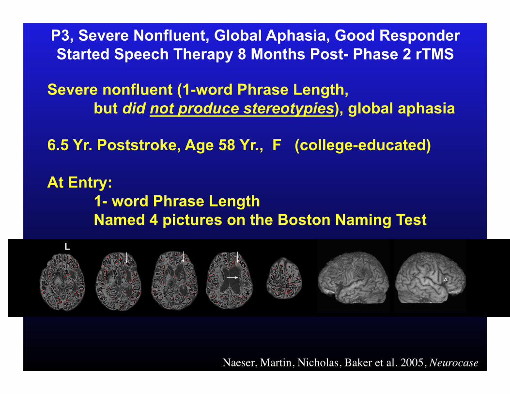
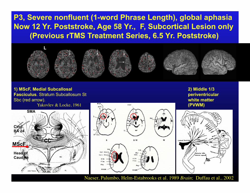
P3, Severe Nonfluent, Global Aphasia, Good Responder Started Speech Therapy 8 Months Post- Phase 2 rTMS
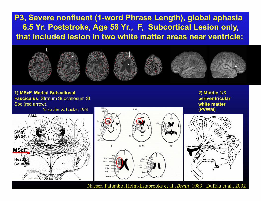
Severe nonfluent (1-word Phrase Length, but did not produce stereotypies), global aphasia
6.5 Yr. Poststroke, Age 58 Yr., F (college-educated)
At Entry: 1- word Phrase Length Named 4 pictures on the Boston Naming Test
Naeser, Martin, Nicholas, Baker et al. 2005, Neurocase
L
SMA
1) MScF, Medial Subcallosal Fasciculus. Stratum Subcallosum St Sbc (red arrow). Yakovlev & Locke, 1961
Cing. BA 24
Head of Caudate
2) Middle 1/3 periventricular white matter (PVWM)
Naeser, Palumbo, Helm-Estabrooks et al., Brain, 1989; Duffau et al., 2002
CC MScF
L
P3, Severe nonfluent (1-word Phrase Length), global aphasia 6.5 Yr. Poststroke, Age 58 Yr., F, Subcortical Lesion only,
that included lesion in two white matter areas near ventricle:
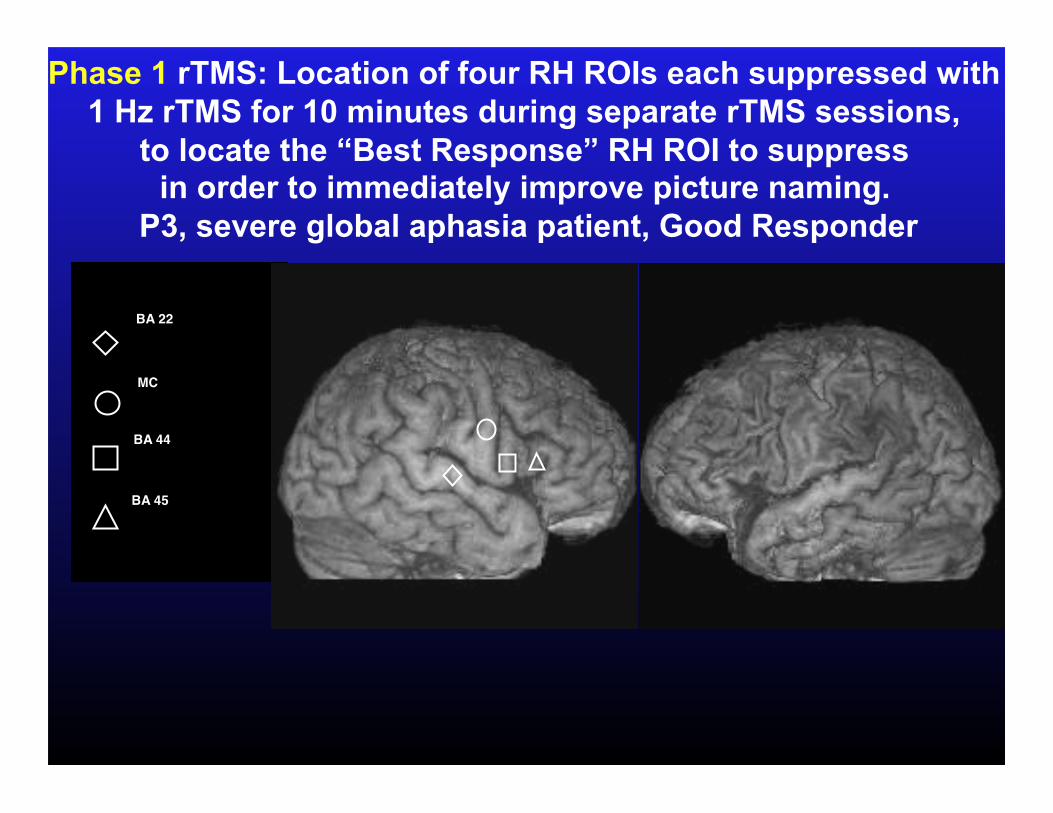
Phase 1 rTMS: Location of four RH ROIs each suppressed with 1 Hz rTMS for 10 minutes during separate rTMS sessions,
to locate the “Best Response” RH ROI to suppress in order to immediately improve picture naming.
P3, severe global aphasia patient, Good Responder
BA 22
BA 44
BA 45
MC
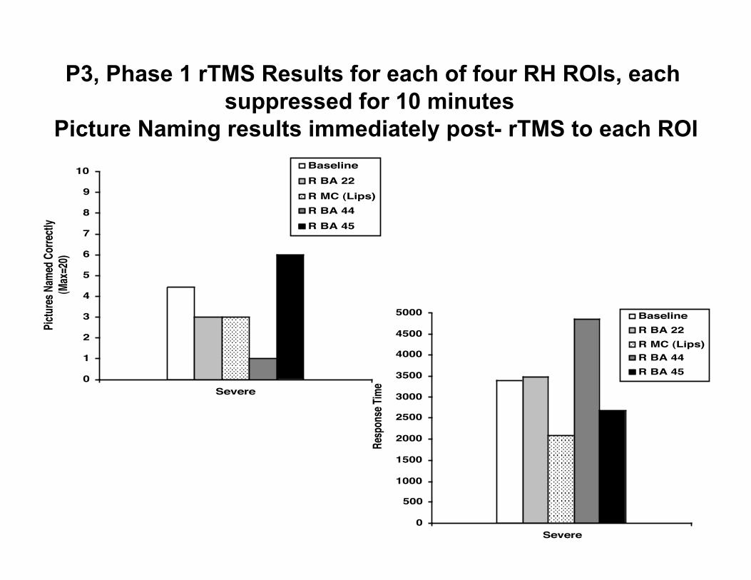
P3, Phase 1 rTMS Results for each of four RH ROIs, each suppressed for 10 minutes
Picture Naming results immediately post- rTMS to each ROI
0
1
2
3
4
5
6
7
8
9
10
Severe
Pictur
es N
amed
Cor
rectly
(Max
=20)
BaselineR BA 22R MC (Lips)R BA 44R BA 45
0
500
1000
1500
2000
2500
3000
3500
4000
4500
5000
Severe
Resp
onse
Time
BaselineR BA 22R MC (Lips)R BA 44R BA 45
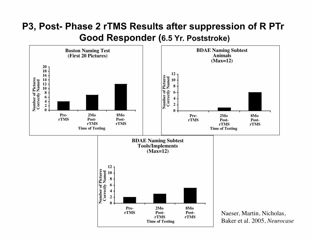
P3, Post- Phase 2 rTMS Results after suppression of R PTr Good Responder (6.5 Yr. Poststroke)
Boston Naming Test(First 20 Pictures)
02468
101214161820
Pre-rTMS
2Mo Post- rTMS
8Mo Post- rTMS
Time of Testing
Num
ber
of P
ictu
res
Cor
rect
ly N
amed
BDAE Naming SubtestAnimals
(Max=12)
02468
1012
Pre-rTMS
2Mo Post-rTMS
8Mo Post-rTMS
Time of Testing
Num
ber
of P
ictu
res
Cor
rect
ly N
amed
BDAE Naming SubtestTools/Implements
(Max=12)
02468
1012
Pre-rTMS
2Mo Post-rTMS
8Mo Post-rTMS
Time of Testing
Num
ber
of P
ictu
res
Cor
rect
ly N
amed
Naeser, Martin, Nicholas, Baker et al. 2005, Neurocase
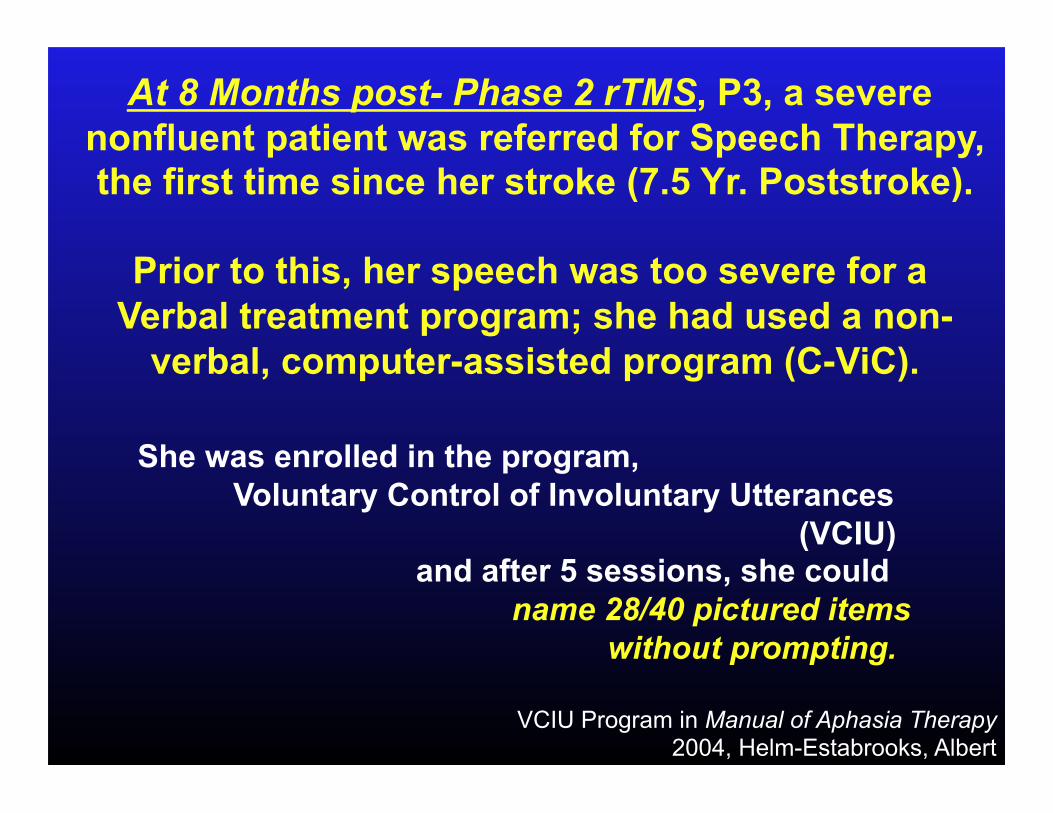
At 8 Months post- Phase 2 rTMS, P3, a severe nonfluent patient was referred for Speech Therapy, the first time since her stroke (7.5 Yr. Poststroke).
Prior to this, her speech was too severe for a Verbal treatment program; she had used a non-
verbal, computer-assisted program (C-ViC).
She was enrolled in the program, Voluntary Control of Involuntary Utterances (VCIU) and after 5 sessions, she could name 28/40 pictured items without prompting.
VCIU Program in Manual of Aphasia Therapy 2004, Helm-Estabrooks, Albert
L
L
R
R
Pre-TMS 17/60 28%
Pictures Named 9 Yr. Poststroke
3 Mo. Post-TMS 25/60 42%
Pictures Named 10;4 Yr. Poststroke
46 Mo. Post-TMS 25/60 42%
Pictures Named 13;11 Yr. Poststroke
16 Mo. Post-TMS 35/60 58%
Pictures Named 11;5 Yr. Poststroke
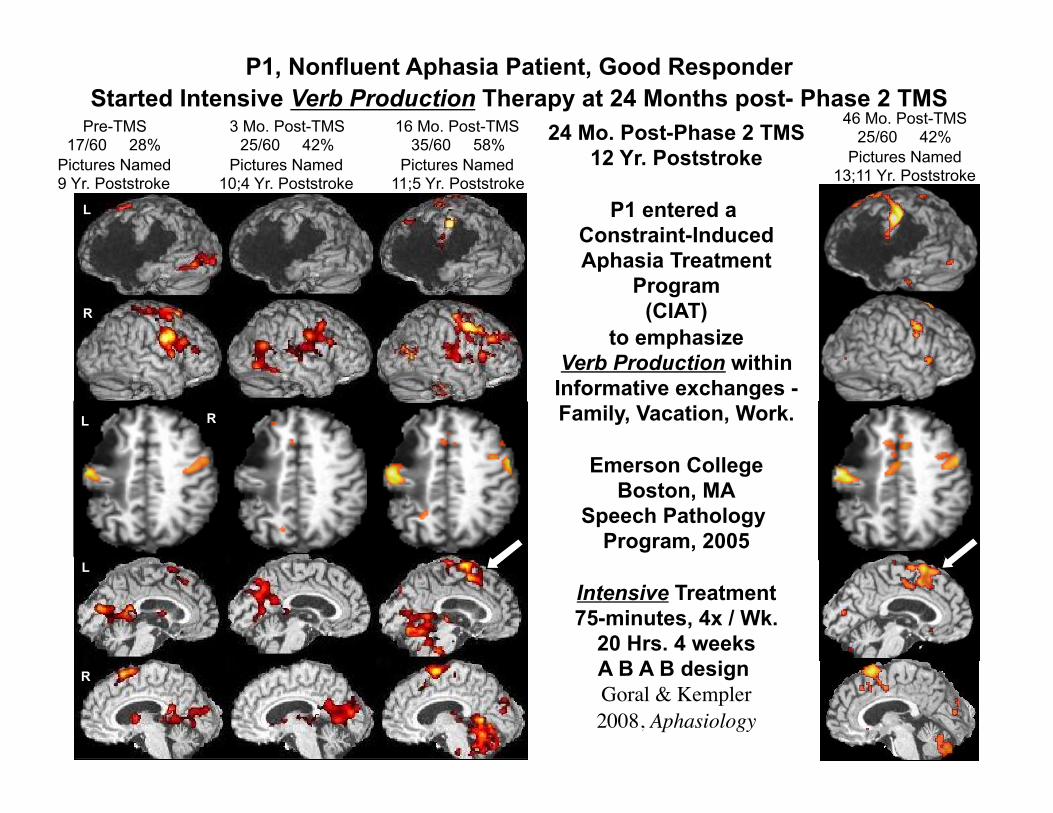
24 Mo. Post-Phase 2 TMS 12 Yr. Poststroke
P1 entered a Constraint-Induced Aphasia Treatment
Program (CIAT)
to emphasize Verb Production within Informative exchanges - Family, Vacation, Work.
Emerson College Boston, MA
Speech Pathology Program, 2005
Intensive Treatment 75-minutes, 4x / Wk.
20 Hrs. 4 weeks A B A B design Goral & Kempler 2008, Aphasiology
P1, Nonfluent Aphasia Patient, Good Responder Started Intensive Verb Production Therapy at 24 Months post- Phase 2 TMS
R
L
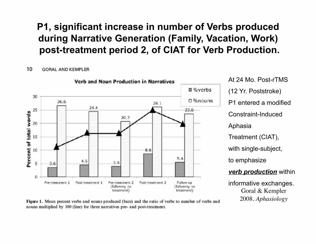
P1, significant increase in number of Verbs produced during Narrative Generation (Family, Vacation, Work) post-treatment period 2, of CIAT for Verb Production.
At 24 Mo. Post-rTMS
(12 Yr. Poststroke)
P1 entered a modified
Constraint-Induced
Aphasia
Treatment (CIAT),
with single-subject,
to emphasize
verb production within
informative exchanges. Goral & Kempler 2008, Aphasiology
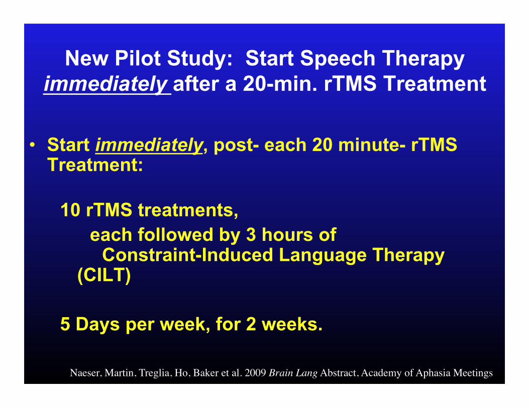
New Pilot Study: Start Speech Therapy immediately after a 20-min. rTMS Treatment
• Start immediately, post- each 20 minute- rTMS Treatment:
10 rTMS treatments, each followed by 3 hours of
Constraint-Induced Language Therapy (CILT)
5 Days per week, for 2 weeks.
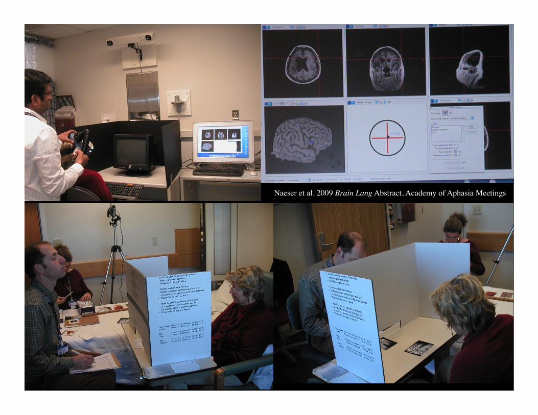
Naeser, Martin, Treglia, Ho, Baker et al. 2009 Brain Lang Abstract, Academy of Aphasia Meetings
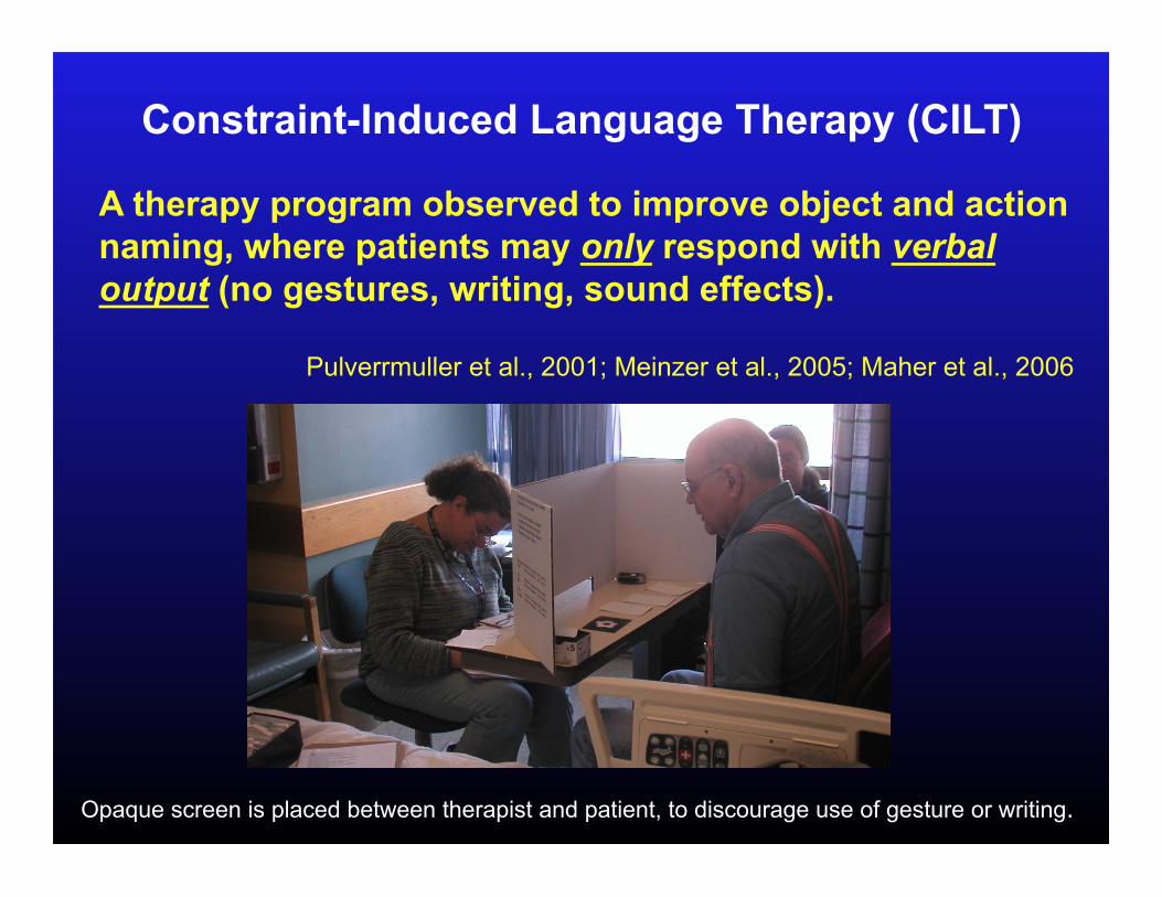
Constraint-Induced Language Therapy (CILT)
A therapy program observed to improve object and action naming, where patients may only respond with verbal output (no gestures, writing, sound effects).
Pulverrmuller et al., 2001; Meinzer et al., 2005; Maher et al., 2006
Opaque screen is placed between therapist and patient, to discourage use of gesture or writing.
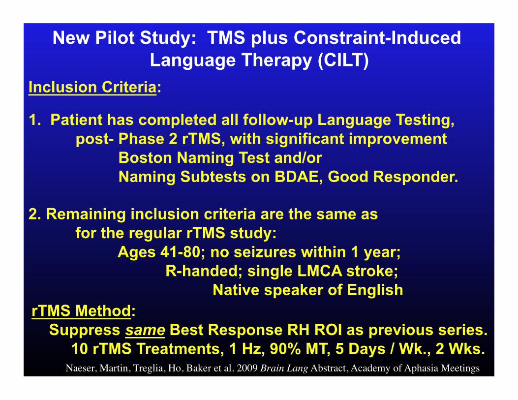
New Pilot Study: TMS plus Constraint-Induced Language Therapy (CILT)
Inclusion Criteria:
1. Patient has completed all follow-up Language Testing, post- Phase 2 rTMS, with significant improvement Boston Naming Test and/or Naming Subtests on BDAE, Good Responder.
2. Remaining inclusion criteria are the same as for the regular rTMS study: Ages 41-80; no seizures within 1 year; R-handed; single LMCA stroke; Native speaker of English
rTMS Method: Suppress same Best Response RH ROI as previous series. 10 rTMS Treatments, 1 Hz, 90% MT, 5 Days / Wk., 2 Wks.
Naeser, Martin, Treglia, Ho, Baker et al. 2009 Brain Lang Abstract, Academy of Aphasia Meetings

TMS plus Constraint-Induced Language Therapy (CILT) Pre- and Post- Language Testing:
Pre- testing: Parts of BDAE, and BNT (x3) (Baseline) Post- testing: at 1 and 6 Mo. post-intervention: Same tests
Language Outcome Measures: Significant improvement defined, >2 SD above Baseline on BDAE, BNT.
To examine for possible changes that might occur during intervention, Naming Probe Testing was administered using BDAE naming subtests (Actions, Animals, Tools/Implements), and BNT:
• Pre-TMS 10-12x, 4-5 week period • Daily, immediately post- each CILT session • Post-TMS 10-12x, 4-5 week period
These time-series data were analyzed using Double Bootstrap method. http://www.stat.wmich.edu/slab/Software/Timeseries.html (McKnight, McKean, Huitema, 2000)
Naeser, Martin, Treglia, Ho, Baker et al. 2009 Brain Lang Abstract, Academy of Aphasia Meetings
SMA
1) MScF, Medial Subcallosal Fasciculus. Stratum Subcallosum St Sbc (red arrow). Yakovlev & Locke, 1961
Cing. BA 24
Head of Caudate
2) Middle 1/3 periventricular white matter (PVWM)
Naeser, Palumbo, Helm-Estabrooks et al. 1989 Brain; Duffau et al., 2002
CC MScF
L
P3, Severe nonfluent (1-word Phrase Length), global aphasia Now 12 Yr. Poststroke, Age 58 Yr., F, Subcortical Lesion only
(Previous rTMS Treatment Series, 6.5 Yr. Poststroke)
Naeser et al. 2009 Brain Lang Abstract, Academy of Aphasia Meetings
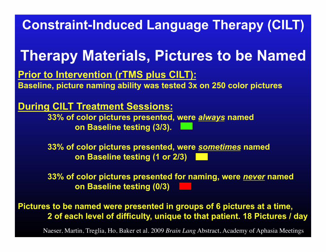
Constraint-Induced Language Therapy (CILT)
Therapy Materials, Pictures to be Named
Prior to Intervention (rTMS plus CILT): Baseline, picture naming ability was tested 3x on 250 color pictures
During CILT Treatment Sessions: 33% of color pictures presented, were always named on Baseline testing (3/3).
33% of color pictures presented, were sometimes named on Baseline testing (1 or 2/3)
33% of color pictures presented for naming, were never named on Baseline testing (0/3)
Pictures to be named were presented in groups of 6 pictures at a time, 2 of each level of difficulty, unique to that patient. 18 Pictures / day
Naeser, Martin, Treglia, Ho, Baker et al. 2009 Brain Lang Abstract, Academy of Aphasia Meetings
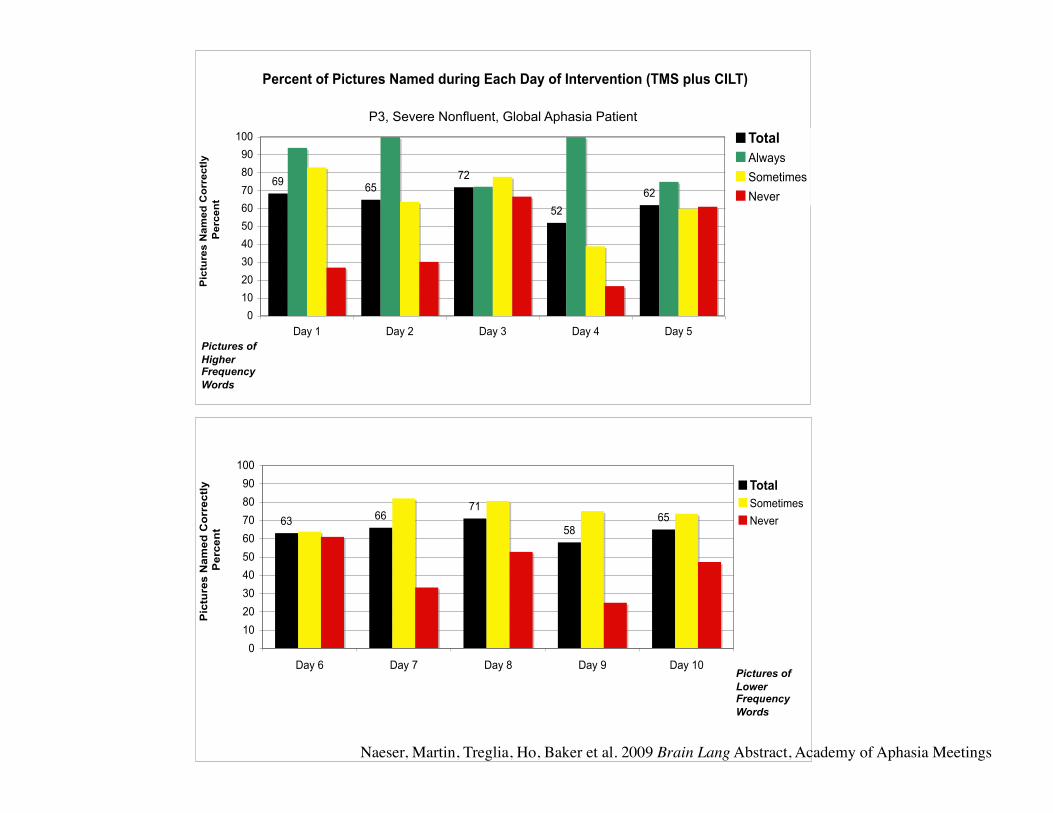
6366
71
5865
0
10
20
30
40
50
60
70
80
90
100
Day 6 Day 7 Day 8 Day 9 Day 10
Pic
ture
s N
am
ed
Co
rrectl
y
Perc
en
t
Total
Sometimes
Never
Percent of Pictures Named during Each Day of Intervention (TMS plus CILT)
6965
72
52
62
0
10
20
30
40
50
60
70
80
90
100
Day 1 Day 2 Day 3 Day 4 Day 5
Pic
ture
s N
am
ed
Co
rrectl
y
Perc
en
t
Total
Always
Sometimes
Never
P3, Severe Nonfluent, Global Aphasia Patient
Pictures of Higher Frequency Words
Pictures of Lower Frequency Words
Naeser, Martin, Treglia, Ho, Baker et al. 2009 Brain Lang Abstract, Academy of Aphasia Meetings
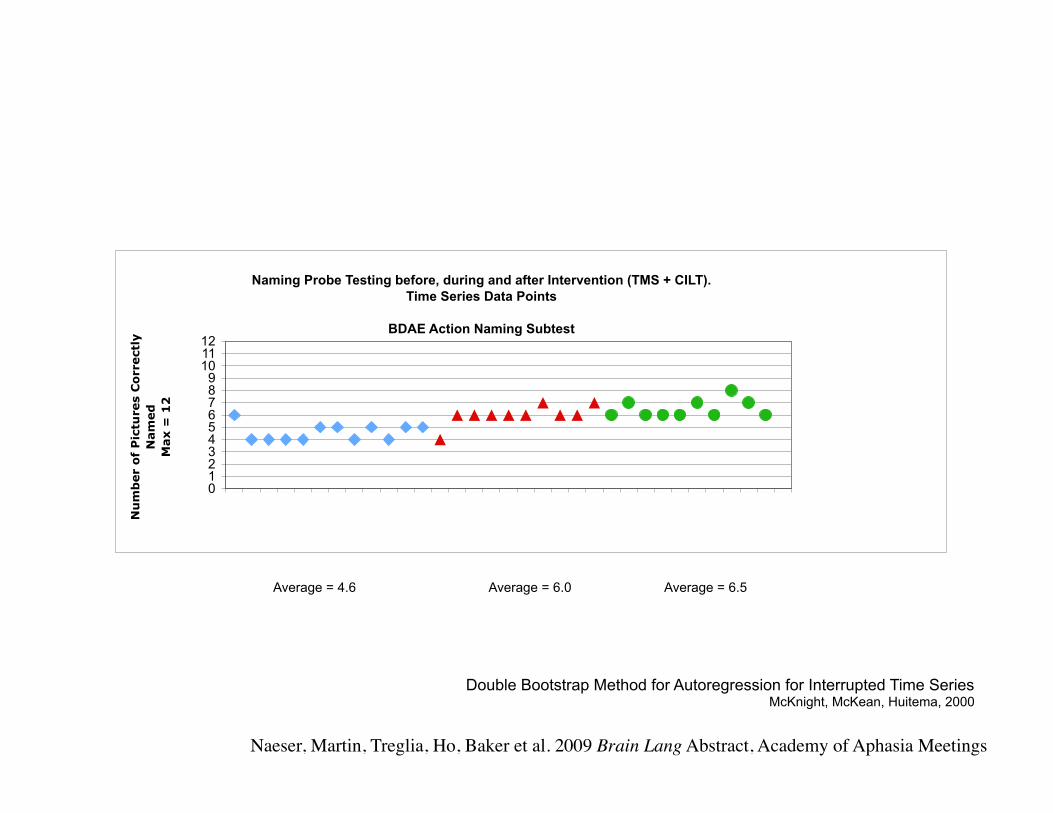
Naming Probe Testing before, during and after Intervention (TMS + CILT).
Time Series Data Points
BDAE Action Naming Subtest
0
1
2
3
4
5
6
7
8
9
10
11
12
Nu
mb
er o
f P
ictu
res C
orrectl
y
Nam
ed
Max =
12
Double Bootstrap Method for Autoregression for Interrupted Time Series McKnight, McKean, Huitema, 2000
Average = 4.6 Average = 6.0 Average = 6.5
Naeser, Martin, Treglia, Ho, Baker et al. 2009 Brain Lang Abstract, Academy of Aphasia Meetings
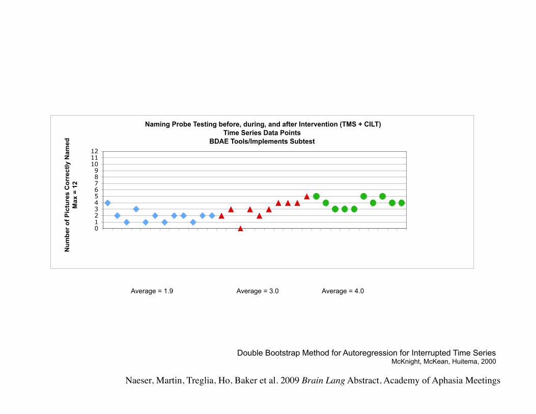
Naming Probe Testing before, during, and after Intervention (TMS + CILT)
Time Series Data Points
BDAE Tools/Implements Subtest
0
1
2
3
4
5
6
7
8
9
10
11
12
Nu
mb
er
of
Pic
ture
s C
orr
ec
tly
Na
me
d
Ma
x =
12
Double Bootstrap Method for Autoregression for Interrupted Time Series McKnight, McKean, Huitema, 2000
Average = 1.9 Average = 3.0 Average = 4.0
Naeser, Martin, Treglia, Ho, Baker et al. 2009 Brain Lang Abstract, Academy of Aphasia Meetings
Naming Probe Testing before, during and after Intervention (TMS + CILT)
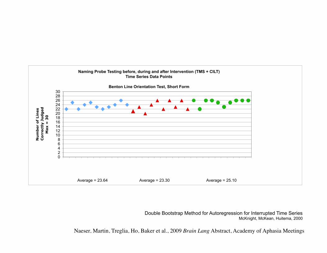
Time Series Data Points
Benton Line Orientation Test, Short Form
0
2
4
6
8
10
12
14
16
18
20
22
24
26
28
30
Nu
mb
er o
f Lin
es
Co
rrectl
y J
ud
ged
Max =
30
Double Bootstrap Method for Autoregression for Interrupted Time Series McKnight, McKean, Huitema, 2000
Average = 23.64 Average = 23.30 Average = 25.10
Naeser, Martin, Treglia, Ho, Baker et al., 2009 Brain Lang Abstract, Academy of Aphasia Meetings
TMS
TMS
+ CILT
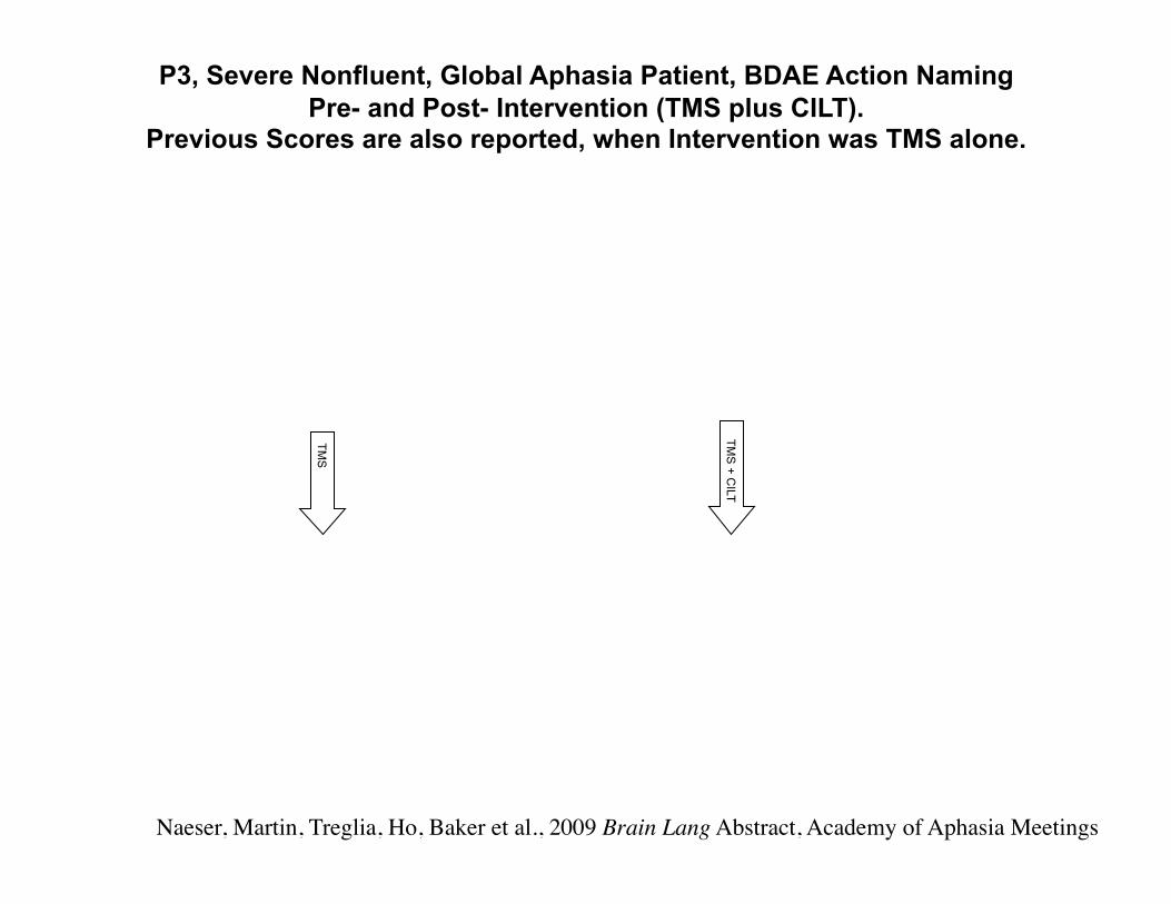
P3, Severe Nonfluent, Global Aphasia Patient, BDAE Action Naming Pre- and Post- Intervention (TMS plus CILT).
Previous Scores are also reported, when Intervention was TMS alone.
Naeser, Martin, Treglia, Ho, Baker et al., 2009 Brain Lang Abstract, Academy of Aphasia Meetings
TMS
TMS
+ CILT
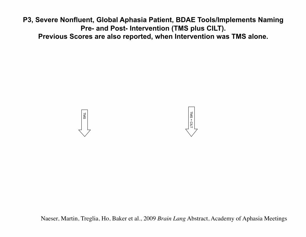
P3, Severe Nonfluent, Global Aphasia Patient, BDAE Tools/Implements Naming Pre- and Post- Intervention (TMS plus CILT).
Previous Scores are also reported, when Intervention was TMS alone.
Naeser, Martin, Treglia, Ho, Baker et al., 2009 Brain Lang Abstract, Academy of Aphasia Meetings
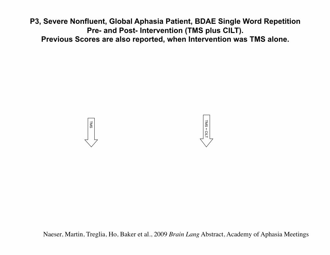
TMS
TMS
+ CILT
P3, Severe Nonfluent, Global Aphasia Patient, BDAE Single Word Repetition Pre- and Post- Intervention (TMS plus CILT).
Previous Scores are also reported, when Intervention was TMS alone.
Naeser, Martin, Treglia, Ho, Baker et al., 2009 Brain Lang Abstract, Academy of Aphasia Meetings
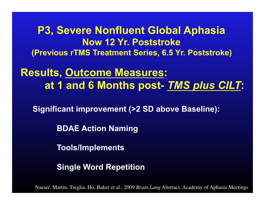
Results, Outcome Measures: at 1 and 6 Months post- TMS plus CILT:
Significant improvement (>2 SD above Baseline):
BDAE Action Naming
Tools/Implements
Single Word Repetition
P3, Severe Nonfluent Global Aphasia Now 12 Yr. Poststroke
(Previous rTMS Treatment Series, 6.5 Yr. Poststroke)
Naeser, Martin, Treglia, Ho, Baker et al., 2009 Brain Lang Abstract, Academy of Aphasia Meetings
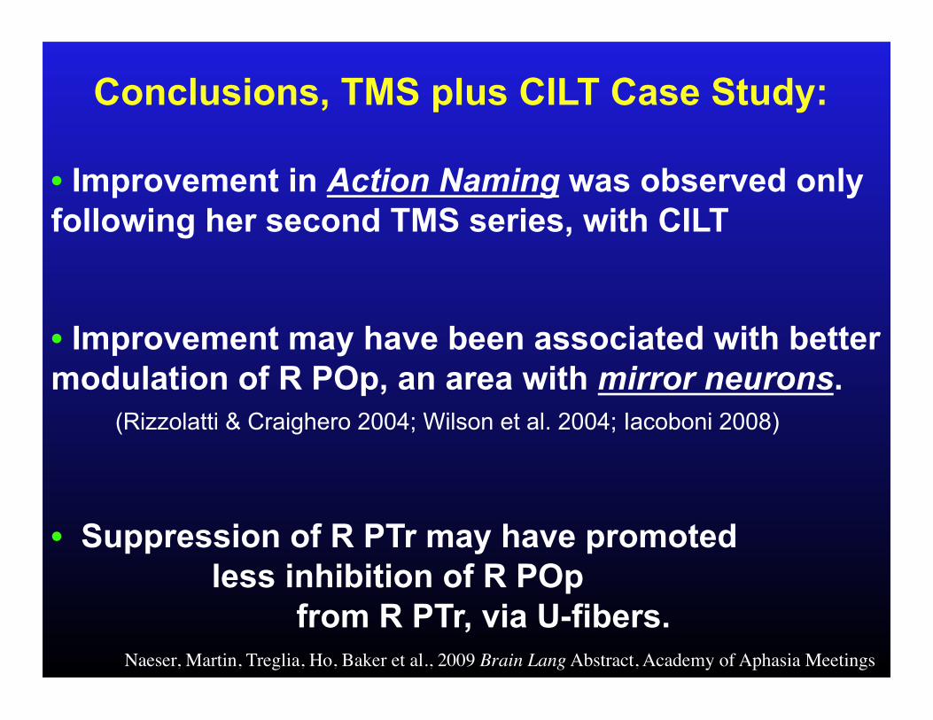
Conclusions, TMS plus CILT Case Study:
• Improvement in Action Naming was observed only following her second TMS series, with CILT
• Improvement may have been associated with better modulation of R POp, an area with mirror neurons. (Rizzolatti & Craighero 2004; Wilson et al. 2004; Iacoboni 2008)
• Suppression of R PTr may have promoted less inhibition of R POp from R PTr, via U-fibers. Naeser, Martin, Treglia, Ho, Baker et al., 2009 Brain Lang Abstract, Academy of Aphasia Meetings
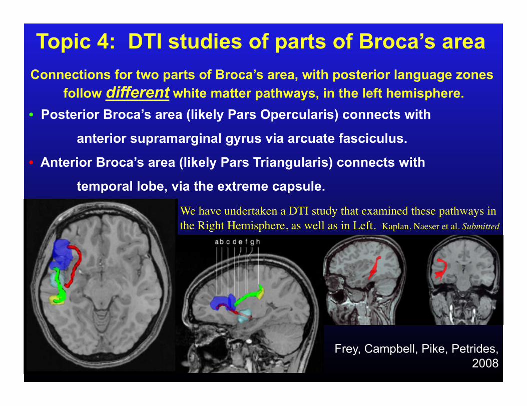
Connections for two parts of Broca’s area, with posterior language zones follow different white matter pathways, in the left hemisphere.
• Posterior Broca’s area (likely Pars Opercularis) connects with
anterior supramarginal gyrus via arcuate fasciculus.
• Anterior Broca’s area (likely Pars Triangularis) connects with
temporal lobe, via the extreme capsule.
Frey, Campbell, Pike, Petrides, 2008
Topic 4: DTI studies of parts of Broca’s area
We have undertaken a DTI study that examined these pathways in the Right Hemisphere, as well as in Left. Kaplan, Naeser et al. Submitted
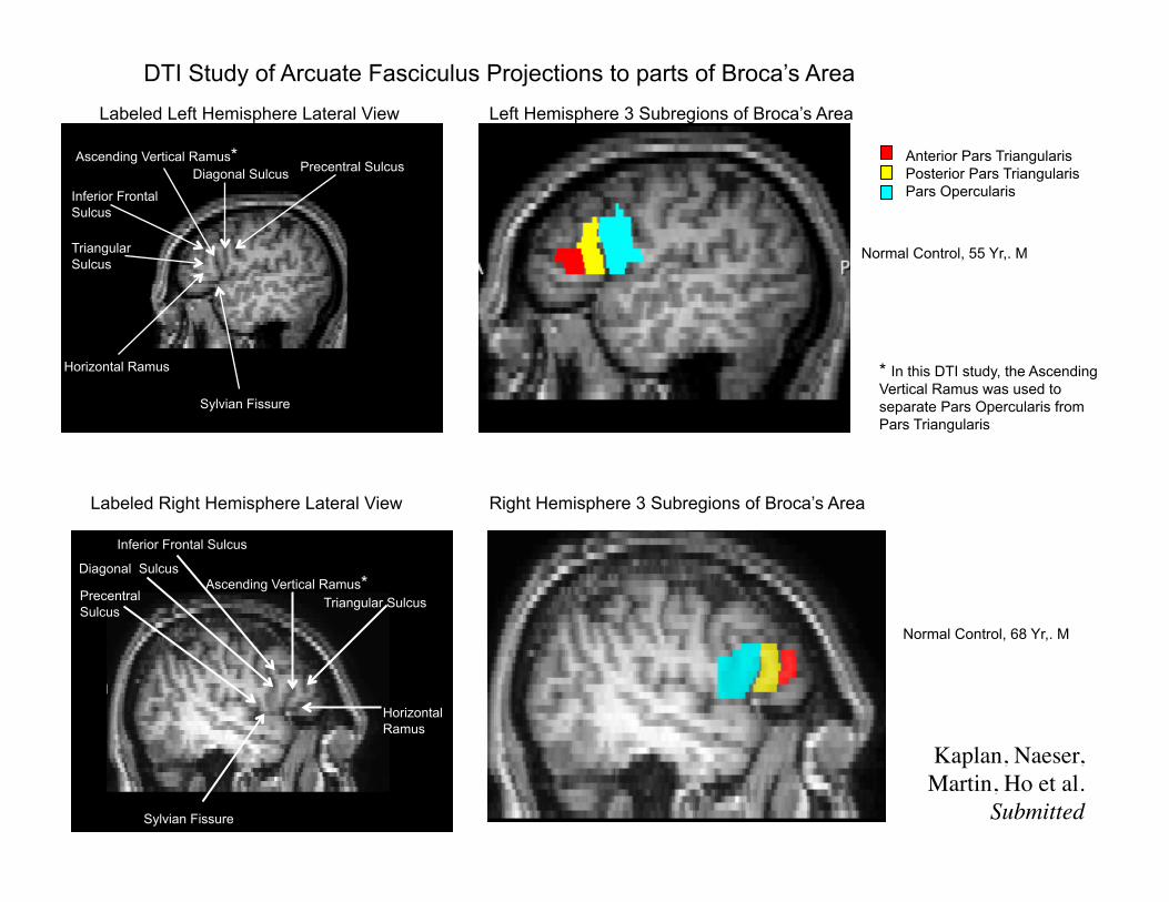
Inferior Frontal Sulcus
Horizontal Ramus
Ascending Vertical Ramus* Diagonal Sulcus
Triangular Sulcus
Sylvian Fissure
Precentral Sulcus
Left Hemisphere 3 Subregions of Broca’s Area
Normal Control, 55 Yr,. M
Labeled Left Hemisphere Lateral View
Normal Control, 68 Yr,. M
Labeled Right Hemisphere Lateral View Right Hemisphere 3 Subregions of Broca’s Area
Kaplan, Naeser, Martin, Ho et al.
Submitted
Precentral Sulcus
Ascending Vertical Ramus* Triangular Sulcus
Horizontal Ramus
Sylvian Fissure
Inferior Frontal Sulcus
Diagonal Sulcus
* In this DTI study, the Ascending Vertical Ramus was used to separate Pars Opercularis from Pars Triangularis
DTI Study of Arcuate Fasciculus Projections to parts of Broca’s Area
Anterior Pars Triangularis Posterior Pars Triangularis Pars Opercularis
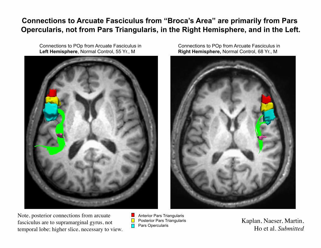
Connections to POp from Arcuate Fasciculus in Left Hemisphere, Normal Control, 55 Yr., M
Connections to POp from Arcuate Fasciculus in Right Hemisphere, Normal Control, 68 Yr., M
Kaplan, Naeser, Martin, Ho et al. Submitted
Anterior Pars Triangularis Posterior Pars Triangularis Pars Opercularis
Connections to Arcuate Fasciculus from “Broca’s Area” are primarily from Pars Opercularis, not from Pars Triangularis, in the Right Hemisphere, and in the Left.
Note, posterior connections from arcuate fasciculus are to supramarginal gyrus, not temporal lobe; higher slice, necessary to view.
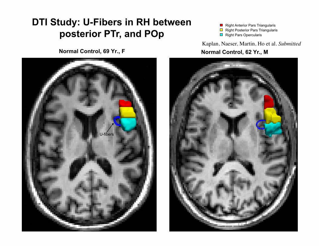
Normal Control, 69 Yr., F
U-fibers
Normal Control, 62 Yr., M
Right Anterior Pars Triangularis Right Posterior Pars Triangularis Right Pars Opercularis
DTI Study: U-Fibers in RH between posterior PTr, and POp
Kaplan, Naeser, Martin, Ho et al. Submitted
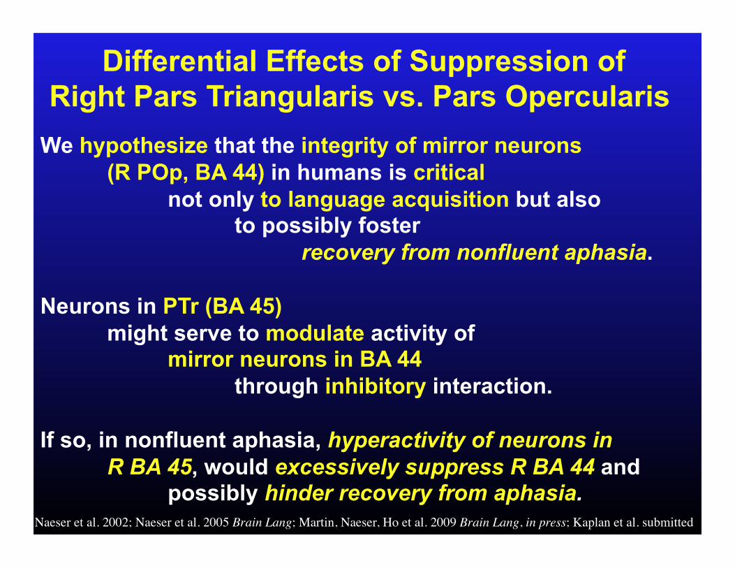
Differential Effects of Suppression of Right Pars Triangularis vs. Pars Opercularis
We hypothesize that the integrity of mirror neurons (R POp, BA 44) in humans is critical not only to language acquisition but also to possibly foster recovery from nonfluent aphasia.
Neurons in PTr (BA 45) might serve to modulate activity of mirror neurons in BA 44 through inhibitory interaction.
If so, in nonfluent aphasia, hyperactivity of neurons in R BA 45, would excessively suppress R BA 44 and possibly hinder recovery from aphasia.
Naeser et al. 2002; Naeser et al. 2005 Brain Lang; Martin, Naeser, Ho et al. 2009 Brain Lang, in press; Kaplan et al. submitted
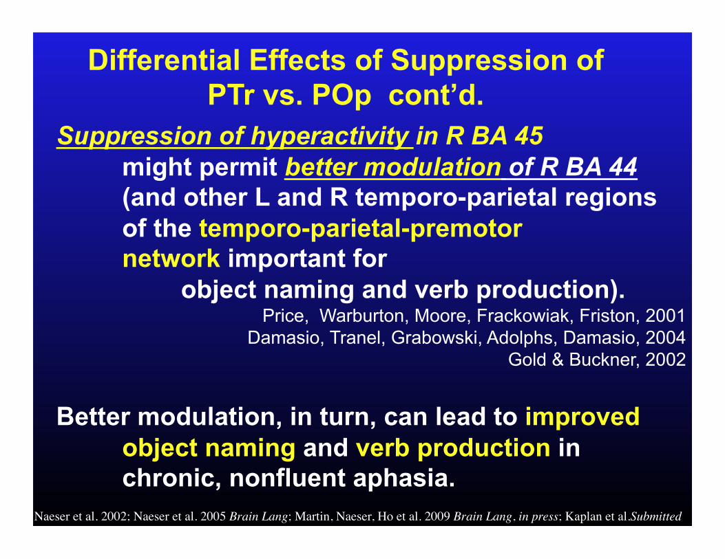
Differential Effects of Suppression of PTr vs. POp cont’d.
Suppression of hyperactivity in R BA 45 might permit better modulation of R BA 44 (and other L and R temporo-parietal regions of the temporo-parietal-premotor network important for object naming and verb production).
Price, Warburton, Moore, Frackowiak, Friston, 2001 Damasio, Tranel, Grabowski, Adolphs, Damasio, 2004
Gold & Buckner, 2002
Better modulation, in turn, can lead to improved object naming and verb production in chronic, nonfluent aphasia.
Naeser et al. 2002; Naeser et al. 2005 Brain Lang; Martin, Naeser, Ho et al. 2009 Brain Lang, in press; Kaplan et al.Submitted
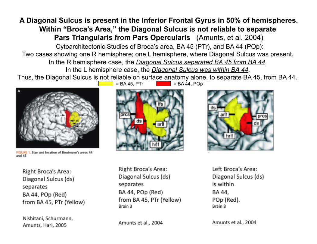
A Diagonal Sulcus is present in the Inferior Frontal Gyrus in 50% of hemispheres. Within “Broca’s Area,” the Diagonal Sulcus is not reliable to separate
Pars Triangularis from Pars Opercularis (Amunts, et al. 2004) Cytoarchitectonic Studies of Broca’s area, BA 45 (PTr), and BA 44 (POp):
Two cases showing one R hemisphere; one L hemisphere, where Diagonal Sulcus was present. In the R hemisphere case, the Diagonal Sulcus separated BA 45 from BA 44.
In the L hemisphere case, the Diagonal Sulcus was within BA 44. Thus, the Diagonal Sulcus is not reliable on surface anatomy alone, to separate BA 45, from BA 44.
= BA 45, PTr = BA 44, POp