Embed Size (px)
Citation preview

J. clin. Path. (1967), 20, 574
'Residual bodies' in sarcoid and sarcoid-likegranulomas
W. JONES WILLIAMS AND D. WILLIAMS
From the Welsh National School of Medicine, Cardiff
SYNOPSIS A morphological and histochemical study was made of epithelioid cell granulomas:(a) classical sarcoid type, namely, sarcoidosis, Kveim tests, tuberculosis, farmer's lung, and Crohn'ssyndrome; (b) sarcoid-like granulomas, often distinguishable from (a) by the presence of extra-cellular mucin or bile, namely, ulcerative colitis, diverticulitis, cholecystitis, cholangitis, carcinomaof the rectum and lymph nodes, draining tumours. All these granulomas showed similar, numerous
cytoplasmic granules in epithelioid and giant cells with the properties of residual bodies, i.e., endproducts of activated lysosomes. The presence of residual bodies demonstrates the following featuresthe morphological similarity of the granulomas studied, and the phagocytic nature of the affectedcells. It suggests a common mechanism of granuloma formation but does not identify any parti-cular exogenous cause. The findings suggest that Boeck's sarcoidosis may be caused by unidentifiedexogenous agents.
A morphological and histochemical study of sarcoidand sarcoid-like granulomas demonstrates thepresence of cytoplasmic granules in epithelioid andgiant cells. They are thought to be 'residual bodies',i.e., end products of activated lysosomes(Weissmann,1965). The significance of these findings is discussedwith particular reference to the aetiology of sar-coidosis.The granulomas in tuberculosis, chronic beryllium
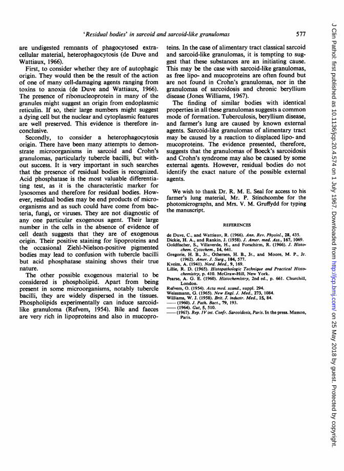
disease (Jones Williams, 1958), Crohn's syndrome(Jones Williams, 1964), the Kveim test (Kveim, 1941),and farmer's lung (Dickie and Rankin, 1958), areoften identical with those of sarcoidosis and maybe called 'classical sarcoid granulomas' (Fig. 1).The rare 'sarcoid-like granuloma' (Fig. 2), as incholecystitis, cholangitis, ulcerative colitis, diverti-culitis, and rectal carcinoma, are distinguishedfrom the above by the frequent presence of extra-cellular mucins and/or bile (Jones Williams, 1967).Sarcoid-like granulomas may also be seen in lymphnodes draining tumours (Gregorie, Othersen, andMoore, 1962).
MATERIAL AND METHODS
Table I shows the diseases studied, the number of cases,and the type of granuloma examined. The site and dis-tribution of the material is as follows: sarcoidosis, 15lungs, 6 lymph nodes; Kveim granulomas, 6, all skin,Received for publication 28 February 1967.
TABLE ITYPES OF GRANULOMA
DISEASES, ANDClassical Sarcoid No. ofCondition Cases
SarcoidKveim testTuberculosisChronic beryllium
disease
Farmer's lungCrohn's syndrome
216
12
7
NUMBER OF CASESSarcoid-likeCondition
Ulcerative colitisDiverticulitisCholecystitisCholangitis
3 Carcinoma rectum14 Lymph nodes
draining tumours
No. ofCases
22
2
tuberculosis, 5 skin, 4 lung, 2 ileum, and 1 brain; chronicberyllium disease, 5 lungs and 2 skin; farmer's lung, 3;Crohn's syndrome, 5 ileum, 3 colon, 3 skin fistulae, and3 lymph nodes; ulcerative colitis, 2; diverticulitis, 2;cholecystitis, 5; cholangitis, 1; carcinoma of rectum 2;lymph nodes draining carcinoma cervix, 1.The material consisted mainly of formalin-fixed tissues,
paraffin blocks, and sections. In one beryllium skingranuloma and two tuberculous lymph nodes freshmaterial was also available. Sections were stained rou-tinely with haematoxylin and eosin. They wereexamined under direct, polarized, phase contrast, andsome under ultra-violet light.The following histochemical tests were performed
periodic acid-Schiff (P.A.S.), to show muco- and glyco-proteins, controlled with diastase; Alcian blue, to demon-strate acid mucopolysaccharides, controlled with hyalase.Sudan black B and luxol fast blue, to demonstrate phos-
574
on 25 May 2018 by guest. P
rotected by copyright.http://jcp.bm
j.com/
J Clin P
athol: first published as 10.1136/jcp.20.4.574 on 1 July 1967. Dow
nloaded from
'Residual bodies' sarcoid and-like granulomas
ki~~~~~~~~~~~~~~~~~~i
* /~#*XdL.x
*X*vJ j ',*xw
%'1 4'i *gR a.t;s^
A' ' ,, 'ea '
a. Aia 4'
44i:*
.:.
'a'-'t/ > 4 t^ A ~ '~'EJR',Q'''~~~~~~~~~~~~~s' -t
S~~-X 4,, , fa.,1.*/4vA *y4<^.#t
i,t, BS ~y Sn,1*"v'
"iK \K wY'.. w * ;
t <'~~~~'a:*IT. #
4.... * ~ 16
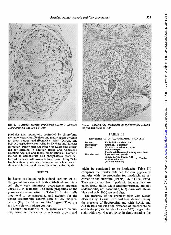
FIG. 1. Classical sarcoid granuloma (Boeck's sarcoid).Haematoxylin and eosin x 350.
pholipids and lipoprotein, controlled by chloroform/methanol extraction; Feulgen and methyl green pyronineto show desoxy- and ribonucleic acids (D.N.A. andR.N.A.) respectively, controlled by D.N.ase and R.N.aseextraction; Perls's stain for iron; Von Kossa and alizarinred for calcium. In addition Barka and Anderson'scoupling Azo dye and Holt's modification of Gomori'smethod to demonstrate acid phosphatases were per-formed on cases with available fresh tissue. Long Ziehl-Neelson staining was also performed on a few cases toshow acid fastness and Sudan stains for neutral lipids.
RESULTS
In haematoxylin-and-eosin-stained sections of allthe granulomas studied, both epithelioid and giantcell show very numerous cytoplasmic granulesabout 1 t in diameter. The main properties of thegranules are summarized in Table II. In giant cellsthey tend to be aggregated and account for thedenser eosinophilic centres seen at low magnifi-cation (Fig. 1). None are birefringent. They areeasily visible with phase contrast.Though the majority of the granules are colour-
less, some are occasionally yellowish brown and
FIG. 2. Sarcoid-like granuloma in cholecystitis. Haema-toxylin and eosin x 100.
TABLE IIPROPERTIES OF INTRACYTOPLASMIC GRANULES
Position Epithelioid and giant cellsMorphology Granular, I u diameterPhysical Colourless or yellowish brown
Not birefringentFaintly autofluorescent in ultra-violet light
Histochemical Lipo- and mucoprotein(S.B.B., L.F.B., P.A.S., A.B.) PositiveAcid phosphatase }(Azo dye, Gomori)
might be considered to be lipofuscin. Table IIIcompares the results obtained for our pigmentedgranules with the properties for lipofuscin as re-corded in the literature (Pearse, 1960; Lillie, 1965).They are distinct from lipofuscin because they arepaler, show bluish white autofluorescence, are notsudanophilic, not basophilic, 60% stain with alcianblue and only 20% are acid fast.The majority of the granules stain with Sudan
black B (Fig. 3.) and Luxol fast blue, demonstratingthe presence of lipoproteins and with P.A.S. andAlcian blue showing the presence of mucoproteinsand acidic polysaccharides. In some 50 % the granulesstain with methyl green pyronin demonstrating the
575
on 25 May 2018 by guest. P
rotected by copyright.http://jcp.bm
j.com/
J Clin P
athol: first published as 10.1136/jcp.20.4.574 on 1 July 1967. Dow
nloaded from

W. Jones Williams and D. Williams
Test
TABLE IIIA COMPARISON OF PIGMENTED RESIDUAL
BODIES AND LIPOFUSCIN
Pigmented Residual Bodies Lipofuscin
Autofluorescence
SudanophiliaBasophiliaAlcian blueAcid fast
Pale yellow or yellowishbrownBluish white
+(60%)-(80%)
Yellow-brown
Yellow-reddishbrown
++
presence of R.N.A. The majority, in the fresh tissueexamined, stain with Azo dye (Fig. 4) and Gomoristain for acid phosphatase. They do not stain forD.N.A., iron, calcium, or neutral lipid.
It is noted that the granules in all the granulomasstudied are histochemically identical, irrespectiveof the type of granuloma, site of the lesion, knownor unknown cause, and duration of the lesion.These properties show the granules to be of the
lysosomal origin. They are distinguishable fromSchaumann bodies (Jones Williams, 1960). Theirproperties also distinguish them from bacteria,viruses, or fungi.
DISCUSSION
Lysosomes participating in a process of digestionare often characterized by the presence of undigestedinert residues, usually lipid, and are then termedresidual bodies, (Weissmann, 1965; de Duve andWattiaux, 1966). Our results show that the cyto-plasmic granules described by virtue of their acidphosphatase, lipid, mucopolysaccharide, and R.N.Acontent are identified as activated lysosomes orresidual bodies. The variation in our histochemicalstaining reflects that, as expected, residual bodiesmay be derived from different sources.
Lipofuscin is described as a type of pigmentedindigestible material sometimes forming residualbodies (Goldfischer, Villaverde, and Forschirm,1966). However, our pigmented residual bodiesdiffer in staining properties and appear to bedistinct from lipofuscin.The presence of residual bodies demonstrates
that the cells are or have been actively phagocytic.There are two alternative modes of formation ofresidual bodies: either they arise from autodigestionof cells by the process of autophagocytosis, or they
......
FIG. 3. FIG. 4.FIG. 3. Sarcoid granuloma in lymph node. Residual bodies in a giant cell. Sudan black B x 886.FIG. 4. Beryllium granuloma of skin. Azo dye x 50.
576
-107W
5ow -,i-l'.4.i. .4:.-v:.- M........V
:M.
:1.:", ..: 4 :-".1
on 25 May 2018 by guest. P
rotected by copyright.http://jcp.bm
j.com/
J Clin P
athol: first published as 10.1136/jcp.20.4.574 on 1 July 1967. Dow
nloaded from
'Residual bodies' in sarcoid and sarcoid-like granulomas
are undigested remnants of phagocytosed extra-cellular material, heterophagocytosis (de Duve andWattiaux, 1966).
First, to consider whether they are of autophagicorigin. They would then be the result of the actionof one of many cell-damaging agents ranging fromtoxins to anoxia (de Duve and Wattiaux, 1966).The presence of ribonucleoprotein in many of thegranules might suggest an origin from endoplasmicreticulin. If so, their large numbers might suggesta dying cell but the nuclear and cytoplasmic featuresare well preserved. This evidence is therefore in-conclusive.
Secondly, to consider a heterophagocytosisorigin. There have been many attempts to demon-strate microorganisms in sarcoid and Crohn'sgranulomas, particularly tubercle bacilli, but with-out success. It is very important in such searchesthat the presence of residual bodies is recognized.Acid phosphatase is the most valuable differentia-ting test, as it is the characteristic marker forlysosomes and therefore for residual bodies. How-ever, residual bodies may be end products of micro-organisms and as such could have come from bac-teria, fungi, or viruses. They are not diagnostic ofany one particular exogenous agent. Their largenumber in the cells in the absence of evidence ofcell death suggests that they are of exogenousorigin. Their positive staining for lipoproteins andthe occasional Ziehl-Nielson-positive pigmentedbodies may lead to confusion with tubercle bacillibut acid phosphatase staining shows their truenature.The other possible exogenous material to be
considered is phospholipid. Apart from beingpresent in some microorganisms, notably tuberclebacilli, they are widely dispersed in the tissues.Phospholipids experimentally can induce sarcoid-like granuloma (Refvem, 1954). Bile and faecesare very rich in lipoproteins and also in mucopro-
teins. In the case of alimentary tract classical sarcoidand sarcoid-like granulomas, it is tempting to sug-gest that these substances are an initiating cause.This may be the case with sarcoid-like granulomas,as free lipo- and mucoproteins are often found butare not found in Crohn's granulomas, nor in thegranulomas of sarcoidosis and chronic berylliumdisease (Jones Williams, 1967).The finding of similar bodies with identical
properties in all these granulomas suggests a commonmode of formation. Tuberculosis, beryllium disease,and farmer's lung are caused by known externalagents. Sarcoid-like granulomas of alimentary tractmay be caused by a reaction to displaced lipo- andmucoproteins. The evidence presented, therefore,suggests that the granulomas of Boeck's sarcoidosisand Crohn's syndrome may also be caused by someexternal agents. However, residual bodies do notidentify the exact nature of the possible externalagents.
We wish to thank Dr. R. M. E. Seal for access to hisfarmer's lung material, Mr. P. Stinchcombe for thephotomicrographs, and Mrs. V. M. Gruffydd for typingthe manuscript.
REFERENCES
de Duve, C., and Wattiaux, R. (1966). Ann. Rev. Physiol., 28, 435.Dickie, H. A., and Rankin, J. (1958). J. Amer. med. Ass., 167, 1069.Goldfischer, S., Villaverde, H., and Forschirm, R. (1966). J. Histo-
chem. Cytochem., 14, 641.Gregorie, H. B., Jr., Othersen, H. B., Jr., and Moore, M. P., Jr.
(1962). Amer. J. Surg., 104, 577.Kveim, A. (1941). Nord. Med., 9, 169.Lillie, R. D. (1965). Histopathologic Technique and Practical Histo-
chemistry, p. 410. McGraw-Hill, New York.Pearse, A. G. E. (1960). Histochemistry, 2nd ed., p. 661. Churchill,
London.Refvem, 0. (1954). Acta med. scand., suppl. 294.Weissmann, G. (1965). New Engi. J. Med., 273, 1084.Williams, W. J. (1958). Brit. J. industr. Med., 15, 84.
(1960). J. Path. Bact., 79, 193.(1964). Gut, 5, 510.(1967). Rep. IV int. Confr. Sarcoidosis, Paris. In the press. Masson,Paris.
577
on 25 May 2018 by guest. P
rotected by copyright.http://jcp.bm
j.com/
J Clin P
athol: first published as 10.1136/jcp.20.4.574 on 1 July 1967. Dow
nloaded from