Embed Size (px)
Citation preview
Review ArticleResin-Dentin Bonding Interface: Mechanisms ofDegradation and Strategies for Stabilization of the Hybrid Layer
D. E. Betancourt ,1 P. A. Baldion ,1 and J. E. Castellanos 2
1Universidad Nacional de Colombia, Facultad de Odontologıa, Departamento de Salud Oral, Colombia2Universidad Nacional de Colombia, Facultad de Odontologıa, Departamento de Ciencias Basicas, Colombia
Correspondence should be addressed to P. A. Baldion; [email protected]
Received 2 July 2018; Revised 3 December 2018; Accepted 28 December 2018; Published 3 February 2019
Academic Editor: Wen-Cheng Chen
Copyright © 2019 D. E. Betancourt et al.This is an open access article distributed under theCreative CommonsAttribution License,which permits unrestricted use, distribution, and reproduction in any medium, provided the original work is properly cited.
Several studies have shown that the dentin-resin interface is unstable due to poor infiltration of resin monomers into thedemineralized dentin matrix. This phenomenon is related to the incomplete infiltration of the adhesive system into the networkof exposed collagen fibrils, mainly due to the difficulty of displacement and subsequent replacement of trapped water betweeninterfibrillar spaces, avoiding adequate hybridization within the network of collagen fibrils. Thus, unprotected fibrils are exposedto undergo denaturation and are susceptible to cyclic fatigue rupture after being subjected to repetitive loads during function.The aqueous inclusions within the hybrid layer serve as a functional medium for the hydrolysis of the resin matrix, giving riseto the activity of esterases and collagenolytic enzymes, such as matrix metalloproteinases, which play a fundamental role in thedegradation process of the hybrid layer. Achieving better interdiffusion of the adhesive system in the network of collagen fibrils andthe substrate stability in the hybrid layer through different strategies are key events for the interfacial microstructure to adequatelyfunction.Hence, it is important to review the factors related to themechanisms of degradation and stabilization of the hybrid layer tosupport the implementation of newmaterials and techniques in the future.The enzymatic degradation of collagen matrix, togetherwith resin leaching, has led to seeking strategies that inhibit the endogenous proteases, cross-linking the denudated collagen fibrilsand improving the adhesive penetration removing water from the interface. Some of dentin treatments have yielded promisingresults and require more research to be validated. A longer durability of adhesive restorations could resolve a variety of clinicalproblems, such as microleakage, recurrent caries, postoperative sensitivity, and restoration integrity.
1. Introduction
Composite resin is a restorative material widely used indentistry for filling dental cavities and cementing indirectrestorations and aesthetic restorations [1, 2].The resin-dentinbond depends on the infiltration of the adhesive systeminto the collagen matrix of the dentin, which is exposedthrough acid conditioning. The resin-dentin interdiffusionzone, called the “hybrid layer,” fulfills a fundamental functionin the micromechanical retention of the restoration [3].It has been established that the infiltration of collagen bythe adhesive is incomplete since its penetration capacity islower than the depth of conditioning of the etching agent.Additionally, removing residual water in the dentin matrixis difficult [4, 5]. Both of these are reasons why a portion ofcollagen remains unprotected, which results in the activation
of endogenous proteases, called extracellular matrix metallo-proteinases (MMPs) and cysteine cathepsins (CTs), present indentin. As collagenolytic enzymes, MMPs and CTs hydrolyzethe organic matrix of demineralized dentin, an event thattriggers hybrid layer degradation [6, 7].
To counteract the effect of MMPs, the use of nonspecificsynthetic inhibitors, such as chlorhexidine (CHX) [8, 9], hasbeen suggested. However, it has been shown to be effectiveonly in short and medium terms due its binding to dentinbeing electrostatic in nature [10]. As a result, its inhibitorycapacity decreases in less than 2 years [11]. On the other hand,Breschi et al. [12] reported the effectiveness of 0,2% CHXto inhibit MMPs activity in acid-etched adhesive-infiltrateddentin aged for 2 years. Recently, the experimental use ofcross-linking agents of collagen has been proposed as astrategy. This technique promotes resistance to enzymatic
HindawiInternational Journal of BiomaterialsVolume 2019, Article ID 5268342, 11 pageshttps://doi.org/10.1155/2019/5268342
2 International Journal of Biomaterials
degradation and has the ability to inhibit the activity ofMMPs. Flavonoid-type polyphenolic compounds, such asproanthocyanidins, quercetin, and curcumin, have chemicalstructures that favor their function as cross-linking agents.However, their benefits in clinically relevant protocols havenot been shown since the application times required andtheir depth of penetration do not make them effective withinclinical protocols [13–15].
This hydrolytic degradation of the adhesive interface gen-erates adverse clinical consequences, such as dentin hyper-sensitivity, marginal pigmentation, and possible secondarycaries, thus decreasing the longevity of the restorations [16].Such events result in high biological and economic costsassociated with the need to replace restorations [4].
The objective of this study was to review the literature toidentify factors that influence degradation of the resin-dentinadhesive interface and strategies that have been proposedto stabilize the hybrid layer and improve the durability ofadhesive restorations.
2. Methods
An electronic search was carried out to identify relevantmanuscripts in the following databases: PubMed, Scielo,Cochrane, Elsevier, EBSCO, LILACS, and Web of Science,using terms selected according to the Medical SubjectHeadings (MeSH): bonding, collagen, cross-linking reagents,matrix metalloproteinases, dentin, dentin bonding agents,endopeptidases, cysteine cathepsins. The reference lists ofthe included articles were also reviewed. There was norestriction of year or language for the publications, and thelast search was conducted in Nov 2018. This review aimedto collect the most outstanding information that describedthe mechanisms that occur during degradation processes ofresin adhesives and collagen matrix, as well as some of theexperimental strategies to stabilize the resin-dentin interface,mainly related to inhibition of MMPs and collagen cross-linking.
2.1. Dentin as a Substrate for Adhesion. Dentin, a biological,complex, and dynamic tissue, underlies the dental enameland is related histologically, embryologically, and function-ally to the dental pulp. Odontoblasts are highly specializedcells that produce both collagen and noncollagen proteinsto build the dentinal extracellular matrix [17]. Dentin iscomposed of 47% (vol) apatite crystals, 20% (vol) water, and33% (vol) organic material. Dentin has a tubular structure ina radial arrangement, where tubules run from the pulp to thedentinoenamel junction (DEJ) surrounded by intertubulardentin. Close to the pulp, the tubule number is 45,000-65,000/mm2 and reaches 22% of the dentinal area, while inouter dentin it is 15,000-20,000/mm2 representing 1% of thedentinal area. In the same way, the diameter of the tubulesclose to the pulp is larger (3-4 𝜇m) and smaller near theDEJ (1,7 𝜇m) [18].The regional differences in the intertubulararea and tubule orientation may influence the efficacy of thedentinal adhesives [19].
Type I collagen makes up 90% of the organic material,with noncollagenous proteins accounting for the remainder
[18, 20]. These proportions vary according to the regionof the tooth and are affected by physiological processes,such as aging, and pathological processes, such as dentalcaries [21]. Dentin conditioning by acid etching also altersthe composition of dentin, whereby the amount of waterpresent changes from 18% to 50-70% by volume, with greatimplications on the physical characteristics of the tissue [22].
Collagen is a protein composed of a sequence of aminoacids: glycine, lysine, proline, and their hydroxylated prod-ucts, mainly hydroxyproline and hydroxylysine. It is syn-thesized from a larger molecule, tropocollagen, which iscleaved at the level of terminal carboxyl and amino groupswith a periodicity of 67-69 nm, which in turn facilitatescross-linking. The intrinsic cross-linking ability of collagen isenhanced by enzymatic and nonenzymatic reactions. Enzy-matic reactions form lysine-lysine covalent bonds, whichare mediated by the hydroxylation of lysine, glycosylation,and molecular turnover rate. Nonenzymatic cross-linkinginvolves oxidation and glycation processes [23, 24].
The quaternary structure of collagen has a triple helixwith an arrangement that makes it very stable and resistantto degradation. Collagen plays a prominent role in the tensilestrength, the elastic modulus and biochemical properties ofdentin, which depend on its degree of cross-linking [25](Figure 1).
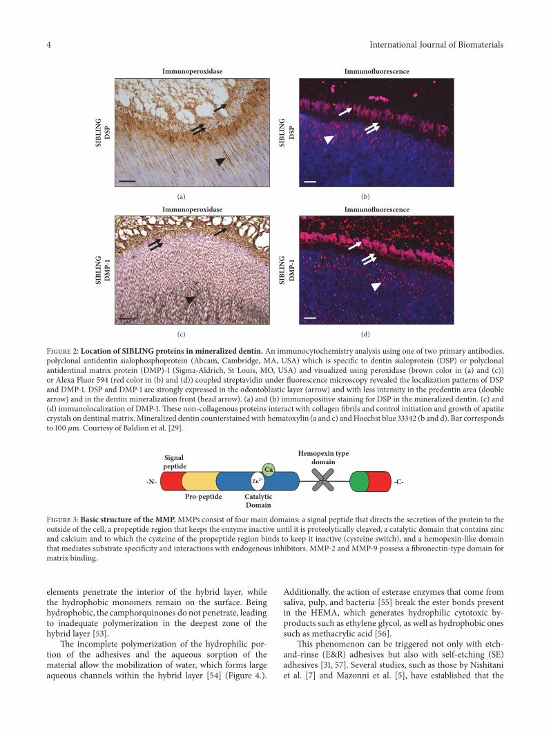
Noncollagenous proteins, such as proteoglycans (PG)(e.g., chondroitin-4/6-sulfate, decorin, biglycan, lumican,and fibromodulin) [26, 27], and small integrin-binding ligandN-linked glycoproteins (SIBLING), such as bone sialopro-tein, osteopontin, protein dentinal-1 matrix, and dentinsialophosphoprotein, play important roles in dentinogenesis,including regulation functions and control of crystal growth,fibrillogenesis, and mineralization [28] (Figure 2).
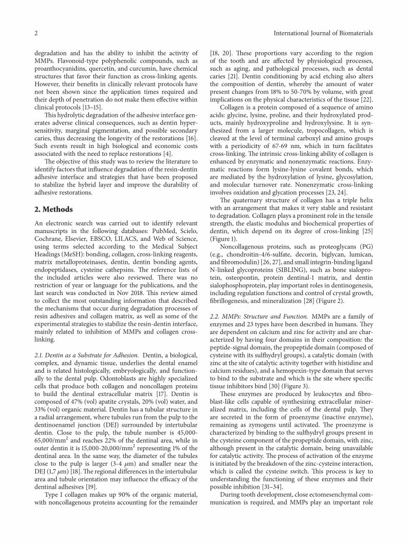
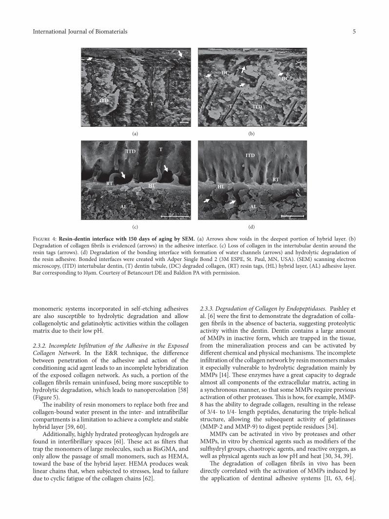
2.2. MMPs: Structure and Function. MMPs are a family ofenzymes and 23 types have been described in humans. Theyare dependent on calcium and zinc for activity and are char-acterized by having four domains in their composition: thepeptide-signal domain, the propeptide domain (composed ofcysteine with its sulfhydryl groups), a catalytic domain (withzinc at the site of catalytic activity together with histidine andcalcium residues), and a hemopexin-type domain that servesto bind to the substrate and which is the site where specifictissue inhibitors bind [30] (Figure 3).
These enzymes are produced by leukocytes and fibro-blast-like cells capable of synthesizing extracellular miner-alized matrix, including the cells of the dental pulp. Theyare secreted in the form of proenzyme (inactive enzyme),remaining as zymogens until activated. The proenzyme ischaracterized by binding to the sulfhydryl groups present inthe cysteine component of the propeptide domain, with zinc,although present in the catalytic domain, being unavailablefor catalytic activity. The process of activation of the enzymeis initiated by the breakdown of the zinc-cysteine interaction,which is called the cysteine switch. This process is key tounderstanding the functioning of these enzymes and theirpossible inhibition [31–34].
During tooth development, close ectomesenchymal com-munication is required, and MMPs play an important role
International Journal of Biomaterials 3
Collagen Helix Collagen Triple Helix
8.7
nm
Gly
Hyp
Pro
Trop
ocol
lage
n
Collagen bundles instepped arrangement
280
nm70
nm
1
1
2
Figure 1: Structure of collagen. Each collagen helix is called the 𝛼 chain, which is a levorotatory strand with about 3 residues per turnof glycine (Gly), proline (Pro), and hydroxyproline (Hyp) sequences. The quaternary structure of the collagen fibril is formed from thesupercoiling of three 𝛼 chains to form a triple dextrorotatory helix.
in this interaction. In early stages of odontogenesis, thisinteraction determines dental morphogenesis and in latestages, determines the differentiation of odontoblasts andameloblasts [35, 36]. Mesenchymal cells express, at least,MMP-1, MMP-2, MMP-3, MMP-8, MMP-9, MMP-14, andMMP-20. Additionally, the role of MMPs in the processof reabsorption of extracellular matrix proteins has beenproposed as a regulatory mechanism necessary for correctmineralization of the dental structure [37]. Some in the formof proenzymes are trapped inside the mineralized dentinmatrix during dentinal development [38], and because oftheir collagenolytic protease activity, they can hydrolyzethe components of the extracellular matrix when activatedby physical or chemical stimuli [39]. These proteases playa central role in several physiological processes, such asdevelopment, tissue remodeling, and angiogenesis [40].
MMPs are involved in different pathological processes,such as periodontal disease and dental caries. Recent studieshave revealed their role in the breakdown of the collagenmatrix in the pathogenesis of caries [33, 41], with potentialand relevant implications for dentin binding [42]. In addition,they may be present in saliva [43], peritubular dentin, andpresumably, dentinal fluid [44].
Of the proposed classifications to name MMPs, the onemost used was established based on the substrate on whichthey have activity. Therefore, they are named as collage-nases (MMP-1, MMP-8, MMP-13, and MMP-18), gelatinases(MMP-2 and MMP-9), stromelysins (MMP-3 and MMP-10), matrilysins (MMP-7 and MMP-26), and membrane-typeMMP (MMP-14, MMP-15, MMP-16, and MMP-24) (Bali etal., 2016). MMPs found in human dentin are MMP-2, MMP-9 [40], MMP-8 [32], MMP-3 [45] and MMP-20 [46].
2.3. Degradation of the Adhesive Interface. Composite resins,as restorative materials, base their retention on an adhesiveprocess that unites them to the dental structure. Adhesionto dentin is a clinical challenge, because it is a tissue ofheterogeneous composition, with high organic content andthe presence of moisture [2, 22]. The adhesion of resinoussystems to the dentin requires the infiltration of collagenfibrils that have been exposed by previous acid conditioning,with the resin monomers present in the adhesive generatinga resin-dentin interdiffusion zone, called the hybrid layer [3].This is necessary for the micromechanical retention of therestoration.
It is widely accepted that after the use of current dentaladhesives, a degradation of the dentin-resin adhesive inter-face occurs [4]. Among the factors that intervene with thedegradation of the adhesive interface, the following have beenproposed:
2.3.1. Degradation of the Adhesive Resin. The use of hydro-philic monomers in adhesive systems, such as 2-hydroxyethylmethacrylate (HEMA), seeks to improve infiltration of theexposed collagen network, which is inherently humid. Thishas been reported to result in an immediate improvementof bond strength [47]; but the longevity of this dentin-resinbond is compromised with the use of these adhesive systems[48–50].
The presence of water in the adhesive interface gen-erates a weak hybrid layer, in which the phenomena ofhydrolysis and leaching of resin adhesives occur [16, 51].Current adhesives include in their formulation hydrophilicand hydrophobic components that, in aqueous solution, pro-duce nanophase separation of adhesives [52].The hydrophilic
4 International Journal of Biomaterials
DSP
Immunoperoxidase
SIBL
ING
(a)
Immunofluorescence
DSP
SIBL
ING
(b)
DM
P-1
Immunoperoxidase
SIBL
ING
(c)
Immunofluorescence
DM
P-1
SIBL
ING
(d)
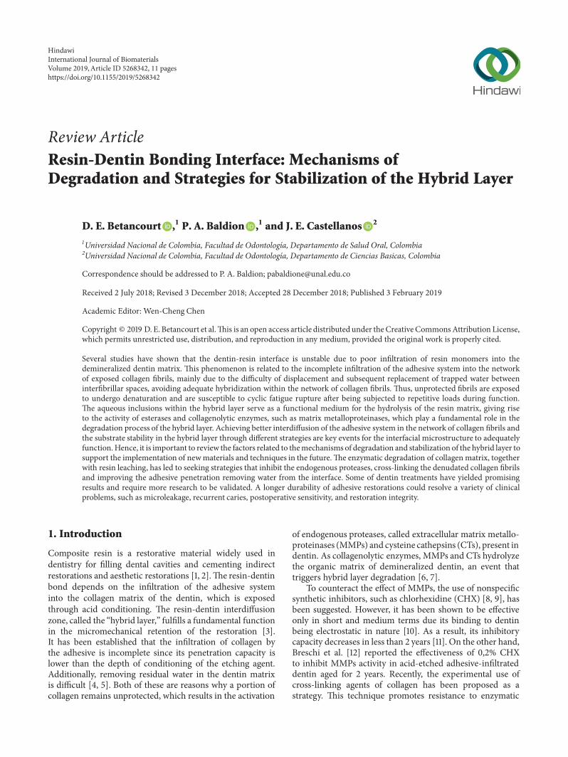
Figure 2: Location of SIBLING proteins in mineralized dentin. An immunocytochemistry analysis using one of two primary antibodies,polyclonal antidentin sialophosphoprotein (Abcam, Cambridge, MA, USA) which is specific to dentin sialoprotein (DSP) or polyclonalantidentinal matrix protein (DMP)-1 (Sigma-Aldrich, St Louis, MO, USA) and visualized using peroxidase (brown color in (a) and (c))or Alexa Fluor 594 (red color in (b) and (d)) coupled streptavidin under fluorescence microscopy revealed the localization patterns of DSPand DMP-1. DSP and DMP-1 are strongly expressed in the odontoblastic layer (arrow) and with less intensity in the predentin area (doublearrow) and in the dentin mineralization front (head arrow). (a) and (b) immunopositive staining for DSP in the mineralized dentin. (c) and(d) immunolocalization of DMP-1. These non-collagenous proteins interact with collagen fibrils and control initiation and growth of apatitecrystals on dentinal matrix.Mineralized dentin counterstainedwith hematoxylin (a and c) andHoechst blue 33342 (b and d). Bar correspondsto 100 𝜇m. Courtesy of Baldion et al. [29].
-N-
Signal peptide
Pro-peptide Catalytic Domain
Ca
Hemopexin typedomain
-C-Zn+
Figure 3: Basic structure of the MMP. MMPs consist of four main domains: a signal peptide that directs the secretion of the protein to theoutside of the cell, a propeptide region that keeps the enzyme inactive until it is proteolytically cleaved, a catalytic domain that contains zincand calcium and to which the cysteine of the propeptide region binds to keep it inactive (cysteine switch), and a hemopexin-like domainthat mediates substrate specificity and interactions with endogenous inhibitors. MMP-2 and MMP-9 possess a fibronectin-type domain formatrix binding.
elements penetrate the interior of the hybrid layer, whilethe hydrophobic monomers remain on the surface. Beinghydrophobic, the camphorquinones do not penetrate, leadingto inadequate polymerization in the deepest zone of thehybrid layer [53].
The incomplete polymerization of the hydrophilic por-tion of the adhesives and the aqueous sorption of thematerial allow the mobilization of water, which forms largeaqueous channels within the hybrid layer [54] (Figure 4.).
Additionally, the action of esterase enzymes that come fromsaliva, pulp, and bacteria [55] break the ester bonds presentin the HEMA, which generates hydrophilic cytotoxic by-products such as ethylene glycol, as well as hydrophobic onessuch as methacrylic acid [56].
This phenomenon can be triggered not only with etch-and-rinse (E&R) adhesives but also with self-etching (SE)adhesives [31, 57]. Several studies, such as those by Nishitaniet al. [7] and Mazonni et al. [5], have established that the
International Journal of Biomaterials 5
ITD T
(a)
ITDT
DCDC
(b)
RT HL
AL
ITD T
(c)
RTHL
AL
ITD
(d)
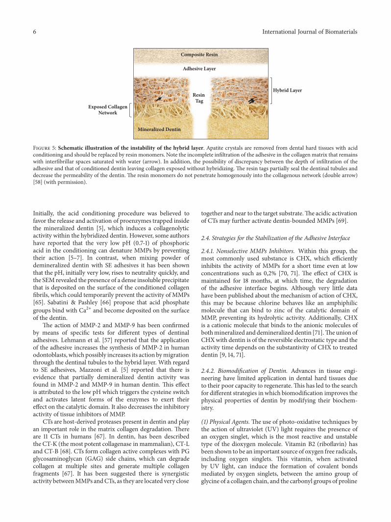
Figure 4: Resin-dentin interface with 150 days of aging by SEM. (a) Arrows show voids in the deepest portion of hybrid layer. (b)Degradation of collagen fibrils is evidenced (arrows) in the adhesive interface. (c) Loss of collagen in the intertubular dentin around theresin tags (arrows). (d) Degradation of the bonding interface with formation of water channels (arrows) and hydrolytic degradation ofthe resin adhesive. Bonded interfaces were created with Adper Single Bond 2 (3M ESPE, St. Paul, MN, USA). (SEM) scanning electronmicroscopy, (ITD) intertubular dentin, (T) dentin tubule, (DC) degraded collagen, (RT) resin tags, (HL) hybrid layer, (AL) adhesive layer.Bar corresponding to 10𝜇m. Courtesy of Betancourt DE and Baldion PA with permission.
monomeric systems incorporated in self-etching adhesivesare also susceptible to hydrolytic degradation and allowcollagenolytic and gelatinolytic activities within the collagenmatrix due to their low pH.
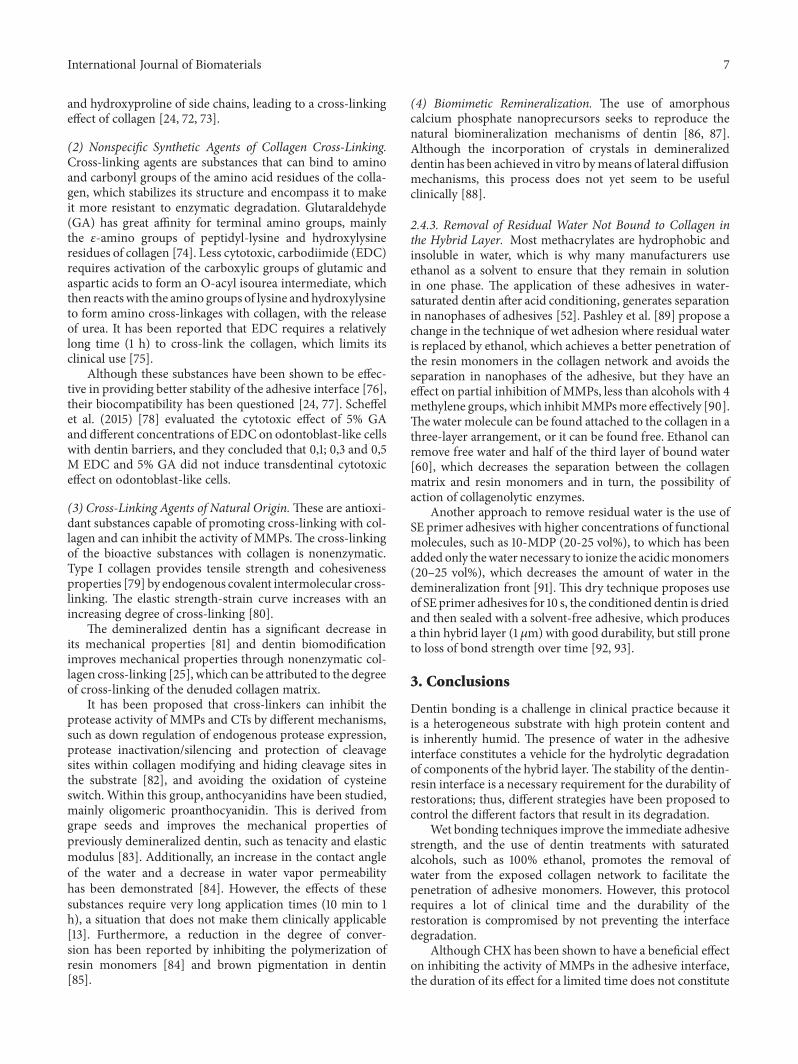
2.3.2. Incomplete Infiltration of the Adhesive in the ExposedCollagen Network. In the E&R technique, the differencebetween penetration of the adhesive and action of theconditioning acid agent leads to an incomplete hybridizationof the exposed collagen network. As such, a portion of thecollagen fibrils remain uninfused, being more susceptible tohydrolytic degradation, which leads to nanopercolation [58](Figure 5).
The inability of resin monomers to replace both free andcollagen-bound water present in the inter- and intrafibrillarcompartments is a limitation to achieve a complete and stablehybrid layer [59, 60].
Additionally, highly hydrated proteoglycan hydrogels arefound in interfibrillary spaces [61]. These act as filters thattrap the monomers of large molecules, such as BisGMA, andonly allow the passage of small monomers, such as HEMA,toward the base of the hybrid layer. HEMA produces weaklinear chains that, when subjected to stresses, lead to failuredue to cyclic fatigue of the collagen chains [62].
2.3.3. Degradation of Collagen by Endopeptidases. Pashley etal. [6] were the first to demonstrate the degradation of colla-gen fibrils in the absence of bacteria, suggesting proteolyticactivity within the dentin. Dentin contains a large amountof MMPs in inactive form, which are trapped in the tissue,from the mineralization process and can be activated bydifferent chemical and physical mechanisms. The incompleteinfiltration of the collagen network by resinmonomersmakesit especially vulnerable to hydrolytic degradation mainly byMMPs [14]. These enzymes have a great capacity to degradealmost all components of the extracellular matrix, acting ina synchronous manner, so that some MMPs require previousactivation of other proteases.This is how, for example, MMP-8 has the ability to degrade collagen, resulting in the releaseof 3/4- to 1/4- length peptides, denaturing the triple-helicalstructure, allowing the subsequent activity of gelatinases(MMP-2 and MMP-9) to digest peptide residues [34].
MMPs can be activated in vivo by proteases and otherMMPs, in vitro by chemical agents such as modifiers of thesulfhydryl groups, chaotropic agents, and reactive oxygen, aswell as physical agents such as low pH and heat [30, 34, 39].
The degradation of collagen fibrils in vivo has beendirectly correlated with the activation of MMPs induced bythe application of dentinal adhesive systems [11, 63, 64].
6 International Journal of Biomaterials
Mineralized Dentin
Composite Resin
Exposed CollagenNetwork
Adhesive Layer
Resin Tag
Hybrid Layer
Figure 5: Schematic illustration of the instability of the hybrid layer. Apatite crystals are removed from dental hard tissues with acidconditioning and should be replaced by resin monomers. Note the incomplete infiltration of the adhesive in the collagen matrix that remainswith interfibrillar spaces saturated with water (arrow). In addition, the possibility of discrepancy between the depth of infiltration of theadhesive and that of conditioned dentin leaving collagen exposed without hybridizing. The resin tags partially seal the dentinal tubules anddecrease the permeability of the dentin. The resin monomers do not penetrate homogenously into the collagenous network (double arrow)[58] (with permission).
Initially, the acid conditioning procedure was believed tofavor the release and activation of proenzymes trapped insidethe mineralized dentin [5], which induces a collagenolyticactivity within the hybridized dentin. However, some authorshave reported that the very low pH (0.7-1) of phosphoricacid in the conditioning can denature MMPs by preventingtheir action [5–7]. In contrast, when mixing powder ofdemineralized dentin with SE adhesives it has been shownthat the pH, initially very low, rises to neutrality quickly, andthe SEM revealed the presence of a dense insoluble precipitatethat is deposited on the surface of the conditioned collagenfibrils, which could temporarily prevent the activity of MMPs[65]. Sabatini & Pashley [66] propose that acid phosphategroups bind with Ca2+ and become deposited on the surfaceof the dentin.
The action of MMP-2 and MMP-9 has been confirmedby means of specific tests for different types of dentinaladhesives. Lehmann et al. [57] reported that the applicationof the adhesive increases the synthesis of MMP-2 in humanodontoblasts, which possibly increases its action bymigrationthrough the dentinal tubules to the hybrid layer. With regardto SE adhesives, Mazzoni et al. [5] reported that there isevidence that partially demineralized dentin activity wasfound in MMP-2 and MMP-9 in human dentin. This effectis attributed to the low pH which triggers the cysteine switchand activates latent forms of the enzymes to exert theireffect on the catalytic domain. It also decreases the inhibitoryactivity of tissue inhibitors of MMP.
CTs are host-derived proteases present in dentin and playan important role in the matrix collagen degradation. Thereare 11 CTs in humans [67]. In dentin, has been describedthe CT-K (the most potent collagenase inmammalian), CT-Land CT-B [68]. CTs form collagen active complexes with PGglycosaminoglycan (GAG) side chains, which can degradecollagen at multiple sites and generate multiple collagenfragments [67]. It has been suggested there is synergisticactivity betweenMMPs andCTs, as they are located very close
together and near to the target substrate.The acidic activationof CTs may further activate dentin-bounded MMPs [69].
2.4. Strategies for the Stabilization of the Adhesive Interface
2.4.1. Nonselective MMPs Inhibitors. Within this group, themost commonly used substance is CHX, which efficientlyinhibits the activity of MMPs for a short time even at lowconcentrations such as 0,2% [70, 71]. The effect of CHX ismaintained for 18 months, at which time, the degradationof the adhesive interface begins. Although very little datahave been published about the mechanism of action of CHX,this may be because chlorine behaves like an amphiphilicmolecule that can bind to zinc of the catalytic domain ofMMP, preventing its hydrolytic activity. Additionally, CHXis a cationic molecule that binds to the anionic molecules ofbothmineralized and demineralized dentin [71].TheunionofCHXwith dentin is of the reversible electrostatic type and theactivity time depends on the substantivity of CHX to treateddentin [9, 14, 71].
2.4.2. Biomodification of Dentin. Advances in tissue engi-neering have limited application in dental hard tissues dueto their poor capacity to regenerate.This has led to the searchfor different strategies in which biomodification improves thephysical properties of dentin by modifying their biochem-istry.
(1) Physical Agents.The use of photo-oxidative techniques bythe action of ultraviolet (UV) light requires the presence ofan oxygen singlet, which is the most reactive and unstabletype of the dioxygen molecule. Vitamin B2 (riboflavin) hasbeen shown to be an important source of oxygen free radicals,including oxygen singlets. This vitamin, when activatedby UV light, can induce the formation of covalent bondsmediated by oxygen singlets, between the amino group ofglycine of a collagen chain, and the carbonyl groups of proline
International Journal of Biomaterials 7
and hydroxyproline of side chains, leading to a cross-linkingeffect of collagen [24, 72, 73].
(2) Nonspecific Synthetic Agents of Collagen Cross-Linking.Cross-linking agents are substances that can bind to aminoand carbonyl groups of the amino acid residues of the colla-gen, which stabilizes its structure and encompass it to makeit more resistant to enzymatic degradation. Glutaraldehyde(GA) has great affinity for terminal amino groups, mainlythe 𝜀-amino groups of peptidyl-lysine and hydroxylysineresidues of collagen [74]. Less cytotoxic, carbodiimide (EDC)requires activation of the carboxylic groups of glutamic andaspartic acids to form an O-acyl isourea intermediate, whichthen reactswith the amino groups of lysine and hydroxylysineto form amino cross-linkages with collagen, with the releaseof urea. It has been reported that EDC requires a relativelylong time (1 h) to cross-link the collagen, which limits itsclinical use [75].
Although these substances have been shown to be effec-tive in providing better stability of the adhesive interface [76],their biocompatibility has been questioned [24, 77]. Scheffelet al. (2015) [78] evaluated the cytotoxic effect of 5% GAand different concentrations of EDC on odontoblast-like cellswith dentin barriers, and they concluded that 0,1; 0,3 and 0,5M EDC and 5% GA did not induce transdentinal cytotoxiceffect on odontoblast-like cells.
(3) Cross-Linking Agents of Natural Origin.These are antioxi-dant substances capable of promoting cross-linking with col-lagen and can inhibit the activity of MMPs.The cross-linkingof the bioactive substances with collagen is nonenzymatic.Type I collagen provides tensile strength and cohesivenessproperties [79] by endogenous covalent intermolecular cross-linking. The elastic strength-strain curve increases with anincreasing degree of cross-linking [80].
The demineralized dentin has a significant decrease inits mechanical properties [81] and dentin biomodificationimproves mechanical properties through nonenzymatic col-lagen cross-linking [25], which can be attributed to the degreeof cross-linking of the denuded collagen matrix.
It has been proposed that cross-linkers can inhibit theprotease activity of MMPs and CTs by different mechanisms,such as down regulation of endogenous protease expression,protease inactivation/silencing and protection of cleavagesites within collagen modifying and hiding cleavage sites inthe substrate [82], and avoiding the oxidation of cysteineswitch.Within this group, anthocyanidins have been studied,mainly oligomeric proanthocyanidin. This is derived fromgrape seeds and improves the mechanical properties ofpreviously demineralized dentin, such as tenacity and elasticmodulus [83]. Additionally, an increase in the contact angleof the water and a decrease in water vapor permeabilityhas been demonstrated [84]. However, the effects of thesesubstances require very long application times (10 min to 1h), a situation that does not make them clinically applicable[13]. Furthermore, a reduction in the degree of conver-sion has been reported by inhibiting the polymerization ofresin monomers [84] and brown pigmentation in dentin[85].
(4) Biomimetic Remineralization. The use of amorphouscalcium phosphate nanoprecursors seeks to reproduce thenatural biomineralization mechanisms of dentin [86, 87].Although the incorporation of crystals in demineralizeddentin has been achieved in vitro bymeans of lateral diffusionmechanisms, this process does not yet seem to be usefulclinically [88].
2.4.3. Removal of Residual Water Not Bound to Collagen inthe Hybrid Layer. Most methacrylates are hydrophobic andinsoluble in water, which is why many manufacturers useethanol as a solvent to ensure that they remain in solutionin one phase. The application of these adhesives in water-saturated dentin after acid conditioning, generates separationin nanophases of adhesives [52]. Pashley et al. [89] propose achange in the technique of wet adhesion where residual wateris replaced by ethanol, which achieves a better penetration ofthe resin monomers in the collagen network and avoids theseparation in nanophases of the adhesive, but they have aneffect on partial inhibition ofMMPs, less than alcohols with 4methylene groups, which inhibitMMPsmore effectively [90].Thewater molecule can be found attached to the collagen in athree-layer arrangement, or it can be found free. Ethanol canremove free water and half of the third layer of bound water[60], which decreases the separation between the collagenmatrix and resin monomers and in turn, the possibility ofaction of collagenolytic enzymes.
Another approach to remove residual water is the use ofSE primer adhesives with higher concentrations of functionalmolecules, such as 10-MDP (20-25 vol%), to which has beenadded only thewater necessary to ionize the acidicmonomers(20–25 vol%), which decreases the amount of water in thedemineralization front [91]. This dry technique proposes useof SE primer adhesives for 10 s, the conditioned dentin is driedand then sealed with a solvent-free adhesive, which producesa thin hybrid layer (1 𝜇m)with good durability, but still proneto loss of bond strength over time [92, 93].
3. Conclusions
Dentin bonding is a challenge in clinical practice because itis a heterogeneous substrate with high protein content andis inherently humid. The presence of water in the adhesiveinterface constitutes a vehicle for the hydrolytic degradationof components of the hybrid layer.The stability of the dentin-resin interface is a necessary requirement for the durability ofrestorations; thus, different strategies have been proposed tocontrol the different factors that result in its degradation.
Wet bonding techniques improve the immediate adhesivestrength, and the use of dentin treatments with saturatedalcohols, such as 100% ethanol, promotes the removal ofwater from the exposed collagen network to facilitate thepenetration of adhesive monomers. However, this protocolrequires a lot of clinical time and the durability of therestoration is compromised by not preventing the interfacedegradation.
Although CHX has been shown to have a beneficial effecton inhibiting the activity of MMPs in the adhesive interface,the duration of its effect for a limited time does not constitute
8 International Journal of Biomaterials
a final solution to the problem of the degradation of thehybrid layer.
The biomodification of dentin is presented as an optionthat shows promising results. Cross-linking of the collagenin demineralized dentin improves the physical properties ofdentin and makes it more resistant to degradation by theaction of collagenolytic enzymes, in addition to the abilityto inhibit the activity of MMPs and CTs by some cross-linking agents. However, all studies have been conductedin vitro since these substances have limitations that do notallow them to be used in a relevant clinical protocol. Due tothis unresolved problem, it is necessary to continue with thesearch for new substances and techniques that can be usedin a clinical protocol to improve the stability of the resin-dentin bonding interface, with which more durable adhesiverestorations can be obtained.
Conflicts of Interest
The authors declare that they have no conflicts of interest.
References
[1] K. L. Van Landuyt, J. Snauwaert, J. De Munck et al., “Systematicreview of the chemical composition of contemporary dentaladhesives,” Biomaterials, vol. 28, no. 26, pp. 3757–3785, 2007.
[2] Y. Liu, L. Tjaderhane, L. Breschi et al., “Limitations in bondingto dentin and experimental strategies to prevent bond degra-dation,” Journal of Dental Research, vol. 90, no. 8, pp. 953–968,2011.
[3] N.Nakabayashi,M.Nakamura, andN.Yasuda, “HybridLayer asa Dentin-Bonding Mechanism,” Journal of Esthetic and Restor-ative Dentistry, vol. 3, no. 4, pp. 133–138, 1991.
[4] D. H. Pashley, F. R. Tay, L. Breschi et al., “State of the art etch-and-rinse adhesives,” Dental Materials, vol. 27, no. 1, pp. 1–16,2011.
[5] A. Mazzoni, P. Scaffa, M. Carrilho et al., “Effects of etch-and-rinse and self-etch adhesives on dentin MMP-2 and MMP-9,”Journal of Dental Research, vol. 92, no. 1, pp. 82–86, 2013.
[6] D. H. Pashley, F. R. Tay, C. Yiu et al., “Collagen degradation byhost-derived enzymes during aging,” Journal ofDental Research,vol. 83, no. 3, pp. 216–221, 2004.
[7] Y. Nishitani, M. Yoshiyama, B. Wadgaonkar et al., “Activationof gelatinolytic/collagenolytic activity in dentin by self-etchingadhesives,” European Journal of Oral Sciences, vol. 114, no. 2, pp.160–166, 2006.
[8] M. R. O. Carrilho, R. M. Carvalho, M. F. de Goes et al., “Chlor-hexidine preserves dentin bond in vitro,” Journal of DentalResearch, vol. 86, no. 1, pp. 90–94, 2007.
[9] M. R.O. Carrilho, S. Geraldeli, F. Tay et al., “In vivo preservationof the hybrid layer by chlorhexidine,” Journal ofDental Research,vol. 86, no. 6, pp. 529–533, 2007.
[10] R. S. Blackburn, A. Harvey, L. L. Kettle, A. P. Manian, J. D.Payne, and S. J. Russell, “Sorption of chlorhexidine on cellulose:Mechanism of binding and molecular recognition,”The Journalof Physical Chemistry B, vol. 111, no. 30, pp. 8775–8784, 2007.
[11] J. De Munck, P. E. Van Den Steen, A. Mine et al., “Inhibition ofenzymatic degradation of adhesive-dentin interfaces,” Journal ofDental Research, vol. 88, no. 12, pp. 1101–1106, 2009.
[12] L. Breschi, A. Mazzoni, F. Nato et al., “Chlorhexidine stabilizesthe adhesive interface: a 2-year in vitro study,”DentalMaterials,vol. 26, no. 4, pp. 320–325, 2010.
[13] V. Hass, I. V. Luque-Martinez, M. F. Gutierrez et al., “Collagencross-linkers on dentin bonding: stability of the adhesiveinterfaces, degree of conversionof the adhesive, cytotoxicity andin situ MMP inhibition,” Dental Materials, vol. 32, no. 6, pp.732–741, 2016.
[14] R. Seseogullari-Dirihan, F. Apollonio, A.Mazzoni et al., “Use ofcrosslinkers to inactivate dentin MMPs,” Dental Materials, vol.32, no. 3, pp. 423–432, 2016.
[15] A. K. B. Bedran-Russo, P. N. R. Pereira, W. R. Duarte, J. L.Drummond, and M. Yamaychi, “Application of crosslinkers todentin collagen enhances the ultimate tensile strength,” Journalof Biomedical Materials Research Part B: Applied Biomaterials,vol. 80, no. 1, pp. 268–272, 2007.
[16] S. Kermanshahi, J. P. Santerre, D. G. Cvitkovitch, and Y. Finer,“Biodegradation of resin-dentin interfaces increases bacterialmicroleakage,” Journal ofDental Research, vol. 89, no. 9, pp. 996–1001, 2010.
[17] A. Linde and M. Goldberg, “Dentinogenesis,” Critical Reviewsin Oral Biology & Medicine, vol. 4, no. 5, pp. 679–728, 2016.
[18] L. Tjaderhane, M. R. Carrilho, L. Breschi, F. R. Tay, and D. H.Pashley, “Dentin basic structure and composition-an overview,”Endodontic Topics, vol. 20, no. 1, pp. 3–29, 2009.
[19] A. F. Montagner, M. P. M. Carvalho, and A. H. Susin, “Micros-hear bonding effectiveness of different dentin regions,” IndianJournal of Dental Research, vol. 26, no. 2, pp. 131–135, 2015.
[20] W. T. Butler, J. C. Brunn, and C. Qin, “Dentin extracellularmatrix (ECM) proteins: comparison to bone ECM and con-tribution to dynamics of dentinogenesis,” Connective TissueResearch, vol. 44, no. 1, pp. 171–178, 2003.
[21] G. W. Marshall Jr., S. J. Marshall, J. H. Kinney, and M. Balooch,“The dentin substrate: structure and properties related tobonding,” Journal of Dentistry, vol. 25, no. 6, pp. 441–458, 1997.
[22] R. M. Carvalho, L. Tjaderhane, A. P. Manso, M. R. Carrilho,andC.A. Carvalho, “Dentin as a bonding substrate,”EndodonticTopics, vol. 21, no. 1, pp. 62–88, 2009.
[23] M. Saito and K. Marumo, “Collagen cross-links as a determi-nant of bone quality: a possible explanation for bone fragilityin aging, osteoporosis, and diabetes mellitus,” OsteoporosisInternational, vol. 21, no. 2, pp. 195–214, 2010.
[24] A. K. Bedran-Russo, G. F. Pauli, S.-N. Chen et al., “Dentinbiomodification: Strategies, renewable resources and clinicalapplications,” Dental Materials, vol. 30, no. 1, pp. 62–76, 2014.
[25] P. H. Dos Santos, S. Karol, and A. K. Bedran-Russo, “Long-term nano-mechanical properties of biomodified dentin-resininterface components,” Journal of Biomechanics, vol. 44, no. 9,pp. 1691–1694, 2011.
[26] G. Embery, R. Hall, R. Waddington, D. Septier, and M. Gold-berg, “Proteoglycans in Dentinogenesis,” Critical Reviews inOral Biology & Medicine, vol. 12, no. 4, pp. 331–349, 2016.
[27] K. A. Staines, V. E. MacRae, and C. Farquharson, “The impor-tance of the SIBLING family of proteins on skeletal mineralisa-tion and bone remodelling,” Journal of Endocrinology, vol. 214,no. 3, pp. 241–255, 2012.
[28] S. Suzuki, N. Haruyama, F. Nishimura, and A. B. Kulkarni,“Dentin sialophosphoprotein and dentin matrix protein-1: twohighly phosphorylated proteins inmineralized tissues,”Archivesof Oral Biolog, vol. 57, no. 9, pp. 1165–1175, 2012.
International Journal of Biomaterials 9
[29] P. A. Baldion, M. L. Velandia-Romero, and J. E. Castellanos,“Odontoblast-like cells differentiated from dental pulp stemcells retain their phenotype after subcultivation,” InternationalJournal of Cell Biology, vol. 2018, Article ID 6853189, 12 pages,2018.
[30] P. Bali, D. Kalaivanan, V. Divater, and . Logarani, “Matrix met-alloproteinases: A double edge sword,” Dentistry and MedicalResearch, vol. 4, no. 1, pp. 3–8, 2016.
[31] S.-C. Zhang and M. Kern, “The role of host-derived dentinalmatrix metalloproteinases in reducing dentin bonding of resinadhesives.,” International Journal of Oral Science, vol. 1, no. 4,pp. 163–176, 2009.
[32] M. Sulkala, T. Tervahartiala, T. Sorsa, M. Larmas, T. Salo, and L.Tjaderhane, “Matrixmetalloproteinase-8 (MMP-8) is themajorcollagenase in human dentin,” Archives of Oral Biolog, vol. 52,no. 2, pp. 121–127, 2007.
[33] C. Chaussain, T. Boukpessi, M. Khaddam, L. Tjaderhane, A.George, and S. Menashi, “Dentin matrix degradation by hostmatrix metalloproteinases: Inhibition and clinical perspectivestoward regeneration,” Frontiers in Physiology, vol. 4, pp. 1–8,2013.
[34] L. Tjaderhane, H. Larjava, T. Sorsa, V.-J. Uitto, M. Larmas, andT. Salo, “The activation and function of hostmatrix metallopro-teinases in dentin matrix breakdown in caries lesions,” Journalof Dental Research, vol. 77, no. 8, pp. 1622–1629, 1998.
[35] A. J. Smith, “Vitality of the dentinpulp complex in health anddisease: growth factors as key mediators,” Journal of DentalEducation, vol. 67, Article ID 678689, 2003.
[36] S. Fanchon, K. Bourd, D. Septier et al., “Involvement of matrixmetalloproteinases in the onset of dentin mineralization,” Euro-pean Journal of Oral Sciences, vol. 112, no. 2, pp. 171–176, 2004.
[37] M. Goldberg, A. B. Kulkarni, M. Young, and A. Boskey,“Dentin: structure, composition and mineralization,” Frontiersin Bioscience—Elite, vol. 3, no. 2, pp. 711–735, 2011.
[38] K. L. Van Landuyt, J. De Munck, J. Snauwaert et al., “Monomer-solvent phase separation in one-step self-etch adhesives,” Jour-nal of Dental Research, vol. 84, no. 2, pp. 183–188, 2005.
[39] C. E. Brinckerhoff and L. M. Matrisian, “Matrix metallopro-teinases: a tail of a frog that became a prince,” Nature ReviewsMolecular Cell Biology, vol. 3, no. 3, pp. 207–214, 2002.
[40] S. Martin-De Las Heras, A. Valenzuela, and C. M. Overall,“Thematrix metalloproteinase gelatinase A in human dentine,”Archives of Oral Biolog, vol. 45, no. 9, pp. 757–765, 2000.
[41] A. J. V. Strijp, D. C. Jansen, J. DeGroot, J. M. Ten Cate, and V.Everts, “Host-derived proteinases and degradation of dentinecollagen in situ,” Caries Research, vol. 37, no. 1, pp. 58–65, 2003.
[42] C. Chaussain-Miller, F. Fioretti, M. Goldberg, and S. Menashi,“The role of matrix metalloproteinases (MMPs) in humancaries,” Journal of Dental Research, vol. 85, no. 1, pp. 22–32, 2006.
[43] M. Sulkala, J. Wahlgren, M. Larmas et al., “The effects ofMMP inhibitors on human salivary MMP activity and cariesprogression in rats,” Journal of Dental Research, vol. 80, no. 6,pp. 1545–1549, 2001.
[44] L. W. Boushell, M. Kaku, Y. Mochida, R. Bagnell, and M.Yamauchi, “Immunohistochemical localization ofmatrixmetal-loproteinase-2 in human coronal dentin,” Archives of OralBiolog, vol. 53, no. 2, pp. 109–116, 2008.
[45] T. Boukpessi, S. Menashi, L. Camoin, J. M. TenCate, M. Gold-berg, and C. Chaussain-Miller, “The effect of stromelysin-1(MMP-3) on non-collagenous extracellular matrix proteins of
demineralized dentin and the adhesive properties of restorativeresins,” Biomaterials, vol. 29, no. 33, pp. 4367–4373, 2008.
[46] Y. Shimada, S. Ichinose, A. Sadr, M. F. Burrow, and J. Tagami,“Localization of matrix metalloproteinases (MMPs-2, 8, 9 and20) in normal and carious dentine,” Australian Dental Journal,vol. 54, no. 4, pp. 347–354, 2009.
[47] A. D. Loguercio, S. K. Moura, A. Pellizzaro et al., “Durabilityof enamel bonding using two-step self-etch systems on groundand unground enamel,” Operative Dentistry, vol. 33, no. 1, pp.79–88, 2008.
[48] M. Hashimoto, H. Ohno, H. Sano et al., “Micromorphologicalchanges in resin-dentin bonds after 1 year of water storage,”Journal of BiomedicalMaterials Research Part B: Applied Bioma-terials, vol. 63, no. 3, pp. 306–311, 2002.
[49] M. Hashimoto, H. Ohno, H. Sano, M. Kaga, and H. Oguchi, “Invitro degradation of resin-dentin bonds analyzed by microten-sile bond test, scanning and transmission electronmicroscopy,”Biomaterials, vol. 24, no. 21, pp. 3795–3803, 2003.
[50] J. Malacarne, R. M. Carvalho, M. F. de Goes et al., “Watersorption/solubility of dental adhesive resins,” Dental Materials,vol. 22, no. 10, pp. 973–980, 2006.
[51] M. Hashimoto, “A review—micromorphological evidence ofdegradation in resin-dentin bonds and potential preventionalsolutions,” Journal of Biomedical Materials Research Part B:Applied Biomaterials, vol. 92, no. 1, pp. 268–280, 2010.
[52] P. Spencer and Y. Wang, “Adhesive phase separation at thedentin interface under wet bonding conditions,” Journal ofBiomedical Materials Research Part B: Applied Biomaterials, vol.62, no. 3, pp. 447–456, 2002.
[53] Y. Wang, P. Spencer, X. Yao, and Q. Ye, “Effect of coinitiatorand water on the photoreactivity and photopolymerization ofHEMA/camphoquinone-based reactant mixtures,” Journal ofBiomedical Materials Research Part A, vol. 78A, no. 4, pp. 721–728, 2006.
[54] J. Tanaka, K. Ishikawa, H. Yatani, A. Yamashita, and K. Suzuki,“Correlation of Dentin Bond Durability withWater Absorptionof Bonding Layer,”DentalMaterials, vol. 18, no. 1, pp. 11–18, 1999.
[55] B. Shokati, L. E. Tam, J. P. Santerre, and Y. Finer, “Effect ofsalivary esterase on the integrity and fracture toughness of thedentin-resin interface,” Journal of BiomedicalMaterials ResearchPart B: Applied Biomaterials, vol. 94, no. 1, pp. 230–237, 2010.
[56] E. L. Kostoryz, K. Dharmala, Q. Ye et al., “Enzymatic biodegra-dation of HEMA/bisGMA adhesives formulated with differentwater content,” Journal of Biomedical Materials Research Part B:Applied Biomaterials, vol. 88B, no. 2, pp. 394–401, 2009.
[57] N. Lehmann, R.Debret, A. Romeas et al., “Self-etching increasesmatrix metalloproteinase expression in the dentin-pulp com-plex,” Journal of Dental Research, vol. 88, no. 1, pp. 77–82, 2009.
[58] P. A. Baldion and C. J. Cortes, “Mathematical models ofpolymer-dentin physicochemical interactions and their biolog-ical effects,” Scientific Journal of Review, vol. 5, pp. 319–330, 2016.
[59] Y. K. Kim, L.-S. Gu, T. E. Bryan et al., “Mineralisation of recon-stituted collagen using polyvinylphosphonic acid/polyacrylicacid templating matrix protein analogues in the presence ofcalcium, phosphate and hydroxyl ions,” Biomaterials, vol. 31, no.25, pp. 6618–6627, 2010.
[60] S. E. Jee, J. Zhou, J. Tan et al., “Investigation of ethanol infiltra-tion into demineralized dentin collagen fibrils using moleculardynamics simulations,” Acta Biomaterialia, vol. 36, pp. 175–185,2016.
10 International Journal of Biomaterials
[61] L. E. Bertassoni, “Dentin on the nanoscale: Hierarchical orga-nization, mechanical behavior and bioinspired engineering,”Dental Materials, vol. 33, no. 6, pp. 637–649, 2017.
[62] D.T. Fung,V.M.Wang,D.M. Laudier et al., “Subrupture tendonfatigue damage,” Journal of Orthopaedic Research, vol. 27, no. 2,pp. 264–273, 2009.
[63] F. R. Tay and D. H. Pashley, “Have dentin adhesives become toohydrophilic?” Journal of Canadian Dental Association, vol. 69,pp. 726–731, 2003.
[64] A. Mazzoni, D. H. Pashley, Y. Nishitani et al., “Reactivationof inactivated endogenous proteolytic activities in phosphoricacid-etched dentine by etch-and-rinse adhesives,” Biomaterials,vol. 27, no. 25, pp. 4470–4476, 2006.
[65] M. Iwasa, K. Tsubota, Y. Shimamura, S. Ando,M.Miyazaki, andJ. A. Platt, “pH changes upon mixing of single-step self-etchingadhesives with powdered dentin,” The Journal of AdhesiveDentistry, vol. 13, no. 3, pp. 207–212, 2011.
[66] C. Sabatini and D. H. Pashley, “Mechanisms regulating thedegradation of dentinmatrices by endogenous dentin proteasesand their role in dental adhesion. A review,” American Journalof Dentistry, vol. 27, no. 4, pp. 203–214, 2014.
[67] V. Turk, V. Stoka, O. Vasiljeva et al., “Cysteine cathepsins: fromstructure, function and regulation to new frontiers,” Biochimicaet Biophysica Acta—Proteins and Proteomics, vol. 1824, no. 1, pp.68–88, 2012.
[68] S. Mahalaxmi, M. M. Madhubala, M. Jayaraman, and S.Sathyakumar, “Evaluation of matrix metalloproteinase andcysteine cathepsin activity in dentin hybrid layer by gelatinzymography,” Indian Journal of Dental Research, vol. 27, no. 6,pp. 652–656, 2016.
[69] P. M. C. Scaffa, L. Breschi, A. Mazzoni et al., “Co-distributionof cysteine cathepsins and matrix metalloproteases in humandentin,” Archives of Oral Biolog, vol. 74, pp. 101–107, 2017.
[70] R. Gendron, D. Grenier, T. Sorsa, and D. Mayrand, “Inhibitionof the activities of matrix metalloproteinases 2, 8, and 9 bychlorhexidine,”Clinical andDiagnostic Laboratory Immunology,vol. 6, no. 3, pp. 437–439, 1999.
[71] M. R. Carrilho, R.M. Carvalho, E. N. Sousa et al., “Substantivityof chlorhexidine to human dentin,”DentalMaterials, vol. 26, no.8, pp. 779–785, 2010.
[72] A. Scott McCall, S. Kraft, H. F. Edelhauser et al., “Mechanismsof corneal tissue cross-linking in response to treatment withtopical riboflavin and long-wavelength ultraviolet radiation(UVA),” Investigative Ophthalmology & Visual Science, vol. 51,no. 1, pp. 129–138, 2010.
[73] E. Choe and D. B. Min, “Chemistry and reactions of reactiveoxygen species in foods,” Critical Reviews in Food Science andNutrition, vol. 46, no. 1, pp. 1–22, 2006.
[74] A. Tezvergil-Mutluay, K. A. Agee, T. Uchiyama et al., “Theinhibitory effects of quaternary ammonium methacrylates onsoluble and matrix-bound MMPs,” Journal of Dental Research,vol. 90, no. 4, pp. 535–540, 2011.
[75] A. Tezvergil-Mutluay, K. A. Agee, T. Hoshika, F. R. Tay, andD. H. Pashley, “The inhibitory effect of polyvinylphosphonicacid on functionalmatrixmetalloproteinase activities in humandemineralized dentin,” Acta Biomaterialia, vol. 6, no. 10, pp.4136–4142, 2010.
[76] A. Tezvergil-Mutluay, M. M. Mutluay, K. A. Agee et al., “Car-bodiimide cross-linking inactivates soluble and matrix-boundMMPs, in vitro,” Journal of Dental Research, vol. 91, no. 2, pp.192–196, 2012.
[77] A. Al-Ammar, J. L. Drummond, and A. K. Bedran-Russo,“The use of collagen cross-linking agents to enhance dentinbond strength,” Journal of Biomedical Materials Research PartB: Applied Biomaterials, vol. 91, no. 1, pp. 419–424, 2009.
[78] D. L. S. Scheffel, L. Bianchi, D. G. Soares et al., “Transdentinalcytotoxicity of carbodiimide (EDC) and glutaraldehyde onodontoblast-like cells,” Operative Dentistry, vol. 40, no. 1, pp.44–54, 2015.
[79] M. Yamauchi and M. Shiiba, “Lysine hydroxylation andcrosslinking of collagen.,”Methods inMolecular Biology (Clifton,N.J.), vol. 194, pp. 277–290, 2002.
[80] F. H. Silver, I. Horvath, and D. J. Foran, “Viscoelasticity ofthe vessel wall: the role of collagen and elastic fibers,” CriticalReviews in Biomedical Engineering, vol. 29, no. 3, pp. 279–301,2001.
[81] Y. Liu, X. Bai, S. Li, Y. Liu, A. Keightley, andY.Wang, “Molecularweight and galloylation affect grape seed extract constituents’ability to cross-link dentin collagen in clinically relevant time,”Dental Materials, vol. 31, no. 7, pp. 814–821, 2015.
[82] A. Jayakrishnan and S. R. Jameela, “Glutaraldehyde as a fixativein bioprostheses and drug delivery matrices,” Biomaterials, vol.17, no. 5, pp. 471–484, 1996.
[83] R.-R. Liu, M. Fang, L. Zhang, C.-F. Tang, Q. Dou, and J.-H. Chen, “Anti-proteolytic capacity and bonding durability ofproanthocyanidin-biomodified demineralized dentin matrix,”International Journal of Oral Science, vol. 6, no. 3, pp. 168–174,2014.
[84] B. Green, X. Yao, A. Ganguly et al., “Grape seed proantho-cyanidins increase collagen biodegradation resistance in thedentin/adhesive interface when included in an adhesive,” Jour-nal of Dentistry, vol. 38, no. 11, pp. 908–915, 2010.
[85] A. Frassetto, L. Breschi, G. Turco et al., “Mechanisms of degra-dation of the hybrid layer in adhesive dentistry and therapeuticagents to improve bond durability - A literature review,”DentalMaterials, vol. 32, no. 2, pp. e41–e53, 2016.
[86] F. R. Tay and D. H. Pashley, “Guided tissue remineralisation ofpartially demineralised human dentine,” Biomaterials, vol. 29,no. 8, pp. 1127–1137, 2008.
[87] L.-N. Niu,W. Zhang, D. H. Pashley et al., “Biomimetic reminer-alization of dentin,” Dental Materials, vol. 30, no. 1, pp. 77–96,2014.
[88] T. Maravic, A. Mazzoni, A. Comba, N. Scotti, V. Checchi, andL. Breschi, “How Stable is Dentin As a Substrate for Bonding?”Current Oral Health Reports, vol. 4, no. 3, pp. 248–257, 2017.
[89] D. H. Pashley, F. R. Tay, R. M. Carvalho et al., “From drybonding to water-wet bonding to ethanol-wet bonding. Areview of the interactions between dentin matrix and solvatedresins using a macromodel of the hybrid layer,” AmericanJournal of Dentistry, vol. 20, no. 1, pp. 7–20, 2007.
[90] A. Tezvergil-Mutluay, K. A. Agee, T. Hoshika et al., “Inhibitionof MMPs by alcohols,” Dental Materials, vol. 27, no. 9, pp. 926–933, 2011.
[91] N. Hiraishi, N. Nishiyama, K. Ikemura et al., “Water concen-tration in self-etching primers affects their aggressiveness andbonding efficacy to dentin,” Journal of Dental Research, vol. 84,no. 7, pp. 653–658, 2005.
[92] F. T. Sadek, C. S. Castellan, R. R. Braga et al., “One-year stabilityof resin-dentin bonds created with a hydrophobic ethanol-wetbonding technique,” Dental Materials, vol. 26, no. 4, pp. 380–386, 2010.

International Journal of Biomaterials 11
[93] L. Breschi, T. Maravic, S. R. Cunha et al., “Dentin bondingsystems: From dentin collagen structure to bond preservationand clinical applications,” Dental Materials, vol. 34, no. 1, pp.78–96, 2018.

CorrosionInternational Journal of
Hindawiwww.hindawi.com Volume 2018
Advances in
Materials Science and EngineeringHindawiwww.hindawi.com Volume 2018
Hindawiwww.hindawi.com Volume 2018
Journal of
Chemistry
Analytical ChemistryInternational Journal of
Hindawiwww.hindawi.com Volume 2018
Scienti�caHindawiwww.hindawi.com Volume 2018
Polymer ScienceInternational Journal of
Hindawiwww.hindawi.com Volume 2018
Hindawiwww.hindawi.com Volume 2018
Advances in Condensed Matter Physics
Hindawiwww.hindawi.com Volume 2018
International Journal of
BiomaterialsHindawiwww.hindawi.com
Journal ofEngineeringVolume 2018
Applied ChemistryJournal of
Hindawiwww.hindawi.com Volume 2018
NanotechnologyHindawiwww.hindawi.com Volume 2018
Journal of
Hindawiwww.hindawi.com Volume 2018
High Energy PhysicsAdvances in
Hindawi Publishing Corporation http://www.hindawi.com Volume 2013Hindawiwww.hindawi.com
The Scientific World Journal
Volume 2018
TribologyAdvances in
Hindawiwww.hindawi.com Volume 2018
Hindawiwww.hindawi.com Volume 2018
ChemistryAdvances in
Hindawiwww.hindawi.com Volume 2018
Advances inPhysical Chemistry
Hindawiwww.hindawi.com Volume 2018
BioMed Research InternationalMaterials
Journal of
Hindawiwww.hindawi.com Volume 2018
Na
nom
ate
ria
ls
Hindawiwww.hindawi.com Volume 2018
Journal ofNanomaterials
Submit your manuscripts atwww.hindawi.com


![IJBM 15.09 sept 2015 [web]](https://img.pdfslide.net/doc/110x75/58a45e411a28abb8288b4b57/ijbm-1509-sept-2015-web.jpg)


![IJBM juni 2015 [v-16]](https://img.pdfslide.net/doc/110x75/55c4e77ebb61eb973f8b45f1/ijbm-juni-2015-v-16.jpg)