Embed Size (px)
Citation preview
JOURNAL OF VIROLOGY, Sept. 1993, p. 5216-5225 Vol. 67, No. 90022-538X/93/095216-10$02.00/0Copyright © 1993, American Society for MicrobiologY
Resistance of a Human Serum-Selected HumanImmunodeficiency Virus Type 1 Escape Mutant to
Neutralization by CD4 Binding Site Monoclonal AntibodiesIs Conferred by a Single Amino Acid Change in gpl20
J. A. MCKEATING,lt*, J. BENNET,2 S. ZOLLA-PAZNER,3 M. SCHUT1rEN,4S. ASHELFORD,S A. LEIGH BROWN,5 AND P. BALFE2
Chester Beatty Laboratories, Institute of Cancer Research, London SW3 6JB, 1 Virology Department,University College London Medical School, London WIP 6DB, 2 and Centre for HIVResearch, University ofEdinburgh, Edinburgh EH9 3JN,5 United Kingdom; Research Service, Veterans Affairs Medical Center,New York, New York 10016, and Department ofPathology, New York University and Medical School,New York, New York 100103; and Laboratory ofImmunology, National Institute of Public Health
and Environmental Protection, 3720 BA Bilthoven, The Netherlands4
Received 1 March 1993/Accepted 2 June 1993
We have selected an HXB2 variant which can replicate in the presence of a neutralizing human serum.Sequencing of the gpl20 region of the env gene from the variant and parental viruses identified a single aminoacid substitution in the third conserved region of gpl20 at residue 375 (AGT-*AAT, Ser--Asn; designated 375S/N). The escape mutant was found to be resistant to neutralization by soluble CD4 (sCD4) and fourmonoclonal antibodies (MAbs), 39.13g, 1.5e, G13, and 448, binding to epitopes overlapping that of the CD4binding site (CD4 b.s.). Introduction of the 375 S/N mutation into HXB2 by site-directed mutagenesisconfirmed that this mutation is responsible for the neutralization-resistant phenotype. Both sCD4 and three ofthe CD4 b.s. MAbs (39.13g, 1.5e, and G13) demonstrated reduced binding to the native 375 S/N mutant gpl20.The ability to select for an escape variant resistant to multiple independent CD4 b.s. MAbs by a human serumconfirms the reports that antibodies to the discontinuous CD4 b.s. are a major component of the group-specificneutralizing activity in human sera.
Neutralizing antibody appears to be an important compo-nent of the protective immune response against humanimmunodeficiency virus type 1 (HIV-1) infection (1, 4-6).Sera from many HIV-1-infected individuals are able toneutralize a broad spectrum of virus isolates, indicating thepresence of conserved neutralization epitopes (28, 37). Theinduction of antibodies with broad neutralizing capacity is aprimary goal of vaccine development strategies. Severalreports have suggested that HIV-1 neutralizing activity isassociated primarily with two regions of the gp120 envelopeglycoprotein: the third hypervariable loop (V3) (8, 10, 29)and the CD4 binding site (CD4 b.s.) (11, 34). Generally,antibodies specific for V3 arise early after HIV infection andexhibit the ability to neutralize a limited number of HIV-1isolates. However, a limited number of V3 monoclonalantibodies (MAbs) recognize conserved features of the loopand subsequently exhibit a broader neutralization profile (7,10, 24). Later in the course of viral infection, antibodiescapable of neutralizing a wider range of HIV-1 isolates aredetectable (20, 28, 37). The appearance of such cross-neutralizing activity coincides with the detection of antibod-ies capable of blocking gp120-soluble CD4 (sCD4) interac-tion (9, 11, 18, 34). A number of neutralizing MAbs derivedboth from infected individuals (9, 12, 26, 30, 36) and fromrecombinant gp120-immunized rats (3) which block gp120-sCD4 binding have been identified, suggesting that such
* Corresponding author.t Present address: Microbiology Department, University of Read-
ing, Whiteknights, P.O. Box 228, Reading RG6 2AJ, United King-dom.
antibodies may play an important role in the cross-neutral-izing response (21).The selection and characterization of neutralization-resis-
tant escape mutants is one strategy for the identification ofamino acid residues critical for the binding of neutralizingantibodies. We have selected escape variants which areresistant to both V3 MAbs and sCD4 (13, 15). To define theepitopes responsible for inducing cross-neutralizing antibod-ies in human sera further, we selected an escape variant thatcould replicate in the presence of a neutralizing humanserum. Characterization of the neutralization sensitivity ofthe escape variant with a panel of MAbs mapping to thesecond variable loop (V2), V3, and the CD4 b.s. demon-strated that the variant was resistant to a number of MAbsmapping to the CD4 b.s., suggesting that antibodies specificfor the CD4 b.s. in human sera are capable of cross-neutralizing a nonhomologous HIV-1 isolate (HXB2) andmay be sufficient to select for neutralization-resistant escapemutants.
MATERIALS AND METHODS
Virus propagation and neutralization assays. (i) Transfec-tion of H9 cells and viral stock production. H9 cells werewashed twice in serum-free medium and resuspended at106/ml in RPMI. Ten micrograms of HXB2 plasmid wastransfected into 2 x 10⁶ H9 cells by the use of Lipofectin asinstructed by the manufacturer (GIBCO BRL, Gaithersburg,Md.). The transfected H9 cultures were monitored for viralinfection by the detection of p24 antigen (as described
5216
RESISTANCE OF AN HIV-1 ESCAPE MUTANT TO NEUTRALIZATION 5217
below) and by the appearance of multinucleated cells. Oncethe cells were producing virus (days 6 to 7 posttransfection),stocks of extracellular virus were made by mixing theinfected cells with uninfected H9 cells in a 1:4 ratio. Forty-eight hours after the cocultivation, the supernatant wascollected, aliquoted, and stored under liquid N2. Infectivitydeterminations were made by performing 10-fold serial dilu-tions of virus (50 pI) and incubation with 100 pI of C8166cells at 2 x 10⁵ cells per ml in microtiter plates at 37°C for 7days. Plates were scored for the presence of p24 antigen, andthe 50% tissue culture infective dose (TCID50) values weredetermined by the Karber formula.
(ii) Neutralization assays. HIV (103 TCID50) in a volume of40 ptl was incubated with 10 RI of a dilution of antibody,serum, or sCD4 under test at 37°C for 1 h. The virus-antibody or virus-sCD4 mixture was then incubated with 100pI of C8166 cells at a concentration of 2 x 10⁵ cells per mlper well in a microtiter plate in triplicate. Five days postin-fection, the wells were scored for the presence of syncytia,and the extracellular supernatant was collected from thewells by centrifugation, inactivated with 1% Empigen, andassayed for soluble p24 antigen as described previously (17).The lowest concentration of antibody or sCD4 resultingeither in a complete blocking of syncytium formation or in a>90% reduction in p24 antigen production was defined as thereciprocal neutralization titer.
(iii) Comparative neutralization of variant and wt virus byhuman sera. Human serum neutralization titers were ob-tained for both variant and wild-type (wt) viruses. Thedifferences in the titers were compared by using a two-tailnonparametric test (Wilcoxon's rank sum) (see Fig. 7).MAbs and human sera. The following antibodies were
used for neutralization studies and/or envelope binding stud-ies: rat MAbs 10/54, 10/36e, and 11/85b, specific for aminoacids 311 to 321 of the HXB10 V3 loop (14); rat MAb 41.1,recognizing a conformation-dependent epitope within the V3loop (14); rat MAb 10/76b, mapping to a linear epitope in theV2 loop (17a); 38.1a, specific for amino acids 425 to 441,involved in CD4 binding (3); 39.13g, a rat MAb specific foran epitope overlapping the CD4 b.s. (3, 19); and humanMAbs 588, 559, 448, 728, 654, 729 (12, 19), 1.5e, 2.1h (9, 35),and G13 (30), binding to closely related epitopes overlappingthe CD4 b.s.; and murine MAb L120, specific for CD4(Medical Research Council [MRC] AIDS Directed Pro-gramme [ADP] Repository).
Sera were collected from 21 healthy CDC stage II and IIIseropositive males attending a sexually transmitted diseaseclinic as part of a longitudinal study of the natural history ofHIV-1 infection (2). These individuals had not receivedantiretroviral therapy at the time of sampling.Antibody and sCD4 binding to detergent-solubilized viral gp
120. Virus-containing supernatants were inactivated with 1%Nonidet P-40 (NP-40), which does not irreversibly denaturegpl20/160 (23). The concentrations of gpl20 present in thepreparations were determined by a twin-site enzyme-linkedimmunosorbent assay (ELISA) as previously described (22,23), using recombinant gpl20 (rgpl2O; BH10 clone), ex-pressed in and purified from CHO cells, as a referencestandard. Briefly, detergent-solubilized gpl20 from wt, vari-ant, and 375 S/N (see Results for definition) site-directedmutant viral stocks was allowed to bind to the solid phase viathe capture antibody D7324 (anti-C terminus; Aalto-Biore-agents, Dublin, Ireland) at an input concentration of 50ng/ml. The abilities of MAbs and sCD4 to bind to thecaptured gpl20 were assessed by using methods previouslypublished (13, 19). In each case, the result is expressed as a
ratio of the optical density of the mutant protein to that of thewt protein and is termed the relative binding index.MAb cross-competition analysis for rgpl2O binding. The
human CD4 b.s. MAbs and three control rat MAbs bindingto the V2, V3, and fourth conserved (C4) regions of gpl20were tested for the ability to compete with iodinated prepa-rations of MAb 39.13g for binding to D7324-bound rgpl20.The unlabeled MAbs, at a concentration sufficient to satu-rate the gpl20 (5.0 ,ug/ml), were mixed with an equal volumeof 39.13g, at a concentration resulting in half-maximal bind-ing (0.16 ,ug/ml), and the mixture was incubated with gpl20for 1 h. The amount of 39.13g bound to gpl20 in the presenceor absence of the test MAbs was determined.
Cell surface MAb and sCD4 binding. Infected H9 cellswere washed twice in complete phosphate-buffered saline(PBS)-1% fetal calf serum-0.05% sodium azide (WB), resus-pended at a concentration of 107 cells per ml in WB, andchilled on ice. MAbs or sCD4 were diluted in WB, and 100 ,ulwas mixed with 100 ,u of cells (106 cells) and incubated in aV-bottom microtiter plate for 30 min. Cells were washedwith WB by centrifugation, and the bound MAb was de-tected with a 1/40 dilution of fluorescein isothiocyanate-conjugated anti-rat immunoglobulin G (IgG) or anti-humanIgG (SeraLabs; Crawley, Sussex, United Kingdom) in WB.Bound sCD4 was detected with CD4-specific MAb L120(MRC ADP Repository) and fluorescein isothiocyanate-con-jugated anti-mouse IgG. After a 30-min incubation on icewith the conjugate, the cells were washed three times withWB and inactivated by resuspension in 500 RIu of 1%paraformaldehyde in PBS at 4°C overnight. Cells wereanalyzed on a FACStar, using Lysis software (Becton Dick-inson). The results shown are the mean fluorescence inten-sities obtained for one experiment. However, all experi-ments were repeated three times.
Cassette vector construction and site-directed mutagenesis.The HXB2 insert of plasmid pHXB2gpt was transferred intoa vector lacking XbaI sites by ligation into the NheI site of apSPTBM20 vector (Boehringer Mannheim), from which thepolylinker had previously been excised by double digestionwith SmaI and EcoRV and self-ligation. A derivative, MCS,was constructed by introducing a silent site-directed muta-tion in the gpl60 gene to generate an XbaI site and twononsilent changes in the nonfunctional vpu gene to generatea NotI site. Virus recovered upon transfection of the modi-fied plasmid was able to infect H9 cells with the sameinfection kinetics as the parental HXB2. Infection of 106 H9cells with viral doses of HXB2 and HXB2-MCS containing200 pg of virion p24 resulted in soluble p24 production at 4,6, 8, and 10 days postinfection of 7.6, 120.0, 390.0, and 510.0ng/ml and 5.0, 185.0, 425.0, and 525.0 ng/ml, respectively. Adeleted form of MCS made by double digestion of theconstruct with NheI and MamI followed by Klenow fill-in ofthe NheI end and self-ligation. This digestion excised a295-bp piece of the envelope gene including the V4, C4, andV5 domains (see Fig. 3), and the plasmid was shown to benoninfectious (data not shown).
Generation and cloning of wild-type and variant gpl20molecules. Single molecules of gpl20 were amplified by alimit dilution method (31) using nested primers specific forthe vpu region (sense) and for the gp4l molecule (antisense).Initial amplification was performed in a volume of 20 ,u withprimers TCA TCA AGT TTC TCT AC/TC AAA GC (5'sense, outer; positions 5568 to 5590 (23a) and TCC CACTCC ATC CAG GTC (3' antisense, outer; positions 7664 to7647) (23a), using 30 cycles of polymerase chain reaction(PCR) amplification (94°C, 40 s; 50°C, 35 s; 72°C, 210 s) with
VOL. 67, 1993

5218 McKEATING ET AL.
TABLE 1. Neutralization sensitivities of wt and variant viruses
Neutralization titerLigand Epitope (,ug/ml or reciprocal dilutiona)
wt Variant
41.1 V3 0.4 + 0.05 0.5 ± 0.0610/54 V3 9.4 ± 1.8 8.4 ± 1.610/36e V3 1.2 + 0.4 0.8 ± 0.311/85b V3 1.3 + 0.5 1.1 ± 0.310/76b V2 0.9 ± 0.2 1.2 ± 0.339.13g CD4 b.s. 5.4 ± 0.8 NRbsCD4 2.5 ± 0.4 NRQC5 (selecting serum) 1/640 1/150
a Reciprocal dilution is given for QC5.b NR, 90% reduction in p24 antigen production was not achieved for the
variant virus with either 39.13g or sCD4. At 20 ,ug/ml, the variant wasneutralized by 10 and 53% by 39.13g and sCD4, respectively.
16 ng of each primer, 200 ,uM each deoxynucleoside triphos-phate (dNTP), 50 ,uM TMAC, 1 x PFU of polymerase buffer3, and 0.5 U of polymerase (Stratagene). To prevent degra-dation, the first-round product was frozen immediately uponcompletion of the reaction. One microliter of this first-roundproduct was transferred to a second tube containing avolume of 100 ,ul with primers GTA GCA TTA GCG GCCGCA ATA ATA ATA GCA ATA G (5' sense, NotI inner;positions 5635 to 5668) (23a) and GTT CTA GAG ATT TATTAC TCC (3' antisense, XbaI inner; positions 7631 to 7611)(23a), using 25 cycles of amplification (94°C, 35 s; 55°C, 25 s;72°C, 150 s) with 80 ng of each primer, 200 ,uM each dNTP,50 ,uM TMAC, lx PFU polymerase buffer 3, and 2.5 U ofPFU polymerase. Ten microliters of the PCR product wasvisualized on an agarose gel, and the remainder was purifiedby a Gene-Clean (Bio 101, La Jolla Calif.) procedure. Afterdigestion with NotI and XbaI (Promega Biotec), and PCRproduct was ligated into NotI-XbaI-digested pHXB2-MCSAenv and cloned into Escherichia coli SURE (Strata-gene). Colonies were screened for full-length clones by PCRand full-length clones were sequenced to confirm the insertgenotype.
Site-directed mutation and cloning of gp120 molecules.Site-directed mutagenesis of the HXB2 envelope was per-formed by a modification of the method of Wolfs et al. (38),using PFU polymerase in place of Taq polymerase. Two20-pl PCR reactions were set up as described above, with 25ng ofpHXB2gpt as the template, using either the 3' antisenseXbaI primer and a sense primer encoding the desired 375mutation or the 5' sense NotI inner primer and a second,antisense, 375 primer. The use of this relatively large amountof template DNA allowed us to reduce the number of cyclesperformed in the PCR and hence the frequency of poly-merase-introduced error; the PCR was run for 10 cycles,using the temperature profile described above for the secondround. The PCR products were gel purified by a Gene-Cleanprocedure, and the two products were pooled into a 100-,ulPCR mix containing the 5' sense Notl inner primer and the 3'antisense XbaI primer; this mixture was reamplified for afurther 10 cycles, using the same temperature profile asbefore. The final, full-length product was separated from thetwo shorter products on a 1.5% agarose gel, purified, cloned,and screened as described above. The presence of thedesired change was verified by sequencing.
RESULTS
Selection procedure. Experiments were designed to moni-tor the kinetics of the appearance of HIV-1 escape mutantspropagated in the presence of a HIV-1-positive humanserum. The serum chosen, QC5 (16), neutralized HXB2 at aserum dilution of 1/640 and contained antibodies capable ofblocking the gpl2O-sCD4 interaction (data not shown). Virus
A -7_
r.
0
m
0.7 - ain
.(A)0.6
0.5-
0.4-
0.3-
0.2-
0.1- ~~~~~-0---<- variant
0.02'...1
C .01 .1 1 1 0 1 00mAb ug/ml
0.7 ,
0.6 -
0.5 -
t 0.4-
0.3-
°0 0 2
0.0-
0.7 -
0.6 -
0.5-
0.40-
0.7
: 0.3-
: 0.2-0
0.1 -
n nA-
0 .01 1 1 1 0 1 0mAb ug/ml
I I~~~~~~~~~~~~~~~~~~~~~~~~~~~~~~~~~~~~~~~~~
0
v v ,1f. . . I. ...I. . . .. .....' . . . .....1.1 .-- .r Vr
0 .01 .1 1 1 0sCD4 pg/ml
FIG. 1. Binding of MAbs and sCD4 to wt and variant gp120/160.Increasing concentrations of MAbs 41.1, mapping to the V3 loop(A), 39.13g, mapping to a discontinuous epitope overlapping that ofthe CD4 b.s. (B), and sCD4 (C) were analyzed for the ability to bindto NP-40-solubilized viral gp120 from wt and variant viral stocks.OD, optical density.
(B)
-*- wt
0- variant
(C)
-0--- wt
0- variant
J. VIROL.

RESISTANCE OF AN HIV-1 ESCAPE MUTANT TO NEUTRALIZATION 5219
obtained by transfection of the HIV-1 molecular cloneHXB2 into C8166 cells was allowed to infect the H9 cell lineat multiplicities of infection of 0.1 and 0.01 for 2 h at 37°C.After viral adsorption, unbound virus was removed bywashing and the cells were cultured in the presence of eithera seronegative human serum (AB') or serum QC5 at finaldilutions of 1/40, 1/80, 1/160, and 1/320. Cultures were fedbiweekly with the human sera and were monitored for bothcytopathic effect and p24 antigen production.
After 2 weeks, virus was recovered as cell-free supema-tant from the cultures containing the lowest serum dilutions(1/160 and 1/320). After 4 weeks of propagation, viral antigen(p24) was detected in the culture containing QC5 at a 1/80dilution. Over a 7-week period, no evidence for viral repli-cation was observed in the highest concentration of selectingserum (1/40). The virus-containing supernatants were testedfor sensitivity to neutralization by the selecting serum. Virusrescued from the cultures containing QC5 at a 1/80 dilutionwas partially resistant to neutralization by QC5 (Table 1).However, virus obtained in the presence of the two lowestconcentrations of QC5 was not resistant to neutralization bythe selecting serum (data not shown).
This partially resistant virus was tested for its sensitivityto neutralization by sCD4 and a number of rat gpl20 MAbs:39.13g, binding to a discontinuous epitope overlapping withthat of CD4 and capable of blocking the gpl2O-sCD4 inter-action (3, 19); 10/54, 10/36e, 11/85b, and 41.1, binding toepitopes in the V3 loop (14); and 10/76b, binding to a linearepitope in the V2 loop (17a). The variant was resistant toneutralization both by MAb 39.13g and by sCD4 (Table 1).However, the variant was as susceptible as the parentalvirus to neutralization by all of the V2 and V3 MAbs (Table1).MAb recognition of variant gp120. We measured the ability
of sCD4 and MAbs 39.13g and 41.1 to bind to nonionicdetergent (NP-40)-solubilized wt and variant viral gpl20 (atan input concentration of 50 ng/ml). The V3 MAb 41.1 boundto the variant and wt gpl20 with comparable affinities (Fig.IA). In contrast, MAb 39.13 g showed a reduced bindingaffinity (10.6-fold) for the variant gpl20 (Fig. 1B), in agree-ment with the reduced ability of the MAb to neutralize thevariant. However, sCD4 bound to the variant gpl20 with anaffinity comparable to that of the wt (Fig. 1C), which wasunexpected given the reduced ability of sCD4 to neutralizethe variant (Table 1). We have previously reported differ-ences in the abilities of both MAbs (14) and sCD4 (22) to bindto native virion gpl20 and detergent-solubilized gpl20. Wetherefore monitored the binding of MAbs and sCD4 to nativeenvelope expressed on the surface of infected cells. sCD4induces gpl20 shedding from the cell surface at 37°C (23);therefore, all ligand titration curves were performed at 4°C toprevent this. In agreement with the ELISA data, MAb39.13g (at a saturating concentration) bound less well tovariant-infected H9 cells than to wt-infected cells, resultingin a mean fluorescence intensity of 50.2 versus 71.5, respec-tively (Fig. 2B). A saturating concentration of sCD4 (10,ug/ml) bound equivalently to wt-infected and variant-in-fected cells, with mean fluorescence intensities of 170 and176, respectively (Fig. 2C). However, concentrations ofsCD4 resulting in half-maximal binding (1.0 ,ug/ml) demon-strated a twofold reduction in binding to variant-infectedcells compared with wt-infected cells, suggesting that thenative variant envelope had a reduced binding affinity forsCD4 (Fig.2C). V3 MAb 41.1 bound to wt-infected andvariant-infected cells with similar affinities, suggesting thatthe level of gpl20 cell surface expression was comparable in
1 50
_4v4 100
50
0
80
60
LL4
40
20
0
-A
200
1 50
100
0 0.3 1 3 10 30sCD4 uglml
FIG. 2. Binding of MAbs and sCD4 to wt- and variant-infectedcells. Increasing concentrations of MAbs 41.1, mapping to the V3loop (A), 39.13g, mapping to a discontinuous epitope overlappingthat of the CD4 b.s. (B), and sCD4 (C) were analyzed for the abilityto bind to wt- and variant-infected H9 cells at 4'C. The meanfluorescence intensity (F.I.) from an individual experiment isshown.
the infected cultures (Fig. 2A). The reduced binding of sCD4to the variant-infected cells compared with the wt-infectedcells was not attributable to differential sCD4-induced gpl20shedding, since the concentration of extracellular soluble
VOL. 67, 1993

5220 McKEATING ET AL.
XmaIIISynthetic sites NotI XbaI
Natural sites BamHI XhoI
\' _-B/4,4I I~~~~~~~glO*~4 5 7 d YKb
env
gpl20 EMS\\\\\p441
"I Aenv ".,
FIG. 3. pHXB2-MCS construct. The diagram shows the locations of the major genes and restriction sites used in this study. The Aenvconstruct is shown by the circle in which the NheI and MamI sites used for the deletion are noted. prot, protease; rt, reverse transcriptase;int, integrase.
gpl20 was found to be identical for the wt- and variant-infected H9 cells (data not shown).
Sequence analysis of variant and site-directed mutagenesis.We have previously identified gp120 residues 113, 117, 256,257, 262, 368, 370, and 421 as being involved in the bindingof MAb 39.13g (19). These regions overlap with thosereported to be important for sCD4 binding (25). We thereforesequenced the gp120 of both the variant and wt viruses andidentified a single amino acid substitution at codon 375(AGT->AAT, Ser--Asn; designated 375 S/N). To confirmthat the gpl20 mutation at 375 is responsible for the resis-tance of the variant to 39.13g neutralization, we cloned boththe wt and variant gpl20 genes into a viable HIV-1 cassettevector, pHXB2-MCS (Fig. 3). In addition, we generated asynthetic mutation at codon 375 by site-directed mutagenesisand also cloned this mutated gpl20 into pHXB2-MCS. Theviral stocks recovered from transfection with several clones,named HXB2-wt, HXB2-var, and HXB2-375 S/N, respec-tively, were tested both for their sensitivities to neutraliza-tion by the original selecting serum QC5 and a range ofneutralizing MAbs and for their abilities to bind MAbs.HXB2-var (clone 23) and HXB2-375 S/N (clone 9) were bothresistant to neutralization by QC5 and 39.13g but remainedsensitive to neutralization by MAbs specific for the V2 andV3 domains (Table 2), confirming that the mutation atresidue 375 is responsible for the neutralization-resistantphenotype. Additional clones of HXB2-var and HXB2-375S/N demonstrated similar neutralization profiles (data notshown).
Neutralization and MAb binding to the 375 S/N site-directedmutant. There are a number of neutralizing human MAbswhich block gpl2O-sCD4 binding and which have similarbinding properties to a panel of site-directed gpl20 mutantsas 39.13g (19, 30, 33, 35). All of the human MAbs tested wereable to cross-compete with 39.13g for gpl20 binding (Fig. 4),suggesting that the epitopes recognized by the human MAbsmay overlap that of 39.13g. We therefore investigated the
sensitivity of HXB2-375 S/N to neutralization by a panel ofnine human MAbs. Both HXB2-var and HXB2-375 S/N wereas sensitive as HXB2-wt to neutralization by the CD4 b.s.MAbs 559, 728, 654, 729, 588 (12), and 2.1h (35) but wereresistant to neutralization by three additional MAbs, 1.5e(9), G13 (30), and 448 (12) (Table 2). MAbs 1.5e and G13showed reduced binding indices for both the cloned variantand the 375 S/N site-directed mutant viral gp120, both byELISA (Fig. 5) and by cell surface fluorescence-activatedcell-sorting (FACS) analysis (data not shown), in goodagreement with the observed resistance to neutralization.However, MAb 448, which demonstrated a reduced ability
TABLE 2. Neutralization sensitivities of HXB2-wt, HXB2-var,and HXB2-375 S/Ne
% NeutralizationLigand Epitope
HXB2-wt HXB2-var HXB2-375 S/N
559 CD4 b.s. 83 14 43 9 54± 10448 CD4 b.s. 72 6 2 0.5 4 0.3728 CD4 b.s. 80 17 59 16 68± 13654 CD4 b.s. 84 18 61 13 78 18729 CD4 b.s. 44 9 54 8 67± 11588 CD4 b.s. 85 14 87 19 93 20G13 CD4 b.s. 95 8 4 0.3 3 0.21.5e CD4 b.s. 56 ± 7 3 ± 0.2 4 ± 0.32.1h CD4 b.s. 67 9 56 7 62 1039.13g CD4 b.s. 94 ± 14 10 ± 0.9 8 ± 0.711/85b V3 96 6 89 14 92 1541.1 V3 98 7 96 6 98 610/76b V2 96 8 94 10 97 9sCD4 94 7 37 ± 2 28 3QC5 (human serum) 94 ± 8 43 ± 6 38 ± 4
a All antibodies and sCD4 were tested at a final concentration of 10.0 pLg/mlwith the exceptions of 559 (0.86 ,ug/ml), 728 (2.20 ,ug/ml), 654 (5.30 ,ug/ml), and729 (1.94 pg/ml), which were tested at the highest concentrations feasible (inparentheses). The selecting serum QC5 was tested at a final dilution of 1/100.
1gag
2 3pol
r/000-0/00/100MNM mi;20 .........prot IF+i-ii-H+t- lnt
rt
4 fo # =4.I I., .I I. .I 4. I a
I
J. VIROL.
RESISTANCE OF AN HIV-1 ESCAPE MUTANT TO NEUTRALIZATION 5221
120-
0 r
sA.0o
E IM04-
cor-7.0 C-4Io
ODi
100 -
80 -
60 -
40 -
20 -
0]
T
W,A , WZIi'AWiI IjA V0[.I
10/54 10/76b 38.la 39.13g 588 448 728 654 729 559 l.5e 2.1h G13
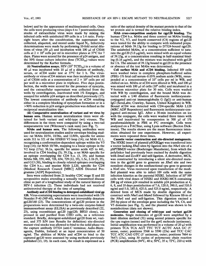
mAbFIG. 4. Cross-competition analysis for MAb 39.13g binding to rgpl20. MAbs (at a saturating concentration of 5.0 jLg/ml) mapping to a
linear V2 epitope (10/76b), to a linear C4 epitope (38.1a), to a linear V3 epitope (10/54), and to epitopes overlapping the CD4 b.s. (588, 448,728, 654, 729, 559, 1.5e, 2.1h, and G13) were tested for their ability to compete with iodinated 39.13g for binding to rgpl20. The resultsrepresent means of triplicate wells and are from one experiment, but similar results were obtained in two separate experiments.
to neutralize both the variant and 375 S/N mutant viruses(Table 2), bound to detergent-solubilized gp120/160 withbinding indices of 0.81 and 0.82, respectively. Furthermore,no detectable difference in the ability of MAb 448 to bind tothe surface of wt- or variant-infected H9 cells was observedby FACS staining (data not shown). The remaining sixhuman MAbs and three control MAbs, mapping to V2, V3,and C4, bound to the variant and 375 S/N site-directedmutant antigens with binding indices ranging from 0.8 to 1.20(Fig. 5).
Since MAbs 39.13g, 1.5e, and G13 still bind to HXB2-375S/N, though with reduced binding indices, we investigatedthe mechanism of neutralization resistance. Since all of theseMAbs block in vitro the gpl20-sCD4 interaction, they pre-sumably neutralize by preventing virion attachment to CD4.We therefore tested the ability of a number of CD4 b.s.MAbs, together with control MAbs specific for the V2, V3,and C4 regions of gpl20, to block sCD4 binding to NP-40-solubilized HXB2-wt, HXB2-var, and HXB2-375 S/N viralgpl20 by ELISA. MAbs 39.13g, 1.5e, and G13 (5 ,ug/ml)blocked sCD4 binding to HXB2-wt gpl20 by 82, 83, and79%, to HXB2-375 S/N mutant by 27, 29, and 20, and toHXB2-var by 24, 22, and 26%, respectively (Fig. 6). Sincethe gpl20-sCD4 binding assay was performed with NP-40-solubilized viral g120, it is not surprising that MAb 448inhibited sCD4 binding to wt and variant gpl20 equivalently(Fig. 6).
Since the 375 S/N mutation was originally selected for byan HIV-1-positive human serum, we investigated the sensi-tivity of the variant to neutralization by a panel of sera fromHIV-1-infected individuals classified in CDC stages II andIII (Fig. 7). Eleven of 21 sera showed a reduced neutraliza-tion titer for the variant, with a median neutralization titer of1/20, compared with 1/40 for HXB2-wt (P = 0.0092, two-tailed Wilcoxon rank test).
DISCUSSIONWe have selected an HIV-1 variant which can replicate in
the presence of a neutralizing human serum (Table 1).
Sequencing of the gpl20 genes from both the variant and thewt virus identified a single amino acid change in the C3region of gpl20, at residue 375 from Ser to Asn. Character-ization of this mutation, both as a cloned variant gpl20(HXB2-var) and as a site-directed mutant (HXB2-375 S/N),demonstrated that the mutation conferred resistance toneutralization by MAbs 39.13g (3), 1.5e (9), G13 (30), and448 (12), binding to epitopes overlapping the CD4 b.s., andby sCD4 (Table 2). In agreement with the reduced sensitivityto neutralization, MAbs 39.13g, 1.5e, and G13 demonstratedreduced binding to NP-40-solubilized HXB2-var and HXB2-375 S/N viral gpl20 with binding indices of 0.48, 0.42, and0.46 and of 0.42, 0.45, and 0.43, respectively (Fig. 5).However, MAb 448 bound to the cloned variant and mutantproteins with binding indices of 0.81 and 0.82. The relativelymodest change in binding index observed with both MAb 448and sCD4 for the variant and the 375 S/N mutant, despite thereduced neutralization sensitivity, suggests that studying thebinding of ligands to detergent-solubilized viral antigen maynot accurately predict the behavior of such ligands with thenative protein. We have previously reported a difference inthe ability of sCD4 to bind to soluble gpl20 and native intactvirion gpl20 (22), suggesting that the tertiary and/or quarter-nary structure of native oligomeric gpl20/41 influences thebinding affinity of sCD4.MAbs 39.13g, 1.5e, and G13 were less efficient at blocking
sCD4 binding to solubilized HXB2-var and HXB2-375 S/Nviral gpl20 than to HXB2-wt gpl20 (Fig. 6), suggesting thatthe efficiency of neutralization by CD4 b.s. antibodies maybe governed by the affinity of the antibody for the envelopeglycoprotein. By using a panel of site-directed gpl20 mu-tants, the epitopes recognized by the CD4 b.s. MAbs, withthe exception of 728 and 654, have been mapped (19, 30, 33,35) to five discontinuous regions including amino acids 113 to117 in Cl, 256 to 262 in C2, 368 to 370 in C3, 421 to 427 in C4,457 in C4, and 475 to 477 in C5. The regions of gpl20recognized by the MAbs differ slightly, but most depend ona core of amino acids including Asp-368, Glu-370, Thr-257,and Lys-421. There is no obvious pattern from the mapping
VOL. 67, 1993

5222 McKEATING ET AL.
i53.S
10/54 10/76b 38.1a 39.13g 588 448 728 654 729 559 1.5e 2. lh G13
mAb
1.2
1.0
0.8
10/54 10176b 38.1a 39.13g 588 448 728 654 729 559 1.5e 2.lh G13
mAbFIG. 5. Binding of MAbs to wt, variant, and 375 S/N mutant gp120/160. MAbs (at a saturating concentration of 5.0 ,ug/ml) mapping to a
linear V2 epitope (10/76b), to a linear C4 epitope (38.la), to a linear V3 epitope (10/54), and to epitopes overlapping the CD4 b.s. (39.13g, 588,448, 728, 654, 729, 559, 1.5e, 2.1h, and G13) were compared for the ability to bind to NP-40-solubilized gp120/160 from HXB2-375 S/N (A)and HXB2-var (B) with HXB2-wt. The results are expressed as the ratio of MAb (optical density) bound to the variant or site-directed mutantrelative to the wt and are termed the relative binding index.
data to explain why MAbs 39.13g, 1.5e, G13, and 448 areunable to neutralize the escape mutant whereas MAbs 559,728, 654, 729, 588, and 2.1h neutralize the mutant as well asthe wt virus. Amino acids from four of these regions (Thr-257, Asp-368, Glu-370, Trp-427, and Asp-457) have beenimplicated in CD4 binding (25). Thus, a number of aminoacid residue changes significantly affect MAb binding with-out affecting sCD4 recognition, suggesting that viral escapefrom antibody neutralization may occur without affecting theability of the virus to bind to its receptor (19). However, the375 S/N variant was also resistant to neutralization by sCD4(Tables 1 and 2). sCD4 demonstrated a reduced bindingaffinity for native envelope but not for detergent-solubilizedgpl20 (Fig. 1 and 2). Olshevsky and colleagues (25) reportedthat mutation of residues 368 and 370 abrogated sCD4binding in the presence or absence of nonionic detergents;however, residue 375 was not studied by these authors.
Reitz and colleagues (27) reported on a serum-selected
escape mutant which had a single change in gp4l at residue582 (Ala-Thr, or A/T), which exhibited a decreased suscep-tibility to neutralization by approximately one-third of hu-man sera tested. Since antibodies to linear epitopes encom-passing residue 582 were not neutralizing, Wilson andcolleagues (37) hypothesized that a neutralization epitope(s)may be affected by the change in gp4l. More recently, we(12b) and others (31a) have reported that the 582 A/T mutantis insensitive to neutralization by a number of CD4 b.s.MAbs, including F105 (26), 1.5e (9), and 39.13g (3). The 582A/T mutant appears to bind the MAbs with an affinityequivalent to that of the wt. This is in contrast to the 375 S/Nmutant, which has a reduced affinity for MAbs 39.13g, G13,and 1.5e (Fig. 5). This finding suggests that escape from CD4b.s. MAbs may occur by one of at least two mechanisms,either by a mutation which directly reduces the affinity of theMAb for gpl20 (Fig. 5) or by a mutation which indirectlyaffects events after MAb binding. Alternatively, a small
J. VIROL.

RESISTANCE OF AN HIV-1 ESCAPE MUTANT TO NEUTRALIZATION 5223
IbL 0 HXB2-375 SIN.880
o 0 ~HXB2-var
.0
60
20
010/54 10J76b 38.la 39.13g 588 448 2.1h l.Se G13
mAbFIG. 6. Abilities of MAbs to inhibit the interaction of HXB2-wt, HXB2-var, and HXB2-375 S/N gpl20/160 with sCD4. MAbs (at a
saturating concentration of 5.0 p.g/ml) mapping to a linear V2 epitope (10/76b), to a linear C4 epitope (38.1a), to a linear V3 epitope (10/54),and to epitopes overlapping the CD4 b.s. (39.13g, 588, 448, 1.5e, 2.1h, and G13) were tested for the ability to inhibit sCD4 (1.0 1Lg/ml) bindingto NP-40-solubilized gpl20/160 (50 ng/ml) from HXB2-wt, HXB2-var, and HXB2-375 S/N viral stocks.
reduction in the affinity of mutant 582 A/T for the MAbs orof mutant 375 S/N for MLAb 448, not measurable by currentassays, may be sufficient to account for the differentialneutralization observed (32).The 375 S/N mutant replicates in CD4+ cells as efficiently
as the wt does, as assessed by measuring the infectivity-to-particle ratio (TCID50 virion p24) (data not shown) (17). Thisfinding is in agreement with the ability of sCD4 and sCD4-IgG neutralization-resistant variants to replicate as well asthe neutralization-sensitive parental virus does (12a, 13).This observation is consistent with reports that replication-competent viruses can exhibit a wide range of CD4-bindingaffinities and may be explained by the greater cooperativity
A1
.
of envelope-receptor interactions during virus-cell or cell-cell fusion compared with the interaction of soluble CD4 andgp120 (32).The ability of a polyclonal serum to select for a mutation
in C3 which affects the sensitivity to neutralization both bysCD4 and CD4 b.s. MAbs confirms that antibodies bindingto the CD4 b.s. contribute to the group-specific neutraliza-tion response (21, 32). However, the 375 S/N mutant wasonly partially resistant to 11 of 21 human sera tested (Fig. 7),suggesting that either the cross-neutralizing activity may beattributed to an epitope(s) in addition to the CD4 b.s. or theCD4 b.s. induces a complex antibody response involvingmany discontinuous overlapping epitopes. The latter sugges-
200 -
180 U wt. var
160-
140-
120 -
100 -
80-
60
40- PI
20
1 2 3 4 5 6 7 8 9 10 11 12 13 14 15 16 17 18 19 20 21
wt
var
Serum numberFIG. 7. Sensitivities of wt and variant viruses to neutralization by sera from HIV-1-infected individuals. Reciprocal neutralization titers
of 21 human sera for wt and variant viruses are shown. Average titers (horizontal lines) were 1/40 for the the wt and 1/20 for the variant(Wilcoxon rank test, P = 0.009).
VOL. 67, 1993

5224 McKEATING ET AL.
tion is supported by the observation that three of nine humanMAbs mapping to the CD4 b.s. failed to neutralize the 375S/N mutant (Table 2). The subdivision of this panel of CD4b.s. MAbs into groups based upon their ability to neutralize375 S/N suggests that current methods of epitope mapping,recognition of synthetic or site-directed gp120 mutants, andcross-competition assays are insufficiently sensitive to defineMAb epitopes completely. These data suggest that singleamino acid changes resulting in resistance to one selectingagent may not lead to complete resistance to a polyclonalmixture of antibodies present in a heterologous serum.
ACKNOWLEDGMENTS
We thank M. Tenant-Flowers and C. Loveday (University Col-lege London Medical School [UCLMS], London, United Kingdom)for the provision of clinical material, H. Holmes for rgpl20 andMAbs from the MRC ADP Repository (NIBSC, Potters Bar, Hert-forshire, United Kingdom), and R. Ward (Genentech, South SanFrancisco, Calif.) for sCDH. We also thank Robin Weiss, ThomasSchulz, Ian Weller, and Richard Tedder for constructive criticism ofthe manuscript.The cohort studies at UCLMS were supported by the MRC, the
Wellcome Trust, and the Frances and Augustus Newman Founda-tion. J.A.M., J.B., S.A., and P.B. are funded by the MRC ADP.S.Z.-P. is funded by the Department of Veterans Affairs and NIHgrant AI 32424.
REFERENCES1. Berman, P., T. Gregory, L. Riddle, G. Nakamura, M. Champe,
J. Porter, F. Wurm, R. Hershberg, E. K. Cobb, and J. Eichberg.1990. Protection of chimpanzees from infection by HIV-1 aftervaccination with recombinant glycoprotein gp120 but not gpl60.Nature (London) 345:622-625.
2. Carne, C., I. V. D. Weller, and A. Johnson. 1987. Prevelance ofantibodies to human immunodeficiency virus, gonorrhoea rate,and changes sexual behaviour in homosexual men in London.Lancet i:656-658.
3. Cordell, J., J. P. Moore, C. J. Dean, P. J. Klasse, R. A. Weiss,and J. A. McKeating. 1991. Rat monoclonal antibodies tononoverlapping epitopes of human immunodeficiency virus typeI gpl20 block CD4 binding in vitro. Virology 185:72-79.
4. Emini, E., P. Nara, W. Schleif, J. Lewis, J. Davide, D. Lee, J.Kessler, S. Conley, M. Matsushita, S. Putney, R. Gerety, and J.Eichberg. 1990. Antibody-mediated in vitro neutralization ofhuman immunodeficiency virus type 1 abolishes infectivity forchimpanzees. J. Virol. 64:3674-3578.
5. Emini, E., W. Schleif, J. Nunberg, A. Conley, Y. Eda, S.Tokiyoshi, S. Putney, S. Matsushita, K. Cobb, C. Jett, J.Eichberg, and K. Murthy. 1992. Prevention of HIV-1 infectionin chimpanzees by gpl20 V3 domain specific monoclonal anti-body. Nature (London) 355:728-730.
6. Girard, M., M. Kieny, A. Pinter, F. Barre-Sinoussi, P. Nara, H.Kolbe, K. Kusumi, A. Chaput, T. Reinhart, E. Muchmore, J.Ronco, M. Kaczorek, E. Gomard, J. C. Gluckman, and P. Fultz.1991. Immunization of chimpanzees confers protection againstchallenge with human immunodeficiency virus. Proc. Natl.Acad. Sci. USA 88:542-546.
7. Gorny, M. K., A. J. Conley, S. Karwowska, A. Buchbinder, J. Y.Xu, E. Emini, S. Koening, and S. Zolla-Pazner. 1992. Neutral-ization of diverse human immunodeficiency virus type 1 vari-ants by an anti-V3 human monoclonal antibody. J. Virol.66:7538-7542.
8. Goudsmit, J., C. Debrouck, R. H. Meloen, L. Smit, E. R.Bakker, D. M. Asher, A. V. Wolff, C. J. Gibbs, and D. C.Gajdusek 1988. Human immunodeficiency virus neutralizationepitope with conserved architecture elicits type-specific anti-bodies in experimentally infected chimpanzees. Proc. Natl.Acad. Sci. USA 85:4478-4482.
9. Ho, D. D., J. A. McKeating, X. Li, T. Moudgil, E. Daar, N. C.Sun, and J. Robinson. 1991. Conformational epitope on gp120important in CD4 binding and human immunodeficiency virus
type 1 neutralization identified by a human monoclonal anti-body. J. Virol. 65:489-493.
10. Javaherian, K., A. J. Langlois, C. McDanal, K. L. Ross, L. I.Eckler, C. L. Jellis, A. T. Profy, J. R. Rusche, D. P. Bolognesi,S. D. Putney, and T. J. Matthews. 1989. Principle neutralizingdomain of the human immunodeficiency virus type I envelopeprotein. Proc. Natl. Acad. Sci. USA 86:6768-6772.
11. Kang, C. Y., P. Nara, S. Chamat, V. Caralli, T. Ryskamp, N.Haigwood, R. Newman, and H. Kohler. 1991. Evidence fornon-V3-specific neutralizing antibodies that interfere withgpl2O/CD4 binding in human immunodeficiency virus-infectedhumans. Proc. Natl. Acad. Sci. USA 88:6171-6175.
12. Karwowska, S., M. K. Gorny, A. Buchbinder, V. Gianakakos, C.Williams, T. Fuerst, and S. Zolla-Pazner. 1992. Production ofhuman monoclonal antibodies specific for conformational andlinear non-V3 epitopes of gp120. AIDS Res. Hum. Retroviruses8:1099-1106.
12a.Klasse, P. J., and J. A. McKeating. Submitted for publication.12b.Klasse, P. J., J. A. McKeating, M. Schutten, M. S. Reitz, Jr.,
and M. Robert-Guroff. An immune-selected point mutation inthe transmembrane protein of human immunodeficiency virustype 1 (HXB2-Env: Ala 582 (- Thr)) decreases viral neutraliza-tion by monoclonal antibodies to the CD4 binding site. Virol-ogy, in press.
13. McKeating, J. A., P. Balfe, P. Clapham, and R. A. Weiss. 1991.Recombinant CD4-selected human immunodeficiency type 1variants with reduced gpl20 affinity for CD4 and increased cellfusion capacity. J. Virol. 65:4777-4785.
14. McKeating, J. A., J. Cordell, C. J. Dean, and P. Balfe. 1992.Synergistic interaction between ligands binding to the CD4binding site and V3 domain of human immunodeficiency virustype I gpl20. Virology 191:732-742.
15. McKeating, J. A., J. Gow, J. Goudsmit, L. H. Pearl, C. Mulder,and R. A. Weiss. 1989. Characterisation of HIV-1 neutralizationescape mutants. AIDS 3:777-784.
16. McKeating, J. A., A. McKnight, K. McIntosh, P. R. Clapham,C. Mulder, and R. A. Weiss. 1989. Evaluation of human andsimian immunodeficiency virus plaque and neutralization as-says. J. Gen. Virol. 70:3327-3333.
17. McKeating, J. A., A. McKnight, and J. P. Moore. 1991. Differ-ential loss of envelope glycoprotein gp120 from virions ofhuman immunodeficiency virus type 1 isolates: effects on infec-tivity and neutralization. J. Virol. 65:852-860.
17a.McKeating, J. A., C. Shotton, J. Cordell, S. Graham, P. Balfe,N. Sullivan, M. Charles, M. Page, A. Bolmstedt, S. Olofsson, S. C.Kayman, Z. Wu, A. Pinter, C. Dean, J. Sodroski, and R. A.Weiss. 1993. Characterization of neutralizing monoclonal anti-bodies to linear and conformation-dependent epitopes withinthe first and second variable domains of human immunodefi-ciency virus type 1 gp120. J. Virol. 67:4932-4944.
18. McKeating, J. A., M. Thali, P. Balfe, J. P. Moore, J. Sodroski,and R. A. Weiss. 1991. Conformational gp120 neutralizationepitopes on HIV-1, p. 199-202. In M. Girard and L. Valette(ed.), Sixieme Colloque des Cent Gardes. Pasteur Vaccins,Paris.
19. McKeating, J. A., M. Thali, C. Furman, S. Karwowska, M. K.Gorney, J. Cordell, S. Zolla-Pazner, J. Sodroski, and R. A.Weiss. 1992. Amino acid residues of the human immunodefi-ciency virus type I gpl20 critical for the binding of rat andhuman monoclonal antibodies that block the gpl2O-sCD4 inter-action. Virology 190:134-142.
20. McKnight, A., P. R. Clapham, J. Goudsmit, R. Cheingsong-Popov, J. Weber, and R. A. Weiss. 1992. Development of HIV-1group-specific neutralizing antibodies after seroconversion.AIDS 6:799-802.
21. Moore, J. P., and D. D. Ho. 1993. Antibodies to discontinuousor conformationally sensitive epitopes on the gpl20 glycopro-tein of human immunodeficiency virus type 1 are highly preva-lent in sera of infected humans. J. Virol. 67:863-875.
22. Moore, J. P., J. A. McKeating, Y. Huang, A. Ashkenazi, andD. D. Ho. 1992. Virions of primary human immunodeficiencyvirus type 1 isolates resistant to soluble CD4 (sCD4) neutraliza-tion differ in sCD4 binding and glycoprotein retention from
J. VIROL.
RESISTANCE OF AN HIV-1 ESCAPE MUTANT TO NEUTRALIZATION 5225
sCD4-sensitive isolates. J. Virol. 66:235-243.23. Moore, J. P., J. A. McKeating, R. A. Weiss, and Q. J. Sattentau.
1990. Dissociation of gp120 from HIV-1 virions induced bysoluble CD4. Science 250:1139-1142.
23a.Myers, G., B. Korber, J. A. Berzofsky, R. F. Smith, and G. N.Pavlakis. 1992. Human retroviruses and AIDS 1992. LosAlamos National Laboratories, Los Alamos, N. Mex.
24. Ohno, T., M. Terada, Y. Yoneda, K. Shea, R. Chambers, D.Stroka, M. Nakamura, and D. Kufe. 1991. A broadly neutraliz-ing monoclonal antibody that recognises the V3 region of humanimmunodeficiency virus type I glycoprotein gpl20. Proc. Natl.Acad. Sci. USA 88:10726-10729.
25. Olshevsky, U., E. Helseth, C. Furman, J. Li, W. Haseltine, andJ. Sodroski. 1990. Identification of individual human immuno-deficiency virus type 1 gpl20 amino acids important for CD4receptor binding. J. Virol. 64:5701-5707.
26. Posner, S. T., T. Hideshima, T. Cannon, M. Mukherjee, K.Mayer, and R. Byrn. 1991. An IgG human monoclonal antibodywhich reacts with HIV-1 gp120, inhibits virus binding to cells,and neutralizes infection. J. Immunol. 146:4325-4332.
27. Reitz, M. S., C. Wilson, C. Naugle, R. C. Gallo, and M.Robert-Guroff. 1988. Generation of a neutralization-resistantvariant of HIV-1 is due to selection for a point mutation in theenvelope gene. Cell 54:57-63.
28. Robert-Guroff, M., M. Brown, and R. C. Gallo. 1985. HTLV-IIIneutralizing antibodies in patients with AIDS and AIDS-relatedcomplex. Nature (London) 316:72-74.
29. Rusche, J. R., K. Javaherian, C. McDanal, J. Petro, D. L. Lynn,R. Grimaila, A. Langlois, R. C. Gallo, L. 0. Arthur, P. J.Fischinger, D. P. Bolognesi, S. D. Putney, and T. J. Matthews.1988. Antibodies that inhibit fusion of human immunodeficiencyvirus-infected cells bind a 24-amino acid sequence of the viralenvelope glycoprotein gp120. Proc. Natl. Acad. Sci. USA85:3198-3202.
30. Schutten, M., A. McKnight, R. C. Huisman, J. P. M. LangedQk,A. C. Andeweg, M. Thali, J. McKeating, R. Meloen, J. Sodroski,J. Goudsmit, and A. D. M. E. Osterhaus. Further characterisa-tion of antigenic sites of HIV-1 gpl20 recognised by virusneutralizing human monoclonal antibodies. AIDS Res. Hum.
Retroviruses, in press.31. Simmonds, P., P. Balfe, C. A. Ludlam, J. 0. Bishop, and A. J.
Leigh-Brown. 1990. Human immunodeficiency virus-infectedindividuals contain provirus in small numbers of peripheralmononuclear cells and at low copy numbers. J. Virol. 64:864-872.
31a.Sodrowski, J. Personal communication.32. Thali, M., C. Furman, D. D. Ho, J. Robinson, S. Tilley, A.
Pinter, and J. Sodroski. 1992. Discontinuous, conserved neu-tralization epitopes overlapping the CD4 binding region ofhuman immunodeficiency virus type 1 gpl20 envelope glyco-protein. J. Virol. 66:5635-5641.
33. Thali, M., U. Olshevsky, C. Furman, D. Gabuzda, J. Li, and J.Sodroski. 1991. Effects of changes in gpl2O-CD4 binding affinityon human immunodeficiency virus type 1 envelope glycoproteinfunction and soluble CD4 sensitivity. J. Virol. 65:5007-5012.
34. Thali, M., U. Olshevsky, C. Furman, D. Gabuzda, M. Posner,and J. Sodroski. 1991. Characterization of a discontinuoushuman immunodeficiency virus type 1 gpl20 epitope recognizedby a broadly reactive neutralizing human monoclonal antibody.J. Virol. 65:6188-6193.
35. Tilley, S. A., W. J. Honnen, M. Racho, M. Hilgartner, and A.Pinter. 1991. A human monoclonal antibody against the CD4binding site of HIV-1 gpl20 exhibits potent, broadly neutralizingactivity. Res. Virol. 142:247-259.
36. Weiss, R. A., P. R. Clapham, J. N. Weber, A. G. Dalgleish, L. A.Lasky, and P. Berman. 1986. Variable and conserved neutral-ization antigens of human immunodeficiency virus. Nature(London) 324:572-575.
37. Wilson, C., M. S. Reitz, K. Aldrich, P. J. Klasse, J. Blomberg,R. C. Gallo, and M. Robert-Guroff. 1990. The site of animmune-selected point mutation in the transmembrane proteinof human immunodeficiency virus type 1 does not constitute aneutralization epitope. J. Virol. 64:3240-3248.
38. Wolfs, T. F. W., G. Zwart, M. Bakker, M. Valk, C. Kuiken, andJ. Goudsmit. 1991. Naturally occurring mutations within HIV-1V3 genomic RNA lead to antigenic variation dependent on asingle amino acid substitution. Virology 185:195-205.
VOL. 67, 1993